
Medical Emergencies in the Dental Office - Barry Krall · PDF fileIntroduction The main...
39
MEDICAL EMERGENCIES IN THE DENTAL OFFICE Barry Krall DDS
-
Upload
dinhkhuong -
Category
Documents
-
view
218 -
download
2
Transcript of Medical Emergencies in the Dental Office - Barry Krall · PDF fileIntroduction The main...

MEDICAL EMERGENCIES IN THE DENTAL OFFICE
Barry Krall DDS

Introduction The main emphasis in the management of medical emergencies in the dental office is preparation. It has been said that “If you are prepared for the emergency, the emergency ceases to exist”. How true that is! The atmosphere surrounding an emergency situation can be stressful and emotionally charged. A well thought out emergency plan, implemented in advance, can reduce the stress associated with the emergency to manageable levels. The well prepared team almost seamlessly goes through the prepared algorithm attending to each step before moving on to the next phase of care.
The question arises as to how often life-threatening emergencies occur in the typical dental office setting? McCarthy estimates that one or two treatment related deaths will occur over the practice lifetime of the typical dental practitioner1. He further estimates that the number of office-related deaths would increase to five if dental patients were observed for seven days following treatment.
The incidence of medical emergencies is unfortunately on the rise. There are many reasons for this increase. First of all patients are living longer due to advances in medical technology and therapeutics. For the same reasons, patients with more medically complex diseases are living longer and surviving decades after diagnosis. These patients’ diseases are often managed with a myriad of medications and increase the likelihood of drug interactions. Thirdly, dental treatment appointments are longer as a result of more complex treatment planning. This is of little concern in the healthy patient, but of much concern in sicker patients with limited physiologic reserve. Lastly, in order to help patients better tolerate longer, more complex appointments; the use of sedation in dentistry is becoming widespread. Sedation is an excellent modality when used appropriately to alleviate treatment induced anxiety. However, deeper than intended levels of central nervous system depression, gone undetected, can lead to significant morbidity or mortality.
ADA Guidelines for Preparation The ADA council on Scientific Affairs Statement and the ADA/PDR Guide to Dental Therapeutics gives the following guidelines to prepare the dentist for the inevitable emergency2, 3.
“Courses on emergency medicine management are included in the curricula of all accredited dental schools”
“The Council on Scientific Affairs recommends that all dental health care professionals receive regular training in BLS, because these skills are maintained only through repetition”
“First and foremost in emergency management is the ability to effectively provide BLS, when appropriate”
“For the practicing dentist, the Council recommends that emergency medicine programs be offered as CE (dental schools, dental societies etc)”
“Didactic and hands-on training in the prevention, recognition and management of common emergencies also recommended”
A quick glance at these guidelines leaves the impression that preparation for emergencies is a continuous process. It is fortunate that emergencies do not happen very often. However, it is difficult to become proficient at something when only encountering these situations infrequently. Therefore, as these guidelines suggest, repetitive practice with simulation is the best way to develop and train the instinct to respond when the ‘moment’ arises. In the following sections, we will address how to prepare the general dental office, as well as offices that provide sedation/general anesthesia, for medical emergencies. The keys for success are proper planning, prevention of avoidable emergencies and a simple approach to managing the inevitable emergency situation.

Emergency Preparedness Emergency Preparedness involves 3 basic steps:
Preparation
Prevention
Management In the following sections, we will look at each of these points individually in the quest for Emergency Preparedness.
1. McCarthy EM: Sudden unexpected death in the dental office, J AM Dent Assoc 83: 1091, 1971. 2. The ADA council on Scientific Affairs Statement; JADA, Vol. 133, March 2002 3. ADA/PDR Guide to Dental Therapeutics. Fourth Edition

A. Preparation Table 1 It is readily apparent by looking at the ADA council on Scientific Affairs Statement and the ADA/PDR Guide to Dental Therapeutics recommendations in the introductory paragraph that preparation for medical emergencies in the dental office is a life-long endeavor. Table 1 represents guidelines that can be followed to prepare an office for medical emergencies. Each dental office is unique and will require different levels of preparedness based on patient type (e.g., pediatrics, medically compromised patients), nature of treatment (complexity/duration) and whether sedation or general anesthesia is administered in the office. Discussion:
1. BLS certification for all office personnel (ACLS/PALS if required by law or practice model)
It is essential that all office personnel are trained to recognize and manage medical emergencies. In the event of a life-threatening emergency, there are many tasks that must be performed and time is of the essence. Therefore, each team member must be well trained in the algorithms of basic life support (BLS) and be prepared to effectively implement these steps effectively.
2. Didactic and clinical courses in emergency medicine Fortunately, the incidences of life-threatening medical emergencies in the dental office are rare. Unfortunately, this is a double- edged sword. These highly critical, low-frequency events typically are managed poorly unless time and resources are committed to rehearsing protocols and keeping up with the latest developments in basic emergency medicine. Ideally, courses that provide opportunities to engage in “hands-on” practice provide the best learning environment. It is one thing to have memorized algorithms on management of medical emergencies and another to actually implement the steps in an organized and effective manner.
Preparation (Modified from: Rosenberg. Preparing for Medical Emergencies: the essential drugs and
equipment for the dental office. JADA 2010;141;14s-19s
1. BLS certification for all office personnel (ACLS/PALS if required by
law or practice model)
2. Didactic and clinical courses in emergency medicine
3. Well-defined roles defined for at least 3 team members (4 is better)
4. Emergency “crash cart” immediately available
5. Emergency oxygen cylinder pressure and supplies checked daily
6. Emergency phone numbers for emergency medical services posted near every phone
7. Periodic emergency drills are scheduled to simulate life-threatening emergencies

3. Well-defined roles defined for at least 3 team members (4 is better) Box 1
In the event of a life-threatening medical emergency, it is readily apparent that this is no time for organization and development of protocols. These protocols should already be in place and well rehearsed. Illustrated in Box 1 is a “job description” for each team member in a life-threatening medical emergency
Team member #1 (dentist, hygienist etc) Assume leadership role
a. The importance of having someone in charge of the emergency is critical as timely decisions and interventions need to be made. This individual must stay with the patient throughout the emergency and create a safe environment for all.
b. They must be open for suggestions from all team members and after considering all information, make decisions regarding definitive care
Implements ABC’s of BLS
Ensures all elements (ABC’s) of basic life support have been considered and if necessary implemented effectively Uses closed loop communication
Avoids “thin air commands”. Examples of thin air commands might include; “someone get the oxygen!” Instructions will be given to a specific person by name and the instruction would be repeated back so the leader knows that the instruction is understood correctly.
Team member #2 Retrieves oxygen
Opens cylinder valve, attaches appropriate oxygen mask/cannula Monitors vitals
It is imperative that blood pressure and heart rate be evaluated and recorded in a time-based fashion. This information, along with baseline values, provides invaluable information for determination of interventions and decision to transport the patient to the emergency room
Documents medications administered Assists with ABC’s as directed Responsible for checking oxygen system daily
a. Pressure gauges on the emergency oxygen system should be checked daily to ensure that adequate volume of oxygen is available in the event of an emergency. The oxygen cylinder should be replaced if the pressure gauge reads less than 1000 psi. An E cylinder reading 1000 psi will provide 30 minutes of oxygen delivery at 10 L/min flows.
b. Stock appropriate masks, nasal cannula and airway adjuncts (if practice appropriate)
Team Member Role Description Team member #1
Assume leadership role Implements ABC’s of BLS Uses closed loop communication
Team member #2 Retrieves oxygen Monitors vitals/charts medications Assists with ABC’s as directed Responsible for checking oxygen system daily
Team member #3 Retrieves “crash cart” Turns on AED and follows prompts Assists with ABC’s as directed Responsible for maintaining “crash cart” Responsible for checking AED (daily)
Team member #4
Activates 911 Assists with ABC’s as directed Meets EMS personnel at entrance and directs them to
emergency Adapted from: Haas D. Preparing dental office staff members for emergencies. JADA 2010; Vol 141
Team member #3 Retrieves “crash cart”
a. Emergency medications b. Monitoring equipment
i. Electronic BP device (gives HR and BP reading) ii. SPO2 (if required for permit)
c. AED Turns on AED and follows prompts
In the case of a cardiac arrest, application of electrical therapy in a timely manner is of utmost importance for long-term survival. The person responsible for retrieving the AED should open the device and attach appropriate pads to device. Scissors must be available in the AED case to cut off clothing to allow for attachment of pads to bare chest.
Assists with ABC’s as directed Responsible for maintaining “crash cart”
The crash cart must be checked on a monthly basis. A checklist is helpful to ensure availability of all medications and supplies. Used and expired medications should be replaced.
Responsible for checking AED (daily) This is a simple and yet important responsibility. Modern AEDS run a self-test daily to check battery life and electrical components
Team member #4 Activates 911
Use a land line if at all possible. Calls made from a cellular phone are routed to a regional call center which will result in delayed response times
Assists with ABC’s as directed Meets EMS personnel at entrance and directs them to emergency

4. Emergency “crash cart” immediately available
Table 2 It is important to have a fully equipped “crash cart” readily available at all times. Depending on the training of the dentist, state dental board requirements, ages of patients treated (pediatric vs adult) and whether sedation or general anesthesia is being administered in the office. There are no firm recommendations for equipment and medication requirements for the general dental office. The ADA Council on scientific affairs states that “no state dental boards have established specific recommendations as to which emergency drugs and equipment a dentist must have available. Proprietary emergency kits are available, but none are compatible with needs of all practitioners. The content and design of these kits should be based upon each practitioner’s training and individual requirements and dentists should design their own emergency kits to meet their own needs. It is also recommended that all dental offices maintain at least the basic recommended emergency equipment and drugs.”2, 3
Table 2 shows a list of emergency armamentarium and medications that are widely recognized as being important for the typical dental office.
I. Oxygen delivery system a. Must be able to deliver O2 under positive pressure!
i. Ambu bag (Fig 1) or manually triggered oxygen delivery device (Fig 2)
Fig 1 Ambu Bag Fig 2 Oxygen powered breathing device
GENERAL DENTAL OFFICE
“Crash Cart”
I. Oxygen Delivery System
II. Automated External Defibrillator (AED)*
III. Blood Pressure Monitor
IV. Syringes a. 1 mL (Tuberculin) b. 3 mL Syringe
V. Medications a. Non-injectable
1. Sugar Source (Juice, Gel, Soda Etc)` 2. Coronary Artery Vasodilator 3. Bronchodilator 4. Platelet Aggregate Inhibitor
b. Injectable 1. Antihistamine 2. Epinephrine
*Optional in most states

b. Nasal cannula (2-6 L/min)- allows delivery of oxygen concentration of 24-44% (Fig 3)
c. Simple mask (6-15 L/min)- allows delivery of oxygen concentration of 40-50% (Fig 3)
Fig 3
Fig 4 II. Automated external defibrillator (AED) (see page 14 for more information on defibrillators)(Fig 4) There are many models on the market and all are FDA approved. There is no evidence to prove that any model is superior. All AED models currently on the market deliver biphasic electrical current. These devices weigh less than 5 pounds and when activated, provide step by step instruction during the resuscitation. Most models can be used on both adults and children and come stocked with both pediatric and adult pads.

Fig 5 III. Electronic blood pressure monitor or sphygmomanometer and stethoscope (fig 5) It is critical to remember to obtain baseline blood pressure and pulse rate values on all patients undergoing dental procedures. Isolated blood pressure readings are of limited value unless they are compared with the baseline. Upper arm, wrist and finger digital devices are available, but inaccuracy is a problem with wrist and finger models.
Fig 6a Fig 6b
IV. Syringes a. 1 mL (tuberculin) syringe-22/ 23 gauge; ¾ - 1 inch needle! (Fig 6a) A larger bore needle (22/23 gauge) and sufficient length (3/4-1 inch) is necessary if a
tuberculin syringe is selected for administration of epinephrine intramuscularly. This allows for easy drawing up of the drug from the ampule and adequate depth of insertion into the muscle. The 1 mL syringe is used for the IM administration of epinephrine.
b. 3 mL syringe- 23 gauge ¾ - 1 inch needle (Fig 6b) The 3 mL syringe is used for the IM administration of Benadryl

V. Medications (see drug monographs for detail pg 22) Box 2 shows the drugs that are considered essential for the general dental office.
Box 2 Emergency Medications for the General Dental Office
Medications Formulation indication Dosage
Sugar
Juice Box/glucose gel
Hypoglycemia
Oral 15-20 Grams Q 15 min
Nitroglycerin 0.4 mg SL Tabs or Translingual spray
Angina 1 Tab SL Q 5 min x 3 doses or 1-2 sprays SL x 3 doses (if SBP >90 mmHg
Albuterol 90 mcg/puff Acute/Severe Asthma Exacerbation
Adult 4-8 puffs Q 20 min (4 hours) Peds 4-8 puffs Q 20 min (Max 3 doses)
Aspirin 81 mg tablets Myocardial Infarction 162-364 mgs (2-4 tabs)
Benadryl 50 mg/mL Allergic rxn
Adult 50-100 mg IM Pediatrics 12.5-25 mg IM
Epinephrine 1:1000 (1mg/mL) or EpiPen/EpiPen jr
Anaphylaxis, Severe Bronchospasm
Adult 0.3 mg IM Peds (<20 kg) 0.15 mg IM
IM- intramuscular SL- sublingual SBP- systolic blood pressure

However, for the office that provides sedation or general anesthesia to patients; state board requirements are well defined. It is important to review individual state board regulations to make sure that the office is in compliance. Listed below is the equipment and medications required by the California State Dental Board for moderate sedation and general anesthesia.
Moderate Sedation
1. Battery powered backup light 2. Suction equipment
i. Tonsillar or pharyngeal type suction tips ii. Backup suction device that operates independent of power supply
3. Oxygen delivery system
a. Must be able to deliver O2 under positive pressure! (Fig 1) i. Ambu bag or manually triggered oxygen delivery device (Fig 2)
b. Nasal cannula (2-6 L/min)- allows delivery of oxygen concentration of 24-44% (Fig 3) c. Simple mask (6-10 L/min)- allows delivery of oxygen concentration of 40-50% (Fig 3)
4. Electronic blood pressure monitor or sphygmomanometer and stethoscope 5. Oral airways 6. Adequate equipment for establishment of IV line 7. Precordial/pretracheal stethoscope 8. Pulse oximeter 9. Medications (required by California State Dental Board) (Box 3) (see drug monographs for
detail) a. Coronary artery vasodilator (nitroglycerin) b. Epinephrine c. Antihistamine (Diphenhydramine) d. Bronchodilator (albuterol) e. Hyperglycemic agent (juice, gel, dextrose 50 %) f. Platelet aggregate-inhibitor (aspirin) g. Anticholinergic (atropine) h. Anticonvulsant (midazolam) i. Corticosteroid (hydrocortisone) j. Vasopressor (ephedrine)

Table 3 Emergency Medications for Moderate Sedation
Medications Concentration Indication Dosage
Epinephrine 1:1000 (1mg/mL)
1 mL vial EpiPen or EpiPen jr
Anaphylaxis, Severe Bronchospasm
Adult 0.3 mg IM Peds 0.15 mg IM
Benadryl 50 mg/mL 1 mL vial
Allergic rxn Adult 50-100 mg IM Pediatrics 25 mg IM
Albuterol 90 mcg/puff Acute/Severe Asthma
Exacerbation Adult 4-8 puffs Q 20 min (4 hours) Peds 4-8 puffs Q 20 min (Max 3 doses)
Aspirin 81 mg tablets Myocardial Infarction 162-364 mgs (2-4 tabs)
Sugar
Juice Box/glucose gel 50 % Dextrose
Hypoglycemia
Oral 15-20 Grams Q 15 min IV Adult 25 Grams (50 mL) IV Peds 1-2 mL/kg
Nitroglycerin
0.4 mg SL Tabs or Translingual spray
Angina
1 Tab SL Q 5 min x 3 doses or 1-2 sprays SL x 3 doses (if SBP >90 mmHg
Atropine 0.4 mg/mL Bradycardia Adult 0.5-1 mg IV Peds 0.01-0.02 mg/kg IV
Flumazenil
0.1 mg/mL
Benzodiazepine Antagonist
IV 0.2mg/min up to 1 mg
Naloxone 0.4 mg/mL Opioid Antagonist IV 0.04 mg/min up to 0.4 mg
Midazolam 5mg/mL Anticonvulsant Adult 5 mg IM; 2 mg IV Peds IM
Hydrocortisone Recurrent Anaphylaxis Adult 100 mg IM/IV
Ephedrine 50 mg/mL Hypotension 5-20 mg IV/IM
Glucagon 1 mg Hypoglycemia (Unconscious Patient)
Adult 1 mg IV/IM Peds 0.5 mg IV/IM
IM- intramuscular; SL- sublingual; IV intravenous SBP- systolic blood pressure

General Anesthesia
1. Battery powered backup light 2. Suction equipment
i. Tonsillar or pharyngeal type suction tips ii. Backup suction device that operates independent of power supply
3. Oxygen delivery system
a. Must be able to deliver O2 under positive pressure! i. Ambu bag or manually triggered oxygen delivery device
b. Nasal cannula (2-6 L/min)- allows delivery of oxygen concentration of 24-44% c. Simple mask (6-10 L/min)- allows delivery of oxygen concentration of 40-50%
4. Electronic blood pressure monitor or sphygmomanometer and stethoscope 5. Oral airways 6. Adequate equipment for establishment of IV line 7. Precordial/pretracheal stethoscope 8. Pulse oximeter 9. Laryngoscope 10. Assorted blades 11. Spare batteries/bulbs 12. Endotracheal tubes and appropriate connectors 13. Endotracheal tube forceps 14. Electrocardioscope (ECG monitor) 15. Defibrillator
Optional (my suggestions) 1. Assorted LMA’s 2. Bougie 3. Intraosseous infusion needle 4. Cricothyrotomy kit
Emergency drugs (required by California Dental Board) (see drug monographs for detail)
1. Oxygen 2. Coronary artery vasodilator (nitroglycerin) 3. Antihistamine (Diphenhydramine) 4. Bronchodilator (albuterol) 5. Hyperglycemic agent (juice, gel, dextrose 50 %) 6. Anticholinergic (atropine) 7. Drug antagonists (Flumazenil, Naloxone) 8. Anticonvulsant (midazolam) 9. Corticosteroid (hydrocortisone) 10. Vasopressor (ephedrine, phenylephrine) 11. Muscle relaxant (succinylcholine) 12. Intravenous medication for treatment of cardiopulmonary arrest (Epinephrine) 13. Antiarrhythmic (lidocaine, amiodarone) 14. Antihypertensive (hydralazine, esmolol)
Optional (my suggestions) 1. Platelet aggregate-inhibitor (aspirin) 2. Dantrolene (if using MH triggering techniques: volatile anesthetics/succinylcholine)
5. Emergency oxygen cylinder pressure and supplies checked daily
Pressure gauges on the emergency oxygen system should be checked daily to ensure that adequate volume of oxygen is available in the event of an emergency. The oxygen cylinder should be replaced if the pressure gauge reads less than 1000 psi. An E cylinder reading 1000 psi will provide 30 minutes of oxygen delivery at 10 L/min flows. Stock appropriate masks, nasal cannula and airway adjuncts (if practice appropriate)
6. Emergency phone numbers for emergency medical services posted near every phone
Immediate recognition and activation of emergency medical services are of paramount importance in the event of a life-threatening emergency. Emergency numbers must be posted near every telephone in the office. Landline phones should be used when possible; calls from cellular phones are routed to a regional call center which results in delays in EMS response.
7. Periodic emergency drills are scheduled to simulate life-threatening
emergencies Certification in BLS provides a framework for managing emergencies, but each office needs to tailor the process to meet their office requirements. For example, each office will have equipment and drugs unique to their training and expertise. Periodic simulations should be planned so as to acquaint each member of the team as to what is expected of them during the emergency crisis. The protocol set up breeds organization rather than confusion. Examples of assigned tasks include; vital sign assessment, retrieving emergency equipment (drugs, oxygen cylinder etc) and calling 911 if necessary. Emphasis on the ABC’s is of paramount importance. Following this acronym forces the emergency team to take care of more important interventions first (e.g., making sure the patient is oxygenating). Each step of the acronym should be evaluated before moving on to the step. For example, the airway must be assessed before checking breathing. It is fairly obvious that if the airway is obstructed, breath sounds will be absent or inadequate.

Defibrillators
Introduction
It is estimated that 330,000 people are stricken with sudden cardiac arrest (SCA) each year. Many of the victims of SCA will develop ventricular fibrillation (VF) at some time during the arrest. Essentially, these individuals are clinically dead and the only effective method of treatment is to deliver electric shocks to the heart using a defibrillator. However, the American Heart Association (AHA) emphasizes that early recognition, early access to EMS, early CPR and early defibrillation as important steps in improving outcomes. Studies have shown that immediate bystander CPR/defibrillation within minutes provides the best chance of survival from sudden VF cardiac arrest in adults. Survival rates of 50-74 % have been reported when adult victims of VF SCA collapse in front of witnesses and receive immediate bystander CPR plus defibrillation within 3-5 minutes of collapse1-8. As each minute passes, the chances of survival for a person suffering SCA drop by 10%. Although some authorities state that rate of survival is fairly constant for everyone with respect to the first 4 minutes. The flatness of this part of the curve, in the early minutes of the cardiac arrest, probably reflects the fact that the body still has oxygenated blood. Once this oxygen is used up-probably after four minutes- the fall in the survival rate begins, and it continues relentlessly, dropping by approximately 5 percent for every minute that goes by. This differential rate of survival- high for the first four minutes, then rapidly dropping- is explained in the three-phase model of cardiac arrest that has been proposed by Weisfeldt and Becker. The first phase lasts approximately four minutes and is called the electrical phase. Defibrillatory shocks are all that is needed during this phase (survival is the same for 4 minutes with a 62% of survival if defibrillation is administered) and CPR is of lesser importance. The second phase is called the circulatory phase. CPR is very important during this phase since it restores oxygenated blood to the brain and other organs. The circulatory phase probably begins after 4 minutes and lasts until ten or twelve minutes into the cardiac arrest. The third phase is called the metabolic phase. Therapy is not effect during this phase as cellular damage has occurred beyond the point where modern science can help15. The AHA strongly advocates that all EMS first response vehicles and ambulances be equipped with a defibrillation device. The AHA also supports placing AED’s in targeted public areas such as sporting arenas, gated communities, office complexes, doctor’s offices, shopping malls, etc. The federal government has mandated that all federal buildings and commercial airlines site AED’s. They have also provided funding to urban and rural communities of limited resources to purchase AEDs and train non-medical personnel in their use.

The placement of defibrillation devices in dental offices is not mandated by state dental boards unless general anesthesia is provided. However, there are certain categories of patients that are prone to SCA and risk assessment of the patient population might be considered for each particular office setting. It should be appreciated that one of the leading causes of death for adults 35-40 years of age is SCA and is the most common cause of death for those > 45 years of age. In the United States each year, SCA occurs with an estimated frequency of ~1:1000 persons > 35 of age per year 9-12. These statistics can be used to estimate the risk of adult SCA for any location on the basis of the number of adults aged 35 and older typically present at that location and the number of hours they are present at that location per year. The following list defines patients that are at increased risk for SCA13:
Pediatrics o abnormal rhythms are typically caused by inherited or congenital cardiac
conditions o Or by acute medical problems that cause inflammation of the heart
Adults o Previous heart attack o Coronary artery disease
Therefore, the decision to site defibrillation devices in the dental office should be determined by several factors:
The volume of patients seen at the office The demographics and medical profile of patients/staff /dentist Complexity and length of procedures
From the above list it is readily apparent that more patients treated in an office per year increases the likelihood of one of those patients who are at risk to visit the office. Also, as the age of the patient/staff as well as dentist increases, the medical complexity is likely to rise. Uncomplicated/short procedures in unhealthy patients are unlikely to cause problems. However, complicated/long procedures in unhealthy patients (or healthy patients) increase the chances dramatically for untoward events. As the intensity of procedural stress increases, patients that have minimal physiologic reserve are candidates for decompensation.
Types of Defibrillators There are basically three types of defibrillators available:
Implantable o These devices are surgically implanted under the skin and the leads placed into the
chambers of the heart o They constantly monitor the patient's heart rhythm, and automatically administer
shocks for various life threatening arrhythmias, according to the device's programming Manual
o Allows operator to read/interpret ECG and select the energy levels to deliver to patients based on prior knowledge and experience
o Generally found only in hospitals and some ambulances Automated
o These simple to use units are based on computer technology which is designed to analyze the heart rhythm itself, and then advise whether a shock is required
o They are designed to be used by lay persons, who require little training o They are usually limited in their interventions to delivering high joule shocks for VF and
VT (ventricular tachycardia) rhythms, making them generally limiting for use by healthcare professionals, who could diagnose and treat a wider range of problems with a manual or semi-automatic unit

The common use of defibrillation technology for VF and VT is a relatively new phenomenon, having been developed only 50 years ago. Manual and automated defibrillators have evolved impressively over the last thirty years since the introduction of the first commercial defibrillator. The first commercial external defibrillators adopted monophasic waveforms which means that the current that is produced by the device flows in one direction (i.e., paddle to paddle). Toward the end of the 1980’s, biphasic technology became available; biphasic defibrillators were developed and are gaining in popularity. The acceptance of biphasic technology is such that all new AED’s (and an increasing number of manual defibrillators) reaching the market have biphasic technology. As opposed to a monophasic defibrillator, biphasic defibrillators deliver current in two directions. In the first phase, the current moves from one paddle to the other as with monophasic defibrillators. During the second phase, the current flow reverses direction. The advantages of using biphasic versus monophasic waveforms are listed below:
1. The bidirectional flow of current allows lower energy levels to be used which lessens the risk of post-shock complications such as myocardial dysfunction and skin burns.
2. Newer biphasic technology allows for measurements of impedance during the first phase of the defibrillation cycle and adjustments in energy levels during the second phase to ensure proper energy levels being delivered to the heart
3. These lower energy devices can be smaller and lighter (more portable) 4. Less expensive 5. Less demanding of batteries 6. Fewer maintenance requirements
To further expand the topic, biphasic defibrillators are available both as manual and automated devices. The popularity of the automated devices is increasing because of its simplicity and effectiveness. Listed below are advantages and disadvantages of AED’s compared to manual devices: Characteristics of AED’s
1. Inexpensive ($1200 to 3500) 2. Small, lightweight, portable (< 5 pounds) 3. Easy to use 4. EKG display (selected AED’s) 5. Manual override option (selected AED’s) 6. CPR technology (objective measurement and corrective feedback on compression depth and
rate) 7. Non-invasive pacing (selected AED’s) 8. Download data to PC (selected AED’s)
Characteristics of manual defibrillators
1. More expensive ($4000 [refurbished] to $10000+) 2. Heavier (10-15 pounds) 3. 5 or 12 lead EKG monitoring 4. SPO2, NIBP, ETCO2 5. Synchronized cardioversion 6. Pads or paddles 7. Some Manual defibrillators now have AED mode!
AED’s
AHA statement “AED’s are computerized medical devices that can check a person’s heart rhythm and recognizes a rhythm that requires a shock. The AED uses voice prompts, lights and text messages to tell the rescuer the steps to take. AED’s are very accurate and easy to use (more accurate than trained emergency medical professionals!). With a few hours of training, anyone can learn to operate an AED safely. There are many different brands of AED’s, but the same basic steps apply to all of them. The AHA does not recommend a specific model”14. When considering an AED model to purchase, the following should be considered:
1. Purchase a model you can afford 2. Purchase an AED the likely rescuer will be comfortable using and has features which are
important 3. Consider the long-term cost of replacement batteries and electrodes
Liability Questions always arise about the liability that accompanies helping victims when the outcome is negative. Each state has its own statutes and should be evaluated. In California, existing law provides immunity for the rescuer using an AED and is summarized below: California Civil Code Section 1714.21, immunizes from civil liability:
Any person who, in good faith and not for compensation, renders emergency treatment by the use of an automated external defibrillator (AED) at the scene of an emergency.
A person or entity (e.g., a building owner) that provides cardiopulmonary resuscitation (CPR) and AED training to a person who renders emergency care pursuant to NO. 1 above. (However, CPR and AED training is not required for the Good Samaritan user's immunity.)
The immunity does not apply in cases of gross negligence or willful or wanton misconduct. A person or entity that acquires an AED for emergency use if the person or entity has complied
with specified training and staffing requirements Existing law, Health and Safety Code Section 1797.196, scheduled for sunset on January 1, 2008,
provides any person or entity that acquires an AED with an immunity from liability for its use by any person rendering emergency care under Civil Code Section 1714.21, if the person or entity ensures all of the following:
That the AED is regularly maintained and regularly tested according to the operation and maintenance guidelines set forth by the manufacturer.
That the AED is checked for readiness after each use and at least once every 30 days if the AED has not been used in the preceding 30 days. Records of these periodic checks are also required.
That any person who renders emergency care or treatment by using an AED activates the emergency medical services system as soon as possible, and reports any use of the AED to the licensed physician and to the local Emergency Medical Services (EMS) agency.

That for every AED unit acquired up to five units, no less than one employee per AED shall complete a CPR training course that complies with specified standards.
That acquirers of AED units shall have trained employees who should be available to respond to an emergency that may involve the use of an AED unit during normal business hours.
That the building owners prepare a written plan describing the procedures to be followed in the event of an emergency requiring the use of an AED. The plan shall require the user to immediately notify "911" and trained office personnel at the start of AED procedures.

Drug Monographs
A. Hyperglycemic agent (Glucose, Dextrose 50 %)
Fig 1a Fig 1b Fig 1c Classification- hyperglycemic agent Indication
Management of hypoglycemia (source of calories) Hyperkalemia- When combined with insulin, stimulates the uptake of potassium by cells
Dosing- may need repeated dosing in severe cases Adult (each gram of dextrose contains 3.4 kcal)
Oral 10-20 grams Conscious
Oral glucose 10-20 g (fig 1a) 1 cup orange juice (25 g) 1 can of soda (40+ g)
Unconscious 50 % Dextrose IV (50 mL) (fig 1b)
o Rule of thumb- I mL will increase blood glucose levels by 2 mg/dl Example: a patient with a blood glucose level of 40 mg/dl will require
30 ml’s (15 grams) of 50 % dextrose to raise glucose levels to 100 mg/dl
Glucagon 1 mg IM/IV/SC (Fig 1c) Pediatrics
Oral- same as adult in patients > 2 years IV (50 % dextrose)
o 0.5- 1 gram/kg (1-2 mL/kg)up to 25 grams Precautions- when using peripheral IV; should dilute dextrose to a 25 % concentration for emergencies.

B. Coronary Artery Vasodilator (Nitroglycerin)
Fig 2a Fig 2b
Classification- coronary Vasodilator, nitrate, antianginal Indication
Acute relief of an attack or prophylaxis of angina pectoris due to coronary artery disease Hypertensive emergencies
Dosing Sublingual 0.4 mg (I tablet) Q 5 min up to a max of 3 tablets (fig 2a) Translingual- 1-2 sprays (0.4-0.8 mg) under the tongue (Fig 2b) IV- 5 mcg/min (increase as up to 20 mcg/min every 3-5 min)
Contraindications Allergy to nitrates (rare)
Precautions Severe hypotension, particularly with upright posture may occur even with small doses of
nitroglycerin Use with caution in subjects who may have volume depletion (diuretic therapy) or those with a
low systolic pressure < 90 mmHg Tolerance to this drug may occur (may need to use nitrate-free interval [10-12 hours/day] to
avoid tolerance development) Physical dependence also occurs since chest pain, acute MI and even sudden death have occurred
during temporary withdrawal of nitroglycerin. Adverse Reactions- dose related
Headache- may be severe and persistent- most commonly reported side effect with the incidence as high as 50 % (dilatation of cerebral vessels)
Dizziness, signs of cerebral ischemia and weakness associated with postural hypotension

Overdosage
1. Nitrate overdose may result in: Severe hypotension Persistent throbbing headache Vertigo Palpitation Visual disturbance Flushing and perspiring skin Syncope Circulatory collapse
Treatment of overdosage: Place patient in recovery position (supine/feet elevated) Administer oxygen and provide artificial ventilation if necessary
C. Bronchodilator (Albuterol)
Fig 3 Classification- adrenergic agonist Mechanism of action
Stimulates β2 receptors which results in relaxation of bronchial smooth muscle Indication
Treatment of reversible airway obstruction (bronchospasm) secondary to asthma/COPD or allergic reaction
Pharmacokinetics Onset or action: Inhalation rapid Peak effect: Inhalation 0.5-2 hours Duration of action: inhalation 3-4 hours
Dosing- metered dose inhaler (MDI) (Fig 3)
Acute Bronchospasm (90 mcg/puff) < 12 years:
Mild: 2 puffs Q 4-6 hrs Severe: 4-8 puffs Q 20 minutes x 3 doses
>12 years: Mild: 2 puffs Q 4-6 hrs Severe: 4-8 puffs Q 20 minutes for up to 4 hours

Contraindications
Hypersensitivity to albuterol or similar adrenergic amines (rare) Precautions
Special care should be used when administering to patients with cardiovascular disorders, especially coronary insufficiency, cardiac arrhythmias, and hypertension:
Clinically significant changes in systolic and diastolic blood pressure have been seen in individual patients
Use with caution if used with additional adrenergic drugs Administer with extreme caution to patients being treated with MAOI’s or TCA’s
Adverse Reactions Tremor, nausea, tachycardia, palpitations, nervousness etc
D. Platelet Aggregate Inhibitor (Aspirin)
Fig 4 Classification- analgesic, anti-inflammatory, anti-pyretic, platelet aggregate inhibitor Mechanism of action
Inhibits prostaglandin synthesis Blocks prostaglandin synthetase action which prevents formation of the platelet-aggregating
substance thromboxane A2 Indication
Prophylaxis of myocardial infarction Treatment of acute myocardial infarction
Dosing Adult
Oral:162-324 mg chew tablet (Fig 4) Contraindications/precautions-
Hypersensitivity to salicylates or NSAIDS Asthmatics/nasal polyps Inherited or acquired bleeding disorders
Adverse Reactions- rare at low doses

E. Antihistamine (Diphenhydramine)
Fig 5 Classification- antihistamine, (also anticholinergic and sedative effects) Mechanism of action
Competes with histamine for cell receptor sites on effector cells Indication
Uncomplicated allergic reactions when oral therapy is impossible or contraindicated Anaphylaxis- use as an adjunct to epinephrine and other standard measures after the acute
symptoms have been controlled Sedative (marked) Antisialagogue (marked) Antiemetic (moderate) Antiparkinsonism Local anesthetic??
Dosing
Adult (maximum daily dose not to exceed 400 mg) IV 10-50 mg (Fig 5) IM 50-100 mg
Pediatric (maximum daily dose not to exceed 300 mg) IV 10- 25 mg IM 25 mg
Pharmacokinetics Duration of action 3-6 hours Contraindications
Hypersensitivity to diphenhydramine or similar chemical structure Precautions
Because of atropine like action should be used in caution in: 1. lower respiratory disease (asthma) (thickening of secretions) 2. Increased intraocular pressure 3. Hyperthyroidism 4. Cardiovascular disease 5. Hypertension

Adverse Reactions- Only most common listed
Atropine-like action causes thickening of bronchial secretions Dizziness, disturbed coordination Cardiac arrhythmias (PVC’s) and excessive rise in blood pressure may occur with systemic
therapeutic doses or with inadvertent overdosage. Epigastric distress
Overdosage Overdosage may cause hallucinations, convulsion or death
F. Epinephrine
Fig 6a Fig 6b Classification- adrenergic agonist Mechanism of action
Stimulates alpha and beta adrenergic receptors Indication
Relieve of respiratory distress secondary to bronchospasm Rapid relief of hypersensitivity reactions to drugs and other allergens Restoring cardiac rhythm in cardiac arrest due to various causes (anesthetic accidents) Prolongs action of local anesthetics Hemostatic agent
(Fig 6c)

Dosing
Adult Bronchospasm
o 0.3 mg (0.3 mL of 1:1000) deep IM (Fig 6c) o EpiPen (0.3 mg) deep IM (Fig 6b) o 0.01mg- 0.05mg IV
Anaphylaxis o 0.3 mg (0.3 mL of 1:1000) o EpiPen (0.3 mg) deep IM o 0.1 mg IV over 5 minutes
Pediatrics Bronchospasm
o 0.15 mg (0.15 mL of 1:1000) deep IM o EpiPen jr (0.15 mg) deep IM o 0.01mg- 0.025mg IV
Anaphylaxis o 0.15 mg (0.15 mL of 1:1000) deep IM o EpiPen jr (0.15 mg) deep IM o 0.05 mg IV over 5 minutes
Contraindications Should not be used in conjunction with agents such as cyclopropane and halothane. These
sensitize the heart to the arrhythmic action of sympathomimetic drugs. Do not use in cardiac failure, traumatic or cardiogenic shock Should not be used (except as an admixture with local anesthetic) in cases where vasopressor
drugs may be contraindicated (e.g., thyrotoxicosis, diabetes, hypertension and other cardiovascular disorders)
Precautions Epinephrine can produce ventricular fibrillation. Its actions in restoring electrical activity in
asystole and in enhancing defibrillation of the fibrillating ventricle are well documented. However, should be used with caution in patients with ventricular fibrillation.
Use cautiously in patients taking MAO inhibitors (these drugs inhibit monoamine oxidase which is responsible for the breakdown of endogenous adrenergic neurotransmitters)
Adverse Reactions Transient/minor side effects such as anxiety, headache, fear and palpitations occur only with
systemic therapeutic doses, especially in hyperthyroid individuals. Cardiac arrhythmias and excessive rise in blood pressure may occur with systemic therapeutic
doses or with inadvertent overdosage. Other adverse reactions include; cerebral/subarachnoid hemorrhage, angina and respiratory
difficulty Overdosage
Erroneous administration of large doses of epinephrine may lead to precordial distress, vomiting, headache, dyspnea as well as unusually high blood pressure
Treatment of overdosage injection of alpha/beta adrenergic blocker and/or vasodilator

G. Anticholinergic (Atropine)
Fig 7
Classification- anticholinergic Mechanism of action
Competitive antagonist of the muscarinic actions of acetylcholine (Vasodilation, antisialogogue, increase in pulse rate, dilation of pupils, inhibition of contractions of GI/GU/bladder)
CNS activity- either stimulation or depressing depending on dose Indication-
Preanesthetic to decrease salivary/bronchial secretions Minimize muscarinic effects of cholinesterase inhibitors Vagally mediated bradycardia
Duration of action: IV vagal blockade 1-2 hours; antisialogogue 4 hours Dosing
Adult Sinus Bradycardia: IV- 0.5-1 mg (Fig 7)
Pediatrics Sinus bradycardia IV: 0.01- 0.02 mg/kg (minimum dose 0.1 mg)
Contraindications
tachycardia glaucoma asthma- excessive drying effect on mucus plugs
Precautions:
Clinical doses are mildly stimulating to the CNS, larger doses (>1 mg) may cause mental disturbances.
Adverse reactions: CVS
Tachycardia (high doses) Bradycardia (low doses)
CNS Confusion Hallucinations Drowsiness Excitement/agitation
GU Urinary hesistancy/retention
Ocular Mydriasis Blurred vision Increased ocular pressure
Management of toxicity Physostigmine 0.5-1mg
H. Anticonvulsant (Midazolam)
Fig 8
Pharmacologic category: benzodiazepines Indications
treatment of seizures, premedication sedation induction agent
Duration of action: IV/IM 15-80 minutes
Dosing: Seizure control (Fig 8)
Adult IV bolus 2-5 mg IM 5-10 mg Pediatrics IV (0.05-0.1 mg/kg) IM (0.2 mg/kg)
Contraindications Hypersensitivity to midazolam
Adverse reactions Respiratory depression and/or arrest may occur Paradoxical reactions such as agitation, involuntary movement and combativeness
might occur Management of toxicity Flumazenil (see below for dosing)
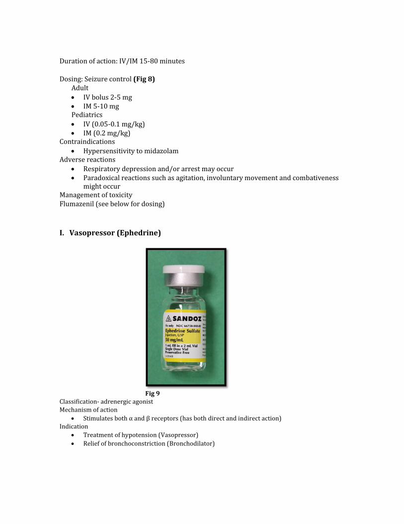
I. Vasopressor (Ephedrine)
Fig 9 Classification- adrenergic agonist Mechanism of action
Stimulates both α and β receptors (has both direct and indirect action) Indication
Treatment of hypotension (Vasopressor) Relief of bronchoconstriction (Bronchodilator)

Pharmacokinetics
Onset or action IV-immediate IM- minutes
Peak effect IV 2-5 minutes IM < 10 minutes
Duration of action: IV/IM 10-60 minutes
Dosing (May repeat dose in 5-10 minutes) (Fig 9) Hypotension: Adult IV 5-20 mg; IM/SC 25-50 mg Pediatric 0.5 mg/kg Bronchospasm: Adult IV 5-20 mg; IM 25-50 mg Pediatric 0.5 mg/kg
Contraindications Hypersensitivity to ephedrine (rare)
Precautions Tolerance may develop- temporary discontinuance restores original effectiveness. Special care should be used when administering to patients with:
Heart disease Angina pectoris Diabetes Hyperthyroidism Hypertension
Monoamine oxidase inhibitors may potentiate the pressor effect of ephedrine, possibly resulting in a hypertensive crisis
Adverse Reactions Nervousness, insomnia, vertigo, headache, tachycardia, palpitation and sweating

J. Corticosteroid (Hydrocortisone)
Fig 10
Classification- Anti-inflammatory Mechanism of action
Decreases inflammation by suppression of migration polymorphonuclear leukocytes and reversal of capillary permeability
It increases blood pressure by increasing the sensitivity of the vasculature to epinephrine and norepinephrine. In the absence of cortisol, widespread vasodilation occurs.
It acts as a physiological antagonist to insulin, thereby increasing blood glucose Indication
Anaphylaxis- management of late phase recurrence of symptoms Management of adrenocortical insufficiency
Pharmacokinetics Onset or action
IV/IM- few minutes Peak effect
IV/IM- < 1 hour Duration of action: short??
Dosing
Allergic reaction: Adult IV 100 mg; Pediatric 1 mg/kg (fig 10) Shock: Adult 100-500 mg IV/IM; Pediatric 1-5 mg/kg IV/IM Q 12 hours
Contraindications/precautions/adverse reactions- few problems in the acute setting

K. Drug Antagonists (Flumazenil)
Fig 11
Classification- Benzodiazepine reversal agent Mechanism of action
Benzodiazepine receptor antagonist Indication
Treatment of benzodiazepine drug overdose Pharmacokinetics
Onset or action: IV 1-2 minutes Peak effect: IV 2-10 minutes Duration of action: 45-90 minutes
Dosing (fig 11) Adult: 0.2 - 1 mg at a rate of 0.2 mg/min; repeat at 20 minute intervals up to 3 mg/hr Pediatrics: 0.01 mg/kg (up to 0.2 mg) over 15 seconds, maximum dose of 0.05 mg/kg or 1 mg Contraindications
Hypersensitivity to Flumazenil or benzodiazepines Patients given benzodiazepines for control of life-threatening conditions (e.g., status epilepticus,
control of intracranial pressure) Precautions
1. Benzodiazepine reversal may result in seizures in some patients 2. Flumazenil does not consistently reverse amnesia (may not recall post-op instructions) 3. Resedation may occur if large single or cumulative doses of benzodiazepines have been used
Adverse Reactions Nausea/vomiting, palpitation,, headache, anxiety, nervousness etc

L. Drug Antagonists (Naloxone)
Fig 12
Classification- opioid reversal agent Mechanism of action
Competitively inhibits opiate agonists at µ, δ, κ receptors Indication
Reverses CNS and respiratory depressant effects of narcotic overdose Pharmacokinetics
Onset or action: IV 1-2 minutes; IM 2-5 minutes Peak effect: IV/IM 5-15 minutes Duration of action: 30-60 minutes
Dosing (Fig 12) Adult: 0.4 - 2 mg every 2-3 minutes as needed; may need to repeat doses every 20-60 minutes. If
no response after 10 mg, question the diagnosis Pediatrics: 0.01 mg/kg repeat every 2-3 minutes based on response
**Use 0.04 mg titration doses in postoperative patients to avoid reversal of analgesia Contraindications
Hypersensitivity to naloxone Precautions
Excessive doses of naloxone may result in reversal of analgesia which may result in significant side effects, such as hypertension, excitement, acute pulmonary edema and cardiac arrhythmia’s in susceptible patients.
Patients who have responded to naloxone should be carefully monitored since the duration of action of some opiates may exceed that of naloxone
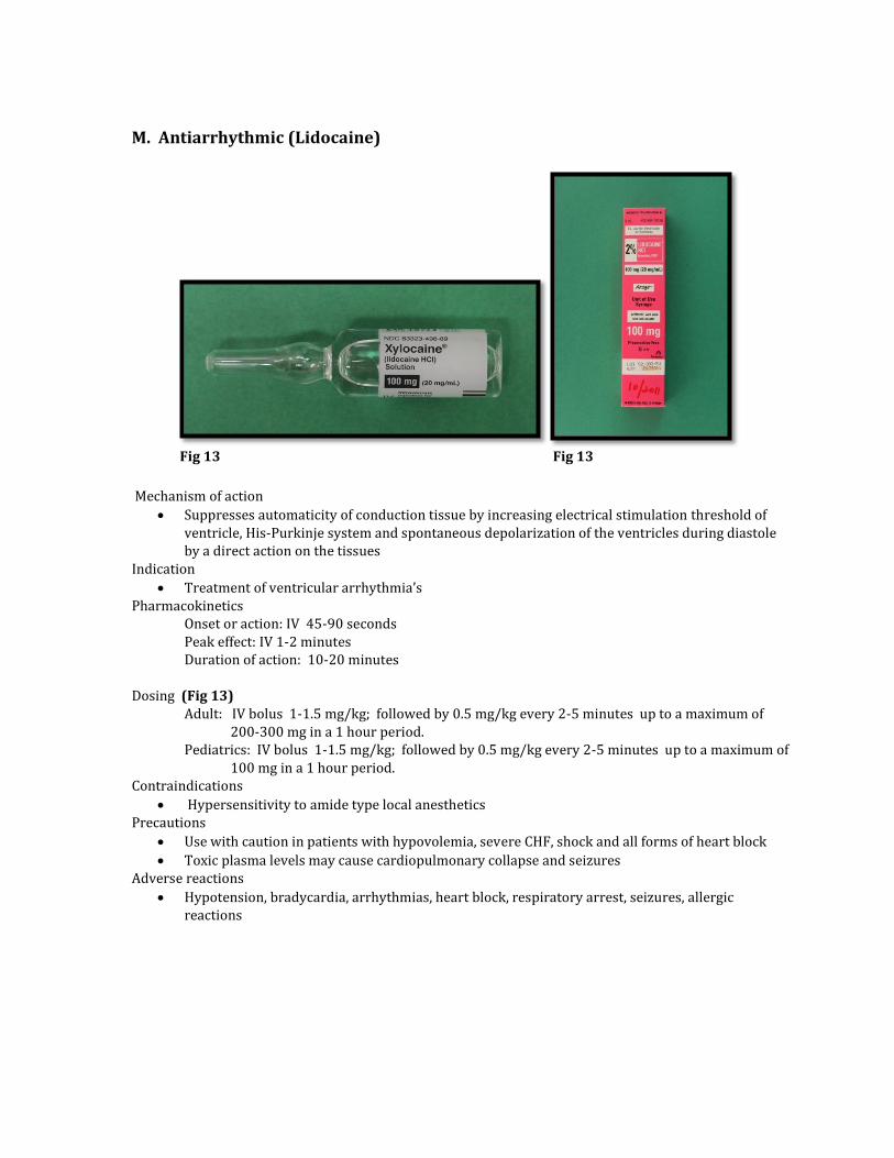
M. Antiarrhythmic (Lidocaine)
Fig 13 Fig 13
Mechanism of action
Suppresses automaticity of conduction tissue by increasing electrical stimulation threshold of ventricle, His-Purkinje system and spontaneous depolarization of the ventricles during diastole by a direct action on the tissues
Indication Treatment of ventricular arrhythmia’s
Pharmacokinetics Onset or action: IV 45-90 seconds Peak effect: IV 1-2 minutes Duration of action: 10-20 minutes
Dosing (Fig 13) Adult: IV bolus 1-1.5 mg/kg; followed by 0.5 mg/kg every 2-5 minutes up to a maximum of
200-300 mg in a 1 hour period. Pediatrics: IV bolus 1-1.5 mg/kg; followed by 0.5 mg/kg every 2-5 minutes up to a maximum of
100 mg in a 1 hour period. Contraindications
Hypersensitivity to amide type local anesthetics Precautions
Use with caution in patients with hypovolemia, severe CHF, shock and all forms of heart block Toxic plasma levels may cause cardiopulmonary collapse and seizures
Adverse reactions Hypotension, bradycardia, arrhythmias, heart block, respiratory arrest, seizures, allergic
reactions

N. Antiarrhythmic (Amiodarone)
Fig 14
Mechanism of action Inhibits adrenergic stimulation (α and β), affects sodium, potassium and calcium channels.
Prolongs the action potential and refractory period in myocardial tissue. Indication
Treatment of ventricular arrhythmia’s Control of rapid ventricular rate due to accessory pathway conduction in pre-excited atrial
arrhythmias Heart rate control in patients with atrial fibrillation (no accessory pathway)
Pharmacokinetics
Onset or action: IV immediate
Dosing (Fig 14) Adult: Pulseless VF/VT- IV push 300 mg in 20-30 ml of NS. If VF or VT recurs, supplemental
doses of 150 mg followed by infusion of 1 mg/min for 6 hours Pediatrics: Pulseless VF/VT 5 mg/kg IV push; repeat up to maximum of 15 mg/kg
Contraindications Hypersensitivity to amiodarone/iodine Severe sinus node dysfunction Second and third degree heart block (except in patients with pacemaker) Patients with preexisting pulmonary disease (e.g., COPD)
Precautions May cause bradycardia or sinus arrest in patients receiving beta-blockers, calcium channel
blockers or lidocaine

O. Skeletal Muscle Relaxant (Dantrolene)
Fig 15
Mechanism of action Acts directly on skeletal muscle by interfering with release of calcium ion from the sarcoplasmic
reticulum One of its most important effects is that cellular metabolism returns to an aerobic process,
respiration normalizes and acidosis is reversed. This rapidly reduces potassium levels because with correction of acidosis. Potassium return to the cells and produces cardiac stability
Indication 1. Treatment of malignant hyperthermia 2. Treatment of spasticity associated with spinal cord injury , stroke, cerebral palsy or multiple
sclerosis
Dosing (Fig 15) 2.5 mg/kg; may repeat dose up to 10 mg/kg ; repeat if MH recurs (can give up to 30 mg/kg) Post-crisis 1-2 mg/kg Q 6 hours for 24 hours Some recommend oral dantrolene 1 mg/kg Q 4-8 hours for 48 hours
Contraindications Avoid combination with calcium channel blockers
Precautions May cause skeletal muscle weakness
How supplied Vials with 20 mg dantrolene and 3000 mg mannitol to be reconstituted in 60 ml sterile water

P. Skeletal Muscle Relaxant (Succinylcholine)
Fig 16
Classification- depolarizing neuromuscular blocker Mechanism of action
Binds cholinergic receptors of the motor end plate to produce depolarization observed as fasciculations
Indication Treatment of laryngospasm To facilitate rapid sequence and routine endotracheal intubation and relax skeletal muscle
during surgery Pharmacokinetics Onset of action
IV- complete relaxation 30-60 seconds IM 2-3 minutes Duration IV 4-6 minutes IM 10-30 minutes
Dosing (Fig 16)
Adult/pediatrics (use ideal body weight) Laryngospasm 10-20 mg (0.2 mg/kg) Routine intubation dose 1-1.5 mg/kg
Precautions Succinylcholine is a triggering agent for malignant hyperthermia Repeated administration at short intervals (<5 minutes) is associated with bradycardia Succinylcholine elevates serum potassium concentration (0.5 mEq/L in normal patients).
Alarming levels of potassium along with cardiovascular collapse may occur when succinylcholine is used in patients with severe burns, electrolyte imbalance, severe trauma, paraplegia, spinal cord injuries, upper motor neuron injury, or degenerative or dystrophic neuromuscular disease

Contraindications
Patients with genetic disorders of plasma pseudocholinesterase Familial history of malignant hyperthermia, or myopathies with elevated creatine
phosphokinase (CPK) values After acute phase of injury following major burns, multiple trauma, extensive denervation of
skeletal muscle or upper motor neuron injury Avoid use in patients with acute narrow angle glaucoma, penetrating eye injuries
R. Antihypertensive (Hydralazine)
Fig 17
Classification- vasodilator Mechanism of action
Direct vasodilation of arterioles(little effect on veins), probably by interference with calcium ion transport in vascular smooth muscle
Indication Management of moderate to severe hypertension Reduction of afterload in treatment of congestive heart failure
Pharmacokinetics Onset of action
IV 5-20 minutes Duration
IV 1-4 hours Dosing (fig 17)
Adults 5mg aliquots (titrate to effect every 5 minutes) maximum dose 20 mg
Pediatrics 0.1 -0.2 mg/kg as above
Precautions Decrease in blood pressure is frequently accompanied by an increase in heart rate and by
increases in cardiac output and stroke volume Contraindications
Hypersensitivity to hydralazine

References
1. Caffrey SL, Willoughby PJ, Pepe PE, et al. Public use of automated external defibrillators. N Engl J Med. 2002; 347: 1242–1247.
2. Robertson RM. Sudden death from cardiac arrest—improving the odds. N Engl J Med. 2000; 343: 1259–1260.
3. Page RL, Joglar JA, Kowal RC, et al. Use of automated external defibrillators by a U.S. airline. N Engl J Med. 2000; 343: 1210–1216.
4. Valenzuela TD, Roe DJ, Nichol G, et al. Outcomes of rapid defibrillation by security officers after cardiac arrest in casinos. N Engl J Med. 2000; 343: 1206–1209.
5. White RD, Asplin BR, Bugliosi TF, et al. High discharge survival rate after out-of-hospital ventricular fibrillation with rapid defibrillation by police and paramedics. Ann Emerg Med. 1996; 28: 480–485.
6. White RD, Hankins DG, Bugliosi TF. Seven years’ experience with early defibrillation by police and paramedics in an emergency medical services system. Resuscitation. 1998; 39: 145–151.
7. White RD, Hankins DG, Atkinson EJ. Patient outcomes following defibrillation with a low energy biphasic truncated exponential waveform in out-of-hospital cardiac arrest. Resuscitation. 2001; 49: 9–14.
8. Bunch TJ, White RD, Gersh BJ, et al. Long-term outcomes of out-of-hospital cardiac arrest after successful early defibrillation. N Engl J Med. 2003; 348: 2626–2633.
9. The American Heart Association in collaboration with the International Liaison Committee on Resuscitation. Part 4: the automated external defibrillator: key link in the chain of survival. In: Guidelines 2000 for Cardiopulmonary Resuscitation and Emergency Cardiovascular Care. Circulation. 2000; 102(suppl 8): I-60–I-76.
10. Kannel WB, Wilson PW, D’Agostino RB, et al. Sudden coronary death in women. Am Heart J. 1998; 136: 205–212.
11. de Vreede-Swagemakers JJ, Gorgels AP, Dubois-Arbouw WI, et al. Out-of-hospital cardiac arrest in the 1990’s: a population-based study in the Maastricht area on incidence, characteristics and survival. J Am Coll Cardiol. 1997; 30: 1500–1505.
12. Kuisma M, Maatta T. Out-of-hospital cardiac arrests in Helsinki: Utstein style reporting. Heart. 1996; 76: 18–23.
13. Liberthson RR. Sudden death from cardiac causes in children and young adults. N Engl J Med. 1996; 334: 1039–1044.
14. www.americanheart.org 15. Eisenberg M. Resuscitate! How your community can improve survival from sudden
cardiac arrest. University of Washington Press 2009 16. Lange RA, Hillis LD. Cardiovascular complications of cocaine use. N Engl J Med
2001;345:351-358 17. Mittlemen MA, et al. Triggering of myocardial infarction by cocaine. Circulation
1999;99:2737-2741 18. Qureshi AI et al. Cocaine use and the likelihood of nonfatal myocardial infarction and
stroke: data from the Third National Health and Nutrition Examination Survey. Circulation 2001; 103:502-506