
Joe Kanaan - OXY USA Dan Holmes - Aqueos Prevention First ...
Upload
melina-purifoyCategory
view
219download
0
Mediastinal StagingMediastinal Staging
Samer Kanaan, M.D.Samer Kanaan, M.D.
OverviewOverview
Importance of accurate nodal stagingImportance of accurate nodal stagingAccuracy of radiographic stagingAccuracy of radiographic stagingMediastinoscopyMediastinoscopyEUSEUSEBUSEBUS

StagingStaging
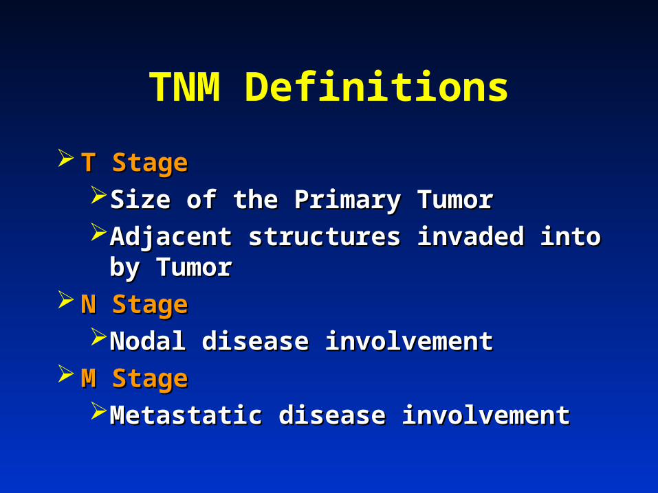
TNM Definitions
T StageT StageSize of the Primary TumorSize of the Primary TumorAdjacent structures invaded into by Adjacent structures invaded into by
TumorTumor N StageN Stage
Nodal disease involvementNodal disease involvement M StageM Stage
Metastatic disease involvementMetastatic disease involvement
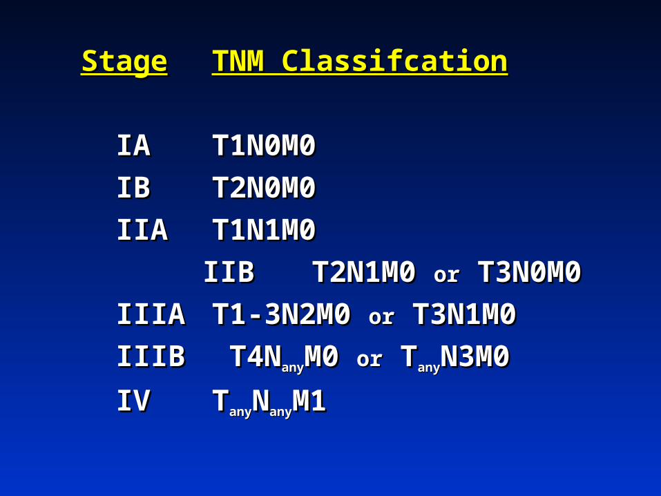
StageStage TNM ClassifcationTNM Classifcation
IAIA T1N0M0T1N0M0
IBIB T2N0M0T2N0M0
IIAIIA T1N1M0T1N1M0
IIBIIB T2N1M0 T2N1M0 oror T3N0M0 T3N0M0
IIIAIIIA T1-3N2M0 T1-3N2M0 oror T3N1M0T3N1M0
IIIBIIIB T4N T4NanyanyM0 M0 oror T TanyanyN3M0N3M0
IVIV TTanyanyNNanyanyM1M1

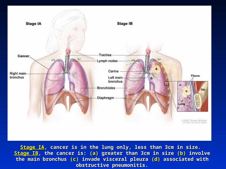
Stage IAStage IA, cancer is in the lung only, less than 3cm in size. , cancer is in the lung only, less than 3cm in size. Stage IBStage IB, the cancer is: , the cancer is: (a)(a) greater than 3cm in size greater than 3cm in size (b)(b) involve the main bronchus involve the main bronchus
(c) (c) invade visceral pleura invade visceral pleura (d)(d) associated with obstructive pneumonitis. associated with obstructive pneumonitis.
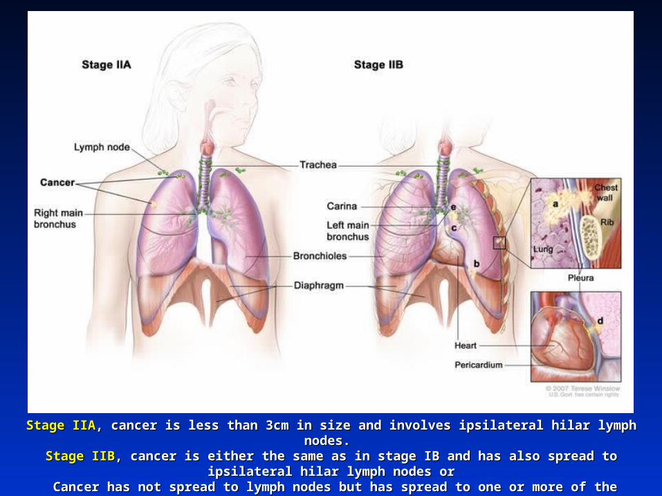
Stage IIAStage IIA, cancer is less than 3cm in size and involves ipsilateral hilar lymph nodes. , cancer is less than 3cm in size and involves ipsilateral hilar lymph nodes. Stage IIBStage IIB, cancer is either the same as in stage IB and has also spread to ipsilateral hilar lymph nodes or, cancer is either the same as in stage IB and has also spread to ipsilateral hilar lymph nodes or Cancer has not spread to lymph nodes but has spread to one or more of the following: Cancer has not spread to lymph nodes but has spread to one or more of the following: (a)(a) the chest wall, the chest wall, (b)(b) the diaphragm, the diaphragm, (c)(c) mediastinal pleura, mediastinal pleura, (d)(d) pericardium, pericardium, (e)(e) the main bronchus less than 2cm from the the main bronchus less than 2cm from the
carina, and/or carina, and/or (f) (f) associated obstructive pneumonitis of the entire lung. associated obstructive pneumonitis of the entire lung.
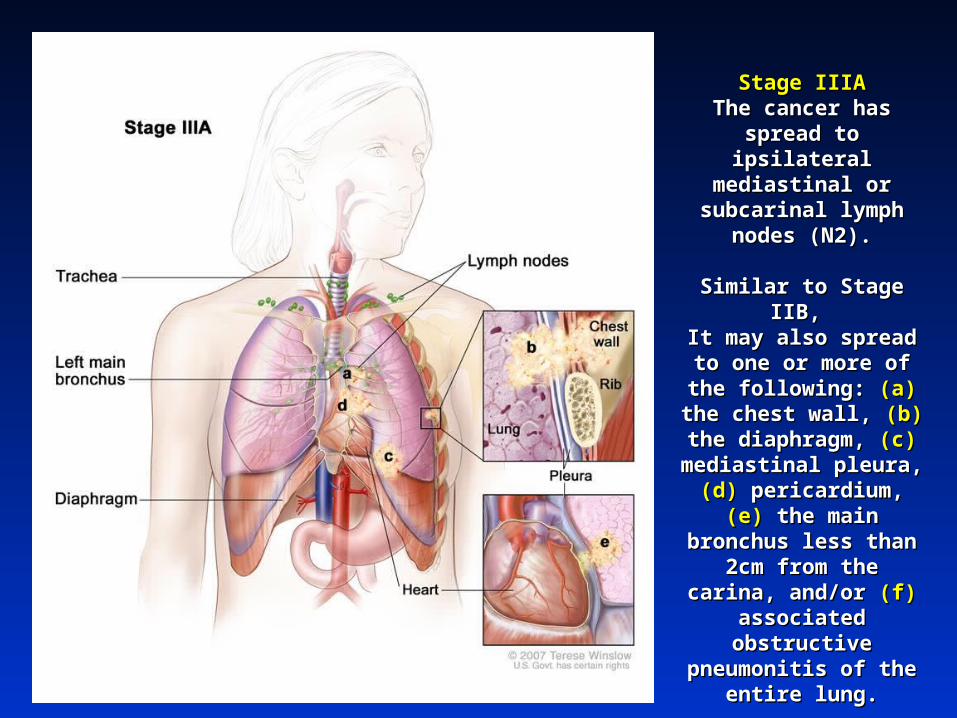
Stage IIIAStage IIIAThe cancer has spread The cancer has spread
to ipsilateral to ipsilateral mediastinal or mediastinal or
subcarinal lymph nodes subcarinal lymph nodes (N2).(N2).
Similar to Stage IIB, Similar to Stage IIB, It may also spread to It may also spread to
one or more of the one or more of the following: following: (a)(a) the chest the chest wall, wall, (b)(b) the diaphragm, the diaphragm, (c)(c) mediastinal pleura, mediastinal pleura, (d)(d) pericardium, pericardium, (e)(e) the the
main bronchus less main bronchus less than 2cm from the than 2cm from the carina, and/or carina, and/or (f) (f)
associated obstructive associated obstructive pneumonitis of the pneumonitis of the
entire lung.entire lung.
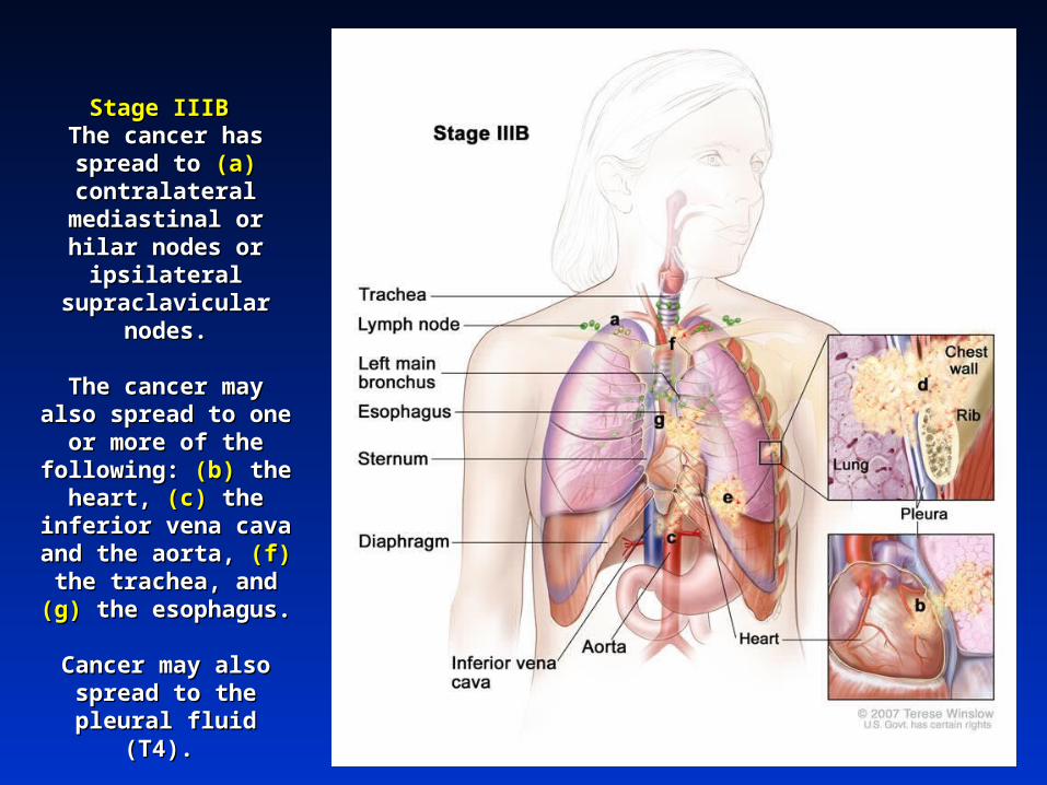
Stage IIIB Stage IIIB The cancer has spread The cancer has spread
to to (a)(a) contralateral contralateral mediastinal or hilar mediastinal or hilar nodes or ipsilateral nodes or ipsilateral
supraclavicular nodes.supraclavicular nodes.
The cancer may also The cancer may also spread to one or more spread to one or more of the following: of the following: (b)(b) the the
heart, heart, (c)(c) the inferior the inferior vena cava and the vena cava and the
aorta, aorta, (f)(f) the trachea, the trachea, and and (g) (g) the esophagus.the esophagus.
Cancer may also Cancer may also spread to the pleural spread to the pleural
fluid (T4). fluid (T4).
Separate nodules in Separate nodules in the same lobe is also the same lobe is also
(T4)*(T4)*

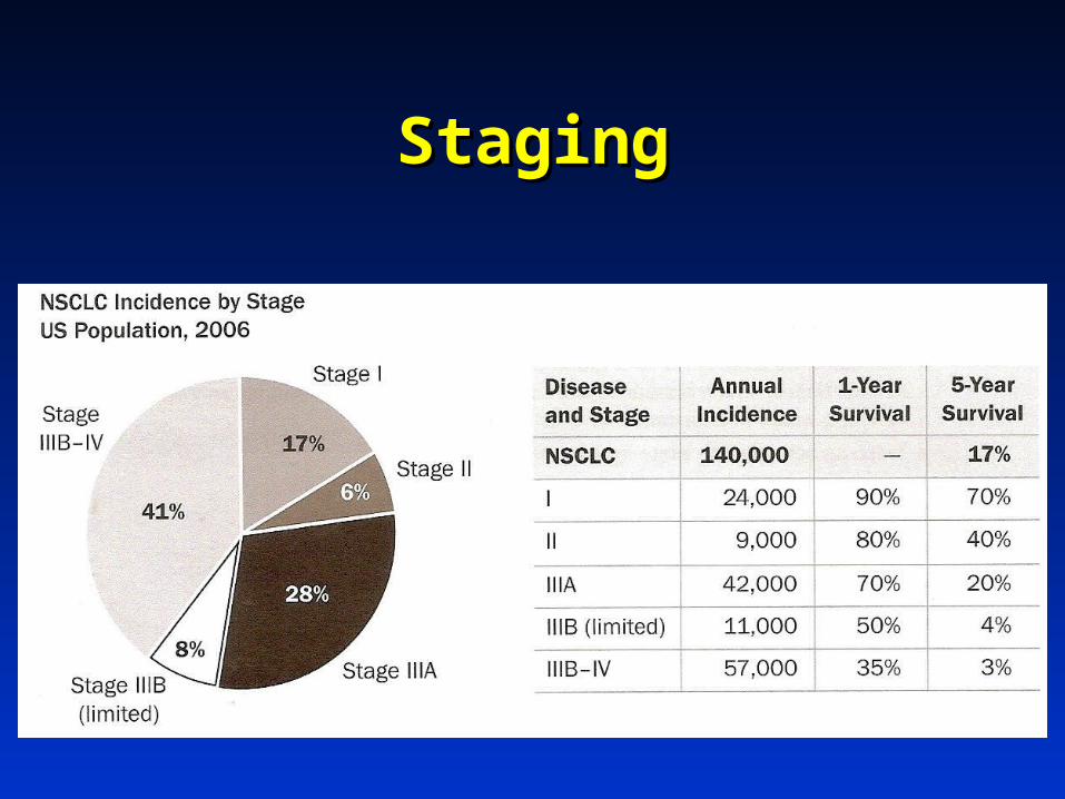
StagingStaging

StageStage TNM ClassifcationTNM Classifcation 5 Year Survival5 Year Survival
IAIA T1N0M0 T1N0M0 6767
IBIB T2N0M0 T2N0M0 5757
IIAIIA T1N1M0 T1N1M0 5555
IIBIIB T2N1M0 T2N1M0 oror T3N0M0 T3N0M0 3939
IIIAIIIA T1-3N2M0 T1-3N2M0 oror T3N1M0 T3N1M0 2323
IIIBIIIB T4N T4NanyanyM0 M0 oror T TanyanyN3M0N3M0 5 5
IVIV T TanyanyNNanyanyM1M1 1 1
Mountain, Mountain, ChestChest 1997 1997
Why is accurate nodal staging Why is accurate nodal staging essential?essential?
N1 diseaseN1 diseaseN2 diseaseN2 diseaseN3 diseaseN3 disease
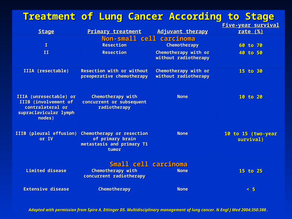
Treatment of Lung Cancer According to StageTreatment of Lung Cancer According to StageStageStage Primary treatmentPrimary treatment Adjuvant therapyAdjuvant therapy Five-year survival rate (%)Five-year survival rate (%)
Non-small cell carcinomaNon-small cell carcinomaII ResectionResection ChemotherapyChemotherapy 60 to 7060 to 70
IIII ResectionResection Chemotherapy with or without Chemotherapy with or without radiotherapyradiotherapy
40 to 5040 to 50
IIIA (resectable)IIIA (resectable) Resection with or without Resection with or without preoperative chemotherapypreoperative chemotherapy
Chemotherapy with or without Chemotherapy with or without radiotherapyradiotherapy
15 to 3015 to 30
IIIA (unresectable) or IIIB IIIA (unresectable) or IIIB (involvement of contralateral or (involvement of contralateral or supraclavicular lymph nodes)supraclavicular lymph nodes)
Chemotherapy with concurrent Chemotherapy with concurrent or subsequent radiotherapyor subsequent radiotherapy
NoneNone 10 to 2010 to 20
IIIB (pleural effusion) or IVIIIB (pleural effusion) or IV Chemotherapy or resection of Chemotherapy or resection of primary brain metastasis and primary brain metastasis and
primary T1 tumorprimary T1 tumor
NoneNone 10 to 15 (two-year survival)10 to 15 (two-year survival)
Small cell carcinomaSmall cell carcinomaLimited diseaseLimited disease Chemotherapy with concurrent Chemotherapy with concurrent
radiotherapyradiotherapyNoneNone 15 to 2515 to 25
Extensive diseaseExtensive disease ChemotherapyChemotherapy NoneNone < 5< 5
Adapted with permission from Spira A, Ettinger DS. Multidisciplinary management of lung cancer. N Engl J Med 2004;350:388Adapted with permission from Spira A, Ettinger DS. Multidisciplinary management of lung cancer. N Engl J Med 2004;350:388..
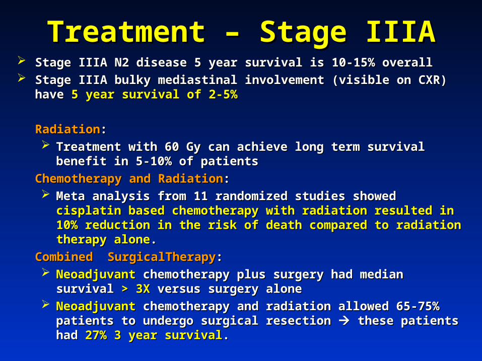
Treatment – Stage IIIATreatment – Stage IIIA Stage IIIA N2 disease 5 year survival is 10-15% overallStage IIIA N2 disease 5 year survival is 10-15% overall Stage IIIA bulky mediastinal involvement (visible on CXR) have Stage IIIA bulky mediastinal involvement (visible on CXR) have 5 year 5 year
survival of 2-5%survival of 2-5%
RadiationRadiation: : Treatment with 60 Gy can achieve long term survival benefit in 5-10% Treatment with 60 Gy can achieve long term survival benefit in 5-10%
of patientsof patients
Chemotherapy and RadiationChemotherapy and Radiation: : Meta analysis from 11 randomized studies showed Meta analysis from 11 randomized studies showed cisplatin based cisplatin based
chemotherapy with radiation resulted in 10% reduction in the risk of chemotherapy with radiation resulted in 10% reduction in the risk of death compared to radiation therapy alonedeath compared to radiation therapy alone..
Combined SurgicalTherapyCombined SurgicalTherapy: : NeoadjuvantNeoadjuvant chemotherapy plus surgery had median survival chemotherapy plus surgery had median survival > 3X > 3X
versus surgery aloneversus surgery alone NeoadjuvantNeoadjuvant chemotherapy and radiation allowed 65-75% patients to chemotherapy and radiation allowed 65-75% patients to
undergo surgical resection undergo surgical resection these patients had these patients had 27% 3 year survival27% 3 year survival..
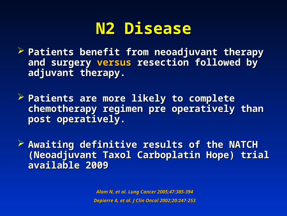
N2 DiseaseN2 Disease Patients benefit from neoadjuvant therapy and Patients benefit from neoadjuvant therapy and
surgery surgery versusversus resection followed by adjuvant resection followed by adjuvant therapy. therapy.
Patients are more likely to complete chemotherapy Patients are more likely to complete chemotherapy regimen pre operatively than post operatively.regimen pre operatively than post operatively.
Awaiting definitive results of the NATCH Awaiting definitive results of the NATCH (Neoadjuvant Taxol Carboplatin Hope) trial available (Neoadjuvant Taxol Carboplatin Hope) trial available 2009 2009
Alam N, et al. Lung Cancer 2005;47:385-394Alam N, et al. Lung Cancer 2005;47:385-394
Depierre A, et al. J Clin Oncol 2002;20:247-253Depierre A, et al. J Clin Oncol 2002;20:247-253
What is the accuracy of What is the accuracy of radiographic staging?radiographic staging?
CT ScanCT Scan
Information gained by CTInformation gained by CT
Tumor sizeTumor size Tumor numberTumor number Central tumor or PeripheralCentral tumor or Peripheral Lymph node enlargement (>1 cm)Lymph node enlargement (>1 cm) Extent Extent
Discrete lymph nodes versus mediastinal Discrete lymph nodes versus mediastinal infiltrationinfiltration
Metastatic diseaseMetastatic disease

Accuracy of CT in Staging
CT scanCT scanTumorTumor
Sensitivity = 63%Sensitivity = 63%Specificity = 84%Specificity = 84%
Mediastinum Mediastinum Sensitivity = 51-75%Sensitivity = 51-75%Specificity = 66-86%Specificity = 66-86%Positive predictive value = 60%Positive predictive value = 60%Negative predictive value = 80%Negative predictive value = 80%
Toloza E, et al. Chest 2003(suppl):137s–146sToloza E, et al. Chest 2003(suppl):137s–146sGould MK, et al. Ann Intern Med 2003; 139:879–892Gould MK, et al. Ann Intern Med 2003; 139:879–892
Dwamena et al. Radiology 1999; 213:530–536Dwamena et al. Radiology 1999; 213:530–536
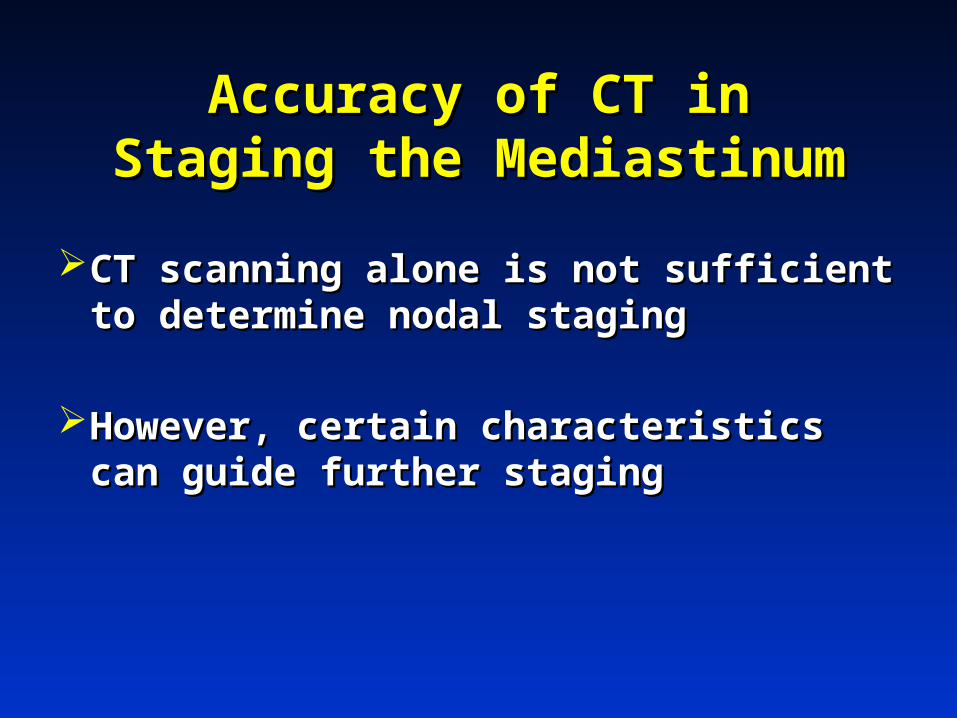
Accuracy of CT in Staging the Accuracy of CT in Staging the MediastinumMediastinum
CT scanning alone is not sufficient to CT scanning alone is not sufficient to determine nodal stagingdetermine nodal staging
However, certain characteristics can However, certain characteristics can guide further stagingguide further staging
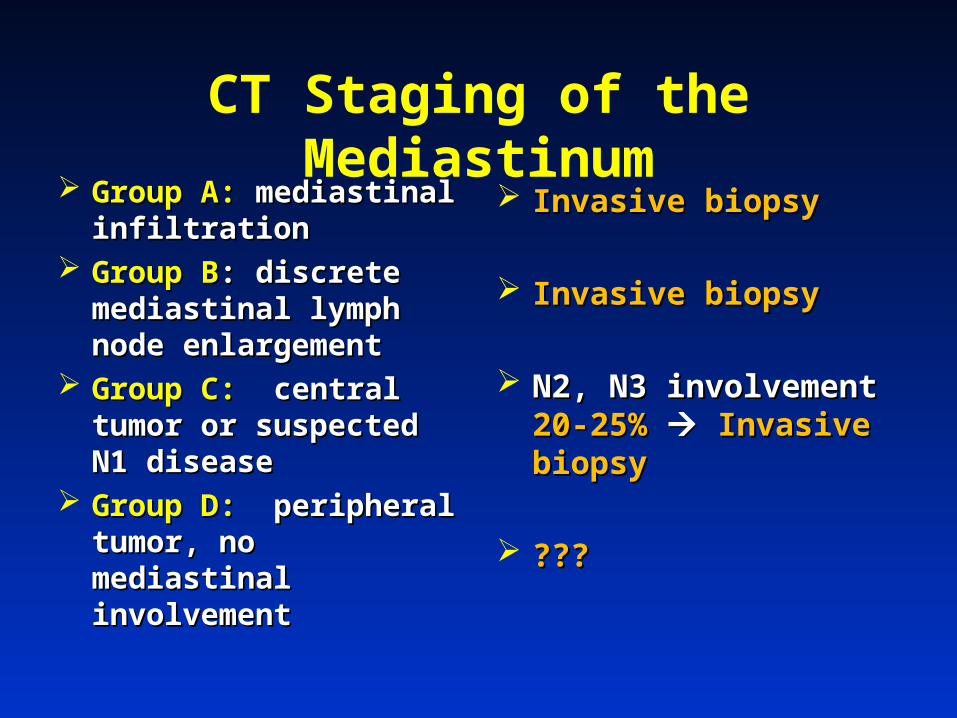
CT Staging of the Mediastinum Group A:Group A: mediastinal mediastinal
infiltrationinfiltration Group BGroup B: discrete : discrete
mediastinal lymph node mediastinal lymph node enlargementenlargement
Group C:Group C: central tumor central tumor or suspected N1 diseaseor suspected N1 disease
Group D:Group D: peripheral peripheral tumor, no mediastinal tumor, no mediastinal involvementinvolvement
Invasive biopsyInvasive biopsy
Invasive biopsyInvasive biopsy
N2, N3 involvement N2, N3 involvement 20-20-25%25% Invasive biopsyInvasive biopsy
??????
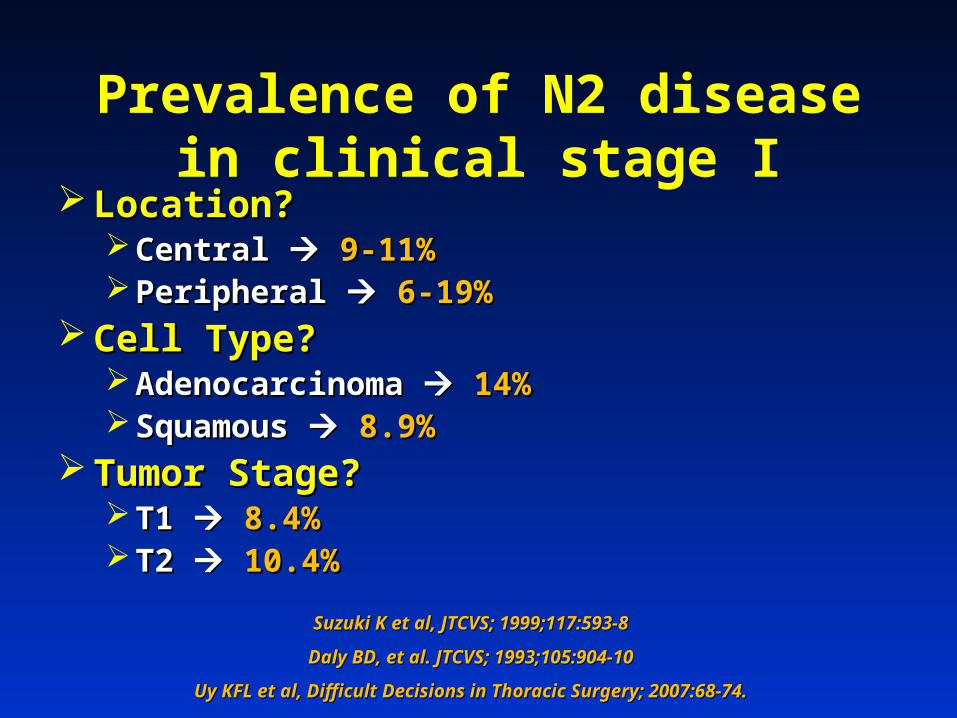
Prevalence of N2 disease in clinical stage I
Location?Location? Central Central 9-11%9-11% Peripheral Peripheral 6-19%6-19%
Cell Type?Cell Type? Adenocarcinoma Adenocarcinoma 14%14% Squamous Squamous 8.9%8.9%
Tumor Stage?Tumor Stage? T1 T1 8.4%8.4% T2 T2 10.4%10.4%
Suzuki K et al, JTCVS; 1999;117:593-8Suzuki K et al, JTCVS; 1999;117:593-8
Daly BD, et al. JTCVS; 1993;105:904-10Daly BD, et al. JTCVS; 1993;105:904-10
Uy KFL et al, Difficult Decisions in Thoracic Surgery; 2007:68-74.Uy KFL et al, Difficult Decisions in Thoracic Surgery; 2007:68-74.
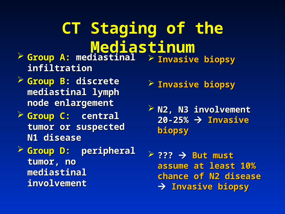
CT Staging of the Mediastinum Group A:Group A: mediastinal mediastinal
infiltrationinfiltration Group BGroup B: discrete : discrete
mediastinal lymph node mediastinal lymph node enlargementenlargement
Group C:Group C: central tumor central tumor or suspected N1 diseaseor suspected N1 disease
Group D:Group D: peripheral peripheral tumor, no mediastinal tumor, no mediastinal involvementinvolvement
Invasive biopsyInvasive biopsy
Invasive biopsyInvasive biopsy
N2, N3 involvement 20-N2, N3 involvement 20-25% 25% Invasive biopsyInvasive biopsy
??? ??? But must But must assume at least 10% assume at least 10% chance of N2 disease chance of N2 disease Invasive biopsyInvasive biopsy
PET ScanPET Scan
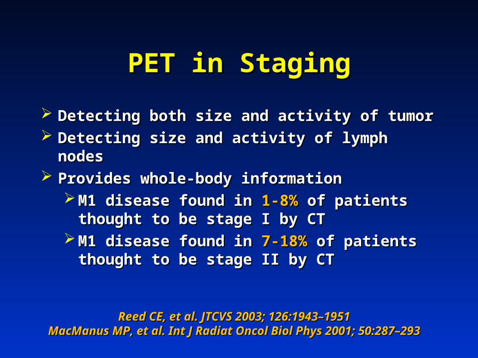
PET in StagingPET in Staging
Detecting both size and activity of tumorDetecting both size and activity of tumor Detecting size and activity of lymph nodesDetecting size and activity of lymph nodes Provides whole-body informationProvides whole-body information
M1 disease found in M1 disease found in 1-8%1-8% of patients thought of patients thought to be stage I by CTto be stage I by CT
M1 disease found in M1 disease found in 7-18%7-18% of patients thought of patients thought to be stage II by CTto be stage II by CT
Reed CE, et al. JTCVS 2003; 126:1943–1951Reed CE, et al. JTCVS 2003; 126:1943–1951MacManus MP, et al. Int J Radiat Oncol Biol Phys 2001; 50:287–MacManus MP, et al. Int J Radiat Oncol Biol Phys 2001; 50:287–
293293
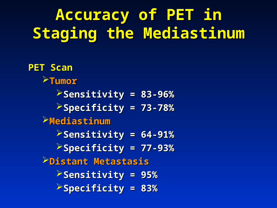
Accuracy of PET in Staging the Accuracy of PET in Staging the MediastinumMediastinum
PET ScanPET ScanTumorTumor
Sensitivity = 83-96%Sensitivity = 83-96%Specificity = 73-78%Specificity = 73-78%
Mediastinum Mediastinum Sensitivity = 64-91%Sensitivity = 64-91%Specificity = 77-93%Specificity = 77-93%
Distant MetastasisDistant MetastasisSensitivity = 95%Sensitivity = 95%Specificity = 83%Specificity = 83%
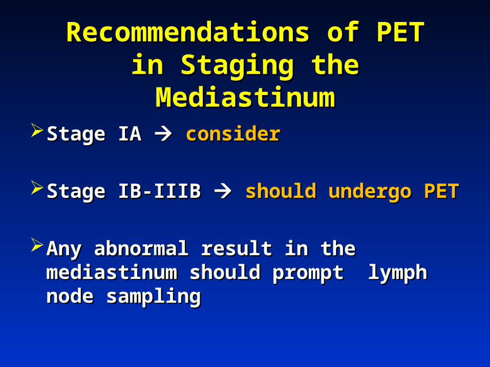
Recommendations of PET in Recommendations of PET in Staging the MediastinumStaging the Mediastinum
Stage IA Stage IA considerconsider
Stage IB-IIIB Stage IB-IIIB should undergo PETshould undergo PET
Any abnormal result in the Any abnormal result in the mediastinum should prompt lymph mediastinum should prompt lymph node samplingnode sampling

PET/CT combinedPET/CT combined
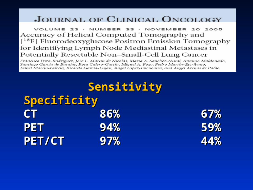
SensitivitySensitivity SpecificitySpecificityCTCT 86%86% 67%67%PETPET 94%94% 59%59%PET/CTPET/CT 97%97% 44%44%
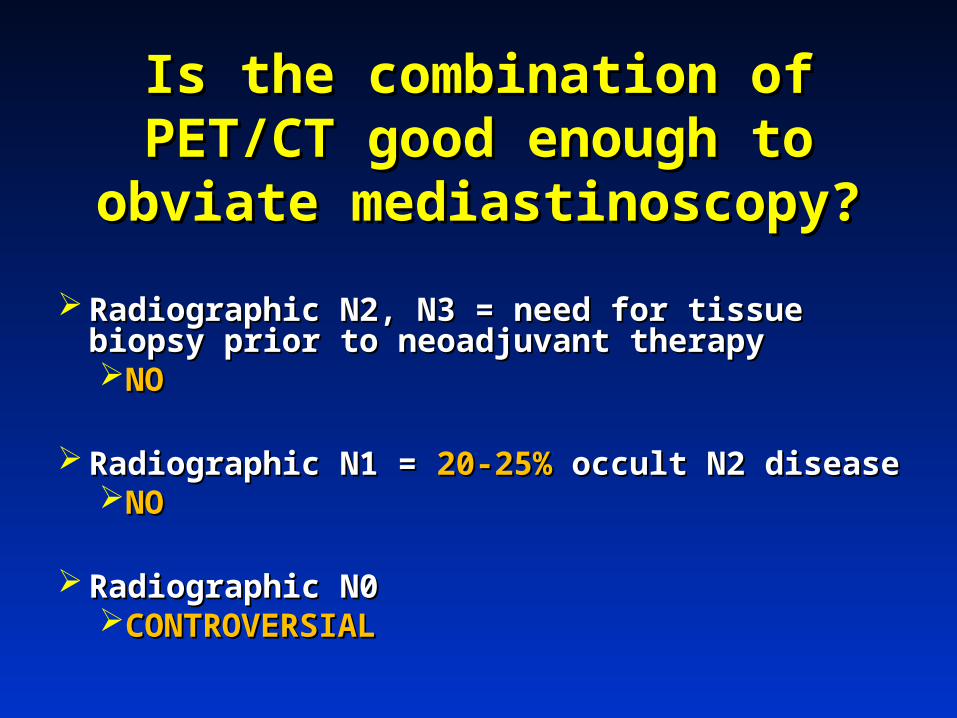
Is the combination of PET/CT Is the combination of PET/CT good enough to obviate good enough to obviate
mediastinoscopy?mediastinoscopy?
Radiographic N2, N3 = need for tissue biopsy Radiographic N2, N3 = need for tissue biopsy prior to neoadjuvant therapy prior to neoadjuvant therapy NONO
Radiographic N1 = Radiographic N1 = 20-25%20-25% occult N2 disease occult N2 disease NONO
Radiographic N0Radiographic N0CONTROVERSIALCONTROVERSIAL
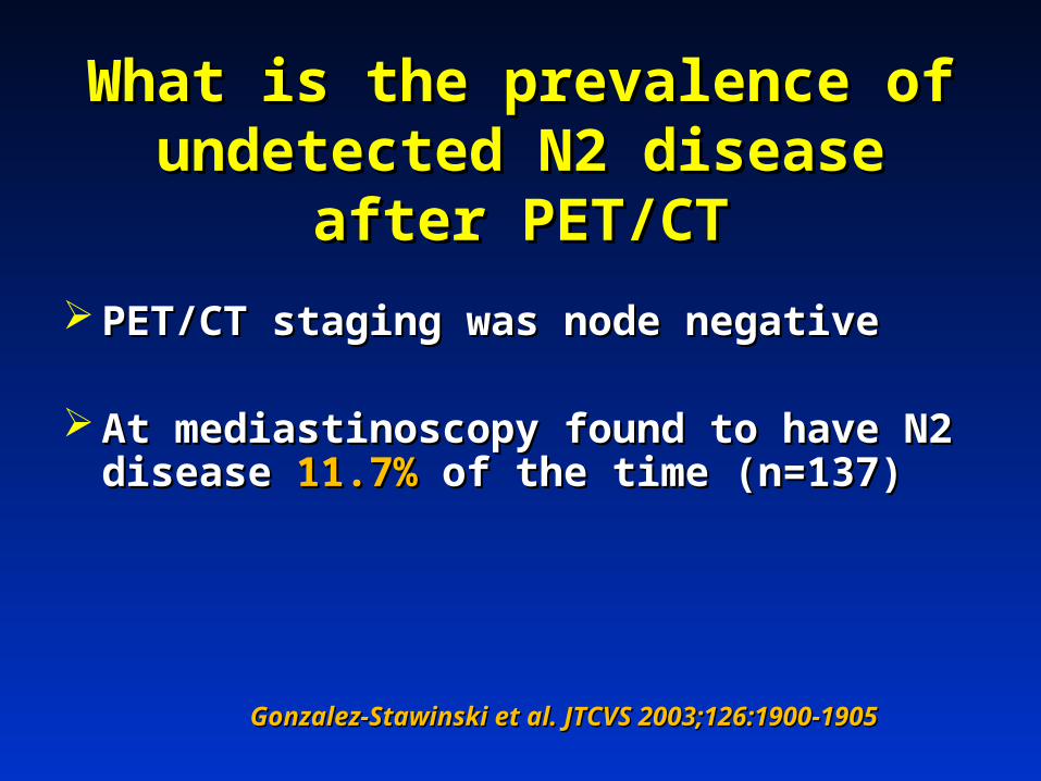
What is the prevalence of What is the prevalence of undetected N2 disease after undetected N2 disease after
PET/CTPET/CT
PET/CT staging was node negativePET/CT staging was node negative
At mediastinoscopy found to have N2 At mediastinoscopy found to have N2 disease disease 11.7%11.7% of the time (n=137) of the time (n=137)
Gonzalez-Stawinski et al. JTCVS 2003;126:1900-1905Gonzalez-Stawinski et al. JTCVS 2003;126:1900-1905
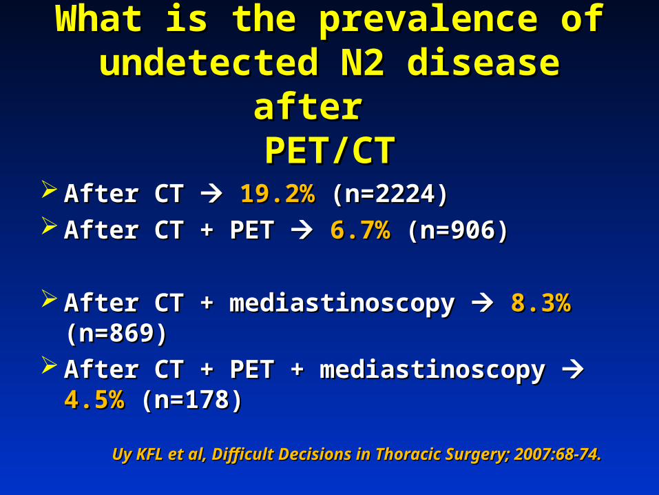
What is the prevalence of What is the prevalence of undetected N2 disease after undetected N2 disease after
PET/CTPET/CT
After CT After CT 19.2%19.2% (n=2224) (n=2224) After CT + PET After CT + PET 6.7%6.7% (n=906) (n=906)
After CT + mediastinoscopy After CT + mediastinoscopy 8.3%8.3% (n=869) (n=869) After CT + PET + mediastinoscopy After CT + PET + mediastinoscopy 4.5%4.5%
(n=178)(n=178)
Uy KFL et al, Difficult Decisions in Thoracic Surgery; 2007:68-74.Uy KFL et al, Difficult Decisions in Thoracic Surgery; 2007:68-74.
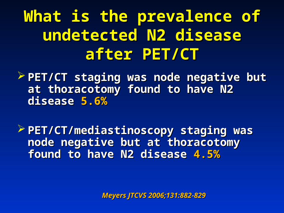
What is the prevalence of What is the prevalence of undetected N2 disease after undetected N2 disease after
PET/CTPET/CT PET/CT staging was node negative but at PET/CT staging was node negative but at
thoracotomy found to have N2 disease thoracotomy found to have N2 disease 5.6%5.6%
PET/CT/mediastinoscopy staging was node PET/CT/mediastinoscopy staging was node negative but at thoracotomy found to have negative but at thoracotomy found to have N2 disease N2 disease 4.5%4.5%
Meyers JTCVS 2006;131:882-829Meyers JTCVS 2006;131:882-829
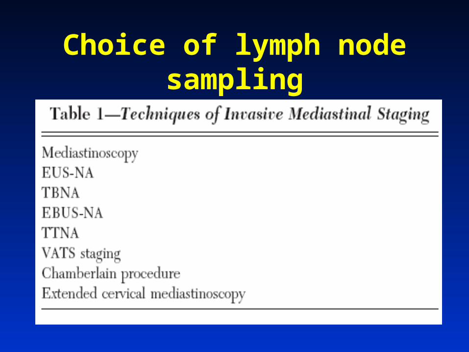
Choice of lymph node sampling
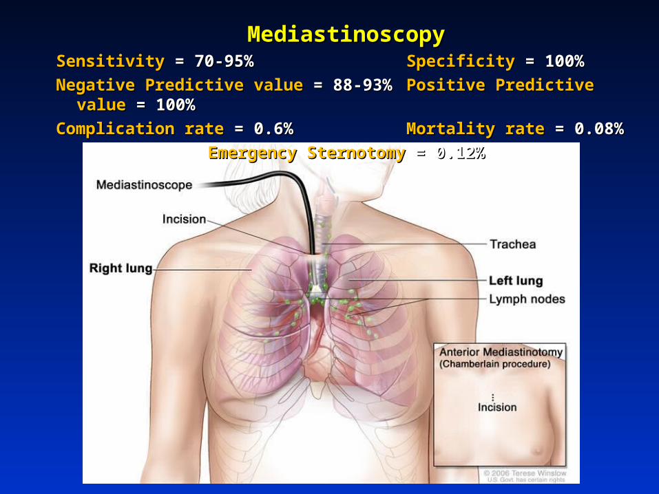
MediastinoscopyMediastinoscopySensitivitySensitivity = 70-95% = 70-95% SpecificitySpecificity = 100% = 100%
Negative Predictive value Negative Predictive value = 88-93%= 88-93% Positive Predictive value Positive Predictive value = 100%= 100%
Complication rate Complication rate = 0.6%= 0.6% Mortality rateMortality rate = 0.08% = 0.08%
Emergency Sternotomy Emergency Sternotomy = 0.12%= 0.12%
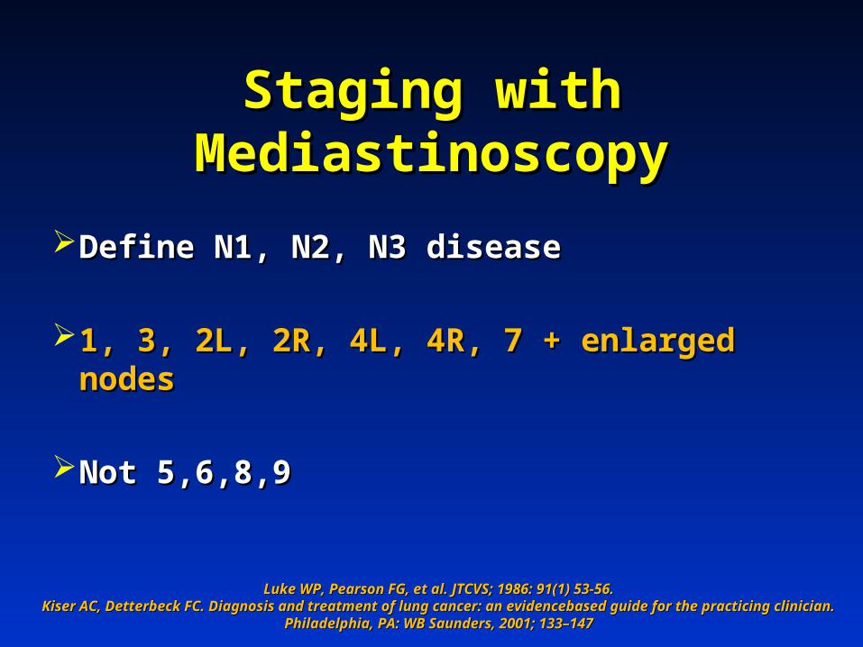
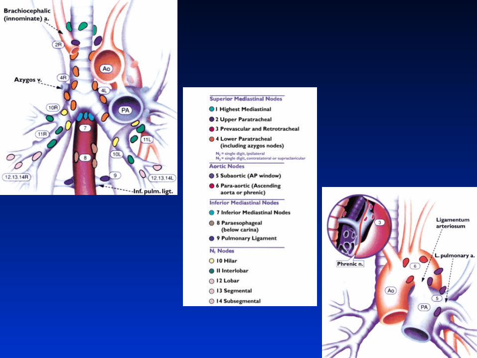
Staging with Staging with MediastinoscopyMediastinoscopy
Define N1, N2, N3 diseaseDefine N1, N2, N3 disease
1, 3, 2L, 2R, 4L, 4R, 7 + enlarged nodes1, 3, 2L, 2R, 4L, 4R, 7 + enlarged nodes
Not 5,6,8,9Not 5,6,8,9
Luke WP, Pearson FG, et al. JTCVS; 1986: 91(1) 53-56.Luke WP, Pearson FG, et al. JTCVS; 1986: 91(1) 53-56.Kiser AC, Detterbeck FC. Diagnosis and treatment of lung cancer: an evidencebased guide for the practicing Kiser AC, Detterbeck FC. Diagnosis and treatment of lung cancer: an evidencebased guide for the practicing
clinician. Philadelphia, PA: WB Saunders, 2001; 133–147clinician. Philadelphia, PA: WB Saunders, 2001; 133–147
What’s the real problem with mediastinoscopy?
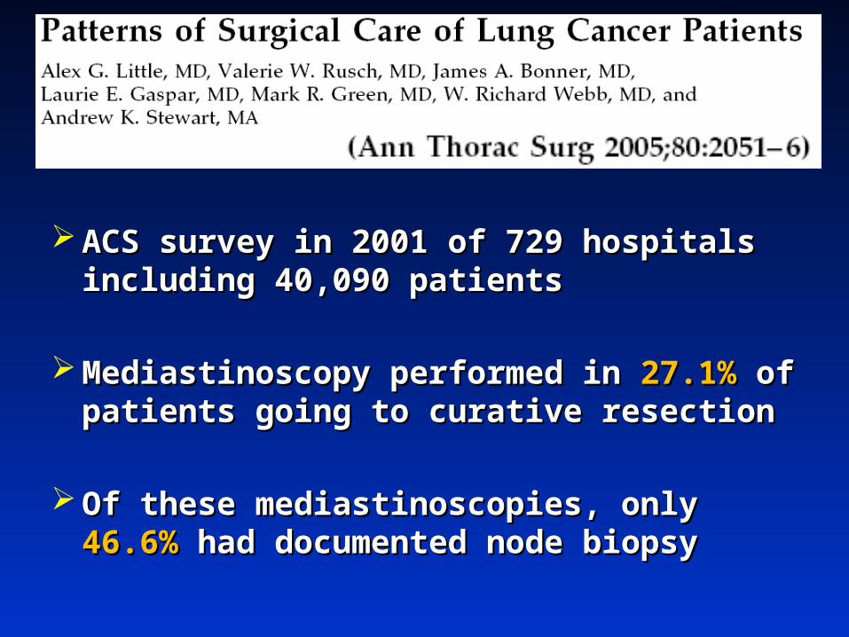
ACS survey in 2001 of 729 hospitals ACS survey in 2001 of 729 hospitals including 40,090 patientsincluding 40,090 patients
Mediastinoscopy performed in Mediastinoscopy performed in 27.1%27.1% of of patients going to curative resectionpatients going to curative resection
Of these mediastinoscopies, only Of these mediastinoscopies, only 46.6%46.6% had had documented node biopsydocumented node biopsy
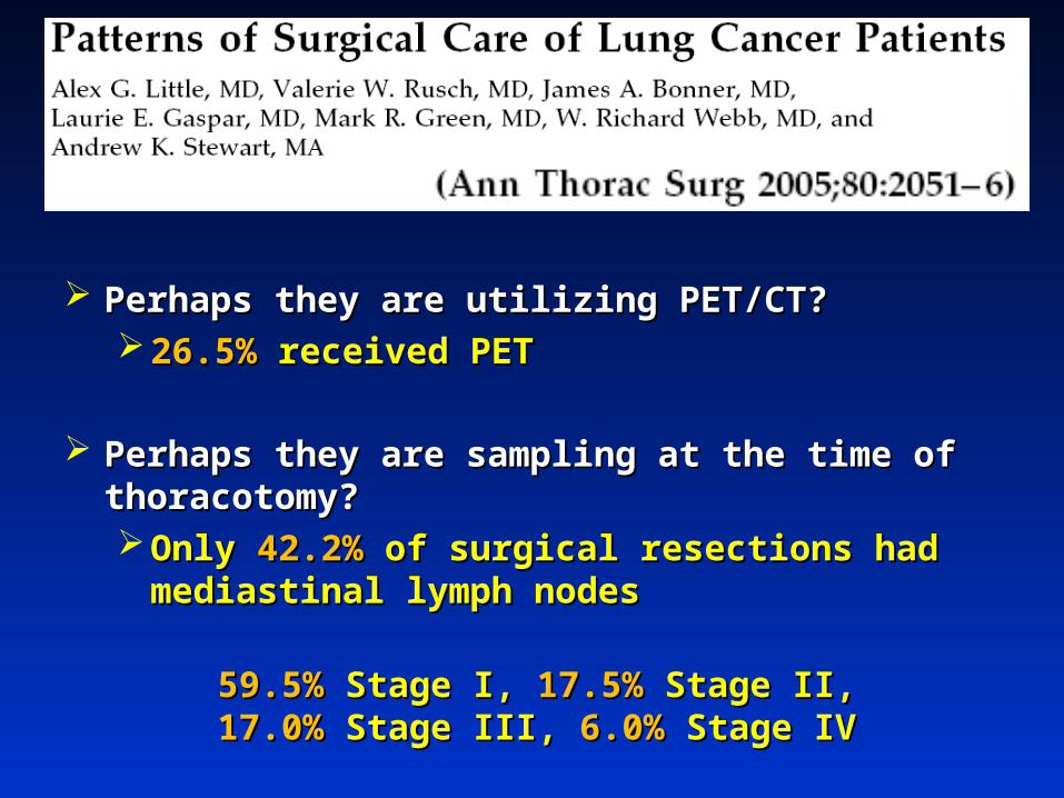
Perhaps they are utilizing PET/CT?Perhaps they are utilizing PET/CT? 26.5%26.5% received PET received PET
Perhaps they are sampling at the time of Perhaps they are sampling at the time of thoracotomy?thoracotomy? Only Only 42.2%42.2% of surgical resections had mediastinal of surgical resections had mediastinal
lymph nodeslymph nodes
59.5% 59.5% Stage I,Stage I, 17.5% 17.5% Stage II,Stage II,17.0% 17.0% Stage III,Stage III, 6.0% 6.0% Stage IVStage IV
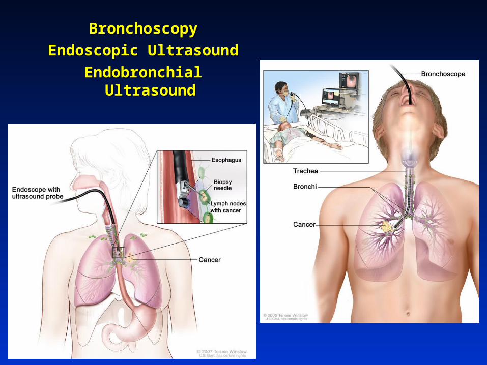
BronchoscopyBronchoscopy
Endoscopic UltrasoundEndoscopic Ultrasound
Endobronchial UltrasoundEndobronchial Ultrasound
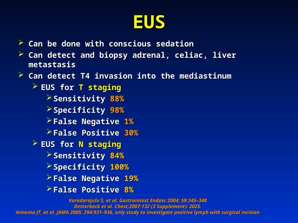
EUSEUS Can be done with conscious sedationCan be done with conscious sedation Can detect and biopsy adrenal, celiac, liver metastasisCan detect and biopsy adrenal, celiac, liver metastasis Can detect T4 invasion into the mediastinumCan detect T4 invasion into the mediastinum
EUS for EUS for T stagingT stagingSensitivity Sensitivity 88%88%Specificity Specificity 98%98%False Negative False Negative 1%1%False Positive False Positive 30%30%
EUS for EUS for N stagingN stagingSensitivity Sensitivity 84%84%Specificity Specificity 100%100%False Negative False Negative 19%19%False Positive False Positive 8%8%
Varadarajulu S, et al. Gastrointest Endosc 2004; 59:345–348Varadarajulu S, et al. Gastrointest Endosc 2004; 59:345–348Detterbeck et al. Chest;2007:132 (3 Supplement): 202S.Detterbeck et al. Chest;2007:132 (3 Supplement): 202S.
Annema JT, et al. JAMA 2005; 294:931–936, only study to investigate positive lymph with surgical Annema JT, et al. JAMA 2005; 294:931–936, only study to investigate positive lymph with surgical incisionincision

EUSEUS
Detterbeck et al. Chest;2007:132 (3 Supplement): 202S. Detterbeck et al. Chest;2007:132 (3 Supplement): 202S.
Pre-selected patient Pre-selected patient populationpopulation
CT evidence of N2, N3 CT evidence of N2, N3 nodal diseasenodal disease

EUS in the Setting of a EUS in the Setting of a Negative CTNegative CT
Saved an inappropriate thoracotomy in 9/67 = Saved an inappropriate thoracotomy in 9/67 = 13%13%
LeBlanc et al. Am J Resp and Crit Care Med. 171 (2): 177. (2005)LeBlanc et al. Am J Resp and Crit Care Med. 171 (2): 177. (2005)
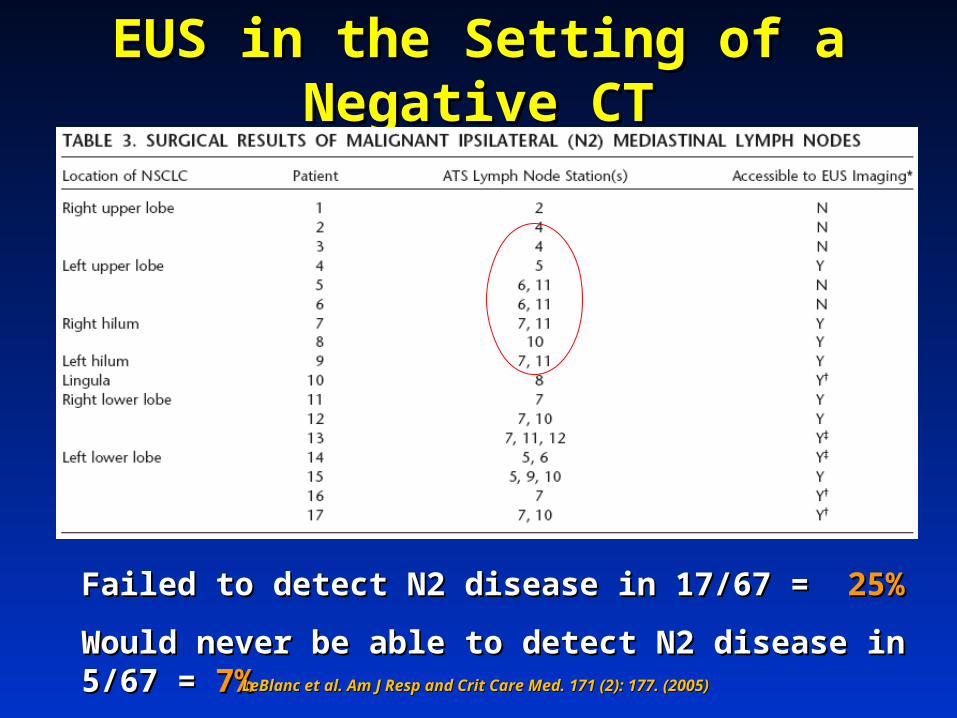
EUS in the Setting of a Negative EUS in the Setting of a Negative CTCT
Failed to detect N2 disease in 17/67 = Failed to detect N2 disease in 17/67 = 25%25%
Would never be able to detect N2 disease in 5/67 = Would never be able to detect N2 disease in 5/67 = 7%7%LeBlanc et al. Am J Resp and Crit Care Med. 171 (2): 177. (2005)LeBlanc et al. Am J Resp and Crit Care Med. 171 (2): 177. (2005)
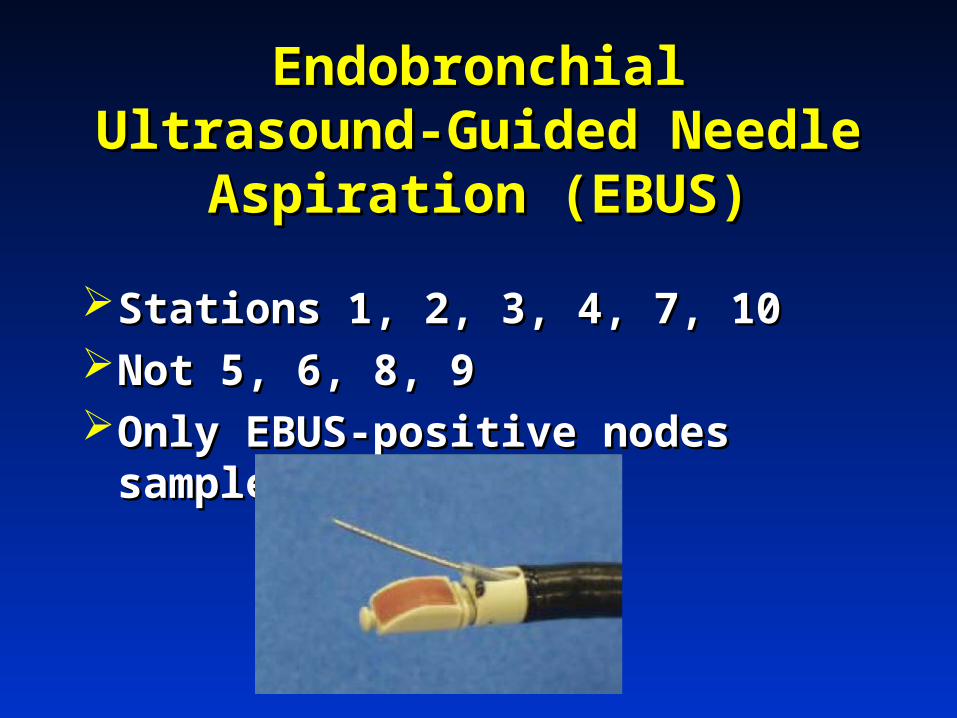
Endobronchial Ultrasound-Endobronchial Ultrasound-Guided Needle Aspiration Guided Needle Aspiration
(EBUS)(EBUS)
Stations 1, 2, 3, 4, 7, 10Stations 1, 2, 3, 4, 7, 10Not 5, 6, 8, 9Not 5, 6, 8, 9Only EBUS-positive nodes sampledOnly EBUS-positive nodes sampled
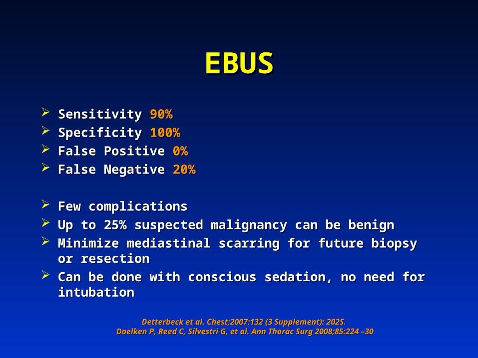
EBUSEBUS
Sensitivity Sensitivity 90%90% Specificity Specificity 100%100% False Positive False Positive 0%0% False Negative False Negative 20%20%
Few complicationsFew complications Up to 25% suspected malignancy can be benignUp to 25% suspected malignancy can be benign Minimize mediastinal scarring for future biopsy or resectionMinimize mediastinal scarring for future biopsy or resection Can be done with conscious sedation, no need for Can be done with conscious sedation, no need for
intubationintubation
Detterbeck et al. Chest;2007:132 (3 Supplement): 202S. Detterbeck et al. Chest;2007:132 (3 Supplement): 202S. Doelken P, Reed C, Silvestri G, et al. Ann Thorac Surg 2008;85:224 –30Doelken P, Reed C, Silvestri G, et al. Ann Thorac Surg 2008;85:224 –30
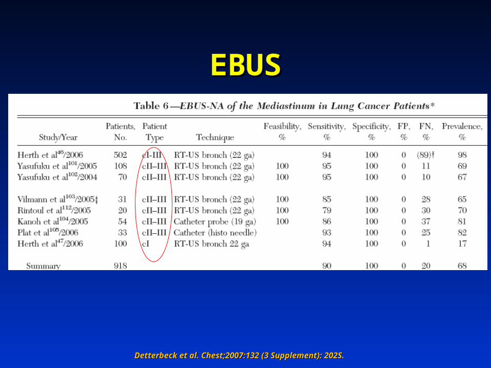
EBUSEBUS
Detterbeck et al. Chest;2007:132 (3 Supplement): 202S. Detterbeck et al. Chest;2007:132 (3 Supplement): 202S.

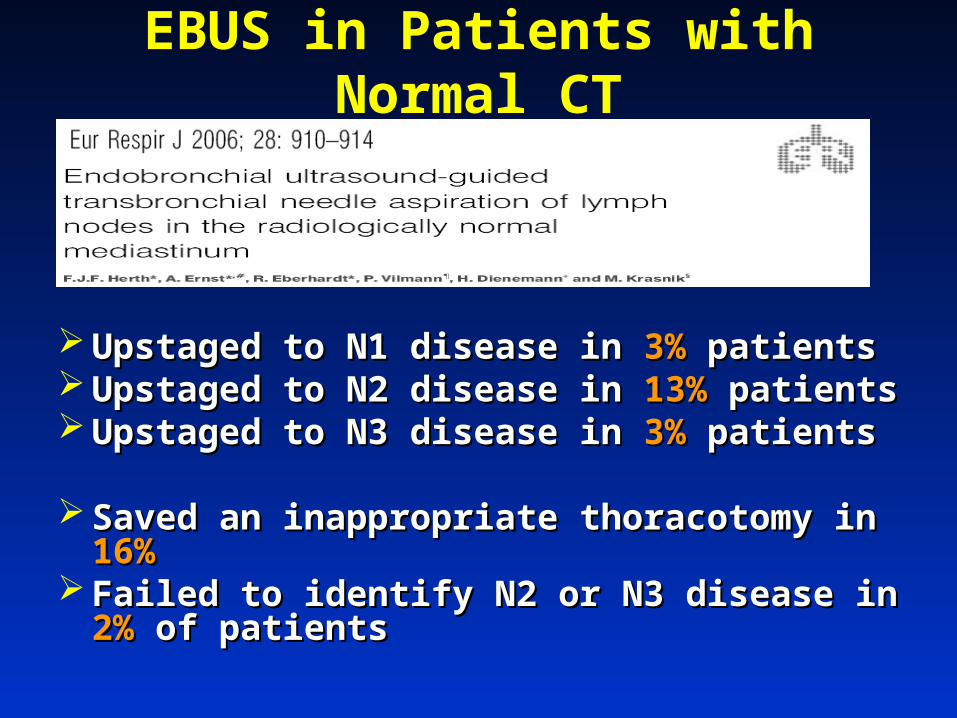
EBUS in Patients with Normal CTEBUS in Patients with Normal CT
100 patients100 patients Biopsies taken from any identifiable lymph node 2, 4, 7, 10, 11Biopsies taken from any identifiable lymph node 2, 4, 7, 10, 11 199 lymph nodes199 lymph nodes Average size 8mmAverage size 8mm Measured against pathologic specimenMeasured against pathologic specimen
Sensitivity Sensitivity 92.3%92.3% Specificity Specificity 100%100% Negative predictive value Negative predictive value 96.3%96.3%
EBUS in Patients with Normal CT
Upstaged to N1 disease in Upstaged to N1 disease in 3%3% patients patients Upstaged to N2 disease in Upstaged to N2 disease in 13%13% patients patients Upstaged to N3 disease in Upstaged to N3 disease in 3%3% patients patients
Saved an inappropriate thoracotomy in Saved an inappropriate thoracotomy in 16%16% Failed to identify N2 or N3 disease in Failed to identify N2 or N3 disease in 2%2% of of
patients patients
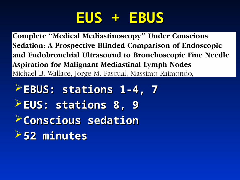
EUS + EBUSEUS + EBUS
EBUS: stations 1-4, 7 EBUS: stations 1-4, 7 EUS: stations 8, 9EUS: stations 8, 9Conscious sedationConscious sedation52 minutes52 minutes
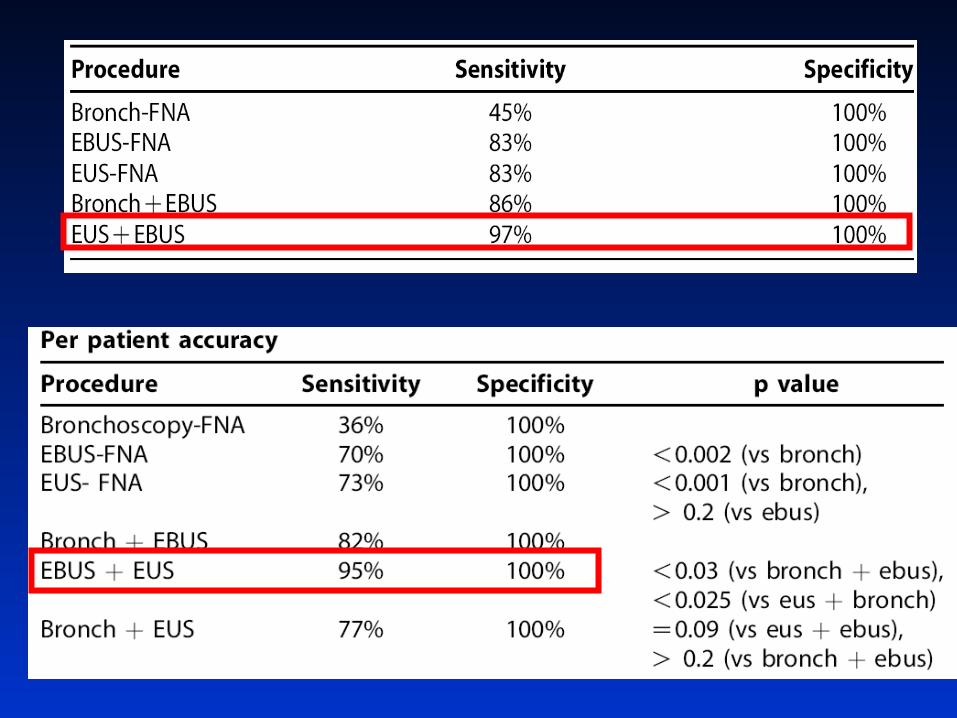
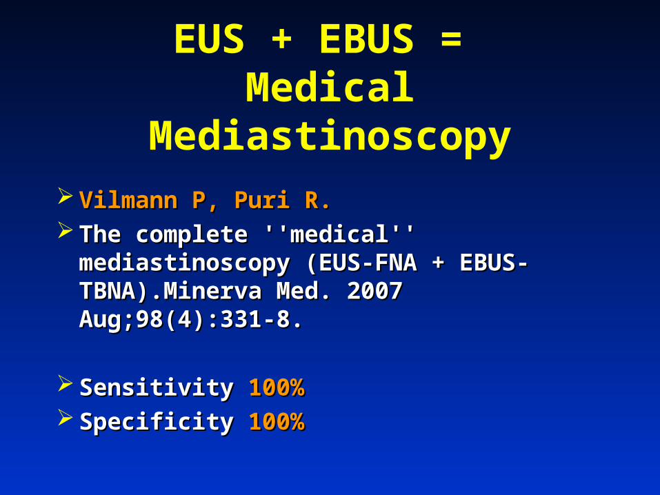
EUS + EBUS = Medical Mediastinoscopy
Vilmann P, Puri R.Vilmann P, Puri R. The complete ''medical'' mediastinoscopy The complete ''medical'' mediastinoscopy
(EUS-FNA + EBUS-TBNA).Minerva Med. (EUS-FNA + EBUS-TBNA).Minerva Med. 2007 Aug;98(4):331-8.2007 Aug;98(4):331-8.
Sensitivity Sensitivity 100%100% Specificity Specificity 100%100%

SensitivitySensitivity SpecificitySpecificity False NegFalse Neg False PosFalse Pos StationStation LimitationsLimitations
MedMed 78-9578-95 100100 1111 00 1, 3, 2, 4, 71, 3, 2, 4, 7 UtilizationUtilization
EUSEUS 8787 100100 1919 88 5, 6, 8, 95, 6, 8, 9 Limited Limited locationlocation
EBUSEBUS 9090 100100 2020 00 1, 3, 2, 4, 1, 3, 2, 4, 7, 107, 10
No standard No standard protocolprotocol
EUS+EUS+
EBUSEBUS
9595 100100 1, 2, 3, 4, 1, 2, 3, 4, 8, 9, 108, 9, 10
No dataNo data

**SensitivitySensitivity SpecificitySpecificity False False
NegNegFalse False PosPos
StationStation LimitationsLimitations
MedMed 4242 78-9578-95 100100 1111 00 1, 3, 2, 4, 71, 3, 2, 4, 7 UtilizationUtilization
EUSEUS 6666 8787 100100 1919 88 5, 6, 8, 95, 6, 8, 9 Limited Limited locationlocation
EBUSEBUS 9494 9090 100100 2020 00 1, 3, 2, 4, 1, 3, 2, 4, 7, 107, 10
No standard No standard protocolprotocol
EUS+EUS+
EBUSEBUS
9797 9595 100100 1, 2, 3, 4, 1, 2, 3, 4, 8, 9, 108, 9, 10
No dataNo data
* * Sensitivity in the setting of radiographic stage 1 diseaseSensitivity in the setting of radiographic stage 1 disease
SummarySummaryMediastinum should be staged Mediastinum should be staged
invasively utilizing mediastinoscopy, invasively utilizing mediastinoscopy, EUS, EBUS or EUS+EBUS.EUS, EBUS or EUS+EBUS.
PET/CT alone will miss N2 disease PET/CT alone will miss N2 disease ((5-12%5-12%))
Perhaps future lies with Perhaps future lies with medical medical mediastinoscopy of EUS+EBUSmediastinoscopy of EUS+EBUS


![Semiconductor Devices [Kanaan Kano]](https://static.fdocuments.us/doc/165x107/55cf931a550346f57b9bb747/semiconductor-devices-kanaan-kano.jpg)
