Mechanobiology of collective cell systems – Biofísica · Biofísica M a g a z i n e COOL...
8
Biofísica M a g a z i n e COOL BIOPHYSICS T Mechanobiology of collective cell systems Raimon Sunyer a and Xavier Trepat b a IBEC, Barcelona and CIBER-BBN Madrid (Spain) b ICREA, IBEC and UB, Barcelona and CIBER-BBN Madrid (Spain) he concept of physical force is deeply integrated in our daily life. We experience it routinely with every heartbeat, walking step, or deep breath. As such, it may seem inconceivable to think that forces are not an integral part of the mechanisms that drive biological function. However, for a long time, modern biology attempted to explain life solely on the basis of the biochemistry of genes and proteins, ignoring any potential role that physical forces could play in biological processes. Yet, it becomes increasingly clear that physical cues are not only as important as biochemical ones, but also that they could help us understand and treat diseases such as atherosclerosis, acute inflammation, fibrosis and cancer. Mechanobiology is the emergent discipline that explores the role of mechanical forces in cell development, physiology and disease. As a multidisciplinary field, it combines concepts from biology, biochemistry and physics. Challenges in mechanobiology cover from the specific mechanisms by which single cells sense and respond to forces (mechanotransduction) to how a tissue monolayer folds into a 3 dimensional structure during organogenesis. Still being a relatively young field, mechanobiology is starting to provide evidence showing that major biological processes are fundamentally ruled by forces. For example, a class of mesenchymal stem cells tends to differentiate into distinct cell types depending on the stiffness of their surroundings [Engler, et al. 2006]. Other examples include force modulation of apoptosis (programed cell death) and cell division [Slattum & Rosenblatt 2014]. Perhaps the collective migration of epithelial monolayers is one of the areas into which mechanobiology has shed more light in the recent years. Cells often move in groups with coordinated polarity without completely disrupting their cell-cell contacts. This harmonic migration is responsible for closing gaps when a monolayer is wounded or determining organ shape during morphogenesis. To fully understand such processes, it is necessary to have access to one fundamental parameter that has been elusive for decades: the physical force. http://biofisica.info/ Mechanobiology of collective cell systems – Biofísica #7, Jan-Apr 2017 1
Transcript of Mechanobiology of collective cell systems – Biofísica · Biofísica M a g a z i n e COOL...

Biofísica M a g a z i n e
COOL BIOPHYSICS
T
Mechanobiology of collective cell systemsRaimon Sunyera and Xavier Trepatb
aIBEC, Barcelona and CIBER-BBN Madrid (Spain)bICREA, IBEC and UB, Barcelona and CIBER-BBN Madrid (Spain)
he concept of physical force is deeply integrated inour daily life. We experience it routinely with everyheartbeat, walking step, or deep breath. As such, it
may seem inconceivable to think that forces are not anintegral part of the mechanisms that drive biological function.However, for a long time, modern biology attempted toexplain life solely on the basis of the biochemistry of genesand proteins, ignoring any potential role that physical forcescould play in biological processes. Yet, it becomesincreasingly clear that physical cues are not only as important
as biochemical ones, but also that they could help us understand and treat diseases such asatherosclerosis, acute inflammation, fibrosis and cancer.
Mechanobiology is the emergent discipline that explores the role of mechanical forces in celldevelopment, physiology and disease. As a multidisciplinary field, it combines concepts from biology,biochemistry and physics. Challenges in mechanobiology cover from the specific mechanisms bywhich single cells sense and respond to forces (mechanotransduction) to how a tissue monolayerfolds into a 3 dimensional structure during organogenesis. Still being a relatively young field,mechanobiology is starting to provide evidence showing that major biological processes arefundamentally ruled by forces. For example, a class of mesenchymal stem cells tends to differentiateinto distinct cell types depending on the stiffness of their surroundings [Engler, et al. 2006]. Otherexamples include force modulation of apoptosis (programed cell death) and cell division [Slattum &Rosenblatt 2014].
Perhaps the collective migration of epithelial monolayers is one of the areas into whichmechanobiology has shed more light in the recent years. Cells often move in groups with coordinatedpolarity without completely disrupting their cell-cell contacts. This harmonic migration is responsiblefor closing gaps when a monolayer is wounded or determining organ shape during morphogenesis.To fully understand such processes, it is necessary to have access to one fundamental parameter thathas been elusive for decades: the physical force.
http://biofisica.info/ Mechanobiology of collective cell systems – Biofísica #7, Jan-Apr 2017
1
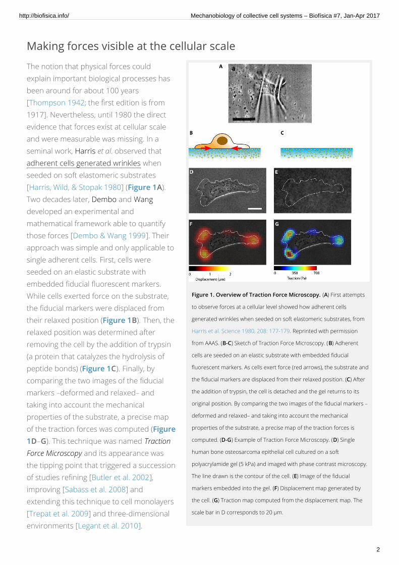
Figure 1. Overview of Traction Force Microscopy. (A) First attempts
to observe forces at a cellular level showed how adherent cells
generated wrinkles when seeded on soft elastomeric substrates, from
Harris et al. Science 1980, 208: 177-179. Reprinted with permission
from AAAS. (B-C) Sketch of Traction Force Microscopy. (B) Adherent
cells are seeded on an elastic substrate with embedded fiducial
fluorescent markers. As cells exert force (red arrows), the substrate and
the fiducial markers are displaced from their relaxed position. (C) After
the addition of trypsin, the cell is detached and the gel returns to its
original position. By comparing the two images of the fiducial markers –
deformed and relaxed– and taking into account the mechanical
properties of the substrate, a precise map of the traction forces is
computed. (D-G) Example of Traction Force Microscopy. (D) Single
human bone osteosarcoma epithelial cell cultured on a soft
polyacrylamide gel (5 kPa) and imaged with phase contrast microscopy.
The line drawn is the contour of the cell. (E) Image of the fiducial
markers embedded into the gel. (F) Displacement map generated by
the cell. (G) Traction map computed from the displacement map. The
scale bar in D corresponds to 20 µm.
Making forces visible at the cellular scale
The notion that physical forces couldexplain important biological processes hasbeen around for about 100 years[Thompson 1942; the first edition is from1917]. Nevertheless, until 1980 the directevidence that forces exist at cellular scaleand were measurable was missing. In aseminal work, Harris et al. observed thatadherent cells generated wrinkles whenseeded on soft elastomeric substrates[Harris, Wild, & Stopak 1980] (Figure 1A).Two decades later, Dembo and Wangdeveloped an experimental andmathematical framework able to quantifythose forces [Dembo & Wang 1999]. Theirapproach was simple and only applicable tosingle adherent cells. First, cells wereseeded on an elastic substrate withembedded fiducial fluorescent markers.While cells exerted force on the substrate,the fiducial markers were displaced fromtheir relaxed position (Figure 1B). Then, therelaxed position was determined afterremoving the cell by the addition of trypsin(a protein that catalyzes the hydrolysis ofpeptide bonds) (Figure 1C). Finally, bycomparing the two images of the fiducialmarkers –deformed and relaxed– andtaking into account the mechanicalproperties of the substrate, a precise mapof the traction forces was computed (Figure1D–G). This technique was named TractionForce Microscopy and its appearance wasthe tipping point that triggered a successionof studies refining [Butler et al. 2002],improving [Sabass et al. 2008] andextending this technique to cell monolayers[Trepat et al. 2009] and three-dimensionalenvironments [Legant et al. 2010].
http://biofisica.info/ Mechanobiology of collective cell systems – Biofísica #7, Jan-Apr 2017
2

Figure 2. Physical forces during collective cell migration. (A-B)
Migration of cell monolayers can be governed by different mechanisms.
(A) Leader cells at the edge pull forward the cells inside the monolayer.
Forces that cells exert on the substrate are depicted in red whereas
forces acting on cells are purple. (B) Alternatively, cell division in the
interior of the monolayer push neighboring cells forward. (C-E) Traction
forces during collective cell migration. (C) Phase contrast image of a
MDCK monolayer cultured in a soft polyacrylamide gel. Tractions
normal (D) and parallel (E) to the edge of the monolayer. (F) The
average normal traction decays slowly with distance from the edge
(filled symbols), whereas the average parallel traction was negligible and
independent of the distance from the edge (open symbols). Error bars
indicate standard errors. C-F are reprinted by permission from
Macmillan Publishers Ltd: Nature Physics (Trepat et al. Nat Phys 5: 426 –
430), copyright 2009. (G) The tug of war illustrates the mechanisms by
which a migrating cell monolayer integrates local tractions (red) into
long-ranged gradients of intra- and inter-cellular tension (purple).
How cell crowds play the tug of war
For a long time, it was unknown whetherthe global motion of cell monolayers wasdriven by the action of leader cells at thefront of the monolayer, pulling the cellsbehind [Poujade et al. 2007], or by internalpressure, due to cell division that pushedthe leading cells forward (Figure 2A and B,respectively). Recent improvements broughtby Traction Force Microscopy have providedsome evidence to elucidate which of thesemechanisms was more plausible [Trepat etal. 2009]. First, the detailed mapping oftraction forces normal and parallel to thecell edge of the monolayer showed thattraction forces were exerted many rowsbehind the leader cells and propagatedover long distances (Figure 2C–E). Thesedata suggested that the idea of leader cellsdragging the passive followers could notfully explain the collective migration.Moreover, force propagation over longdistances established that collectivemigration not only involved interactions withthe substrate but also interactions withneighboring cells. Consistent with this view,the average traction force in the monolayerwas not concentrated at the edges butdecayed slowly keeping the values largerthan zero (Figure 2F).
These findings implied that cell sheets playa global tug of war that requires cell-celljunctions (Figure 2G). Interestingly, byapplying Newton’s 2nd law, the cell stresswithin the monolayer can be calculated[Tambe 2011]. The stress transmittedthrough cell-cell junctions increased as afunction of the distance to the monolayeredge. Such a tensile stress ruled out theidea that cell division and proliferation
http://biofisica.info/ Mechanobiology of collective cell systems – Biofísica #7, Jan-Apr 2017
3

Figure 3. Durotaxis in single cells and multicellular clusters. (A) Phase contrast
image of human mammary epithelial cells (MCF-10A) cultured in isolation on a gel with
graded stiffness. Numbers at the top indicate Young’s modulus values measured with
AFM. (B) Distribution of the angle θ between the instantaneous velocity vector and the
x-axis for isolated cells. The inset shows a cell trajectory (blue) and the definition of the
angle θ. (C) A representative cell cluster expanding on a soft uniform gel of 6.6 kPa.
The gray transparent area indicates initial the cluster position (t = 0h) and the phase
contrast image shows the cluster after 10 h. Gray lines indicate cluster edges at 10 h.
(D) Example of a cell cluster expanding on a graded stiffness gel. The gel stiffness
increases towards the right of the panel. Numbers at the bottom indicate Young’s
modulus values measured with AFM. (E-F) Distribution of the angle θ between the
instantaneous velocity vector and the x-axis (see inset) for the experiments displayed
in panels C and D, respectively. Figure adapted from Sunyer et al. Science 2016, 353:
1157-1161. Reprinted with permission from AAAS.
pushed the monolayer forward. This kind of tug of war motion has been observed in other contexts,such as wound healing [Brugués et al. 2014, Vedula et al. 2014] and cancer progression [Wagstaff,Kolahgar & Piddini 2013].
Moving together towardsstiff: how the “tug of war”guides cell groups
A remaining question in epithelial monolayermigration is how the tug of war is modulatedby external cues, such as the stiffness of theenvironment. In single cells, it is wellestablished that cells tend to move from softto stiff regions when seeded on a rigiditygradient matrix [Lo et al. 2000]. Thismigration was termed durotaxis, after de Latindurus (hard) and the Greek taxis (regulararrangement). Such a guided motion,however, is very small and only appreciablewhen averaged over many cells or in thepresence of very steep gradients (Figure 3Aand B). Surprisingly, when cell clusters wereseeded on matrices with graded stiffness,durotaxis was far more efficient than in singlecells (Figure 3C–F). Again, the measure oftraction force turns out to be the key tounderstand this phenomenon.
Measuring traction forces in matrices of non-uniform stiffness is not straightforward.Conventional Traction Force Microscopyalgorithms are designed for uniform stiffnesssubstrates in which the mechanicalproperties are constant along all coordinates.Conversely, on matrices of graded stiffnessthe application of a given force will producedifferent displacements depending onwhether this force is applied to a soft or astiff region. To extend Traction ForceMicroscopy to arbitrary stiffness profiles, theuse of finite element methods (FEM) is
http://biofisica.info/ Mechanobiology of collective cell systems – Biofísica #7, Jan-Apr 2017
4

Figure 4. Traction force microscopy on gradient gels shows long range
intercellular force transmission within the clusters. (A-B) Phase contrast
images of clusters migrating on a uniform gel (A) and on a gradient gel ( B). (C-D)
Maps of the traction component Tx and (E-F) maps of the substrate
displacement component ux. Adapted from Sunyer et al. Science 2016, 353:
1157-1161. Reprinted with permission from AAAS.
required [Sunyer et al. 2016]. Using suchmethods, it was found that multicellularclusters migrating on gels of gradedstiffness exhibit traction force maps withhighest tractions localized at the edges andpointing towards the midline of the cluster,whereas relatively lower tractions in thebulk showed no particular orientation(Figure 4A–D). Unlike traction forces,substrate displacements on gradient gelsare larger on the soft edge than on the stiffone (Figure 4E–F). These forcemeasurements establish that themonolayer expands by generatingcontractile traction forces of equalmagnitude at both edges, and that theseforces are transmitted across the cluster.The net advance of the monolayer istherefore a consequence of the differencebetween the soft and the stiff deformation.
These results can also explain why durotaxisis less efficient in single cells whencompared to multicellular clusters. In single cells, the difference in stiffness across a cell length is notlarge enough to trigger durotaxis. Multicellular clusters, however, act like a super-cell: cells within it areconnected through cell-cell junctions and capable to transmit forces. Due to the large size of clusters,the variation in stiffness across its length will be much larger than in single cells. Consequently,durotaxis will be stronger. The fact that collectives are more efficient at responding to environmentalgradients than their isolated constituents is often referred as collective intelligence. This phenomenonhas been observed in cell clusters during chemotaxis [Camley et al. 2016, Mayor & Etienne-Manneville2016], fish schools during phototaxis [Berdahl et al. 2013], and human groups during online gaming[Krafft et al. 2015].
Conclusion: moving forward in cell mechanobiology
The idea that living cells sense and exert physical forces has been around for a long time. However,until the last two decades the measurement of those forces has been elusive. Today it is widelyaccepted that mechanical cues are fundamental to fully explain biological processes in health anddisease. The migration of epithelial monolayers is just an example of how a simple mechanical concept–the tug of war– can help us to understand a universal migratory mechanism present in complexbiological processes such as morphogenesis or wound healing, as well as in diseases such as fibrosis
http://biofisica.info/ Mechanobiology of collective cell systems – Biofísica #7, Jan-Apr 2017
5

and cancer. As the field of mechanobiology continues to grow, it also faces new challenges. One of themost exciting ones is to translate the basic findings of mechanobiology to clinical applications. The firstpromising attempts to diagnose pathologies, such as malignant transformations with mechanicalphenotypes are already on the way [Tse et al. 2013, Otto et al. 2015].
ReferencesBerdahl A, Torney CJ, Ioannou CC, Faria JJ, Couzin ID. “Emergent Sensing of Complex Environments by Mobile Animal
Groups”. Science, 2013, 339: 574. DOI: 10.1126/science.1225883.
Brugués A, Anon E, Conte V, Veldhuis JH, Gupta M, Colombelli J, Muñoz JJ, Brodland JW, Ladoux B, Trepat X. “Forces
Driving Epithelial Wound Healing”. Nat Phys, 2014, 10: 683. DOI: 10.1038/nphys3040.
Butler JP, Tolić-Nørrelykke IM, Fabry B, Fredberg JJ. “Traction Fields, Moments, and Strain Energy That Cells Exert on
Their Surroundings”. Am J Physiol, 2002, 282: C595. DOI: 10.1152/ajpcell.00270.2001.
Camley BA, Zimmermann J, Levine H, Rappel WJ. “Emergent Collective Chemotaxis without Single-Cell Gradient
Sensing”. Phys Rev Lett, 2016, 116: 98101. DOI: 10.1103/PhysRevLett.116.098101.
Dembo M, Wang YL. “Stresses at the Cell-to-Substrate Interface during Locomotion of Fibroblasts”. Biophys J, 1999, 76:
2307. DOI: 10.1016/S0006-3495(99)77386-8.
Engler A J, Sen S, Sweeney HL, Discher DE. “Matrix Elasticity Directs Stem Cell Lineage Specification”. Cell, 2006, 126:
677. DOI: 10.1016/j.cell.2006.06.044.
RAIMON SUNYER
Institute for Bioengineering of Catalonia (IBEC), Barcelona (Spain).
Centro de Investigación Biomédica en Red en Bioingeniería, Biomateriales y Nanomedicina, Madrid (Spain).
XAVIER TREPAT
Institute for Bioengineering of Catalonia (IBEC), Institució Catalana de Recerca i Estudis Avançats (ICREA) and
University of Barcelona (UB), Barcelona (Spain).
Centro de Investigación Biomédica en Red en Bioingeniería, Biomateriales y Nanomedicina, Madrid (Spain).
http://biofisica.info/ Mechanobiology of collective cell systems – Biofísica #7, Jan-Apr 2017
6

Harris AK, Wild P, Stopak D. “Silicone Rubber Substrata: A New Wrinkle in the Study of Cell Locomotion”. Science, 1980,
208: 177. DOI: 10.1126/science.6987736 .
Kra t PM, Hawkins RXD, Pentland AS, Goodman ND, Tenenbaum JB. “Emergent Collective Sensing in Human Groups”.
Proceedings of the 37th Conference of the Cognitive Science Society, 2015, 208: 177. available from standford.edu.
Legant WR, Miller JS, Blakely BL, Cohen DM, Genin GM,Chen CS. “Measurement of Mechanical Tractions Exerted by
Cells in Three-Dimensional Matrices”. Nat Methods, 2010, 7: 969. DOI: 10.1038/nmeth.1531.
Lo CM, Wang HB, Dembo M, Wang YL. “Cell Movement Is Guided by the Rigidity of the Substrate”. Biophys J, 2000, 79:
144. DOI: 10.1016/S0006-3495(00)76279-5.
Mayor R, Etienne-Manneville S. “The Front and Rear of Collective Cell Migration”. Nat Rev Mol Cell Biol, 2016, 17: 97.
DOI: doi:10.1038/nrm.2015.14.
Otto O, Rosendahl P, Mietke A, Gol er S, Herold C, Klaue D, Girardo S, et al.. “Real-Time Deformability Cytometry: On-
the-Fly Cell Mechanical Phenotyping”. Nat Methods, 2015, 12: 199. DOI: 10.1038/nmeth.3281.
Poujade M, Grasland-Mongrain E, Hertzog A, Jouanneau J, Chavrier P, Ladoux B, Buguin A, Silberzan P. “Collective
Migration of an Epithelial Monolayer in Response to a Model Wound”. PNAS, 2007, 104: 15988. DOI:
10.1073/pnas.0705062104.
Sabass B, Gardel ML, Waterman CM, Schwarz US. “High Resolution Traction Force Microscopy Based on Experimental
and Computational Advances”. Biophys J, 2008, 94: 207. DOI: 10.1529/biophysj.107.113670.
Slattum GM, Rosenblatt J. “Tumour Cell Invasion: An Emerging Role for Basal Epithelial Cell Extrusion”. Nat Rev Cancer,
2014, 14: 495. DOI: 10.1038/nrc3767.
Sunyer R, Conte V, Escribano J, Elosegui-Artola A, Labernadie A, Valon L, Navajas D, García-Aznar JM, Muñoz JL, Roca-
Cusachs P, Trepat X. “Collective Cell Durotaxis Emerges from Long-Range Intercellular Force Transmission”.
Science, 2016, 353: 1157. DOI: 10.1126/science.aaf7119.
Tambe DT, Hardin CC, Angelini TE, Rajendran K, Park CY, Serra-Picamal X, Zhou EH, Zaman MH, Butler JP, Weitz DA,
Fredberg JJ, Trepat X. “Collective Cell Guidance by Cooperative Intercellular Forces”. Nat Mater, 2011, 10: 469.
DOI: 10.1038/nmat3025.
Thompson D’AW. On Growth and Form, Cambridge University Press, 1942.
Trepat X, Wasserman MR, Angelini TE, Millet E, Weitz DA, Butler JP, Fredberg JJ. “Physical Forces During Collective Cell
Migration”. Nat Phys, 2009, 5: 426. DOI: 10.1038/nphys1269.
Tse HTK, Gossett DR, Moon YS, Masaeli M, Sohsman M, Ying Y, Mislick K, Adams RP, Rao J, di Caro D. “Quantitative
Diagnosis of Malignant Pleural Effusions by Single-Cell Mechanophenotyping”. Sci Transl Med, 2013, 5:
212ra163. DOI: 10.1126/scitranslmed.3006559.
http://biofisica.info/ Mechanobiology of collective cell systems – Biofísica #7, Jan-Apr 2017
7

EDITORS
Jesús Salgado
Jorge Alegre-Cebollada
Xavier Daura
Teresa Giráldez
ISSN 2445-4311
SPONSORS
CONTACT
SBE - Sociedad de Biofísica de España
Secretaria SBE, IQFR-CSIC,
C/Serrano 119, 28006 Madrid
Email: [email protected]
WEB: http://www.sbe.es
Biofísica: Biophysics Magazine by SBE - Sociedad de Biofísica de España.Design based on a Theme by Alx. Powered by WordPress. PDF export using wkhtmltopdf.
Vedula SRK, Hirata H, Nai MH, Brugués A, Toyama Y, Trepat X, Lim CT, Ladoux B. “Epithelial Bridges Maintain Tissue
Integrity during Collective Cell Migration”. Nat Mat, 2014, 13: 87. DOI: 10.1038/nmat3814.
Wagsta L, Kolahgar G, Piddini E. “Competitive Cell Interactions in Cancer: A Cellular Tug of War”. Trends Cell Biol, 2013,
23: 160. DOI: 10.1016/j.tcb.2012.11.002.
http://biofisica.info/ Mechanobiology of collective cell systems – Biofísica #7, Jan-Apr 2017
8