Mechanistic pathways of mercury removal from the ... · Mechanistic pathways of mercury ......
18
Submitted 18 March 2015 Accepted 3 July 2015 Published 28 July 2015 Corresponding author Pedro J. Silva, [email protected] Academic editor Li Zuo Additional Information and Declarations can be found on page 15 DOI 10.7717/peerj.1127 Copyright 2015 Silva and Rodrigues Distributed under Creative Commons CC-BY 4.0 OPEN ACCESS Mechanistic pathways of mercury removal from the organomercurial lyase active site Pedro J. Silva and Viviana Rodrigues FP-ENAS/Fac. de Ciˆ encias da Sa ´ ude, Universidade Fernando Pessoa, Porto, Portugal ABSTRACT Bacterial populations present in Hg-rich environments have evolved biological mechanisms to detoxify methylmercury and other organometallic mercury compounds. The most common resistance mechanism relies on the H + -assisted cleavage of the Hg–C bond of methylmercury by the organomercurial lyase MerB. Although the initial reaction steps which lead to the loss of methane from methylmercury have already been studied experimentally and computationally, the reaction steps leading to the removal of Hg 2+ from MerB and regeneration of the active site for a new round of catalysis have not yet been elucidated. In this paper, we have studied the final steps of the reaction catalyzed by MerB through quantum chemical computations at the combined MP2/CBS//B3PW91/6-31G(d) level of theory. While conceptually simple, these reaction steps occur in a complex potential energy surface where several distinct pathways are accessible and may operate concurrently. The only pathway which clearly emerges as forbidden in our analysis is the one arising from the sequential addition of two thiolates to the metal atom, due to the accumulation of negative charges in the active site. The addition of two thiols, in contrast, leads to two feasible mechanistic possibilities. The most straightforward pathway proceeds through proton transfer from the attacking thiol to Cys159 , leading to its removal from the mercury coordination sphere, followed by a slower attack of a second thiol, which removes Cys96. The other pathway involves Asp99 in an accessory role similar to the one observed earlier for the initial stages of the reaction and affords a lower activation enthalpy, around 14 kcal mol −1 , determined solely by the cysteine removal step rather than by the thiol ligation step. Addition of one thiolate to the intermediates arising from either thiol attack occurs without a barrier and produces an intermediate bound to one active site cysteine and from which Hg(SCH 3 ) 2 may be removed only after protonation by solvent-provided H 3 O + . Thiolate addition to the active site (prior to any attack by thiols) leads to pathways where the removal of the first cysteine becomes the rate-determining step, irrespective of whether Cys159 or Cys96 leaves first. Comparisons with the recently computed mechanism of the related enzyme MerA further underline the important role of Asp99 in the energetics of the MerB reaction. Kinetic simulation of the mechanism derived from our computations strongly suggests that in vivo the thiolate-only pathway is operative, and the Asp-assisted pathway (as well as the conversion of intermediates of the thiolate pathway into intermediates of the Cys-assisted pathway) is prevented by steric factors absent from our model and related to the precise geometry of the organomercurial binding-pocket. How to cite this article Silva and Rodrigues (2015), Mechanistic pathways of mercury removal from the organomercurial lyase active site. PeerJ 3:e1127; DOI 10.7717/peerj.1127
Transcript of Mechanistic pathways of mercury removal from the ... · Mechanistic pathways of mercury ......

Submitted 18 March 2015Accepted 3 July 2015Published 28 July 2015
Corresponding authorPedro J. Silva, [email protected]
Academic editorLi Zuo
Additional Information andDeclarations can be found onpage 15
DOI 10.7717/peerj.1127
Copyright2015 Silva and Rodrigues
Distributed underCreative Commons CC-BY 4.0
OPEN ACCESS
Mechanistic pathways of mercuryremoval from the organomercurial lyaseactive sitePedro J. Silva and Viviana Rodrigues
FP-ENAS/Fac. de Ciencias da Saude, Universidade Fernando Pessoa, Porto, Portugal
ABSTRACTBacterial populations present in Hg-rich environments have evolved biologicalmechanisms to detoxify methylmercury and other organometallic mercurycompounds. The most common resistance mechanism relies on the H+-assistedcleavage of the Hg–C bond of methylmercury by the organomercurial lyaseMerB. Although the initial reaction steps which lead to the loss of methane frommethylmercury have already been studied experimentally and computationally, thereaction steps leading to the removal of Hg2+ from MerB and regeneration of theactive site for a new round of catalysis have not yet been elucidated. In this paper,we have studied the final steps of the reaction catalyzed by MerB through quantumchemical computations at the combined MP2/CBS//B3PW91/6-31G(d) level oftheory. While conceptually simple, these reaction steps occur in a complex potentialenergy surface where several distinct pathways are accessible and may operateconcurrently. The only pathway which clearly emerges as forbidden in our analysisis the one arising from the sequential addition of two thiolates to the metal atom, dueto the accumulation of negative charges in the active site. The addition of two thiols,in contrast, leads to two feasible mechanistic possibilities. The most straightforwardpathway proceeds through proton transfer from the attacking thiol to Cys159 ,leading to its removal from the mercury coordination sphere, followed by a slowerattack of a second thiol, which removes Cys96. The other pathway involves Asp99in an accessory role similar to the one observed earlier for the initial stages of thereaction and affords a lower activation enthalpy, around 14 kcal mol−1, determinedsolely by the cysteine removal step rather than by the thiol ligation step. Additionof one thiolate to the intermediates arising from either thiol attack occurs withouta barrier and produces an intermediate bound to one active site cysteine and fromwhich Hg(SCH3)2 may be removed only after protonation by solvent-providedH3O+. Thiolate addition to the active site (prior to any attack by thiols) leads topathways where the removal of the first cysteine becomes the rate-determiningstep, irrespective of whether Cys159 or Cys96 leaves first. Comparisons with therecently computed mechanism of the related enzyme MerA further underline theimportant role of Asp99 in the energetics of the MerB reaction. Kinetic simulationof the mechanism derived from our computations strongly suggests that in vivothe thiolate-only pathway is operative, and the Asp-assisted pathway (as well asthe conversion of intermediates of the thiolate pathway into intermediates of theCys-assisted pathway) is prevented by steric factors absent from our model andrelated to the precise geometry of the organomercurial binding-pocket.
How to cite this article Silva and Rodrigues (2015), Mechanistic pathways of mercury removal from the organomercurial lyase activesite. PeerJ 3:e1127; DOI 10.7717/peerj.1127

Subjects Biochemistry, BiophysicsKeywords Reaction mechanism, Computational chemistry, Enzymology, MerB, Densityfunctional theory, Organomercurial lyase, MerA
INTRODUCTIONMercury is naturally present in the environment, especially at specific geologically enriched
regions along tectonical plate boundaries (Varekamp & Buseck, 1986), where it can be
found as the characteristically colored cinnabar ores (HgS). Though quite insoluble in
water (≈10 µg/L), the solubilized species (Hg2+) may be readily uptaken by methanogens
and sulfate-reducing bacteria, which then methylate it to methylmercury (Barkay,
Miller & Summers, 2003; Lin, Yee & Barkay, 2012) through the combined action of the
reductive acetyl-CoA pathway (Choi, Chase & Bartha, 1994) and two novel proteins: a
methyl-binding corrinoid-containing protein (HgcA) and a corrinoid-reducing protein
with unknown physiological function(Parks et al., 2013). The methylmercury thus formed
is highly soluble in lipids and therefore tends to accumulate in living tissues and to be
concentrated along the food chain. The solution reactivity of mercury towards soft ligands
(Riccardi et al., 2013) like the thiols present in cysteine-containing proteins is responsible
for the high toxicity of methylmercury (Eto, Marumoto & Takeya, 2010), especially towards
lipid-enriched cells (like those of the nervous system) where its solubility is the highest.
Bacterial populations present in Hg-rich environments have therefore evolved biological
mechanisms to detoxify methylmercury and other organometallic mercury compounds.
The most common resistance mechanism relies on proton-assisted cleavage of the Hg–C
bond of methylmercury by the organomercurial lyase MerB (Begley, Walts & Walsh, 1986a;
Begley, Walts & Walsh, 1986b), and sequential transfer of the remaining Hg2+ ion to a
flavoprotein (MerA) which reduces the cation to its metallic form (Fox & Walsh, 1982;
Ledwidge et al., 2005; Ledwidge et al., 2010).
Extensive experimental studies (Begley, Walts & Walsh, 1986a; Begley, Walts & Walsh,
1986b; Pitts & Summers, 2002; Di Lello et al., 2004; Lafrance-Vanasse et al., 2009) have
elucidated the structure of MerB and established that this enzyme does not require any
cofactors and uses two thiols (like cysteine or glutathione, but not dithiothreytol (Pitts
& Summers, 2002)) as co-reactants for every mercury organic compound cleaved. A
pioneering computational study (Parks et al., 2009) has shown that in the active site
any one of two conserved Cys residues (Cys 96 and Cys 159) may, upon deprotonation,
complex the Hg moiety of the substrate. A proton is then transferred from the other
conserved Cys to a conserved acidic residue (Asp 99), which subsequently acts as a proton
donor to the leaving alkyl or aryl group. That study did not, however, address the reaction
steps leading to the loss of Hg2+ from MerB and regeneration of the active site for a new
round of catalysis. In this contribution, we use quantum chemical computations to analyze
the possible reaction mechanisms. The application of these methods to protein active
sites (reviewed e.g., in Himo & Siegbahn, 2003; Ramos & Fernandes, 2008) allows the
characterization of enzyme-bound intermediates and transition states which (due to their
transient nature) are not amenable to experimental characterization and the evaluation
Silva and Rodrigues (2015), PeerJ, DOI 10.7717/peerj.1127 2/18

of thermodynamic and kinetic feasibility of postulated mechanisms. The results show
that complexation of Hg2+ by extraneous thiols is most likely to proceed through the
attack by one protonated and one deprotonated thiol, rather than by two deprotonated
thiols. The precise order of attack seems to be arbitrary, and an important role of the
Hg2+-coordinating Asp99 residue in lowering the reaction energy was found.
METHODSThe active site geometry was built from PDB:3F0P, the crystal structure of the mercury-
bound form of MerB (Lafrance-Vanasse et al., 2009). The active site included the conserved
residues Cys96, Asp 99, Cys 159, the mercury ion and Hg-complexing water molecule. To
prevent unrealistic motions of the active site, the Cα and Cβ atoms of every aminoacid
were constrained to their crystallographic positions. All calculations were performed
at the B3PW91 level of theory (Perdew, 1991; Becke, 1993), which has been commonly
used in the study of Hg-containing complexes (Barone et al., 1997; Ni et al., 2006; Parks
et al., 2009; Li et al., 2010; Riccardi et al., 2013). Autogenerated delocalized coordinates
(Baker, Kessi & Delley, 1996) were used for geometry optimizations, using the SDD effective
core-potential and associated basis set (Haussermann et al., 1993) for Hg and the 6-31G(d)
basis set for all other atoms. More accurate DFT energies of the optimized geometries
were calculated with a triple-ζ quality basis set, 6-311 + G(d). Zero point (ZPE) and
thermal effects (T = 298.15 K, P = 1 bar) were evaluated using a scaling factor of 0.9804
for the computed frequencies. All computations were performed with the Firefly quantum
chemistry package, which is partially based on the GAMESS (US) (Schmidt et al., 1993)
source code. Environmental contributions to the energies of the stationary points and
transition states were computed with the polarizable conductor model (Tomasi & Persico,
1994; Mennucci & Tomasi, 1997; Cossi et al., 1998), with dielectric constants ranging from
4 (usually chosen for protein-embedded active sites) to 78.36 (mimicking a completely
exposed active site). Dispersion and repulsion effects were evaluated as described by
Amovilli and Mennucci (Amovilli & Mennucci, 1997). MP2 single-point energies were
computed on the optimized geometries using the aug-cc-pVDZ-PP (or aug-ccpVTZ-PP)
basis set (Peterson & Puzzarini, 2005) for mercury and cc-pVDZ (or cc-pVTZ basis sets)
for all other elements, and extrapolated to the complete basis set limit (CBS-MP2) as
described by Truhlar (Truhlar, 1998). Solution MP2 values were obtained by applying the
DFT solvation energies to the gas-phase CBS-MP2 energies. Numerical integration of the
rate equations of the kinetic model derived in this work was performed using a self-built
program, which is available for download, together with respective outputs at http://dx.
doi.org/10.6084/m9.figshare.1433993.
RESULTSA large number of mechanistic pathways for Hg2+ removal from the active site of MerB
is possible (Fig. 1), depending on the protonation state of each mercury-attacking ligand
(thiol vs. thiolate), on whether Cys96 or Cys159 is first ejected from the coordination
sphere of the Hg ion, and on whether the protonation state of Asp99 changes throughout
the cycle. Our density-functional computations show that extraneous methanethiol is
Silva and Rodrigues (2015), PeerJ, DOI 10.7717/peerj.1127 3/18

Figure 1 Pathways for Hg removal from MerB, starting from an attacking thiol (“thiol-based” mecha-nism) or an attacking thiolate (“thiolate-based” mechanism). In both mechanisms, primed-numberedintermediates arise from the attack of a thiol and a thiolate, whereas intermediates numbered withunprimed numbers arise from the attack of two species with the same protonation state (either two thiolsor two thiolates).
not nucleophilic enough to directly the attack of the enzyme-bound Hg2+. The moderate
acidity of the thiol, however, allows it to transfer a proton to one of the Hg2+ ligands
(either Cys159 or Asp 99), in a process which both weakens the ligand-to-metal bond and
transforms the thiol into a (much more nucleophilic) thiolate (Fig. 2). Proton transfer
to Cys159 (Fig. 2B) occurs with a small barrier (12.3–12.8 kcal mol−1 in MP2, 7.8–8.0
kcal mol−1 using DFT) and is moderately exergonic by 7–9 kcal mol−1. This activation
barrier is very similar to the barrier found experimentally (Hong et al., 2010) for the initial
attack of MerB-bound mercury by free glutathione (2.5 × 10⁴ M−1 s−1, which translates to
11.4 kcal mol−1).
Silva and Rodrigues (2015), PeerJ, DOI 10.7717/peerj.1127 4/18

Figure 2 Cys-assisted thiol addition to Hg2+. (A) Pre-reactional complex (Int1); (B) H+ trans-fer to Cys 159 (transition state); (C) thiol-based Int2 (Cys96-bound); (D) thiol-based Int2 (Cys96-bound) + CH3SH; (E) H+ transfer to Cys 96 (transition state); (F) thiol-based Int4 (Asp99-bound).Relevant distances (in Angstrom) are highlighted. Molecules (D–E) have been rotated counterclockwiseca. 90◦ around the z-axis, relative to the orientation of molecules (A–C).
The addition of a second thiol to the singly-cysteinated Hg2+ is quite similar to that of
the first thiol, as expected from the identical composition of coordination sphere around
the metal atom (a carboxylate and two thiols). The most interesting difference arises
from the possibility of proton transfer to Cys96 (in the Cys96-bound Int2) due to the
newly-found flexibility of the freed Cys159 sidechain. This step (Figs. 2D–2F) has a larger
barrier (15.8–18.1 kcal mol−1 using MP2, 14.5–14.8 kcal mol−1 in DFT) than the addition
of the first thiol because the larger thiol(ate)-Hg distance in the latter transition state (2.875
vs. 2.685 A) entails a smaller stabilization due to lower overlap between thiol(ate) and Hg
orbitals. In the gas phase, regeneration of the active site through the removal of Hg(SCH3)2
from Asp99 leads to a continuous increase in electronic energy of approximately 26 kcal
mol−1. In solution, however, the reaction is only moderately endergonic (1–6 kcal mol−1,
depending on the dielectric constant) since the presence of a compact negative charge
in the Asp99 residue in the product state leads to a stronger solvation of the separated
fragments, which largely offsets the gas-phase energy increase due to the severing of the
Hg-carboxylate bond.
If the initial conformation of the attacking thiol, in contrast to that depicted in Fig. 2,
has the S–H bond aligned towards Asp99, H+-transfer to Asp99 occurs instead, without
any thermodynamic barrier (Fig. 3A). This transfer is favorable by 15 kcal mol−1 and may
be followed by a further movement of the proton from Asp99 to the distal Cys96 Hg-ligand
(Fig. 3B), which is thus released from the metal (Fig. 3C). This proton-transfer step has a
moderate barrier around 12–14 kcal mol−1, and should therefore occur at a rate similar to
that of the direct protonation and removal of the Cys159 ligand depicted in the alternative
Silva and Rodrigues (2015), PeerJ, DOI 10.7717/peerj.1127 5/18

Figure 3 Asp99-assisted thiol addition to Hg2+. (A) Asp 99 receives H+ from the attacking thiol;(B) H+ transfer from Asp99 to Cys96 (transition state); (C) thiol-based Int2 (Cys159-bound); (D)thiol-based Int2 (Cys159-bound) + CH3SH; (E) H+ transfer from thiol to Asp99 (transition state);(F) thiol-based Int4 (Cys159-bound). Molecules (D–F) are depicted as seen from a point of viewapproximately opposite that used in the depiction of molecules (A–C). Relevant distances (in Angstrom)are highlighted.
mechanism above (Figs. 2A–2C). The addition of a second thiol may again proceed in an
Asp99-assisted fashion (Figs. 3D–3F): proton transfer from the thiol to the Asp99 ligand
of the Cys159-bound Int2 is favored by 10–11 kcal-mol−1 but must now overcome a
small barrier (4 kcal mol−1), in contrast to the barrier-free process observed when this
movement is the first step of the reaction sequence.
In contrast to the addition of cysteine thiols analyzed above, addition of a cysteine
thiolate to the MerB-bound Hg2+ (Fig. 4) proceeds unhindered, i.e., without any energetic
barrier. The tetra-coordinated intermediate formed (Int1) lies 12–13 kcal mol−1 below
the infinitely-separated reactants (in MP2; 6–7 kcal mol−1 below reactants in DFT),
and may then shed any of its Cys-ligands upon overcoming a moderate 14.0–15.5 kcal
mol−1 barrier. The addition of a second thiolate to this complex, however, is much
costlier due to the electrostatic repulsion between the freed, deprotonated, Cys and the
negatively-charged thiolate. The precise cost depends very steeply on the chosen dielectric
constant (Table 3), as expected for a reaction involving highly localized charges, but the
transition state for this step always remains more than 25 kcal mol−1 above Int1, far above
the 16–20 kcal mol−1 expected (Parks et al., 2009) for the rate-limiting step of this enzyme
from the application of the Eyring equation, kcat =kBT
h e−ΔG‡RT , to the experimentally
observed reaction rate (Begley, Walts & Walsh, 1986b). Since methanethiol is weakly acidic
(pKa = 10.4) and is almost completely protonated at physiological pH, the initial cost of
deprotonating it must be added to the computations above. This cost amounts to −RT ln
10pH-pKa (Ullmann & Knapp, 1999), or 4.6 kcal mol−1 at pH = 7.
Silva and Rodrigues (2015), PeerJ, DOI 10.7717/peerj.1127 6/18

Figure 4 Addition of deprotonated thiol to MerB-bound Hg2+. (A) Thiolate-based Int1; (B) breakingthe Cys159-Hg bond (transition state); (C) thiolate-based Int2 (C96-bound); (D) breaking the Cys96-Hgbond (transition state); (E) thiolate-based Int2 (C159-bound). Molecules (D–E) are depicted as seen froma point of view approximately opposite that used in the depiction of molecules (A–C). Relevant distances(in Angstrom) are highlighted.
So far, we have only described the reaction mechanism arising from the addition of
two thiols with the same protonation state. We now turn to the analysis of mechanism
involving distinct protonation states of the attacking thiols: inded, the two N-terminal
cysteines of MerA which catalyze removal of Hg2+ from MerB in vivo (Ledwidge et al.,
2005) have been shown to possess widely separated pKa’s (Ledwidge et al., 2010) which
entail that at physiological pH one of them is expected to remain mostly unprotonated
while the other only deprotonates at high pH.
The addition of a thiolate to any of the forms of thiol-based intermediate 2 (where Hg2+
is bound to either of Cys159 or Cys96) occurs spontaneously without any energetic barrier.
In the Cys159-bound form, the reaction product has a slightly lower energy than in the
Cys96-bound form and adopts a more exposed conformation (Fig. 5D). The metal ion in
the resulting intermediate 3′ has a sulfur-only coordination sphere in both instances, as the
interactions with Asp99 have disappeared (Figs. 5B and 5D).
Reaction of a thiol with the thiolate-based C96-bound/C159-deprotonated intermediate
proceeds readily through proton transfer from the attacking thiol to the deprotonated
Cys159 and immediate thiolate attack of the metal atom. This process occurs without
an electronic barrier and yields C96-bound Int3′ (Fig. 5B). On the other hand, the
thiolate-based C159-bound intermediate 2 is susceptible to attack by a thiol in an Asp99-
dependent manner (Fig. 5F). As in the other Asp99-assisted thiol attacks analyzed above,
the electronic barrier to this process is negligible (Fig. 5E) and yields an intermediate where
Asp99 is protonated and the mercury ion remains coordinated by three ligands (two ex-
ternal thiolates and Cys159). This Cys159-bound/Cys96-deprotonated/Asp99-protonated
Silva and Rodrigues (2015), PeerJ, DOI 10.7717/peerj.1127 7/18

Figure 5 MP2/CBS//B3PW91/6-31G(d) energetic profiles (with ε = 20) and representative structuresof intermediates arising from attack of Hg2+ by a thiol and a thiolate. (A) Energetic profile of Cys159-assisted thiol attack followed by thiolate addition; (B) structure of Int3′ arising from Cys159-assisted thiolattack; (C) energetic profile of Asp99-assisted thiol attack followed by thiolate addition; (D) structure ofInt3′ arising from Asp99-assisted thiol attack followed by thiolate addition; (E) energetic profile of aninitial thiolate attack followed by Asp99-assisted thiol addition to Hg2+; (F) structure of the transitionstate of Asp99-assisted thiol addition to thiolate-based Int 2. Relevant distances (in Angstrom) arehighlighted.
intermediate (Int3′a in Fig. 5E) spontaneously decays, through a negligible energetic barrier
(<1 kcal mol−1), to the Cys159 bound/ Cys96-protonated/Asp99-deprotonated state.
The overall reaction barrier for all of the mechanisms involving attack of the Hg2+ ion
by a thiol and a thiolate therefore depends on the barrier of the first attack, which was
computed above (Tables 1–3) to lie between 12 and 15 kcal mol−1 in all instances. Since
the 3–4 kcal mol−1 difference between the barriers of these alternatives is equivalent to the
intrinsic error of the computational protocols used, further discrimination between these
three possibilities is unfortunately not possible at this stage.
Silva and Rodrigues (2015), PeerJ, DOI 10.7717/peerj.1127 8/18

Table 1 Relative enthalpies (kcal mol−1) of the reaction intermediates in the Cys-assisted thioladdition to MerB-bound Hg2+, computed at the MP2/CBS//B3PW91/6-31G(d) level of theory.
ε = 4 ε = 10 ε = 20 ε = 78.36
Reagent + CH3 SH 0.0 0.0 0.0 0.0
Int 1 −0.6 −1.2 −1.5 −1.7
TS Int1 to Int2 (Cys-96 bound) 11.7 11.4 11.3 11.1
Int2 (Cys-96 bound) −8.3 −9.3 −10.0 −10.8
Int2 (Cys-96 bound) + CH3 SH −11.8 −12.4 −13.1 −14.0
TS Int2 to Int4 4.1 4.4 4.4 4.2
Thiol-based Int4 (Asp99-bound) −12.8 −12.8 −13.5 −14.3
Infinitely separated products −7.3 −10.7 −12.1 −13.3
Figure 6 H3O+-assisted removal of Hg(SCH3)2 from the MerB active site (compact conforma-tion). (A) Cys96-bound Int3′ surrounded by water-solvated H3O+; (B) proton transfer from H3O+
to Cys96 (transition state); (C) Asp-bound Hg(SCH3)2 (Int4). Relevant distances (in Angstrom) arehighlighted.
Regeneration of the initial state of the active site from the Int3′ intermediate now
requires the severing of the remaining Hg–Cys bond. Preliminary attempts at the
characterization of this reaction step showed that direct stretching of the Hg–Cys bond
is energetically quite costly. Our results above (Table 2), however, show that protonation of
the metal-bound Cys dramatically weakens the Hg–S bond. We have therefore analyzed the
feasibility of removing Hg(SCH3)2 from the active site cysteine through direct protonation
by solvent-provided H3O+. A few explicit water molecules were also added to the model to
provide an appropriate description of the solvated hydronium ion (Fig. 6).
As mentioned above, two different conformations of the Int3′ intermediate exist: an
extended conformation (Fig. 5D) where Hg(SCH3)2 is bound to Cys159 and a compact
conformation where the product is bound to Cys96, instead (Fig. 5B). In the compact
conformation (Fig. 6) this proton transfer is spontaneous by 6.6 kcal mol−1 (according to
MP2; 2.7 kcal mol−1 according to DFT) and diffusion-controlled: the very small energetic
barrier found during the geometry optimization completely disappears upon inclusion
of solvation, zero-point and vibrational effects. Upon removal of Cys96, Asp99 weakly
attaches to the mercury ion, preventing the product from freely diffusing away from the
Silva and Rodrigues (2015), PeerJ, DOI 10.7717/peerj.1127 9/18

Table 2 Relative enthalpies (kcal mol−1) of the reaction intermediates in the Asp99-assisted thioladdition to MerB-bound Hg2+, computed at the MP2/CBS//B3PW91/6-31G(d) level of theory.
ε = 4 ε = 10 ε = 20 ε = 78.36
Reagent +CH3SH 0.0 0.0 0.0 0.0
Int 1 (protonated Asp99) −15.4 −15.2 −15.2 −15.2
TS Int1 → Int2 (H+ moves from Asp99 to Cys96) −1.7 −2.4 −2.8 −3.2
Int 2 (Cys-159 bound) −1.0 −0.9 −1.0 −1.1
Int 2 (Cys-159 bound) + CH3SH −9.6 −8.6 −8.3 −8.2
TS Int2 (Cys-159 bound) to Int4 (Cys159-bound) −5.5 −4.7 −4.5 −4.4
Thiol-based Int4 (Cys159-bound) −19.8 −19.4 −19.4 −19.5
Table 3 Relative enthalpies (kcal mol−1) of the reaction intermediates in thiolate addition to MerB-bound Hg2+, computed at the MP2/CBS//B3PW91/6-31G(d) level of theory.
ε = 4 ε = 10 ε = 20 ε = 78.36
Reactant + CH3S− 0.0 0.0 0.0 0.0
Thiolate-based Int1 −13.1 −12.6 −12.3 −12.1
Thiolate-based TS 1 →2 (C96-bound) 1.8 1.8 1.8 1.9
Thiolate-based Int2 (C96-bound) −3.7 −2.6 −2.2 −1.9
Thiolate-based Int2 (C96-bound) + CH3S− TS 26.8 17.1 14.0 11.8
Thiolate-based Int3 (C96-bound) 10.6 1.5 −1.3 −3.2
Reactant + CH3S− 0.0 0.0 0.0 0.0
Thiolate-based Int1 −13.1 −12.6 −12.3 −12.1
Thiolate-based TS 1 →2 (C159-bound) 1.5 2.4 2.8 3.2
Thiolate-based Int2 (C159-bound) −0.6 0.5 1.0 1.5
Thiolate-based Int2 (C159-bound) + CH3S− TS 32.3 21.0 17.3 14.6
Thiolate-based Int3 (C159-bound) 14.3 3.4 −0.1 −2.5
active site. Complete removal of Hg(SCH3)2 occurs upon stretching this very weak Asp-Hg
bond, at a cost of only 4.1 kcal mol−1.
In the “extended” conformation of Int3′, the Hg(SCH3)2 moiety lies quite far
from Asp99, which modifies the mechanistic analysis due to the impossibility of
Asp99-attachment to the metal upon the release of Cys159. In contrast to the previous
analysis, in this conformation the solvated H3O+ is unstable even before including bulk
solvation effects implicitly through the PCM model. Instead, two separate minima arise:
an unproductive intermediate featuring a proton on the Asp99 residue (Fig. 7A), and the
Cys159-protonated product featuring a free Hg(SCH3)2 (Fig. 7C). Both minima lie ≈10
kcal mol−1 below the postulated initial (meta-stable) conformation featuring a solvated
H3O+ (Fig. 7B).
Kinetic simulationsExtensive experimental analysis of the reaction of Hg-bound MerB with glutathione
or the physiological partner (Hong et al., 2010) has shown that MerA is able to effect
Silva and Rodrigues (2015), PeerJ, DOI 10.7717/peerj.1127 10/18

Figure 7 H3O+-assisted removal of Hg(SCH3)2 from the MerB active site (extended conforma-tion) (A) Asp99-protonated Int3′ surrounded by water molecules; (B) proton transfer from Asp99 toCys159 (transition state); (C) regenerated active site with released Hg(SCH3)2. Relevant distances (inAngstrom) are highlighted.
complete metal removal even at very low concentrations (50 µM), whereas concentrations
of monothiols below 10 mM afford only partial protein demetallation. Numerical
simulation of the complete reaction mechanism described in this work (Fig. 8) reveals
a very good agreement with experiment, provided that a protonated thiol is prevented
from performing the initial attack on the mercury ion (Fig. 9A and 9B): operation of
the Asp-assisted pathway (either alone or in concert with other pathways) would always
lead to complete removal of mercury from the MerB active site (Fig. 9C) due to the high
exergonicity of the initial formation of the Asp-protonated form of Int1 intermediate
(Fig. 3A and Table 2). Simultaneous operation of the Cys-assisted pathways would in
turn allow the C96-bound Int3′ intermediate (formed mainly in the thiolate pathway,
which has a more exergonic first reaction than the Cys-assisted thiol attack pathway) to be
diverted through thiolate loss (reaction k6 in Fig. 8) to the Cys-assisted pathway, yielding a
complex kinetic profile which ultimately leads to total mercury removal from MerB (Fig.
9D). In turn, setting the reaction rate of the k5 and k6 steps to zero (i.e., preventing the
conversion of Int2 (C96-bound) into Int3′ (C96-bound, and vice-versa)), while keeping
the thiolate-only pathway and the rest of the Cys-assisted pathway operative yields a
kinetic profile indistinguishable from that of the thiolate-only pathway. Interestingly,
identical kinetic simulations using the DTT (which is a known inhibitor of MerB) failed
to show any inhibition. The agreement of our model with the experimental observations
therefore requires that the formation of Int1 (protonated Asp) (Fig. 8, reaction k15/k16),
the conversion of Int2 (C96-bound) into Int3′ (C96-bound) (Fig. 8, reaction k5/k6),
and the release of the Hg-DTT complex from the active site, which are predicted by our
small-model QM computations to be thermodynamically and kinetically feasible, are
prevented in the enzyme, most likely due to the intervention of steric factors arising from
the rest of the protein. The proposed role of steric factors in the overall kinetic profile of
MerB is consistent with other experimental observation: for example, though the trigonal
complex of Hg bound by both sulfur atoms of DTT and by Cys96 is long-lived in the
absence of added thiols, Hg can be removed after a few minutes of incubation with MerA or
Silva and Rodrigues (2015), PeerJ, DOI 10.7717/peerj.1127 11/18
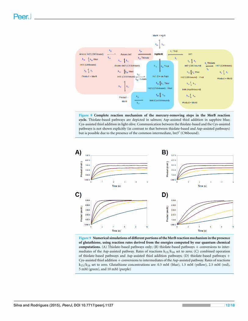
Figure 8 Complete reaction mechanism of the mercury-removing steps in the MerB reactioncycle. Thiolate-based pathways are depicted in salmon; Asp-assisted thiol addition in sapphire blue;Cys-assisted thiol addition in light olive. Communication between the thiolate-based and the Cys-assistedpathways is not shown explicitly (in contrast to that between thiolate-based and Asp-assisted pathways)but is possible due to the presence of the common intermediate, Int3′ (C96bound).
Figure 9 Numerical simulations of different portions of the MerB reaction mechanism in the presenceof glutathione, using reaction rates derived from the energies computed by our quantum chemicalcomputations. (A) Thiolate-based pathways only; (B) thiolate-based pathways + conversions to inter-mediates of the Asp-assisted pathway. Rates of reactions k15/k16 set to zero; (C) combined operationof thiolate-based pathways and Asp-assisted thiol addition pathways; (D) thiolate-based pathways +
Cys-assisted thiol addition + conversions to intermediates of the Asp-assisted pathway. Rates of reactionsk15/k16 set to zero. Glutathione concentrations are: 0.5 mM (blue), 1.5 mM (yellow), 2.5 mM (red),5 mM (green), and 10 mM (purple)
Silva and Rodrigues (2015), PeerJ, DOI 10.7717/peerj.1127 12/18

with incubation with very high concentrations of glutathione (Benison et al., 2004). Since
the chemically reactive portion in all these MerB co-substrates is the same, this implies
that the differences in behavior should not be based in purely electronic factors but in the
presence of intermolecular interactions between Hg-DTT and MerB (but not between
Hg-MerA and MerB, or between Hg-glutathione and MerB) which prevent its exit from the
active site. Indeed, preliminary experimental evidence (J Omichinski, pers. comm., 2015)
suggests that the N-terminal portion of MerB is responsible for trapping the Hg-DTT
complex and the observed partial inhibition of MerB activity by DTT.
DISCUSSIONOur computations show that the final steps of the reaction catalyzed by MerB, while
conceptually simple, occur in a complex potential energy surface where several distinct
pathways are accessible and may operate concurrently. The only pathway which clearly
emerges as forbidden in our quantum chemical analysis is the one arising from the
sequential addition of two thiolates to the metal atom, due to the accumulation of negative
charges in the active site. The addition of two thiols, in contrast, leads to two feasible
mechanistic possibilities. The most straightforward pathway proceeds through proton
transfer from the attacking thiol to Cys159 (activation ΔH = 13 kcal mol−1), leading to
its removal from the mercury coordination sphere, followed by a slower attack of a second
thiol, which removes Cys96 (activation ΔH = 16–18 kcal mol−1). Entropic effects, which
we could not analyze due to the need of enforcing geometric constraints on our active
site model, may, however, easily place this pathway above the experimentally determined
activation ΔG (16–20 kcal mol−1). The other pathway involves Asp99 in an accessory
role similar to the one observed earlier for the initial stages of the reaction (Parks et al.,
2009) and affords a lower activation enthalpy, around 14 kcal mol−1, determined solely
by the removal of the first cysteine ligand rather than by the ligation of the second thiol.
Unlike this Asp99-assisted mechanism, the Cys-assisted pathway predicts the addition of
the second thiol to be rate-limiting, in marked disagreement with previous interpretations
of experimental results (Hong et al., 2010). In contrast, addition of one thiolate to the
intermediates arising from either thiol leads to pathways where the later reaction steps
have negligible barriers. The intermediate formed in this reaction (Int3′) remains bound
to one active site cysteine and may shed Hg(SCH3)2 after protonation of this cysteine by
solvent-provided H3O+. The activation energy of this step in solution therefore depends
on the solution pH according to equation:
ΔG‡= ΔG0‡
+ RT ln1
[H+].
This protonation event is quite spontaneous and occurs without an energetic barrier
(ΔG0‡= 0), leading to an effective ΔG‡
= −RT ln10−pH, or 9.5 kcal mol−1 at pH = 7.
Thiolate addition to the active site (prior to any attack by thiols) leads to pathways where
the removal of the first cysteine becomes the rate-determining step (activation ΔH =
14–15 kcal mol−1, irrespective of whether Cys159 or Cys96 leaves first). Asp99-assisted
addition of a thiol to this intermediate then occurs without an energy barrier and yields the
Silva and Rodrigues (2015), PeerJ, DOI 10.7717/peerj.1127 13/18

familiar Int3′ intermediate discussed above. A comparison of these results with the recently
published computational analysis of the transfer of Hg2+ from the C-terminal cysteine
pair of MerA to the buried cysteine pair in the active site of MerA (Lian et al., 2014) affords
additional insights on the relative importance of the mercury-coordinating aminoacids.
In that work, which included (unlike ours) the influence of the remainder of the enzyme
through a QM/MM formalism, thiol addition to a Hg2+ ion coordinated by two cysteines
was observed to proceed through a relatively high-energy transition state (20.4 kcal mol−1)
and to be endergonic by 9.0 kcal mol−1, in contrast to the 12–13 kcal mol−1 barrier and
9–10 kcal mol−1 exergonicity we computed for the related addition of a thiol to the MerB
active site. Whereas the change in activation energy may be attributed to our neglect of
the surrounding protein environment and to the absence, in the MerA study, of a direct
proton transfer from the attacking thiol to the leaving Cys residue, further analysis points
to another reason. Indeed, the QM-only results reported by Lian et al. in their Fig. 7 show
that neglect of the electrostatic influence of the protein brings the activation energy down
to 12.6 kcal mol−1 (in perfect agreement with our data) but only lowers the reaction energy
by 10 kcal mol−1 (instead of the 19 kcal mol−1 computed for the MerB reaction in our
study). This observation allows us to attribute this 9 kcal mol−1 energy difference to the
additional interaction, in MerB, of Asp99 with the mercury ion. The influence of Asp99 is
also noticeable in the steps involving thiolate addition to mercury, which occur without a
barrier in MerB but have an activation energy of 9 kcal mol−1 in the QM-only MerA model
and (in a smaller extent) in the removal of a cysteine from a thiolate-attacked mercury,
which has an activation energy of 11 kcal mol−1 in the QM-only MerA model, compared to
15 kcal mol−1 in MerB.
In spite of the similarity of the kinetic barriers of the different tested pathways, we
were able to discriminate between the reaction pathways by comparing the kinetic profiles
predicted by the quantum-chemical computations with the wealth of experimental data
obtained by Hong et al. (2010) and Benison et al. (2004). This analysis strongly suggests
that in vivo the thiolate-only pathway is operative, and the Asp-assisted pathway (as
well as the conversion of intermediates of the thiolate pathway into intermediates of the
Cys-assisted pathway) is prevented by steric factors absent from our model and related to
the precise geometry of the organomercurial binding-pocket. Considering the success
we obtained with the combined used of quantum chemical and kinetic simulations,
we strongly recommend that kinetic simulations be used (in addition to QM methods
and experimental analysis of reaction time-coursed) whenever the analysis of reaction
mechanism by quantum chemical methods does not afford a clearly preferred pathway.
ACKNOWLEDGEMENTSThe authors thank Jan H. Jensen (University of Copenhagen) for helpful comments on this
manuscript and James G. Omichinski (University of Montreal) for extensive discussions on
the mechanism of MerB inhibition by DTT.
Silva and Rodrigues (2015), PeerJ, DOI 10.7717/peerj.1127 14/18

ADDITIONAL INFORMATION AND DECLARATIONS
FundingSome portions of the work were performed using computational resources acquired
under project PTDC/QUI-QUI/111288/2009, funded by the Portuguese Fundacao
para a Ciencia e Tecnologia and FEDER through Programa Operacional Factores de
Competitividade–COMPETE. The funders had no role in study design, data collection
and analysis, decision to publish, or preparation of the manuscript.
Grant DisclosuresThe following grant information was disclosed by the authors:
Portuguese Fundacao para a Ciencia e Tecnologia: PTDC/QUI-QUI/111288/2009.
FEDER through Programa Operacional Factores de Competitividade–COMPETE.
Competing InterestsThe authors declare there are no competing interests.
Author Contributions• Pedro J. Silva conceived and designed the experiments, performed the experiments,
analyzed the data, contributed reagents/materials/analysis tools, wrote the paper,
prepared figures and/or tables, reviewed drafts of the paper.
• Viviana Rodrigues performed the experiments, analyzed the data.
Data DepositionThe following information was supplied regarding the deposition of related data:
Complete data files for the quantum-chemical computations (including input, output
and checkkpoint files) are available at:
http://dx.doi.org/10.6084/m9.figshare.991852.
The source code, compiled Windows executable and outputs of the kinetic simulation
program used are available at:
http://dx.doi.org/10.6084/m9.figshare.1433993.
Supplemental InformationSupplemental information for this article can be found online at http://dx.doi.org/
10.7717/peerj.1127#supplemental-information.
REFERENCESAmovilli C, Mennucci B. 1997. Self-consistent-field calculation of Pauli repulsion and dispersion
contributions to the solvation free energy in the polarizable continuum model. The Journal ofPhysical Chemistry B 5647:1051–1057 DOI 10.1021/jp9621991.
Baker J, Kessi A, Delley B. 1996. The generation and use of delocalized internal coordinates ingeometry optimization. Journal of Chemical Physics 105:192–212 DOI 10.1063/1.471864.
Silva and Rodrigues (2015), PeerJ, DOI 10.7717/peerj.1127 15/18

Barkay T, Miller SM, Summers AO. 2003. Bacterial mercury resistance from atoms to ecosystems.FEMS Microbiology Reviews 27:355–384 DOI 10.1016/S0168-6445(03)00046-9.
Barone V, Bencini A, Totti F, Uytterhoeven MG. 1997. Comparison between post-Hartree–Fockand DFT methods for the study of strength and mechanism of cleavage ofHg(SINGLE BOND)C bond. International Journal of Quantum Chemistry 61:361–367DOI 10.1002/(SICI)1097-461X(1997)61:3<361::AID-QUA1>3.0.CO;2-U.
Becke AD. 1993. Density-functional thermochemistry. III. The role of exact exchange. The Journalof Chemical Physics 98:5648–5652 DOI 10.1063/1.464913.
Begley TP, Walts AE, Walsh CT. 1986a. Bacterial organomercurial lyase: overproduction,isolation, and characterization. Biochemistry 25:7186–7192 DOI 10.1021/bi00370a063.
Begley TP, Walts AE, Walsh CT. 1986b. Mechanistic studies of a protonolytic organomercurialcleaving enzyme: bacterial organomercurial lyase. Biochemistry 25:7192–7200DOI 10.1021/bi00370a064.
Benison GC, Di Lello P, Shokes JE, Cosper NJ, Scott RA, Legault P, Omichinski JG. 2004. Astable mercury-containing complex of the organomercurial lyase MerB: catalysis, productrelease, and direct transfer to MerA. Biochemistry 43:8333–8345 DOI 10.1021/bi049662h.
Choi SC, Chase T, Bartha R. 1994. Metabolic pathways leading to mercury methylation indesulfovibrio desulfuricans LS. Applied and Environmental Microbiology 60:4072–4077.
Cossi M, Mennucci B, Pitarch J, Tomasi J. 1998. Correction of cavity-induced errors inpolarization charges of continuum solvation models. Journal of Computational Chemistry19:833–846 DOI 10.1002/(SICI)1096-987X(199806)19:8<833::AID-JCC3>3.0.CO;2-Q.
Di Lello P, Benison GC, Valafar H, Pitts KE, Summers AO, Legault P, Omichinski JG. 2004.NMR structural studies reveal a novel protein fold for MerB, the organomercurial lyase involvedin the bacterial mercury resistance system. Biochemistry 43:8322–8332 DOI 10.1021/bi049669z.
Eto K, Marumoto M, Takeya M. 2010. The pathology of methylmercury poisoning (Minamatadisease). Neuropathology 30:471–479.
Fox B, Walsh C. 1982. Mercuric reductase. Purification and characterization of a transposon-encoded flavoprotein containing an oxidation–reduction-active disulfide. The Journal ofBiological Chemistry 257:2498–2503.
Haussermann U, Dolg M, Stoll H, Preuss H, Schwerdtfeger P, Pitzer RM. 1993. Accuracy ofenergy-adjusted quasirelativistic ab initio pseudopotentials. Molecular Physics 78:1211–1224DOI 10.1080/00268979300100801.
Himo F, Siegbahn PEM. 2003. Quantum chemical studies of radical-containing enzymes.Chemical Reviews 103:2421–2456 DOI 10.1021/cr020436s.
Hong B, Nauss R, Harwood IM, Miller SM. 2010. Direct measurement of mercury(II) removalfrom organomercurial lyase (MerB) by tryptophan fluorescence: NmerA domain of coevolvedγ -proteobacterial mercuric ion reductase (MerA) is more efficient than MerA catalytic core orglutathione. Biochemistry 49:8187–8196 DOI 10.1021/bi100802k.
Lafrance-Vanasse J, Lefebvre M, Di Lello P, Sygusch J, Omichinski JG. 2009. Crystal structuresof the organomercurial lyase MerB in its free and mercury-bound forms: insights into themechanism of methylmercury degradation. The Journal of Biological Chemistry 284:938–944DOI 10.1074/jbc.M807143200.
Ledwidge R, Hong B, Dotsch V, Miller SM. 2010. NmerA of Tn501 mercuric ion reductase:structural modulation of the pKa values of the metal binding cysteine thiols. Biochemistry49:8988–8998 DOI 10.1021/bi100537f.
Silva and Rodrigues (2015), PeerJ, DOI 10.7717/peerj.1127 16/18

Ledwidge R, Patel B, Dong A, Fiedler D, Falkowski M, Zelikova J, Summers AO, Pai EF,Miller SM, Conditions G. 2005. NmerA, the metal binding domain of mercuric ion reductase,removes Hg2+ from proteins, delivers it to the catalytic core, and protects cells underglutathione-depleted conditions. Biochemistry 44:11402–11416 DOI 10.1021/bi050519d.
Li X, Liao R-Z, Zhou W, Chen G. 2010. DFT studies of the degradation mechanism of methylmercury activated by a sulfur-rich ligand. Physical Chemistry Chemical Physics 12:3961–3971DOI 10.1039/b918402c.
Lian P, Guo H-B, Riccardi D, Dong A, Parks JM, Xu Q, Pai EF, Miller SM, Wei D-Q, Smith JC,Guo H. 2014. X-ray structure of a Hg(2+) complex of mercuric reductase (MerA) and quantummechanical/molecular mechanical study of Hg(2+) transfer between the C-terminal and buriedcatalytic site cysteine pairs. Biochemistry 53:7211–7222 DOI 10.1021/bi500608u.
Lin C-C, Yee N, Barkay T. 2012. Microbial transformations in the mercury cycle. In: Liu G, Cai Y,O’Driscoll N, eds. Environmental chemistry and toxicology of mercury. Hoboken: Wiley, 155–192.
Mennucci B, Tomasi J. 1997. Continuum solvation models: a new approach to the problem ofsolute’s charge distribution and cavity boundaries. Journal of Chemical Physics 106:5151–5158DOI 10.1063/1.473558.
Ni B, Kramer JR, Bell RA, Werstiuk NH. 2006. Protonolysis of the Hg–C bond ofchloromethylmercury and dimethylmercury. A DFT and QTAIM study. The Journal of PhysicalChemistry A 110:9451–9458 DOI 10.1021/jp061852+.
Parks JM, Guo H, Momany C, Liang L, Miller SM, Summers AO, Smith JC. 2009. Mechanism ofHg–C protonolysis in the organomercurial lyase MerB. Journal of the American Chemical Society131:13278–13285 DOI 10.1021/ja9016123.
Parks JM, Johs A, Podar M, Bridou R, Hurt RA, Smith SD, Tomanicek SJ, Qian Y, Brown SD,Brandt CC, Palumbo AV, Smith JC, Wall JD, Elias DA, Liang L. 2013. The genetic basis forbacterial mercury methylation. Science 339:1332–1335 DOI 10.1126/science.1230667.
Perdew JP. 1991. Unified theory of exchange and correlation beyond the local densityapproximation. In: Ziesche P, Eschrig H, eds. Electronic structure of solids ’91. Physical research.Berlin: Akademie Verlag, 11–20.
Peterson KA, Puzzarini C. 2005. Systematically convergent basis sets for transition metals.II. Pseudopotential-based correlation consistent basis sets for the group 11 (Cu,Ag, Au) and 12 (Zn, Cd, Hg) elements. Theoretical Chemistry Accounts 114:283–296DOI 10.1007/s00214-005-0681-9.
Pitts KE, Summers AO. 2002. The roles of thiols in the bacterial organomercurial lyase (MerB).Biochemistry 41:10287–10296 DOI 10.1021/bi0259148.
Ramos MJ, Fernandes PA. 2008. Computational enzymatic catalysis. Accounts of ChemicalResearch 41:689–698 DOI 10.1021/ar7001045.
Riccardi D, Guo H-B, Parks JM, Gu B, Summers AO, Miller SM, Liang L, Smith JC. 2013.Why mercury prefers soft ligands? The Journal of Physical Chemistry Letters 4:2317–2322DOI 10.1021/jz401075b.
Schmidt MW, Baldridge KK, Boatz JA, Elbert ST, Gordon MS, Jensen JH, Koseki S,Matsunaga N, Nguyen KA, Su S, Windus TL, Dupuis M, Montgomery JA. 1993. Generalatomic and molecular electronic structure system. Journal of Computational Chemistry14:1347–1363 DOI 10.1002/jcc.540141112.
Tomasi J, Persico M. 1994. Molecular interactions in solution: an overview of methodsbased on continuous distributions of the solvent. Chemical Reviews 94:2027–2094DOI 10.1021/cr00031a013.
Silva and Rodrigues (2015), PeerJ, DOI 10.7717/peerj.1127 17/18
Truhlar DG. 1998. Basis-set extrapolation. Chemical Physics Letters 294:45–48DOI 10.1016/S0009-2614(98)00866-5.
Ullmann GM, Knapp EW. 1999. Electrostatic models for computing protonation and redoxequilibria in proteins. European Biophysics Journal 28:533–551 DOI 10.1007/s002490050236.
Varekamp JC, Buseck PR. 1986. Global mercury flux from volcanic and geothermal sources.Applied Geochemistry 1:65–73 DOI 10.1016/0883-2927(86)90038-7.
Silva and Rodrigues (2015), PeerJ, DOI 10.7717/peerj.1127 18/18


![Metathesis Reactions in Total Synthesis · carbenoid mechanistic pathways.[17] This type of transforma-tion forms an important subset of a larger class of transition-metal-mediated](https://static.fdocuments.us/doc/165x107/5f02bc8a7e708231d405c1ca/metathesis-reactions-in-total-synthesis-carbenoid-mechanistic-pathways17-this.jpg)