Mechanisms of top-down attention - Welcome to...
15
Feature Review Mechanisms of top-down attention Farhan Baluch 1 and Laurent Itti 1, 2 1 Neuroscience Graduate Program, University of Southern California, Los Angeles, CA, USA 2 Department of Computer Science, University of Southern California, Los Angeles, CA, USA Attention exhibits characteristic neural signatures in brain regions that process sensory signals. An important area of future research is to understand the nature of top- down signals that facilitate attentional guidance to- wards behaviorally relevant locations and features. In this review, we discuss recent studies that have made progress towards understanding: (i) the brain structures and circuits involved in attentional allocation; (ii) top- down attention pathways, particularly as elucidated by microstimulation and lesion studies; (iii) top-down mod- ulatory influences involving subcortical structures and reward systems; (iv) plausible substrates and embodi- ments of top-down signals; and (v) information proces- sing and theoretical constraints that might be helpful in guiding future experiments. Understanding top-down attention is crucial for elucidating the mechanisms by which we can filter sensory information to pay attention to the most behaviorally relevant events. Introduction Language is infused with idiomatic expressions that make explicit the distinction between bottom-up (BU) and top- down (TD) processes of attention. We might ask someone to ‘pay attention to the road’ while driving, which implies a voluntary choice to allocate resources to a subset of the perceptual input. Alternatively, we might remark that the orange sports car really ‘caught our attention’. In this case, the resource has been involuntarily captured rather than voluntarily allocated. The distinction is not limited to idiomatic expressions, but rather stems from disparate modes of attentional processing [1]. BU attention is deployed very rapidly and depends exclusively on the properties of a sensory stimulus. By contrast, TD attention is slower and requires more effort to engage. In the modality of vision, the two modes (BU and TD) give rise to the psychophysical phenomenon of pop-out and set-size effects. In a typical visual search experiment, a subject is presented with a number of items on a display and is asked to find a target item within this display, such as a bar with a particular orientation, or color, or a combi- nation of the two. Pop-out occurs when the target item is significantly distinct from the surrounding items (distrac- tors), such as a horizontal bar among several vertical bars. This different item automatically attracts BU attention (or pops-out) rapidly and independently of the number of distractors [2,3]. By contrast, when the target item is distinguished only by taking into account the conjunction of its features, such as color and orientation, BU cues alone cannot efficiently guide attention and TD attention must be recruited to scan the display. This gives rise to search times that increase with the number of distractors; in other words, a set-size effect is observed. In most real-life situa- tions, the responses of the nervous system to a sensory input depend on both BU influences driven by the sensory stimulus and TD influences shaped by extra-retinal factors such as the current state and goal of the organism [4,5]. A distinction is also made between two types of TD mechanisms. The first type is intuitively associated with TD and is called the volitional TD process, which can exert its influence through acts of will. The second type is known as a mandatory TD process and it is an automatic, percept- modifying TD mechanism that is pervasive and that voli- tion cannot completely eliminate. The latter TD process can develop through experience-dependent plasticity or during development, and includes contextual modulation Review Glossary BU influence: influence on the nervous system due to extrinsic properties of the stimuli. Conjunction search: search task in which a subject is required to find a target item among several distractors, and the target is defined by a unique conjunction of features. In this type of search task, locating the target is more difficult because distractors share some of the features of the target and thus the target does not obviously stand or pop out. Covert attention: attention paid to a subset of the sensory inputs through mental focusing. Feed-forward sweep: first epoch of neural activity that travels from lower to higher visual areas on the onset of a visual stimulus via feed-forward connections. Mandatory TD process: attentional process that influences sensory processing in an automatic and persistent manner. Overt attention: attention paid through orienting of sensory organs toward a sensory input of interest. Percept: mental impression of an external stimulus. Pop-out search: search task in which a subject is required to find a target item among several distractors, and the target is defined by a unique visual feature not shared with any of the distractors. The target thus stands or pops out and is easy to find. Priority map: map of visual space constructed from a combination of properties of the external stimuli, and intrinsic expectations, knowledge and current behavioral goals. Recurrent epoch: second epoch of neural activity that occurs after an initial response to onset of a stimulus and is mediated by intra-cortical horizontal connections and inter-cortical feedback connections. Saliency map: map of stimulus conspicuity over visual space. Set-size effect: in search tasks, a set-size effect is observed if the time required to find the target depends on the total number of items in the display (the set size). Task-relevance map: map of behaviorally relevant locations over visual space. TD influence: influence on the nervous system due to extra-retinal effects such as intrinsic expectations, knowledge and goals. Volitional TD process: attentional process that exerts influence on sensory processing through an act of volition, such as willfully shifting attention to the right part of space. Corresponding author: Itti, L. ([email protected]). 210 0166-2236/$ – see front matter ß 2011 Elsevier Ltd. All rights reserved. doi:10.1016/j.tins.2011.02.003 Trends in Neurosciences, April 2011, Vol. 34, No. 4
Transcript of Mechanisms of top-down attention - Welcome to...

Feature Review
Mechanisms of top-down attentionFarhan Baluch1 and Laurent Itti1,2
1 Neuroscience Graduate Program, University of Southern California, Los Angeles, CA, USA2 Department of Computer Science, University of Southern California, Los Angeles, CA, USA
Review
Glossary
BU influence: influence on the nervous system due to extrinsic properties of
the stimuli.
Conjunction search: search task in which a subject is required to find a target
item among several distractors, and the target is defined by a unique
conjunction of features. In this type of search task, locating the target is more
difficult because distractors share some of the features of the target and thus
the target does not obviously stand or pop out.
Covert attention: attention paid to a subset of the sensory inputs through
mental focusing.
Feed-forward sweep: first epoch of neural activity that travels from lower to
higher visual areas on the onset of a visual stimulus via feed-forward
connections.
Mandatory TD process: attentional process that influences sensory processing
in an automatic and persistent manner.
Overt attention: attention paid through orienting of sensory organs toward a
sensory input of interest.
Percept: mental impression of an external stimulus.
Pop-out search: search task in which a subject is required to find a target item
among several distractors, and the target is defined by a unique visual feature
not shared with any of the distractors. The target thus stands or pops out and is
easy to find.
Priority map: map of visual space constructed from a combination of
properties of the external stimuli, and intrinsic expectations, knowledge and
current behavioral goals.
Recurrent epoch: second epoch of neural activity that occurs after an initial
response to onset of a stimulus and is mediated by intra-cortical horizontal
connections and inter-cortical feedback connections.
Saliency map: map of stimulus conspicuity over visual space.
Set-size effect: in search tasks, a set-size effect is observed if the time required
to find the target depends on the total number of items in the display (the set
size).
Task-relevance map: map of behaviorally relevant locations over visual space.
TD influence: influence on the nervous system due to extra-retinal effects such
as intrinsic expectations, knowledge and goals.
Volitional TD process: attentional process that exerts influence on sensory
Attention exhibits characteristic neural signatures inbrain regions that process sensory signals. An importantarea of future research is to understand the nature of top-down signals that facilitate attentional guidance to-wards behaviorally relevant locations and features. Inthis review, we discuss recent studies that have madeprogress towards understanding: (i) the brain structuresand circuits involved in attentional allocation; (ii) top-down attention pathways, particularly as elucidated bymicrostimulation and lesion studies; (iii) top-down mod-ulatory influences involving subcortical structures andreward systems; (iv) plausible substrates and embodi-ments of top-down signals; and (v) information proces-sing and theoretical constraints that might be helpful inguiding future experiments. Understanding top-downattention is crucial for elucidating the mechanisms bywhich we can filter sensory information to pay attentionto the most behaviorally relevant events.
IntroductionLanguage is infused with idiomatic expressions that makeexplicit the distinction between bottom-up (BU) and top-down (TD) processes of attention.Wemight ask someone to‘pay attention to the road’ while driving, which implies avoluntary choice to allocate resources to a subset of theperceptual input. Alternatively, we might remark that theorange sports car really ‘caught our attention’. In this case,the resource has been involuntarily captured rather thanvoluntarily allocated. The distinction is not limited toidiomatic expressions, but rather stems from disparatemodes of attentional processing [1]. BU attention isdeployed very rapidly and depends exclusively on theproperties of a sensory stimulus. By contrast, TD attentionis slower and requires more effort to engage.
In the modality of vision, the two modes (BU and TD)give rise to the psychophysical phenomenon of pop-out andset-size effects. In a typical visual search experiment, asubject is presented with a number of items on a displayand is asked to find a target item within this display, suchas a bar with a particular orientation, or color, or a combi-nation of the two. Pop-out occurs when the target item issignificantly distinct from the surrounding items (distrac-tors), such as a horizontal bar among several vertical bars.This different item automatically attracts BU attention (orpops-out) rapidly and independently of the number ofdistractors [2,3]. By contrast, when the target item isdistinguished only by taking into account the conjunction
Corresponding author: Itti, L. ([email protected]).
210 0166-2236/$ – see front matter � 2011 Elsevier Ltd. All rights res
of its features, such as color and orientation, BU cues alonecannot efficiently guide attention and TD attention mustbe recruited to scan the display. This gives rise to searchtimes that increase with the number of distractors; in otherwords, a set-size effect is observed. In most real-life situa-tions, the responses of the nervous system to a sensoryinput depend on both BU influences driven by the sensorystimulus and TD influences shaped by extra-retinal factorssuch as the current state and goal of the organism [4,5].
A distinction is also made between two types of TDmechanisms. The first type is intuitively associated withTD and is called the volitional TD process, which can exertits influence through acts of will. The second type is knownas a mandatory TD process and it is an automatic, percept-modifying TD mechanism that is pervasive and that voli-tion cannot completely eliminate. The latter TD processcan develop through experience-dependent plasticity orduring development, and includes contextual modulation
processing through an act of volition, such as willfully shifting attention to the
right part of space.
erved. doi:10.1016/j.tins.2011.02.003 Trends in Neurosciences, April 2011, Vol. 34, No. 4
[()TD$FIG]
(a) (b)
TRENDS in Neurosciences
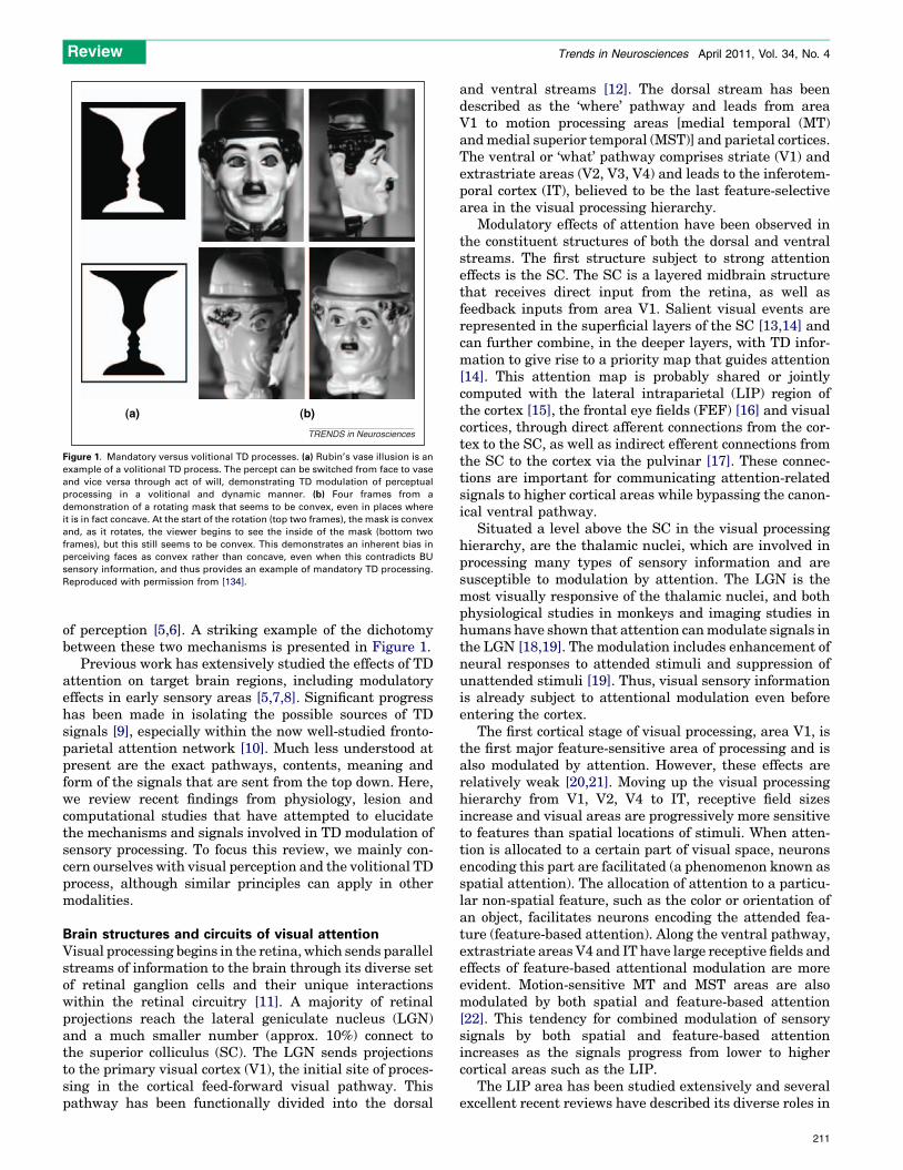
Figure 1. Mandatory versus volitional TD processes. (a) Rubin’s vase illusion is an
example of a volitional TD process. The percept can be switched from face to vase
and vice versa through act of will, demonstrating TD modulation of perceptual
processing in a volitional and dynamic manner. (b) Four frames from a
demonstration of a rotating mask that seems to be convex, even in places where
it is in fact concave. At the start of the rotation (top two frames), the mask is convex
and, as it rotates, the viewer begins to see the inside of the mask (bottom two
frames), but this still seems to be convex. This demonstrates an inherent bias in
perceiving faces as convex rather than concave, even when this contradicts BU
sensory information, and thus provides an example of mandatory TD processing.
Reproduced with permission from [134].
Review Trends in Neurosciences April 2011, Vol. 34, No. 4
of perception [5,6]. A striking example of the dichotomybetween these two mechanisms is presented in Figure 1.
Previous work has extensively studied the effects of TDattention on target brain regions, including modulatoryeffects in early sensory areas [5,7,8]. Significant progresshas been made in isolating the possible sources of TDsignals [9], especially within the now well-studied fronto-parietal attention network [10]. Much less understood atpresent are the exact pathways, contents, meaning andform of the signals that are sent from the top down. Here,we review recent findings from physiology, lesion andcomputational studies that have attempted to elucidatethe mechanisms and signals involved in TD modulation ofsensory processing. To focus this review, we mainly con-cern ourselves with visual perception and the volitional TDprocess, although similar principles can apply in othermodalities.
Brain structures and circuits of visual attentionVisual processing begins in the retina, which sends parallelstreams of information to the brain through its diverse setof retinal ganglion cells and their unique interactionswithin the retinal circuitry [11]. A majority of retinalprojections reach the lateral geniculate nucleus (LGN)and a much smaller number (approx. 10%) connect tothe superior colliculus (SC). The LGN sends projectionsto the primary visual cortex (V1), the initial site of proces-sing in the cortical feed-forward visual pathway. Thispathway has been functionally divided into the dorsal
and ventral streams [12]. The dorsal stream has beendescribed as the ‘where’ pathway and leads from areaV1 to motion processing areas [medial temporal (MT)andmedial superior temporal (MST)] and parietal cortices.The ventral or ‘what’ pathway comprises striate (V1) andextrastriate areas (V2, V3, V4) and leads to the inferotem-poral cortex (IT), believed to be the last feature-selectivearea in the visual processing hierarchy.
Modulatory effects of attention have been observed inthe constituent structures of both the dorsal and ventralstreams. The first structure subject to strong attentioneffects is the SC. The SC is a layered midbrain structurethat receives direct input from the retina, as well asfeedback inputs from area V1. Salient visual events arerepresented in the superficial layers of the SC [13,14] andcan further combine, in the deeper layers, with TD infor-mation to give rise to a priority map that guides attention[14]. This attention map is probably shared or jointlycomputed with the lateral intraparietal (LIP) region ofthe cortex [15], the frontal eye fields (FEF) [16] and visualcortices, through direct afferent connections from the cor-tex to the SC, as well as indirect efferent connections fromthe SC to the cortex via the pulvinar [17]. These connec-tions are important for communicating attention-relatedsignals to higher cortical areas while bypassing the canon-ical ventral pathway.
Situated a level above the SC in the visual processinghierarchy, are the thalamic nuclei, which are involved inprocessing many types of sensory information and aresusceptible to modulation by attention. The LGN is themost visually responsive of the thalamic nuclei, and bothphysiological studies in monkeys and imaging studies inhumans have shown that attention canmodulate signals inthe LGN [18,19]. The modulation includes enhancement ofneural responses to attended stimuli and suppression ofunattended stimuli [19]. Thus, visual sensory informationis already subject to attentional modulation even beforeentering the cortex.
The first cortical stage of visual processing, area V1, isthe first major feature-sensitive area of processing and isalso modulated by attention. However, these effects arerelatively weak [20,21]. Moving up the visual processinghierarchy from V1, V2, V4 to IT, receptive field sizesincrease and visual areas are progressively more sensitiveto features than spatial locations of stimuli. When atten-tion is allocated to a certain part of visual space, neuronsencoding this part are facilitated (a phenomenon known asspatial attention). The allocation of attention to a particu-lar non-spatial feature, such as the color or orientation ofan object, facilitates neurons encoding the attended fea-ture (feature-based attention). Along the ventral pathway,extrastriate areasV4 and IT have large receptive fields andeffects of feature-based attentional modulation are moreevident. Motion-sensitive MT and MST areas are alsomodulated by both spatial and feature-based attention[22]. This tendency for combined modulation of sensorysignals by both spatial and feature-based attentionincreases as the signals progress from lower to highercortical areas such as the LIP.
The LIP area has been studied extensively and severalexcellent recent reviews have described its diverse roles in
211
Review Trends in Neurosciences April 2011, Vol. 34, No. 4
attention, reward, and occulomotor behavior [15,23,24]. Itis important to point out that responses in area LIP can bedriven by both BU factors, such as stimulus salience, andTD factors, such has behavioral relevance of stimuli [25],the current locus of attention [26] and occulomotor plan-ning [15]. Therefore, the LIP is another candidate struc-ture (beyond the SC described above) where BU and TDinfluences can combine to give rise to a spatial prioritymap[15]. The many facets of observed responses in the LIP canbe attributed to the fact that both BU and a diverse set ofTD influences can give rise to behavioral priority, and thusmodulate LIP responses, which suggests that the LIPencodes priority in a manner largely agnostic to the factorsthat caused the priority [15]. Through direct feedbackconnections [27] or connections via the pulvinar to visualareas (see below), the LIP can communicate the fusedsignals to other brain areas for biasing or further atten-tional processing.
FEF neurons also represent salient stimuli, specificallystimuli that vary significantly from surrounding items in avisual display (known as odd-ball stimuli). The FEF hasalso been described as a region with neural responsescharacteristic of a priority map [16]. Single-unit responsesin monkey FEF exhibit transients on stimulus onset,followed by a later response (latency of �100 ms) thatdiscriminates an odd-ball stimulus from surrounding dis-
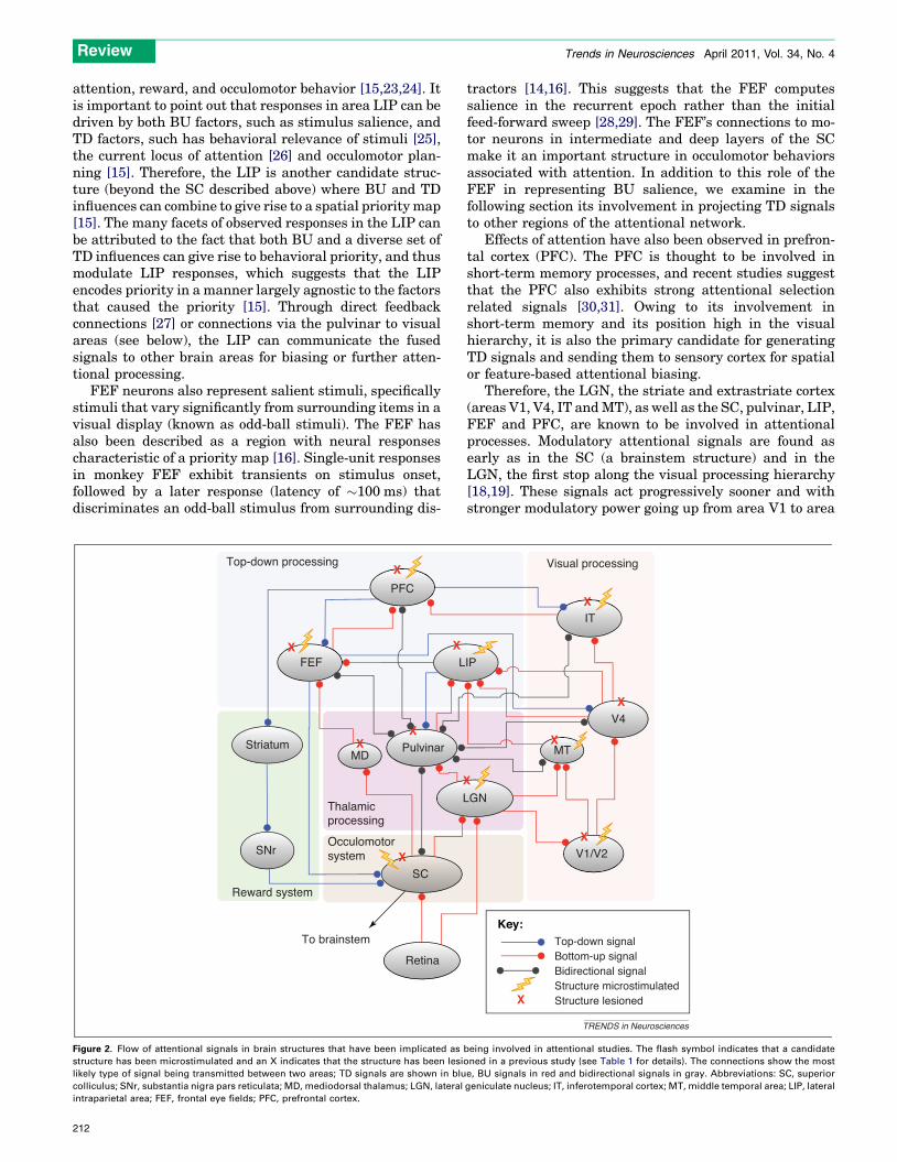
[()TD$FIG]PFC
FEF
Pulvinar
SC
L
L
Striatum
SNr
Retina
MD
X
X
X
X
X
X
X
Top-down processing
Thalamicprocessing
Reward system
To brainstem
Occulomotorsystem
Figure 2. Flow of attentional signals in brain structures that have been implicated as
structure has been microstimulated and an X indicates that the structure has been lesi
likely type of signal being transmitted between two areas; TD signals are shown in blu
colliculus; SNr, substantia nigra pars reticulata; MD, mediodorsal thalamus; LGN, lateral
intraparietal area; FEF, frontal eye fields; PFC, prefrontal cortex.
212
tractors [14,16]. This suggests that the FEF computessalience in the recurrent epoch rather than the initialfeed-forward sweep [28,29]. The FEF’s connections to mo-tor neurons in intermediate and deep layers of the SCmake it an important structure in occulomotor behaviorsassociated with attention. In addition to this role of theFEF in representing BU salience, we examine in thefollowing section its involvement in projecting TD signalsto other regions of the attentional network.
Effects of attention have also been observed in prefron-tal cortex (PFC). The PFC is thought to be involved inshort-term memory processes, and recent studies suggestthat the PFC also exhibits strong attentional selectionrelated signals [30,31]. Owing to its involvement inshort-term memory and its position high in the visualhierarchy, it is also the primary candidate for generatingTD signals and sending them to sensory cortex for spatialor feature-based attentional biasing.
Therefore, the LGN, the striate and extrastriate cortex(areas V1, V4, IT andMT), as well as the SC, pulvinar, LIP,FEF and PFC, are known to be involved in attentionalprocesses. Modulatory attentional signals are found asearly as in the SC (a brainstem structure) and in theLGN, the first stop along the visual processing hierarchy[18,19]. These signals act progressively sooner and withstronger modulatory power going up from area V1 to area
IP
GN
V1/V2
IT
X
V4X
MT
Top-down signalBottom-up signalBidirectional signalStructure microstimulatedStructure lesioned
Visual processing
X
X
X
Key:
TRENDS in Neurosciences
being involved in attentional studies. The flash symbol indicates that a candidate
oned in a previous study (see Table 1 for details). The connections show the most
e, BU signals in red and bidirectional signals in gray. Abbreviations: SC, superior
geniculate nucleus; IT, inferotemporal cortex; MT, middle temporal area; LIP, lateral
Review Trends in Neurosciences April 2011, Vol. 34, No. 4
IT [20]. These signals can bias attention for particularvisual locations [32], visual features [33–36], or both.The characteristic signature of these attentional modula-tions onto target sensory areas includes heightened gain,sharpened tuning and other end-effects, as reviewed pre-viously [8,37,38]. In the following section, we examine theareas that are specifically involved in mediating TD atten-tional signals.
Pathways of TD attentionIn this section, we focus on lesion and electrophysiologicalstudies, particularly those using methods of microstimula-tion and simultaneous recordings in the brain areas iden-tified in the previous section. These areas form anattentional network (Figure 2) and we consider how TDinformation is relayed in this network. Microstimulation,together with reversible inactivation [using either phar-macological agents such as muscimol or transcranial mag-netic stimulation (TMS)] and permanent lesion studies,have enabled researchers to go from correlation to causa-tion in the study of perception and attention (Table 1).
It has been suggested that all sensory stimuli competefor entry into working memory [39]. Working memory notonly stores information, but also enhances this informationand actively generates TD attentional signals that biasfeature-sensitive brain regions, and is thus vital for accom-plishing behavioral goals [39]. An elegant study demon-strated that the PFC transmits the contents of workingmemory to the visual system by using a posterior-split-brain paradigm [40]. In this study, monkeys were pre-sented with a visual cue in either the left or right hemifield,followed by a probe stimulus. The task was to respond tothe appearance of the probe that had previously beenassociated with the cued item. BU signals were recordedby presenting the cue in the hemifield ipsilateral to therecording site in the IT (i.e. direct BU path from the retinaup to IT), whereas TD signals could be recorded from areaIT by presenting the cue in the visual hemifield contralat-eral to the recording site in the IT. The posterior callosumtransection precluded direct communication between visu-al cortices from both sides of the brain, so it was hypothe-sized that the TD signals were fed back from the PFC toarea IT (Figure 3a). To move to a more causal explanation,the next experiment involved transection of the anteriorcorpus callosum (thereby cutting that hypothetical path-way), which resulted in a lack of response from the IT cells[40]. These results demonstrated that TD signals correlat-ing with working memory emanate from the PFC and feedback into the ventral stream. Amore recent study also usedthe posterior-split-brain paradigm in conjunction with uni-lateral PFC removal and demonstrated that performanceon a search task was mainly impaired when the goal of thesearchwas switched on a regular basis [41]. This study thushighlighted the importance of the PFC in switching the TDcontext. It has also been found that microstimulation of thePFC leads to biases in target selection towards or awayfrom the stimulation field, which demonstrates how TDsignals can affect occulomotor behavior [42]. Furthermore,the sheer connectedness of the PFC suggests that its effectsare pervasive and are driven by a combination of goals,rewards, salience, and planning of motor actions [9,39].
The next area proximal to the PFC, and an importantplayer in TD attention, is the FEF. Sub-threshold FEFstimulation enhances responses of V4 neurons in the pres-ence of a stimulus in their receptive field (Figure 4a) [43].This demonstrates that descending TD signals from theFEF bias processing in area V4. These results were repli-cated in analogous regions of the barn owl [44]. The com-parison of local field potentials (LFP, which may bestrongly driven by afferent inputs from other brain regions)and spiking activity in the FEF (which represents intrinsicactivity of FEF neurons) revealed that target-selectivesignals appeared in spiking activity before showing adifference in the LFP, which suggests that spatial selectionwas computed locally in the FEF [29]. There is speculationthat this emergence of selection is communicated down toventral regions through a synchronization of gamma-bandactivity between the FEF and area V4 [45]. However, alesion study demonstrated that temporary inactivation ofthe FEF (using a GABA-A receptor agonist, muscimol) ledto deficits not only in visually guided saccades, but also inshifts of attention during either pop-out or conjunctionvisual searches [46]. Contrary to an earlier study [29],these findings suggested that the FEF, although involvedin covert attention, does not locally compute the selectionbut is rather a participant in a network with heavy in-volvement of the LIP.
Area LIP is strongly connected to the FEF and is inte-gral to the attentional network through both anatomicaland functional characterization. Suprathreshold microsti-mulation in the posterior parietal cortex (PPC), whichincludes both area LIP and the ventral intraparietal area(VIP), induces saccades; however, the current required toinduce saccades is significantly higher compared to thatrequired when microstimulating the FEF, which suggeststhat the connection from the PPC to the occulomotorsystemmight not be a direct one. Subthreshold stimulationresults in a shift of covert attention [47]. Interestingly, anon-spatial effect was also found whereby reaction times indetecting a target decreased irrespective of whether avalid, invalid, or no cue was presented [47]. This suggestedthat microstimulation of the LIP can override the cuesignal and orient attention to the visual location corre-sponding to the site of stimulation. Evidence from lesionstudies demonstrates that damage or inactivation of theLIP causes deficits only in the presence of multiple stimuli[48,49]. These results point to an additional role of the LIPin resolving competition among stimuli represented atlower levels through TD connections to these levels [7,50].
The aforementioned studies did not, however, differen-tiate between the dorsal (LIPd) and ventral (LIPv) sub-divisions of the LIP. In a more recent study, the effects oflocal reversible inactivation (using a GABA-A receptoragonist) in areas LIPd and LIPv have been studied sepa-rately [51]. Interestingly, the many dimensions of LIPresponses demonstrated previously [23] were shown toreside in disparate subdivisions of the LIP. Inactivationof the LIPd affected performance on simple saccade tasksbut left visual search intact, whereas temporary lesions ofthe LIPv led to deficits in both search and saccadic perfor-mance [51]. The authors stressed that deficits in saccadicperformance after LIPd inactivation were far smaller than
213
Table 1. Microstimulation and lesion studies of different brain structures involved in attention.a,b
Brain region Microstimulation studies Refs Lesion studies Refs
Implications for attentional processing Implications for attentional processing
SC Shift of spatial attention [55] Deficit in target selection [58,68]
Perceptual facilitation at site of stimulation [56] Deficit in perceptual decision in
presence of distractors
[60]
Selection of target independent of motor plan [57]
Signal transmitted to MT via Pulvinar [70]
LGN Elicits visual percepts [111] Eliminates residual visual responses in
extrastriate cortex after V1 lesion
[112]
Disruption of smooth pursuit eye movements [113]
Deficits in target detection (human) [110]
Pulvinar
–
No deficit in saccadic behavior [67]
No deficit in visual search [68]
Deficit in suppression of distractors during
search (human)
[65]
Spatial and temporal attention deficits with
anterior and posterior lesions respectively
(human)
[66]
V1 Target selection disrupted with upper layer
stimulation, facilitated with lower layer stimulation
[114] Deficit in motion detection and discrimination [117]
Lower current thresholds needed for evoking
saccades in lower layers
[115] Deficit in saccade targeting [118]
Median current of 5.2 mA (6.6 mA) required for
behavioral detection of stimulationc
[116]
V4
–
Deficit in distractor suppression when
target and distractor are inside RF of neuron
[119]
Deficit in distractor suppression [133]
IT/TE Biases perceptual judgement in visual classification [53] No behavioral deficit when lesion is made in
infantile monkeys
[120]
Bias in selection of stimulus category [54] Deficit in distractor suppression [119]
Median current of 10.3 mA (11.3 mA) required for
behavioral detection of stimulationc
[116]
MT Bias in motion direction discrimination [122] Loss in perception of motion [124]
Bias in motion direction during stimulus
presentation but not during memorizing
period
[123] Loss in perception of motion more evident in
noisy conditions
[132]
Median current of 10.1 mA required for behavioral
detection of stimulationc
[116]
LIP Sub and suprathreshold stimulation lead to covert
and over shifts of attention respectively
[47] Deficit in distractor suppression even when
stimuli are non overlapping within RF,
contrast with [121]
[49]
Bias in visual selection [125] Dorsal lesion leads to occulomotor deficits
ventral lesion leads to attentional and
occulomotor deficit
[51]
Affects performance in tasks requiring spatial
attention
[48]
FEF Enhanced response elicited in V4 [43] Deficit in target detection [46]
Facilitation akin to allocation of covert attention [126] Enhanced contrast sensitivity in fovea but
not periphery (human)
[128]
Bias toward direction of saccade plan rather
than location of attention
[127] Disruption of facilitation by saccade plan to
location corresponding with stimulation site
(human)
[129]
PFC Bias in target selection [42] Loss of TD signal recorded in IT [40]
Disruption in saccadic activity [130] Decrease in behavioral performance when cue
is frequently switched
[41]
Elimination of acetylcholine release in sensory
cortex after stimulus presentation (rat)
[131]
aAll studies have been conducted in monkeys unless otherwise denoted.
bRF, receptive field; SC, superior colliculus; LGN, lateral geniculate nucleus; IT, inferotemporal cortex; MT, middle temporal area; LIP, lateral intraparietal area; FEF, frontal
eye fields; PFC, prefrontal cortex.
cStimulation current values reported in two monkeys (see [116] for details).
Review Trends in Neurosciences April 2011, Vol. 34, No. 4
214
[()TD$FIG]
‘Electrode’ ‘Electrode’
Anterior CC
CUE CUE
V1
V4
LIP
MTFEF
IT
PFC
n = 43 (cells) n = 28 (cells)
Bottom-upcondition
Top-downcondition
Posterior-split
AC • AC •
CC CC
A P
Full-split
(b)
500(ms)CUE0500(ms)CUE00
(a)
60
0
60
BUresponse
TDresponse
Spi
kes
s–1
TD signal
TRENDS in Neurosciences
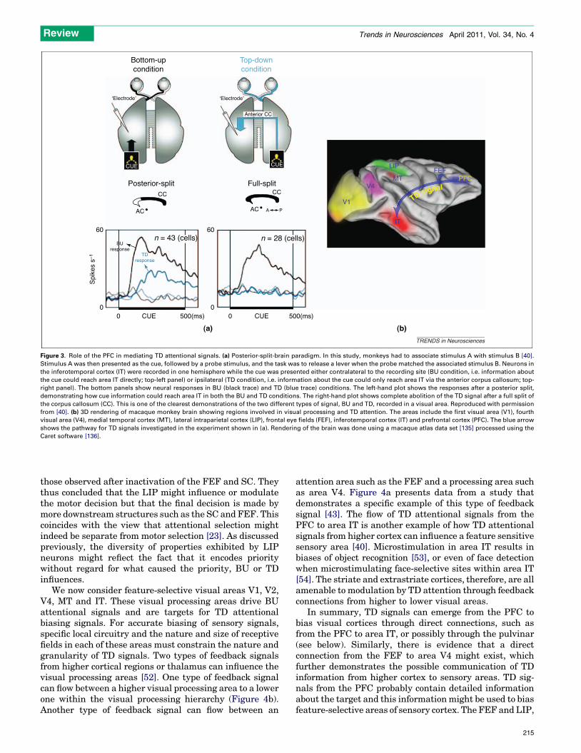
Figure 3. Role of the PFC in mediating TD attentional signals. (a) Posterior-split-brain paradigm. In this study, monkeys had to associate stimulus A with stimulus B [40].
Stimulus A was then presented as the cue, followed by a probe stimulus, and the task was to release a lever when the probe matched the associated stimulus B. Neurons in
the inferotemporal cortex (IT) were recorded in one hemisphere while the cue was presented either contralateral to the recording site (BU condition, i.e. information about
the cue could reach area IT directly; top-left panel) or ipsilateral (TD condition, i.e. information about the cue could only reach area IT via the anterior corpus callosum; top-
right panel). The bottom panels show neural responses in BU (black trace) and TD (blue trace) conditions. The left-hand plot shows the responses after a posterior split,
demonstrating how cue information could reach area IT in both the BU and TD conditions. The right-hand plot shows complete abolition of the TD signal after a full split of
the corpus callosum (CC). This is one of the clearest demonstrations of the two different types of signal, BU and TD, recorded in a visual area. Reproduced with permission
from [40]. (b) 3D rendering of macaque monkey brain showing regions involved in visual processing and TD attention. The areas include the first visual area (V1), fourth
visual area (V4), medial temporal cortex (MT), lateral intraparietal cortex (LIP), frontal eye fields (FEF), inferotemporal cortex (IT) and prefrontal cortex (PFC). The blue arrow
shows the pathway for TD signals investigated in the experiment shown in (a). Rendering of the brain was done using a macaque atlas data set [135] processed using the
Caret software [136].
Review Trends in Neurosciences April 2011, Vol. 34, No. 4
those observed after inactivation of the FEF and SC. Theythus concluded that the LIP might influence or modulatethe motor decision but that the final decision is made bymore downstream structures such as the SC and FEF. Thiscoincides with the view that attentional selection mightindeed be separate from motor selection [23]. As discussedpreviously, the diversity of properties exhibited by LIPneurons might reflect the fact that it encodes prioritywithout regard for what caused the priority, BU or TDinfluences.
We now consider feature-selective visual areas V1, V2,V4, MT and IT. These visual processing areas drive BUattentional signals and are targets for TD attentionalbiasing signals. For accurate biasing of sensory signals,specific local circuitry and the nature and size of receptivefields in each of these areas must constrain the nature andgranularity of TD signals. Two types of feedback signalsfrom higher cortical regions or thalamus can influence thevisual processing areas [52]. One type of feedback signalcan flow between a higher visual processing area to a lowerone within the visual processing hierarchy (Figure 4b).Another type of feedback signal can flow between an
attention area such as the FEF and a processing area suchas area V4. Figure 4a presents data from a study thatdemonstrates a specific example of this type of feedbacksignal [43]. The flow of TD attentional signals from thePFC to area IT is another example of how TD attentionalsignals from higher cortex can influence a feature sensitivesensory area [40]. Microstimulation in area IT results inbiases of object recognition [53], or even of face detectionwhen microstimulating face-selective sites within area IT[54]. The striate and extrastriate cortices, therefore, are allamenable to modulation by TD attention through feedbackconnections from higher to lower visual areas.
In summary, TD signals can emerge from the PFC tobias visual cortices through direct connections, such asfrom the PFC to area IT, or possibly through the pulvinar(see below). Similarly, there is evidence that a directconnection from the FEF to area V4 might exist, whichfurther demonstrates the possible communication of TDinformation from higher cortex to sensory areas. TD sig-nals from the PFC probably contain detailed informationabout the target and this informationmight be used to biasfeature-selective areas of sensory cortex. The FEF and LIP,
215
[()TD$FIG]
FEFmicrostimulation
80
0 250 500 750
Time (ms)
Spi
kes
s-1
1.0
2.0
3.0
4.0
FEF microstimulation
V4 recording
(a) (b)
V5 - TMS suprathreshold
V1 - TMS subthreshold
-100 100V5-V1 asynchrony (ms)
Conditioning to test stimulus asynchrony (ms)
-80 80-60 60-40 40-20 200
Pho
sphe
ne r
epor
t
TRENDS in Neurosciences
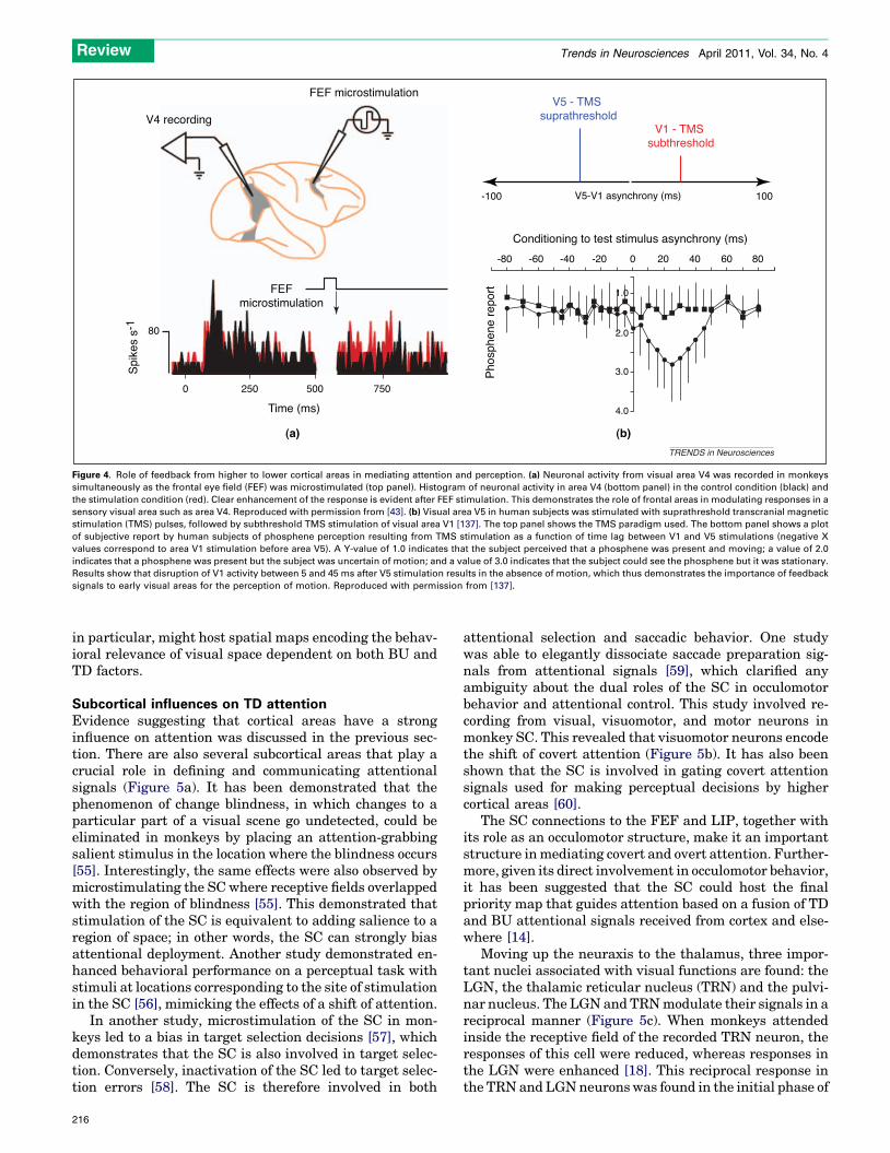
Figure 4. Role of feedback from higher to lower cortical areas in mediating attention and perception. (a) Neuronal activity from visual area V4 was recorded in monkeys
simultaneously as the frontal eye field (FEF) was microstimulated (top panel). Histogram of neuronal activity in area V4 (bottom panel) in the control condition (black) and
the stimulation condition (red). Clear enhancement of the response is evident after FEF stimulation. This demonstrates the role of frontal areas in modulating responses in a
sensory visual area such as area V4. Reproduced with permission from [43]. (b) Visual area V5 in human subjects was stimulated with suprathreshold transcranial magnetic
stimulation (TMS) pulses, followed by subthreshold TMS stimulation of visual area V1 [137]. The top panel shows the TMS paradigm used. The bottom panel shows a plot
of subjective report by human subjects of phosphene perception resulting from TMS stimulation as a function of time lag between V1 and V5 stimulations (negative X
values correspond to area V1 stimulation before area V5). A Y-value of 1.0 indicates that the subject perceived that a phosphene was present and moving; a value of 2.0
indicates that a phosphene was present but the subject was uncertain of motion; and a value of 3.0 indicates that the subject could see the phosphene but it was stationary.
Results show that disruption of V1 activity between 5 and 45 ms after V5 stimulation results in the absence of motion, which thus demonstrates the importance of feedback
signals to early visual areas for the perception of motion. Reproduced with permission from [137].
Review Trends in Neurosciences April 2011, Vol. 34, No. 4
in particular, might host spatial maps encoding the behav-ioral relevance of visual space dependent on both BU andTD factors.
Subcortical influences on TD attentionEvidence suggesting that cortical areas have a stronginfluence on attention was discussed in the previous sec-tion. There are also several subcortical areas that play acrucial role in defining and communicating attentionalsignals (Figure 5a). It has been demonstrated that thephenomenon of change blindness, in which changes to aparticular part of a visual scene go undetected, could beeliminated in monkeys by placing an attention-grabbingsalient stimulus in the location where the blindness occurs[55]. Interestingly, the same effects were also observed bymicrostimulating the SC where receptive fields overlappedwith the region of blindness [55]. This demonstrated thatstimulation of the SC is equivalent to adding salience to aregion of space; in other words, the SC can strongly biasattentional deployment. Another study demonstrated en-hanced behavioral performance on a perceptual task withstimuli at locations corresponding to the site of stimulationin the SC [56], mimicking the effects of a shift of attention.
In another study, microstimulation of the SC in mon-keys led to a bias in target selection decisions [57], whichdemonstrates that the SC is also involved in target selec-tion. Conversely, inactivation of the SC led to target selec-tion errors [58]. The SC is therefore involved in both
216
attentional selection and saccadic behavior. One studywas able to elegantly dissociate saccade preparation sig-nals from attentional signals [59], which clarified anyambiguity about the dual roles of the SC in occulomotorbehavior and attentional control. This study involved re-cording from visual, visuomotor, and motor neurons inmonkey SC. This revealed that visuomotor neurons encodethe shift of covert attention (Figure 5b). It has also beenshown that the SC is involved in gating covert attentionsignals used for making perceptual decisions by highercortical areas [60].
The SC connections to the FEF and LIP, together withits role as an occulomotor structure, make it an importantstructure inmediating covert and overt attention. Further-more, given its direct involvement in occulomotor behavior,it has been suggested that the SC could host the finalpriority map that guides attention based on a fusion of TDand BU attentional signals received from cortex and else-where [14].
Moving up the neuraxis to the thalamus, three impor-tant nuclei associated with visual functions are found: theLGN, the thalamic reticular nucleus (TRN) and the pulvi-nar nucleus. The LGN and TRNmodulate their signals in areciprocal manner (Figure 5c). When monkeys attendedinside the receptive field of the recorded TRN neuron, theresponses of this cell were reduced, whereas responses inthe LGN were enhanced [18]. This reciprocal response inthe TRNandLGNneuronswas found in the initial phase of

[()TD$FIG]
ATTin
ATTout
LGNm
0
100
300
200
Stimulus on
TRN
200
400
2001000
0
ATTin
ATTout
400 800 1200
Time (ms)
(b)
(a)
(c)
100
80
60
40
20
00
Spi
kes
s-1
Spi
kes
s-1
Spi
kes
s-1
Time (ms)
Cortex
(+)
(+)
SC(-)
(-)
SNr
CD
Attention shift response
Key:
Key:
TRENDS in Neurosciences
Figure 5. Role of subcortical structures in attention. (a) Schematic drawing of circuitry that has been proposed to be involved in the generation of eye movements towards
locations of reward ([78]). The cortex sends excitatory inputs to both the superior colliculus (SC) and the caudate nucleus (CD). The CD in turn inhibits the substantia nigra
pars reticulata (SNr), which then reduces its tonic inhibition on the SC. A disinhibited SC enables eye movements to be made. Reproduced with permission from [78]. (b)
Neuronal activity from a visuomotor cell in the SC. Monkeys were first presented with a spatial cue, followed by an oriented stimulus at the cued location. Monkeys then
made a saccade in the direction corresponding to the orientation of the stimulus. The orientation was always orthogonal to the location of the cue, and this dissociates
shifts of attention from saccadic behavior. The plot shows responses of a visuomotor SC cell, which shows significant activity in the attention shift period (between the
dashed lines) that occurs immediately after presentation of the cue, whereas purely motor cells in the deeper layers of the SC did not show such a response (data not
shown). Reproduced with permission from [59]. (c) Neuronal activity recorded from the thalamus in awake behaving monkeys. The monkeys were presented with a central
cue that instructed them to attend to one of two peripheral oriented bar stimuli, one inside the receptive field (RF) of a recorded neuron and one outside the RF. The top
shows the spike density of a magnocellular lateral geniculate nucleus (LGNm) neuron that exhibits an enhanced response when the monkey attends to a stimulus inside the
RF (ATTin condition) of the neuron compared to when the monkey attends to a stimulus outside the RF (ATTout condition). The bottom shows responses in the thalamic
reticular nucleus (TRN), which responds in a reciprocal manner to the LGNm neuron exhibiting an enhanced response when attention is allocated to a stimulus outside the
RF. Therefore, the TRN might gate responses in the LGN. Reproduced with permission from [18].
Review Trends in Neurosciences April 2011, Vol. 34, No. 4
the response to a visual stimulus. In a later phase, the TRNresponse remained unchanged, but attention further en-hanced responses in LGN. These results suggest that (i)the TRN serves as the initiator of modulation in the LGNand (ii) attentional modulation begins at an early stage inthe LGN. The TRN therefore plays a crucial role in modu-lating visual signals at a very early stage of processing.
The pulvinar is a hyperconnected nucleus of the thala-mus that has been implicated in the function of visualattention based on anatomical [17,61–63], physiological[64], lesion [65–68] and computational [69] studies. Ithas been shown that a monkey’s ability to suppress dis-tractors is diminished when the pulvinar is pharmacologi-cally inactivated via administration of muscimol [64].Relay neurons have also been identified in the pulvinarby microstimulating the SC and area MT while simulta-neously recording from cells in the pulvinar [70]. Thisstudy adds to evidence of a subcortical route for visualsignals to reach higher cortex via the pulvinar. At the same
time, its bidirectional connections with higher corticalareas make it a potentially important structure in mediat-ing TD signals. However, the pulvinar remains an under-studied nucleus, and further studies on this particularbrain nucleus are warranted.
Subcortical structures, therefore, both modulate signalsin areas encoding BU and TD information, such as the LIPand FEF, and receive TD information from higher corticalareas, directly or possibly through the pulvinar. The SCitself is believed to host a priority map, but this prioritymap might have closer correspondence to representationsneeded formotor decisions, including occulomotor behaviorand head movements. Thalamic nuclei, including the LGNand TRN, modulate visual signals early on, before theyreach cortex, and the pulvinar might be a key relay incommunicating attentional signals from one region toanother. Subcortical structures are also heavily involvedand influenced by reward and emotion, as discussed in thefollowing section.
217
Review Trends in Neurosciences April 2011, Vol. 34, No. 4
One emerging theme is that disparate modes of proces-sing might exist in the different brain regions identifiedabove. Areas such as the LIP and FEF, and subcorticalstructures such as the SC, might normally operate in afeature-agnostic mode, encoding salience and facilitatingor inhibiting regions of visual space according to behavioralgoals, but without regard to detailed visual features. (Thisdoes not, however, preclude these areas from developingfeature selectivity through operant training [16], condi-tioning [71] or task demands [72]). Conversely, visualcortices (areas V1, V2, V4), the IT and the PFC might beoperating in a feature-committed mode, modulatingresponses depending on the exact visual features that giverise to BU salience and/or TD relevance. The pulvinarmight then serve as a bidirectional translator, convertingfine-grained, feature-committed TD signals to coarser,feature-agnostic TD signals and vice versa. This dichotomybetween feature-agnostic and feature-committed TD sig-nals gives rise to interesting hypotheses about possiblemechanisms in which TD attention exerts its influence onneural responses in sensory cortex, and thus affects atten-tional allocation and gaze behavior.
The role of reward and emotion in TD attentionUntil recently, studies of visual attention have tradition-ally tended to avoid non-visual aspects of cortical andsubcortical neuronal responses to manipulations of atten-tion. This has begun to change with a small number ofpsychophysical and electrophysiological studies that haveexplored the interplay between reward and attention.
To investigate the role of reward in modulating atten-tion-related responses in the LIP, stimulus selection hasbeen dissociated from motor selection in monkeys [71].With training, LIP neurons exhibit a strong sustained biastoward the location of a conditioned stimulus, even when asaccade in the opposite direction was required to reveal theoutcome of the trial. This suggests that LIP neurons encode‘the value of information’ [23] and prioritize spatial loca-tions based on this value.
Studies using operant conditioning paradigms demon-strate effects related to improvements in the volitional TDprocess. However, learning is also the primary method foraugmenting the mandatory TD process. It has been shownthat the FEF develops systematic biases, akin to a man-datory TD signal, thereby facilitating shifts of attention inthe direction of the feature when it is present at anylocation [73,74]. More recently, a similar tendency wasfound in humans performing a visual search task in whichthe target changed on every trial, which therefore preclud-ed subjects from simply learning a limited set of targetfeatures [75]. Subjects’ performance improved, demon-strating an improved ability to quickly extract informationfrom a brief preview of the target before each trial, and tothen use this information to shape TD signals and guideattention. Learning and reward paradigms can thereforeinfluence ability to both generate TD biasing signals (i.e.volitional TD process) and introduce systematic biases (i.e.mandatory TD process).
Reward plays an important role in modulating atten-tional signals, and the basal ganglia, which consist ofdopaminergic nuclei in the substantia nigra pars reticulata
218
(SNr), the caudate and the putamen, are essential inencoding reward signals [76]. The basal ganglia are inte-grally connected to the occulomotor system through theconnection of the SC to the SNr [77]. Reward signals (TD)from frontal cortices are transmitted to the caudate, whichthen inhibits the SNr, which in turn pauses the tonicinhibition from the SNr to the SC, releasing it from inhibi-tion and enabling saccades [78]. This follows a more gen-eral scheme in the CNS in which the basal ganglia circuitcontinually inhibits movement of all limbs until an explicitcommand to make a motor movement is received fromcortical or subcortical regions. Furthermore, it is alsopossible that reward plays a strong role in influencing asubcortical salience map that can cause instant occulomo-tor reflexes.
A recent study has shed new light on the SNr to SCconnection by demonstrating that SNr fibers connect notonly to excitatory neurons in the SC, but also to localGABAergic neurons in the intermediate layers of the SC[79]. Therefore, the SNr is involved in shaping the balanceof inhibition and excitation in the local SC circuit. SCinvolvement in attentional selection and the strong roleof the SNr in reward render the SNr–SC connection animportant one because in most studies, especially physio-logical studies in monkeys, paradigms are based on theelements of operant conditioning and reinforcement learn-ing with a crucial role for reward (see [78,80] for moredetailed discussions).
Sensory processing is also amenable to modulation bybrain regions encoding emotions. In particular, it is knownthat the amygdala has reciprocal connections with bothearly and late visual areas and can thus give priority,through modulation, to stimuli of ecological relevance[81]. Using a combination of functionalmagnetic resonanceimaging (fMRI) and a study of lesion patients, it was foundthat visual areas such as the fusiform gyrus receive inputfrom the amygdala and exhibit enhanced responses toaffective stimuli [82]. Suchmodulation by emotionmatchesresponse enhancement observed through attentional allo-cation. Furthermore, it has been shown that emotional andattentional modulations can act independently, as ob-served in patients with lesions of the amygdala, whosefusiform cortex exhibited responses modulated by atten-tion but not emotion [82]. Affective stimuli can thereforeimpinge on sensory signals independently of attention;however, the very enhancement due to emotional valencemight render the stimuli salient and thus draw moreattention. Attention and emotion might thus act indepen-dently on the sensory signals and the behavioral relevanceof these sensory inputs might be determined by the cumu-lative effects of both attention and emotion.
One proposal for neural mechanisms and regions in-volved in fusion of affective inputs with purely visualaspects driving attention has recently been suggestedbased on a search task in human subjects using fMRI[83]. The frontoparietal spatial attention network, consist-ing of the superior parietal lobule (SPL), the inferiorparietal lobule (IPL) and the FEF, was activated whenthe cue was purely spatial. However, when the cue con-tained both spatial and emotional information, limbic andsubcortical structures including the posterior cingulate
Review Trends in Neurosciences April 2011, Vol. 34, No. 4
cortex (PCC), the amygdala and the orbitofrontal cortexwere activated, in addition to the frontoparietal network.This study also found selectivity in the PCC for respondingonly to cues that had emotional valence [83]. These resultssuggest that the cingulate gyrus, which receives inputsfrom the amygdala and sends outputs to the frontoparietalnetwork, might serve as the gateway for affective inputs tofuse with spatial biasing signals. This gives rise to a TDsalience map in the frontoparietal network, complete withaffective and spatial priority information.
Although evidence remains limited, a number of studieshave demonstrated links between the attentional networkand reward and emotional centers. Such connections mustbe taken into account when considering TD networks,because most experimental paradigms involving TD atten-tion to date have used reward and/or emotional valence totrain and motivate human or animal participants.
The role of oscillatory activity and neuromodulation inTD attentionIt has recently been suggested that synchronous activity (inthe gammarange, 50–80 Hz) between cortical regionsmightserve as the basis for attentional facilitation and corticalcomputations [84]. In this proposal, neuronal populationsrepresenting inputs anddecision centers all consist of rhyth-mically activeneural ensembleswithdistinct excitatory andinhibitoryphases. Inhibitory interneurons ineachensemblerhythmically inhibit excitatory pyramidal neurons, therebyestablishing a rhythm. Two neural ensembles can thensynchronize through phase-locking. This gives rise to awinner-take-all mechanism among two competing inputsfeeding into a single higher cortical decision area, throughsynchronization between the higher area and one selectedinput. Synchrony between the input andhigher areas can beestablished in a TD or BUmanner. In the TD case, a regionin higher cortical regionsmight establish a gamma synchro-ny with a lower sensory area by phase-locking.
Data from several studies demonstrate that gammaoscillations in the cortex are correlated with attention[45,85]. Disparate brain regions might synchronize theiractivity in the gamma band when an animal is attending toa particular stimulus. A specific example of this type ofcoupling is that observed between the FEF and area V4 inmonkeys. When attending to a stimulus, coupling throughgamma oscillations during attention was observed be-tween neurons in the FEF and V4 [45]. Oscillations inlower frequency bands, such as the alpha and delta bands,have also been implicated in sensory selection [86]. Spe-cifically, in the presence of rhythmic stimuli, delta bandoscillations in visual cortex entrain to the rhythm of thestimuli [86]. In doing so, periods of excitability in sensorycortex are aligned with events in the attended stream. Inthis manner, behaviorally relevant events in the input canbe detectedmore reliably. The same study also showed thatthe phase of the low-frequency band can modulate ampli-tudes in higher-frequency bands, such as the gamma bandessential for attention. Thus, oscillations in both the gam-ma and lower-frequency bands are essential neuralmechanisms for sensory selection and attention.
The neurochemical basis for attention further supportsthe notion that synchrony is a possible mechanism for TD
attention. Several studies have described acetylcholine(ACh) as the major neurotransmitter involved in mediat-ing attention at the neuronal level [87]. Using pharmaco-logical manipulations, it was found that attentionalmodulation in area V1 could be enhanced by low dosesof ACh [88]. Furthermore, injection of a muscarinic AChreceptor (mAchR) antagonist eliminated such facilitation,but a nicotinic ACh receptor (nAChR) antagonist did not.This demonstrates that ACh acts through mAChRs tomodulate attention. Such modulation might enhance pro-cessing in sensory areas, a property of TD attention. It hasbeen demonstrated that pharmacological modulation ofglutamatergic transmission in the PFC causes an increasein cholinergic release in the PPC [89]. Given the evidencefrom the studies discussed above, it is reasonable to hy-pothesize that one neurochemical process by which thePFC could be involved in TD biasing is modulation of AChrelease in sensory areas.
One method that has been suggested for achievinggamma synchrony is the disinhibition of pyramidal cellsfrom inhibitory interneuron activity through cholinergicinputs [90]. This suggests that the cholinergic systemmight also give rise to the gamma synchrony correlatedwith attention [84]. Taken together, this evidence suggeststhat one possible mechanism involved in the selection ofrelevant sensory stimuli is via modulation of ACh byhigher cortical regions, such as the PFC, onto sensorycortical regions, which in turn would induce more powerfulgamma synchronies between sensory and higher corticalregions. However, it is currently unclear whether gammasynchrony modulation or firing rate modulation is the coremechanism involved in TD attention. This question wasaddressed using a biophysically realistic computationalmodel of a single layer of visual cortex receiving attentionalinputs [91]. The model of the visual cortex consisted ofneurons with glutamatergic synapses. These synapseswere modeled with two types of glutamate receptors,AMPA and NMDA. Modulation of the ratio of AMPA toNMDA receptor conductance gave rise to both firing rateand gamma synchrony modulation in an independentmanner. This suggests that TD attention might be ableto regulate these two systems in an independentmanner toset or modify gain in sensory areas. Despite the paucity ofconclusive empirical evidence, neural gamma synchronyand the concept of glutamatergic modulation in PFC givingrise to ACh modulation in sensory areas provide a compel-ling potential neural mechanism for TD attention.
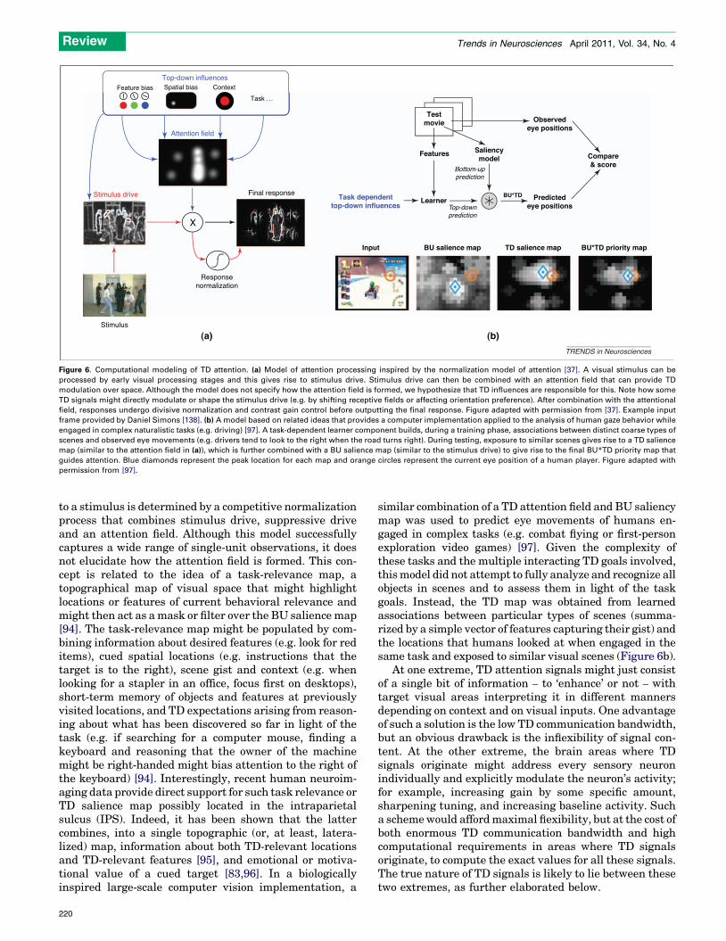
Computational modelingPhysiological studies have guided several theoretical andcomputational models of attention. Building on the influ-ential feature integration theory [2], guided search theoryhypothesizes thatmassively parallel pre-attentive process-es can be guided by TD biasing for features and locations[92]. This theory brings TD elements to a basic BUmodel ofattention [93], which computes individual features at dif-ferent scales and then combines these features to form asaliency map. A unifying normalization model of attentionhas recently been proposed and accounts formany effects ofTD attention onto visual areas (Figure 6a) [37]. In thismodel, the neuronal population response of sensory cortex
219
[()TD$FIG]
Feature bias Spatial bias Context
X
Stimulus
Stimulus drive
Attention field
Final response
Responsenormalization
Top-down influences
Task ...
(a) (b)
Task dependenttop-down influences
Input BU salience map TD salience map BU*TD priority map
Testmovie
Features Saliencymodel
Learner Predictedeye positions
Compare& score
Observedeye positions
BU*TD
TRENDS in Neurosciences
Bottom-upprediction
Top-downprediction
Figure 6. Computational modeling of TD attention. (a) Model of attention processing inspired by the normalization model of attention [37]. A visual stimulus can be
processed by early visual processing stages and this gives rise to stimulus drive. Stimulus drive can then be combined with an attention field that can provide TD
modulation over space. Although the model does not specify how the attention field is formed, we hypothesize that TD influences are responsible for this. Note how some
TD signals might directly modulate or shape the stimulus drive (e.g. by shifting receptive fields or affecting orientation preference). After combination with the attentional
field, responses undergo divisive normalization and contrast gain control before outputting the final response. Figure adapted with permission from [37]. Example input
frame provided by Daniel Simons [138]. (b) A model based on related ideas that provides a computer implementation applied to the analysis of human gaze behavior while
engaged in complex naturalistic tasks (e.g. driving) [97]. A task-dependent learner component builds, during a training phase, associations between distinct coarse types of
scenes and observed eye movements (e.g. drivers tend to look to the right when the road turns right). During testing, exposure to similar scenes gives rise to a TD salience
map (similar to the attention field in (a)), which is further combined with a BU salience map (similar to the stimulus drive) to give rise to the final BU*TD priority map that
guides attention. Blue diamonds represent the peak location for each map and orange circles represent the current eye position of a human player. Figure adapted with
permission from [97].
Review Trends in Neurosciences April 2011, Vol. 34, No. 4
to a stimulus is determined by a competitive normalizationprocess that combines stimulus drive, suppressive driveand an attention field. Although this model successfullycaptures a wide range of single-unit observations, it doesnot elucidate how the attention field is formed. This con-cept is related to the idea of a task-relevance map, atopographical map of visual space that might highlightlocations or features of current behavioral relevance andmight then act as amask or filter over the BU salience map[94]. The task-relevance map might be populated by com-bining information about desired features (e.g. look for reditems), cued spatial locations (e.g. instructions that thetarget is to the right), scene gist and context (e.g. whenlooking for a stapler in an office, focus first on desktops),short-term memory of objects and features at previouslyvisited locations, and TD expectations arising from reason-ing about what has been discovered so far in light of thetask (e.g. if searching for a computer mouse, finding akeyboard and reasoning that the owner of the machinemight be right-handed might bias attention to the right ofthe keyboard) [94]. Interestingly, recent human neuroim-aging data provide direct support for such task relevance orTD salience map possibly located in the intraparietalsulcus (IPS). Indeed, it has been shown that the lattercombines, into a single topographic (or, at least, latera-lized) map, information about both TD-relevant locationsand TD-relevant features [95], and emotional or motiva-tional value of a cued target [83,96]. In a biologicallyinspired large-scale computer vision implementation, a
220
similar combination of a TD attention field and BU saliencymap was used to predict eye movements of humans en-gaged in complex tasks (e.g. combat flying or first-personexploration video games) [97]. Given the complexity ofthese tasks and the multiple interacting TD goals involved,thismodel did not attempt to fully analyze and recognize allobjects in scenes and to assess them in light of the taskgoals. Instead, the TD map was obtained from learnedassociations between particular types of scenes (summa-rized by a simple vector of features capturing their gist) andthe locations that humans looked at when engaged in thesame task and exposed to similar visual scenes (Figure 6b).
At one extreme, TD attention signals might just consistof a single bit of information – to ‘enhance’ or not – withtarget visual areas interpreting it in different mannersdepending on context and on visual inputs. One advantageof such a solution is the low TD communication bandwidth,but an obvious drawback is the inflexibility of signal con-tent. At the other extreme, the brain areas where TDsignals originate might address every sensory neuronindividually and explicitly modulate the neuron’s activity;for example, increasing gain by some specific amount,sharpening tuning, and increasing baseline activity. Sucha schemewould affordmaximal flexibility, but at the cost ofboth enormous TD communication bandwidth and highcomputational requirements in areas where TD signalsoriginate, to compute the exact values for all these signals.The true nature of TD signals is likely to lie between thesetwo extremes, as further elaborated below.

Box 1. Outstanding questions
� What is the bandwidth of the TD signal transmitted from one
region of the brain to the next? Figure I illustrates the two types of
signal. A narrow-bandwidth signal (yellow arrow) defines single
weights for individual features, whereas a broadband signal (blue
arrow) defines the distribution of gain and tuning over the feature
space, as well as the interactions within a feature dimension.
� Are TD signals relayed to visual areas through a central hub (e.g.
the pulvinar) or does a more distributed mechanism reflect the
reality of communication of TD signals to sensory areas?
� What is the representation or encoding of TD signals? In concrete
terms, how are behavioral goals represented and communicated
to sensory neurons that are tuned to specific features?
� What (if any) computations take place subcortically, independent
of the cortex, that would influence attention modulation of
sensory perception?
[()TD$FIG]
A
B
?
Tun
ing
(σ)
G
ain
InteractionsT
unin
g (σ
)
Gai
n
Interactions
I t ti
Tun
ing
(σ)
Gai
n
Interactions
Hue
Orientation
Intensity
Red
Blue Shallow
SteepWei
ght
Dark
Bright
OrientationHue Intensity
0
1
0
1
0
1
Narrow-bandsignal
Broad-band ?Signal
TRENDS in Neurosciences
Figure I. Narrow-band versus broad-band TD biasing signals. The TD biasing
signals transmitted from one area ‘A’ of the brain to another area ‘B’ can be
either narrow-band (yellow arrow) or broad-band (blue arrow) in nature.
Narrow-band signals consist of a small set of weights that bias feature
preferences in a coarse manner. The bar graph shows a signal that applies a
higher gain to neurons tuned to red rather than blue in the color feature
dimension, neurons tuned to shallow rather than steep orientations, and
neurons tuned to brighter rather than darker stimuli. Broad-band biasing
signals (bottom) contain a greater amount of information and might facilitate
biasing of features in a detailed manner, weighing gain, tuning and feature
interactions independently. Rather than simply setting a weight along a feature
dimension, as is the case in the narrow-band example, broad-band TD signals
might set a biasing profile along the feature dimension, as shown in the
example graphs. Green curves show a biasing profile for gains of neurons
along a feature dimension. Blue curves show a biasing profile of tuning of
neurons; a peak here would indicate a bias or shift of tuning of neurons for the
particular feature value. The interaction triangles on the right show biases for
feature interactions. For example, along the hue dimension, there are two hot
spots, one indicating a preference for simultaneous occurrence of yellow and
red hues and another indicating a preference for red and blue hues.
Review Trends in Neurosciences April 2011, Vol. 34, No. 4
The Guided Search model lies towards the low-band-width end of the spectrum, with TD signals imposingspatial attention modulation over coarse regions of visualspace and coarse visual features (e.g. a single TD attentionweight for each of red, green, blue or yellow colors, or steep,shallow, left or right orientations) [92]. Two recent studieshave refined this proposal. First, in human eye-trackingexperiments it has been shown that attention and gaze caneffectively be guided towards rather fine sub-bands of basicvisual features, such as mid-luminance items among low-and high-luminance items, and similarly for size and colorsaturation [98]. Furthermore, these results have beenformalized with a signal-to-noise ratio (SNR)-maximizingmodel for feature search, whereby the TD gain applied toeach sensory neuron is proportional to its ability to distin-guish the target of behavioral interest from backgroundclutter [99]. Taken together, these two studies suggest thatthe bandwidth or granularity of TD signals is unlikely to beextremely low, but rather might consist of at least a fewbits for each fine-grained feature sub-band, sufficient toconvey optimal biases from the top down. The bandwidth(and number of descending connections) might be higher ifdifferent biases can be communicated to different locationsof sensory space. At the high extreme, the aforementionednormalization model of attention assumes a highly de-tailed attention field over space and features [37], implyinghigh-bandwidth TD signals.
Beyond the nature and bandwidth of information con-veyed from the top down, computational models haveproposed a number of connectivity styles that might beembodied in the biological reality of TD connections. Onthe one hand, one model has identified a specific dedicatedstructure (the pulvinar) as a hub or relay for TD signals toreach target visual areas [69]. On the other hand, a moredistributed model suggests that TD signals are embeddedwithin the visual areas themselves [100]. In this model, astimulus is selected at the top level based on an initialsweep of feed-forward information. The spatial selectionsignals then propagate back and tune lower levels of the(cortical) visual processing hierarchy through a cascade ofwinner-take-all mechanisms. This view involves retro-grade propagation of signals over the processing hierarchyas opposed to direct connections (or through one or a fewrelays) between top and bottom. A number of models alsogive specific roles to direct or indirect connections amongdifferent levels of the hierarchy, for example between thePFC, FEF, TE and V4 [101]. These models are importantbecause they develop hypotheses for the meaning of large-scale connectivity between brain areas, and these arebeginning to be explicitly tested in biological networksusing graph-theoretic analyses [102]. Nevertheless, thereis a clear lack of specific computational (and experimental)studies that systematically investigate the granularity,bandwidth and specific wiring of TD signals.
Finally, computational theories and models havestarted to provide hypotheses for the meaning of TD sig-nals. For example, models based on feedback connectionsfrom higher cortical areas have been placed in a Bayesianframework, with the suggestion of a generative model thatproduces a hypothesis about a percept (the prior), thencombines this with evidence from BU information to make
a final decision on the percept [103]. This approach hasbeen formalized in a hierarchical Bayesian framework[104]. Although these ideas have so far been explored morein the context of the mandatory TD process, they can also
221

Review Trends in Neurosciences April 2011, Vol. 34, No. 4
be placed in the context of the volitional TD process.Volitional TD control could then be understood as updat-ing, biasing or disambiguating the prior based on high-level tasks, contextual cues or behavioral goals. In com-puter-vision models using these principles, it has indeedbeen shown that TD attention provides great benefit overpure BU processing [105]. For example, TD informationcan more effectively guide visual search for specific objectsin natural scenes (e.g. pedestrians in street scenes) bylimiting the search to spatial locations of high prior orposterior probabilities [106–108]. Although computationalmodels have made some headway in both incorporatingexperimental data and generating predictions to guidefurther experiments, much remains to be done both exper-imentally and theoretically to unravel the mechanisms bywhich TD attentional mechanisms influence BU proces-sing (Box 1).
ConclusionAttention modulates sensory signals early in the process,exerting its influence on the SC and the thalamus beforefurther modulating signals in cortex. The cumulativeeffects of this modulation based on both TD and BUinfluences might be represented by a priority map overvisual space. Although there is some debate about theexact locus of the priority map, it is clear that the LIP,FEF and SC exhibit properties that are compatible withthe existence of a spatial map encoding behavioral rele-vance of spatial locations. These three regions might joint-ly compute or host such a map that is agnostic to thefeatures that caused the priority. Thus, the map fuses bothBU and TD influences and drives motor output.
Higher cortical areas such as the PFC send detailed TDsignals to sensory areas for biasing of spatial and non-spatial features. Such signals fuse together with reward-related and emotional signals to form the TD influence onattention, which might be reflected in the priority map.Subcortical regions, through their close connection to thereward systems in the brain and their coupling with motorsystems, exert strong influences on attentional signals, inaddition to being major targets of attentional modulationfor motor output. Feedback connections are both pervasiveand crucial for the transmission of biasing signals ema-nating from higher brain regions, especially the frontalcortices that are involved in working memory processesand send descending reward signals. Computational stud-ies highlight the important constraints on the nature,granularity, bandwidth and connectivity style of TD con-nections. There is a pressing need to buildmodels that takeinto account physiological data, particularly from micro-stimulation and lesion studies, which could help to deter-mine the contributions of specific areas to thecomputations necessary for attentional guidance.
Although the exact mechanisms of TD attention haveyet to be completely delineated, there are sufficient dataavailable to demonstrate that attention is mediated by themerging of TD and BU information. As William Jameseloquently stated, ‘ The attentive process, therefore, at itsmaximum may be physiologically symbolized, by a brain-cell played on in two ways from without and from within’[109].
222
AcknowledgementsThis work was supported by the Defense Advanced Research ProjectsAgency (government contract no. HR0011-10-C-0034), the NationalScience Foundation (CRCNS grant number BCS-0827764), GeneralMotors Corporation, and the Army Research Office (grant no. W911NF-08-1-0360). The authors affirm that the views expressed herein are solelytheir own, and do not represent the views of the United Statesgovernment or any agency thereof. We would also like to thank RobertDesimone, Jack Gallant, Jacqueline Gottlieb and the anonymousreviewers for their helpful comments and suggestions.
References1 James, W. (1890) The Principles of Psychology, Harvard University
Press2 Treisman, A. and Gelade, G. (1980) A feature-integration theory of
attention. Cogn. Psychol. 12, 97–1363 Wolfe, J.M. and Horowitz, T.S. (2004) What attributes guide the
deployment of visual attention and how do they do it? Nat. Rev.Neurosci. 5, 495–501
4 Itti, L. and Koch, C. (2001) Computational modelling of visualattention. Nat. Rev. Neurosci. 2, 194–203
5 Gilbert, C. and Sigman,M. (2007) Brain states: top-down influences insensory processing. Neuron 54, 677–696
6 Chun, M.M. and Jiang, Y. (1998) Contextual cueing: implicit learningand memory of visual context guides spatial attention. Cogn. Psychol.36, 28–71
7 Desimone, R. and Duncan, J. (1995) Neural mechanisms of selectivevisual attention. Annu. Rev. Neurosci. 18, 193–222
8 Noudoost, B. et al. (2010) Top-down control of visual attention. Curr.Opin. Neurobiol. 20, 183–190
9 Miller, E.K. and Cohen, J.D. (2001) An integrative theory of prefrontalcortex function. Annu. Rev. Neurosci. 24, 167–202
10 Corbetta, M. and Shulman, G.L. (2002) Control of goal-directed andstimulus-driven attention in the brain.Nat. Rev. Neurosci. 3, 201–215
11 Werblin, F. et al. (2001) Parallel processing in the mammalian retina:lateral and vertical interactions across stacked representations. Prog.Brain Res. 131, 229–238
12 Goodale, M.A. and Milner, A.D. (1992) Separate visual pathways forperception and action. Trends Neurosci. 15, 20–25
13 Wurtz, R. and Mohler, C. (1976) Enhancement of visual responses inmonkey striate cortex and frontal eye fields. J. Neurophysiol. 39, 766–
77214 Fecteau, J. and Munoz, D. (2006) Salience, relevance, and firing: a
priority map for target selection. Trends Cogn. Sci. 10, 382–39015 Bisley, J.W. and Goldberg, M.E. (2010) Attention, intention, and
priority in the parietal lobe. Annu. Rev. Neurosci. 33, 1–2116 Thompson, K.G. and Bichot, N.P. (2005) A visual salience map in the
primate frontal eye field. Prog. Brain Res. 147, 251–26217 Shipp, S. (2003) The functional logic of cortico-pulvinar connections.
Philos. Trans. R. Soc. Lond. B Biol. Sci. 358, 1605–162418 McAlonan, K. et al. (2008) Guarding the gateway to cortex with
attention in visual thalamus. Nature 456, 391–39419 O’Connor, D. et al. (2002) Attention modulates responses in the
human lateral geniculate nucleus. Nat. Neurosci. 5, 1203–120920 Buffalo, E.A. et al. (2010) A backward progression of attentional
effects in the ventral stream. Proc. Natl. Acad. Sci. U.S.A. 107,361–365
21 McAdams, C.J. and Maunsell, J.H. (1999) Effects of attention onorientation-tuning functions of single neurons in macaque corticalarea V4. J. Neurosci. 19, 431–441
22 Martinez-Trujillo, J.C. and Treue, S. (2004) Feature-based attentionincreases the selectivity of population responses in primate visualcortex. Curr. Biol. 14, 744–751
23 Gottlieb, J. and Balan, P. (2010) Attention as a decision in informationspace. Trends Cogn. Sci. 14, 240–248
24 Gottlieb, J. (2007) From thought to action: the parietal cortex as abridge between perception, action, and cognition. Neuron 53, 9–16
25 Gottlieb, J. et al. (1998) The representation of visual salience inmonkey parietal cortex. Nature 391, 481–484
26 Bisley, J. and Goldberg, M. (2003) Neuronal activity in the lateralintraparietal area and spatial attention. Science 299, 81–86
27 Ungerleider, L.G. et al. (2008) Cortical connections of area V4 in themacaque. Cereb. Cortex 18, 477–499
Review Trends in Neurosciences April 2011, Vol. 34, No. 4
28 Lamme, V.A. and Roelfsema, P.R. (2000) The distinct modes of visionoffered by feedforward and recurrent processing. Trends Neurosci. 23,571–579
29 Monosov, I. et al. (2008) Measurements of simultaneously recordedspiking activity and local field potentials suggest that spatial selectionemerges in the frontal eye field. Neuron 57, 614–625
30 Buschman, T. and Miller, E. (2007) Top-down versus bottom-upcontrol of attention in the prefrontal and posterior parietal cortices.Science 315, 1860–1862
31 Lebedev, M.A. et al. (2004) Representation of attended versusremembered locations in prefrontal cortex. PLoS Biol. 2, e365
32 Luck, S.J. et al. (1997) Neural mechanisms of spatial selectiveattention in areas V1, V2, and V4 of macaque visual cortex. J.Neurophysiol. 77, 24–42
33 Bichot, N.P. et al. (2005) Parallel and serial neural mechanisms forvisual search in macaque area V4. Science 308, 529–534
34 Saenz, M. et al. (2003) Global feature-based attention for motion andcolor. Vis. Res. 43, 629–637
35 Treue, S. and Martinez Trujillo, J.C. (1999) Feature-based attentioninfluences motion processing gain in macaque visual cortex. Nature399, 575–579
36 Motter, B.C. (1994) Neural correlates of attentive selection for color orluminance in extrastriate area V4. J. Neurosci. 14, 2178–2189
37 Reynolds, J.H. and Heeger, D.J. (2009) The normalization model ofattention. Neuron 61, 168–185
38 Reynolds, J. and Chelazzi, L. (2004) Attentional modulation of visualprocessing. Annu. Rev. Neurosci. 27, 611–647
39 Knudsen, E.I. (2007) Fundamental components of attention. Annu.Rev. Neurosci. 30, 57–78
40 Tomita, H. et al. (1999) Top-down signal from prefrontal cortex inexecutive control of memory retrieval. Nature 401, 699–703
41 Rossi, A. et al. (2007) Top down attentional deficits in macaques withlesions of lateral prefrontal cortex. J. Neurosci. 27, 11306–11314
42 Opris, I. et al. (2005) Microstimulation of the dorsolateral prefrontalcortex biases saccade target selection. J. Cogn. Neurosci. 17, 893–904
43 Moore, T. and Armstrong, K. (2003) Selective gating of visual signalsby microstimulation of frontal cortex. Nature 421, 370–373
44 Winkowski, D. and Knudsen, E. (2008) Distinct mechanisms for top-down control of neural gain and sensitivity in the owl optic tectum.Neuron 60, 698–708
45 Gregoriou, G.G. et al. (2009) High-frequency, long-range couplingbetween prefrontal and visual cortex during attention. Science 324,1207–1210
46 Wardak, C. et al. (2006) Contribution of themonkey frontal eye field tocovert visual attention. J. Neurosci. 26, 4228–4235
47 Cutrell, E. and Marrocco, R. (2002) Electrical microstimulation ofprimate posterior parietal cortex initiates orienting and alertingcomponents of covert attention. Exp. Brain Res. 144, 103–113
48 Balan, P.F. and Gottlieb, J. (2009) Functional significance ofnonspatial information in monkey lateral intraparietal area. J.Neurosci. 29, 8166–8176
49 Friedman-Hill, S. et al. (2003) Posterior parietal cortex and thefiltering of distractors. Proc. Natl. Acad. Sci. U.S.A. 100, 4263–
426850 Saalmann, Y. et al. (2007) Neuralmechanisms of visual attention: how
top-down feedback highlights relevant locations. Science 316, 161251 Liu, Y. et al. (2010) Intention and attention: different functional roles
for LIPD and LIPV. Nat. Neurosci. 13, 495–50052 Sherman, S. (2007) The thalamus is more than just a relay. Curr.
Opin. Neurobiol. 17, 417–42253 Kawasaki, K. and Sheinberg, D.L. (2008) Learning to recognize visual
objects with microstimulation in inferior temporal cortex. J.Neurophysiol. 100, 197–211
54 Afraz, S-R. et al. (2006) Microstimulation of inferotemporal cortexinfluences face categorization. Nature 442, 692–695
55 Cavanaugh, J. and Wurtz, R. (2004) Subcortical modulation ofattention counters change blindness. J. Neurosci. 24, 11236–11243
56 Muller, J. et al. (2005) Microstimulation of the superior colliculusfocuses attention without moving the eyes. Proc. Natl. Acad. Sci.U.S.A. 102, 524–529
57 Carello, C. and Krauzlis, R., (2004)Manipulating intent evidence for acausal role of the superior colliculus in target selection. Neuron 43,575–583
58 McPeek, R. and Keller, E. (2004) Deficits in saccade target selectionafter inactivation of superior colliculus. Nat. Neurosci. 7, 757–763
59 Ignashchenkova, A. et al. (2003) Neuron-specific contribution of thesuperior colliculus to overt and covert shifts of attention. Nat.Neurosci. 7, 56–64
60 Lovejoy, L. and Krauzlis, R. (2009) Inactivation of primate superiorcolliculus impairs covert selection of signals for perceptual judgments.Nat. Neurosci. 13, 261–266
61 Kaas, J.H. and Lyon, D.C. (2007) Pulvinar contributions to the dorsaland ventral streams of visual processing in primates. Brain Res. Rev.55, 285–296
62 Leh, S. et al. (2008) The connectivity of the human pulvinar: adiffusion tensor imaging tractography study. Int. J. Biomed.Imaging 2008, 1–5
63 Robinson, D.L. and Petersen, S.E. (1992) The pulvinar and visualsalience. Trends Neurosci. 15, 127–132
64 Desimone, R. et al. (1990) Attentional control of visual perception:cortical and subcortical mechanisms. Cold Spring Harb. Symp.Quant. Biol. 55, 963–971
65 Snow, J. et al. (2009) Impaired attentional selection following lesionsto human pulvinar: evidence for homology between human andmonkey. Proc. Natl. Acad. Sci. U.S.A. 106, 4054–4059
66 Arend, I. et al. (2008) Spatial and temporal deficits are regionallydissociable in patients with pulvinar lesions. Brain 131, 2140–2152
67 Bender, D.B. and Baizer, J.S. (1990) Saccadic eye movementsfollowing kainic acid lesions of the pulvinar in monkeys. Exp.Brain Res. 79, 467–478
68 Bender, D.B. and Butter, C.M. (1987) Comparison of the effects ofsuperior colliculus and pulvinar lesions on visual search andtachistoscopic pattern discrimination in monkeys. Exp. Brain Res.69, 140–154
69 Olshausen, B.A. et al. (1993) A neurobiological model of visualattention and invariant pattern recognition based on dynamicrouting of information. J. Neurosci. 13, 4700–4719
70 Berman, R.A. and Wurtz, R.H. (2010) Functional identification of apulvinar path from superior colliculus to cortical areaMT. J. Neurosci.30, 6342–6354
71 Peck, C.J. et al. (2009) Reward modulates attention independently ofaction value in posterior parietal cortex. J. Neurosci. 29, 11182–
1119172 Toth, L.J. and Assad, J.A. (2002) Dynamic coding of behaviourally
relevant stimuli in parietal cortex. Nature 415, 165–16873 Bichot, N.P. and Schall, J.D. (1999) Effects of similarity and history on
neural mechanisms of visual selection. Nat. Neurosci. 2, 549–55474 Bichot, N.P. et al. (1996) Visual feature selectivity in frontal eye fields
induced by experience in mature macaques. Nature 381, 697–69975 Baluch, F. and Itti, L. (2010) Training top-down attention improves
performance on a triple-conjunction search task. PLoS ONE 5, e912776 Graybiel, A. (2005) The basal ganglia: learning new tricks and loving
it. Curr. Opin. Neurobiol. 15, 638–64477 Boehnke, S.E. and Munoz, D.P. (2008) On the importance of the
transient visual response in the superior colliculus. Curr. Opin.Neurobiol. 18, 544–551
78 Hikosaka, O. et al. (2006) Basal ganglia orient eyes to reward. J.Neurophysiol. 95, 567–584
79 Kaneda, K. et al. (2008) Nigral inhibition of GABAergic neurons inmouse superior colliculus. J. Neurosci. 28, 11071–11078
80 Shires, J. et al. (2010) Shedding new light on the role of the basalganglia-superior colliculus pathway in eye movements. Curr. Opin.Neurobiol. 20, 717–725
81 Pessoa, L. (2008) On the relationship between emotion and cognition.Nat. Rev. Neurosci. 9, 148–158
82 Vuilleumier, P. et al. (2004) Distant influences of amygdala lesion onvisual cortical activation during emotional face processing. Nat.Neurosci. 7, 1271–1278
83 Mohanty, A. et al. (2009) Search for a threatening target triggerslimbic guidance of spatial attention. J. Neurosci. 29, 10563–10572
84 Fries, P. (2009) Neuronal gamma-band synchronization as afundamental process in cortical computation. Annu. Rev. Neurosci.32, 209–224
85 Fries, P. et al. (2002) Oscillatory neuronal synchronization in primaryvisual cortex as a correlate of stimulus selection. J. Neurosci. 22,3739–3754
223
Review Trends in Neurosciences April 2011, Vol. 34, No. 4
86 Lakatos, P. et al. (2008) Entrainment of neuronal oscillations as amechanism of attentional selection. Science 320, 110–113
87 Sarter, M. et al. (2005) Unraveling the attentional functions of corticalcholinergic inputs: interactions between signal-driven and cognitivemodulation of signal detection. Brain Res. Rev. 48, 98–111
88 Herrero, J.L. et al. (2008) Acetylcholine contributes throughmuscarinic receptors to attentional modulation in V1. Nature 454,1110–1114
89 Nelson, C.L. et al. (2005) Prefrontal cortical modulation ofacetylcholine release in posterior parietal cortex. Neuroscience 132,347–359
90 Deco, G. and Thiele, A. (2009) Attention: oscillations andneuropharmacology. Eur. J. Neurosci. 30, 347–354
91 Buehlmann, A. and Deco, G. (2008) The neuronal basis of attention:rate versus synchronization modulation. J. Neurosci. 28, 7679–
768692 Wolfe, J. (1994) Guided search 2.0. A revised model of visual search.
Psychonom. Bull. Rev. 1, 202–23893 Koch, C. and Ullman, S. (1985) Shifts in selective visual attention:
towards the underlying neural circuitry. Hum. Neurobiol. 4, 219–
22794 Navalpakkam, V. and Itti, L. (2005) Modeling the influence of task on
attention. Vis. Res. 45, 205–23195 Egner, T. et al. (2008) Neural integration of top-down spatial and
feature-based information in visual search. J. Neurosci. 28, 6141–
615196 Mohanty, A. et al. (2008) The spatial attention network interacts with
limbic and monoaminergic systems to modulate motivation-inducedattention shifts. Cereb. Cortex 18, 2604–2613
97 Peters, R. and Itti, L. (2007) Beyond bottom-up: incorporating task-dependent influences into a computational model of spatial attention.In Proceedings of the 2007 IEEE Conference on Computer Vision andPattern Recognition, pp. 1–8
98 Navalpakkam, V. and Itti, L. (2006) Top-down attention selection isfine-grained. J. Vis. 6, 1180–1193
99 Navalpakkam, V. and Itti, L. (2007) Search goal tunes visual featuresoptimally. Neuron 53, 605–617
100 Tsotsos, J.K. et al. (1995) Modeling visual-attention via selectivetuning. Artif. Intell. 78, 507–545
101 Hamker, F.H. (2006) Modeling feature-based attention as an activetop-down inference process. Biosystems 86, 91–99
102 Bullmore, E. and Sporns, O. (2009) Complex brain networks: graphtheoretical analysis of structural and functional systems. Nat. Rev.Neurosci. 10, 186–198
103 Gregory, R. (1970) The Intelligent Eye, McGraw-Hill104 Lee, T. and Mumford, D. (2003) Hierarchical Bayesian inference in
the visual cortex. J. Opt. Soc. Am. A 20, 1434–1448105 Frintrop, S. et al. (2010) Computational visual attention systems and
their cognitive foundation: a survey. ACM Trans. Appl. Percept. 7, 6106 Najemnik, J. and Geisler, W.S. (2005) Optimal eye movement
strategies in visual search. Nature 434, 387–391107 Torralba, A. et al. (2006) Contextual guidance of eye movements and
attention in real-world scenes: the role of global features in objectsearch. Psychol. Rev. 113, 766–786
108 Ehinger, K.A. et al. (2009) Modeling search for people in 900 scenes: acombined source model of eye guidance. Vis. Cogn. 17, 945–978
109 James, W. (1892) Talks to Teachers on Psychology and to Students onSome of Life’s Ideals, Adamant Media Corporation
110 Rafal, R.D. and Posner, M.I. (1987) Deficits in human visual spatialattention following thalamic lesions. Proc. Natl. Acad. Sci. U.S.A. 84,7349–7353
111 Pezaris, J.S. and Reid, R.C. (2007) Demonstration of artificial visualpercepts generated through thalamic microstimulation. Proc. Natl.Acad. Sci. U.S.A. 104, 7670–7675
112 Schmid, M.C. et al. (2010) Blindsight depends on the lateralgeniculate nucleus. Nature 466, 373–377
113 Page, W.K. et al. (1994) Magnocellular or parvocellular lesions in thelateral geniculate nucleus of monkeys cause minor deficits of smoothpursuit eye movements. Vis. Res. 34, 223–239
224
114 Tehovnik, E.J. et al. (2003) Differential effects of laminar stimulationof V1 cortex on target selection by macaque monkeys. Eur. J.Neurosci. 16, 751–760
115 Tehovnik, E.J. et al. (2003) Saccadic eye movements evoked bymicrostimulation of striate cortex. Eur. J. Neurosci. 17, 870–878
116 Murphey, D.K. and Maunsell, J.H.R. (2007) Behavioral detection ofelectrical microstimulation in different cortical visual areas. Curr.Biol. 17, 862–867
117 Moore, T. et al. (2001) Direction of motion discrimination after earlylesions of striate cortex (V1) of themacaquemonkey. Proc. Natl. Acad.Sci. U.S.A. 98, 325–330
118 Yoshida, M. et al. (2008) Striate cortical lesions affect deliberatedecision and control of saccade: implication for blindsight. J.Neurosci. 28, 10517–10530
119 Buffalo, E. et al. (2005) Impaired filtering of distracter stimuli by TEneurons following V4 and TEO lesions in macaques. Cereb. Cortex 15,141–151
120 Bachevalier, J. and Mishkin, M. (1994) Effects of selective neonataltemporal lobe lesions on visual recognition memory in rhesusmonkeys. J. Neurosci. 14, 2128–2139
121 Petrides, M. (2000) Dissociable roles of mid-dorsolateral prefrontaland anterior inferotemporal cortex in visual working memory. J.Neurosci. 20, 7496–7503
122 Nichols, M.J. and Newsome, W.T. (2002) Middle temporal visual areamicrostimulation influences veridical judgments of motion direction.J. Neurosci. 22, 9530–9540
123 Bisley, J.W. et al. (2001) Microstimulation of cortical area MT affectsperformance on a visual working memory task. J. Neurophysiol. 85,187–196
124 Newsome, W.T. and Pare, E.B. (1988) A selective impairment ofmotion perception following lesions of the middle temporal visualarea (MT). J. Neurosci. 8, 2201–2211
125 Hanks, T.D. et al. (2006) Microstimulation of macaque area LIPaffects decision-making in a motion discrimination task. Nat.Neurosci. 9, 682–689
126 Moore, T. and Fallah, M. (2004) Microstimulation of the frontal eyefield and its effects on covert spatial attention. J. Neurophysiol. 91,152–162
127 Juan, C. et al. (2004) Dissociation of spatial attention and saccadepreparation. Proc. Natl. Acad. Sci. U.S.A. 101, 15541–15544
128 Ruff, C.C. et al. (2006) Concurrent TMS-fMRI and psychophysicsreveal frontal influences on human retinotopic visual cortex. Curr.Biol. 16, 1479–1488
129 Neggers, S.F.W. et al. (2007) TMS pulses on the frontal eye fieldsbreak coupling between visuospatial attention and eyemovements. J.Neurophysiol. 98, 2765–2778
130 Wegener, S.P. et al. (2008) Microstimulation of monkey dorsolateralprefrontal cortex impairs antisaccade performance. Exp. Brain Res.190, 463–473
131 Rasmusson, D.D. et al. (2007) Inactivation of prefrontal cortexabolishes cortical acetylcholine release evoked by sensory orsensory pathway stimulation in the rat. Neuroscience 149, 232–241
132 Rudolph, K. and Pasternak, T. (1999) Transient and permanentdeficits in motion perception after lesions of cortical areas MT andMST in the macaque monkey. Cereb. Cortex 9, 90–100
133 De Weerd, P. et al. (1999) Loss of attentional stimulus selection afterextrastriate cortical lesions in macaques. Nat. Neurosci. 2, 753–758
134 Gregory, R. (1997) Knowledge in perception and illusion. Philos.Trans. R. Soc. B Biol. Sci. 352, 1121–1127
135 Van Essen, D.C. (2002) Windows on the brain: the emerging role ofatlases and databases in neuroscience. Curr. Opin. Neurobiol. 12,574–579
136 Van Essen, D.C. et al. (2001) An integrated software suite for surface-basedanalyses of cerebral cortex.J.Am.Med. Inform.Assoc.8, 443–459
137 Pascual-Leone, A. andWalsh, V. (2001) Fast backprojections from themotion to the primary visual area necessary for visual awareness.Science 292, 510–512
138 Simons, D.J. and Chabris, C.F. (1999) Gorillas in ourmidst: sustainedinattentional blindness for dynamic events. Perception 28, 1059–1074