FGF signaling in specification of hESC- derived definitive endoderm
RESEARCH ARTICLE
Mechanisms of endoderm formation in a cartilaginous fish revealancestral and homoplastic traits in jawed vertebrates
Benoit G. Godard1, Marion Coolen2,7, Sophie Le Panse3, Aurelie Gombault2,9, Susana Ferreiro-Galve2,8,Laurent Laguerre1, Ronan Lagadec1, Patrick Wincker4, Julie Poulain4, Corinne Da Silva4, Shigehiro Kuraku5,Wilfrid Carre6, Agnes Boutet1 and Sylvie Mazan1,*
ABSTRACT
In order to gain insight into the impact of yolk increase on endoderm
development, we have analyzed the mechanisms of endoderm
formation in the catshark S. canicula, a species exhibiting telolecithal
eggs and a distinct yolk sac. We show that in this species, endoderm
markers are expressed in two distinct tissues, the deepmesenchyme,
a mesenchymal population of deep blastomeres lying beneath the
epithelial-like superficial layer, already specified at early blastula
stages, and the involuting mesendoderm layer, which appears at the
blastoderm posterior margin at the onset of gastrulation. Formation of
the deep mesenchyme involves cell internalizations from the
superficial layer prior to gastrulation, by a movement suggestive of
ingressions. These cell movements were observed not only at the
posterior margin, where massive internalizations take place prior to
the start of involution, but also in the center of the blastoderm, where
internalizations of single cells prevail. Like the adjacent involuting
mesendoderm, the posterior deep mesenchyme expresses anterior
mesendoderm markers under the control of Nodal/activin signaling.
Comparisons across vertebrates support the conclusion that
endoderm is specified in two distinct temporal phases in the
catshark as in all major osteichthyan lineages, in line with an
ancient origin of a biphasic mode of endoderm specification in
gnathostomes. They also highlight unexpected similarities with
amniotes, such as the occurrence of cell ingressions from the
superficial layer prior to gastrulation. These similarities may
correspond to homoplastic traits fixed separately in amniotes and
chondrichthyans and related to the increase in egg yolk mass.
KEY WORDS: endoderm, telolecithal egg, chondrichthyan, Nodal
signalling
INTRODUCTIONSpectacular expansions of the egg yolk mass have taken place
several times during vertebrate evolution, extreme examples of
this evolutionary trend being observed in amniotes, cartilaginous
fishes and myxinoids. These adaptations have gone together with
transitions from holoblastic to meroblastic cleavage modes and
major changes in the early embryo architecture (Arendt and
Nubler-Jung, 1999). One of the most visible examples of
such changes is the presence of endodermal components
morphologically distinct from the embryonic gut and
traditionally referred to as extraembryonic, such as the early
lower layer of birds (hypoblast and endoblast), primitive
endoderm of mammals or yolk syncytial layer (YSL) of
teleosts. The cellular organization and mode of formation of
these tissues have been extensively studied in the mouse, chick
and zebrafish and they appear highly divergent between these
species. For instance, in the mouse, the primitive endoderm forms
a morphologically distinct polarized epithelium, which arises
from the blastocyst inner cell mass by a cell sorting mechanism,
shortly after blastocoel formation. It later subdivides into two
components, the parietal endoderm and visceral endoderm,
which contrary to the traditional view of an absolute early
segregation between embryonic and extraembryonic tissues, is
now known to contribute to the gut (Rossant and Tam, 2009;
Kwon et al., 2008). In the chick, hypoblast formation has long
been thought to involve the merging of cell clusters, originating
from the early epiblast by poly-ingression, and the occurrence of
single cell ingressions from the epiblast prior to gastrulation has
recently been confirmed at stages preceding primitive streak
formation (Stern and Downs, 2012; Voiculescu et al., 2014).
Finally, the zebrafish YSL forms by a collapse of marginal
blastomeres with the yolk cell cytoplasmic cortex between the
512- to 1024-cell stage (Carvalho and Heisenberg, 2010).
Despite these differences in morphogenesis, the amniote
hypoblast/AVE (anterior visceral endoderm) and teleost dorsal
YSL share expression of signaling molecules and transcription
factors known as components of AME (anterior mesendoderm)
genetic programs, a similarity proposed to be related to
independent recruitments in the amniote and actinopterygian
lineages (Stern and Downs, 2012).
How the increase in egg yolk amount may affect early
endoderm formation and patterning is poorly known outside
osteichthyans (bony fishes and their descendants, including
tetrapods), which comprise all established vertebrate model
organisms. Cartilaginous fishes or chondrichthyans, which form
one of the three major vertebrate phyla and comprise about 1100
extant species, are of interest to address this issue for two reasons.
First, as the closest outgroup to osteichthyans, the other major
1Sorbonne Universites, UPMC Univ Paris 06, CNRS, UMR 7150, 29688 Roscoff,France. 2Universite d’Orleans-CNRS, UMR 6218, 45070 Orleans, France.3Plateforme d’Imagerie, Sorbonne Universites, UPMC Univ Paris 06, CNRS, FR2424, Station Biologique, 29688 Roscoff, France. 4CEA-Institut de Genomique-Genoscope, 2 rue Gaston-Cremieux, 91057 Evry, France. 5Genome Resourceand Analysis Unit (GRAS), Center for Developmental Biology, RIKEN.2-2-3Minatojima-minami, Chuo-KU, Kobe 650-0047, Japan. 6ABiMS, SorbonneUniversites, UPMC Univ Paris 06, CNRS, FR 2424, 29688 Roscoff, France.7Present address: CNRS UPR 3294, Institute of Neurobiology Alfred Fessard,91198 Gif-sur-Yvette, France. 8Present address: Instituto de Neurociencias,Consejo Superior de Investigaciones Cientıficas y Universidad MiguelHernandez, Campus San Juan de Alicante, 03550 Alicante, Spain. 9Presentaddress: UMR 7355, Universite d’Orleans-CNRS, 45071 Orleans, France.
This is an Open Access article distributed under the terms of the Creative Commons AttributionLicense (http://creativecommons.org/licenses/by/3.0), which permits unrestricted use, distributionand reproduction in any medium provided that the original work is properly attributed.
Received 21 February 2014; Accepted 8 October 2014
*Corresponding author ([email protected])
� 2014. Published by The Company of Biologists Ltd | Biology Open (2014) 000, 1–10 doi:10.1242/bio.20148037
1
BiologyOpen
by guest on May 30, 2019http://bio.biologists.org/Downloaded from
phylum of gnathostomes (jawed vertebrates), they are essential toreconstruct gnathostome ancestral characteristics, through
comparisons with other vertebrate models (Coolen et al.,2008a). Second, a lecithotrophic mode of embryonic nutrition islikely to be ancestral in chondrichthyans and most elasmobranchsdevelop from large telolecithal eggs, endowed with a distinct yolk
sac (Blackburn, 2014). This is in particular the case of thecatshark Scyliorhinus canicula, one of the most extensivelystudied representatives of chondrichthyans (Coolen et al., 2008a).
This species develops from large, telolecithal eggs, whichundergo a discoidal meroblastic cleavage and are laid at earlystages of blastocoel formation (Ballard et al., 1993). Following
egg deposition, the blastoderm consists of two cell layers, asuperficial one, exhibiting an epithelial-like morphology, and aninner cell population of dispersed blastomeres, referred to as deep
mesenchyme (Ballard et al., 1993; Coolen et al., 2007). Thisbilayered structure persists for about seven days, a periodcharacterized morphologically by a size expansion of theblastoderm (Ballard et al., 1993). A marked change is observed
at stage 11, which is considered as the start of gastrulation. At thisstage, a novel cell population identified as mesendoderm, basedboth on molecular characterization and histology, starts to
involute along the blastoderm posterior margin, adjacent to thedeep mesenchyme (Ballard et al., 1993; Coolen et al., 2007). Thisinvolution movement results in the formation of a posterior
overhang, which initially elongates over the yolk from anterior toposterior and is later found lining the developing archenteron(Coolen et al., 2007). Concomitantly, lateral and anterior regions
of the blastoderm become thinner and spread over the yolk tolater form a distinct yolk sac, connected to the embryo via avascularized stalk. From the morphological appearance of theembryonic axis (stage 12) and until neural tube closure, the
developing embryo thus appears strictly restricted to the posteriorpart of a growing, flattened disc (Ballard et al., 1993; Coolenet al., 2007). This posterior restriction of embryo formation is a
major difference with teleosts and was previously suggested tobe related to the increase in egg yolk mass and the rise of adistinct yolk sac (Arendt and Nubler-Jung, 1999). The timing of
specification and mode of formation of early endodermal tissuesare completely unknown in the catshark. Here, we address theseprocesses, thus providing the first characterization of endodermformation in a chondrichthyan. Comparisons with osteichthyan
model organisms highlight characteristics likely to correspond toancestral traits of jawed vertebrates, and homoplastic featureswith amniotes, possibly indicative of conserved developmental
constraints.
MATERIALS AND METHODSEmbryo production and maintenance, staging and nomenclatureS. canicula eggs were produced by the Biological Marine Resources
facility of Roscoff Marine Station and kept in 17 C oxygenated sea water
until the desired stages were obtained. This study was performed on
catshark embryos prior to formation of the nervous system and of any
other organ and is therefore exempt from a special license under the
terms of institutional and national regulations. Embryos were staged after
Ballard et al. (Ballard et al., 1993) and a description of the stages studied
is provided in supplementary material Movies 1 and 2. Stage 11 is
considered as the start of gastrulation, based on two criteria (1) the
appearance of a distinct mesendoderm layer (Ballard et al., 1993) and (2)
the onset of Brachyury expression (Coolen et al., 2007). Prior to stage 11,
the anterior to posterior polarity of the blastoderm refers to
the orientation of the future elongating embryonic axis, and
corresponds to the ventral to dorsal, or ab-organizer to organizer
polarity of amphibians and teleosts.
Probe isolation and characterizationThe S. canicula Dkk1 probe was amplified by degenerate RT-PCR from
stage 9–15 cDNA by a nested PCR, successively using the following
pairs of primers: 59-GAYGCNATGTGYTGYCC and 39-ATYTT-
RCTCCARAARTG, respectively encoding the conserved DAMCCP
and HFWSKI amino acid motifs of the Dkk1 peptide of other vertebrates,
and 59-GCNATGTGYTGYCCNGG and 39-ARRCACATRTCNCCYTC,
respectively encoding the conserved motifs AMCCPG and EGDMCL.
The amplified cDNA fragments were subcloned in the pGEM-T easy
vector and sequenced. ScSox17, ScHex, ScLeftyB, ScFgf17, ScShh and
ScChd probes were obtained from a large-scale cDNA sequencing project
described (Coolen et al., 2007). Novel sequences were included in
molecular phylogenetic trees to confirm their identity (supplementary
material Fig. S1). ScT, ScOtx5, ScGata6 and ScLim1 probes were
reported in previous studies (Coolen et al., 2007; Plouhinec et al., 2005;
Sauka-Spengler et al., 2003).
In situ hybridization and histological analysesWhole-mount in situ hybridizations were conducted using standard
protocols adapted to the catshark and followed by embryo embedding in
paraffin and sectioning, as described previously (Derobert et al., 2002).
For semi-thin sections, embryos were fixed in 4% glutaraldehyde, 0.25 M
sucrose in 0.2 M cacodylate buffer pH 7.4, post-fixed in 1% OsO4 and
embedded in Epon. 0.5 mm sections were cut and stained with toluidine
blue.
DiI cell labelingStage 8 to 10 embryos were removed from the shell and transferred to
0.45 mm filtered sea water. CellTracker CM-DiI (Invitrogen) was diluted
(1/10) in 0.3M sucrose from a 5 mg/ml stock solution in ethanol and
applied to embryo territories by ejection from a capillary tube. A control
was also performed after one hour of culture, in order to check the
absence of internal labeling due to tissue disruption related to the process
of dye application. Labeled embryos were cultured in filtered sea water
for 24 hours, prior to fixation, paraffin embedding and sectioning
(12 mm). Sections were stained with DAPI, mounted and photographed
using a Leica SP5 confocal microscope. The presence or absence of
labeled cells was assessed in the deep mesenchyme or involuted
mesendoderm, taking into account heavily labeled cells.
Pharmacological treatmentsPharmacological treatments were conducted by in ovo injection of 200 ml
of a 500 mM dilution of the Alk4/5/7 inhibitor SB-505124 in 0.01%
DMSO in stage 8/9 catshark embryos. This solution was replaced by the
same volume of 0.01% DMSO in control embryos. Following injections,
eggs were maintained for 3 days in oxygenated sea water at 17 C, with
viabilities higher than 90%. They were then dissected, fixed in PFA 4%,
dehydrated and stored at 220 C in methanol 100% prior to in situ
hybridization.
RESULTSTwo distinct phases of ScSox17, ScGata6 and ScHexexpressions in the early catshark embryoIn order to unambiguously identify endodermal cell populations in
the catshark, we analyzed expression of homologues of three genesknown to be expressed in extraembryonic endoderm in amniotesand additional mesendoderm territories, ScSox17, ScGata6, and
ScHex. Expression was observed at the earliest blastula stagesstudied (stages 4–6), less than 48 hours following egg deposition(supplementary material Fig. S2A). At stages 7 to 10, all three were
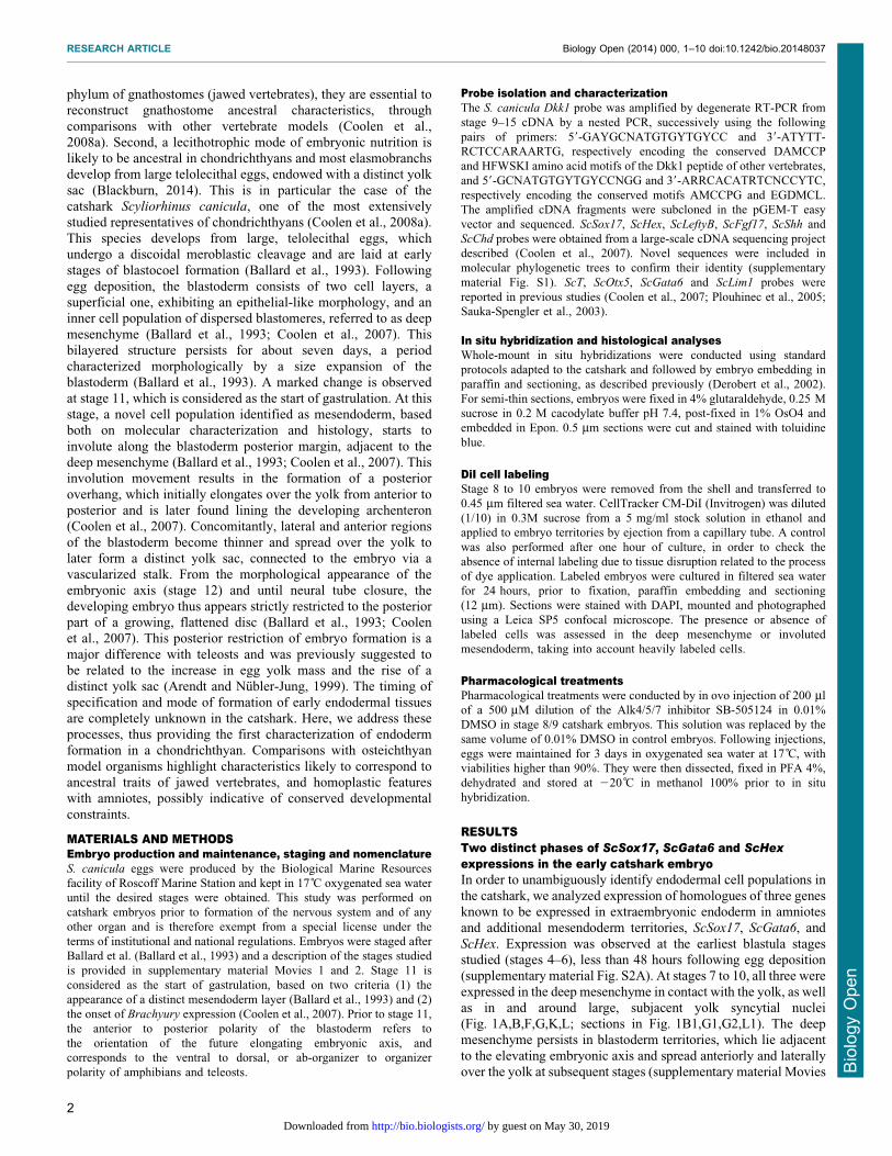
expressed in the deep mesenchyme in contact with the yolk, as wellas in and around large, subjacent yolk syncytial nuclei(Fig. 1A,B,F,G,K,L; sections in Fig. 1B1,G1,G2,L1). The deepmesenchyme persists in blastoderm territories, which lie adjacent
to the elevating embryonic axis and spread anteriorly and laterallyover the yolk at subsequent stages (supplementary material Movies
RESEARCH ARTICLE Biology Open (2014) 000, 1–10 doi:10.1242/bio.20148037
2
BiologyOpen
by guest on May 30, 2019http://bio.biologists.org/Downloaded from
1 and 2; Fig. S2F,G). Expressions of ScSox17, ScGata6, and ScHex
in this tissue were maintained from stage 11 to at least stage 14(Fig. 1C–E,H–J,M–Q; sections in Fig. 1D1,E1,J1,O1,Q1–Q3; see
also supplementary material Fig. S2B,D–G). Starting from stage11, expression of all three genes was also observed in differentterritories of the involuting AME layer. ScSox17 transcripts
accumulated in the involuted mesendoderm along a 60˚ crescentof the posterior margin (Fig. 1C,D,D1; supplementary materialFig. S2D). ScGata6 territory largely overlapped with ScSox17, thesignal extending further laterally in marginal territories known to
express lateral mesoderm markers (Fig. 1H,I; supplementarymaterial Fig. S2E). ScHex expression appeared in the involutingAME at stage 11, initially confined to the midline of the posterior
margin (Fig. 1M) and progressively displaced to the anterior aspectof the involuting layer and adjacent deep mesenchyme asinvolution proceeded (Fig. 1N–P,O1). An additional signal was
also transiently observed more posteriorly, in the prospectiveprechordal mesendoderm (Fig. 1P; supplementary material Fig.S2C). At stages 13–14, expression of all three genes wasmaintained in the ventral and lateral parts of the forming foregut
of the embryonic axis as it elevates (Fig. 1E,J,Q and correspondingsections Fig. 1E1,J1,Q1–Q3).
Deep mesenchyme formation in the catsharkIn order to gain insight into the mode of formation of the deepmesenchyme, we next conducted a histological description, based
on analysis of semi-thin sections from stage 9 to stage 11. Thisanalysis highlighted the presence of several populations of inner
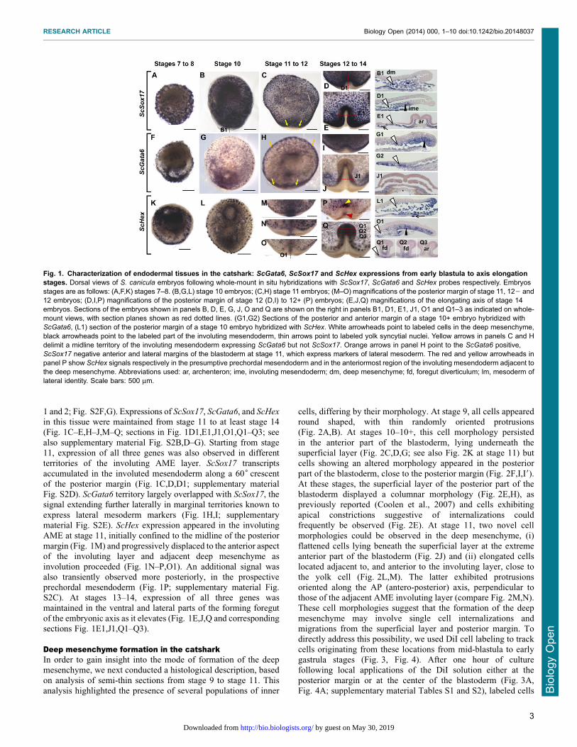
cells, differing by their morphology. At stage 9, all cells appearedround shaped, with thin randomly oriented protrusions(Fig. 2A,B). At stages 10–10+, this cell morphology persisted
in the anterior part of the blastoderm, lying underneath thesuperficial layer (Fig. 2C,D,G; see also Fig. 2K at stage 11) butcells showing an altered morphology appeared in the posterior
part of the blastoderm, close to the posterior margin (Fig. 2F,I,I9).At these stages, the superficial layer of the posterior part of theblastoderm displayed a columnar morphology (Fig. 2E,H), aspreviously reported (Coolen et al., 2007) and cells exhibiting
apical constrictions suggestive of internalizations couldfrequently be observed (Fig. 2E). At stage 11, two novel cellmorphologies could be observed in the deep mesenchyme, (i)
flattened cells lying beneath the superficial layer at the extremeanterior part of the blastoderm (Fig. 2J) and (ii) elongated cellslocated adjacent to, and anterior to the involuting layer, close to
the yolk cell (Fig. 2L,M). The latter exhibited protrusionsoriented along the AP (antero-posterior) axis, perpendicular tothose of the adjacent AME involuting layer (compare Fig. 2M,N).These cell morphologies suggest that the formation of the deep
mesenchyme may involve single cell internalizations andmigrations from the superficial layer and posterior margin. Todirectly address this possibility, we used DiI cell labeling to track
cells originating from these locations from mid-blastula to earlygastrula stages (Fig. 3, Fig. 4). After one hour of culturefollowing local applications of the DiI solution either at the
posterior margin or at the center of the blastoderm (Fig. 3A,Fig. 4A; supplementary material Tables S1 and S2), labeled cells
Fig. 1. Characterization of endodermal tissues in the catshark: ScGata6, ScSox17 and ScHex expressions from early blastula to axis elongationstages. Dorsal views of S. canicula embryos following whole-mount in situ hybridizations with ScSox17, ScGata6 and ScHex probes respectively. Embryosstages are as follows: (A,F,K) stages 7–8. (B,G,L) stage 10 embryos; (C,H) stage 11 embryos; (M–O) magnifications of the posterior margin of stage 11, 122 and12 embryos; (D,I,P) magnifications of the posterior margin of stage 12 (D,I) to 12+ (P) embryos; (E,J,Q) magnifications of the elongating axis of stage 14embryos. Sections of the embryos shown in panels B, D, E, G, J, O and Q are shown on the right in panels B1, D1, E1, J1, O1 and Q1–3 as indicated on whole-mount views, with section planes shown as red dotted lines. (G1,G2) Sections of the posterior and anterior margin of a stage 10+ embryo hybridized withScGata6, (L1) section of the posterior margin of a stage 10 embryo hybridized with ScHex. White arrowheads point to labeled cells in the deep mesenchyme,black arrowheads point to the labeled part of the involuting mesendoderm, thin arrows point to labeled yolk syncytial nuclei. Yellow arrows in panels C and Hdelimit a midline territory of the involuting mesendoderm expressing ScGata6 but not ScSox17. Orange arrows in panel H point to the ScGata6 positive,ScSox17 negative anterior and lateral margins of the blastoderm at stage 11, which express markers of lateral mesoderm. The red and yellow arrowheads inpanel P show ScHex signals respectively in the presumptive prechordal mesendoderm and in the anteriormost region of the involuting mesendoderm adjacent tothe deep mesenchyme. Abbreviations used: ar, archenteron; ime, involuting mesendoderm; dm, deep mesenchyme; fd, foregut diverticulum; lm, mesoderm oflateral identity. Scale bars: 500 mm.
RESEARCH ARTICLE Biology Open (2014) 000, 1–10 doi:10.1242/bio.20148037
3
BiologyOpen
by guest on May 30, 2019http://bio.biologists.org/Downloaded from
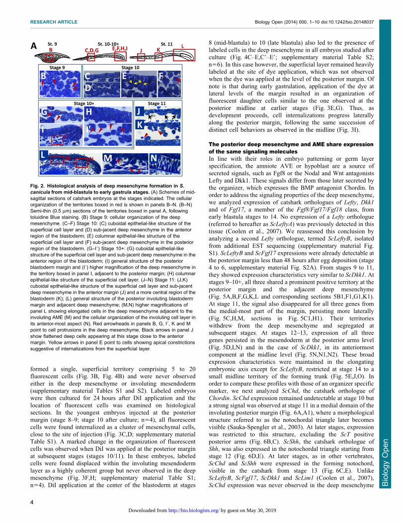
formed a single, superficial territory comprising 5 to 20fluorescent cells (Fig. 3B, Fig. 4B) and were never observed
either in the deep mesenchyme or involuting mesendoderm(supplementary material Tables S1 and S2). Labeled embryoswere then cultured for 24 hours after DiI application and the
location of fluorescent cells was examined on histologicalsections. In the youngest embryos injected at the posteriormargin (stage 8–9; stage 10 after culture; n54), all fluorescent
cells were found internalized as a cluster of mesenchymal cells,close to the site of injection (Fig. 3C,D; supplementary materialTable S1). A marked change in the organization of fluorescentcells was observed when DiI was applied at the posterior margin
at subsequent stages (stages 10/11). In these embryos, labeledcells were found displaced within the involuting mesendodermlayer as a highly coherent group but never observed in the deep
mesenchyme (Fig. 3F,H; supplementary material Table S1;n54). DiI application at the center of the blastoderm at stages
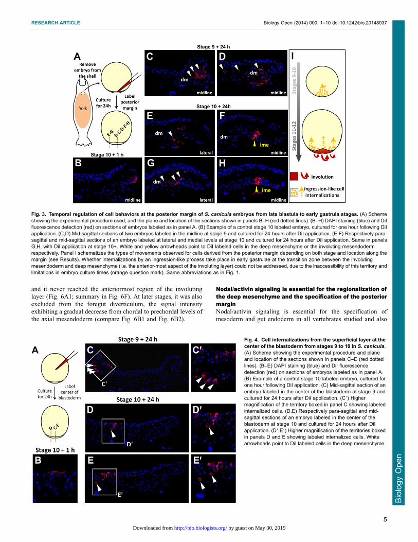
8 (mid-blastula) to 10 (late blastula) also led to the presence oflabeled cells in the deep mesenchyme in all embryos studied after
culture (Fig. 4C–E,C9–E9; supplementary material Table S2;n56). In this case however, the superficial layer remained heavilylabeled at the site of dye application, which was not observedwhen the dye was applied at the level of the posterior margin. Of
note is that during early gastrulation, application of the dye atlateral levels of the margin resulted in an organization offluorescent daughter cells similar to the one observed at the
posterior midline at earlier stages (Fig. 3E,G). Thus, asdevelopment proceeds, cell internalizations progress laterallyalong the posterior margin, following the same succession of
distinct cell behaviors as observed in the midline (Fig. 3I).
The posterior deep mesenchyme and AME share expressionof the same signaling moleculesIn line with their roles in embryo patterning or germ layerspecification, the amniote AVE or hypoblast are a source ofsecreted signals, such as Fgf8 or the Nodal and Wnt antagonists
Lefty and Dkk1. These signals differ from those later secreted bythe organizer, which expresses the BMP antagonist Chordin. Inorder to address the signaling properties of the deep mesenchyme,
we analyzed expression of catshark orthologues of Lefty, Dkk1
and of Fgf17, a member of the Fgf8/Fgf17/Fgf18 class, fromearly blastula stages to 14. No expression of a Lefty orthologue
(referred to hereafter as ScLeftyA) was previously detected in thistissue (Coolen et al., 2007). We reassessed this conclusion byanalyzing a second Lefty orthologue, termed ScLeftyB, isolated
from additional EST sequencing (supplementary material Fig.S1). ScLeftyB and ScFgf17 expressions were already detectable atthe posterior margin less than 48 hours after egg deposition (stage4 to 6, supplementary material Fig. S2A). From stages 9 to 11,
they showed expression characteristics very similar to ScDkk1. Atstages 9–10+, all three shared a prominent positive territory at theposterior margin and the adjacent deep mesenchyme
(Fig. 5A,B,F,G,K,L and corresponding sections 5B1,F1,G1,K1).At stage 11, the signal also disappeared for all three genes fromthe medial-most part of the margin, persisting more laterally
(Fig. 5C,H,M, sections in Fig. 5C1,H1). Their territorieswithdrew from the deep mesenchyme and segregated atsubsequent stages. At stages 12–13, expression of all threegenes persisted in the mesendoderm at the posterior arms level
(Fig. 5D,I,N) and in the case of ScDkk1, in its anteriormostcomponent at the midline level (Fig. 5N,N1,N2). These broadexpression characteristics were maintained in the elongating
embryonic axis except for ScLeftyB, restricted at stage 14 to asmall midline territory of the forming trunk (Fig. 5E,J,O). Inorder to compare these profiles with those of an organizer specific
marker, we next analyzed ScChd, the catshark orthologue ofChordin. ScChd expression remained undetectable at stage 10 buta strong signal was observed at stage 11 in a medial domain of the
involuting posterior margin (Fig. 6A,A1), where a morphologicalstructure referred to as the notochordal triangle later becomesvisible (Sauka-Spengler et al., 2003). At later stages, expressionwas restricted to this structure, excluding the ScT positive
posterior arms (Fig. 6B,C). ScShh, the catshark orthologue ofShh, was also expressed in the notochordal triangle starting fromstage 12 (Fig. 6D,E). At later stages, as in other vertebrates,
ScChd and ScShh were expressed in the forming notochord,visible in the catshark from stage 13 (Fig. 6C,E). UnlikeScLeftyB, ScFgf17, ScDkk1 and ScLim1 (Coolen et al., 2007),
ScChd expression was never observed in the deep mesenchyme
Fig. 2. Histological analysis of deep mesenchyme formation in S.
canicula from mid-blastula to early gastrula stages. (A) Schemes of mid-sagittal sections of catshark embryos at the stages indicated. The cellularorganization of the territories boxed in red is shown in panels B–N. (B–N)Semi-thin (0.5 mm) sections of the territories boxed in panel A, followingtoluidine Blue staining. (B) Stage 9: cellular organization of the deepmesenchyme. (C–F) Stage 10: (C) cuboidal epithelial-like structure of thesuperficial cell layer and (D) sub-jacent deep mesenchyme in the anteriorregion of the blastoderm; (E) columnar epithelial-like structure of thesuperficial cell layer and (F) sub-jacent deep mesenchyme in the posteriorregion of the blastoderm. (G–I9) Stage 10+: (G) cuboidal epithelial-likestructure of the superficial cell layer and sub-jacent deep mesenchyme in theanterior region of the blastoderm; (I) general structure of the posteriorblastoderm margin and (I9) higher magnification of the deep mesenchyme inthe territory boxed in panel I, adjacent to the posterior margin; (H) columnarepithelial-like structure of the superficial cell layer. (J–N) Stage 11: (J,K)cuboidal epithelial-like structure of the superficial cell layer and sub-jacentdeep mesenchyme in the anterior margin (J) and a more central region of theblastoderm (K); (L) general structure of the posterior involuting blastodermmargin and adjacent deep mesenchyme; (M,N) higher magnifications ofpanel L showing elongated cells in the deep mesenchyme adjacent to theinvoluting AME (M) and the cellular organization of the involuting cell layer inits anterior-most aspect (N). Red arrowheads in panels B, G, I9, K and Mpoint to cell protrusions in the deep mesenchyme. Black arrows in panel Jshow flattened deep cells appearing at this stage close to the anteriormargin. Yellow arrows in panel E point to cells showing apical constrictionssuggestive of internalizations from the superficial layer.
RESEARCH ARTICLE Biology Open (2014) 000, 1–10 doi:10.1242/bio.20148037
4
BiologyOpen
by guest on May 30, 2019http://bio.biologists.org/Downloaded from
and it never reached the anteriormost region of the involuting
layer (Fig. 6A1; summary in Fig. 6F). At later stages, it was alsoexcluded from the foregut diverticulum, the signal intensityexhibiting a gradual decrease from chordal to prechordal levels ofthe axial mesendoderm (compare Fig. 6B1 and Fig. 6B2).
Nodal/activin signaling is essential for the regionalization ofthe deep mesenchyme and the specification of the posteriormarginNodal/activin signaling is essential for the specification ofmesoderm and gut endoderm in all vertebrates studied and also
Fig. 3. Temporal regulation of cell behaviors at the posterior margin of S. canicula embryos from late blastula to early gastrula stages. (A) Schemeshowing the experimental procedure used, and the plane and location of the sections shown in panels B–H (red dotted lines). (B–H) DAPI staining (blue) and DiIfluorescence detection (red) on sections of embryos labeled as in panel A. (B) Example of a control stage 10 labeled embryo, cultured for one hour following DiIapplication. (C,D) Mid-sagittal sections of two embryos labeled in the midline at stage 9 and cultured for 24 hours after DiI application. (E,F) Respectively para-sagittal and mid-sagittal sections of an embryo labeled at lateral and medial levels at stage 10 and cultured for 24 hours after DiI application. Same in panelsG,H, with DiI application at stage 10+. White and yellow arrowheads point to DiI labeled cells in the deep mesenchyme or the involuting mesendodermrespectively. Panel I schematizes the types of movements observed for cells derived from the posterior margin depending on both stage and location along themargin (see Results). Whether internalizations by an ingression-like process take place in early gastrulae at the transition zone between the involutingmesendoderm and deep mesenchyme (i.e. the anterior-most aspect of the involuting layer) could not be addressed, due to the inaccessibility of this territory andlimitations in embryo culture times (orange question mark). Same abbreviations as in Fig. 1.
Fig. 4. Cell internalizations from the superficial layer at thecenter of the blastoderm from stages 9 to 10 in S. canicula.(A) Scheme showing the experimental procedure and planeand location of the sections shown in panels C–E (red dottedlines). (B–E) DAPI staining (blue) and DiI fluorescencedetection (red) on sections of embryos labeled as in panel A.(B) Example of a control stage 10 labeled embryo, cultured forone hour following DiI application. (C) Mid-sagittal section of anembryo labeled in the center of the blastoderm at stage 9 andcultured for 24 hours after DiI application. (C9) Highermagnification of the territory boxed in panel C showing labeledinternalized cells. (D,E) Respectively para-sagittal and mid-sagittal sections of an embryo labeled in the center of theblastoderm at stage 10 and cultured for 24 hours after DiIapplication. (D9,E9) Higher magnification of the territories boxedin panels D and E showing labeled internalized cells. Whitearrowheads point to DiI labeled cells in the deep mesenchyme.
RESEARCH ARTICLE Biology Open (2014) 000, 1–10 doi:10.1242/bio.20148037
5
BiologyOpen
by guest on May 30, 2019http://bio.biologists.org/Downloaded from
required for AVE specification in the mouse (Mesnard et al.,2006). In order to analyze the role of Nodal/activin signaling inendoderm development in the catshark, we conducted an in ovo
pharmacological approach, using SB-505124, a selective inhibitorof activin Alk4/5/7 receptors. The drug or control DMSO wasinjected inside the eggshell at stages 8 to 9, reached about 5 days
following egg deposition. Eggs were then maintained for threedays in 17 C oxygenated sea water prior to embryo fixation anddissection. DMSO injected control embryos appeared normal andtheir stages ranged between 11 (n521) and 12 (n57), as expected
for uninjected embryos at this temperature. SB-505124 treatedembryos could be classified into two classes based on theirgeneral morphology (supplementary material Table S3). The
majority, referred to as class 1 embryos, appeared as flattenedblastodiscs, without evidence of posterior fold formation(Fig. 7B,D,F,H,J,P). A minority of treated embryos, referred to
as class 2 embryos, showed similarities to stage 12 embryos, inthat they exhibited distinct posterior arms on each side of theforming embryonic axis (Fig. 7L,N). In order to assess the loss of
Nodal/activin signaling in the experimental conditions tested, wefirst focused on expression of the feedback antagonist ScLeftyB inSB-505124-treated and control embryos (supplementary materialTable S3; Fig. 7A,B). While present at the posterior margin in all
control embryos tested (n53, Fig. 7A), ScLeftyB expressionremained undetectable in all treated embryos (class 1, n53,Fig. 7B), in line with a loss of Nodal/activin signaling. We next
analyzed expression of the general mesoderm marker ScT
(Fig. 7C,D) and of the notochordal triangle marker ScChd
(Fig. 7E,F). In both cases, control embryos exhibited the
expected signals (n55 for ScT, n52 for ScChd) around thewhole margin (ScT, Fig. 7C) or in the involuting axialmesendoderm (ScChd, Fig. 7E). In contrast, ScT and ScChd
expressions were abolished in all treated embryos analyzed (n54
and n53 respectively, Fig. 7D,F). Similarly, ScOtx5 and ScLim1
signals were observed, in the involuting mesendoderm andadjacent deep mesenchyme of control embryos (n54 for ScOtx5,same for ScLim; Fig. 7G,I), but lost in all treated embryos (n52
and n54 for ScOtx5 and ScLim1 respectively; Fig. 7H,J). Finally,we analyzed the effect of the drug on three genes characterized byan early expression in the deep mesenchyme and syncytial nuclei,
and a later expression phase at different levels of the involutingmargin as shown above, ScSox17, ScGata6 and ScHex (Fig. 7K–P). In all cases (total of 11 embryos tested), the signal in the
deep mesenchyme and yolk syncytial nuclei was maintained incontrol and SB-505124 treated embryos (compare Fig. 7K andFig. 7L, Fig. 7M and Fig. 7N, Fig. 7O and Fig. 7P; see alsosupplementary material Fig. S3A,B). In contrast, no marginal
expression was observed in any of the treated embryos (n513,Fig. 7L,N,P), while it was present in control embryos(Fig. 7K,M,O; compare Fig. 7K1 and Fig. 7L1, Fig. 7M1 and
Fig. 7N1, Fig. 7M19 and Fig. 7N19; see also supplementarymaterial Fig. S3C1,C2,D1,D2). Histological sections showed thatthe deep mesenchyme was maintained in class 1 and class 2
embryos (supplementary material Fig. S3). An inner layer waspresent at the posterior margin of class 2 embryos but it appearedless expanded and thinner than in control embryos (compareFig. 7M19 and Fig. 7N19).
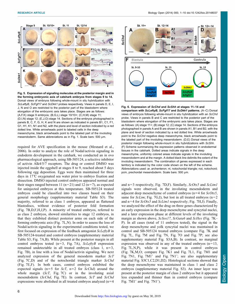
Fig. 5. Expression of signaling molecules at the posterior margin and inthe forming embryonic axis of catshark embryos from stages 9 to 14.Dorsal views of embryos following whole-mount in situ hybridization withScLeftyB, ScFgf17 and ScDkk1 probes respectively. Views in panels D, E, I,J, N and O are restricted to the posterior part of the blastoderm whereelongation of the embryonic axis takes place. Stages are as follows:(A,F,K) stage 9 embryos; (B,G,L) stage 10/10+; (C,H,M) stage 11;(D,I,N) stage 12; (E,J,O) stage 14. Sections of the embryos photographed inpanels B, C, F, G, H, K and N are shown as indicated in panels B1, C1, F1,G1, H1, K1, N1 and N2, with the plane and level of section indicated by a reddotted line. White arrowheads point to labeled cells in the deepmesenchyme, black arrowheads point to the labeled part of the involutingmesendoderm. Same abbreviations as in Fig. 1. Scale bars: 500 mm.
Fig. 6. Expression of ScChd and ScShh at stages 11–14 andcomparison with ScLeftyB, ScFgf17 and ScDkk1 patterns. (A–C) Dorsalviews of embryos following whole-mount in situ hybridization with an ScChd
probe. Views in panels B and C are restricted to the posterior part of theblastoderm where elongation of the embryonic axis takes place. Stages areas follows: (A) stage 11+; (B) stage 12; (C) stage 14. Sections of the embryosphotographed in panels A and B are shown in panels A1, B1 and B2, with theplane and level of section indicated by a red dotted line. White arrowheadspoint to the ScChd negative deep mesenchyme, black arrowheads point tothe labeled part of the involuting mesendoderm. (D,E) Dorsal views of theposterior margin following whole-mount in situ hybridizations with ScShh.(F) Scheme summarizing the expression patterns observed in endodermaltissues in the catshark. Dotted areas indicate signals in the deepmesenchyme, uniformly colored areas indicate signals in the involutingmesendoderm and at the margin. A dotted black line delimits the extent of theinvoluting mesendoderm. The combination of genes expressed in eachterritory is indicated by the color code shown on the left of the scheme.Abbreviations used: ar, archenteron; nt, notochordal triangle; not, notochord;pcm, prechordal mesendoderm. Scale bars: 500 mm.
RESEARCH ARTICLE Biology Open (2014) 000, 1–10 doi:10.1242/bio.20148037
6
BiologyOpen
by guest on May 30, 2019http://bio.biologists.org/Downloaded from
DISCUSSIONEndoderm is specified in two phases in the catshark as inosteichthyansAnalysis of Hex, Gata6 and Sox17 orthologues in the catsharkshow that not only the anteriormost part of the involuting layer
but also the deep mesenchyme is endowed with an endodermalidentity. The timing of their specification appears as a majordifference between these two tissues, since the latter already
expresses Sox17, Hex and Gata6 at the earliest stages accessible,which shortly follow blastocoel formation and precedegastrulation by more than seven days (Ballard et al., 1993;Coolen et al., 2007; Sauka-Spengler et al., 2003). As previously
noted (Godard and Mazan, 2013), two phases of endodermspecification are most obvious in mammals, birds and teleosts, allendowed with distinct extraembryonic tissues, but have also been
reported in some amphibians exhibiting no evidence for twomorphologically distinct endoderm components, includingxenopus. While a key role of Nodal in mesendoderm formation
has been demonstrated in amniotes, amphibians and teleosts(Conlon et al., 1994; Erter et al., 1998; Feldman et al., 1998;Schier, 2009; Schier and Shen, 2000; Steiner et al., 2006), themechanisms controlling the earliest phase of endoderm
specification appear to vary extensively across vertebrates. Inthe catshark, formation of the involuting layer and expression of
all mesoderm and mesendoderm markers were abolishedfollowing SB-505124 treatments, in line with a conservation ofthe role of Nodal/activin signaling in mesendoderm specification.In contrast, no evidence for a loss of the endodermal identity was
observed in the deep mesenchyme following abrogation of Nodalsignaling activity. This observation cannot rule out a role ofNodal signaling in the initial steps of deep mesenchyme
specification, as ScSox17, ScHex and ScGata6 expressions werealready established at the time of egg laying, making it difficult toassess the effect of the drug prior to the onset of their expression.
However, it argues against a major role of the pathway in themaintenance of the deep mesenchyme endodermal identity. Thisconclusion is also supported by the localized deep mesenchyme
expression of the catshark orthologue of Lefty, a feedbackantagonist of Nodal signaling (Chen and Schier, 2002; Juan andHamada, 2001; Meno et al., 1999), which suggests a posteriorrestriction of Nodal signaling activity at all stages studied. From
an evolutionary standpoint, the reiteration of a biphasic mode ofendoderm specification now found in chondrichthyans as in allmajor osteichthyan lineages supports the hypothesis that it may
be an ancestral characteristic of jawed vertebrates (Godardand Mazan, 2013). However, in the absence of mechanisticarguments, it remains difficult to formally exclude independent
rises of the earliest specification event in the different vertebratephyla. Finally it should be noted that a partitioning of nutritivetissues into a vegetal mass without contribution to the gut and an
embryonic component derived from the blastopore margin hasalso been proposed in the lamprey (Takeuchi et al., 2009).However, in this species, this distinction primarily relied on theabsence of endoderm marker expression in the vegetal mass, a
criterion, which argues against homology with the early specifiedtissue identified in the catshark.
Cell internalizations suggestive of ingression movementsprecede gastrulation in the catsharkWe had previously observed that the physical continuity between
the posterior deep mesenchyme and the adjacent AME goestogether with a molecular continuity, both expressing not onlyendoderm but also AME regional markers, such as Lim1 or Gsc
(Coolen et al., 2007). This study extends this conclusion to
signaling molecules such as the catshark orthologues of Lefty,Fgf17, a member of the Fgf8/17/18 family, or Dkk1, known to bespecifically expressed in AME as well as in endodermal
extraembryonic components of amniotes and teleosts (Stern andDowns, 2012). We further show that this regional identity is lostfollowing abrogation of Nodal/activin signaling, which as
reviewed previously (Godard and Mazan, 2013) is reminiscentof the molecular phenotype observed in the mouse embryonicvisceral endoderm (Mesnard et al., 2006). Finally, we find that
massive cell internalizations from the posterior margin take placeprior to gastrulation and contribute to posterior deep mesenchymeformation. The posterior deep mesenchyme and involutingmesendoderm thus lie adjacent to each other, exhibit the same,
Nodal-dependent, AME regional identity (Fig. 8A,B), and arealso related by their embryonic origin. However, the cellinternalizations from the posterior margin, which contribute to
their formation, differ by their timing and the cell movementsinvolved. The low cohesion of cells internalized at the posteriormargin from stages 8 to 10, prior to the appearance of a bilayered
overhang, contrasts with the tightly clustered cell organization
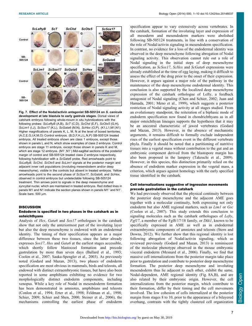
Fig. 7. Effect of the Nodal/activin antagonist SB-505124 on S. canicula
development at late blastula to early gastrula stages. Dorsal views ofcatshark embryos following whole-mount in situ hybridizations with thefollowing probes: ScLeftyB (A,B), ScT (C,D), ScChd (E,F), ScOtx5 (G,H),ScLim1 (I,J), ScSox17 (K,L), ScGata6 (M,N), ScHex (O,P). (K1,L1,M1,N1)Higher magnifications of panels K, L, M, N at the level of boxed territories.(A,C,E,G,I,K,M,O) Control embryos. (B,D,F,H,J,L,N,P) SB-505124 treatedembryos. All treated embryos shown are class 1 embryos, except thoseshown in panels L and N, which show examples of class 2 embryos. Controlembryos are stage 11 embryos, except those shown in panels K and M,which are stage 12 embryos. (M19,N19) Mid-sagittal sections of the posteriormargin of control and SB-505124 treated class 2 embryos respectively,following hybridization with a ScGata6 probe. Red arrowheads point toScLeftyB, ScChd, ScOtx5 and ScLim1 signals at the posterior margin andadjacent inner cell populations (involuting mesendoderm and/or deepmesenchyme), visible in the controls but absent in treated embryos. Yellowarrowheads point to the second phase of ScSox17, ScGata6, and ScHex,observed in control embryos but undetectable following SB-505124treatment. Thin arrows point to signals in the deep mesenchyme andsyncytial nuclei, which are maintained in treated embryos. Red dotted lines inpanels M1 and N1 indicate the section planes shown in panels M19 and N19.Scale bars: 500 mm.
RESEARCH ARTICLE Biology Open (2014) 000, 1–10 doi:10.1242/bio.20148037
7
BiologyOpen
by guest on May 30, 2019http://bio.biologists.org/Downloaded from
observed at later stages, and suggests that massive ingression-likemovements precede involution at the posterior margin (Fig. 8C).
In addition to these cell movements taking place at the posteriormargin prior to gastrulation, we also obtained evidence for cellinternalizations in the center of the blastoderm. Theseinternalizations differed from the former in that a prominent
labeling persisted in the superficial cell layer following DiIapplication and embryo culture, suggesting that they may onlyconcern small clusters of cells or individuals cells, as suggested
by histological analyses. Taken together, these data provideevidence that early development in the catshark involves cellinternalizations from the superficial layer, taking place prior to
gastrulation. These movements and the shift in cell cohesionobserved at the posterior margin at gastrulation are likely toinvolve a highly dynamic regulation of cell properties such as
adhesiveness, shape, polarity and motility but the underlyingmechanisms remain completely unknown. In line with theconservation of its role in mesendoderm formation across jawedvertebrates, Nodal signaling appeared essential for the formation
of the involuting layer. Together with the major Lefty expressionobserved at the posterior margin since early blastula stages, theabsence of a posterior thickening in class 1 SB-505124 treated
embryos suggests that it may also control the earlier ingression-like cell movements taking place at the posterior margin.However, we could not directly assess this possibility due to
the impossibility to conduct in ovo injections and DiI labelingconcomitantly. Other candidate mechanisms include FGF orBMP, respectively known to regulate epithelium–mesenchyme
transitions in the context of the primitive streak and the mode of
migration of lateral mesoderm in the zebrafish, or Wnt-PCP andSphingosine-1-phosphate signaling, which control the choice
between individual and collective cell migrations in the zebrafishprechordal mesendoderm (Ciruna et al., 1997; Hardy et al., 2011;Kai et al., 2008; Luxardi et al., 2010; von der Hardt et al., 2007).More detailed analyses of cellular phenotypes coupled with
pharmacological treatments directed against these pathways willbe crucial to address this point.
Evolutionary implications: homoplastic traits betweenchondrichthyans and amniotesFrom an evolutionary standpoint, the cell movements, shown here
to occur during catshark early development, strikingly recallsome aspects of amniote development. Firstly, as already noted(Godard and Mazan, 2013) and confirmed in this study, the
catshark posterior deep mesenchyme and involuting AME appearto differ by their cell organization rather than their regionalidentity. The establishment of fate maps in the catshark iscurrently hampered by difficulties to maintain embryo viability
during extended durations following cell marking procedures butbased on its cell organization and location relative to thedeveloping embryonic axis, the deep mesenchyme is likely to
have a major contribution to extraembryonic structures, such asthe yolk sac, syncytial nuclei (Lechenault and Mellinger, 1993) orstalk connecting the embryo to the yolk sac. Deep mesenchymal
cells thus persist until at least somite stages in blastodermterritories spreading over the yolk, at increasing distances fromthe site where involution and embryo formation take place
(Ballard et al., 1993; Coolen et al., 2007; this study)
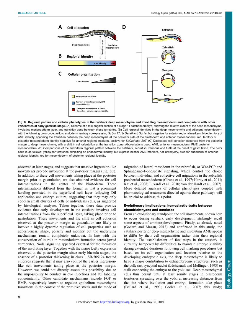
Fig. 8. Regional pattern and cellular phenotypes in the catshark deep mesenchyme and involuting mesendoderm and comparison with othervertebrates at early gastrula stage. (A) Scheme of a mid-sagittal section of a stage 11 catshark embryo, showing the relative extent of the deep mesenchyme,involuting mesendoderm layer, and transition zone between these territories. (B) Cell regional identities in the deep mesenchyme and adjacent mesendodermwith the following color code: yellow, endoderm territory co-expressing ScSox17, ScGata6 and ScHex but negative for anterior regional markers; blue, territory ofAME identity, spanning the transition between the deep mesenchyme at the posterior side of the blastoderm and anterior mesendoderm; red, territory ofposterior mesendoderm identity negative for anterior regional markers, positive for ScChd and ScT. (C) Decreased cell cohesion observed from the posteriormargin to deep mesenchyme, with a shift in cell orientation at the transition zone. Abbreviations used: AME, anterior mesendoderm; PME posteriormesendoderm. (D) Comparisons of the endoderm regional pattern between the catshark, zebrafish, xenopus and turtle at the onset of gastrulation. The colorcode is as follows: yellow for territories exhibiting an endodermal identity, but express neither AME markers, nor Brachyury, blue for endoderm of anteriorregional identity, red for mesendoderm of posterior regional identity.
RESEARCH ARTICLE Biology Open (2014) 000, 1–10 doi:10.1242/bio.20148037
8
BiologyOpen
by guest on May 30, 2019http://bio.biologists.org/Downloaded from
(supplementary material Movie 2; Fig. S2B–G). The molecularsimilarity between endoderm components exhibiting very
different cell organizations recalls that observed in mammalsbetween tissues traditionally considered as extraembryonic, suchas the AVE, and the adjacent AME (Fig. 8D) (Stern and Downs,2012). The parallel is most obvious with turtles, which have
retained involution as the primary mode of mesendoderminternalization a feature likely to be ancestral in amniotes(Fig. 8D) (Bertocchini et al., 2013; Coolen et al., 2008b).
Secondly, the catshark also exhibits unexpected similaritieswith amniotes in the cell movements, which take place prior togastrulation. In the chick, a recent study has thus demonstrated
that ingressions of individual cells from the epiblast layer takeplace prior to the onset of gastrulation and that suchmovements are able to promote massive cell ingressions and
induction of mesoderm markers, mimicking the formation ofthe primitive streak in a Nodal-dependent manner (Voiculescuet al., 2014). The early internalizations observed in the centerof the blastoderm in the catshark are reminiscent of these cell
movements, even though it remains unclear where they couldsimilarly promote more massive internalizations at the posteriormargin, a territory where Nodal signaling is likely to be active,
based on Lefty expression. Such early cell movements, nowfound in the catshark as in the chick, are unlikely to represent agnathostome ancestral characteristic since they have no
equivalent either in amphibians or in teleosts. They are thuslikely to correspond to a homoplastic feature (Wake et al.,2011), separately fixed in amniotes and chondrichthyans and
possibly associated to the yolk increase, which took place inthese lineages. Whether their rise may result from convergentevolution, involving different mechanisms, or parallelevolution, relying on the deployment of the same mechanisms
and possibly indicative of developmental constraints, remainsan open question. Deciphering the underlying mechanism in thechick and in the catshark will be crucial to address this issue.
ConclusionIn conclusion, our data support the view that the blueprint ofendoderm formation in jawed vertebrates involves a conserved
biphasic mechanism, which tolerates an extensive variability incell movements and individual behaviors. Such an uncouplingbetween highly conserved patterning mechanisms and morerapidly diverging morphogenetic processes also applies to
mesoderm formation and may reflect a general evolutionarytrend (Alev et al., 2013; Shook and Keller, 2008). They also pointto unexpected similarities in cell movements preceding
gastrulation with amniotes, likely to correspond to homoplasticfeatures. Deciphering the mechanisms controlling thecoordination between early patterning mechanisms, cell fate
determination and cell behaviors in different vertebrate lineageswill be essential to understand the molecular basis for theevolvability of endoderm morphogenesis across vertebrates. The
catshark should be a valuable model to address these aspects, notonly by its phylogenetic position, but also by the characteristicsof its early development, which involves complex cellmovements, remarkable temporal and spatial resolutions of the
processes involved and allows straightforward comparisons withall major vertebrate model organisms.
AcknowledgementsEST sequences were obtained with the support of Genoscope, Evry, France. Weare grateful to Sebastien Henry, Ronan Garnier, Regis Lasbleiz and Sebastien
Paturance for their help in embryo maintenance, Diane Schausi for excellenttechnical help, J. Andrew Gillis for sharing his expertise in DiI labeling, AitanaPerea-Gomez, Anne Camus, Jerome Collignon, De-Li Shi, Helene Mayeur andWilliam Norton for helpful comments on the manuscript.
Competing interestsAll authors declare that they have no financial and competing interests.
Author contributionsSM designed the study; BGG, MC, SLP, AG, SF-G, LL, RL performedexperiments; PW, JP, CDS; SK and WC contributed to sequences and analyses;BGG, AB and SM analyzed the results and wrote the manuscript.
FundingThis work was funded by Region Centre, Region Bretagne (EVOVERT grantnumber 049755; PEPTISAN project), National Research Agency (grant ANR-09-BLAN-026201), CNRS, Universite d’Orleans and Universite Pierre et Marie Curie.BGG benefited from a Region Bretagne fellowship.
ReferencesAlev, C., Wu, Y., Nakaya, Y. and Sheng, G. (2013). Decoupling of amniotegastrulation and streak formation reveals a morphogenetic unity in vertebratemesoderm induction. Development 140, 2691-2696.
Arendt, D. and Nubler-Jung, K. (1999). Rearranging gastrulation in the nameof yolk: evolution of gastrulation in yolk-rich amniote eggs. Mech. Dev. 81, 3-22.
Ballard, W. W., Mellinger, J. and Lechenault, H. (1993). A series of normalstages for development of Scyliorhinus canicula, the lesser spotted dogfish(Chondrichthyes: Scyliorhinidae). J. Exp. Zool. 267, 318-336.
Bertocchini, F., Alev, C., Nakaya, Y. and Sheng, G. (2013). A little winningstreak: the reptilian-eye view of gastrulation in birds. Dev. Growth Differ. 55, 52-59.
Blackburn, D. G. (2014). Evolution of vertebrate viviparity and specializations forfetal nutrition: A quantitative and qualitative analysis. J. Morphol. (Epub ahead ofPrint) doi: 10.1002/jmor.20272.
Carvalho, L. and Heisenberg, C. P. (2010). The yolk syncytial layer in earlyzebrafish development. Trends Cell Biol. 20, 586-592.
Chen, Y. and Schier, A. F. (2002). Lefty proteins are long-range inhibitors ofsquint-mediated nodal signaling. Curr. Biol. 12, 2124-2128.
Ciruna, B. G., Schwartz, L., Harpal, K., Yamaguchi, T. P. and Rossant, J.(1997). Chimeric analysis of fibroblast growth factor receptor-1 (Fgfr1) function:a role for FGFR1 in morphogenetic movement through the primitive streak.Development 124, 2829-2841.
Conlon, F. L., Lyons, K. M., Takaesu, N., Barth, K. S., Kispert, A., Herrmann, B.and Robertson, E. J. (1994). A primary requirement for nodal in the formationand maintenance of the primitive streak in the mouse. Development 120, 1919-1928.
Coolen, M., Sauka-Spengler, T., Nicolle, D., Le-Mentec, C., Lallemand, Y., DaSilva, C., Plouhinec, J.-L., Robert, B., Wincker, P., Shi, D.-L. et al. (2007).Evolution of axis specification mechanisms in jawed vertebrates: insights from achondrichthyan. PLoS ONE 2, e374.
Coolen, M., Menuet, A., Chassoux, D., Compagnucci, C., Henry, S., Leveque,L., Da Silva, C., Gavory, F., Samain, S., Wincker, P. et al. (2008a). The dogfishScyliorhinus canicula: a reference in jawed vertebrates. CSH Protoc. 2008,pdb.emo111.
Coolen, M., Nicolle, D., Plouhinec, J.-L., Gombault, A., Sauka-Spengler, T.,Menuet, A., Pieau, C. and Mazan, S. (2008b). Molecular characterization of thegastrula in the turtle Emys orbicularis: an evolutionary perspective ongastrulation. PLoS ONE 3, e2676.
Derobert, Y., Plouhinec, J. L., Sauka-Spengler, T., Le Mentec, C., Baratte, B.,Jaillard, D. and Mazan, S. (2002). Structure and expression of three Emxgenes in the dogfish Scyliorhinus canicula: functional and evolutionaryimplications. Dev. Biol. 247, 390-404.
Erter, C. E., Solnica-Krezel, L. and Wright, C. V. (1998). Zebrafish nodal-related2 encodes an early mesendodermal inducer signaling from the extraembryonicyolk syncytial layer. Dev. Biol. 204, 361-372.
Feldman, B., Gates, M. A., Egan, E. S., Dougan, S. T., Rennebeck, G., Sirotkin,H. I., Schier, A. F. and Talbot, W. S. (1998). Zebrafish organizer developmentand germ-layer formation require nodal-related signals. Nature 395, 181-185.
Godard, B. G. and Mazan, S. (2013). Early patterning in a chondrichthyan model,the small spotted dogfish: towards the gnathostome ancestral state. J. Anat.222, 56-66.
Guindon, S. and Gascuel, O. (2003). A simple, fast, and accurate algorithm toestimate large phylogenies by maximum likelihood. Syst. Biol. 52, 696-704.
Hardy, K. M., Yatskievych, T. A., Konieczka, J., Bobbs, A. S. and Antin, P. B.(2011). FGF signalling through RAS/MAPK and PI3K pathways regulates cellmovement and gene expression in the chicken primitive streak without affectingE-cadherin expression. BMC Dev. Biol. 11, 20.
Juan, H. and Hamada, H. (2001). Roles of nodal-lefty regulatory loops inembryonic patterning of vertebrates. Genes Cells 6, 923-930.
RESEARCH ARTICLE Biology Open (2014) 000, 1–10 doi:10.1242/bio.20148037
9
BiologyOpen
by guest on May 30, 2019http://bio.biologists.org/Downloaded from
Kai, M., Heisenberg, C. P. and Tada, M. (2008). Sphingosine-1-phosphatereceptors regulate individual cell behaviours underlying the directed migration ofprechordal plate progenitor cells during zebrafish gastrulation. Development135, 3043-3051.
Katoh, K., Kuma, K., Toh, H. and Miyata, T. (2005). MAFFT version 5:improvement in accuracy of multiple sequence alignment. Nucleic Acids Res.33, 511-518.
Kwon, G. S., Viotti, M. and Hadjantonakis, A.-K. (2008). The endoderm of themouse embryo arises by dynamic widespread intercalation of embryonic andextraembryonic lineages. Dev. Cell 15, 509-520.
Lechenault, H. and Mellinger, J. (1993). Dual origin of yolk nuclei in the lesserspotted dogfish, Scyliorhinus canicula (Chondrichthyes). J. Exp. Zool. 265, 669-678.
Luxardi, G., Marchal, L., Thome, V. and Kodjabachian, L. (2010). DistinctXenopus Nodal ligands sequentially induce mesendoderm and controlgastrulation movements in parallel to the Wnt/PCP pathway. Development137, 417-426.
Meno, C., Gritsman, K., Ohishi, S., Ohfuji, Y., Heckscher, E., Mochida, K.,Shimono, A., Kondoh, H., Talbot, W. S., Robertson, E. J. et al. (1999). MouseLefty2 and zebrafish antivin are feedback inhibitors of nodal signaling duringvertebrate gastrulation. Mol. Cell 4, 287-298.
Mesnard, D., Guzman-Ayala, M. and Constam, D. B. (2006). Nodal specifiesembryonic visceral endoderm and sustains pluripotent cells in the epiblastbefore overt axial patterning. Development 133, 2497-2505.
Plouhinec, J. L., Leconte, L., Sauka-Spengler, T., Bovolenta, P., Mazan, S. andSaule, S. (2005). Comparative analysis of gnathostome Otx gene expressionpatterns in the developing eye: implications for the functional evolution of themultigene family. Dev. Biol. 278, 560-575.
Rossant, J. and Tam, P. P. L. (2009). Blastocyst lineage formation, early embryonicasymmetries and axis patterning in the mouse. Development 136, 701-713.
Sauka-Spengler, T., Baratte, B., Lepage, M. and Mazan, S. (2003).Characterization of Brachyury genes in the dogfish S. canicula and thelamprey L. fluviatilis. Insights into gastrulation in a chondrichthyan. Dev. Biol.263, 296-307.
Schier, A. F. (2009). Nodal morphogens. Cold Spring Harb. Perspect. Biol. 1,a003459.
Schier, A. F. and Shen, M. M. (2000). Nodal signalling in vertebrate development.Nature 403, 385-389.
Shook, D. R. and Keller, R. (2008). Epithelial type, ingression, blastoporearchitecture and the evolution of chordate mesoderm morphogenesis. J. Exp.Zool. B 310, 85-110.
Steiner, A. B., Engleka, M. J., Lu, Q., Piwarzyk, E. C., Yaklichkin, S., Lefebvre,J. L., Walters, J. W., Pineda-Salgado, L., Labosky, P. A. and Kessler, D. S.(2006). FoxD3 regulation of Nodal in the Spemann organizer is essential forXenopus dorsal mesoderm development. Development 133, 4827-4838.
Stern, C. D. and Downs, K. M. (2012). The hypoblast (visceral endoderm): anevo-devo perspective. Development 139, 1059-1069.
Takeuchi, M., Takahashi, M., Okabe, M. and Aizawa, S. (2009). Germ layerpatterning in bichir and lamprey; an insight into its evolution in vertebrates. Dev.Biol. 332, 90-102.
Voiculescu, O., Bodenstein, L., Lau, I. J. and Stern, C. D. (2014). Local cellinteractions and self-amplifying individual cell ingression drive amniotegastrulation. eLife 3, e01817.
von der Hardt, S., Bakkers, J., Inbal, A., Carvalho, L., Solnica-Krezel, L.,Heisenberg, C.-P. and Hammerschmidt, M. (2007). The Bmp gradient of thezebrafish gastrula guides migrating lateral cells by regulating cell-cell adhesion.Curr. Biol. 17, 475-487.
Wake, D. B., Wake, M. H. and Specht, C. D. (2011). Homoplasy: from detectingpattern to determining process and mechanism of evolution. Science 331, 1032-1035.
RESEARCH ARTICLE Biology Open (2014) 000, 1–10 doi:10.1242/bio.20148037
10
BiologyOpen
by guest on May 30, 2019http://bio.biologists.org/Downloaded from