Mechanisms of Disease Atopic Dermatitis -...
12
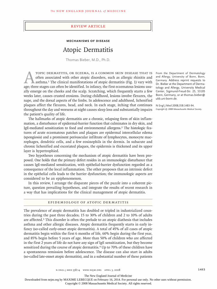
The new england journal of medicine n engl j med 358;14 www.nejm.org april 3, 2008 1483 review article Mechanisms of Disease Atopic Dermatitis Thomas Bieber, M.D., Ph.D. From the Department of Dermatology and Allergy, University of Bonn, Bonn, Germany. Address reprint requests to Dr. Bieber at the Department of Derma- tology and Allergy, University Medical Center, Sigmund-Freud-Str. 25, 53105 Bonn, Germany, or at thomas.bieber@ ukb.uni-bonn.de. N Engl J Med 2008;358:1483-94. Copyright © 2008 Massachusetts Medical Society. A topic dermatitis, or eczema, is a common skin disease that is often associated with other atopic disorders, such as allergic rhinitis and asthma. 1 The clinical manifestations of atopic dermatitis (Fig. 1) vary with age; three stages can often be identified. In infancy, the first eczematous lesions usu- ally emerge on the cheeks and the scalp. Scratching, which frequently starts a few weeks later, causes crusted erosions. During childhood, lesions involve flexures, the nape, and the dorsal aspects of the limbs. In adolescence and adulthood, lichenified plaques affect the flexures, head, and neck. In each stage, itching that continues throughout the day and worsens at night causes sleep loss and substantially impairs the patient’s quality of life. The hallmarks of atopic dermatitis are a chronic, relapsing form of skin inflam- mation, a disturbance of epidermal-barrier function that culminates in dry skin, and IgE-mediated sensitization to food and environmental allergens. 2 The histologic fea- tures of acute eczematous patches and plaques are epidermal intercellular edema (spongiosis) and a prominent perivascular infiltrate of lymphocytes, monocyte mac- rophages, dendritic cells, and a few eosinophils in the dermis. In subacute and chronic lichenified and excoriated plaques, the epidermis is thickened and its upper layer is hypertrophied. Two hypotheses concerning the mechanism of atopic dermatitis have been pro- posed. One holds that the primary defect resides in an immunologic disturbance that causes IgE-mediated sensitization, with epithelial-barrier dysfunction regarded as a consequence of the local inflammation. The other proposes that an intrinsic defect in the epithelial cells leads to the barrier dysfunction; the immunologic aspects are considered to be an epiphenomenon. In this review, I arrange the disparate pieces of the puzzle into a coherent pic- ture, question prevailing hypotheses, and integrate the results of recent research in a way that has implications for the clinical management of atopic dermatitis. Epidemiology of Atopic Dermatitis The prevalence of atopic dermatitis has doubled or tripled in industrialized coun- tries during the past three decades; 15 to 30% of children and 2 to 10% of adults are affected. 3 This disorder is often the prelude to an atopic diathesis that includes asthma and other allergic diseases. Atopic dermatitis frequently starts in early in- fancy (so-called early-onset atopic dermatitis). A total of 45% of all cases of atopic dermatitis begin within the first 6 months of life, 60% begin during the first year, and 85% begin before 5 years of age. More than 50% of children who are affected in the first 2 years of life do not have any sign of IgE sensitization, but they become sensitized during the course of atopic dermatitis. 4 Up to 70% of these children have a spontaneous remission before adolescence. The disease can also start in adults (so-called late-onset atopic dermatitis), and in a substantial number of these patients The New England Journal of Medicine Downloaded from nejm.org by MAXIME LEBECQUE on February 16, 2014. For personal use only. No other uses without permission. Copyright © 2008 Massachusetts Medical Society. All rights reserved.
Transcript of Mechanisms of Disease Atopic Dermatitis -...
T h e n e w e ng l a nd j o u r na l o f m e dic i n e
n engl j med 358;14 www.nejm.org april 3, 2008 1483
review article
Mechanisms of Disease
Atopic DermatitisThomas Bieber, M.D., Ph.D.
From the Department of Dermatology and Allergy, University of Bonn, Bonn, Germany. Address reprint requests to Dr. Bieber at the Department of Derma-tology and Allergy, University Medical Center, Sigmund-Freud-Str. 25, 53105 Bonn, Germany, or at [email protected].
N Engl J Med 2008;358:1483-94.Copyright © 2008 Massachusetts Medical Society.
A topic dermatitis, or eczema, is a common skin disease that is often associated with other atopic disorders, such as allergic rhinitis and asthma.1 The clinical manifestations of atopic dermatitis (Fig. 1) vary with
age; three stages can often be identified. In infancy, the first eczematous lesions usu-ally emerge on the cheeks and the scalp. Scratching, which frequently starts a few weeks later, causes crusted erosions. During childhood, lesions involve flexures, the nape, and the dorsal aspects of the limbs. In adolescence and adulthood, lichenified plaques affect the flexures, head, and neck. In each stage, itching that continues throughout the day and worsens at night causes sleep loss and substantially impairs the patient’s quality of life.
The hallmarks of atopic dermatitis are a chronic, relapsing form of skin inflam-mation, a disturbance of epidermal-barrier function that culminates in dry skin, and IgE-mediated sensitization to food and environmental allergens.2 The histologic fea-tures of acute eczematous patches and plaques are epidermal intercellular edema (spongiosis) and a prominent perivascular infiltrate of lymphocytes, monocyte mac-rophages, dendritic cells, and a few eosinophils in the dermis. In subacute and chronic lichenified and excoriated plaques, the epidermis is thickened and its upper layer is hypertrophied.
Two hypotheses concerning the mechanism of atopic dermatitis have been pro-posed. One holds that the primary defect resides in an immunologic disturbance that causes IgE-mediated sensitization, with epithelial-barrier dysfunction regarded as a consequence of the local inflammation. The other proposes that an intrinsic defect in the epithelial cells leads to the barrier dysfunction; the immunologic aspects are considered to be an epiphenomenon.
In this review, I arrange the disparate pieces of the puzzle into a coherent pic-ture, question prevailing hypotheses, and integrate the results of recent research in a way that has implications for the clinical management of atopic dermatitis.
Epidemiol o gy of At opic Der m ati tis
The prevalence of atopic dermatitis has doubled or tripled in industrialized coun-tries during the past three decades; 15 to 30% of children and 2 to 10% of adults are affected.3 This disorder is often the prelude to an atopic diathesis that includes asthma and other allergic diseases. Atopic dermatitis frequently starts in early in-fancy (so-called early-onset atopic dermatitis). A total of 45% of all cases of atopic dermatitis begin within the first 6 months of life, 60% begin during the first year, and 85% begin before 5 years of age. More than 50% of children who are affected in the first 2 years of life do not have any sign of IgE sensitization, but they become sensitized during the course of atopic dermatitis.4 Up to 70% of these children have a spontaneous remission before adolescence. The disease can also start in adults (so-called late-onset atopic dermatitis), and in a substantial number of these patients
The New England Journal of Medicine Downloaded from nejm.org by MAXIME LEBECQUE on February 16, 2014. For personal use only. No other uses without permission.
Copyright © 2008 Massachusetts Medical Society. All rights reserved.

T h e n e w e ng l a nd j o u r na l o f m e dic i n e
n engl j med 358;14 www.nejm.org april 3, 20081484
there is no sign of IgE-mediated sensitization.5 The lower prevalence of atopic dermatitis in rural as compared with urban areas suggests a link to the “hygiene hypothesis,” which postulates that the absence of early childhood exposure to infectious agents increases susceptibility to allergic diseases.6 This concept has recently been questioned with re-gard to atopic dermatitis, however.3,7
Gene tic s of At opic Der m ati tis
The concordance rate for atopic dermatitis is high-er among monozygotic twins (77%) than among dizygotic twins (15%).8 Allergic asthma or aller-
gic rhinitis in a parent appears to be a minor fac-tor in the development of atopic dermatitis in the offspring, suggesting atopic dermatitis–specific genes.9
Genomewide scans10 have highlighted several possible atopic dermatitis–related loci on chromo-somes 3q21,11 1q21, 16q, 17q25, 20p,12 and 3p26.13 The region of highest linkage was identified on chromosome 1q21, which harbors a family of epi-thelium-related genes called the epidermal differ-entiation complex.14 Most of the genetic regions associated with atopic dermatitis correspond to loci associated with psoriasis, although these two diseases are rarely linked. Also, the genomic as-
33p9
AUTHOR
FIGURE
JOB: ISSUE:
4-CH/T
RETAKE 1st
2nd
SIZE
ICM
CASE
EMail LineH/TCombo
Revised
AUTHOR, PLEASE NOTE: Figure has been redrawn and type has been reset.
Please check carefully.
REG F
FILL
TITLE3rd
Enon ARTIST:
Bieber
1a-e
4-3-08
mst
35814
A B C
ED
*
*
Figure 1. Clinical, Histologic, and Immunohistochemical Aspects of Atopic Dermatitis.
Panel A shows initial lesions of early-onset atopic dermatitis involving the cheek and scalp in an infant at 4 months of age. Panel B shows classic head and neck manifestations of atopic dermatitis in an adult. Panel C shows typical chronic, lichenified flexural lesions in an adult. The arrow in Panel D (hematoxylin and eosin), which shows the typi-cal histologic aspects of acute lesions, indicates a spongiotic area within the epidermis. The asterisk indicates the prominent perivascular infiltrate. Panel E (hematoxylin and eosin) shows a chronic lesion with thickening of the epi-dermis. The asterisk indicates the prominent perivascular infiltrate.
The New England Journal of Medicine Downloaded from nejm.org by MAXIME LEBECQUE on February 16, 2014. For personal use only. No other uses without permission.
Copyright © 2008 Massachusetts Medical Society. All rights reserved.

mechanisms of disease
n engl j med 358;14 www.nejm.org april 3, 2008 1485
sociations revealed by these scans do not overlap with allelic variants that are frequent in allergic asthma15; this finding is consistent with epidemio-logic data.3,4,16
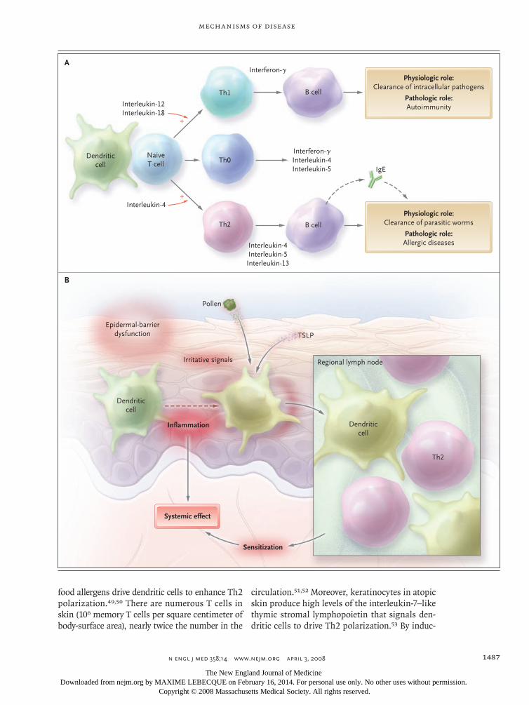
Several candidate genes have been identified in atopic dermatitis,9,17 notably on chromosome 5q31-33. All of them encode cytokines involved in the regulation of IgE synthesis: interleukin-4, interleukin-5, interleukin-12, interleukin-13, and granulocyte–macrophage colony-stimulating fac-tor (GM-CSF). These and other cytokines are pro-duced by two main types of T lymphocytes. Type 2 helper T cells (Th2) produce interleukin-4 as well as interleukin-5 and interleukin-13, two cyto-kines that up-regulate the production of IgE. Type 1 helper T cells (Th1) produce mainly interleu-kin-12 and interferon-γ, which suppresses produc-tion of IgE and stimulates production of IgG antibodies (Fig. 2A). Mutations that affect the function of the promoter region of the lympho-cyte-attracting chemokine RANTES (regulated on activation, normal T-cell expressed and secreted) (17q11) and gain-of-function polymorphisms in the α subunit of the interleukin-4 receptor (16q12) have been identified in patients with atopic der-matitis. Polymorphisms of the gene encoding the cytokine interleukin-18,18 which contributes to the shift of Th1 and Th2 cross-regulation toward Th1-mediated responses (so-called Th1 polariza-tion), or polymorphisms of the genes encoding receptors of the innate immune system19,20 may contribute to the imbalance between Th1 and Th2 immune responses in atopic dermatitis. In persons with atopic dermatitis, a genetically determined dominance of Th2 cytokines affects the matura-tion of B cells and a genomic rearrangement in these cells that favors isotype class switching from IgM to IgE.
Since dry and scaly skin is a symptom of both atopic dermatitis and ichthyosis vulgaris, the most common autosomal dominant disorder of kerati-nization, both diseases might overlap genetically. After the filaggrin gene (FLG) on chromosome 1q21.3, which encodes a key protein in epidermal differentiation, was identified as the gene involved in ichthyosis vulgaris,21 several loss-of-function mutations of the gene were identified in Europe-an patients with atopic dermatitis,22-25 and other, distinctive FLG mutations in Japanese patients have been reported.25,26 Mutations of FLG occur main-ly in early-onset atopic dermatitis and indicate a propensity toward asthma. There is, however, no
association between mutant FLG and allergic air-way diseases without atopic dermatitis. Since FLG mutations are identified in only 30% of European patients with atopic dermatitis, genetic variants of other epidermal structures, such as the stratum corneum tryptic enzyme or a new epidermal col-lagen, may be important.27,28
Atopic dermatitis is a complex genetic disease that arises from gene–gene and gene–environment interactions. The disease emerges in the context of two major groups of genes: genes encoding epi-dermal or other epithelial structural proteins, and genes encoding major elements of the immune system.
B a r r ier F unc tion of the Sk in
Physical Barrier
An intact epidermal compartment is a prerequi-site for the skin to function as a physical and chem-ical barrier. The barrier itself is the stratum cor-neum, the brick and mortar–like structure of the upper epidermal layer.29 An alteration of the bar-rier that causes increased transepidermal water loss is a hallmark of atopic dermatitis. Intercellu-lar lipids of the epidermal horny layers are pro-vided by lamellar bodies, which are produced by exocytosis from upper keratinocytes. Changes in skin ceramides that are secondary to variations in the pH of the stratum corneum can disturb mat-uration of lamellar bodies and impair the barri-er.30 Alterations in the expression of enzymes in-volved in the subtle balance of epidermal adhesion structures are also likely to contribute to the break-down of the epidermal barrier in patients with atopic dermatitis.27,31
Whether these epidermal alterations are pri-mary or are secondary to the underlying inflam-mation remained unclear until immunohisto-chemical32 and genetic studies highlighted the importance of FLG mutations in atopic dermatitis. FLG contributes to the keratin cytoskeleton by act-ing as the template for the assembly of the corni-fied envelope; moreover, breakdown products of FLG contribute to the water-binding capacity of the stratum corneum.33 Genetic variants of FLG in atopic dermatitis that lack the capacity to be pro-teolytically cleaved have been identified,22 but other genetically determined alterations of the epidermis (e.g., changes in the cornified envelope proteins involucrin and loricrin) or lipid composi-tion are also likely to contribute to barrier dysfunc-
The New England Journal of Medicine Downloaded from nejm.org by MAXIME LEBECQUE on February 16, 2014. For personal use only. No other uses without permission.
Copyright © 2008 Massachusetts Medical Society. All rights reserved.

T h e n e w e ng l a nd j o u r na l o f m e dic i n e
n engl j med 358;14 www.nejm.org april 3, 20081486
tion.14 Underlying inflammation can alter the ex-pression of genes such as FLG that are involved in epidermal-barrier function,34 allowing increased transepidermal penetration of environmental al-lergens35,36 and, in collaboration with pruritus, fostering inflammation and sensitization.36,37
The Innate Immune System
Epithelial cells at the interface between the skin and the environment are the first line of defense of the innate immune system.38 They are equipped with a variety of sensing structures, which include the toll-like receptors (TLRs),39 C-type lectins, nu-cleotide-binding oligomerization domain–like re-ceptors, and peptidoglycan-recognition proteins.40 At least 10 different TLRs have been described in humans; they bind to bacterial, fungal (both cell walls), or viral structures (DNA or RNA with so-called cytosine phosphate guanidine [CpG] mo-tifs), and to other microbial structures termed the pathogen-associated molecular patterns. TLR-mediated activation of epithelial cells induces the production of defensins and cathelicidins — fam-ilies of antimicrobial peptides.38
The skin produces the cathelicidin LL-37; the human β-defensins HBD-1, HBD-2, and HBD-3; and dermcidin. The inflammatory micromilieu initiated by interleukin-4, interleukin-13, and in-terleukin-10 down-regulates these antimicrobial peptides in the skin of patients with atopic der-matitis.40-43 For these reasons, it is difficult to manage microbial infections of the skin in pa-tients with atopic dermatitis. Lesional and normal-looking skin is extensively colonized by bacteria such as Staphylococcus aureus or fungi such as malas-sezia. Patients with atopic dermatitis are predis-posed to eczema herpeticum and eczema vaccina-tum because of a reduced production of cathelicidin, which has potent antiviral activity.44,45
Immunopathol o gic Mech a nisms of At opic Der m ati tis
Initial Mechanisms of Skin Inflammation
Early-onset atopic dermatitis usually emerges in the absence of detectable IgE-mediated allergic sensi-tization,4 and in some children — mostly girls — such sensitization never occurs.5 The initial mech-anisms that induce skin inflammation in patients with atopic dermatitis are unknown. They could entail neuropeptide-induced, irritation-induced, or pruritus-induced scratching, which releases pro-
inflammatory cytokines from keratinocytes, or they could be T-cell–mediated but IgE-independent reactions to allergens present in the disturbed epi-dermal barrier or in food (so-called food-sensitive atopic dermatitis). Allergen-specific IgE is not a prerequisite, however, because the atopy patch test can show that aeroallergens applied under occlud-ed skin induce a positive reaction in the absence of allergen-specific IgE.46,47
The Initiation Site of Sensitization
In patients with early-onset atopic dermatitis, IgE-mediated sensitization often occurs several weeks or months after the lesions appear,4 suggesting that the skin is the site of the sensitization. In ani-mal models, repeated epidermal challenge with ovalbumin induces ovalbumin-specific IgE, respi-ratory allergy, and eczematous lesions at the ap-plication site.48 A similar process is likely in hu-mans (Fig. 2B).
Epidermal-barrier dysfunction is a prerequisite for the penetration of high-molecular-weight al-lergens in pollens, house-dust-mite products, mi-crobes, and food. Molecules in pollens and some
Figure 2 (facing page). The Th1–Th2 Paradigm and Its Role in Allergy and the Skin as the Site of Initiation for Sensitization.
Panel A shows that the outcome of helper T cell (Th) differentiation is dictated by the type of dendritic cell, the microenvironment, or both. On antigen presenta-tion, naive T cells are subjected to either interleukin-12 and interleukin-18 or interleukin-4, which polarizes them to Th1 or Th2 helper cells, respectively. Th1 cells produce interferon-γ, whereas Th2 cells produce inter-leukin-4, interleukin-5, and interleukin-13. Th0 cells produce both Th1 and Th2 cytokines, probably in re-sponse to less stringent polarizing signals. Both helper T-cell types have distinct physiologic roles, and it is assumed that a subtle balance between Th1 and Th2 cells is provided under normal conditions. However, a strong Th2 predominance leads to pathologic condi-tions such as overproduction of IgE and allergic diseas-es. Panel B shows non–IgE-mediated inflammation. Epidermal-barrier dysfunction, mechanical irritative signals, or T-cell–mediated events that do not involve IgE lead to an initial inflammatory reaction accompa-nied by an alteration of the function of resident den-dritic cells. These cells are also subjected to locally produced cytokine thymic stromal lymphopoietin (TSLP) and the pollen-derived mediators. As a result, the dendritic cells migrate to the regional lymph nodes and induce an allergen-specific Th2 polarization. The inflammatory reaction may also have a substantial sys-temic effect on the adaptive immune system, favoring the development of IgE-mediated sensitization.
The New England Journal of Medicine Downloaded from nejm.org by MAXIME LEBECQUE on February 16, 2014. For personal use only. No other uses without permission.
Copyright © 2008 Massachusetts Medical Society. All rights reserved.
mechanisms of disease
n engl j med 358;14 www.nejm.org april 3, 2008 1487
food allergens drive dendritic cells to enhance Th2 polarization.49,50 There are numerous T cells in skin (106 memory T cells per square centimeter of body-surface area), nearly twice the number in the
circulation.51,52 Moreover, keratinocytes in atopic skin produce high levels of the interleukin-7–like thymic stromal lymphopoietin that signals den-dritic cells to drive Th2 polarization.53 By induc-
Dendriticcell
Inflammation
Sensitization
Dendriticcell
Dendriticcell
NaiveT cell
Th1
Interleukin-12Interleukin-18
Interleukin-4+
+
Interleukin-4Interleukin-5Interleukin-13
Interferon-γInterleukin-4Interleukin-5
Interferon-γA
Th0
Th2
Th2
Regional lymph node
IgE
Pollen
Epidermal-barrierdysfunction
Irritative signals
TSLP
B cell
B cell
Physiologic role:Clearance of parasitic worms
Pathologic role:Allergic diseases
Physiologic role:Clearance of intracellular pathogens
Pathologic role:Autoimmunity
COLOR FIGURE
AUTHOR PLEASE NOTE:Figure has been redrawn and type has been reset
Please check carefully
03/05/08Draft 4
2
SBL
BieberAuthor
Fig #Title
ME
DEArtist
Issue date
B
Systemic effect
The New England Journal of Medicine Downloaded from nejm.org by MAXIME LEBECQUE on February 16, 2014. For personal use only. No other uses without permission.
Copyright © 2008 Massachusetts Medical Society. All rights reserved.

T h e n e w e ng l a nd j o u r na l o f m e dic i n e
n engl j med 358;14 www.nejm.org april 3, 20081488
ing the production of large amounts of cytokines such as GM-CSF or chemokines, widespread skin inflammation can affect adaptive immunity,54 al-ter the phenotype of circulating monocytes,55-57 and increase the production of prostaglandin E2
58 in atopic dermatitis. All these factors provide sig-nals required for strong skin-driven Th2 polariza-tion, and for this reason, the skin acts as the point of entry for atopic sensitization and may even de-liver signals required for allergenic sensitization in the lung or the gut. The development of sen-sitization and atopic dermatitis in a bone marrow recipient after engraftment of hematopoietic stem cells from an atopic donor59 provides support for the role of the hematopoietic system as a factor in addition to the genetically determined epider-mal-barrier dysfunction in atopic dermatitis.
Antigen-specific IgE is the major recognition structure for allergens on mast cells and baso-phils. It may also be instrumental for the induc-tion of allergen-specific tolerance or in antiinflam-matory mechanisms,60 but whether such events underlie spontaneous remissions of atopic derma-titis remains to be explored.
Dendritic Cells
Epidermal dendritic cells in atopic dermatitis bear IgE61 and express its high-affinity receptor (FcεRI).62-64 In skin lesions, dendritic cells of the plasmacytoid lineage,65 which have potent antivi-ral activity by virtue of interferon-α production, are almost absent.66 In contrast, two populations of myeloid dendritic cells are present: Langerhans’ cells and inflammatory dendritic epidermal cells.67 In atopic dermatitis, but not in other conditions, there is a high-density display of FcεRI by both types of cells. Langerhans’ cells occur in normal skin, but inflammatory dendritic epidermal cells appear only in inflamed skin. They take up and present allergens to Th1 and Th2 cells, and pos-sibly also to regulatory T cells.60 On ligation of FcεRI by IgE, Langerhans’ cells produce interleu-kin-16, which recruits CD4+ T cells to the skin.68 Apart from interleukin-16, Langerhans’ cells pro-duce only a limited range of chemokines and al-most no proinflammatory cytokines.69 On aller-gen capture, Langerhans’ cells contribute to Th2 polarization by an unknown mechanism, and in-flammatory dendritic epidermal cells lead to Th1 polarization by producing interleukin-12 and in-terleukin-18 and releasing proinflammatory cyto-
kines. In the atopy patch test, high numbers of in-flammatory dendritic epidermal cells invade the epidermis 72 hours after allergen challenge, and they and Langerhans’ cells up-regulate their dis-play of FcεRI.70
A Biphasic T-Cell–Mediated Disease
Allergen-specific CD4+ and CD8+ T cells can be isolated from the skin lesions of patients with atop-ic dermatitis. Inflammation in atopic dermatitis is biphasic: an initial Th2 phase precedes a chronic phase in which Th0 cells (cells that share some activities of both Th1 and Th2 cells) and Th1 cells are predominant (Fig. 3).71 The Th2 cytokines in-terleukin-4, interleukin-5, and interleukin-13 pre-dominate in the acute phase of the lesions, and in chronic lesions there is an increase of interfer-on-γ, interleukin-12, interleukin-5, and GM-CSF72; these changes are characteristic of Th1 and Th0 predominance. Th0 cells can differentiate into ei-ther Th1 or Th2 cells, depending on the predomi-nant cytokine milieu. The increased expression of interferon-γ messenger RNA by Th1 cells follows a peak of interleukin-12 expression, which co-incides with the appearance of inflammatory den-dritic epidermal cells in the skin. Normal-looking skin in patients with atopic dermatitis harbors a mild infiltrate, strongly suggesting the presence of residual inflammation between flares.73
Recruitment of T cells into the skin is orches-trated by a complex network of mediators that contribute to chronic inflammation. Homeostatic and inflammatory chemokines produced by skin cells are involved in this process.74,75 Inflamma-tory cells and keratinocytes in the skin lesions ex-press high levels of chemoattractants,76-78 and the keratinocyte-derived thymic stromal lymphopoi-etin induces dendritic cells to produce the Th2-cell–attracting thymus and activation-regulated chemokine, TARC/CCL17. In this way, they may amplify and sustain the allergic response and the generation of interferon-γ–producing cytotoxic T cells,79 as suggested by in vitro studies. Inter-feron-γ produced by Th1 cells has been implicated in the apoptosis of keratinocytes induced by the cell-death receptor Fas.80
The role of regulatory T cells in atopic derma-titis81 has also been examined. High levels of ex-pression of the alpha chain of the interleukin-2 receptor (CD25) and the transcription factor FOXP3 are characteristic of these cells. There is an in-
The New England Journal of Medicine Downloaded from nejm.org by MAXIME LEBECQUE on February 16, 2014. For personal use only. No other uses without permission.
Copyright © 2008 Massachusetts Medical Society. All rights reserved.

mechanisms of disease
n engl j med 358;14 www.nejm.org april 3, 2008 1489
creased pool of circulating regulatory T cells in atopic dermatitis,82 but the skin lesions are devoid of functional regulatory T cells.83 The complexity of the regulatory T-cell compartment is not yet fully understood, and the role of regulatory T cells in the regulation of chronic inflammatory skin disease is elusive.
Staphylococcus aureus
Suppression of the innate immune system of the skin by the inflammatory micromilieu of atopic dermatitis explains the colonization of the skin by S. aureus in more than 90% of patients with atopic dermatitis.83 This feature contributes to allergic sensitization and inflammation (Fig. 4). Scratch-ing increases the binding of S. aureus to the skin, and the increased amount of S. aureus–derived ce-ramidase can aggravate the defect in the skin
barrier. S. aureus enterotoxins84 increase the inflam-mation in atopic dermatitis and provoke the gen-eration of enterotoxin-specific IgE, which corre-lates with the severity of the disease.85 These enterotoxins interact directly with class II mol-ecules of the major histocompatibility complex and the beta chain of the T-cell receptor to induce an antigen-independent proliferation of T cells. They also up-regulate the expression of the skin-homing receptor cutaneous lymphocyte-associated antigen on T cells and the production of keratinocyte-derived chemokines that recruit T cells. By induc-ing the competing β-isoform of the glucocorticoid receptor in mononuclear cells, enterotoxins con-tribute to the emergence of a resistance to local corticosteroid treatment. S. aureus enterotoxins also induce the expression of the glucocorticoid-induced protein ligand related to the tumor necrosis fac-
Langerhans’cell
Aeroallergens
Acute atopicdermatitis
Chronic atopicdermatitis
Th2
Th2
IDEC
Th1/0
Monocyte
Interleukin-16MCP-1
IgE
Interleukin-1Interleukin-6
TNF-α
Interleukin-12Interleukin-18
Interleukin-4Interleukin-5Interleukin-13Interleukin-31
Interferon-γInterleukin-5Interleukin-31
COLOR FIGURE
AUTHOR PLEASE NOTE:Figure has been redrawn and type has been reset
Please check carefully
03/03/08Draft 3
3
SBL
BieberAuthor
Fig #Title
ME
DEArtist
Issue date
Figure 3. Acute and Chronic Phases of IgE and T-Cell–Mediated Atopic Dermatitis.
In the acute phase of atopic dermatitis, Langerhans’ cells are activated on binding of allergens by means of specific IgE and FcεRI. They produce monocyte chemotactic protein 1 (MCP-1) and interleukin-16. Allergen-derived peptides are presented to T cells by Langerhans’ cells that induce a Th2 profile. After migration into the skin, the recruited monocytes differentiate into inflammatory dendritic epidermal cells (IDEC) and produce the proinflammatory cytokines interleukin-1, interleukin-6, and tumor necrosis factor α (TNF-α). Their secretion of interleukin-12 and interleukin-18 contributes to the switch from Th2 to Th1/0 and thereby leads to the chronic phase of the disease.
The New England Journal of Medicine Downloaded from nejm.org by MAXIME LEBECQUE on February 16, 2014. For personal use only. No other uses without permission.
Copyright © 2008 Massachusetts Medical Society. All rights reserved.

T h e n e w e ng l a nd j o u r na l o f m e dic i n e
n engl j med 358;14 www.nejm.org april 3, 20081490
tor receptor on antigen-presenting cells, result-ing in an inhibition of the suppressive activity of regulatory T cells.86
Mechanism of Pruritus
The most important symptom in atopic dermati-tis is persistent pruritus, which impairs the pa-tient’s quality of life. The lack of effect of antihis-tamines argues against a role of histamine in causing atopic dermatitis–related pruritus.87 Neu-ropeptides, proteases, kinins, and cytokines induce itching. Interleukin-31 is a cytokine produced by T cells that increases the survival of hematopoi-etic cells and stimulates the production of inflam-
matory cytokines by epithelial cells. It is strongly pruritogenic, and both interleukin-31 and its re-ceptor are overexpressed in lesional skin.88-90 More-over, interleukin-31 is up-regulated by exposure to staphylococcal exotoxins in vitro. These findings implicate interleukin-31 as a major factor in the genesis of pruritus in atopic dermatitis.
Au t oimmuni t y in At opic Der m ati tis
In addition to IgE antibodies against food and aeroallergens, serum specimens from patients with severe atopic dermatitis contain IgE antibodies
COLOR FIGURE
AUTHOR PLEASE NOTE:Figure has been redrawn and type has been reset
Please check carefully
03/05/08Draft 3
4
SBL
BieberAuthor
Fig #Title
ME
DEArtist
Issue date
Polyclonalstimulation
Dendriticcell
IgE-mediatedreaction
Inductionof CLA
Corticosteroidresistance
Staphylococcusaureus
ChemokinesTSLP
T cell
T cell
T cell
T cell
Figure 4. Multiple Pathways of Staphylococcus aureus–Driven Sensitization and Inflammation.
By virtue of several mechanisms, S. aureus and its products provide signals that favor sensitization and inflamma-tion. S. aureus–derived ceramidase increases the permeability of the stratum corneum, and the superantigenic ca-pacity of S. aureus enterotoxins activates T cells in an allergen-independent manner. S. aureus induces the expres-sion of the skin-homing receptor cutaneous lymphocyte-associated antigen (CLA) on T cells. Keratinocyte-derived chemokines, thymic stromal lymphopoietin (TSLP), and interleukin-31 secretion are induced and augmented by S. aureus enterotoxins. They also contribute to corticosteroid resistance in T cells and alter the activity of regulatory T cells. S. aureus–specific IgE generated by the immune system can bind to FcεRI receptors on dendritic cells and initiate an IgE-mediated reaction to this microbe.
The New England Journal of Medicine Downloaded from nejm.org by MAXIME LEBECQUE on February 16, 2014. For personal use only. No other uses without permission.
Copyright © 2008 Massachusetts Medical Society. All rights reserved.

mechanisms of disease
n engl j med 358;14 www.nejm.org april 3, 2008 1491
against proteins from keratinocytes and endothe-lial cells such as manganese superoxide dismutase and calcium-binding proteins.91,92 The serum lev-els of these IgE autoantibodies correlate with dis-ease severity. Scratching probably releases intracel-lular proteins from keratinocytes. These proteins could be molecular mimics of microbial structures and thus could induce IgE autoantibodies.93 About 25% of adults with atopic dermatitis have IgE an-tibodies against self-proteins.94 In these patients, early-onset atopic dermatitis, intense pruritus, re-current bacterial skin infections, and high serum IgE levels are hallmarks of the disease. Further-more, IgE antibodies against self-proteins can be detected in patients with atopic dermatitis as early as 1 year of age.94 Some autoallergens in skin are also strong inducers of Th1 responses.92 IgE anti-bodies in atopic dermatitis can be induced by en-vironmental allergens, but IgE antibodies against autoantigens in the skin can perpetuate the aller-gic inflammation. Thus, atopic dermatitis seems
to stand at the frontier between allergy and auto-immunity.
A Unif y ing H y po thesis
One classification distinguishes an IgE-associat-ed form of atopic dermatitis (i.e., true atopic der-matitis, formerly called extrinsic atopic dermatitis) from a non–IgE-associated form (“nonatopic” der-matitis, formerly called intrinsic atopic dermati-tis).95 This division implies that nonatopic der-matitis and atopic dermatitis are two different diseases. However, since dry skin is an important sign of both conditions, and the absence of IgE-mediated sensitization may be only a transient fac-tor, there is a need to reconcile these divergent hypotheses. A new picture emerges from recent findings, in which the natural history of atopic dermatitis has three phases (Fig. 5). The initial phase is the nonatopic form of dermatitis in early infancy, when sensitization has not yet occurred.
Allergens
Environment
Genes
Staphylococcusaureus
Scratching,tissue damage
Environmentalfactors
Nonatopicdermatitis
Sensitizationto allergens
Sensitizationto self-proteins
Atopicdermatitis
Autoallergicatopic dermatitis
Impairedepidermal barrier
Tissue-relatedgenes
Atopic genes(cytokines, receptors)
Receptors,cytokines, etc.
COLOR FIGURE
AUTHOR PLEASE NOTE:Figure has been redrawn and type has been reset
Please check carefully
03/03/08Draft 3
5
SBL
BieberAuthor
Fig #Title
ME
DEArtist
Issue date
Figure 5. Gene–Gene and Gene–Environment Interactions in the Natural History of Atopic Dermatitis.
As a result of genetically determined epidermal-barrier dysfunction and the effect of environmental factors, non-atopic dermatitis is the first manifestation of atopic dermatitis. Subsequently, because of their genetic predisposi-tion for IgE-mediated sensitization, patients become sensitized. This phenomenon is favored by Staphylococcus aureus enterotoxin products. Finally, scratching leads to tissue damage and the release of structural proteins, trig-gering an IgE response in patients with atopic dermatitis. This sensitization to self-proteins can be due to the ho-mology of allergen-derived epitopes and human proteins in the context of molecular mimicry.
The New England Journal of Medicine Downloaded from nejm.org by MAXIME LEBECQUE on February 16, 2014. For personal use only. No other uses without permission.
Copyright © 2008 Massachusetts Medical Society. All rights reserved.
T h e n e w e ng l a nd j o u r na l o f m e dic i n e
n engl j med 358;14 www.nejm.org april 3, 20081492
Next, in 60 to 80% of patients, genetic factors in-fluence the induction of IgE-mediated sensitiza-tion to food, environmental allergens, or both — this is the transition to true atopic dermatitis. Third, scratching damages skin cells, which release autoantigens that induce IgE autoantibodies in a substantial proportion of patients with atopic der-matitis.
CL INIC A L IMPL IC ATIONS
Since the barrier dysfunction of the skin and chron-ic inflammation are characteristic of atopic der-matitis, long-term clinical management should emphasize prevention, intensified and individual-ly adapted skin care, reduction of bacterial colo-nization by means of local application of lotions containing antiseptics such as triclosan and chlorhexidine, and — most important — the con-trol of inflammation by the regular use of topical corticosteroids or topical calcineurin inhibitors. In children, before and after the diagnosis of IgE-mediated sensitization, measures that prevent exposure to allergens should be beneficial. The current therapy of atopic dermatitis is reactive — treating the flares — but management should include early and proactive intervention with ef-fective and continuous control of the skin inflam-mation and S. aureus colonization. This strategy
has proved to be effective in reducing the number of flares.96 When applied early in infancy, it could potentially help to reduce later sensitization to en-vironmental antigens and autoallergens.
CONCLUSIONS
Recent insights into the genetic and immunologic mechanisms that drive cutaneous inflammation in atopic dermatitis have led to a better understanding of the natural history of this disease and have high-lighted the critical role of the epidermal-barrier function and the immune system. Both contribute to IgE-mediated sensitization and should be con-sidered as major targets for therapy. New develop-ments aimed specifically at the molecular defects in the stratum corneum could provide a customized way to improve the barrier function. Early and pro-active management could improve the outcome and quality of life for patients with atopic dermatitis.
Supported by grants from the German Research Council and the Atopic Dermatitis and Vaccinia Immunization Network of the National Institute of Allergy and Infectious Diseases.
Dr. Bieber reports receiving consulting fees from Novartis. No other potential conflict of interest relevant to this article was reported.
I thank Drs. W. Burgdorf, M. Noethen, and H. Williams for their review of an earlier version of the manuscript and helpful discussion, and I thank all colleagues, fellows, and students who have studied the genetics and inflammatory mechanisms of atopic dermatitis with me.
References
Akdis CA, Akdis M, Bieber T, et al. Diagnosis and treatment of atopic derma-titis in children and adults: European Academy of Allergology and Clinical Im-munology/American Academy of Allergy, Asthma and Immunology/PRACTALL Con-sensus Report. J Allergy Clin Immunol 2006;118:152-69. [Erratum, J Allergy Clin Immunol 2006;118:724.]
Leung DY, Bieber T. Atopic dermati-tis. Lancet 2003;361:151-60.
Williams H, Flohr C. How epidemiol-ogy has challenged 3 prevailing concepts about atopic dermatitis. J Allergy Clin Im-munol 2006;118:209-13.
Illi S, von Mutius E, Lau S, et al. The natural course of atopic dermatitis from birth to age 7 years and the association with asthma. J Allergy Clin Immunol 2004; 113:925-31.
Novak N, Bieber T. Allergic and non-allergic forms of atopic diseases. J Allergy Clin Immunol 2003;112:252-62.
Strachan DP. Hay fever, hygiene, and household size. BMJ 1989;299:1259-60.
Zutavern A, Hirsch T, Leupold W,
1.
2.
3.
4.
5.
6.
7.
Weiland S, Keil U, von Mutius E. Atopic dermatitis, extrinsic atopic dermatitis and the hygiene hypothesis: results from a cross-sectional study. Clin Exp Allergy 2005;35:1301-8.
Schultz Larsen FV, Holm NV. Atopic dermatitis in a population based twin se-ries: concordance rates and heritability estimation. Acta Derm Venereol Suppl (Stockh) 1985;114:159.
Morar N, Willis-Owen SA, Moffatt MF, Cookson WO. The genetics of atopic dermatitis. J Allergy Clin Immunol 2006; 118:24-34.
Palmer LJ, Cardon LR. Shaking the tree: mapping complex disease genes with linkage disequilibrium. Lancet 2005;366: 1223-34.
Lee YA, Wahn U, Kehrt R, et al. A ma-jor susceptibility locus for atopic dermati-tis maps to chromosome 3q21. Nat Genet 2000;26:470-3.
Cookson WO, Ubhi B, Lawrence R, et al. Genetic linkage of childhood atopic dermatitis to psoriasis susceptibility loci. Nat Genet 2001;27:372-3.
8.
9.
10.
11.
12.
Haagerup A, Bjerke T, Schiøtz PO, et al. Atopic dermatitis — a total genome-scan for susceptibility genes. Acta Derm Venereol 2004;84:346-52.
Cookson W. The immunogenetics of asthma and eczema: a new focus on the epithelium. Nat Rev Immunol 2004;4:978-88.
Bowcock AM, Cookson WO. The ge-netics of psoriasis, psoriatic arthritis and atopic dermatitis. Hum Mol Genet 2004;13 Spec No 1:R43-R55.
Flohr C, Johansson SG, Wahlgren CF, Williams H. How atopic is atopic derma-titis? J Allergy Clin Immunol 2004;114: 150-8.
Hoffjan S, Epplen JT. The genetics of atopic dermatitis: recent findings and fu-ture options. J Mol Med 2005;83:682-92.
Novak N, Kruse S, Potreck J, et al. Single nucleotide polymorphisms of the IL18 gene are associated with atopic ec-zema. J Allergy Clin Immunol 2005;115: 828-33. [Erratum, J Allergy Clin Immunol 2006;118:1319.]
Lange J, Heinzmann A, Zehle C,
13.
14.
15.
16.
17.
18.
19.
The New England Journal of Medicine Downloaded from nejm.org by MAXIME LEBECQUE on February 16, 2014. For personal use only. No other uses without permission.
Copyright © 2008 Massachusetts Medical Society. All rights reserved.
mechanisms of disease
n engl j med 358;14 www.nejm.org april 3, 2008 1493
Kopp M. CT genotype of promotor poly-morphism C159T in the CD14 gene is as-sociated with lower prevalence of atopic dermatitis and lower IL-13 production. Pe-diatr Allergy Immunol 2005;16:456-7.
Ahmad-Nejad P, Mrabet-Dahbi S, Breuer K, et al. The toll-like receptor 2 R753Q polymorphism defines a subgroup of patients with atopic dermatitis having severe phenotype. J Allergy Clin Immunol 2004;113:565-7.
Smith FJ, Irvine AD, Terron-Kwiat-kowski A, et al. Loss-of-function muta-tions in the gene encoding filaggrin cause ichthyosis vulgaris. Nat Genet 2006;38: 337-42.
Palmer CN, Irvine AD, Terron-Kwiat-kowski A, et al. Common loss-of-function variants of the epidermal barrier protein filaggrin are a major predisposing factor for atopic dermatitis. Nat Genet 2006;38: 441-6.
Weidinger S, Illig T, Baurecht H, et al. Loss-of-function variations within the fil-aggrin gene predispose for atopic derma-titis with allergic sensitizations. J Allergy Clin Immunol 2006;118:214-9. [Errata, J Allergy Clin Immunol 2006;118:724, 922.]
Marenholz I, Nickel R, Rüschendorf F, et al. Filaggrin loss-of-function muta-tions predispose to phenotypes involved in the atopic march. J Allergy Clin Im-munol 2006;118:866-71.
Sandilands A, Terron-Kwiatkowski A, Hull PR, et al. Comprehensive analysis of the gene encoding filaggrin uncovers prevalent and rare mutations in ichthyosis vulgaris and atopic eczema. Nat Genet 2007;39:650-4.
Nomura T, Sandilands A, Akiyama M, et al. Unique mutations in the filaggrin gene in Japanese patients with ichthyosis vulgaris and atopic dermatitis. J Allergy Clin Immunol 2007;119:434-40.
Vasilopoulos Y, Cork MJ, Murphy R, et al. Genetic association between an AACC insertion in the 3′UTR of the stratum cor-neum chymotryptic enzyme gene and atop-ic dermatitis. J Invest Dermatol 2004; 123:62-6.
Söderhäll C, Marenholz I, Kerscher T, et al. Variants in a novel epidermal colla-gen gene (COL29A1) are associated with atopic dermatitis. PLoS Biol 2007;5(9):e242.
Proksch E, Jensen JM, Elias PM. Skin lipids and epidermal differentiation in atopic dermatitis. Clin Dermatol 2003;21: 134-44.
Schmid-Wendtner MH, Korting HC. The pH of the skin surface and its impact on the barrier function. Skin Pharmacol Physiol 2006;19:296-302.
Hansson L, Bäckman A, Ny A, et al. Epidermal overexpression of stratum cor-neum chymotryptic enzyme in mice: a model for chronic itchy dermatitis. J In-vest Dermatol 2002;118:444-9.
Seguchi T, Cui CY, Kusuda S, Taka-
20.
21.
22.
23.
24.
25.
26.
27.
28.
29.
30.
31.
32.
hashi M, Aisu K, Tezuka T. Decreased ex-pression of filaggrin in atopic skin. Arch Dermatol Res 1996;288:442-6.
Scott IR, Harding CR. Filaggrin breakdown to water binding compounds during development of the rat stratum corneum is controlled by the water activ-ity of the environment. Dev Biol 1986; 115:84-92.
Howell MD, Kim BE, Gao P, et al. Cy-tokine modulation of atopic dermatitis filaggrin skin expression. J Allergy Clin Immunol 2007;120:150-5.
Proksch E, Fölster-Holst R, Jensen JM. Skin barrier function, epidermal prolifer-ation and differentiation in eczema. J Der-matol Sci 2006;43:159-69.
Hudson TJ. Skin barrier function and allergic risk. Nat Genet 2006;38:399-400.
Cork MJ, Robinson DA, Vasilopoulos Y, et al. New perspectives on epidermal barrier dysfunction in atopic dermatitis: gene-environment interactions. J Allergy Clin Immunol 2006;118:3-21.
Braff MH, Gallo RL. Antimicrobial peptides: an essential component of the skin defensive barrier. Curr Top Microbiol Immunol 2006;306:91-110.
Trinchieri G, Sher A. Cooperation of Toll-like receptor signals in innate im-mune defence. Nat Rev Immunol 2007;7: 179-90.
McGirt LY, Beck LA. Innate immune defects in atopic dermatitis. J Allergy Clin Immunol 2006;118:202-8.
Ong PY, Ohtake T, Brandt C, et al. En-dogenous antimicrobial peptides and skin infections in atopic dermatitis. N Engl J Med 2002;347:1151-60.
Rieg S, Steffen H, Seeber S, et al. De-ficiency of dermcidin-derived antimicro-bial peptides in sweat of patients with atopic dermatitis correlates with an im-paired innate defense of human skin in vivo. J Immunol 2005;174:8003-10.
Howell MD, Gallo RL, Boguniewicz M, et al. Cytokine milieu of atopic derma-titis skin subverts the innate immune re-sponse to vaccinia virus. Immunity 2006; 24:341-8.
Peng WM, Jenneck C, Bussmann C, et al. Risk factors of atopic dermatitis pa-tients for eczema herpeticum. J Invest Dermatol 2007;127:1261-3.
Howell MD, Wollenberg A, Gallo RL, et al. Cathelicidin deficiency predisposes to eczema herpeticum. J Allergy Clin Im-munol 2006;117:836-41.
Ingordo V, D’Andria G, D’Andria C, Tortora A. Results of atopy patch tests with house dust mites in adults with ‘in-trinsic’ and ‘extrinsic’ atopic dermatitis. J Eur Acad Dermatol Venereol 2002;16: 450-4.
Kerschenlohr K, Darsow U, Burgdorf WH, Ring J, Wollenberg A. Lessons from atopy patch testing in atopic dermatitis. Curr Allergy Asthma Rep 2004;4:285-9.
33.
34.
35.
36.
37.
38.
39.
40.
41.
42.
43.
44.
45.
46.
47.
Spergel JM, Mizoguchi E, Brewer JP, Martin TR, Bhan AK, Geha RS. Epicuta-neous sensitization with protein antigen induces localized allergic dermatitis and hyperresponsiveness to methacholine af-ter single exposure to aerosolized antigen in mice. J Clin Invest 1998;101:1614-22.
Traidl-Hoffmann C, Mariani V, Hoch-rein H, et al. Pollen-associated phytopros-tanes inhibit dendritic cell interleukin-12 production and augment T helper type 2 cell polarization. J Exp Med 2005;201:627-36. [Erratum, J Exp Med 2005;201:1347.]
Shreffler WG, Castro RR, Kucuk ZY, et al. The major glycoprotein allergen from Arachis hypogaea, Ara h 1, is a ligand of dendritic cell-specific ICAM-grabbing non-integrin and acts as a Th2 adjuvant in vitro. J Immunol 2006;177:3677-85.
Kupper TS, Fuhlbrigge RC. Immune surveillance in the skin: mechanisms and clinical consequences. Nat Rev Immunol 2004;4:211-22.
Clark RA, Chong B, Mirchandani N, et al. The vast majority of CLA+ T cells are resident in normal skin. J Immunol 2006; 176:4431-9.
Soumelis V, Reche PA, Kanzler H, et al. Human epithelial cells trigger den-dritic cell mediated allergic inflammation by producing TSLP. Nat Immunol 2002;3: 673-80.
Denburg JA, van Eeden SF. Bone mar-row progenitors in inflammation and repair: new vistas in respiratory biology and pathophysiology. Eur Respir J 2006;27: 441-5.
Bratton DL, Hamid Q, Boguniewicz M, Doherty DE, Kailey JM, Leung DY. Granu-locyte macrophage colony-stimulating fac-tor contributes to enhanced monocyte survival in chronic atopic dermatitis. J Clin Invest 1995;95:211-8.
Novak N, Kruse S, Kraft S, et al. Di-chotomic nature of atopic dermatitis re-flected by combined analysis of monocyte immunophenotyping and single nucleo-tide polymorphisms of the interleukin-4/interleukin-13 receptor gene: the dichoto-my of extrinsic and intrinsic atopic derma-titis. J Invest Dermatol 2002;119:870-5.
von Bubnoff D, Scheler M, Hinz T, Matz H, Koch S, Bieber T. Comparative immunophenotyping of monocytes from symptomatic and asymptomatic atopic individuals. Allergy 2004;59:933-9.
Chan SC, Kim JW, Henderson WR Jr, Hanifin JM. Altered prostaglandin E2 regulation of cytokine production in atopic dermatitis. J Immunol 1993;151: 3345-52.
Hallstrand TS, Sprenger JD, Agosti JM, Longton GM, Witherspoon RP, Hen-derson WR Jr. Long-term acquisition of allergen-specific IgE and asthma follow-ing allogeneic bone marrow transplanta-tion from allergic donors. Blood 2004; 104:3086-90.
48.
49.
50.
51.
52.
53.
54.
55.
56.
57.
58.
59.
The New England Journal of Medicine Downloaded from nejm.org by MAXIME LEBECQUE on February 16, 2014. For personal use only. No other uses without permission.
Copyright © 2008 Massachusetts Medical Society. All rights reserved.

n engl j med 358;14 www.nejm.org april 3, 20081494
mechanisms of disease
Bieber T. The pro- and anti-inflam-matory properties of human antigen-pre-senting cells expressing the high affinity receptor for IgE (Fc epsilon RI). Immuno-biology 2007;212:499-503.
Bruynzeel-Koomen C, van Wichen DF, Toonstra J, Berrens L, Bruynzeel PL. The presence of IgE molecules on epidermal Langerhans cells in patients with atopic dermatitis. Arch Dermatol Res 1986;278: 199-205.
Bieber T, de la Salle H, Wollenberg A, et al. Human epidermal Langerhans cells express the high affinity receptor for im-munoglobulin E (Fc epsilon RI). J Exp Med 1992;175:1285-90.
Wang B, Rieger A, Kilgus O, et al. Epi-dermal Langerhans cells from normal hu-man skin bind monomeric IgE via Fc ep-silon RI. J Exp Med 1992;175:1353-65.
Novak N, Bieber T. The role of den-dritic cell subtypes in the pathophysiolo-gy of atopic dermatitis. J Am Acad Derma-tol 2005;53:Suppl 2:S171-S176.
Cao W, Liu YJ. Innate immune func-tions of plasmacytoid dendritic cells. Curr Opin Immunol 2007;19:24-30.
Wollenberg A, Wagner M, Gunther S, et al. Plasmacytoid dendritic cells: a new cutaneous dendritic cell subset with dis-tinct role in inflammatory skin diseases. J Invest Dermatol 2002;119:1096-102.
Wollenberg A, Kraft S, Hanau D, Bieber T. Immunomorphological and ul-trastructural characterization of Langer-hans cells and a novel, inflammatory den-dritic epidermal cell (IDEC) population in lesional skin of atopic eczema. J Invest Dermatol 1996;106:446-53.
Reich K, Heine A, Hugo S, et al. Engage-ment of the Fc epsilon RI stimulates the production of IL-16 in Langerhans cell-like dendritic cells. J Immunol 2001;167:6321-9.
Novak N, Valenta R, Bohle B, et al. FcepsilonRI engagement of Langerhans cell-like dendritic cells and inflammatory dendritic epidermal cell-like dendritic cells induces chemotactic signals and different T-cell phenotypes in vitro. J Allergy Clin Immunol 2004;113:949-57.
Kerschenlohr K, Decard S, Przybilla B, Wollenberg A. Atopy patch test reac-tions show a rapid influx of inflammatory dendritic epidermal cells in patients with extrinsic atopic dermatitis and patients with intrinsic atopic dermatitis. J Allergy Clin Immunol 2003;111:869-74.
Grewe M, Walther S, Gyufko K, Czech W, Schöpf E, Krutmann J. Analysis of the cytokine pattern expressed in situ in in-halant allergen patch test reactions of atopic dermatitis patients. J Invest Der-matol 1995;105:407-10.
60.
61.
62.
63.
64.
65.
66.
67.
68.
69.
70.
71.
Taha RA, Leung DY, Ghaffar O, Bo-guniewicz M, Hamid Q. In vivo expres-sion of cytokine receptor mRNA in atopic dermatitis. J Allergy Clin Immunol 1998; 102:245-50.
Mihm MC Jr, Soter NA, Dvorak HF, Austen KF. The structure of normal skin and the morphology of atopic eczema. J Invest Dermatol 1976;67:305-12.
Homey B, Steinhoff M, Ruzicka T, Leung DY. Cytokines and chemokines or-chestrate atopic skin inflammation. J Al-lergy Clin Immunol 2006;118:178-89.
Nomura I, Gao B, Boguniewicz M, Darst MA, Travers JB, Leung DY. Distinct patterns of gene expression in the skin lesions of atopic dermatitis and psoriasis: a gene microarray analysis. J Allergy Clin Immunol 2003;112:1195-202.
Morales J, Homey B, Vicari AP, et al. CTACK, a skin-associated chemokine that preferentially attracts skin-homing mem-ory T cells. Proc Natl Acad Sci U S A 1999; 96:14470-5.
Homey B, Alenius H, Muller A, et al. CCL27-CCR10 interactions regulate T cell-mediated skin inflammation. Nat Med 2002;8:157-65.
Gombert M, Dieu-Nosjean MC, Win-terberg F, et al. CCL1-CCR8 interactions: an axis mediating the recruitment of T cells and Langerhans-type dendritic cells to sites of atopic skin inflammation. J Immunol 2005;174:5082-91. [Erratum, J Immunol 2005;174:8219.]
Gilliet M, Soumelis V, Watanabe N, et al. Human dendritic cells activated by TSLP and CD40L induce proallergic cytotoxic T cells. J Exp Med 2003;197:1059-63.
Trautmann A, Akdis M, Kleemann D, et al. T cell-mediated Fas-induced kerati-nocyte apoptosis plays a key pathogenetic role in eczematous dermatitis. J Clin In-vest 2000;106:25-35.
Ziegler SF. FOXP3: of mice and men. Annu Rev Immunol 2006;24:209-26.
Ou LS, Goleva E, Hall C, Leung DY. T regulatory cells in atopic dermatitis and subversion of their activity by superanti-gens. J Allergy Clin Immunol 2004;113: 756-63.
Verhagen J, Akdis M, Traidl-Hoff-mann C, et al. Absence of T-regulatory cell expression and function in atopic der-matitis skin. J Allergy Clin Immunol 2006;117:176-83.
Cardona ID, Cho SH, Leung DY. Role of bacterial superantigens in atopic der-matitis: implications for future therapeu-tic strategies. Am J Clin Dermatol 2006;7: 273-9.
Bunikowski R, Mielke M, Skarabis H, et al. Prevalence and role of serum IgE
72.
73.
74.
75.
76.
77.
78.
79.
80.
81.
82.
83.
84.
85.
antibodies to the Staphylococcus aureus-derived superantigens SEA and SEB in children with atopic dermatitis. J Allergy Clin Immunol 1999;103:119-24.
Cardona ID, Goleva E, Ou LS, Leung DY. Staphylococcal enterotoxin B inhibits regulatory T cells by inducing glucocorti-coid-induced TNF receptor-related pro-tein ligand on monocytes. J Allergy Clin Immunol 2006;117:688-95.
Diepgen TL. Long-term treatment with cetirizine of infants with atopic der-matitis: a multi-country, double-blind, randomized, placebo-controlled trial (the ETAC trial) over 18 months. Pediatr Al-lergy Immunol 2002;13:278-86.
Sonkoly E, Muller A, Lauerma AI, et al. IL-31: a new link between T cells and pruritus in atopic skin inflammation. J Allergy Clin Immunol 2006;117:411-7.
Paus R, Schmelz M, Bíró T, Steinhoff M. Frontiers in pruritus research: scratch-ing the brain for more effective itch ther-apy. J Clin Invest 2006;116:1174-86.
Neis MM, Peters B, Dreuw A, et al. Enhanced expression levels of IL-31 cor-relate with IL-4 and IL-13 in atopic and allergic contact dermatitis. J Allergy Clin Immunol 2006;118:930-7.
Mittermann I, Aichberger KJ, Bünder R, Mothes N, Renz H, Valenta R. Autoim-munity and atopic dermatitis. Curr Opin Allergy Clin Immunol 2004;4:367-71.
Aichberger KJ, Mittermann I, Reininger R, et al. Hom s 4, an IgE-reactive autoanti-gen belonging to a new subfamily of cal-cium-binding proteins, can induce Th cell type 1-mediated autoreactivity. J Immunol 2005;175:1286-94.
Schmid-Grendelmeier P, Flückiger S, Disch R, et al. IgE-mediated and T cell-mediated autoimmunity against manga-nese superoxide dismutase in atopic der-matitis. J Allergy Clin Immunol 2005;115: 1068-75.
Mothes N, Niggemann B, Jenneck C, et al. The cradle of IgE autoreactivity in atopic eczema lies in early infancy. J Al-lergy Clin Immunol 2005;116:706-9.
Johansson SG, Bieber T, Dahl R, et al. Revised nomenclature for allergy for glob-al use: report of the Nomenclature Review Committee of the World Allergy Organiza-tion, October 2003. J Allergy Clin Immunol 2004;113:832-6.
Berth-Jones J, Damstra RJ, Golsch S, et al. Twice weekly f luticasone propio-nate added to emollient maintenance treatment to reduce risk of relapse in atopic dermatitis: randomised, double blind, parallel group study. BMJ 2003; 326:1367.Copyright © 2008 Massachusetts Medical Society.
86.
87.
88.
89.
90.
91.
92.
93.
94.
95.
96.
The New England Journal of Medicine Downloaded from nejm.org by MAXIME LEBECQUE on February 16, 2014. For personal use only. No other uses without permission.
Copyright © 2008 Massachusetts Medical Society. All rights reserved.