mechanisms of disease Antiinflammatory Action of ... · mechanisms of disease Antiinflammatory...
13
n engl j med 353;16 www.nejm.org october 20, 2005 The new england journal of medicine 1711 review article mechanisms of disease Antiinflammatory Action of Glucocorticoids — New Mechanisms for Old Drugs Turk Rhen, Ph.D., and John A. Cidlowski, Ph.D. From the Department of Biology, Univer- sity of North Dakota, Grand Forks (T.R.); the Laboratory of Signal Transduction, National Institute of Environmental Health Sciences, Research Triangle Park, N.C. (J.A.C.); and the Department of Health and Human Services, National Institutes of Health, Bethesda, Md. (J.A.C.). Address re- print requests to Dr. Cidlowski at the Lab- oratory of Signal Transduction, National Institute of Environmental Health Sciences, 111 T.W. Alexander Dr., Research Triangle Park, NC 27709, or at cidlows1@niehs. nih.gov. N Engl J Med 2005;353:1711-23. Copyright © 2005 Massachusetts Medical Society. nflammation is a reflexive response to infection, the binding of antibodies to antigens within the body, mechanical irritation, or injury. 1 Mi- crobes that breach epithelial barriers, for instance, directly activate complement and toll-like receptors, two principal components of the innate immune system. The activation of these sentinels triggers the synthesis and release of inflammatory media- tors with acute effects on the vasculature. Localized vasodilation, increased vascular permeability, extravasation of plasma (and humoral) proteins, and migration of leuko- cytes into the affected tissue produce the classic signs of inflammation: calor, dolor, rubor, tumor, and functio laesa. A positive feedback loop initiates the production of ad- ditional inflammatory cytokines once infiltrating leukocytes become activated. Antiin- flammatory homeostatic mechanisms reverse these processes as the infectious agent is cleared by the innate and adaptive immune systems. The hypothalamic–pituitary– adrenal axis and glucocorticoids in particular are essential in limiting and resolving the inflammatory process. 2 Whereas restricted inflammation is beneficial, excessive or persistent inflammation incites tissue destruction and disease. Together, inflammatory disorders such as aller- gies, asthma, autoimmune diseases, and sepsis are a major cause of illness and death. Asthma affects approximately 21.9 million adults and 8.9 million children in the Unit- ed States alone. The prevalence of autoimmune diseases, which affect 8.5 million Americans, 3,4 is also noteworthy. Rheumatoid arthritis, Graves’ disease, glomerulone- phritis, type 1 diabetes mellitus, multiple sclerosis, thyroiditis, pernicious anemia, sys- temic lupus erythematosus, psoriasis, and vitiligo account for most of these autoim- mune diseases. Sepsis is fatal for roughly 30 percent of the 700,000 patients affected annually in the United States. 5-7 Glucocorticoids are indicated for the treatment of many of these diverse conditions. The efficacy of glucocorticoids in alleviating inflam- matory disorders results from the pleiotropic effects of the glucocorticoid receptor on multiple signaling pathways. Pleiotropy can, however, also have adverse effects: growth retardation in children, immunosuppression, hypertension, inhibition of wound re- pair, osteoporosis, and metabolic disturbances. All these harmful properties contrain- dicate prolonged glucocorticoid therapy. Here, we review mechanisms whereby gluco- corticoids inhibit inflammation and the therapeutic limitations of these hormones. We then provide a prospectus for research on drugs that dissociate the beneficial and det- rimental effects of glucocorticoids. The hypothalamic–pituitary–adrenal axis plays a central role in regulating signaling by the glucocorticoid receptor, which is expressed in virtually all cells. In brief, neural, en- docrine, and cytokine signals converge at the level of the periventricular nucleus of the i basic actions of endogenous glucocorticoids The New England Journal of Medicine Downloaded from nejm.org at UNIVERSITY OF CHICAGO LIBRARIES on May 8, 2013. For personal use only. No other uses without permission. Copyright © 2005 Massachusetts Medical Society. All rights reserved.
Transcript of mechanisms of disease Antiinflammatory Action of ... · mechanisms of disease Antiinflammatory...

n engl j med
353;16
www.nejm.org october
20, 2005
The
new england journal
of
medicine
1711
review article
mechanisms of disease
Antiinflammatory Action of Glucocorticoids — New Mechanisms for Old Drugs
Turk Rhen, Ph.D., and John A. Cidlowski, Ph.D.
From the Department of Biology, Univer-sity of North Dakota, Grand Forks (T.R.);the Laboratory of Signal Transduction,National Institute of Environmental HealthSciences, Research Triangle Park, N.C.(J.A.C.); and the Department of Health andHuman Services, National Institutes ofHealth, Bethesda, Md. (J.A.C.). Address re-print requests to Dr. Cidlowski at the Lab-oratory of Signal Transduction, NationalInstitute of Environmental Health Sciences,111 T.W. Alexander Dr., Research TrianglePark, NC 27709, or at [email protected].
N Engl J Med 2005;353:1711-23.
Copyright © 2005 Massachusetts Medical Society.
nflammation is a reflexive response to infection, the
binding
of antibodies to antigens within the body, mechanical irritation, or injury.
1
Mi-crobes that breach epithelial barriers, for instance, directly activate complement
and toll-like receptors, two principal components of the innate immune system. Theactivation of these sentinels triggers the synthesis and release of inflammatory media-tors with acute effects on the vasculature. Localized vasodilation, increased vascularpermeability, extravasation of plasma (and humoral) proteins, and migration of leuko-cytes into the affected tissue produce the classic signs of inflammation: calor, dolor,rubor, tumor, and functio laesa. A positive feedback loop initiates the production of ad-ditional inflammatory cytokines once infiltrating leukocytes become activated. Antiin-flammatory homeostatic mechanisms reverse these processes as the infectious agentis cleared by the innate and adaptive immune systems. The hypothalamic–pituitary–adrenal axis and glucocorticoids in particular are essential in limiting and resolving theinflammatory process.
2
Whereas restricted inflammation is beneficial, excessive or persistent inflammationincites tissue destruction and disease. Together, inflammatory disorders such as aller-gies, asthma, autoimmune diseases, and sepsis are a major cause of illness and death.Asthma affects approximately 21.9 million adults and 8.9 million children in the Unit-ed States alone. The prevalence of autoimmune diseases, which affect 8.5 millionAmericans,
3,4
is also noteworthy. Rheumatoid arthritis, Graves’ disease, glomerulone-phritis, type 1 diabetes mellitus, multiple sclerosis, thyroiditis, pernicious anemia, sys-temic lupus erythematosus, psoriasis, and vitiligo account for most of these autoim-mune diseases. Sepsis is fatal for roughly 30 percent of the 700,000 patients affectedannually in the United States.
5-7
Glucocorticoids are indicated for the treatment ofmany of these diverse conditions. The efficacy of glucocorticoids in alleviating inflam-matory disorders results from the pleiotropic effects of the glucocorticoid receptor onmultiple signaling pathways. Pleiotropy can, however, also have adverse effects: growthretardation in children, immunosuppression, hypertension, inhibition of wound re-pair, osteoporosis, and metabolic disturbances. All these harmful properties contrain-dicate prolonged glucocorticoid therapy. Here, we review mechanisms whereby gluco-corticoids inhibit inflammation and the therapeutic limitations of these hormones. Wethen provide a prospectus for research on drugs that dissociate the beneficial and det-rimental effects of glucocorticoids.
The hypothalamic–pituitary–adrenal axis plays a central role in regulating signaling bythe glucocorticoid receptor, which is expressed in virtually all cells. In brief, neural, en-docrine, and cytokine signals converge at the level of the periventricular nucleus of the
i
basic actions of endogenous glucocorticoids
The New England Journal of Medicine Downloaded from nejm.org at UNIVERSITY OF CHICAGO LIBRARIES on May 8, 2013. For personal use only. No other uses without permission.
Copyright © 2005 Massachusetts Medical Society. All rights reserved.

n engl j med
353;16
www.nejm.org october
20
,
2005
The
new england journal
of
medicine
1712
hypothalamus to control the secretion of cortico-tropin-releasing hormone into the hypophyseal por-tal system (Fig. 1).
2,8
In turn, corticotropin-releas-ing hormone stimulates the release of corticotropinfrom the anterior pituitary. Corticotropin inducesthe synthesis and secretion of cortisol by the adre-nal cortex. Most of the secreted cortisol (approxi-mately 90 percent) is bound to corticosteroid-bind-ing globulins in the blood.
9
Free cortisol is thebiologically active form of the hormone and is con-verted to cortisone by type 2 11
b
-hydroxysteroiddehydrogenase.
10
Conversely, type 1 11
b
-hydroxy-steroid dehydrogenase converts cortisone intocortisol.
10
The glucocorticoid receptor is a member of thesteroid-hormone–receptor family of proteins.
11,12
It binds with high affinity to cortisol; the bound cor-tisol promotes the dissociation of molecular chap-erones, including heat–shock proteins, from thereceptor (Fig. 2). Within the cell, cortisol acts inthree ways. First, the cortisol–glucocorticoid recep-tor complex moves to the nucleus, where it binds asa homodimer (see the Glossary) to DNA sequencescalled glucocorticoid-responsive elements. The re-sulting complex recruits either coactivator or core-pressor proteins that modify the structure of chro-matin, thereby facilitating or inhibiting assemblyof the basal transcription machinery and the initia-tion of transcription by RNA polymerase II.
13
Thisprocess is highly dynamic in cell culture and is pre-sumably so in vivo.
14
Second, regulation of otherglucocorticoid-responsive genes involves interac-tions between the cortisol–glucocorticoid receptorcomplex and other transcription factors, such asnuclear factor-
k
B (NF-
k
B) (Fig. 2).
15,16
These lat-ter actions seem to occur at lower cortisol levelsthan the cortisol–glucocorticoid receptor–glucocor-ticoid-responsive element complex needs to changetranscription. The third mechanism is glucocorti-coid signaling through membrane-associated re-ceptors and second messengers (so-called non-genomic pathways) (Fig. 2).
17,18
Evidence indicatesthat the glucocorticoid receptor inhibits inflam-mation through all three mechanisms: direct andindirect genomic effects and nongenomic mecha-nisms.
The human glucocorticoid receptor (
GR
) gene isone locus on chromosome 5q31–32 (Fig. 3). Even
so, variation in the structure and the expression ofthe gene generates diversity in glucocorticoid sig-naling.
19
The genomic structure includes three tran-scription-initiation sites; each produces an alter-native first exon that is spliced to a common exon 2(Fig. 3). Though the first exon is not translated,there is a potential for functional differences amongexons 1A, 1B, and 1C because dexamethasone up-regulates all three
GR
transcripts to a similar degreein acute lymphoblastic leukemia T cells but depress-es these transcripts to different degrees in a B-cellline.
20,21
These observations highlight the impor-tance of understanding the regulation of the ex-pression of the glucocorticoid receptor in healthand disease.
Human
GR
messenger RNA (mRNA) has alter-native splice variants.
19
Whereas exons 2 through8 are constant components of
GR
mRNA, there aretwo exon 9 isoforms that can be spliced to producemature mRNA. Splicing of exon 9
a
produces
GR
a
mRNA, which is translated into a protein with aunique sequence of 50 amino acids at its carboxyend (Fig. 3). The glucocorticoid receptor
a
isoformbinds cortisol, DNA, and other transcription fac-tors, thereby modifying transcriptional activity oftarget genes. Limited evidence suggests gluco-corticoid receptor
a
may act through nongenom-ic pathways. Splicing of exon 9
b
produces
GR
b
mRNA, which is translated into a protein with 15distinct amino acids at its carboxy end (Fig. 3).
19
Although glucocorticoid receptor
b
protein formshomodimers that bind DNA, it does not bind anyligands examined so far and fails to activate tran-scription. Glucocorticoid receptor
b
can also formheterodimers with glucocorticoid receptor
a
andinterfere with the function of this protein. The rel-ative levels of glucocorticoid receptor
a
and
b
in acell influence the cell’s sensitivity to glucocorticoid,with higher levels of glucocorticoid receptor
b
lead-ing to glucocorticoid resistance.
22
The inflamma-tory cytokines tumor necrosis factor
a
(TNF-
a
) andinterleukin-1 can selectively up-regulate the levelsof glucocorticoid receptor
b
, suggesting its role ininflammation.
23-25
Alternative translation-initiation sites withinexon 2 produce additional isoforms of the gluco-corticoid receptor
19
(Fig. 3). Translation from thefirst methionine codon in
GR
a
and
GR
b
mRNAproduces proteins that consist of 777 amino acids(glucocorticoid receptor
a
-A) and 742 amino acids(glucocorticoid receptor
b
-A). Translation from asecond methionine produces proteins with 751 ami-
structure of
the glucocorticoid receptor
The New England Journal of Medicine Downloaded from nejm.org at UNIVERSITY OF CHICAGO LIBRARIES on May 8, 2013. For personal use only. No other uses without permission.
Copyright © 2005 Massachusetts Medical Society. All rights reserved.

n engl j med
353;16
www.nejm.org october
20, 2005
mechanisms of disease
1713
Figure 1. Pathways of Communication among the Immune System, the Hypothalamic–Pituitary–Adrenal Axis, and Other Tissues Influenced by Immune Signals and Glucocorticoids.
The diagram also shows other important influences on the hypothalamic–pituitary–adrenal axis. Red lines denote inhibition, and blue and black arrows activation.
The New England Journal of Medicine Downloaded from nejm.org at UNIVERSITY OF CHICAGO LIBRARIES on May 8, 2013. For personal use only. No other uses without permission.
Copyright © 2005 Massachusetts Medical Society. All rights reserved.

n engl j med
353;16
www.nejm.org october
20
,
2005
The
new england journal
of
medicine
1714
no acids (glucocorticoid receptor
a
-B) and 716 ami-no acids (glucocorticoid receptor
b
-B), respectively.There are important functional differences betweenthe two isoforms: glucocorticoid receptor
a
-B hasroughly twice the biologic activity of glucocorti-coid receptor
a
-A in gene-expression studies invitro.
26
The finding that the two isoforms are ex-pressed at different ratios in various types of cellsand tissues also suggests that they may have dis-tinct functions in vivo.
27
The human glucocorticoid receptor has five serineresidues that are phosphorylated under differentconditions by cyclin-dependent kinases and mito-gen-activated protein kinases (MAPKs) (Fig. 3).
28
The phosphorylation of several of the serines is de-pendent on the binding of ligands such as cortisolto the glucocorticoid receptor, whereas other serinesare phosphorylated in a ligand-independent man-ner. The specific combination of serines that arephosphorylated has distinct effects on its transcrip-tional activity. For example, the glucocorticoid re-ceptor is found primarily in the cytoplasm and isinactive when phosphorylated at serine 203, but itactively transcribes DNA when phosphorylated atserine 211.
28
Another important modification ofthe glucocorticoid receptor that cortisol bindinginduces is the covalent attachment of ubiquitin tothe receptor (Fig. 3), thus marking it for degradationby the proteasome
29
; however, this process can becell-type specific.
30
Recent studies show that sumoy-lation (the attachment of small, ubiquitin-relatedmodifiers) of the glucocorticoid receptor potenti-ates its transcriptional activity.
31,32
Little is knownabout the effect of post-translational modificationson the repression of gene transcription, interactionswith other transcription factors, or nongenomic sig-naling pathways.
Interactions among the nervous system, the hypo-thalamic–pituitary–adrenal axis, and componentsof the innate and adaptive immune system play akey role in the regulation of inflammation and im-munity (Fig. 1).
2,8
For instance, cytokines and in-
flammatory mediators activate peripheral pain re-ceptors whose axons project to the dorsal horn andsynapse with the lemniscal tract, which in turn car-ries pain signals to the thalamus and the somatosen-sory cortex. Activation of this nociceptive pathwayultimately stimulates hypothalamic–pituitary–adre-nal activity. Glucocorticoids inhibit the synthesis ofcytokines and inflammatory mediators, thus form-ing a negative feedback loop. Cytokines can alsoact directly on the brain to activate the hypotha-lamic–pituitary–adrenal axis. Dysregulation of thisneuroendocrine loop by hyperactivity or hypoac-tivity of the hypothalamic–pituitary–adrenal axiscauses systemic changes in inflammation and im-munity.
2,8
Hyperactivity of the hypothalamic–pituitary–adrenal axis in the absence of inflammation, as inCushing’s syndrome, causes immunosuppressionand increased susceptibility to infection.
33
Physi-cal pain, emotional trauma, and caloric restrictionalso activate the hypothalamic–pituitary–adrenalaxis and cause immunosuppression.
34,35
In con-trast, decreased activity of the axis and low levelsof glucocorticoids increase susceptibility to andthe severity of inflammation. Patients with Addi-son’s disease, for example, require supplementalglucocorticoids during infection and inflamma-tion to prevent the toxic effects of cytokines.
36
Dys-regulation of the hypothalamic–pituitary–adrenalaxis by inflammation is associated with adverseoutcomes among patients with the acute respira-tory distress syndrome.
37
Likewise, acquired glu-cocorticoid resistance is a common occurrence inpatients with severe rheumatoid arthritis.
38
Gluco-corticoid resistance is a common finding and canbe due to decreased expression of glucocorticoidreceptor
a
, increased expression of glucocorticoidreceptor
b
, or activation of MAPK, which phos-phorylates the glucocorticoid receptor and therebyinhibits glucocorticoid signaling.
39,40
post-translational
modifications of the
glucocorticoid receptor
neuroendocrine regulation
of inflammation
Figure 2 (facing page). Three General Mechanisms of Action of Glucocorticoids and the Glucocorticoid Receptor in the Inhibition of Inflammation.
TNF-
a
denotes tumor-necrosis factor
a
, HSP heat-shock protein, mRNA messenger RNA, and P phosphate. The three mechanisms are nongenomic activation, DNA-dependent regulation, and protein interference mecha-nisms (e.g., NF-
k
B elements). Black arrows denote acti-vation, the red line inhibition, the red dashed arrow repression, and the red X lack of product (i.e., no mRNA).
The New England Journal of Medicine Downloaded from nejm.org at UNIVERSITY OF CHICAGO LIBRARIES on May 8, 2013. For personal use only. No other uses without permission.
Copyright © 2005 Massachusetts Medical Society. All rights reserved.

n engl j med
353;16
www.nejm.org october
20, 2005
mechanisms of disease
1715
The New England Journal of Medicine Downloaded from nejm.org at UNIVERSITY OF CHICAGO LIBRARIES on May 8, 2013. For personal use only. No other uses without permission.
Copyright © 2005 Massachusetts Medical Society. All rights reserved.

n engl j med
353;16
www.nejm.org october
20
,
2005
The
new england journal
of
medicine
1716
Glucocorticoids and the glucocorticoid receptorreside at the apex of a regulatory network that blocksseveral inflammatory pathways (Fig. 4). For exam-ple, glucocorticoids can inhibit prostaglandin pro-duction through three independent mechanisms:the induction and activation of annexin I, the in-duction of MAPK phosphatase 1, and the repres-sion of transcription of cyclooxygenase 2. AnnexinI (also called lipocortin-1) is an antiinflammatoryprotein that physically interacts with and inhibitscytosolic phospholipase A
2
a
(cPLA
2
a
).
41-44
Thiscalcium-binding protein requires elevated calciumlevels and phosphorylation by the protein kinasesMAPK, calcium/calmodulin–dependent kinase II,and MAPK interacting kinase to exert its enzymat-
antiinflammatory signaling
mechanisms
ic activity.
45
The activation of cPLA
2
a
by inflam-matory stimuli begins with the movement of thephospholipase from the cytosol to the perinuclearmembrane, where it hydrolyzes phospholipids con-taining arachidonic acid. Glucocorticoids induceannexin I, which by inhibiting cPLA
2
a
, blocks therelease of arachidonic acid and its subsequent con-version to eicosanoids (i.e., prostaglandins, throm-boxanes, prostacyclins, and leukotrienes). Micelacking annexin I have elevated levels of cPLA
2
a
,an exaggerated inflammatory response, and par-tial resistance to the antiinflammatory action ofglucocorticoids.
46-48
A strong correlation existsbetween basal and corticotropin-stimulated corti-sol levels and the expression of annexin I in neu-trophils in humans, but the clinical importance ofannexin I as an antiinflammatory protein is un-known.
49
A second antiinflammatory protein inducedby glucocorticoids is MAPK phosphatase 1 (Fig.4).
50-52
Cytokines, bacterial and viral infections,and ultraviolet radiation are but a few of the inflam-matory signals that activate MAPK cascades.
16
Ul-traviolet light triggers a kinase cascade that phos-phorylates and activates Jun N-terminal kinase,which in turn phosphorylates the transcriptionfactor c-Jun. Phosphorylated c-Jun homodimersand c-Jun–Fos heterodimers bind DNA sequenc-es called activator protein 1 response elementsand induce the transcription of inflammatory andimmune genes.
16
Glucocorticoid-induced MAPKphosphatase 1 dephosphorylates and inactivatesJun N-terminal kinase, thereby inhibiting c-Jun–mediated transcription. MAPK phosphatase 1 alsodephosphorylates and inactivates all members ofthe MAPK family of proteins, including Jun N-ter-minal kinase, extracellular-signal–related kinase 1and 2, and p38 kinase. Consequently, MAPK phos-phatase 1 may also inhibit cPLA
2
a
activity by block-ing its phosphorylation by MAPKs and MAPK-inter-acting kinase. In addition to blocking an essentialupstream component of the c-Jun pathway, glu-cocorticoids and the glucocorticoid receptor di-rectly interfere with c-Jun–mediated transcription(Fig. 4). Transcriptional interference between theglucocorticoid receptor and c-Jun homodimers (andc-Jun–Fos heterodimers) results from protein–pro-tein interactions and has proved to be a major anti-inflammatory mechanism.
16
The cortisol–glucocorticoid receptor complexalso physically interacts with NF-
k
B to block itstranscriptional activity.
15,16
In its inactive state,
Glossary
Activator protein 1:
An inflammatory transcription factor composed of two polypeptide subunits (c-Jun homodimers or c-Jun–Fos heterodimers)
Coactivator :
A class of eukaryotic proteins that modulate the structure of chromatin or serve as a bridge between transcription factors and RNA po-lymerase II to facilitate the transcription of target genes
Corepressor:
A class of eukaryotic proteins that modulate the structure of chromatin or inhibit RNA polymerase II to suppress transcription of target genes
Glucocorticoid-responsive element:
A DNA motif usually found in the promot-er of glucocorticoid-responsive genes that binds glucocorticoid receptor and enhances transcription
Heterodimer:
A complex of two different protein chains held together by non-covalent bonds
Homodimer:
A complex of two identical protein chains held together by non-covalent bonds
Negative glucocorticoid-responsive element:
A DNA motif usually found in the promoter of glucocorticoid-responsive genes that binds glucocorticoid receptor and represses transcription
Nuclear factor-
k
B (NF-
k
B):
An inflammatory transcription factor composed of two polypeptides from the Rel family of proteins
Pleiotropy:
The ability of one gene to have multiple phenotypic effects
Transcription:
The synthesis of RNA from a DNA template
Transactivation: The stimulation of transcription by a specific protein (i.e., transcription factor)
Transrepression: The inhibition of transcription by a specific protein (i.e., tran-scription factor)
The New England Journal of Medicine Downloaded from nejm.org at UNIVERSITY OF CHICAGO LIBRARIES on May 8, 2013. For personal use only. No other uses without permission.
Copyright © 2005 Massachusetts Medical Society. All rights reserved.

n engl j med 353;16 www.nejm.org october 20, 2005
mechanisms of disease
1717
NF-kB is sequestered in the cytoplasm by an inhib-itory protein named IkB. TNF-a, interleukin-1, mi-crobial pathogens, viral infections, and other in-flammatory signals trigger signaling cascades thatactivate IkB kinases (Fig. 2).53 Phosphorylation ofIkB leads to its ubiquination and degradation bythe proteasome, unmasking a nuclear localizationsignal on NF-kB. In the nucleus, NF-kB binds DNAsequences called NF-kB elements and stimulatesthe transcription of cytokines, chemokines, cell-adhesion molecules, complement factors, and re-ceptors for these molecules. NF-kB also induces thetranscription of cyclooxygenase 2, an enzyme es-sential for prostaglandin production.54 Thus, gluco-corticoid-induced antagonism of NF-kB and re-pression of cyclooxygenase 2 is the third mechanismfor the inhibition of prostaglandin synthesis after
the induction of the antagonists of cPLA2a, annex-in I, and MAPK phosphatase 1 (Fig. 4). Direct inter-actions between the glucocorticoid receptor andNF-kB probably account for most of the inhibitoryeffects of glucocorticoids on NF-kB signaling.15,55
Despite the analogous nature of glucocorticoid re-ceptor–mediated repression of activator protein 1and NF-kB, different parts of the surface of the glu-cocorticoid receptor contact each transcription fac-tor.56 Glucocorticoids and the glucocorticoid recep-tor also modulate the activity of other transcriptionfactors.57
Recent work suggests that glucocorticoids canhave rapid effects on inflammation that are notmediated by changes in gene expression. The best-described nongenomic mechanism involves theactivation of endothelial nitric oxide synthetase
Figure 3. Genomic Location and Organization of the Human Glucocorticoid Receptor (GR).
Diversity in the expression and function of the glucocorticoid receptor results from alternative sites for the initiation of transcription (exon 1A, 1B, or 1C), as well as alternative splicing of pre–messenger RNA (mRNA) at exon 9a or 9b. Additional variation in the structure and function of the protein results from alternative sites for the initiation of translation within exon 2 and post-translational modifications in the form of phosphorylation (P), ubiquination (Ub), and sumoylation (Sumo). DBD denotes DNA-binding domain, LBD ligand-binding domain, and hGR human GR.
5q31
1
1A
P
1 420 488 527 777
27 420 488 527 777
1 420 488 527 742
27 420 488 527 742
P PPPSumo Sumo
1B 1C
2 3 4 5 6 7 8 9b9a
2 3 4 5 6 7 8 9
Chromosome 5
Human GR gene
Human GR proteins
hGRa-A
hGRa-B
hGRb-A
hGRb-B
NH2-terminal DBD LBD
Exons
5q32
Ub Sumo
Human GR mRNA
The New England Journal of Medicine Downloaded from nejm.org at UNIVERSITY OF CHICAGO LIBRARIES on May 8, 2013. For personal use only. No other uses without permission.
Copyright © 2005 Massachusetts Medical Society. All rights reserved.

n engl j med 353;16 www.nejm.org october 20, 2005
The new england journal of medicine
1718
(eNOS).17 Glucocorticoids stimulate the activity ofphosphatidylinositol-3-hydroxykinase (PI3K) in aglucocorticoid receptor–dependent, but transcrip-tion-independent, manner in human endothelial
cells. Activation of PI3K leads to phosphorylationof Akt. Phosphorylated Akt then phosphorylatesand activates eNOS, resulting in the production ofnitric oxide. In mice, glucocorticoid-induced acti-
Figure 4. Partial Molecular Architecture Underlying the Glucocorticoid-Induced Antagonism of Inflammation.
Inflammatory pathways are characterized by positive feedback loops (i.e., cytokines activate NF-kB, which in turn stimulates the synthesis of more cytokines) and by redundancy (i.e., cytokines also activate c-Jun–Fos). The glucocorticoid receptor inhibits these pathways at multiple points by directly blocking the transcription of inflammatory proteins by NF-kB and activator protein 1 and by inducing the expression of antiinflammatory proteins such as IkB, annexin I, and MAPK phosphatase I. 5-LOX denotes 5-lipoxygenase, and COX-2 cyclooxygenase 2. Red lines denote in-hibition, and black arrows activation. An interactive version of this figure is available with the full text of the article at www.nejm.org.
Glucocorticoidreceptor
Annexin I
cPLA2a
COX-2
NF-kB
c-Jun Fos
Jun N-terminalkinase
MAPKphosphatase I
MAPK-interactingkinase
IkB kinase
IkB
MAPKs
Calcium kinase II5-LOX
Cortisol
Arachidonic acid
Prostaglandins
Inflammation
Leukotrienes
Phospholipids
Protein kinase Inflammatorytranscription factor
EnzymeMinor pathways
Core pathways
Inhibitory proteinProteinphosphatase
CytokinesBacteriaViruses
Free radicalsUltraviolet radiation
CytokinesCytokine receptors
Chemotactic proteinsAdhesion molecules
CytokinesHormonesMitogensEndotoxinAntigen
CytokinesGrowth factors
MitogensBacteriaViruses
Ultraviolet radiation
CytokinesCytokine receptors
Chemotactic proteinsAdhesion molecules
CollagenasesMatrix metalloproteinases
Repression by means of negative glucocorticoid-responsive elements
Corticotropin-releasing hormonePro-opiomelanocortin
OsteocalcinProliferinKeratins
Interleukin-1b
Calcium/calmodulin–dependent kinase II
Calcium
HOC
CH2OH
OOH
O
The New England Journal of Medicine Downloaded from nejm.org at UNIVERSITY OF CHICAGO LIBRARIES on May 8, 2013. For personal use only. No other uses without permission.
Copyright © 2005 Massachusetts Medical Society. All rights reserved.
n engl j med 353;16 www.nejm.org october 20, 2005
mechanisms of disease
1719
vation of the PI3K–Akt–eNOS pathway protectsagainst ischemia- or reperfusion-induced injury inthe heart and the cremaster muscle. This finding issurprising because the production of nitric oxide isgenerally associated with vasodilation and inflam-mation.58 More research is needed to clarify therole of nontranscriptional mechanisms in the inhi-bition of vasodilation, vascular permeability, andmigration of leukocytes across endothelium.59 An-other mechanism of the glucocorticoid-inducedinhibition of inflammation involves decreased sta-bility of mRNA for genes for inflammatory pro-teins such as vascular endothelial growth factorand cyclooxygenase 2.51,60,61 Glucocorticoids clear-ly act on diverse targets through multiple mecha-nisms to control inflammation.
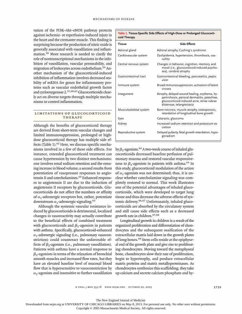
Although the benefits of glucocorticoid therapyare derived from short-term vascular changes andlimited immunosuppression, prolonged or high-dose glucocorticoid therapy has multiple side ef-fects (Table 1).62 Here, we discuss specific mecha-nisms involved in a few of these side effects. Forinstance, extended glucocorticoid treatment cancause hypertension by two distinct mechanisms:one involves renal sodium retention and the ensu-ing increase in blood volume; a second results frompotentiation of vasopressor responses to angio-tensin II and catecholamines.63 Enhanced respons-es to angiotensin II are due to the induction ofangiotensin II receptors by glucocorticoids. Glu-cocorticoids do not affect the numbers or affinityof a1-adrenergic receptors but, rather, potentiatedownstream a1-adrenergic signaling.63
Although the systemic vascular resistance in-duced by glucocorticoids is detrimental, localizedchanges in vasoreactivity may actually contributeto the beneficial effects of combined treatmentwith glucocorticoids and b2-agonists in patientswith asthma. Specifically, glucocorticoid-enhanceda1-adrenergic signaling (i.e., pulmonary vasocon-striction) could counteract the unfavorable ef-fects of b2-agonists (i.e., pulmonary vasodilation).Patients with asthma have a normal response tob2-agonists in terms of the relaxation of bronchialsmooth muscles and increased flow rates, but theyhave an elevated baseline level of mucosal bloodflow that is hypersensitive to vasoconstriction bya1-agonists and insensitive to further vasodilation
by b2-agonists.64 A two-week course of inhaled glu-cocorticoids decreased baseline perfusion of pul-monary mucosa and restored vascular responsive-ness to b2-agonists in patients with asthma.65 Inthis study, glucocorticoid modulation of the actionof a1-agonists was not determined; thus, it is un-clear whether catecholamine signaling was com-pletely restored to normal. This work illustratesone of the potential advantages of inhaled gluco-corticoids, which were developed to target lungtissue and thus decrease the adverse effects of sys-temic delivery.66,67 Unfortunately, inhaled gluco-corticoids are absorbed by the circulatory systemand still cause side effects such as a decreasedgrowth rate in children.67,68
Longitudinal growth in children is a result of theorganized proliferation and differentiation of chon-drocytes and the subsequent ossification of theextracellular matrix laid down in the growth platesof long bones.69 Stem cells reside at the epiphyse-al end of the growth plate and give rise to proliferat-ing chondrocytes. Moving toward the metaphysealbone, chondrocytes slow their rate of proliferation,begin to hypertrophy, and produce extracellularmatrix proteins and matrix metalloproteinases. Aschondrocytes synthesize this scaffolding, they takeup calcium and secrete calcium phosphate and hy-
limitations of glucocorticoid
therapy
Table 1. Tissue-Specific Side Effects of High-Dose or Prolonged Glucocorti-coid Therapy.
Tissue Side Effects
Adrenal gland Adrenal atrophy, Cushing’s syndrome
Cardiovascular system Dyslipidemia, hypertension, thrombosis, vas-culitis
Central nervous system Changes in behavior, cognition, memory, and mood (i.e., glucocorticoid-induced psycho-ses), cerebral atrophy
Gastrointestinal tract Gastrointestinal bleeding, pancreatitis, peptic ulcer
Immune system Broad immunosuppression, activation of latent viruses
Integument Atrophy, delayed wound healing, erythema, hy-pertrichosis, perioral dermatitis, petechiae, glucocorticoid-induced acne, striae rubrae distensae, telangiectasia
Musculoskeletal system Bone necrosis, muscle atrophy, osteoporosis, retardation of longitudinal bone growth
Eyes Cataracts, glaucoma
Kidney Increased sodium retention and potassium ex-cretion
Reproductive system Delayed puberty, fetal growth retardation, hypo-gonadism
The New England Journal of Medicine Downloaded from nejm.org at UNIVERSITY OF CHICAGO LIBRARIES on May 8, 2013. For personal use only. No other uses without permission.
Copyright © 2005 Massachusetts Medical Society. All rights reserved.

n engl j med 353;16 www.nejm.org october 20, 2005
The new england journal of medicine
1720
droxyapatite. Chondrocytes ultimately undergo ap-optosis, leaving behind mineralized bone. Gluco-corticoids slow longitudinal growth by reducing theproliferation of chondrocytes and inducing apopto-sis of these cells. Inhibition of insulin-like growthfactor I signaling is one mechanism underlying de-creased chondrocyte proliferation.69 In contrast totheir effects in the circulatory system, glucocorti-coid-induced apoptosis in chondrocytes involvesthe suppression of signaling through the Akt path-way.70 Interestingly, insulin-like growth factor I in-creases the phosphorylation of Akt and acts as asurvival factor in glucocorticoid-treated chondro-cytes. Although there is generally a period of catch-up growth once glucocorticoid therapy is stopped,sustained treatment with substantive amounts ofglucocorticoids during childhood is often associat-ed with decreased adult stature.69
Glucocorticoids also have damaging effects onbone in adults. Osteoporosis and an increased riskof fractures are the main side effects of glucocorti-coid therapy.62 Osteoporosis is mediated in part bythe binding of glucocorticoid receptors to nega-tive glucocorticoid-responsive elements that inhib-it transcription of osteocalcin in osteoblasts; osteo-calcin is an important extracellular matrix proteinthat promotes bone mineralization.71,72 Severalother side effects of glucocorticoids, including theinhibition of corticotropin-releasing hormone andthe expression of pro-opiomelanocortin, are alsomediated by negative glucocorticoid-responsive el-ements (Fig. 4). Glucocorticoids exacerbate osteo-porosis by inducing apoptosis in osteoblasts andby increasing the activity of osteoclasts. Some ofthese effects are directly mediated by glucocorti-coid receptors in bone cells, whereas indirect ef-fects are mediated by interactions with other en-docrine signals.73
The repair of aseptic wounds is also inhibitedby glucocorticoids. For example, fractures triggerinflammation and the production of cytokines cru-cial for the healing and remodeling of bone.74-76
In addition to blocking cytokine signaling, gluco-corticoids inhibit the synthesis of matrix metal-loproteinases and collagen, which are importantfactors in wound repair.77-80 Glucocorticoids alsopromote gluconeogenesis in the liver, the degra-dation of proteins to free amino acids in muscle(and muscle atrophy), and lipolysis,81-83 ultimatelyproducing hyperglycemia. There are currently nomeans of ameliorating the side effects of prolongedglucocorticoid therapy that function at the level of
the glucocorticoid receptor or the glucocorticoid-responsive elements; rather, treatments such as in-sulin (or its analogues) for glucocorticoid-induceddiabetes, bisphosphonates for osteoporosis, andstandard lipid regulators for dyslipidemia are oftenused.84 This problem has led to research that hasidentified potentially selective glucocorticoids.
The pleiotropic effects of glucocorticoids lie be-tween two theoretical extremes. On the one hand,their manifold effects could be inseparable. Alter-natively, each effect could be fully dissociated. Ithas been posited that the antiinflammatory effectsof glucocorticoids are primarily mediated by the in-hibition of NF-kB and activator protein 1, whereastheir side effects result from the activation of tran-scription. Although this hypothesis is overly sim-plistic, a recent study described a novel glucocorti-coid (ZK216348) with a pattern of repression andactivation of transcription that was dramaticallydifferent from that of known glucocorticoids.85 Thelevel of glucocorticoid required to repress interleu-kin-8 in monocytes was 8 to 12 times as high as thatrequired to induce tyrosine aminotransferase in liv-er cells. In contrast, the ratio of ZK216348 requiredto repress interleukin-8 to the level required to acti-vate tyrosine aminotransferase was just 0.4, whichwas reflected in a better therapeutic index in vivo.Thus, it is possible to develop ligands that inhibitNF-kB–induced expression of inflammatory genesand activate transcription by means of glucocorti-coid-responsive elements much more selectivelythan do currently available glucocorticoids.
Mechanistically, ligands with different struc-tures induce different receptor conformations; forexample, the position of helix 12 differs betweenglucocorticoid receptors bound to agonists and re-ceptors bound to antagonists. Helix 12 closes be-hind dexamethasone as it sits in the hormone-bind-ing pocket.86,87 In this position, helix 12 recruitscoactivators required for ligand-dependent tran-scription.86 The antagonist mifepristone residesin the same pocket as dexamethasone but causeshelix 12 to assume a position that precludes coac-tivator binding87 and results in the recruitment oftranscriptional corepressors.88 Further compari-son of glucocorticoid analogues reveals that theyhave divergent transcriptional activities.89 It is alsoimportant that subtle mutations in the GR gene
selective glucocorticoids
and future therapy
The New England Journal of Medicine Downloaded from nejm.org at UNIVERSITY OF CHICAGO LIBRARIES on May 8, 2013. For personal use only. No other uses without permission.
Copyright © 2005 Massachusetts Medical Society. All rights reserved.

n engl j med 353;16 www.nejm.org october 20, 2005
mechanisms of disease
1721
can be used to differentiate the repression of tran-scription by activator protein 1 from repression byNF-kB56 and that different glucocorticoids vary intheir capacity to activate genomic and nongenomicmechanisms.90 These observations highlight thepotential for the development of selective gluco-corticoids with improved therapeutic profiles.
The rational development of compounds thatdissociate the effects of glucocorticoids will requireintricate knowledge of the structure of receptorsbound to various ligands and an understanding ofthe way different isoforms of the glucocorticoid re-ceptor activate each signaling pathway. Several com-mercial entities are actively pursuing these goals.91
Despite our optimism, it would be naive to suggestthat therapeutic effects and side effects are medi-ated by separate mechanisms or that one coulddevelop ligands that exclusively activate one mo-lecular mechanism. Consequently, it will also beimportant to optimize the pharmacokinetic andpharmacodynamic properties of new drugs and todevelop novel ways to target these drugs to in-flamed tissues, as is the case with inhaled gluco-corticoids.67,92
The potency of glucocorticoids as inhibitors ofdiverse inflammatory disorders guarantees theircontinued use as therapeutic agents. The antiinflam-
matory and immunosuppressive effects of gluco-corticoids rely on several molecular mechanisms,which have been elucidated by basic research. Threemain mechanisms include direct effects on gene ex-pression by the binding of glucocorticoid receptorsto glucocorticoid-responsive elements (i.e., theinduction of annexin I and MAPK phosphatase 1),indirect effects on gene expression through the in-teractions of glucocorticoid receptors with othertranscription factors (i.e., NF-kB and activator pro-tein 1), and glucocorticoid receptor–mediated ef-fects on second-messenger cascades (i.e., the PI3K–Akt–eNOS pathway). Unfortunately, because someof these mechanisms are also involved in physio-logic signaling rather than inflammatory signaling,the therapeutic effects of glucocorticoids in inflam-mation are often accompanied by clinically signifi-cant side effects. It is unclear whether isoforms ofthe glucocorticoid receptor are differentially in-volved in signaling through each of these mecha-nisms. Similarly, we do not know whether gluco-corticoid-induced activation of certain mechanismsalleviates specific diseases or causes particular sideeffects. If this sort of signaling specificity exists invivo, there will be tremendous potential for the de-velopment of synthetic ligands that activate antiin-flammatory mechanisms but do not affect otherpathways. Such drugs would in essence mimic thebeneficial effects of natural glucocorticoids withouttheir detrimental side effects.
conclusions
references
1. Gallin JI, Goldstein IM, Snyderman R.Overview. In: Gallin JI, Goldstein IM, Snyder-man R, eds. Inflammation: basic principlesand clinical correlates. 2nd ed. New York:Raven Press, 1992:1-4.2. Webster JI, Tonelli L, Sternberg EM.Neuroendocrine regulation of immunity.Annu Rev Immunol 2002;20:125-63.3. Jacobson DL, Gange SJ, Rose NR, Gra-ham NM. Epidemiology and estimated pop-ulation burden of selected autoimmune dis-eases in the United States. Clin ImmunolImmunopathol 1997;84:223-43.4. Cooper GS, Stroehla BC. The epidemi-ology of autoimmune diseases. AutoimmunRev 2003;2:119-25.5. Angus DC, Linde-Zwirble WT, LidickerJ, Clermont G, Carcillo J, Pinsky MR. Epide-miology of severe sepsis in the United States:analysis of incidence, outcome, and associ-ated costs of care. Crit Care Med 2001;29:1303-10.6. Riedemann NC, Guo RF, Ward PA. Theenigma of sepsis. J Clin Invest 2003;112:460-7.
7. Martin GS, Mannino DM, Eaton S, MossM. The epidemiology of sepsis in the UnitedStates from 1979 through 2000. N Engl J Med2003;348:1546-54.8. Rivest S. How circulating cytokines trig-ger the neural circuits that control the hy-pothalamic-pituitary-adrenal axis. Psycho-neuroendocrinology 2001;26:761-88.9. Breuner CW, Orchinik M. Plasma bind-ing proteins as mediators of corticosteroidaction in vertebrates. J Endocrinol 2002;175:99-112.10. Yang S, Zhang L. Glucocorticoids andvascular reactivity. Curr Vasc Pharmacol 2004;2:1-12.11. Laudet V, Hanni C, Coll J, Catzeflis F,Stehelin D. Evolution of the nuclear recep-tor gene superfamily. EMBO J 1992;11:1003-13.12. Rhen T, Cidlowski JA. Steroid hormoneaction. In: Strauss JF III, Barbieri RL, eds.Yen and Jaffe’s reproductive endocrinology.5th ed. Philadelphia: Elsevier Saunders, 2004:155-74.13. Hebbar PB, Archer TK. Chromatin re-
modeling by nuclear receptors. Chromo-soma 2003;111:495-504.14. Nagaich AK, Rayasam GV, Martinez ED,et al. Subnuclear trafficking and gene tar-geting by steroid receptors. Ann N Y AcadSci 2004;1024:213-20.15. McKay LI, Cidlowski JA. Molecular con-trol of immune/inflammatory responses:interactions between nuclear factor-kappa Band steroid receptor-signaling pathways.Endocr Rev 1999;20:435-59.16. De Bosscher K, Vanden Berghe W,Haegeman G. Interplay between the gluco-corticoid receptor and nuclear factor-kB oractivator protein-1: molecular mechanismsfor gene repression. Endocr Rev 2003;24:488-522.17. Hafezi-Moghadam A, Simoncini T,Yang Z, et al. Acute cardiovascular protec-tive effects of corticosteroids are mediated bynon-transcriptional activation of endothelialnitric oxide synthase. Nat Med 2002;8:473-9.18. Cato AC, Nestl A, Mink S. Rapid actionsof steroid receptors in cellular signalingpathways. Sci STKE 2002;2002(138):RE9.
The New England Journal of Medicine Downloaded from nejm.org at UNIVERSITY OF CHICAGO LIBRARIES on May 8, 2013. For personal use only. No other uses without permission.
Copyright © 2005 Massachusetts Medical Society. All rights reserved.

n engl j med 353;16 www.nejm.org october 20, 2005
The new england journal of medicine
1722
19. Lu NZ, Cidlowski JA. The origin andfunctions of multiple human glucocorticoidreceptor isoforms. Ann N Y Acad Sci 2004;1024:102-23.20. Zhang T, Haws P, Wu Q. Multiple varia-ble first exons: a mechanism for cell- andtissue-specific gene regulation. Genome Res2004;14:79-89.21. Pedersen KB, Vedeckis WV. Quantifica-tion and glucocorticoid regulation of gluco-corticoid receptor transcripts in two humanleukemic cell lines. Biochemistry 2003;42:10978-90.22. Pujols L, Mullol J, Perez M, et al. Expres-sion of the human glucocorticoid receptoralpha and beta isoforms in human respira-tory epithelial cells and their regulation bydexamethasone. Am J Respir Cell Mol Biol2001;24:49-57.23. Webster JC, Oakley RH, Jewell CM, Cid-lowski JA. Proinflammatory cytokines regu-late human glucocorticoid receptor gene ex-pression and lead to the accumulation of thedominant negative beta isoform: a mecha-nism for the generation of glucocorticoid re-sistance. Proc Natl Acad Sci U S A 2001;98:6865-70.24. Gagliardo R, Vignola AM, Mathieu M. Isthere a role for glucocorticoid receptor betain asthma? Respir Res 2001;2:1-4.25. Torrego A, Pujols L, Roca-Ferrer J, MullolJ, Xaubet A, Picado C. Glucocorticoid recep-tor isoforms alpha and beta in in vitro cyto-kine-induced glucocorticoid insensitivity.Am J Respir Crit Care Med 2004;170:420-5.26. Yudt MR, Cidlowski JA. Molecular iden-tification and characterization of A and Bforms of the glucocorticoid receptor. MolEndocrinol 2001;15:1093-103.27. Lu NZ, Cidlowski JA. Translational reg-ulatory mechanisms generate N-terminalglucocorticoid receptor isoforms with uniquetranscriptional target genes. Mol Cell 2005;18:331-42.28. Ismaili N, Garabedian MJ. Modulationof glucocorticoid receptor function via phos-phorylation. Ann N Y Acad Sci 2004;1024:86-101.29. Wallace AD, Cidlowski JA. Proteasome-mediated glucocorticoid receptor degrada-tion restricts transcriptional signaling byglucocorticoids. J Biol Chem 2001;276:42714-21.30. Wang X, Pongrac JL, DeFranco DB. Glu-cocorticoid receptors in hippocampal neu-rons that do not engage proteasomes escapefrom hormone-dependent down-regulationbut maintain transactivation activity. MolEndocrinol 2002;16:1987-98.31. Le Drean Y, Mincheneau N, Le Goff P,Michel D. Potentiation of glucocorticoid re-ceptor transcriptional activity by sumoyla-tion. Endocrinology 2002;143:3482-9.32. Holmstrom S, Van Antwerp ME, Iniguez-Lluhi JA. Direct and distinguishable inhibi-tory roles for SUMO isoforms in the controlof transcriptional synergy. Proc Natl AcadSci U S A 2003;100:15758-63.33. Lionakis MS, Kontoyiannis DP. Gluco-
corticoids and invasive fungal infections.Lancet 2003;362:1828-38.34. Delbende C, Delarue C, Lefebvre H, etal. Glucocorticoids, transmitters, and stress.Br J Psychiatry 1992;Suppl 15:24-35.35. Miller DB, O’Callaghan JP. Neuroendo-crine aspects of the response to stress. Me-tabolism 2002;51:Suppl 1:5-10.36. Kapcala LP, Chautard T, Eskay RL. Theprotective role of the hypothalamic-pitu-itary-adrenal axis against lethality producedby immune, infectious, and inflammatorystress. Ann N Y Acad Sci 1995;771:419-37.37. Meduri GU, Yates CR. Systemic inflam-mation-associated glucocorticoid resistanceand outcome of ARDS. Ann N Y Acad Sci2004;1024:24-53.38. Chikanza IC. Mechanisms of corticoste-roid resistance in rheumatoid arthritis: a pu-tative role for the corticosteroid receptorbeta isoform. Ann N Y Acad Sci 2002;966:39-48.39. Tsitoura DC, Rothman PB. Enhance-ment of MEK/ERK signaling promotes glu-cocorticoid resistance in CD4+ T cells. J ClinInvest 2004;113:619-27.40. Li LB, Goleva E, Hall CF, Ou LS, LeungDY. Superantigen-induced corticosteroid re-sistance of human T cells occurs throughactivation of the mitogen-activated proteinkinase kinase/extracellular signal-regulatedkinase (MEK-ERK) pathway. J Allergy ClinImmunol 2004;114:1059-69.41. Solito E, de Coupade C, Parente L, Flow-er RJ, Russo-Marie F. IL-6 stimulates annex-in 1 expression and translocation and sug-gests a new biological role as class II acutephase protein. Cytokine 1998;10:514-21.42. Mizuno H, Uemura K, Moriyama A, etal. Glucocorticoid induced the expression ofmRNA and the secretion of lipocortin 1 inrat astrocytoma cells. Brain Res 1997;746:256-64.43. Antonicelli F, De Coupade C, Russo-Marie F, Le Garrec Y. CREB is involved inmouse annexin A1 regulation by cAMP andglucocorticoids. Eur J Biochem 2001;268:62-9.44. Kim SW, Rhee HJ, Ko J, et al. Inhibitionof cytosolic phospholipase A2 by annexin I:specific interaction model and mapping ofthe interaction site. J Biol Chem 2001;276:15712-9.45. Hirabayashi T, Murayama T, Shimizu T.Regulatory mechanism and physiologicalrole of cytosolic phospholipase A2. BiolPharm Bull 2004;27:1168-73.46. Roviezzo F, Getting SJ, Paul-Clark MJ, etal. The annexin-1 knockout mouse: what ittells us about the inflammatory response.J Physiol Pharmacol 2002;53:541-53.47. Lim LH, Solito E, Russo-Marie F, FlowerRJ, Perretti M. Promoting detachment ofneutrophils adherent to murine postcapil-lary venules to control inflammation: effectof lipocortin 1. Proc Natl Acad Sci U S A 1998;95:14535-9.48. Pitzalis C, Pipitone N, Perretti M. Regu-lation of leukocyte-endothelial interactions
by glucocorticoids. Ann N Y Acad Sci 2002;966:108-18.49. Mulla A, Leroux C, Solito E, Bucking-ham JC. Correlation between the anti-inflammatory protein annexin 1 (lipocortin1) and serum cortisol in subjects with normaland dysregulated adrenal function. J Clin En-docrinol Metab 2005;90:557-62.50. Kassel O, Sancono A, Kratzschmar J,Kreft B, Stassen M, Cato AC. Glucocorticoidsinhibit MAP kinase via increased expres-sion and decreased degradation of MKP-1.EMBO J 2001;20:7108-16.51. Lasa M, Abraham SM, Boucheron C,Saklatvala J, Clark AR. Dexamethasone caus-es sustained expression of mitogen-acti-vated protein kinase (MAPK) phosphatase 1and phosphatase-mediated inhibition ofMAPK p38. Mol Cell Biol 2002;22:7802-11.52. Toh ML, Yang Y, Leech M, Santos L, Mo-rand EF. Expression of mitogen-activatedprotein kinase phosphatase 1, a negativeregulator of the mitogen-activated proteinkinases, in rheumatoid arthritis: up-regu-lation by interleukin-1beta and glucocorti-coids. Arthritis Rheum 2004;50:3118-28.53. Rhen T, Cidlowski JA. Nuclear factor-kBand glucocorticoid receptors. In: Martini L,ed. Encyclopedia of endocrine diseases.Vol. 3. Boston: Elsevier Academic Press,2004:391-8.54. Tanabe T, Tohnai N. Cyclooxygenaseisozymes and their gene structures and ex-pression. Prostaglandins Other Lipid Medi-at 2002;68-69:95-114.55. Nissen RM, Yamamoto KR. The gluco-corticoid receptor inhibits NFkappaB by in-terfering with serine-2 phosphorylation ofthe RNA polymerase II carboxy-terminal do-main. Genes Dev 2000;14:2314-29.56. Bladh LG, Liden J, Dahlman-Wright K,Reimers M, Nilsson S, Okret S. Identifica-tion of endogenous glucocorticoid repressedgenes differentially regulated by a gluco-corticoid receptor mutant able to separatebetween nuclear factor-kappaB and activa-tor protein-1 repression. Mol Pharmacol2005;67:815-26.57. Barnes PJ, Adcock IM. Transcription fac-tors and asthma. Eur Respir J 1998;12:221-34.58. Ortiz PA, Garvin JL. Cardiovascular andrenal control in NOS-deficient mouse mod-els. Am J Physiol Regul Integr Comp Physiol2003;284:R628-R638.59. Perretti M, Ahluwalia A. The microcir-culation and inflammation: site of action forglucocorticoids. Microcirculation 2000;7:147-61.60. Saklatvala J, Dean J, Clark A. Control ofthe expression of inflammatory responsegenes. Biochem Soc Symp 2003;70:95-106.61. Gille J, Reisinger K, Westphal-VargheseB, Kaufmann R. Decreased mRNA stabilityas a mechanism of glucocorticoid-mediatedinhibition of vascular endothelial growthfactor gene expression by cultured keratino-cytes. J Invest Dermatol 2001;117:1581-7.62. Schacke H, Docke WD, Asadullah K.
The New England Journal of Medicine Downloaded from nejm.org at UNIVERSITY OF CHICAGO LIBRARIES on May 8, 2013. For personal use only. No other uses without permission.
Copyright © 2005 Massachusetts Medical Society. All rights reserved.

n engl j med 353;16 www.nejm.org october 20, 2005
mechanisms of disease
1723
Mechanisms involved in the side effects ofglucocorticoids. Pharmacol Ther 2002;96:23-43.63. Ullian ME. The role of corticosteroids inthe regulation of vascular tone. CardiovascRes 1999;41:55-64.64. Brieva J, Wanner A. Adrenergic airwayvascular smooth muscle responsiveness inhealthy and asthmatic subjects. J Appl Phys-iol 2001;90:665-9.65. Brieva JL, Danta I, Wanner A. Effect ofan inhaled glucocorticosteroid on airwaymucosal blood flow in mild asthma. Am JRespir Crit Care Med 2000;161:293-6.66. Umland SP, Schleimer RP, Johnston SL.Review of the molecular and cellular mecha-nisms of action of glucocorticoids for use inasthma. Pulm Pharmacol Ther 2002;15:35-50.67. Allen DB, Bielory L, Derendorf H, DluhyR, Colice GL, Szefler SJ. Inhaled cortico-steroids: past lessons and future issues.J Allergy Clin Immunol 2003;112:Suppl 3:S1-S40.68. Lipworth BJ. Systemic adverse effects ofinhaled corticosteroid therapy: a systematicreview and meta-analysis. Arch Intern Med1999;159:941-55.69. van der Eerden BC, Karperien M, WitJM. Systemic and local regulation of thegrowth plate. Endocr Rev 2003;24:782-801.70. Chrysis D, Zaman F, Chagin AS, Taki-gawa M, Savendahl L. Dexamethasone in-duces apoptosis in proliferative chondro-cytes through activation of caspases andsuppression of the Akt-phosphatidylinosi-tol 3'-kinase signaling pathway. Endocrinol-ogy 2005;146:1391-7.71. Dostert A, Heinzel T. Negative glucocor-ticoid receptor response elements and theirrole in glucocorticoid action. Curr PharmDes 2004;10:2807-16.72. Iwamoto J, Takeda T, Sato Y. Effects ofvitamin K2 on osteoporosis. Curr Pharm Des2004;10:2557-76.73. Canalis E, Delany AM. Mechanisms of
glucocorticoid action in bone. Ann N Y AcadSci 2002;966:73-81.74. Sato S, Kim T, Arai T, Maruyama S, Taji-ma M, Utsumi N. Comparison between theeffects of dexamethasone and indomethacinon bone wound healing. Jpn J Pharmacol1986;42:71-8.75. Seidenberg AB, An YH. Is there an in-hibitory effect of COX-2 inhibitors on bonehealing? Pharmacol Res 2004;50:151-6.76. Beer HD, Fassler R, Werner S. Glucocor-ticoid-regulated gene expression during cu-taneous wound repair. Vitam Horm 2000;59:217-39.77. Chakraborti S, Mandal M, Das S, Man-dal A, Chakraborti T. Regulation of matrixmetalloproteinases: an overview. Mol CellBiochem 2003;253:269-85.78. Richardson DW, Dodge GR. Dose-dependent effects of corticosteroids on theexpression of matrix-related genes in nor-mal and cytokine-treated articular chondro-cytes. Inflamm Res 2003;52:39-49.79. Cutroneo KR. How is Type I procollagensynthesis regulated at the gene level duringtissue fibrosis. J Cell Biochem 2003;90:1-5.80. Nuutinen P, Riekki R, Parikka M, et al.Modulation of collagen synthesis and mRNAby continuous and intermittent use of topi-cal hydrocortisone in human skin. Br J Der-matol 2003;148:39-45.81. Dallman MF, Strack AM, Akana SF, et al.Feast and famine: critical role of glucocor-ticoids with insulin in daily energy flow.Front Neuroendocrinol 1993;14:303-47.82. Mitch WE. Mechanisms acceleratingmuscle atrophy in catabolic diseases. TransAm Clin Climatol Assoc 2000;111:258-69.83. Leal-Cerro A, Soto A, Martinez MA, Di-eguez C, Casanueva FF. Influence of cortisolstatus on leptin secretion. Pituitary 2001;4:111-6.84. Trence DL. Management of patients onchronic glucocorticoid therapy: an endocrineperspective. Prim Care 2003;30:593-605.85. Schacke H, Schottelius A, Docke WD,
et al. Dissociation of transactivation fromtransrepression by a selective glucocorticoidreceptor agonist leads to separation of ther-apeutic effects from side effects. Proc NatlAcad Sci U S A 2004;101:227-32.86. Bledsoe RK, Montana VG, Stanley TB, etal. Crystal structure of the glucocorticoid re-ceptor ligand binding domain reveals a nov-el mode of receptor dimerization and coacti-vator recognition. Cell 2002;110:93-105.87. Kauppi B, Jakob C, Farnegardh M, et al.The three-dimensional structures of antag-onistic and agonistic forms of the gluco-corticoid receptor ligand-binding domain:RU-486 induces a transconformation thatleads to active antagonism. J Biol Chem 2003;278:22748-54.88. Garside H, Stevens A, Farrow S, et al.Glucocorticoid ligands specify different in-teractions with NF-kappaB by allostericeffects on the glucocorticoid receptor DNAbinding domain. J Biol Chem 2004;279:50050-9.89. Coghlan MJ, Elmore SW, Kym PR, KortME. The pursuit of differentiated ligands forthe glucocorticoid receptor. Curr Top MedChem 2003;3:1617-35.90. Croxtall JD, van Hal PT, Choudhury Q,Gilroy DW, Flower RJ. Different glucocorti-coids vary in their genomic and nongenom-ic mechanism of action in A549 cells. Br JPharmacol 2002;135:511-9.91. Einstein M, Greenlee M, Rouen G, et al.Selective glucocorticoid receptor nonsteroi-dal ligands completely antagonize the dexa-methasone mediated induction of enzymesinvolved in gluconeogenesis and glutaminemetabolism. J Steroid Biochem Mol Biol2004;92:345-56.92. Kemp JE. Expected characteristics of anideal, all-purpose inhaled corticosteroid forthe treatment of asthma. Clin Ther 2003;25:Suppl C:C15-C27.Copyright © 2005 Massachusetts Medical Society.
apply for jobs electronically at the nejm careercenter
Physicians registered at the NEJM CareerCenter can apply for jobs electronically using their own cover letters and CVs. You can keep track of your job-application history with a personal account that is created when you register with the CareerCenter and apply for jobs seen online at our Web site. Visit www.nejmjobs.org for more information.
The New England Journal of Medicine Downloaded from nejm.org at UNIVERSITY OF CHICAGO LIBRARIES on May 8, 2013. For personal use only. No other uses without permission.
Copyright © 2005 Massachusetts Medical Society. All rights reserved.