Mechanism-based pharmacodynamic interactions of ...
14
Indian Journal of Inflammation Research Page 1 of 14 IJIR. 2018;(2)1:RA1 Mechanism-based pharmacodynamic interacons of glucocorcoids and disease-modifying anrheumac drugs: A review 1 Director, ChanRe Rheumatology and Immunology Center, Bangalore, India Introductıon The management of rheumatoid arthritis (RA) has witnessed a gradual shift towards the use of combined disease- modifying antirheumatic drugs (DMARD). There are trials demonstrating the benefits of combining the steroid along with DMARDs. However, there are observations indicating increased mortality in RA patients treated with steroids in combination with DMARD. 1-3 Higher glucocorticoid dose was associated with all-cause and cardiovascular mortality in RA. But certain researchers believe that the increased mortality could be due to the bias in selecting patients to receive steroids. A recent analysis has clarified and demonstrated that the steroid use is directly responsible for increased mortality. The study has also shown that the use of only DMARD therapy is associated with reduced risk than the combination therapy. 4, 5 The steroid use for managing RA was more prevalent in earlier days due to the lack of in-depth understanding of the immunopathogenesis of RA, and pharmacokinetic and pharmacodynamics of these drugs. 6 Steroid and several conventional DMARDs has been in clinical use for managing RA, with proven clinical efficacy, much before understanding their pharmacodynamics. There is limited literature evidence on the pharmacodynamic effects of DMARDs and immunosuppressive drugs. The in-depth knowledge on the immune response, both in normal and in pathologic processes like RA, has contributed to further understand the drug mechanisms. The DMARD and steroids confer both direct and indirect effects in altering the functioning of the immune and inflammatory pathways to achieve the effective control of autoimmune rheumatic diseases. The immunomodulators like steroids and methotrexate (MTX) Abstract Combinaon of convenonal disease-modifying anrheumac drugs (cDMARDs), which assist in achieving effecve disease control and funconal outcomes in rheumatoid arthris (RA), is widely accepted as an effecve treatment regimen. Steroid is oſten used in combinaon with DMARDs and there are several uncertaines associated with the regimen, including the therapeuc benefits of the combinaon. Further understanding the pharmacodynamics and mechanism of acons of steroids and possible drug interacons may assist in improving the treatment outcomes. While the treatment has shown to be effecve with beer outcome, the use of steroids in the combinaon regime has been a topic of contradicon. The pharmacodynamic acons of corcosteroids and cDMARDs is variable and overlapping in some domains. In a highly dynamic and constantly adopng immune system, the expression of receptors and signal pathways depends on various factors including the status of cytokine and angen load. The paern of introducon of the drugs is one of the major challenges that hampers the effecve use of synergism and other interacons associated with the combinaon. Understanding the drug mechanism and interacon pathways and their variability is essenal for customizing the treatment strategies. This review arcle addresses the current trends of combined treatment in the management of RA. It is also aimed at providing consolidated informaon on the pharmacodynamic interacons of glucocorcoids and DMARDs in RA. Keywords: Pharmacodynamics, RA, DMARDs, glucocorcoids, NF-κB, T cells Chandrashekara S 1* REVIEWS DOI:10.15305/ijir/v2i1/268
Transcript of Mechanism-based pharmacodynamic interactions of ...

Indian Journal of Inflammation Research Page 1 of 14
IJIR. 2018;(2)1:RA1
Mechanism-based pharmacodynamic interactions of glucocorticoids and disease-modifying antirheumatic drugs: A review
1Director, ChanRe Rheumatology and Immunology Center, Bangalore, India
Introductıon The management of rheumatoid arthritis (RA) has witnessed a gradual shift towards the use of combined disease-modifying antirheumatic drugs (DMARD). There are trials demonstrating the benefits of combining the steroid along with DMARDs. However, there are observations indicating increased mortality in RA patients treated with steroids in combination with DMARD.1-3 Higher glucocorticoid dose was associated with all-cause and cardiovascular mortality in RA. But certain researchers believe that the increased mortality could be due to the bias in selecting patients to receive steroids. A recent analysis has clarified and demonstrated that the steroid use is directly responsible for increased mortality. The study has also shown that the use of only DMARD therapy is associated with reduced risk than the combination therapy.4, 5 The steroid use for managing RA was more prevalent in earlier days due to the
lack of in-depth understanding of the immunopathogenesis of RA, and pharmacokinetic and pharmacodynamics of these drugs.6
Steroid and several conventional DMARDs has been in clinical use for managing RA, with proven clinical efficacy, much before understanding their pharmacodynamics. There is limited literature evidence on the pharmacodynamic effects of DMARDs and immunosuppressive drugs. The in-depth knowledge on the immune response, both in normal and in pathologic processes like RA, has contributed to further understand the drug mechanisms. The DMARD and steroids confer both direct and indirect effects in altering the functioning of the immune and inflammatory pathways to achieve the effective control of autoimmune rheumatic diseases. The immunomodulators like steroids and methotrexate (MTX)
Abstract Combination of conventional disease-modifying antirheumatic drugs (cDMARDs), which assist in achieving effective disease control and functional outcomes in rheumatoid arthritis (RA), is widely accepted as an effective treatment regimen. Steroid is often used in combination with DMARDs and there are several uncertainties associated with the regimen, including the therapeutic benefits of the combination. Further understanding the pharmacodynamics and mechanism of actions of steroids and possible drug interactions may assist in improving the treatment outcomes. While the treatment has shown to be effective with better outcome, the use of steroids in the combination regime has been a topic of contradiction.
The pharmacodynamic actions of corticosteroids and cDMARDs is variable and overlapping in some domains. In a highly dynamic and constantly adopting immune system, the expression of receptors and signal pathways depends on various factors including the status of cytokine and antigen load. The pattern of introduction of the drugs is one of the major challenges that hampers the effective use of synergism and other interactions associated with the combination. Understanding the drug mechanism and interaction pathways and their variability is essential for customizing the treatment strategies. This review article addresses the current trends of combined treatment in the management of RA. It is also aimed at providing consolidated information on the pharmacodynamic interactions of glucocorticoids and DMARDs in RA.
Keywords: Pharmacodynamics, RA, DMARDs, glucocorticoids, NF-κB, T cells
Chandrashekara S1*
REVIEWS
DOI:10.15305/ijir/v2i1/268
Indian Journal of Inflammation Research Page 2 of 14
mediate the changes in immunological function through altering the gene expression or altering the function of primary or secondary cellular signaling pathways.7, 8 There are studies reporting the in vivo and in vitro interactions of these drugs, and these interferences could be synergistic and/or antagonistic. For instance, there is a theoretical possibility for reduced efficiency of MTX-steroid combination therapy, as the MTX acts by inducing apoptosis through IκB kinase complex (IKK) pathway in activated lymphocytes.9
Whereas, the steroids act by reducing the number of activated lymphocytes.10 However, there is no adequate evidence on the reduced effectiveness of combination therapy in clinical studies. There are still more unanswered questions on the pharmacodynamic effects of these drugs including the drug interactions associated with sequential and concomitant use.
The major areas where the interactions of immunomodulators drugs can alter pharmacodynamics, and thereby the effectiveness, can be grouped into three domains. They are: 1. metabolic alterations, 2. immunological effects of the drugs and 3. gene induction or interference in gene expression of alternative or interdependent pathways. Within these three domains, interactions can be competitively exclusive or synergistically beneficial. The current review predominantly focuses on the molecular aspects of the pharmacodynamics of steroids and three commonly used DMARDs namely MTX, leflunomide and hydroxychloroquine. The metabolic interactions like enzyme induction are not included in the discussion.
The pharmacodynamics of steroidsThe pharmacokinetics and pharmacodynamics of both oral and intravenous prednisolone are dose dependent and have complex effects on the human immune response. The mechanism of the corticosteroid-induced immunoregulation is poorly understood. However, in recent years, there is better understanding of glucocorticoid (GC)-mediated immunosuppression with increased knowledge on the molecular, cellular, and pharmacological properties.
Administration of GC in a sustained fashion, throughout the course of the disease is considered to be highly beneficial in subjects with systemic inflammation. The timing of administration of GC is crucial and can explain some of the harmful effects associated with it.11 Alternate- day corticosteroid therapy is widely used and has become a highly effective way of treating various conditions
like childhood nephrotic syndrome, chronic asthma, renal transplantation, and various inflammatory and autoimmune diseases.12-17 This particular form of therapy minimizes the serious Cushingoid side effects and the incidence of infectious complications associated with daily prednisone therapy.18, 19 However, the cutaneous delayed hypersensitivity remains intact.
Currently, the intracytoplasmic corticosteroid-specific receptors are considered as important pathways for steroid-induced changes.20 Both endogenous and exogenous corticosteroids cause immunosuppression and increased incidence of infectious diseases.21 Lymphopenia and eosinopenia are common findings after GC exposure.22
Monocyte depletion in the blood stream is caused due to redistribution of cells into lymphoid tissues.23
A study by Fauci and Dale has addressed several concerns related to the circulating lymphocytes and monocytes of patients on alternate-day prednisone therapy. On the day of administration, the total circulating lymphocyte and monocyte counts were found to be normal in all the patients and in all the three dosage categories, immediately before the administration of prednisone at 8 am. After the administration of prednisone, transient but profound lymphocytopenia and monocytopenia were noted and were maximum after 4 hrs. These counts returned to normal 24 hrs and remained normal until the next dose of the alternate day. Thus, a transient cyclic decrease in circulating counts, persisting only for few hours, was noted every 2 days after the therapy. Same type of transient lymphocytopenia/monocytopenia was observed in normal volunteers after intravenous administration of a single dose of hydrocortisone.24
In an effort to study the effects of methylprednisolone on immune mechanisms in the absence of other immunologically mediated diseases or immunosuppressive agents, 17 normal adult male volunteers were administered with methylprednisolone 96 mg daily for 3-5 days in 6 divided doses and the results were compared with 12 untreated controls. Within several days after the methylprednisolone therapy, decrease in serum IgG concentration was noted in 12 subjects, decreased serum IgA in 6 and serum IgM in only 2 of the 14 treated volunteers. The rate of decrease of serum IgG declined after a week of cessation of corticosteroid treatment, suggesting that recovery began shortly after discontinuing the treatment. After 2-4 weeks of methyl prednisolone therapy, the treated volunteers had
Indian Journal of Inflammation Research Page 2 of 14 Page 3 of 14
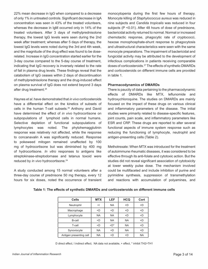
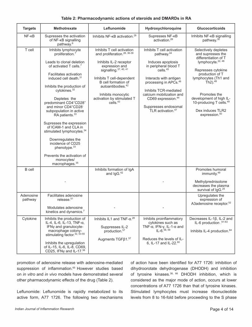
Table 1: The effects of synthetic DMARDs and corticosteroids on different immune cells
22% mean decrease in IgG when compared to a decrease of only 1% in untreated controls. Significant decrease in IgA concentration was seen in 43% of the treated volunteers, whereas the decrease in IgM occurred only in 14% of the treated volunteers. After 3 days of methylprednisolone therapy, the lowest IgG levels were seen during the 2nd week after treatment; whereas after 5 days of therapy, the lowest IgG levels were noted during the 3rd and 4th week, and the magnitude of the drug effect was found to be dose-related. Increase in IgG concentration started earlier for the 3-day course compared to the 5-day course of treatment, indicating that IgG recovery is inversely related to the rate of fall in plasma drug levels. These findings reveal that the catabolism of IgG ceases within 2 days of discontinuation of methylprednisolone therapy and the drug-induced effect on plasma survival of IgG does not extend beyond 3 days after drug treatment.25
Haynes et al. have demonstrated that in vivo corticosteroids have a differential effect on the kinetics of subsets of cells in the human T-cell subsets.26 Anthony and David have determined the effect of in vivo hydrocortisone on subpopulations of lymphoid cells in normal humans. Selective depletion of functional subpopulations of lymphocytes was noted. The phytohemagglutinin response was relatively not affected, while the response to concanavalin A was significantly reduced. Response to pokeweed mitogen remained unaffected by 100 mg of hydrocortisone but was diminished by 400 mg of hydrocortisone. In vitro responses to antigens like streptokinase-streptodornase and tetanus toxoid were reduced by in vivo hydrocortisone.24
A study conducted among 15 normal volunteers after a three-day course of prednisone 50 mg therapy, every 12 hours for six doses, noted the occurrence of transient
monocytopenia during the first few hours of therapy. Monocyte killing of Staphylococcus aureus was reduced in nine subjects and Candida tropicalis was reduced in four subjects (P <0.01). After 48 hours of dose of prednisone, bactericidal activity returned to normal. Normal or increased chemotactic response, phagocytic rate of cryptococci, hexose monophosphate-shunt response to phagocytosis and ultrastructural characteristics were seen with the same monocyte preparations. The impairment of bactericidal and fungicidal activity may be attributed to the development of infectious complications in patients receiving comparable doses of corticosteroids.27 The effects of synthetic DMARDs and corticosteroids on different immune cells are provided in table 1.
Pharmacodynamics of DMARDsThere is paucity of data pertaining to the pharmacodynamic effects of DMARDs like MTX, leflunomide and hydroxychloroquine. The studies on DMARDs are mainly focused on the impact of these drugs on various clinical and inflammatory parameters of the disease. The initial studies were primarily related to disease-specific features, joint counts, pain scale, and inflammatory parameters like ESR and CRP. These drugs are reported to alter several functional aspects of immune system response such as reducing the functioning of lymphocyte, neutrophil and antigen-presenting cells (Table 2).
Methotrexate: When MTX was introduced for the treatment of autoimmune rheumatic diseases, it was considered to be effective through its anti-folate and cytotoxic action. But the studies did not reveal significant association of cytotoxicity at lower weekly pulse dose. The mechanism involved could be multifaceted and include inhibition of purine and pyrimidine synthesis, suppression of transmethylation and reactions with accumulation of polyamines, and
Cells MTX LEF HCQ Cort Neutrophil +I NA +D +D
Macrophage +D +D +D +D
Lymphocyte NA NA +D +D
B-cell +D NA NA +D
T-cell +D +D* NA +D
Synoviocyte NA +D NA +D
Antigen presenting cell NA +D +D NA
D direct effect, I indirect effect, NA data not available, + effect, * inhibit TH2>TH1
Indian Journal of Inflammation Research Page 4 of 14
promotion of adenosine release with adenosine-mediated suppression of inflammation.65 However studies based on in vitro and in vivo models have demonstrated several other pharmacodynamic effects of the drug (Table 2).
Leflunomide: Leflunomide is rapidly metabolized to its active form, A77 1726. The following two mechanisms
of action have been identified for A77 1726: inhibition of dihydroorotate dehydrogenase (DHODH) and inhibition of tyrosine kinases.38, 66 DHODH inhibition, which is considered as the major mode of action, occurs at lower concentrations of A77 1726 than that of tyrosine kinases. Stimulated lymphocytes must increase ribonucleotide levels from 8 to 16-fold before proceeding to the S phase
Targets Methotrexate Leflunomide Hydroxychloroquine Glucocorticoids
NF-κB Supresses the activation of NF-κB signalling
pathway.9
Inhibits NF-κB activation.28 Supresses NF-κB activation.29
Inhibits NF-κB signalling pathway.30
T cell Inhibits lymphocyte proliferation.7
Leads to clonal deletion of activated T cells.7
Facilitates activation induced cell death.31
Inhibits the production of cytokines.32
Depletes the predominant CD4+CD28+
and minor CD4+CD28- subpopulation in active
RA patients.33
Supresses the expression of ICAM-1 and CLA in
stimulated lymphocytes.34
Downregulates the incidence of CD25
phenotype.33
Prevents the activation of monocytes/
macrophages.35
Inhibits T cell activation and proliferation.28, 36-39
Inhibits IL-2 receptor expression and signalling.37, 40, 41
Inhibits T cell-dependent B cell formation of autoantibodies.42
Inhibits monocytic activation by stimulated T
cells.43
Inhibits T cell activation pathway.44
Induces apoptosis in peripheral blood T
cells.45
Interacts with antigen processing in APCs.46
Inhibits TCR-mediated calcium mobilization and
CD69 expression.44
Suppresses endosomal TLR activation.47
Selectively depletes and supresses the differentiation of T lymphocyte.22, 48
Represses cytokine production of T
lymphocytes (Th1 and Th2).48
Promotes the development of high IL-10-producing T cells.49
Dex induces TLR2 expression.50
B cell
-
Inhibits formation of IgA and IgG.42
-
Promotes humoral immunity.49
Methylprednisolone decreases the plasma
survival of IgG.25
Adenosine pathway
Facilitates adenosine release.51
Modulates adenosine kinetics and dynamics.7
- -
Upregulates the expression of
A3adenosine receptor.52
Cytokine Inhibits the production of IL-4, IL-6, IL-13, TNF-α, IFNγ and granulocyte-macrophage colony-
stimulating factor.32, 53-55
Inhibits the upregulation of IL-15, IL-6, IL-8, CD69, CD25, IFNγ and IL-17.56
Inhibits IL1 and TNF-α.28
Suppresses IL-2 production.57
Augments TGFβ1.57
Inhibits proinflammatory cytokines such as
TNF-α, IFN-γ, IL-1-α and IL-6.58, 59
Reduces the levels of IL-6, IL-17 and IL-22.60
Decreases IL-1β, IL-2 and IL-6 production .61-63
Inhibits IL-4 production.64
Table 2: Pharmacodynamic actions of steroids and DMARDs in RA

Indian Journal of Inflammation Research Page 4 of 14 Page 5 of 14
Table 3: Mechanism of action of steroids and DMARDs on NF-κB, T cell and adenosine pathway
Targets Drugs Mechanism of actionNF-κB and activator
protein-1 (AP-1)
Glucocorticoids
Binds to the p65 subunit of DNA-bound NF-κB molecule and inhibits the signalling pathway.30
Decreases the activity of Akt and IκB kinase, essential for the phosphorylation
and activation of NF-κB by associating with p85α/P13K.70
Inhibits NF-κB, by interacting with PKAc, crucial for the maximum transactivation capacity of NF-κB.71, 72
Inhibits NF-κB signalling pathway by upregulating IκBα, cytoplasmic sequestration of p65, HDAC2 recruitment and interfering the phosphorylation
status of RNA polymerase II.73
Binds to c-Fos/c-Jun dimers of AP-1, thereby inhibiting their DNA binding and transactivation capacities.74
Represses AP-1 activity by inhibiting the subsequent phosphorylation of c-Jun on S63/73.73
Associates with JNK and loads inactive JNK to AP-1, while masking AP-1 from active JNK generated by MAPK activation.75
The interaction of GR with MSK1 shifts the nuclear to cytoplasmic MSK1 ratio, thereby inhibiting the NF-κB activity.76
GR dimers activate anti-inflammatory genes (Gilz, Annexin-1 and Mkp1) by binding to their respective GR response elements.77
Interacts with coactivator molecules like CBP, pCAF, or SRCs, in the nucleus and activates anti-inflammatory genes (including Slpi, Mkp1/Dusp1, IκB-α,
and Gilz).78, 79
GR dimerization inhibits JNK2, a crucial mediator of TNF-induced inflammation, through Mkp1.80
Peroxisome proliferator-activated receptor γ (PPARγ) interacts with GR and they conjointly act as immunosuppressors.75
During stressful events, GR induces c-Fos and early growth response protein (Egr-1) by increasing the phosphorylation of ERK.81
The simultaneous activation of PPARα and GR enhances transrepression of NF-κB-driven gene expression and represses cytokine production.82
Downregulates NF-κB activity and subsequent generation of cytokines (IL-1β, IL-2, IL-6), by modulating the expression of zinc transporter.61-63
Competes with NF-κB for coactivators like CBP and SRC-1, required for their induction of downstream genes/transactivation.83
The binding of GR with corepressors like GRIP1, results in the transrepression of the target proinflammatory cytokine genes of AP-1 and NF-κB.84
Methotrexate
Prevents the activation of NF-κB signalling pathway by inhibiting the degradation and suppressing the phosphorylation of IκBα.9
Supresses the expression of receptor activator of NF-κB ligand, RANKL and RANKL mRNA, thereby indirectly inhibiting osteoclast formation.85
Leflunomide A77 1726 (active metabolite of leflunomide), inhibits NF-κB activation by targeting the degradation of inhibitory protein IκBα.28
Hydroxychloroquine Hydroxychloroquine HCQ suppresses IL-1β-induced activation of NF-κB.29

Indian Journal of Inflammation Research Page 6 of 14
Targets Drugs Mechanism of actionT cell
Glucocorticoids
The binding of GCs to the GR limits the availability of unliganded GR, essential for the efficient TcR signalling and Tc R-dependent LCK/FYN activation.86
GR interacts with NF-AT (nuclear factor of activated T cells), and inhibits the 1L-4 production of T cells .64
GCs interferes with T-bet and GATA-3 and downregulates the cytokine production of Th1 and Th2 respectively.48
GR interferes with the TLR3/4 signalling cascade by competing with IRF3 for binding to GRIP1.87
Methylprednisolone facilitates the catabolism of IgG, thereby decreasing its plasma survival.25
Dex induces TLR2 expression by recruiting GR and STAT5 at the TLR2 promoter in vivo.50
Methotrexate
Prevents the de novo pyrimidine and purine synthesis, thereby inhibiting the cellular proliferation of T cells.7
MTX and/or MTX-polyglutamates inhibit enzymes such as dihydrofolate reductase, thymidylate synthase and 5-aminoimidazole-4-carboxamide ribonucleotide
transformylase.88, 89
Facilitates clonal deletion of activated T cells via ROS-dependent and mitochondria-mediated pathways.7, 90
Increases the homocysteine levels and subsequently reduces the methylation of Ras protein, thereby executing anti-proliferative effect.91
Increases CD95 sensitivity and facilitates activation-induced cell death.31
Inhibits the upregulation of IL-15, IL-6, IL-8, CD69, CD25, IFNγ and IL-17, and disrupts the interaction between synovial fibroblasts and T lymphocytes.56
Interacts with several agonists and suppress IL-6 production in osteoblastic cell lines.92
Leflunomide
Prevents T cell division primarily by inhibiting de novo pyrimidine ribonucleotide synthesis in the late G1 phase and at higher doses by inhibiting tyrosine kinases.93,
39
Non-competitively inhibits dihydroorotate dehydrogenase, thereby preventing de novo pyrimidine synthesis.42
Causes cell cycle arrest at the late G1 phase by decreasing rUMP levels and upregulating p53 and p21.94, 95
Inhibits the activities of the Src-related tyrosine kinases p56lck (reduces IL-2 production) and p59fyn, in Jurkat T cells.40
Effectively inhibits the levels of tyrosine phosphorylated proteins, in mouse leukemia cell line (LSTRA) cells, which overexpress p56lck.96
Inhibits the phosphorylation of Jak1 and Jak3 tyrosine kinases, which are necessary for IL2 receptor signalling.41
Hydroxychloroquine
Inhibits T cell stimulation by inhibiting the digestion of antigenic proteins and the assembly of peptides with the α and β chains of MHC class II proteins in APCs.46,
97
Blocks the proliferative responses of T-cell mitogens and alloantigens.44
Decreases the production of TNF-α in human macrophages, and not of monocytes and T cells.98, 99

Indian Journal of Inflammation Research Page 6 of 14 Page 7 of 14
Induces apoptosis in peripheral blood T lymphocyte through caspase cascades.45
Decreases the levels of IL-6, IL-17 and IL 22, possibly by reducing Th17 cells, through a decrease in antigen presentation.60
Inhibits TNFα, IL-1β and aPL-mediated induction of endosomal NOX, in human monocytes and MonoMac1 cells by inhibiting the translocation of gp91phox,
catalytic subunit of NOX2.100
Adenosine pathway
GlucocorticoidsGCs up-regulates the expression of A3 adenosine receptor, resulting in the activation of extracellular signal regulated kinase (ERK)1/2 phosphorylation,
thereby promoting survival of anti-inflammatory macrophages.52
Methotrexate
Increases adenosine levels by decreasing the activity of purine enzymes adenosine deaminase, purine-nucleoside phosphorylase and hypoxanthine-
guanine-phosphoribosyl transferase.101
Stimulate A2a-receptor through adenosine release, resulting in anti-inflammatory effects.102
Significantly suppresses NURR1 expression in patients with psoriatic arthritis, mediated through the adenosine receptor A2.7, 103
Reduces the adherence of T lymphocytes to synovial fibroblasts, mediated by adenosine release.56
Leflunomide ---
Hydroxychloroquine ---
from G1. Increased levels of ribonucleotides can only be met by de novo ribonucleotide synthesis. At low levels of ribonucleotides, p53, the activation of a ‘sensor’ molecule prevents progression through the cell cycle. Therefore, an inhibitor of de novo uridine monophosphate synthesis would predictably arrest stimulated cells at the G1 phase.67 Mice model studies suggest the inhibition of tyrosine kinase as the more active mechanism than dihydroorotate dehydrogenase (DHODH) inhibition.68 Inhibition of pyrimidine synthesis promotes TH2 cell activation and inhibition of TH1 cell activity.40 Its effects on different cells are summarized in table 2.
Hydroxychloroquine is considered as a potential immunomodulator interfering the antigen presentation and expression of several receptors (Table 2). The drug is effective after 6 to 8 weeks of loading dose, suggesting the cumulative effect of the drug.69 Though the exact mechanism of the drug is uncertain, clinical studies have demonstrated the advantage of combining the drug with other DMARDs. The molecular mechanism of the drug is given in table 3.
Drug interactions in pharmacodynamicsThe drugs like glucocorticoids, MTX, leflunomide and
hydroxychloroquine alter the immune system function by interfering both metabolic and secondary signal pathways, thereby altering the gene expression. The mechanism of interference in cell signaling as well as in metabolic processes of individual drug is shown in table 3. Here we have attempted to show the three major pathways namely NF-κB (Fig. 1), T cell (Fig. 2A & 2B) and adenosine pathways (Fig. 3). The figures also depict the sites of convergence of actions of different drugs. Both MTX and leflunomide act at different levels of metabolic pathways of purine and pyrimidine synthesis (Fig. 2A).104 The interactions seem to be clinically synergistic.105 The interactions of DMARDs and steroid suggest initial advantage in improving the disease control. However, with respect to improved survival, there may not be significant advantage in long term.
Combination pharmacodynamicsWith respect to the use of combination drugs, one of the major challenges confronted by clinicians is whether to introduce drugs simultaneously or in step-wise manner. In combination treatment, the doses can be altered to receive the optimal dose of each component, to retain the efficacy and to reduce the incidence of adverse events associated with the individual use. For predicting the quantitative effects of synergisms of the drugs, methods

Indian Journal of Inflammation Research Page 8 of 14
+ induction, -- / * inhibition, effect
GR binds to the p65 subunit of DNA-bound NFκB molecule and inhibits the signalling; GR associates with P13K/p85α and decreases the activity of Akt
and IκB kinase, which is essential for the phosphorylation and activation of NFκB; GR interacts with PKAc and inhibits NFκB; GR inhibits NF-κB pathway
by upregulating IκBα and by facilitating cytoplasmic sequestration of p65; GR and Lef inhibit the proteasomal degradation of phosphorylated IκBα; MTX
inhibits the degradation and suppresses the phosphorylation of IκBα; HCQ supresses IL-1β-induced activation of NF-κB.
Fig. 1: Major targets of action of steroids and DMARDs on NFκB signaling pathway

Indian Journal of Inflammation Research Page 8 of 14 Page 9 of 14
Fig. 2A: Targets of action of steroids and DMARDs on T cells
+ induction, - / * inhibition, effect
GCs inhibits TCR signalling and TCR-dependent LCK/FYN activation by binding to the free GR required for efficient signalling; GCs downregulates the
cytokine production in Th1 and Th2 cells by interfering with T-bet and GATA-3 respectively; GR inhibits IL-4 production in T cells by interacting with NF-
AT; MTX increases the homocysteine level and subsequently reduces the methylation of Ras proteins; MTX facilitates activation-induced cell death; MTX
and Lef inhibit the expression of IL-2R gene; Lef prevents IL2 receptor signalling by inhibiting the phosphorylation of JAK 1 and JAK 3 tyrosine kinases;
Lef inhibits the activity of p56lck in Jurkat T cells and also inhibits cytokines such as IL1 and TNF-α, and supresses IL2 production; HCQ inhibits TCR-
mediated calcium mobilization and induces apoptosis in peripheral blood T cells through caspase cascade; HCQ also inhibits cytokines like TNF-α,
IFN-γ, IL6 and IL-1-α.
Indian Journal of Inflammation Research Page 10 of 14
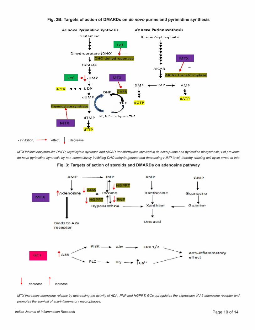
Fig. 2B: Targets of action of DMARDs on de novo purine and pyrimidine synthesis
Fig. 3: Targets of action of steroids and DMARDs on adenosine pathway
- inhibition, effect, decrease
MTX inhibits enzymes like DHFR, thymidylate synthase and AICAR transformylase involved in de novo purine and pyrimidine biosynthesis; Lef prevents
de novo pyrimidine synthesis by non-competitively inhibiting DHO dehydrogenase and decreasing rUMP level, thereby causing cell cycle arrest at late
decrease, increase
MTX increases adenosine release by decreasing the activity of ADA, PNP and HGPRT; GCs upregulates the expression of A3 adenosine receptor and
promotes the survival of anti-inflammatory macrophages.

Indian Journal of Inflammation Research Page 10 of 14 Page 11 of 14
such as combination index are used.106 The simple principle of the combination treatment is that if the drugs are competing for same pathways, the efficacy is not the sum of their individual effect and the combined use may not confer any advantage. Evaluation of the mechanisms and pharmacodynamic effects of the steroid and other DMARDs reveals several instances of overlap in gross pharmacodynamic action on lymphocyte, inhibition of NF-κB and other intervention pathways. But the sites of action in the cell signal pathways of these drugs are different with a few convergent locations (Fig. 1) . Analysis of intervention in NF-κB pathway of steroid, leflunomide and MTX shows that the step interfered by leflunomide is prior to that of MTX, whereas steroids act at multiple steps. Blocking of the downstream pathways by the drugs is expected to have different impact when compared to upstream pathway blocking. For instance, if the blocking at downstream pathway increases the number of functional receptors through counter-regulatory mechanism, higher concentrations of drugs levels are needed to prevent the repercussions. Whereas the downstream blocking reduces the number of receptors, it may enhance the response even at lower concentrations. However, the available evidence on the intervention of upstream/downstream pathways from the in vitro and in vivo studies is very limited, further research in this area is warranted to improve the use of combination and also for personalizing the DMARD and steroid prescriptions. Studies involving in silico and experimental models are necessary to clearly understand these drugs interactions, which may facilitate the improvement of treatment strategies and customization of treatment approaches.
AbbreviationsMTX, methotrexate; LEF/Lef, leflunomide; HCQ, hydroxychloroquine; Cort, corticosteroids; GR, glucocorticoid receptor; GC, glucocorticoids; Dex, Dexamethasone; PKAc, catalytic subunit of protein kinase A; Th 1, type 1 T helper cells; Th 2, type 2 T helper cells; TCR, T cells receptor; DHFR, dihydrofolate reductase; AICAR transformylase, 5-aminoimidazole-4-carboxamide ribonucleotide transformylase; dTTP, deoxythymidine triphosphate; dCTP, deoxycytidine triphosphate; dGTP, deoxyguanosine triphosphate; dATP, deoxyadenosine triphosphate; rUMP, ribonucleotide uridine monophosphate; ADA, adenosine deaminase; HGPRT, hypoxanthine-guanine phosphoribosyltransferase; PNP, purine nucleoside phosphorylase; A3R, adenosine 3 receptor; A2a receptor, adenosine 2a receptor.
Competing interestsThe author declares that he has no competing interests.
CitationChandrashekara S. Mechanism-based pharmacodynamic interactions of glucocorticoids and disease-modifying antirheumatic drugs: A review. IJIR.
2018;(2)1:RA1.
Submitted: 14 June 2018, Accepted: 17 September 2018, Published: 26 September 2018
*Correspondence: Dr. Chandrashekara [email protected]
References1. Mikuls TR, Fay BT, Michaud K, et al. Associations of disease
activity and treatments with mortality in men with rheumatoid arthritis: results from the VARA registry. Rheumatology (Oxford). 2011;50(1):101–9.
2. Caplan L, Wolfe F, Russell AS, et al. Corticosteroid use in rheumatoid arthritis: prevalence, predictors, correlates, and outcomes. J Rheumatol. 2007;34(4):696–705.
3. del Rincón I, Battafarano DF, Restrepo JF, et al. Glucocorticoid dose thresholds associated with all-cause and cardiovascular mortality in rheumatoid arthritis. Arthritis Rheumatol. 2014;66(2):264–72.
4. Wasko MC, Dasgupta A, Ilse Sears G, et al. Prednisone use and risk of mortality in patients with rheumatoid arthritis: moderation by use of disease-modifying antirheumatic drugs. Arthritis Care Res (Hoboken). 2016;68(5):706–10.
5. Chandrashekara S. Pharmacokinetic consideration of synthetic DMARDs in rheumatoid arthritis. Expert Opin Drug Metab Toxicol. 2013;9(8):969–81.
6. Honoré PM, Jacobs R, De Waele E, et al. What do we know about steroids metabolism and “PK/PD approach” in AKI and CKD especially while on RRT--current status in 2014. Blood Purif. 2014;38(2):154–7.
7. Wessels J a. M, Huizinga TWJ, Guchelaar H-J. Recent insights in the pharmacological actions of methotrexate in the treatment of rheumatoid arthritis. Rheumatology (Oxford). 2008;47(3):249–55.
8. Chikanza IC. Mechanisms of Corticosteroid Resistance in Rheumatoid Arthritis. Ann N Y Acad Sci. 966(1):39–48.
9. Majumdar S, Aggarwal BB. Methotrexate Suppresses NF-κB Activation Through Inhibition of IκBα Phosphorylation and Degradation. J Immunol. 2001;167(5):2911–20.
10. Hedman LA, Röckert L-L, Lundin PM. The effect of steroids on the circulating lymphocyte population — VI. Studies of the thoracic duct T- and B-lymphocyte populations after neonatal thymectomy and prednisolone treatment. An immunofluorescence. In J Immunopharmacol. 1984;6(4):357–63.
11. Galon J, Franchimont D, Hiroi N, et al. Gene profiling reveals unknown enhancing and suppressive actions of glucocorticoids on immune cells. FASEB J. 2002;16(1):61–71.
12. Soyka LF. Treatment of the Nephrotic Syndrome in Childhood: Use of an Alternate-Day Prednisone Regimen. Am J Dis Child. 1967;113(6):693–701.
13. Portner MM, Thayer KH, Harter JG, et al. Successful initiation of alternate-day prednisone in chronic steroid dependent asthmatic patients. J Allergy Clin Immunol. 1972;49(1):16-26.
14. Reed W, Lucas ZJ, Cohn R. Alternate-day prednisone therapy after renal transplantation. Lancet. 1970;295(7650):747–9.
15. McEnery PT, Gonzalez LL, Martin LW, et al. Growth and development of children with renal transplants. Use of alternate-day steroid therapy. J Pediatr 1973;83(5):806–14.
16. Cocco AE, Mendeloff Al. An evaluation of intermittent corticosteroid therapy in the management of ulcerative colitis. Johns Hopkins Med J. 1967;120(3):162-9.
17. Hess EV, Goldman JA: Corticosteroids and corticotropin in therapy of rheumatoid arthritis. In: Hollander JL, Mccarty DJ: Arthritis and

Indian Journal of Inflammation Research Page 12 of 14
Allied conditions. 8th ed. Philadelphia, Lea & Febiger, 1972.18. Harter JG, Reddy WJ, Thorn GW. Studies on an intermittent
corticosteroid dosage regimen. N Engl J Med. 1963;269(12):591-6.19. MacGregor RR, Sheagren JN, Lipsett MB, et al. Alternate-day
prednisone therapy. Evaluation of delayed hypersensitivity responses, control of disease and steroid side effects. N Engl J Med. 1969;280(26):1427–31.
20. Cupps TR, Fauci AS. Corticosteroid-mediated immunoregulation in man. Immunol Rev. 1982;65:133–55.
21. Nara PL, Krakowka S, Poweres TE. Effects of prednisolone on the development of immune responses to canine distemper virus in beagle pups. Am J Vet Res. 1979;40(12):1742-7.
22. Thorne GW, Forsham PH, Prunty FTG, et al. A test for adrenal corticle insufficiency. JAMA. 1948;137:1005-9.
23. Cohn LA. The influence of corticosteroids on host defense mechanisms. J Vet Intern Med. 1991;5(2):95–104.
24. Fauci AS, Dale DC. The effect of in vivo hydrocortisone on subpopulations of human lymphocytes. J Clin Invest. 1974;53(1):240–6.
25. Butler WT, Rossen RD. Effects of corticosteroids on immunity in man. I. Decreased serum IgG concentration caused by 3 or 5 days of high doses of methylprednisolone. J Clin Invest. 1973;52(10):2629–40.
26. Haynes BF, Fauci AS. The Differential Effect of In Vivo Hydrocortisone on the Kinetics of Subpopulations of Human Peripheral Blood Thymus-Derived Lymphocytes. J Clin Invest. 1978;61(3):703–7.
27. Rinehart JJ, Sagone AL, Balcerzak SP, et al. Effects of Corticosteroid Therapy on Human Monocyte Function. N Engl J Med. 1975;292(5):236–41.
28. Breedveld FC, Dayer JM. Leflunomide: mode of action in the treatment of rheumatoid arthritis. Ann Rheum Dis. 2000;59(11):841–9.
29. Vuolteenaho K, Kujala P, Moilanen T, et al. Aurothiomalate and hydroxychloroquine inhibit nitric oxide production in chondrocytes and in human osteoarthritic cartilage. Scand J Rheumatol. 2005;34(6):475–9.
30. Ray A, Prefontaine KE. Physical association and functional antagonism between the p65 subunit of transcription factor NF-kappa B and the glucocorticoid receptor. Proc Natl Acad Sci U S A. 1994;91(2):752–6.
31. Strauss G, Osen W, Debatin KM. Induction of apoptosis and modulation of activation and effector function in T cells by immunosuppressive drugs. Clin Exp Immunol. 2002;128(2):255–66.
32. Gerards AH, de Lathouder S, de Groot ER, et al. Inhibition of cytokine production by methotrexate. Studies in healthy volunteers and patients with rheumatoid arthritis. Rheumatology (Oxford). 2003;42(10):1189–96.
33. Herman S, Zurgil N, Langevitz P, et al. The immunosuppressive effect of methotrexate in active rheumatoid arthritis patients vs. its stimulatory effect in nonactive patients, as indicated by cytometric measurements of CD4+ T cell subpopulations. Immunol Invest. 2004;33(3):351–62.
34. Johnston A, Gudjonsson JE, Sigmundsdottir H, et al. The anti-inflammatory action of methotrexate is not mediated by lymphocyte apoptosis, but by the suppression of activation and adhesion molecules. Clin Immunol. 2005;114(2):154–63.
35. Wijngaarden S, van Roon JA, van de Winkel JG, et al. Down-regulation of activating Fcgamma receptors on monocytes of patients with rheumatoid arthritis upon methotrexate treatment. Rheumatology (Oxford). 2005;44(6):729–34.
36. Cherwinski HM, McCarley D, Schatzman R, et al. The immunosuppressant leflunomide inhibits lymphocyte progression
through cell cycle by a novel mechanism. J Pharmacol Exp Ther. 1995;272(1):460–8.
37. Bartlett RR, Anagnostopulos H, Zielinski T, et al. Effects of leflunomide on immune responses and models of inflammation. Springer Semin Immunopathol. 1993;14(4):381–394.
38. Cherwinski HM, Cohn RG, Cheung P, et al. The immunosuppressant leflunomide inhibits lymphocyte proliferation by inhibiting pyrimidine biosynthesis. J Pharmacol Exp Ther. 1995;275(2):1043–9.
39. Cherwinski HM, Byars N, Ballaron SJ, et al. Leflunomide interferes with pyrimidine nucleotide biosynthesis. Inflamm Res. 1995;44(8):317–322.
40. Xu X, Williams JW, Bremer EG, et al. Inhibition of protein tyrosine phosphorylation in T cells by a novel immunosuppressive agent, leflunomide. J Biol Chem. 1995;270(21):12398–403.
41. Elder RT, Xu X, Williams JW, et al. The immunosuppressive metabolite of leflunomide, A77 1726, affects murine T cells through two biochemical mechanisms. J Immunol. 1997;159(1):22–7.
42. Siemasko KF, Chong AS, Williams JW, et al. Regulation of B cell function by the immunosuppressive agent leflunomide. Transplantation. 1996;61(4):635–42.
43. Deage V, Burger D, Dayer J-M. Exposure of T lymphocytes to leflunomide but not to dexamethasone favors the production by monocytic cells of interleukin-1 receptor antagonist and the tissue-inhibitor of metalloproteinases-1 over that of interleukin-1 beta and metalloproteinases. Eur Cytokine Netw. 1998;9(4):663–8.
44. Goldman FD, Gilman AL, Hollenback C, et al. Hydroxychloroquine inhibits calcium signals in T cells: a new mechanism to explain its immunomodulatory properties. Blood. 2000;95(11):3460–6.
45. Meng XW, Feller JM, Ziegler JB, et al. Induction of apoptosis in peripheral blood lymphocytes following treatment in vitro with hydroxychloroquine. Arthritis Rheum. 1997;40(5):927–35.
46. Fox RI. Mechanism of action of hydroxychloroquine as an antirheumatic drug. Semin Arthritis Rheum. 1993;23(2, Supplement 1):82–91.
47. Kuznik A, Bencina M, Svajger U, et al. Mechanism of endosomal TLR inhibition by antimalarial drugs and imidazoquinolines. J Immunol. 2011;186(8):4794–804.
48. Liberman AC, Druker J, Refojo D, et al. Glucocorticoids inhibit GATA-3 phosphorylation and activity in T cells. FASEB J. 2009;23(5):1558–71.
49. Franchimont D. Overview of the actions of glucocorticoids on the immune response: a good model to characterize new pathways of immunosuppression for new treatment strategies. Ann N Y Acad Sci. 2004;1024(1):124–37.
50. Hermoso MA, Matsuguchi T, Smoak K, et al. Glucocorticoids and tumor necrosis factor alpha cooperatively regulate Toll-like receptor 2 gene expression. Mol Cell Biol. 2004;24(11):4743–756.
51. van Ede AE, Laan RF, De Abreu RA, et al. Purine enzymes in patients with rheumatoid arthritis treated with methotrexate. Ann Rheum Dis. 2002;61(12):1060–4.
52. Barczyk K, Ehrchen J, Tenbrock K, et al. Glucocorticoids promote survival of anti-inflammatory macrophages via stimulation of adenosine receptor A3. Blood. 2010;116(3):446–55.
53. de Lathouder S, Gerards AH, Dijkmans BA et al. Two inhibitors of DNA synthesis lead to inhibition of cytokine production via a different mechanism. Nucleosides Nucleotides Nucleic Acids. 2004;23(8-9):1089–100.
54. Kraan M, Smeets T, van Loon MJ, et al. Differential effects of leflunomide and methotrexate on cytokine production in rheumatoid arthritis. Ann Rheum Dis. 2004;63(9):1056–61.
55. de Lathouder S, Gerards AH, de Groot ER, et al. Mycophenolic acid and methotrexate inhibit lymphocyte cytokine production via

Indian Journal of Inflammation Research Page 12 of 14 Page 13 of 14
different mechanisms. Eur Cytokine Netw. 2002;13(3):317–23.56. Miranda-Carus ME, Balsa A, Benito-Miguel M, et al. IL-15 and the
initiation of cell contact-dependent synovial fibroblast-T lymphocyte cross-talk in rheumatoid arthritis: effect of methotrexate. J Immunol. 2004;173(2):1463–76.
57. Cao WW, Kao PN, Aoki Y, et al. A novel mechanism of action of the immunomodulatory drug, leflunomide: augmentation of the immunosuppressive cytokine, TGF-beta 1, and suppression of the immunostimulatory cytokine, IL-2. Transplant Proc. 1996;28(6):3079–80.
58. van den Borne BE, Dijkmans BA, de Rooij HH, et al. Chloroquine and hydroxychloroquine equally affect tumor necrosis factor-alpha, interleukin 6, and interferon-gamma production by peripheral blood mononuclear cells. J Rheumatol. 1997;24(1):55–60.
59. Jeong JY, Choi JW, Jeon KI, et al. Chloroquine decreases cell-surface expression of tumour necrosis factor receptors in human histiocytic U-937 cells. Immunology. 2002;105(1):83–91.
60. da Silva JC, Mariz HA, da Rocha Júnior LF, et al. Hydroxychloroquine decreases Th17-related cytokines in systemic lupus erythematosus and rheumatoid arthritis patients. Clinics (Sao Paulo). 2013;68(6):766–71.
61. Guo L, Lichten LA, Ryu MS, et al. STAT5-glucocorticoid receptor interaction and MTF-1 regulate the expression of ZnT2 (Slc30a2) in pancreatic acinar cells. Proc Natl Acad Sci U S A. 2010;107(7):2818–23.
62. Foster M, Samman S. Zinc and regulation of inflammatory cytokines: implications for cardiometabolic disease. Nutrients. 2012;4(7):676–94.
63. Liu MJ, Bao S, Galvez-Peralta M, et al. ZIP8 regulates host defense through zinc-mediated inhibition of NF-κB. Cell Rep. 2013;3(2):386–400.
64. Chen R, Burke TF, Cumberland JE, et al. Glucocorticoids inhibit calcium- and calcineurin-dependent activation of the human IL-4 promoter. J Immunol. 2000;164(2):825–832.
65. Tian H, Cronstein BN. Understanding the mechanisms of action of methotrexate: implications for the treatment of rheumatoid arthritis. Bull NYU Hosp Jt Dis 2007;65(3):168–73.
66. Davis JP, Cain GA, Pitts WJ, Magolda RL, Copeland RA. The immunosuppressive metabolite of leflunomide is a potent inhibitor of human dihydroorotate dehydrogenase. Biochemistry. 1996;35(4):1270–3.
67. Fox RI. Mechanism of action of leflunomide in rheumatoid arthritis. J Rheumatol Suppl. 1998;53:20–6.
68. Xu X, Blinder L, Shen J, Gong H, Finnegan A, Williams JW, et al. In vivo mechanism by which leflunomide controls lymphoproliferative and autoimmune disease in MRL/MpJ-lpr/lpr mice. J Immunol. 1997;159(1):167–74.
69. Furst DE, Lindsley H, Baethge B, Botstein GR, Caldwell J, Dietz F, et al. Dose-loading with hydroxychloroquine improves the rate of response in early, active rheumatoid arthritis: a randomized, double-blind six-week trial with eighteen-week extension. Arthritis Rheum. 1999;42(2):357–65.
70. Leis H, Page A, Ramirez A, et al. Glucocorticoid receptor counteracts tumorigenic activity of Akt in skin through interference with the phosphatidylinositol 3-kinase signaling pathway. Mol Endocrinol. 2004;18(2):303–11.
71. Haske T, Nakao M, Moudgil VK. Phosphorylation of immunopurified rat liver glucocorticoid receptor by the catalytic subunit of cAMP-dependent protein kinase. Mol Cell Biochem. 1994;132(2):163–71.
72. Doucas V, Shi Y, Miyamoto S, et al. Cytoplasmic catalytic subunit of protein kinase A mediates cross-repression by NF-κB and the glucocorticoid receptor. Proc Natl Acad Sci U S A. 2000;97(22):11893–8.
73. Petta I, Dejager L, Ballegeer M. The Interactome of the Glucocorticoid Receptor and Its Influence on the Actions of Glucocorticoids in Combatting Inflammatory and Infectious Diseases. Microbiol Mol Biol Rev. 2016;80(2):495–522.
74. Herdegen T, Leah JD. Inducible and constitutive transcription factors in the mammalian nervous system: control of gene expression by Jun, Fos and Krox, and CREB/ATF proteins. Brain Res Brain Res Rev. 1998;28(3):370–490.
75. Bruna A, Nicolas M, Munoz A, et al. Glucocorticoid receptor-JNK interaction mediates inhibition of the JNK pathway by glucocorticoids. EMBO J. 2003;22(22):6035–44.
76. Beck IM, Berghe WV, Vermeulen L, et al. Altered subcellular distribution of MSK1 induced by glucocorticoids contributes to NF-κB inhibition. EMBO J. 2008;27(12):1682–93.
77. Vandevyver S, Dejager L, Tuckermann J, et al. New insights into the anti-inflammatory mechanisms of glucocorticoids: an emerging role for glucocorticoid-receptor-mediated transactivation. Endocrinology. 2013;154(3):993–1007.
78. Barnes PJ. Corticosteroid effects on cell signalling. Eur Respir J. 2006;27(2):413–26.
79. Li X, Wong J, Tsai SY, et al. Progesterone and glucocorticoid receptors recruit distinct coactivator complexes and promote distinct patterns of local chromatin modification. Mol Cell Biol. 2003;23(11):3763–73.
80. Vandevyver S, Dejager L, Van Bogaert T, et al. Glucocorticoid receptor dimerization induces MKP1 to protect against TNF-induced inflammation. J Clin Invest. 2012;122(6):2130–40.
81. Gutierrez-Mecinas M, Trollope AF, Collins A, et al. Long-lasting behavioral responses to stress involve a direct interaction of glucocorticoid receptors with ERK1/2-MSK1-Elk-1 signaling. Proc Natl Acad Sci U S A. 2011;108(33):13806–11.
82. Bougarne N, Paumelle R, Caron S, et al. PPARalpha blocks glucocorticoid receptor alpha-mediated transactivation but cooperates with the activated glucocorticoid receptor alpha for transrepression on NF-kappaB. Proc Natl Acad Sci U S A. 2009;106(18):7397–402.
83. Sheppard KA, Phelps KM, Williams AJ, et al. Nuclear integration of glucocorticoid receptor and nuclear factor-kappaB signaling by CREB-binding protein and steroid receptor coactivator-1. J Biol Chem. 1998;273(45):29291–4.
84. Gupte R, Muse GW, Chinenov Y, et al. Glucocorticoid receptor represses proinflammatory genes at distinct steps of the transcription cycle. Proc Natl Acad Sci U S A. 2013;110(36):14616–21.
85. Lee CK, Lee EY, Chung SM, et al. Effects of disease-modifying antirheumatic drugs and antiinflammatory cytokines on human osteoclastogenesis through interaction with receptor activator of nuclear factor kappaB, osteoprotegerin, and receptor activator of nuclear factor kappaB ligand. Arthritis Rheum. 2004;50(12):3831–43.
86. Lowenberg M, Verhaar AP, van den Brink GR, et al. Glucocorticoid signaling: a nongenomic mechanism for T-cell immunosuppression. Trends Mol Med. 2007;13(4):158–63.
87. Reily MM, Pantoja C, Hu X, et al. The GRIP1:IRF3 interaction as a target for glucocorticoid receptor-mediated immunosuppression. EMBO J. 2006;25(1):108–117.
88. Ranganathan P, McLeod HL. Methotrexate pharmacogenetics: the first step toward individualized therapy in rheumatoid arthritis. Arthritis Rheum 2006;54(5):1366–77.
89. Kremer JM. Toward a better understanding of methotrexate. Arthritis Rheum. 2004;50(5):1370–82.
90. Herman S, Zurgil N, Deutsch M. Low dose methotrexate induces apoptosis with reactive oxygen species involvement in T

Indian Journal of Inflammation Research Page 14 of 14
lymphocytic cell lines to a greater extent than in monocytic lines. Inflamm Res. 2005;54(7):273–80.
91. Winter-Vann AM, Kamen BA, Bergo MO, et al. Targeting Ras signaling through inhibition of carboxyl methylation: an unexpected property of methotrexate. Proc Natl Acad Sci USA. 2003;100(11):6529–34.
92. Giacomelli R, Cipriani P, Cerinic MM, et al. Combination therapy with cyclosporine and methotrexate in patients with early rheumatoid arthritis soon inhibits TNFalpha production without decreasing TNF alpha mRNA levels. An in vivo and in vitro study. Clin Exp Rheumatol. 2002;20(3):365–72.
93. Sawah S Al, Zhang X, Zhu B, et al. Effect of corticosteroid use by dose on the risk of developing organ damage over time in systemic lupus erythematosus-the Hopkins Lupus Cohort. Lupus Sci Med. 2015;2(1):e000066.
94. Greene S, Watanabe K, Braatz-Trulson J, et al. Inhibition of dihydroorotate dehydrogenase by the immunosuppressive agent leflunomide. Biochem Pharmacol. 1995;50(6):861–7.
95. Herrmann M, Frangou CG, Kirschbaum B. Cell cycle control of the de novo pyrimidine synthesis inhibitor leflunomide through the p53 and p21WAF-1 pathways. Arthritis Rheum. 1997;40(9):S177-S177.
96. Xu X, Williams JW, Gong H, et al. Two activities of the immunosuppresive metabolite of leflunomide, A77 1726: inhibition of pyrimidine nucleotide synthesis and protein tyrosine phosphorylation. Biochem Pharmacol. 1996;52(4):527–34.
97. Poole B, Ohkuma S. Effect of weak bases on the intralysosomal pH in mouse peritoneal macrophages. J Cell Biol. 1981;90(3):665-9.
98. Sperber K, Quraishi H, Kalb TH, et al. Selective regulation of cytokine secretion by hydroxychloroquine: Inhibition of interleukin 1 alpha (IL-1-alpha) and IL-6 in human monocytes and T cells. J
Rheumatol. 1993;20(5):803-8. 99. Picot S, Peyton F, Vuillez JP. Chloroquine inhibits tumor necrosis
factor production by human macrophages in vitro. J Infect Dis. 1991;164(4):830.
100. Müller-Calleja N, Manukyan D, Canisius A, et al. Hydroxychloroquine inhibits proinflammatory signalling pathways by targeting endosomal NADPH oxidase. Ann Rheum Dis. 2017;76(5):891–7.
101. Hasko G, Cronstein BN. Adenosine: an endogenous regulator of innate immunity. Trends Immunol. 2004;25(1):33–9.
102. Montesinos MC, Takedachi M, Thompson LF, et al. The antiinflammatory mechanism of methotrexate depends on extracellular conversion of adenine nucleotides to adenosine by ecto-5’-nucleotidase: findings in a study of ecto-5’-nucleotidase gene-deficient mice. Arthritis Rheum. 2007;56(5):1440–5.
103. Ralph JA, McEvoy AN, Kane D, et al. Modulation of orphan nuclear receptor NURR1 expression by methotrexate in human inflammatory joint disease involves adenosine A2A receptor-mediated responses. J Immunol. 2005;175(1):555–65.
104. Kremer JM. Methotrexate and leflunomide: Biochemical basis for combination therapy in the treatment of rheumatoid arthritis. Semin Arthritis Rheum. 1999;29(1):14–26.
105. Weinblatt ME, Kremer JM, Coblyn JS, et al. Pharmacokinetics, safety, and efficacy of combination treatment with methotrexate and leflunomide in patients with active rheumatoid arthritis. Arthritis Rheum. 1999;42(7):1322–8.
106. Chou TC. Theoretical Basis, Experimental Design, and Computerized Simulation of Synergism and Antagonism in Drug Combination Studies. Pharmacol Rev. 2006;58 (3):621-81.