Measuring Bohler’s Angle with Oblique Lateral Radiographs ... · Measuring Bohler’s Angle with...
6
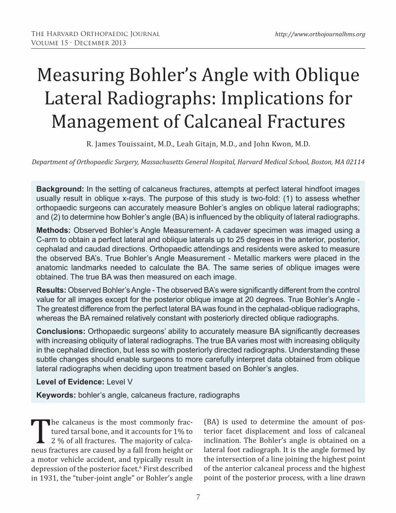
7 The Harvard Orthopaedic Journal Volume 15 · December 2013 http://www.orthojournalhms.org Measuring Bohler’s Angle with Oblique Lateral Radiographs: Implications for Management of Calcaneal Fractures R. James Touissaint, M.D., Leah Gitajn, M.D., and John Kwon, M.D. Department of Orthopaedic Surgery, Massachusetts General Hospital, Harvard Medical School, Boston, MA 02114 Background: In the setting of calcaneus fractures, attempts at perfect lateral hindfoot images usually result in oblique x-rays. The purpose of this study is two-fold: (1) to assess whether orthopaedic surgeons can accurately measure Bohler’s angles on oblique lateral radiographs; and (2) to determine how Bohler’s angle (BA) is influenced by the obliquity of lateral radiographs. Methods: Observed Bohler’s Angle Measurement- A cadaver specimen was imaged using a C-arm to obtain a perfect lateral and oblique laterals up to 25 degrees in the anterior, posterior, cephalad and caudad directions. Orthopaedic attendings and residents were asked to measure the observed BA’s. True Bohler’s Angle Measurement - Metallic markers were placed in the anatomic landmarks needed to calculate the BA. The same series of oblique images were obtained. The true BA was then measured on each image. Results: Observed Bohler’s Angle - The observed BA’s were significantly different from the control value for all images except for the posterior oblique image at 20 degrees. True Bohler’s Angle - The greatest difference from the perfect lateral BA was found in the cephalad-oblique radiographs, whereas the BA remained relatively constant with posteriorly directed oblique radiographs. Conclusions: Orthopaedic surgeons’ ability to accurately measure BA significantly decreases with increasing obliquity of lateral radiographs. The true BA varies most with increasing obliquity in the cephalad direction, but less so with posteriorly directed radiographs. Understanding these subtle changes should enable surgeons to more carefully interpret data obtained from oblique lateral radiographs when deciding upon treatment based on Bohler’s angles. Level of Evidence: Level V Keywords: bohler’s angle, calcaneus fracture, radiographs T he calcaneus is the most commonly frac- tured tarsal bone, and it accounts for 1% to 2 % of all fractures. The majority of calca- neus fractures are caused by a fall from height or a motor vehicle accident, and typically result in depression of the posterior facet. 6 First described in 1931, the “tuber-joint angle” or Bohler’s angle (BA) is used to determine the amount of pos- terior facet displacement and loss of calcaneal inclination. The Bohler’s angle is obtained on a lateral foot radiograph. It is the angle formed by the intersection of a line joining the highest point of the anterior calcaneal process and the highest point of the posterior process, with a line drawn
Transcript of Measuring Bohler’s Angle with Oblique Lateral Radiographs ... · Measuring Bohler’s Angle with...
7
The Harvard Orthopaedic JournalVolume 15 · December 2013
http://www.orthojournalhms.org
Measuring Bohler’s Angle with Oblique Lateral Radiographs: Implications for Management of Calcaneal Fractures
R. James Touissaint, M.D., Leah Gitajn, M.D., and John Kwon, M.D. Department of Orthopaedic Surgery, Massachusetts General Hospital, Harvard Medical School, Boston, MA 02114
Background: In the setting of calcaneus fractures, attempts at perfect lateral hindfoot images usually result in oblique x-rays. The purpose of this study is two-fold: (1) to assess whether orthopaedic surgeons can accurately measure Bohler’s angles on oblique lateral radiographs; and (2) to determine how Bohler’s angle (BA) is influenced by the obliquity of lateral radiographs.
Methods: Observed Bohler’s Angle Measurement- A cadaver specimen was imaged using a C-arm to obtain a perfect lateral and oblique laterals up to 25 degrees in the anterior, posterior, cephalad and caudad directions. Orthopaedic attendings and residents were asked to measure the observed BA’s. True Bohler’s Angle Measurement - Metallic markers were placed in the anatomic landmarks needed to calculate the BA. The same series of oblique images were obtained. The true BA was then measured on each image.
Results: Observed Bohler’s Angle - The observed BA’s were significantly different from the control value for all images except for the posterior oblique image at 20 degrees. True Bohler’s Angle - The greatest difference from the perfect lateral BA was found in the cephalad-oblique radiographs, whereas the BA remained relatively constant with posteriorly directed oblique radiographs.
Conclusions: Orthopaedic surgeons’ ability to accurately measure BA significantly decreases with increasing obliquity of lateral radiographs. The true BA varies most with increasing obliquity in the cephalad direction, but less so with posteriorly directed radiographs. Understanding these subtle changes should enable surgeons to more carefully interpret data obtained from oblique lateral radiographs when deciding upon treatment based on Bohler’s angles.
Level of Evidence: Level V
Keywords: bohler’s angle, calcaneus fracture, radiographs
T he calcaneus is the most commonly frac-tured tarsal bone, and it accounts for 1% to 2 % of all fractures. The majority of calca-
neus fractures are caused by a fall from height or a motor vehicle accident, and typically result in depression of the posterior facet.6 First described in 1931, the “tuber-joint angle” or Bohler’s angle
(BA) is used to determine the amount of pos-terior facet displacement and loss of calcaneal inclination. The Bohler’s angle is obtained on a lateral foot radiograph. It is the angle formed by the intersection of a line joining the highest point of the anterior calcaneal process and the highest point of the posterior process, with a line drawn
8
The Harvard Orthopaedic JournalVolume 15 · December 2013
http://www.orthojournalhms.org
projecting from the superior posterior calcaneal tuberosity.2 It is widely accepted that in the unin-jured adult population, a normal Bohler’s angle is between 25 and 40 degrees.2, 1 However, numer-ous papers suggest that there is variation among populations, with a range of values between 14 degrees in Malawians and up to 50 degrees in Ugandan subjects.15, 8, 14, 7, 13
Bohler’s angle has been shown to have prognos-tic value in determining morbidity and outcomes following calcaneus fractures.4 This radiograph-ic parameter is often used to guide treatment, and the need for obtaining further imaging such as computed tomography (CT) scans. Unfortu-nately, lateral radiographs obtained in the trau-ma setting are often oblique due to difficulties in positioning the traumatized extremity, or due to limited positioning secondary to splint materials. These detail of these radiographs may influence the accuracy of the measured Bohler’s angle, as skewed views can lead to more bony overlap that blurs the proper anatomic landmarks. Inaccurate assessment of Bohler’s angles may lead to under- or overtreatment of patients with intra-articular calcaneus fractures.
The purpose of this study is two-fold: (1) to assess whether orthopaedic surgeons can accu-rately measure Bohler’s angles on oblique lateral radiographs; and (2) to determine how Bohler’s angle, as measured using consistent anatomic landmarks, is influenced by the obliquity of the lateral radiograph. We hypothesize that ortho-paedic surgeons will inaccurately measure Bohler’s angle on oblique lateral x-rays. We also hypothesize that the true Bohler’s angle will vary based on the magnitude of the obliquity of the lateral image.
Materials and MethodsObserved Bohler’s Angles
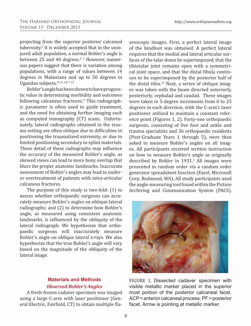
A fresh-frozen cadaver specimen was imaged using a large C-arm with laser positioner (Gen-eral Electric, Fairfield, CT) to obtain multiple flu-
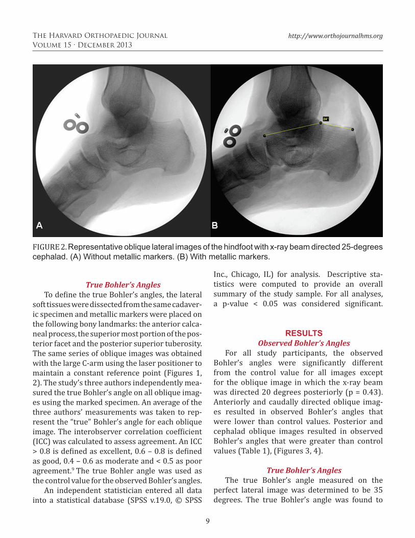
oroscopic images. First, a perfect lateral image of the hindfoot was obtained. A perfect lateral requires that the medial and lateral articular sur-faces of the talar dome be superimposed, that the tibiotalar joint remains open with a symmetri-cal joint space, and that the distal fibula contin-ues to be superimposed by the posterior half of the distal tibia.12 Next, a series of oblique imag-es was taken with the beam directed anteriorly, posteriorly, cephalad and caudad. These images were taken in 5-degree increments from 0 to 25 degrees in each direction, with the C-arm’s laser positioner utilized to maintain a constant refer-ence point (Figures 1, 2). Forty-one orthopaedic surgeons, consisting of five foot and ankle and trauma specialists and 36 orthopaedic residents (Post-Graduate Years 1 through 5), were then asked to measure Bohler’s angles on all imag-es. All participants received written instruction on how to measure Bohler’s angle as originally described by Bohler in 1931.2 All images were presented in random order via a random order generator spreadsheet function (Excel, Microsoft Corp, Redmond, WA). All study participants used the angle-measuring tool found within the Picture Archiving and Communication System (PACS).
FIGURE 1. Dissected cadaver specimen with visible metallic marker placed in the superior most portion of the posterior calcaneal facet. ACP = anterior calcaneal process. PF = posterior facet. Arrow is pointing at metallic marker.
9
The Harvard Orthopaedic JournalVolume 15 · December 2013
http://www.orthojournalhms.org
True Bohler’s AnglesTo define the true Bohler’s angles, the lateral
soft tissues were dissected from the same cadaver-ic specimen and metallic markers were placed on the following bony landmarks: the anterior calca-neal process, the superior most portion of the pos-terior facet and the posterior superior tuberosity. The same series of oblique images was obtained with the large C-arm using the laser positioner to maintain a constant reference point (Figures 1, 2). The study’s three authors independently mea-sured the true Bohler’s angle on all oblique imag-es using the marked specimen. An average of the three authors’ measurements was taken to rep-resent the “true” Bohler’s angle for each oblique image. The interobserver correlation coefficient (ICC) was calculated to assess agreement. An ICC > 0.8 is defined as excellent, 0.6 – 0.8 is defined as good, 0.4 – 0.6 as moderate and < 0.5 as poor agreement.9 The true Bohler angle was used as the control value for the observed Bohler’s angles.
An independent statistician entered all data into a statistical database (SPSS v.19.0, © SPSS
Inc., Chicago, IL) for analysis. Descriptive sta-tistics were computed to provide an overall summary of the study sample. For all analyses, a p-value < 0.05 was considered significant.
RESULTSObserved Bohler’s Angles
For all study participants, the observed Bohler’s angles were significantly different from the control value for all images except for the oblique image in which the x-ray beam was directed 20 degrees posteriorly (p = 0.43). Anteriorly and caudally directed oblique imag-es resulted in observed Bohler’s angles that were lower than control values. Posterior and cephalad oblique images resulted in observed Bohler’s angles that were greater than control values (Table 1), (Figures 3, 4).
True Bohler’s AnglesThe true Bohler’s angle measured on the
perfect lateral image was determined to be 35 degrees. The true Bohler’s angle was found to
FIGURE 2. Representative oblique lateral images of the hindfoot with x-ray beam directed 25-degrees cephalad. (A) Without metallic markers. (B) With metallic markers.

10
The Harvard Orthopaedic JournalVolume 15 · December 2013
http://www.orthojournalhms.org
TABLE 1. Observed and true Bohler’s angles
Image Obliquity True Bohler’s Angles Observed Bohler’s Angles SD p-ValuePerfect Lateral 35 37 5.1 p = 0.007Anterior 5 36 35 4.5 p = 0.037Anterior 10 37 32 6.6 p < 0.001Anterior 15 38 30 7.2 p < 0.001Anterior 20 39 30 8.5 p < 0.001Anterior 25 40 31 3.9 p < 0.001Caudad 5 37 34 3.0 p < 0.001Caudad 10 38 33 3.3 p < 0.001Caudad 15 38 34 2.8 p < 0.001Caudad 20 39 33 4.3 p < 0.001Caudad 25 39 34 3.0 p < 0.001Cepahald 5 34 37 6.5 p = 0.039Cephalad 10 32 36 4.4 p < 0.001Cephalad 15 29 32 5.9 p < 0.001Cephalad 20 27 36 6.0 p < 0.001Cephalad 25 24 35 5.0 p < 0.001Posterior 5 35 41 5.9 p < 0.001Posterior 10 34 36 4.7 p = 0.004Posterior 15 35 37 3.6 p = 0.002Posterior 20 37 37 3.9 p = 0.43Posterior 25 37 39 4.1 p < 0.001
p-Values < 0.05 are statistically significant. Image obliquity, true Bohler’s angle and observed Bohler’s angle values are listed in degrees. True Bohler’s angles represent the mean of the three authors’ measurements. The interobserver correlation was excellent, ICC = 0.985 (p < 0.001). Observed Bohler’s angles are listed as the mean value of all observers. SD = standard deviation.
FIGURE 3. Observed and true Bohler’s angles with changes in the obliquity of lateral radiographs in the anterior (positive values on x-axis) and posterior (negative values on x-axis) directions.
FIGURE 4. Observed and true Bohler’s angles with changes in the obliquity of lateral radiographs in the cephalad (positive values on x-axis) and caudad (negative values on x-axis) directions.
11
The Harvard Orthopaedic JournalVolume 15 · December 2013
http://www.orthojournalhms.org
increase with increasing obliquity as the x-ray beam was directed anteriorly and caudally, whereas the angle remained relatively constant when directed posteriorly. The true Bohler’s angle decreased as the x-ray beam was directed cephalad. With increasing obliquity, the differ-ence between the true Bohler’s angle on the per-fect lateral and oblique laterals was found to be smallest in the posterior direction (Figures 3, 4).
There was no significant difference between the true Bohler’s angle values for the three authors. The agreement between the authors was excellent with an ICC of 0.985 (p < 0.001) for the mean of the true Bohler’s angles.
DiscussionThe study findings highlight the difficulty of
accurately measuring Bohler’s angle on oblique images. The results also highlighted that the true Bohler’s angle varied based on the obliqui-ty of the lateral fluoroscopic image. These find-ings are notable given that in the trauma setting, oblique lateral radiographs are often the norm due to difficulties in proper positioning of the traumatized extremity or limitations from splint materials. Numerous papers direct clinicians to the importance of Bohler’s angle as a prognostic indicator for patient outcomes.4, 3, 10 The random-ized controlled study performed by Buckley et al. in 2002 suggests that anatomic or near ana-tomic reduction has a positive effect on patient outcomes.4 Buckley and Meek in 1992 demon-strated that malreduced intra-articular fractures fared no better than those treated nonoperative-ly.3 An accurate assessment of Bohler’s angle and displacement of the posterior facet will avoid the over- or under-treatment of calcaneus fractures.
Observed Bohler’s AnglesAs demonstrated in Table 1, there was a statis-
tically significant difference for nearly all oblique images obtained, including the perfect lateral image. Willmott et al. showed that Bohler’s angle has good interobserver reliability and can be
easily measured on a plain lateral radiograph.15
Clint et al. also demonstrated excellent interob-server agreement in assessing Bohler’s angle in children.5 The work done by Willmott et al. and Clint et al., suggests that high quality lateral radiographs are needed to confidently estimate Bohler’s angle, and accurately detect posterior facet displacement.
Our findings further illustrate that subopti-mal imaging in the form of oblique hindfoot lat-eral x-rays will result in inaccurate calculation of Bohler’s angle. The inability of the study partici-pants to accurately measure Bohler’s angle was a result of several factors. Oblique radiographs change the relationship between the three ana-tomic landmarks used to measure Bohler’s angle, the anterior calcaneal process, the superior most portion of the posterior facet and the posterior superior tuberosity. A “flattening” of the poste-rior facet appears to occur with x-rays directed cephalad, whereas this relationship is reversed in the caudal and anteriorly directed x-rays. Oblique radiographs result in the three anatomic land-marks not easily visualized “en face” as would be expected in the perfect lateral x-ray. Instead, a dou-ble shadow of the landmarks is seen rendering it difficult for clinicians to estimate Bohler’s angle.
True Bohler’s AnglesAs previously noted, the true Bohler’s angle
was found to vary based on the obliquity of the lateral fluoroscopic image, which is in direct contrast to the results of Malissard et al. In their study, Malissard et al demonstrated that Bohler’s angle was constant despite an obliquity of up to 15 degrees in all planes.11 Our findings suggest that Bohler’s angle does indeed vary with a dif-ference of up to 6 degrees when the x-ray beam is directed 15 degrees cephalad. This difference is increased to 11 degrees when the x-ray beam is directed 25 degrees cephalad. However even con-sidering this information, the data still show that Bohler’s angle does not vary significantly with posteriorly directed oblique radiographs – only a two-degree change with 25-degree oblique lat-
12
The Harvard Orthopaedic JournalVolume 15 · December 2013
http://www.orthojournalhms.org
erals. This would imply that although posteriorly directed oblique images may be acceptable, cli-nicians should avoid oblique radiographs in the cephalad direction.
The results of our study should be interpret-ed in the following context: There were only 41 participants (five attending orthopaedic physi-cians and 36 orthopaedic residents in all levels of training) involved. A broader sample size of par-ticipants may change the direction of the results.
ConclusionThe study findings reveal that orthopaedic
surgeons’ ability to accurately measure Bohler’s angle significantly decreases with increasing obliquity of lateral radiographs. In addition, the
true Bohler’s angle varies most with increasing obliquity in the cephalad direction, but less so with posteriorly directed x-rays. Although perfect hindfoot laterals are ideal, posteriorly directed oblique images may be acceptable. Oblique later-al radiographs in the cephalad direction should be avoided. Surgeons should be aware of varia-tions in true Bohler’s angle, as well as difficulties with accurately measuring this angle with oblique lateral x-rays when evaluating patients with intra-articular calcaneus fractures. Understand-ing these subtle changes should enable surgeons to more carefully interpret data obtained from oblique lateral radiographs, both when deciding upon the need for CT and/or when determining operative versus nonoperative treatment based solely on Bohler’s angles.
1. Banerjee R. Florian N. Easley M. et al. Foot Injuries. In: Browner BD. Skeletal Trauma. 4th ed. Philadelphia, PA: Saunders; 2008:2585–2747.2. Bohler L. Diagnosis, pathology, and treatment of fractures of the os calcis. J Bone Joint Surg Am. 13(1):75-89, 1931.3. Buckley RE, Meek RN. Comparison of open versus closed reduction of intraarticular calcaneal fractures: a matched cohort in workmen. J Orthop Trauma. 6(2):216-22, 1992.4. Buckley R, Tough S, McCormack R, et al. Operative compared with nonoperative treatment of displaced intra-articular calcaneal fractures: a prospective, randomized, controlled multicenter trial. J Bone Joint Surg Am. 84-A(10):1733-44, 2002.5. Clint SA, Morris TP, Shaw OM, et al. The reliability and variation of measurements of the os calcis angles in children. J Bone Joint Surg Br. 92(4):571-5, 2010.6. Essex-Lopresti P. The mechanism, reduction technique, and results in fractures of the os calcis. Br J Surg. 39(157):395–419, 1952.7. Igbigbi PS, Msamati BC. The calcaneal angle in indigenous Malawian subjects. The Foot. 12(1):27-31, 2002.
8. Igbigbi PS, Mutesasira AN. Calcaneal angle in Ugandans. Clin Anat. 16(4):328-30, 2003.9. Landis JR, Koch GG. The measurement of observer agreement for categorical data. Biometrics. 33(1):159-74, 1977.10. Loucks C, Buckley R. Bohler’s angle: correlation with outcome in displaced intra-articular calcaneal fractures. J Orthop Trauma. 13(8):554-8, 1999.11. Malissard M, Gaisne E, Barsotti J. Radio-anatomical study of the calcaneus. Accuracy of Böhler’s angle measurement [French]. Rev Chir Orthop Reparatrice Appar Mot. 77(7):462-6, 1991.12. McQuillen-Martensen, K. Radiographic Critique. Philadelphia, PA: Saunders; 1996.13. Seyahi A, Uludağ S, Koyuncu LO, et al. The calcaneal angles in the Turkish population [Turkish]. Acta Orthop Traumatol Turc. 43(5):406–11, 2009.14. Shoukry FA, Aref YK, Sabry A. Evaluation of the normal calcaneal angles in Egyptian population. Alexandria Journal of Medicine. 48(2):91-97, 2012.15. Willmott H, Stanton J, Southgate C. Böhler’s angle - What is normal in the uninjured British population? Foot Ankle Surg. 18(3):187-9, 2012.
References