Measurement of Cortisol - Sage Publications
37
37 3 Measurement of Cortisol NANCY A. NICOLSON INTRODUCTION TO THE HYPOTHALAMIC-PITUITARY- ADRENOCORTICAL AXIS The hypothalamic-pituitary-adrenocortical (HPA) axis and its end product, cortisol, are thought to be important mediators of the relationship between stressful life experi- ences and health outcomes. The HPA response is a component of the organism’s adaptive system for maintaining function under changing environmental circum- stances. Over the long term, however, chronic overactivation following repeated stressors can give rise to wear and tear or allostatic load (McEwen, 2003). Both mal- adaptive responses to stress and distur- bances in the functioning of the HPA axis have been implicated in a wide variety of syndromes and illnesses, including cardio- vascular illness, insulin resistance syndrome and diabetes, cognitive decline during aging, fatigue and pain syndromes, and psychiatric disorders such as depression and posttrau- matic stress disorder (PTSD), among others (Charmandari, Tsigos, & Chrousos, 2005). As the name indicates, the main compo- nents of the HPA axis are the hypothalamus, the pituitary gland, and the adrenal cortex (see Figure 3.1). The hypothalamus releases corticotropin-releasing hormone (CRH, also known as CRF) into the portal blood vessels connecting the hypothalamus to the anterior pituitary. CRH, which works synergisti- cally with arginine vasopressin (AVP) released from the hypothalamus, then trig- gers the pituitary to secrete adrenocorticotropic hormone (ACTH) into the bloodstream. After reaching the adrenal cortex, ACTH stimulates the release of glucocorticoids (GCs)—in humans, cortisol. This entire pro- cess takes place within a matter of minutes. The HPA axis is regulated by a complex neg- ative feedback system, with circulating gluco- corticoids inhibiting activity at the level of the hippocampus, the hypothalamus, and the pituitary. In general, hippocampal structures exert inhibitory influences on the axis at the level of the hypothalamus, whereas the amygdala plays an activating role (Herman & Cullinan, 1997). Mineralocorticoid (MR) and glucocorticoid (GR) receptors in the brain are thought to play different but com- plementary roles in regulating normal circa- dian activity, preparing the organism to respond to external stimuli, and facilitating recovery of disturbed homeostasis after acutely stressful situations (de Kloet, 1991). CHAPTER 03 - Luecken - 45370.qxd 9/12/2007 4:45 PM Page 37
Transcript of Measurement of Cortisol - Sage Publications

37
3Measurement of Cortisol
NANCY A. NICOLSON
INTRODUCTION TO THEHYPOTHALAMIC-PITUITARY-ADRENOCORTICAL AXIS
The hypothalamic-pituitary-adrenocortical(HPA) axis and its end product, cortisol, arethought to be important mediators of therelationship between stressful life experi-ences and health outcomes. The HPAresponse is a component of the organism’sadaptive system for maintaining functionunder changing environmental circum-stances. Over the long term, however,chronic overactivation following repeatedstressors can give rise to wear and tear orallostatic load (McEwen, 2003). Both mal-adaptive responses to stress and distur-bances in the functioning of the HPA axishave been implicated in a wide variety ofsyndromes and illnesses, including cardio-vascular illness, insulin resistance syndromeand diabetes, cognitive decline during aging,fatigue and pain syndromes, and psychiatricdisorders such as depression and posttrau-matic stress disorder (PTSD), among others(Charmandari, Tsigos, & Chrousos, 2005).
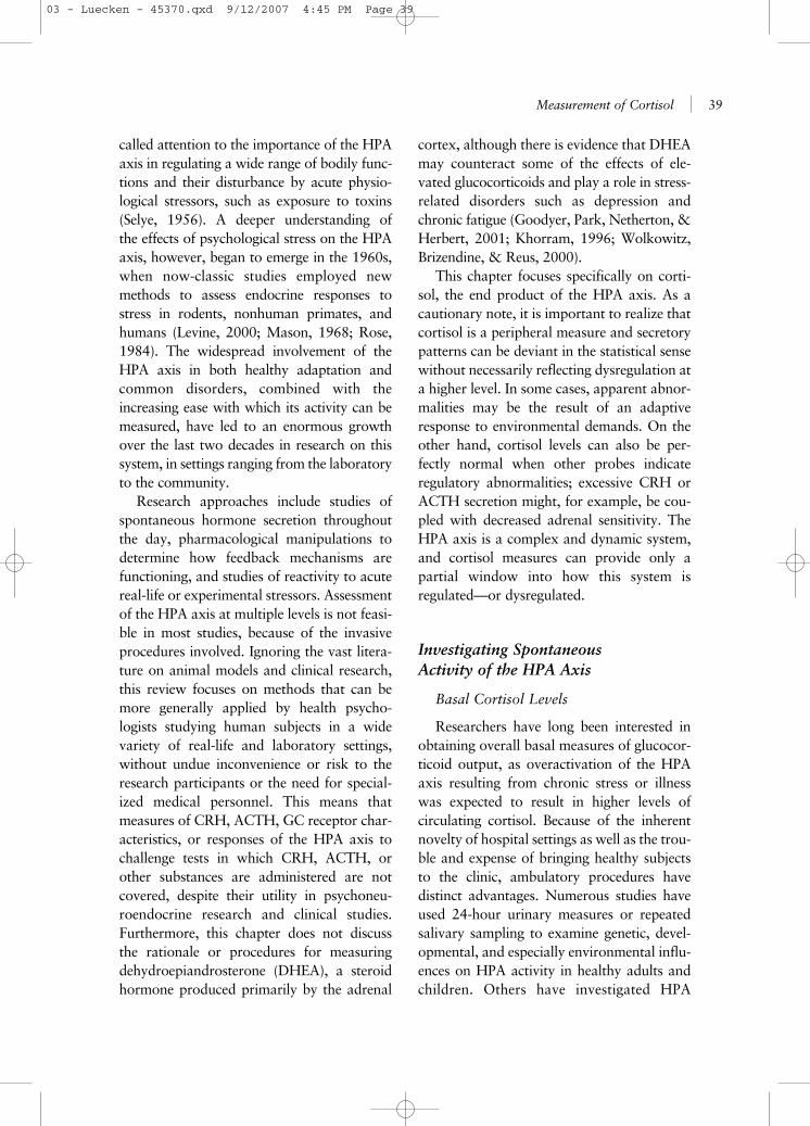
As the name indicates, the main compo-nents of the HPA axis are the hypothalamus,the pituitary gland, and the adrenal cortex
(see Figure 3.1). The hypothalamus releasescorticotropin-releasing hormone (CRH, alsoknown as CRF) into the portal blood vesselsconnecting the hypothalamus to the anteriorpituitary. CRH, which works synergisti-cally with arginine vasopressin (AVP)released from the hypothalamus, then trig-gers the pituitary to secrete adrenocorticotropichormone (ACTH) into the bloodstream.After reaching the adrenal cortex, ACTHstimulates the release of glucocorticoids(GCs)—in humans, cortisol. This entire pro-cess takes place within a matter of minutes.The HPA axis is regulated by a complex neg-ative feedback system, with circulating gluco-corticoids inhibiting activity at the level ofthe hippocampus, the hypothalamus, and thepituitary. In general, hippocampal structuresexert inhibitory influences on the axis at the level of the hypothalamus, whereas theamygdala plays an activating role (Herman& Cullinan, 1997). Mineralocorticoid (MR)and glucocorticoid (GR) receptors in thebrain are thought to play different but com-plementary roles in regulating normal circa-dian activity, preparing the organism torespond to external stimuli, and facilitatingrecovery of disturbed homeostasis afteracutely stressful situations (de Kloet, 1991).
C H A P T E R
03 - Luecken - 45370.qxd 9/12/2007 4:45 PM Page 37

Activity of the HPA axis shows apronounced circadian rhythm, controlled by the primary endogenous pacemaker, thesuprachiasmatic nucleus. ACTH and cortisolare secreted in short pulsatile episodes, con-centrated in the morning hours in humans,but occurring throughout the day, even in the absence of stressors. In a 24-hour cycle,approximately 15 to 18 ACTH pulses can beidentified. In people who have a normal rou-tine of nocturnal sleep and daytime activity,cortisol levels are lowest between 10 p.m.and 4 a.m. After a quiescent period of HPA activity lasting from 2.5 to 6 hours(Linkowski et al., 1985), cortisol levels beginto rise several hours before awakening, withan additional sharp increase in the 30 to
40 minutes following awakening. Thereafter,cortisol levels steadily decrease, except for amoderate rise following lunch. Although cor-tisol levels decline over the rest of the after-noon and throughout the evening until sleeponset, the slope of the diurnal curve is rela-tively flat compared to the morning hours.
A Brief Overview ofResearch Approaches
Because of its central role in regulating the psychobiological stress response, the HPAaxis is one of the most heavily investigatedphysiological systems in health psychologyand psychiatry. Hans Selye’s conception of thegeneral adaptation syndrome, in particular,
PART II: PHYSIOLOGICAL SYSTEMS AND ASSESSMENTS: HORMONAL38
Hippocampus
Hypothalamus
Pituitary
Adrenal cortex
↑Cortisol
Target organs/metabolic effects
↑CRH ↑AVP
↑ACTH
−
−
−
−
Figure 3.1 Schematic Overview of the Hypothalamic-Pituitary-Adrenocortical (HPA) Axis
NOTE: CRH = corticotropin-releasing hormone, AVP = arginine vasopressin, ACTH = adrenocorticotropichormone. Dashed lines indicate negative feedback effects.
03 - Luecken - 45370.qxd 9/12/2007 4:45 PM Page 38
called attention to the importance of the HPAaxis in regulating a wide range of bodily func-tions and their disturbance by acute physio-logical stressors, such as exposure to toxins(Selye, 1956). A deeper understanding of the effects of psychological stress on the HPAaxis, however, began to emerge in the 1960s,when now-classic studies employed newmethods to assess endocrine responses tostress in rodents, nonhuman primates, andhumans (Levine, 2000; Mason, 1968; Rose,1984). The widespread involvement of theHPA axis in both healthy adaptation andcommon disorders, combined with theincreasing ease with which its activity can bemeasured, have led to an enormous growthover the last two decades in research on thissystem, in settings ranging from the laboratoryto the community.
Research approaches include studies ofspontaneous hormone secretion throughoutthe day, pharmacological manipulations todetermine how feedback mechanisms arefunctioning, and studies of reactivity to acutereal-life or experimental stressors. Assessmentof the HPA axis at multiple levels is not feasi-ble in most studies, because of the invasiveprocedures involved. Ignoring the vast litera-ture on animal models and clinical research,this review focuses on methods that can bemore generally applied by health psycho-logists studying human subjects in a widevariety of real-life and laboratory settings,without undue inconvenience or risk to theresearch participants or the need for special-ized medical personnel. This means thatmeasures of CRH, ACTH, GC receptor char-acteristics, or responses of the HPA axis tochallenge tests in which CRH, ACTH, orother substances are administered are notcovered, despite their utility in psychoneu-roendocrine research and clinical studies.Furthermore, this chapter does not discuss the rationale or procedures for measuringdehydroepiandrosterone (DHEA), a steroidhormone produced primarily by the adrenal
cortex, although there is evidence that DHEAmay counteract some of the effects of ele-vated glucocorticoids and play a role in stress-related disorders such as depression andchronic fatigue (Goodyer, Park, Netherton, &Herbert, 2001; Khorram, 1996; Wolkowitz,Brizendine, & Reus, 2000).
This chapter focuses specifically on corti-sol, the end product of the HPA axis. As acautionary note, it is important to realize thatcortisol is a peripheral measure and secretorypatterns can be deviant in the statistical sensewithout necessarily reflecting dysregulation ata higher level. In some cases, apparent abnor-malities may be the result of an adaptiveresponse to environmental demands. On theother hand, cortisol levels can also be per-fectly normal when other probes indicateregulatory abnormalities; excessive CRH orACTH secretion might, for example, be cou-pled with decreased adrenal sensitivity. TheHPA axis is a complex and dynamic system,and cortisol measures can provide only apartial window into how this system isregulated—or dysregulated.
Investigating SpontaneousActivity of the HPA Axis
Basal Cortisol Levels
Researchers have long been interested inobtaining overall basal measures of glucocor-ticoid output, as overactivation of the HPAaxis resulting from chronic stress or illnesswas expected to result in higher levels ofcirculating cortisol. Because of the inherentnovelty of hospital settings as well as the trou-ble and expense of bringing healthy subjectsto the clinic, ambulatory procedures havedistinct advantages. Numerous studies haveused 24-hour urinary measures or repeatedsalivary sampling to examine genetic, devel-opmental, and especially environmental influ-ences on HPA activity in healthy adults andchildren. Others have investigated HPA
Measurement of Cortisol 39
03 - Luecken - 45370.qxd 9/12/2007 4:45 PM Page 39

abnormalities in stress-related disorders. It isnow clear that not only hypercortisolism butalso hypocortisolism can occur, for examplein PTSD, pain, and fatigue syndromes. Theprocesses by which stress could lead to suchdivergent outcomes are still poorly under-stood, but recent reviews have summarized a number of hypotheses (Fries, Hesse,Hellhammer, & Hellhammer, 2005; Gunnar& Vazquez, 2001; Heim, Ehlert, &Hellhammer, 2000; Yehuda, 2002). Forexample, hypocortisolism could be the long-term effect of adverse early experiences that permanently alter the axis. Down-regulation might even be seen as a protectivemechanism, set in motion following long-term hyperactivation to reduce the negative effects of allostatic load. Alternatively, hypocor-tisolism might represent a preexisting riskfactor, of genetic or early developmental ori-gin, which later undermines the individual’sability to respond adaptively to trauma orchronic stressors.
Circadian Rhythm and DiurnalPatterns of HPA Axis Activity
In addition to overall cortisol levels, thediurnal patterning of hormone secretion canprovide important clues to HPA axis dys-regulation. Sophisticated chronobiologicalanalyses of circadian rhythms (see, e.g.,Posener et al., 2000; Van Cauter, Leproult,& Kupfer, 1996) require more frequent sam-pling than is feasible in ambulatory settings,not to mention the problem of obtaining noc-turnal measures. For this reason, simplermeasures of the shape of the diurnal curveare more frequently employed, in particularthe steepness of the decline in cortisol levelsfrom morning to evening. Loss of diurnalvariation, as reflected in flatter slopes, hasbeen reported in various disorders and at-risk groups (Bower et al., 2005; Sephton,Sapolsky, Kraemer, & Spiegel, 2000).
Even if the diurnal slope is not of directrelevance to the goals of a study, collectingseveral samples over the course of a day isgood practice; differences between groupsbeing compared may be restricted to acertain time of day, which often cannot bepredicted on theoretical grounds. For thisreason, studies with only a single diurnalsampling time will inevitably raise questionsabout how results generalize to the rest ofthe day.
Cortisol Response to Awakening
In recent years, interest has been growingin the cortisol awakening response (CAR).Cortisol levels rise sharply (50-160% insaliva) during the first 30 to 40 minutes afterwakeup, returning to the awakening baselinewithin 60 to 75 minutes, and declining moregradually thereafter (Clow, Thorn, Evans, &Hucklebridge, 2004; Pruessner et al., 1997;Wüst et al., 2000). The function of the CARis not yet clear, but general agreement is thatthis response is a discrete aspect of cortisol’scircadian rhythm, with its own regulatoryprocesses (Clow et al., 2004; Schmidt-Reinwald et al., 1999).
The CAR appears to be moderately stablewithin persons, from day to day and overlonger periods of several weeks to months,and it has a clear genetic component (Wüst et al., 2000). Nevertheless, it can vary in relation to short-term influences, such as the stressfulness of a workday compared to a weekend (Kunz-Ebrecht, Kirschbaum,Marmot, & Steptoe, 2004), or an early-shiftcompared to a late-shift workday (Williams,Magid, & Steptoe, 2005). In addition, theCAR may be either enhanced or blunted inchronic stress, burnout, depression, andother disorders (e.g., Bhagwagar, Hafizi, &Cowen, 2005; Grossi et al., 2005; Pruessner,Hellhammer, Pruessner, & Lupien, 2003;Stetler & Miller, 2005).
PART II: PHYSIOLOGICAL SYSTEMS AND ASSESSMENTS: HORMONAL40
03 - Luecken - 45370.qxd 9/12/2007 4:45 PM Page 40

Within-Person Variability
One important aspect of spontaneouscortisol secretion that has received relativelylittle research attention, despite its potentialsignificance as an index of HPA dysregulation,is within-person variability. Greater irregular-ity in within-day cortisol measures has beenobserved in affective disorders, even whenoverall levels are normal (Peeters, Nicolson, &Berkhof, 2004; Posener et al., 2004; Yehuda,Teicher, Trestman, Levengood, & Siever,1996), and may predict worse clinical out-comes (Goodyer, Tamplin, Herbert, &Altham, 2000). There is some evidence that asubset of individuals lacks a consistent diurnalslope pattern (Smyth et al., 1997), but day-to-day variation in cortisol measures remainslargely unexplored. One major obstacle isthat investigating within-person variabilityrequires many more samples per person.
Summary
The degree of detail with which a givenstudy is able to characterize spontaneouscortisol secretory patterns depends on its spe-cific goals, but also on the available budget andlogistical considerations. Thus, large epidemio-logical surveys are often restricted to obtainingonly a few samples per subject and may haveto choose between the response to awakeningand/or a diurnal slope measure (either of whichcan be estimated with a minimum of two salivasamples; see, e.g., Young & Breslau, 2004),perhaps in combination with a urinary mea-sures if nighttime or total cortisol secretion are of interest. At the other extreme, intensivedaily process designs may collect 60 or moresaliva samples per subject in order to estimatenot only overall levels and diurnal slopes, butalso the association between cortisol at a par-ticular point in time with current mood, symp-toms, daily hassles, and uplifts (Smyth et al.,1998; van Eck, Berkhof, Nicolson, & Sulon,1996). As we don’t yet know which measures
of spontaneous cortisol secretion are most relevant for understanding disease processes, aconservative approach would be to obtain reli-able measures of cortisol basal levels, diurnalslopes, and the CAR in the same protocol (see Sampling Strategy under A Framework for Designing a Study and Interpreting theResults).
The availability of noninvasive sam-pling methods (described in MeasuringActivity of the HPA Axis) has greatlyincreased the range of research applications.These include cross-cultural field studies(Flinn, 1999; Hruschka, Kohrt, & Worthman,2005), large-scale longitudinal studies in thecommunity (Rosmalen et al., 2005), interven-tion studies (Carlson, Speca, Patel, & Goodey,2004; Gaab et al., 2003), and prediction ofdisease outcomes (Sephton et al., 2000).
Sensitivity of the HPAAxis to Negative Feedback
Measuring the response of the HPA axis tosynthetic glucocorticoids provides a measureof the strength of negative feedback inhibition.Following an oral dose of 1 mg dexametha-sone late in the evening, cortisol levels are nor-mally suppressed the next day; incompletesuppression or early escape from suppressionindicates deficits in feedback regulatory mech-anisms. The dexamethasone suppression test(DST) was originally developed as a diag-nostic tool in major depression, a disorder inwhich hypercortisolism is often observed(Carroll et al., 1981). A low-dose (0.25–0.5mg) version of the DST has been used to inves-tigate more subtle deficits in feedback regula-tion in individuals with chronic stress (Powellet al., 2002) or to determine whether sensitiv-ity of the HPA axis to glucocorticoid negativefeedback is heightened in disorders in whichhypocortisolism is more frequently observed,such as PTSD (Yehuda et al., 1993) or chronicfatigue syndrome (Gaab et al., 2002).
Measurement of Cortisol 41
03 - Luecken - 45370.qxd 9/12/2007 4:45 PM Page 41

Response of the HPAAxis to Acute Stressors
Early studies of the HPA axis focused onthe hormonal response to acute stressors,and this remains one of the primary interestsof health psychologists. In humans, the corti-sol response to stress can be studied in reallife or under more controlled conditions inthe laboratory. Compared to the quick butshort-lived response of the catecholamines,the cortisol response to acute stress is rela-tively slow. Within minutes of the onset of adiscrete stressful stimulus or event, such aspublic speaking, cortisol levels begin to rise,superimposed on the diurnal profile of basalHPA activity. After termination of the stres-sor, cortisol levels gradually return to theirprestress baseline; full recovery can take anhour or more, in part reflecting the approxi-mately one-hour half-life of cortisol in bloodor saliva.
Basal levels of glucocorticoids act permis-sively to prepare the individual to respond toa stressful episode. The cortisol response tostress mobilizes energy for coping with thestressor, but also shuts down the initial fightor flight responses of the sympathetic ner-vous and immune systems to prevent themfrom overshooting and damaging the organ-ism (Munck, 2000). Glucocorticoid releaseduring stress is thus primarily a protectiveresponse. If, however, cortisol levels aredelayed in their poststress recovery, orrepeated stress exposures result in sensitiza-tion instead of habituation of the HPA axis,a chronic hyperactivation of this system canbe maladaptive, leading to stress-related dis-orders (McEwen, 2003).
What Kinds of StimuliActivate the HPA Axis?
It is a common misconception, probablygoing back to the work of Hans Selye (1956),that the HPA axis will respond to all types of
stressful experiences and the acute cortisolresponse can therefore serve as the gold stan-dard for determining whether a particularexperience was stressful. Many physiologicalsystems are involved in stress responses, andeach system varies in terms of the types ofstressors that activate it, its temporal dynam-ics, and its relations to other systems (Baum& Grunberg, 1995). For example, aversivestimuli that activate the sympathetic nervoussystem and adrenal medulla, producingelevations in heart rate, blood pressure, andcatecholamines, do not necessarily lead tomeasurable changes in cortisol.
Certain types of psychosocial stressors dohave consistent effects. Reviews of early stud-ies in humans, rodents, and nonhuman pri-mates concluded that situations characterizedby novelty, unpredictability, or low perceivedcontrol were most likely to activate the HPAaxis (Mason, 1968; Rose, 1984). A recentmetaanalysis of experimental studies showedthat social-evaluative threat during task per-formance and low control over the situationwere the two best predictors of acute cortisolresponses in humans (Dickerson & Kemeny,2004). Although an individual’s appraisal ofthe stressor, coping, and degree of distress arepredicted, on the basis of transactional stresstheory (Lazarus & Folkman, 1984), to mod-erate or (in the case of distress) mediate thecortisol response, laboratory studies haveshown surprisingly low correlations betweenindividual self-reports of these variables andcortisol measures.
Physical stressors such as intense exercisealso activate the HPA axis. The observationthat cortisol elevations are often greater dur-ing competitive sports than during training atthe same level of physical exertion (Cook, Ng, Read, Harris, & Riad-Fahmy, 1987)indicates that physical and psychosocial com-ponents of competition have additive effects.Cortisol levels also increase following experi-mentally induced pain (al’Absi, Petersen, &Wittmers, 2002).
PART II: PHYSIOLOGICAL SYSTEMS AND ASSESSMENTS: HORMONAL42
03 - Luecken - 45370.qxd 9/12/2007 4:45 PM Page 42

MEASURING ACTIVITYOF THE HPA AXIS
Salivary Cortisol
Background
The first assays for salivary steroids weredescribed in 1959, but this method did notgain widespread acceptance until researchersat the Tenovus Institute in Wales developedreliable assays for steroids in small volumes ofwhole saliva (Riad-Fahmy, Read, Walker, &Griffiths, 1982; Walker, Riad-Fahmy, &Read, 1978). Over the past 20 years, there hasbeen an explosive growth in the number ofstudies using salivary measures to assess corti-sol levels in a wide variety of applications inpsychology, psychiatry, endocrinology, andbeyond. The advantages of salivary cortisolsampling, compared to traditional proceduresfor blood sampling, have been summarized inseveral reviews (Kirschbaum & Hellhammer,1989, 1994; Vining, McGinley, Maksvytis, &Ho, 1983). In addition to the ease and non-invasive nature of sample collection, the fact that salivary cortisol is “free,” unboundby corticosteroid-binding globulin (CBG) orother carriers, is advantageous, as free cortisolthus represents the biologically active fractionof the hormone (Mendel, 1989).
As noted earlier, salivary cortisol is idealfor assessing acute responses to experimentalstressors. In addition, repeated measurementby subjects in their daily environment allowsa good estimate of basal levels, diurnal vari-ation, and response to awakening; somenaturalistic designs also permit individualestimates of day-to-day variability and stressreactivity.
Comparison With Blood Measures
Cortisol levels measured in saliva correlatehighly with free cortisol in blood. However,because of partial conversion of cortisol tocortisone during passage through the salivary
glands, the absolute level of free cortisol in saliva is 10% to 35% lower than it is inblood (Vining et al., 1983). Correlationswith total blood concentrations (bound andfree fractions) are also high, but the slope ofthe regression line becomes steeper at highercortisol concentrations, after CBG-bindingsites in blood are fully occupied. CBG levelscan vary both within and between individu-als, for example during pregnancy or withoral contraceptive use.
Movement of cortisol from blood to salivaoccurs by passive diffusion, so that salivarylevels are independent of the flow rate ofsaliva (Vining et al., 1983). Changes in plasmaand salivary cortisol levels are closely synchro-nized. After injections of cortisol, salivarylevels increased within 1 minute (Walker,1984), and peak concentrations in blood areseen 2 to 3 minutes later in saliva (Kirschbaum& Hellhammer, 2000). Cortisol responses toawakening and to meals appear to be morepronounced in salivary than in plasma mea-sures, and salivary cortisol returns to baselinemore slowly after psychosocial stressors(Kirschbaum & Hellhammer, 2000).
Collection
The popularity of salivary cortisol mea-sures is largely due to the ease of collectingsamples from participants in both labora-tory and field settings. A number of differenttechniques for collecting saliva samples havebeen described; which is most appropriatefor a given research question will depend oncharacteristics of the participants, the setting,and frequency with which samples will becollected, and whether other substances willbe measured in the same samples.
Saliva samples are usually obtained frominfants and toddlers with pipettes or otherdevices that aspirate saliva from the floor ofthe mouth, cotton ropes, swabs, or spongesheld by the researcher or parent (Gunnar &Talge, 2007). In older children and adults,
Measurement of Cortisol 43
03 - Luecken - 45370.qxd 9/12/2007 4:45 PM Page 43

cotton dental rolls—including the widelyused salivettes® (Sarstedt, Nümbrecht,Germany)—have convenient features forboth research participants and laboratorypersonnel. Because of aspecific binding to theswabs or interference by other substancessuch as phytoestrogens that may be pre-sent, cotton salivettes or swabs should not be used when other steroids (e.g., DHEA,testosterone, progesterone) or salivaryimmunoglobulin A (IgA) are also being measured (Shirtcliff, Granger, Schwartz, &Curran, 2001). In 2007 Sarstedt began pro-duction of a new synthetic salivette, designedto eliminate the risk of batch-to-batch varia-tion in the performance of cotton swabs.Swabs are unnecessary if participants cancollect saliva by drooling into a tube, eitherdirectly or through a straw. Drooling may beless acceptable in studies where repeatedsamples need to be collected as rapidly andunobtrusively as possible, for example, dur-ing participants’ daily activities outside thehome. In one comparison, subjects collectedadequate amounts of saliva in 1 to 2 minuteswith cotton salivettes or cellulose-cotton tip“eyespears,” whereas passive drooling tookfrom 1 to 15 minutes to produce the sameamount (Strazdins et al., 2005). For all collec-tion methods, it is important that the plasticstorage tubes and stoppers are made of mate-rials, such as unrecycled polypropylene (IBL, 2006), that do not absorb the hormone.Stoppers also need to fit tightly, because evaporation of saliva will lead to inaccuratecortisol results.
Most cortisol assays require only 20 to50 µl of saliva per tube, and therefore twicethese amounts for a duplicate assay. Inpractice, larger volumes of saliva need to becollected when cotton-based methods areused, because up to 450 µl of saliva canremain in the cotton after centrifuging (deWeerth, Graat, Buitelaar, & Thijssen, 2003).Specialized techniques make it possible toextract cortisol from smaller sample volumes,
which may be a great advantage in studies ofinfants (de Weerth et al., 2003). In subgroupswith low spontaneous flow rates (e.g., babiesand small children, depressed patients, theelderly), saliva flow can be stimulated withpowdered drink mix crystals, candies con-taining citric acid, or lemon juice. Salivettesprepared with citric acid are also meant tostimulate salivary flow. Extreme caution iswarranted in using such procedures, how-ever, as they can lower the pH of the resultingsaliva sample. Many currently availableimmunoassays produce false high valueswhen sample pH is lower than 3.5 to 4(Kirschbaum & Hellhammer, 2000; Schwartz,Granger, Susman, Gunnar, & Laird, 1998;Talge, Donzella, Kryzer, Gierens, & Gunnar,2005; Vialard-Miguel, Belaidi, Lembeye, &Corcuff, 2005). Chewing on an inert sub-stance (e.g., plain salivette, sugarless chewinggum, parafilm) or just making chewing move-ments are good alternatives for stimulatingsalivary flow.
Instructions to Subjects
Subjects should be trained how to collectsaliva samples and given the opportunity topractice under supervision to ensure that they collect adequate volumes of saliva. Withsalivettes, subjects should be instructed tochew lightly on the swab and to keep it fullyinside the mouth until it feels saturated. (Thiscan take 1 to 2 minutes, depending on salivaryflow rate.) It is standard practice to ask sub-jects not to brush their teeth in the 30 minutesbefore scheduled collection of a salivary sam-ple. Acidic drinks, milk, and use of inhaledsteroids (as examples of substances that couldinterfere with assay performance) should beavoided shortly before taking a saliva sample.If rinsing with water is considered necessary, itshould be done at least 10 minutes beforesaliva collection to avoid diluting the cortisolconcentration. Recent food intake and smok-ing can influence cortisol responses to acute
PART II: PHYSIOLOGICAL SYSTEMS AND ASSESSMENTS: HORMONAL44
03 - Luecken - 45370.qxd 9/12/2007 4:45 PM Page 44

stressors and possibly to morning awakeningand should be avoided in the hour before sam-pling. It is crucial that subjects close the tubestightly and label them with the exact time the sample was collected! Clear instructionsshould be given concerning storage (i.e.,whether tubes should be kept at room temper-ature, in the refrigerator, or in the homefreezer); samples should not be left exposed toheat or sunlight.
Storage and Handling
Saliva samples can be stored at room tem-perature (RT) or in participants’ home refrig-erator or freezer until they are mailed ordelivered to the lab. Estimates of how longcortisol is stable at RT range from 7 days(Groschl, Wagner, Rauh, & Dorr, 2001) toat least 4 weeks (Kirschbaum & Hellhammer,2000). Centrifuging samples before storageappears to prolong the stable period (Groschlet al., 2001); nevertheless, increasing vari-ance as well as decreasing levels over timeindicate that storage at RT for more than 2 to maximally 4 weeks should be avoided(Garde & Hansen, 2005). Salivette samplesdevelop mold and a bad odor after about 4days at RT; this does not affect the cortisolconcentrations, but makes the work of labtechnicians unpleasant.
The benefits of refrigeration at 4° to 5ºC,compared to RT, are unclear. In one study(Groschl et al., 2001), cortisol levels decreasedin samples refrigerated for 11 days or longer;in contrast, Garde and Hansen (2005) foundno change in cortisol levels in polyestersalivettes refrigerated up to 3 months.Freezing clearly prolongs the stability of sali-vary cortisol. In samples frozen at either –20ºor –80ºC, cortisol concentrations remain sta-ble for 9 months (Aardal & Holm, 1995) to 1 year (Garde & Hansen, 2005); freezing foras long as 2 years is probably possible.
In settings where there is no access torefrigerators or freezers, stability of samples
can be prolonged by adding preservativessuch as sodium azide (Groschl et al., 2001),citric acid (alone or with sodium benzoate),or ethyl and propyl paraben (Nimmagudda,Ramanathan, & Putcha, 1997). Cortisol insamples treated with citric acid and sodiumbenzoate remained stable for 180 days at RT(Nimmagudda et al., 1997). As noted earlier,preservatives, especially those that lower pH,may invalidate certain assays. Blood spotsoffer an alternative to saliva when extendedstorage at RT is necessary (Worthman &Stallings, 1997). (See Blood Spot Measures,below.)
Salivary cortisol levels are relatively insen-sitive to repeated thawing and refreezing; inrecent studies, cortisol levels remained stablein samples undergoing up to three (Groschl et al., 2001) or four (Garde & Hansen, 2005)freeze/thaw cycles prior to assay. In the labo-ratory, samples collected by passive drool arefrozen and thawed at least once before assay-ing in order to break down mucins that caninterfere with pipetting (Vining & McGinley,1986). Centrifuging helps remove particulatematter that can interfere with immunoassay.In salivettes, clear saliva collects in the bottomof the outer tube after centrifuging.
There is normally no need to transportsamples to the laboratory on ice (Clements &Parker, 1998). However, when the time intransit is more than a few days, shipping ondry ice will prevent molding and may berequired by some laboratories. (For infor-mation on international shipping, see Inter-national Air Transport Association IATAregulations; adjustments made in 2005exempt saliva samples from regulations forhazardous biological substances.)
Prior to assay, saliva samples should bechecked for blood contamination, as this canartificially elevate the cortisol concentration.Deficient diet, poor oral hygiene, and overlystrenuous toothbrushing can cause bleed-ing gums. In a recent study (Kivlighan et al.,2004), subjects first brushed their teeth
Measurement of Cortisol 45
03 - Luecken - 45370.qxd 9/12/2007 4:45 PM Page 45

vigorously and then collected saliva by directdrool. Minor injuries to the oral cavity led todetectable blood leakage in the samples, asassessed by three different methods: trans-ferrin immunoassay, dipsticks for detectinghemoglobin in urine, and visual inspection. A moderate degree of blood contamination(samples visibly pink) had a negligible effecton cortisol levels, but darker saliva sampleswere more problematic. Visual inspectionand discarding of saliva samples that are dis-colored therefore appears to be adequate tocontrol this source of error under normal cir-cumstances. This is good news, because assayof transferrin—the most accurate method forassessing blood contamination—is relativelyexpensive, and dipsticks can yield false-posi-tive results (Worthman & Stallings, 1997).
Types of Assays
Free cortisol in the blood represents only4% to 5% of total cortisol released; more-over, during passive diffusion into thesalivary glands, approximately one-third ofthe free cortisol is lost through conversion to cortisone. Sensitive assay procedures aretherefore necessary. Several methods cur-rently allow reliable measurement of salivarycortisol without the necessity of extractionprocedures. These include radioimmuno-assay (RIA), enzyme-linked immunosorbentassay (ELISA), fluorescence immunoassay(FIA), and chemiluminescence immunoassay(LIA). The last three are nonradioactiveassays in microtiter plate format, which canbe run either manually or on automatedequipment. Special laboratory equipment isrequired. Before the late 1990s, salivary cor-tisol assays were often adaptations of proto-cols designed for plasma/serum measures.Currently, assay kits developed for salivarydeterminations have standards suspended ina saliva matrix (in contrast to a serum orbuffer matrix). Regardless of the assay used,
the following procedures are recommended:(1) assay samples in duplicate and use themean value in statistical analyses,1 (2) repeatthe assay for samples with duplicate valuesthat differ by more than 20%, and (3) mea-sure all samples from a given subject in thesame assay run.
Choosing a Lab
Over the last several years, commerciallabs have proliferated, in some cases offeringassay services for salivary cortisol as well askits for use in the investigator’s own lab.Details are available through the laboratories’websites, for example www.salimetrics.com, www.ibl-hamburg.com, and www.dslabs.com. In addition, many hospital and researchlabs have expertise in salivary assays; someuse commercially available kits, while othershave developed their own in-house assays.Price is an important consideration; the costsof a duplicate cortisol determination canrange from roughly $6 to $30, often with adiscount for large quantities. Quality shouldalso enter into the choice, as not all assays areequally sensitive or reliable. Fortunately,assay quality and cost are likely to beinversely related, because laboratories withtailored salivary assays also tend to havemore experience, higher volumes, and auto-mated procedures.
Sensitivity refers to the minimum concen-tration of cortisol that can be distinguishedfrom zero. Salivary cortisol assays generallyhave a lower detection limit of less than .01µg/dL, which is below the concentration nor-mally observed until late in the evening whenthe HPA axis becomes quiescent. The relia-bility of an assay, which is even more crucialfor most research questions, is reflected inthe intra- and interassay coefficients of varia-tion (CV). The intra-assay CV can be calcu-lated by dividing the standard deviation bythe mean and then multiplying this figure by
PART II: PHYSIOLOGICAL SYSTEMS AND ASSESSMENTS: HORMONAL46
03 - Luecken - 45370.qxd 9/12/2007 4:45 PM Page 46

100, over a representative subsample (high,medium, and low cortisol concentrations) ofduplicate measures from the same assay run.The interassay CV is calculated over severalassay runs; laboratories should be able toprovide this information on request. BothCVs should be under 12% to 15% (most arelower). In general, CVs are higher for corti-sol concentrations in the lower part of therange. Even when intra- and interassaycoefficients of variation are acceptable, somelaboratories may obtain higher or lowerabsolute cortisol values than others (Hansen,Garde, Christensen, Eller, & Netterstrom,2003; Kraemer et al., 2006). For this reason,it is inadvisable to switch from one type ofassay to another or from one laboratory toanother during the same study.
Other indicators of assay performance arerange of calibration, range of linearity of theassay (the linear region of the standard curveshould cover the range in which mostsalivary cortisol values are found), spikerecovery, and specificity (the percentage ofcrossreactivity with other endogenous orexogenous substances, like cortisone or pred-nisone, that are or may be present in saliva).Laboratories should be able to provide thisinformation for their assays. Reviewers arelikely to request more detailed informationfor in-house assays. Recent implementationof voluntary quality assessment programs for salivary assays (IBL, 2005) will hopefullymake it easier for researchers to evaluate andcompare laboratories.
Urinary Cortisol
Background
Urinary measures of glucocorticoidmetabolites (17-hydroxycorticoids) wereamong the first techniques available forstudying activity of the HPA axis in humans,going back to the 1950s. An impressivebody of knowledge emerged from the early
psychoendocrine studies of 17-HOCs levels(Mason, 1968). New techniques soon allowedresearchers to measure cortisol directly, assmall amounts are excreted as free cortisolin the urine (UFC). Urine samples collectedover 24 hours provide an integrated measureof total free cortisol excretion. Mean UFCvalues are approximately 20µg/24 h (range3-43 µg/24 h) in healthy adult women(Murphy, 2003).
To reduce participant burden, collectionover shorter periods may prove adequate fora specific research question. In addition,urine collection can be scheduled in such away that more refined analyses are possible.As an example, Jerjes and colleagues wereable to investigate diurnal patterns of HPAactivity by having subjects collect urine every3 hours for 15 hours (Jerjes et al., 2006).Another recent study compared women’s uri-nary cortisol levels when they were at home,at work, or asleep (Dettenborn et al., 2005).
A distinct advantage of urinary measuresis that they allow assessment of nighttimecortisol levels, which may be crucial in cer-tain disorders in which daytime levels areoften normal (anxiety: Abelson & Curtis,1996; PTSD: Yehuda, 2002). Urinary mea-sures also have some disadvantages, whichexplain why they are less popular than sali-vary cortisol. First, integrated measures arenot very informative for research questionsconcerning acute stress responses. Second,the burden of collecting complete urine sam-ples should not be underestimated, as it canlead to low participation in studies or poorcompliance. Finally, transporting large vol-umes of urine from field to laboratory iscumbersome.
Collection, Storage, and Handling
At the beginning of the sampling period,subjects void and discard the first urine. Allurine produced thereafter is collected in large
Measurement of Cortisol 47
03 - Luecken - 45370.qxd 9/12/2007 4:45 PM Page 47

plastic containers designed for this purpose, orseveral containers if the study entails separatemeasurements. Samples can be kept at roomtemperature, without preservatives, for atleast 24 hours without degradation of gluco-corticoids (Gouarne, Foury, & Duclos, 2004).
Assays
Urinary free cortisol represents a small fraction of total cortisol released by the adrenalcortex. Commercially available RIA kits formeasuring UFC may yield falsely high values,as results can be influenced by the presence ofcortisol metabolites as well as other interferingsubstances; UFC values obtained with theseassays are potentially two to four times higherthan the true values established with chro-matography (Murphy, 2002). Immunoassayshave been reported to show particularly lowspecificity and poor precision at low cortisolconcentrations, leading to widely discrepantresults in studies of adrenal suppression (Finket al., 2002). In choosing a laboratory, it istherefore important to make sure that theassay has been validated and is monitoredaccording to established standards for UFC;details concerning the assay (accuracy, recov-ery, precision, antibody used, crossreactivity,extraction method) should also be reported inpublications. Accurate methods, for exampleliquid chromatography/tandem mass spec-trometry (McCann, Gillingwater, & Keevil,2005; Turpeinen & Stenman, 2003), arebecoming more widely available and afford-able. Because the tiny percentage (2-3%) ofUFC in relation to total urinary cortisolmetabolites may vary due to changes in steroidmetabolism, measuring urinary cortisone, theratio of cortisone to cortisol, or total cortisolmetabolites may provide additional insightsinto HPA axis function (Gouarne, Groussard,Gratas-Delamarche, Delamarche, & Duclos,2005; Jerjes et al., 2006). UFC results are oftencorrected for creatinine levels.
Blood Spot Measures
Background
Finger-prick blood spot sampling providesan alternative to salivary measures of cortisol;this technique combines the advantages oftraditional blood samples, in terms of therange of substances that can be measured,with greater ease of sample collection andmore convenient storage and handling pro-cedures (Wong, Yan, Donald, & McLean,2004; Worthman & Stallings, 1997). Usingdevices designed to allow diabetics to monitortheir own glucose levels, collection of finger-prick samples in capillary blood is quick andminimally invasive. Because of the tinyamount of blood required, obtaining repeatedsamples from an individual is feasible. Bloodspot cortisol is highly correlated with serumlevels. The method also has some disadvan-tages: not all participants can be trained tocollect their own samples, so that researchpersonnel may have to be involved; finger-pricks are not entirely painless, and recruit-ment of subjects may be more difficult for thisreason; subjects may be concerned about thesafety of the procedure; and so on.
Collection, Handling, and Storage
Capillary blood from a finger-prick isdropped, without blotting or smearing, ontospecially designed filter paper of the sortwidely used in neonatal screening programs.One drop (yielding a blood spot of approxi-mately 50 µL of whole blood) is sufficient forcortisol determination. After samples on fil-ter paper are air-dried for several hours, theycan be easily stored in plastic bags for trans-port and even be mailed directly to the lab byordinary post.
Assays
Special, highly sensitive assays have beendeveloped to determine cortisol levels in
PART II: PHYSIOLOGICAL SYSTEMS AND ASSESSMENTS: HORMONAL48
03 - Luecken - 45370.qxd 9/12/2007 4:45 PM Page 48

blood spots. These assays are currentlyperformed by an increasing number of labo-ratories, including commercial laboratoriessuch as Salimetrics and DSL. Additional hor-mones and other substances can be measuredin blood spots, including those not measur-able in saliva, such as prolactin and markersof immune function (McDade et al., 2000).Accurate measures of estradiol and proges-terone can also be obtained, enabling theresearcher reliably to assess the stage of themenstrual cycle (Shirtcliff, Reavis, Overman,& Granger, 2001), for example.
A FRAMEWORK FORDESIGNING A STUDY ANDINTERPRETING THE RESULTS
The first key to designing an effective studywith clear results is awareness of the tempo-ral dynamics of HPA axis activity, as thesewill dictate the sampling strategy. For studiesof stress reactivity, the choice of a stressor canhave a major impact on the results and theirinterpretation. Finally, the design should takeinto consideration the range of possiblemoderators, mediators, and confounders thatmight affect the hypothesized relationshipbetween HPA measures and biopsychosocialvariables of interest. This review attempts tosummarize current recommendations andpractice, without claiming that evidence in all cases is so consistent and complete thatresearchers have reached a consensus.
Sampling Strategy
As previously described, integrated (inurine, UFC) and momentary (in saliva orblood) measures of cortisol are available. ForUFC, the main decision is whether to collectsamples over 24 hours or shorter time peri-ods; the choice should be based on theoreti-cal grounds, but subject burden and logistics
often play a role. For salivary cortisol andblood spots, the optimal number and timingof samples depends on the aspects of HPA activity being investigated (e.g., basallevels, diurnal variation, response to awaken-ing, negative feedback inhibition, or responseto acute stressors) and the stability of thesemeasures over time.
Basal Cortisol Levelsand Diurnal Variation
Although investigators seem to agree thatcortisol should be measured several times aday for a number of days to get reliable esti-mates of mean basal levels and diurnal slope(Goodyer et al., 2001; Stewart & Seeman,2000), clear recommendations with support-ing data are difficult to find. Hruschka andcolleagues (2005) recently presented formu-las for determining these figures on the basisof variance estimates from multilevel regres-sion models. Based on data from a number ofstudies using different sampling protocols,these calculations suggested that—dependingon the spacing of the samples in time—as fewas four samples taken on one day might beadequate for estimating individual meanlevels, but that 14 or more days of samplingwith four to five samples per day might benecessary to obtain a reliable estimate of an individual’s diurnal slope. In contrast, astudy in an older population with very goodprotocol compliance found that five samplesper day for 3 days provided a reliable esti-mate of daytime slope; moreover, slopesbased on as few as two daily time points(wake and 9:00 p.m.) correlated highly withthose based on four points, and additionaldays did not substantially increase reliability(Kraemer et al., 2006). These findings under-score the need for more analyses of existing“daily profile” datasets. To establish anoptimal sampling protocol for a specificpopulation, a pilot study with at least
Measurement of Cortisol 49
03 - Luecken - 45370.qxd 9/12/2007 4:45 PM Page 49

50 participants is desirable (Kramer et al.,2006). Until more empirical results are avail-able, a conservative recommendation wouldbe to collect three to five samples a day for atleast 3 days if basal levels are of primaryinterest and for 6 to 7 days if diurnal varia-tion is a major focus. Increasing the numberof subjects can increase statistical powerwhen reliability of the cortisol measures isnot optimal.
Because individual differences in sleeppatterns may be associated with shifts in thecircadian cycle, some researchers have cho-sen to collect samples at fixed intervals fromthe habitual time of awakening, instead of atfixed times of day. Another elegant but logis-tically simpler design is to sample at fixedtimes of day and then model effects of timesince awakening statistically (Cohen et al.,2006). In all cases, efforts should be madeto obtain accurate information concerningthe actual sample collection times. The mostfoolproof method is some form of elec-tronic monitoring, for example, devices thatrecord whenever a participant opens a vial toremove a cotton swab (Broderick, Arnold,Kudielka, & Kirschbaum, 2004; Jacobset al., 2005; Kudielka, Broderick, &Kirschbaum, 2003) or handheld computersthat generate time stamps with whichparticipants must label their tubes (Stetler,Dickerson, & Miller, 2004). Awareness thatcompliance is being monitored increases theprobability that samples will be taken asdirected (Kudielka et al., 2003). Promptingparticipants with an audible or vibrating sig-nal can also help. Finally, instructions to par-ticipants should emphasize the importance ofaccuracy and honesty in reporting actual col-lection times. In older adults, self-reportedcollection times were close to automaticallyrecorded times, and test-retest reliability ofslope estimates was actually slightly betterwhen based on self-reported times (Kraemeret al., 2006).
Cortisol Awakening Response (CAR)
The time course of this response has beenwell characterized. The peak response occurs30 to 45 minutes after awakening; by 60 min-utes, cortisol levels are decreasing and may nolonger be reliably distinguishable from thelevels at awakening. At least two samples (atawakening and either 30 or 45 minutes later)are needed to characterize the response; moresamples (e.g., at 0, 30, 45, and 60 minutes)may increase reliability and allow calculationof AUC measures (see Statistical Analysis).The CAR should preferably be measured onat least 2 days. Given the narrow window ofresponse, accurate timing of samples is cru-cial. Because participants appear to havedifficulty in taking early morning samples asdirected (Kudielka et al., 2003), it may bewise to reduce the sample burden to the min-imum, emphasizing quality rather than quan-tity. Some kind of alarm device is useful toremind the participant to collect samples atthe appropriate times, and compliance shouldbe monitored electronically (see above) if thisis possible. Activity monitors can be helpful inconfirming time of awakening, but this is notconsidered essential for all studies.
Methodological issues relevant to studydesign have been summarized by Clow andcolleagues (2004). Instructions to subjectsshould be standardized along the followinglines:
• Place all materials next to your bed beforegoing to sleep.
• Take the first sample in bed immediatelyafter awakening, with lights on and eyes open.
• Do not go back to sleep; get out of bed(within 15 minutes) before taking anothersample.
• The second sample should be taken [n]minutes after awakening (and so on foreach sample).
• Do not brush your teeth, smoke, eat, ordrink anything except water until you havefinished taking the [n] morning samples.
PART II: PHYSIOLOGICAL SYSTEMS AND ASSESSMENTS: HORMONAL50
03 - Luecken - 45370.qxd 9/12/2007 4:45 PM Page 50
• Remember to record the exact time eachsample was taken on the tube, even if thiswas not the scheduled time.
Dexamethasone Suppressionas a Measure of NegativeFeedback Sensitivity
Dexamethasone (DEX) can be safelyingested by participants at home and itseffects measured in salivary cortisol (Lindley,Carlson, & Benoit, 2004; Powell et al.,2002). In most studies, cortisol measures ona control day are compared with measurestaken at the same times of day followingintake of 0.25 to 0.5 mg (low dose) or 1.0 mg(high dose) dexamethasone late the previ-ous evening (at 11 p.m. or an agreed-on bed-time). The original dexamethasone suppressiontest (DST), developed as a diagnostic test formajor depression, was scored as positive if,following administration of 1 mg DEX at 11p.m. on day 1, cortisol at 4 p.m. on day 2was above an established cutoff point(Carroll et al., 1981). For research purposes,analyzing the cortisol results as continuousinstead of dichotomized measures yieldsmore information. The optimal timing of thesamples depends on the DEX dosage, as cor-tisol will “escape” from suppression earlierwith lower doses. Collecting a number ofpost-DEX saliva samples at intervals of sev-eral hours will increase reliability of theresults and gives added information aboutthe time course of feedback inhibition.
The Cortisol Responseto Acute Stressors
Cortisol responses to acute stressors can be studied in the laboratory and in real life,where anticipated as well as unanticipatedstressors occur. Design issues vary accordingto the setting. In the laboratory, importantdecisions include the best time of day to
schedule the experiment, how many samplesare needed to characterize the stress response,the timing of these samples in relation to thestress task, the nature of the task, how to con-trol for effects of novelty and anticipation,and habituation to repeated stressors. For adetailed overview of many of these designissues, see Dickerson and Kemeny (2004).
Time of Day. Although the HPA axis is capa-ble of responding to acute stress at any pointin the diurnal cycle (Kudielka, Schommer,Hellhammer, & Kirschbaum, 2004), schedul-ing experiments in the mid to late afternoon(roughly between 3 and 6 p.m.) has advan-tages. First, the cortisol response to stress ismore readily distinguishable in the afternoonthan in the morning from background noisein the form of spontaneous pulsatile episodesand the natural decline in basal levels over themorning hours; the metaanalysis performedby Dickerson and Kemeny (2004) showedmoderate effect sizes for cortisol response tostress tasks performed in the afternoon, com-pared to small effect sizes in the morning.Second, cortisol responses are easier to pro-voke in the afternoon than in the late evening,when the HPA axis becomes quiescent. Third,effects of potential confounders such as recentawakening and lunch (see below) are easierto exclude. Thus far, study design has beeninfluenced mainly by such practical consider-ations, and little attention has been paidto theoretically important issues, such asthe consequences of differential activation ofMR and GR systems by stressors occurringat the trough versus the peak of the diurnalcycle (Dallman, Akana, Bhatnagar, Bell, &Strack, 2000).
Number and Timing of Samples. To charac-terize the cortisol response to an acute stres-sor, samples are taken at fixed intervalsduring baseline (30-40 minutes), stress expo-sure (10-20 minutes), and recovery (40-60
Measurement of Cortisol 51
03 - Luecken - 45370.qxd 9/12/2007 4:45 PM Page 51

minutes) periods. Participants thus need toremain at the laboratory for a total of 1½ to2 hours. The stress exposure includes boththe preparation period, if there is one, andthe actual task performance. Peak cortisollevels are usually observed 20 to 40 minutesafter task onset, depending on the intensityand duration of the task, with a gradualreturn to baseline levels over the next hour orlonger (Dickerson & Kemeny, 2004). Evenwith an identical task, however, there aremarked individual differences in latency topeak response (Gunnar & Talge, 2007). Iftime and budget allow, an optimal designwould include two to three baseline mea-sures, one to two measures shortly after andpossibly during the stress task, and two tothree measures during the recovery period.Minimalistic designs with one prestress andone poststress measure run the risk of miss-ing the peak response and provide no infor-mation about speed of recovery.
Controlling for Novelty and Anticipation.Prestress baseline measures are highly sensi-tive to the novelty of the setting. Previousvisits to the lab or an extended acclimationperiod (30 minutes or more) after arrival canreduce the probability of elevated baselinelevels. Anxiety in anticipation of the taskremains difficult to control. Because infor-mation provided earlier as part of informedconsent and pretask instructions can eitherheighten or reduce anxiety, procedures needto be fully standardized in terms of both con-tent and timing. Obtaining a saliva sample athome, at the same time on another day, isvery useful in determining whether lab base-line levels are elevated (Nicolson, Storms,Ponds, & Sulon, 1997). This is important toknow, because high baseline cortisol is oftenassociated with a blunted response to stress(Kudielka, Schommer, et al., 2004; Young &Nolen-Hoeksema, 2001). Interestingly, infantsand young children tend to show lower
cortisol levels at lab arrival than at home(Gunnar & Talge, 2007).
Type of Stressor. Health psychology studiesmost frequently apply psychosocial stresstasks, as these are thought to have the greatestecological validity. The HPA axis can beactivated by physical stressors such as intenseexercise or pain, or by pharmacologicalchallenges; individuals’ responses to differentclasses of stressors, however, do not appear to be highly intercorrelated. Among thepsychosocial stressors, performance tasks withelements of social-evaluative threat, uncon-trollability, or both produce the largest andmost consistent increases in cortisol. Less con-sistent results are found for passive tasks (e.g.,watching a film or other emotion inductionprocedures, noise exposure) and performancetasks without evaluative threat or uncontrolla-bility (Dickerson & Kemeny, 2004).
Only a few stress tasks have beendescribed in sufficient detail that results canbe compared across studies and populations.The best known of these is the Trier SocialStress Test (TSST) (Kirschbaum, Pirke, &Hellhammer, 1993). The widespread use ofthe TSST reflects the fact that it has beenextensively studied, can be applied in sub-jects varying in age and educational status,and induces a cortisol response in the major-ity of participants. For the TSST and othertasks with a combination of social-evaluativethreat and uncontrollability, effect sizes, onaverage, are large (Dickerson & Kemeny,2004). Results are sensitive to changes in the procedure, however: when the intervalbetween instructions to subjects about thetask and performance was extended from 10 minutes to 1 hour, the cortisol response tothe task was obliterated (Young & Nolen-Hoeksema, 2001).
Interpretation of Results. Although labora-tory stress tasks are probably the single most
PART II: PHYSIOLOGICAL SYSTEMS AND ASSESSMENTS: HORMONAL52
03 - Luecken - 45370.qxd 9/12/2007 4:45 PM Page 52

common approach to investigating the HPA axis in health psychology, results can be difficult to interpret. Failure to detect astatistically significant cortisol response is a rather common occurrence, even when thetask appears to be experienced as stressful.Weaknesses in study design are responsiblefor some of these negative findings. With ade-quate sample size and the right choice ofstress task and timing of measures, however,the majority of subjects are likely to show anincrease in cortisol from baseline to posttask.Theoretically, one would expect the magni-tude of this response to reflect the individ-ual’s experience of the situation, in terms ofappraised threat, coping possibilities, and theintensity of emotional distress. Unfortunately,this is rarely the case with laboratory tasks,possibly because self-report instruments arenot sensitive to the relevant processes orbecause other aspects of the situation—forexample, its novelty—are more salient.Another discouraging finding is that cortisolresponses often show no correlation withpersonality traits linked to stress reactivity,such as neuroticism (Schommer, Kudielka,Hellhammer, & Kirschbaum, 1999). Further-more, little is known about the generaliz-ability of lab reactivity measures to real-lifesituations (for studies involving cortisol, seeHoutman & Bakker, 1987; Lundberg, Melin,Fredrikson, Tuomisto, & Frankenhaeuser,1990; van Eck, Nicolson, Berkhof, & Sulon,1996). Giving a speech on a particular topicor performing mental arithmetic before anaudience may also have different meaningsfor individuals or groups, depending on vari-ables such as cognitive ability, occupation,and cultural background. The challenge forresearchers is to design laboratory stressorsthat convincingly tap into the processes ofinterest in a given population and also reli-ably activate the HPA axis. Examples includea task involving competition with an antago-nistic peer in children (van Goozen et al.,
1998) and a standardized lecture in studentteachers (Houtman & Bakker, 1987).
Test-retest reliability of acute stressresponse measures appears to be low, proba-bly because of both the underestimated noiseintroduced by spontaneous pulsatile activity(Young, Abelson, & Lightman, 2004) andthe tendency of the cortisol response tohabituate following repeated exposures. Lowreliability of cortisol outcome measuresmeans that laboratory stress experiments areparticularly vulnerable to Type 2 error. Thisissue is important in all studies, but especiallyneeds to be taken into account in inter-vention studies, where stress reactivity iscompared pre- and postintervention. Moreanalyses are needed to determine how manycortisol measures per session and how manyrepeated sessions are necessary to obtain reli-able measures of stress reactivity for differentsubject populations and stressors (Gunnar &Talge, 2007; Hruschka et al., 2005).
Habituation and Sensitization. If the cortisolresponse to experimental stressors is to beconsidered a valid indicator of what goes onin daily life, it is important to know whathappens following repetitive stressful experi-ences. The acute response is adaptive, but isexpected to habituate over repeated expo-sures as novelty decreases and controlincreases. Failure to habituate or sensitizationto repeated stressors, in contrast, is regardedas maladaptive, contributing in the long runto allostatic load. Habituation versus sensiti-zation of the HPA response has been exten-sively examined in animal models (e.g.,Pitman, Ottenweller, & Natelson, 1990), butcomparatively little research has been con-ducted in humans (exceptions include al’Absiet al., 1997; Epel et al., 2000; Gerra et al.,2001; Gunnar, Hertsgaard, Larson, &Rigatuso, 1991; Kirschbaum, Prüssner et al.,1995; Wüst, Federenko, van Rossum, Koper,& Hellhammer, 2005). Findings indicate
Measurement of Cortisol 53
03 - Luecken - 45370.qxd 9/12/2007 4:45 PM Page 53

rapid habituation from the first to the secondand later exposures, but also show markedindividual differences, with a subset of indi-viduals failing to habituate after repeatedexposures. In addition, correlations betweenthe cortisol response and trait characteristicsmay increase (Kirschbaum, Prüssner et al.,1995) or decrease (al’Absi & Lovallo, 1993)over successive task performances. Thesefindings suggest that repeated testing (forexample, three times in a week, or once aweek for 3 weeks) is much more informativethan single exposures in elucidating individ-ual differences in stress reactivity that are rel-evant to long-term health outcomes.
Naturalistic Experiments. Responses of theHPA axis can also be investigated in responseto real-life activities that entail some level ofchallenge or threat. Examples include exams,parachuting, sports competitions, musicalperformances, and occupational stressors, toname just a few. Tension-reducing activitiessuch as yoga, meditation, and massage, onthe other hand, may lower cortisol levels.Like lab experiments, these activities are usually scheduled or at least anticipated in advance, so that baseline, response, andrecovery measures can be obtained. Certainactivities, like parachute jumping, also lendthemselves to studies of the habituationprocess (Deinzer, Kirschbaum, Gresele,& Hellhammer, 1997; Levine, 1978).Correlations between cortisol responses andsubjective distress measures may be higherfor real-life than for laboratory stressors (Nicolson, 1992).
HPA axis responses to life events can alsobe investigated, although prestress baselinemeasures are rarely available because of theunpredictable nature of individual life events(e.g., rape, sudden death of a family member)and natural or manmade catastrophes (e.g.,earthquakes, hurricanes, war). In this case,cortisol levels in exposed individuals arecompared with those in an unexposed
comparison group and are often examinedlongitudinally, in relation to symptoms.
Daily Hassles and Emotions. Combiningrepeated self-reports with salivary measures,experience sampling or ecological momentaryassessment studies have investigated cortisolreactivity to daily life hassles and uplifts andaccompanying emotions. Real-life stressorsvary widely in duration, and participants areoften unable to report exactly when a stressfulsituation began or when it ended. Althoughthe timing of cortisol measures in relation todaily hassles is therefore imprecise, multilevelregression analyses can assess associationsbetween the two. The association betweendaily events and cortisol is probably mediatedby changes in negative affect. The finding ofhigher salivary cortisol in association withdaily hassles or negative affects has been repli-cated in several samples of adults (Hanson,Maas, Meijman, & Godaert, 2000; Peeters,Nicholson, & Berkhof, 2003; Smyth et al.,1998; van Eck, Berkhof et al., 1996) andchildren (Adam, 2006). In some but not allstudies, positive affects were associated withlower cortisol (Adam, 2005; Polk, Cohen,Doyle, Skoner, & Kirschbaum, 2005).
Moderators and Confounders
Interpretation of cortisol results can befacilitated by considering a number ofbetween- and within-individual factors thatcan influence HPA axis activity. Theseinclude age, gender-related variables, andethnicity; somatic variables such as illness,medications, and obesity; daily lifestyle vari-ables such as food intake, smoking, and sleeppatterns; psychosocial variables related tostress; and genetic differences.
Age and Gender-Related Variables
Findings concerning the effects of age andgender on the HPA axis vary from study to
PART II: PHYSIOLOGICAL SYSTEMS AND ASSESSMENTS: HORMONAL54
03 - Luecken - 45370.qxd 9/12/2007 4:45 PM Page 54

study in magnitude and sometimes even indirection. It remains essential to consider andif necessary to control for the independenteffects of these two variables and their possi-ble interactions. In brief summary of themost consistent findings, cortisol levelsincrease with age, especially in the very old,and changes also occur, sometimes depen-dent on gender, in circadian amplitude andphase (Van Cauter et al., 1996). Age-relateddifferences in acute stress reactivity have also been reported (Nicolson et al., 1997;Otte et al., 2005).
Gender appears to have a negligible effecton basal cortisol levels or diurnal slopes, but males and females often show differentresponses to experimental stressors. As sum-marized in recent reviews (Kajantie &Phillips, 2006; Kudielka & Kirschbaum,2005), gender differences in cortisol reactiv-ity have been attributed to the influence offemale reproductive hormones as well asexogenous estrogens, but also to differ-ences in cognitive, emotional, and behav-ioral responses to specific stressors (e.g.,Kirschbaum, Klauer, Filipp, & Hellhammer,1995; Stroud, Salovey, & Epel, 2002). Basallevels remain stable throughout the men-strual cycle, but both menstrual phase andoral contraceptive use may influence cortisolreactivity to stressors. Although findings areconflicting, current evidence indicates thatsalivary cortisol responses to psychosocialstress are blunted during the follicular ascompared to the luteal phase of the men-strual cycle, whereas responses of women inthe luteal phase are similar to those of men.Oral contraceptive users show responsessimilar to those observed in the follicularphase (Kajantie & Phillips, 2006; Kudielka& Kirschbaum, 2005). Pubertal developmentin girls (Netherton, Goodyer, Tamplin, &Herbert, 2004), pregnancy (de Weerth &Buitelaar, 2005), and menopause (Kajantie& Phillips, 2006; Kudielka, Buske-Kirschbaum, Hellhammer, & Kirschbaum,
2004) have all been reported to moderateeither basal cortisol levels or stress reactivity.Female hormonal and reproductive statusshould therefore be taken into account instudy design and analysis.
Race/Ethnicity
Studies examining ethnic differences incortisol measures have produced mixedresults (Bennett, Merritt, & Wolin, 2004;Cohen et al., 2006; Polk et al., 2005;Reynolds et al., 2006). Given race differencesin other physiological measures and their rel-evance to disparities in health outcomes,additional research is needed.
Somatic Variables
Illness. Participants who are acutely ill, with fever and malaise, should be excludedor rescheduled after full recovery. Chronicdisorders such as Type 1 diabetes and otherendocrine disorders, epilepsy, autoimmunedisorders, and severe psychiatric disordersare often excluded because of their known orsuspected direct effects on the HPA axis oreffects of associated medications. Adrenaldisorders should clearly be excluded, and it is standard practice to exclude other severeor unmanaged chronic disorders. For moreprevalent and manageable disorders, exclu-sion criteria should be considered in light ofstudy objectives and population. In a com-munity sample of older men and women, forexample, exclusion of all with hyperten-sion, Type 2 diabetes, asthma, fibromyalgia,osteoarthritis, or a lifetime or family historyof psychiatric disorder would leave few par-ticipants, and results of such a study wouldnot be generalizable. Self-reports of theseconditions are also unreliable, as many ofthese illnesses remain undiagnosed.
Medication. A similar situation applies toconfounding effects of medications. These
Measurement of Cortisol 55
03 - Luecken - 45370.qxd 9/12/2007 4:45 PM Page 55

should not be underestimated, and a conser-vative approach (as in most clinical studies ofthe HPA axis) would require all subjects to bedrug free. However, due to the range of med-ications in widespread use and their small oras yet unknown effects of the HPA axis, it isnot feasible in health psychology studies toexclude all subjects who take any medication.All medications should therefore be carefullyrecorded. Some classes of drugs, in particularsystemic GCs like prednisone, predniso-lone, and hydrocortisone, can have long-termeffects on the HPA feedback system, and indi-viduals who have used them in the past 6months should be excluded. Anticonvulsantssuch as phenytoin and carbamazepine shouldalso be excluded (Kunzel et al., 2003), as wellas pure agonist opioids (Hibel et al., 2006).Use of low-dose GC inhalers, intranasalsprays, and topically applied creams can leadto mild suppression of the HPA axis in someindividuals (Masharani et al., 2005), but thisis unlikely to be a serious confounder (Hibelet al., 2006). Clinical dosages of zolpidem,a frequently used non-benzodiazepinehypnotic, does not alter cortisol rhythms(Copinschi et al., 1995). Use of antidepres-sants, low-dose benzodiazepines, non-steroidal anti-inflammatory drugs (NSAIDs),antihypertensives, and even over-the-counterdrugs such as acetylsalicylic acid andacetaminophen (Hibel et al., 2006) should beevaluated in light of study goals and con-trolled for as necessary.
Body Weight. A comprehensive review of the literature on cortisol in human obesity(Björntorp & Rosmond, 2000) indicates thatcortisol secretion rate is elevated in obesity,but cortisol is removed more rapidly fromthe circulation; the net effect is normal orlower-than-normal basal levels. However,studies that take the type of obesity intoaccount have found that hypercortisolemiaand dysregulation of the HPA axis are morecommon in central, abdominal obesity than
in peripheral obesity. Men and women withcentral obesity often show elevated cortisolresponses to laboratory stressors and to foodintake, but there is marked heterogeneity,also in patterns of diurnal salivary cortisolsecretion (Rosmond & Björntorp, 2001).Depending on the population and researchquestions, waist-to-hip ratio (WHR) may bea more informative measure than body massindex (BMI) (Epel et al., 2000; Ljung et al.,2000). BMI is a useful index of abnormallylow body weight due to fasting or malnutri-tion, which has also been associated withHPA axis irregularities.
Daily Activities and Lifestyle
Sleep Patterns. The circadian cycle is sensi-tive to disturbances and individual differ-ences in the sleep-wake cycle. Studies ofdiurnal variation or the CAR should cer-tainly control for effects of wake-up time,sleep duration, acute sleep loss (Leproult,Copinschi, Buxton, & Van Cauter, 1997),and disturbances in the sleep-wake pattern,including those due to jet lag and shiftwork. Related variables include individualdifferences in morningness-eveningness andseasonal changes in zeitgebers that affectHPA axis activity directly or throughchanges in sleep and activity patterns (Polket al., 2005; Touitou et al., 1983). Seasonaleffects may be more pronounced in certaindisorders (Sher et al., 2005).
Food Intake. Controlled experiments haveshown that food intake, particularly at lunch,increases cortisol secretion. In studies wheresalivary cortisol was repeatedly sampled overthe day under naturalistic conditions, recentfood intake at any time of day was associatedwith higher cortisol (Peeters et al., 2003; vanEck, Berkhof, et al., 1996). Studies haveshown that the magnitude of the responsedepends on the macronutrient compositionof the meal. Protein-rich meals lead to an
PART II: PHYSIOLOGICAL SYSTEMS AND ASSESSMENTS: HORMONAL56
03 - Luecken - 45370.qxd 9/12/2007 4:45 PM Page 56

increase of 50% to 100% in cortisol concen-trations, with levels starting to rise approxi-mately 30 minutes after meal onset, peakingaround 60 minutes, and returning to base-line within 2 hours (Gibson et al., 1999;Slag, Ahmed, Gannon, & Nuttall, 1981).Glucose intake enhances cortisol responseto acute stressors (Gonzalez-Bono, Rohleder,Hellhammer, Salvador, & Kirschbaum,2002), which implies that food intake prior toundergoing an experimental stressor shouldbe carefully controlled. Researchers mightconsider offering participants a standardizedsnack, with uniform caloric and carbohydratecontent, an hour before baseline measures orelse ask participants to refrain from eating.Similarly, subjects should not eat before com-pleting all assessments of the CAR. For assess-ment of diurnal profiles with fixed schedules,researchers should choose sampling timeslong enough after usual meal times to mini-mize the effects of food intake (e.g., 11 a.m.,4 p.m., and 9 p.m. instead of 9 a.m., 2 p.m.,and 7 p.m.). In studies with more frequentmeasures over the entire day, participantsshould be asked to record whether they haveeaten in the past hour.
Caffeine Intake. Dietary doses of caffeinehave been shown to increase cortisol secre-tion under experimental conditions (Lovallo,Al’Absi, Blick, Whitsett, & Wilson, 1996). Ina recent study, acute cortisol responses tocumulative caffeine administration during asingle day were reduced but not eliminatedwhen subjects had consumed caffeine on the preceding 5 days, compared to a placebocondition (Lovallo et al., 2005). This sug-gests that even regular coffee drinkers maydisplay some degree of HPA axis activation,especially in the afternoon. A single cup ofcoffee or tea, on the other hand, is unlikely totrigger an acute cortisol response (Quinlan,Lane, & Aspinall, 1997). Taken together,these results suggest that habitual caffeineconsumption may be a relevant trait variable
to assess, especially if one suspects thatgroups being compared might differ in per-centage of coffee drinkers. Caffeine intake inthe past hour is not likely to be a serious con-founder in experimental studies.
Smoking. A number of studies in different agegroups have reported higher cortisol levels inhabitual smokers than in nonsmokers. Forexample, teenagers who smoked more than10 cigarettes a day had higher basal cortisollevels than nonsmokers or light smokers; thiseffect was especially pronounced in girls(Canals, Colomina, Domingo, & Domenech,1997). In college student smokers, serum cor-tisol levels were higher than in nonsmokers(Gilbert, Stunkard, Jensen, Detwiler, &Martinko, 1996). Habitual smoking was sim-ilarly associated with higher serum cortisollevels in postmenopausal women; this affectwas not attributable to acute effects of smok-ing cigarettes during the test day (Baron,Comi, Cryns, Brinck Johnsen, & Mercer,1995). In middle-aged men and women, sali-vary cortisol levels were higher throughoutthe day in smokers than in nonsmokers, andsmokers’ cortisol responses to awakeningwere also greater (Steptoe & Ussher, 2006).Smokers in the process of quitting show anacute reduction in cortisol levels in the earlyweeks, with levels gradually returning tosomewhat under the preabstinence baselineafter 4 to 6 weeks (Frederick et al., 1998;Steptoe & Ussher, 2006). Because smokingstatus can be an important confounder andmay also partially mediate effects of othervariables of interest (for examples, see Cohenet al., 2006; Olff et al., 2006), it shouldalways be assessed, including number ofcigarettes or other sources of nicotine per dayand recent cessation or reduction.
Smoking may have greater trait than stateinfluences on cortisol. One-day abstinencecompared to ad libitum smoking had noeffect on cortisol measures in habitual smok-ers (al’Absi, Amunrud, & Wittmers, 2002).
Measurement of Cortisol 57
03 - Luecken - 45370.qxd 9/12/2007 4:45 PM Page 57
On the other hand, several studies havereported that recent smoking can cause tran-sient cortisol elevations (Baron et al., 1995;Kirschbaum, Wust, & Strasburger, 1992) andattenuates the cortisol response to acute psy-chosocial stressors (Kirschbaum, Strasburger,& Langkrar, 1993; Rohleder & Kirschbaum,2006; Tsuda, Steptoe, West, Fieldman, &Kirschbaum, 1996). Subjects should thereforebe asked to refrain from smoking for at leastan hour prior to cortisol measurements in thelaboratory or scheduled sampling in real life.With random sampling, participants shouldrecord whether they have smoked in the lasthour so that possible effects can be controlledfor in the statistical analysis.
Alcohol Intake. Acute alcohol intake is asso-ciated with an increase in cortisol in lightdrinkers; this response is attenuated in heavydrinkers (King, Houle, de Wit, Holdstock, &Schuster, 2002). In a study of non–alcohol-dependent binge drinkers, moderate consump-tion of white wine (4 glasses over roughly 2 hours) reduced the cortisol response to foodintake (Kokavec & Crowe, 2001). Moderatealcohol consumption does not appear to sig-nificantly influence basal cortisol levels, butalcohol dependence can alter both basal activ-ity and reactivity of the HPA axis. Comparedto alcohol-dependent individuals who werecurrently abstinent, those who were recentlyintoxicated displayed elevated cortisol, whichincreased further during withdrawal (Adinoff,Ruether, Krebaum, Iranmanesh, & Williams,2003). Depending on study goals and popula-tion, these findings suggest that subjectsshould be screened for alcohol or other sub-stance dependence. This is often an exclusioncriterion.
Physical Activity. Both recent physical exer-tion and habitual athletic training can influ-ence HPA activity. Experiments have shownthat an hour of high-intensity exercise
(at 70% VO2 peak) leads to pronouncedincreases in cortisol levels, whereas lowerintensities or shorter durations had no sig-nificant effects (Jacks, Sowash, Anning,McGloughlin, & Andres, 2002). Moderateincreases in physical activity are thus unlikelyto have a measurable effect on cortisol.Postural changes (e.g., from supine to standing)do not affect cortisol measures (Hucklebridge,Mellins, Evans, & Clow, 2002).
Psychosocial Variables
Past and current exposure to life eventsand chronic stressors are known to moderatebasal cortisol levels and stress reactivity, asdo individual traits like neuroticism andhabitual coping styles. Depending on theresearch question, it may be useful to assesschildhood adversity, recent life events,chronic stress (low socioeconomic status,difficulties with work, family, or other lifedomains), neuroticism, trait positive affect,coping styles, and current symptoms ofdepression, anxiety, or fatigue. Psychologi-cal state at the time samples are collected isalso important to assess, including separatemeasures of positive and negative affect, andrecent as well as anticipated stressors.
Genetic Polymorphisms
There is growing evidence of individualdifferences in genetic vulnerability to stress,as well as gene-environment interactions(Caspi et al., 2003). Polymorphisms in genesinvolved in HPA axis regulation (DeRijk,Schaaf, & de Kloet, 2002; Wüst, DeRijk, et al., 2005) or in other stress-sensitivesystems may predispose individuals to showdifferent patterns of cortisol response to envi-ronmental stressors. DNA can be obtainednoninvasively from buccal swabs, salivettes,or mouth rinses (Etter, Neidhart, Bertrand,Malafosse, & Bertrand, 2005).
PART II: PHYSIOLOGICAL SYSTEMS AND ASSESSMENTS: HORMONAL58
03 - Luecken - 45370.qxd 9/12/2007 4:45 PM Page 58

STATISTICAL ANALYSISOF CORTISOL DATA
Preparing the Data for Analysis
Checking the Cortisol Distributions
Before statistical analysis, cortisol datashould be screened to eliminate outliers, inparticular those values exceeding the normalphysiological range. The highest unstimulatedsalivary cortisol levels observed in healthysubjects are approximately 45 to 50 nmol/L,most likely to occur in the early morning.Cortisol outliers can also be defined statisti-cally, for example, as values greater than 4standard deviations above the mean. Theresearcher should also define criteria forexcluding individuals who have a relativelyhigh percentage of suspect measures, even ifsome samples are in the normal range.
Even after excluding physiologicallyabnormal data points, cortisol values displayskewed distributions, especially in the earlymorning and evening hours. Data are usuallylogarithmically transformed prior to analy-sis, to avoid violating assumptions of com-mon statistical procedures. Transformation,when necessary, should be done on the mea-sure (e.g., area under the curve, or AUC)actually being entered into the analysis.
Checking ComplianceWith Timing of Samples
If saliva collection is unsupervised, it maybe necessary to exclude samples not takenclose enough to the intended collection time.This is essential for accurate measurement ofthe cortisol response to awakening, as thepeak response occurs within a narrow timewindow (30-45 minutes after awakening);participants who collect saliva samples toolate will thus falsely appear to have a bluntedresponse. Later in the day, when cortisol levelsare fairly stable over periods of a few hours,
deviations from planned collection times are less likely to have a serious impact on theresults; here, it is appropriate to exclude sam-ples outside preestablished windows of 30 to60 minutes on either side of the scheduled collection time. Some researchers define evenlarger windows of acceptability (Cohen et al.,2006). In multilevel regression approaches,the diurnal curve can be more accuratelymodeled using actual collection times (self-reported or electronically monitored) (Ranjitet al., 2005), and in this case, cortisol mea-sures taken later than scheduled need not beexcluded. Intensive, semi-random samplingschedules appear to enhance participant com-pliance and improve the reliability of cortisolresults obtained over the day, even when par-ticipants believe that their compliance is notbeing monitored (Jacobs et al., 2005).
Statistical Methods
Total Cortisol Concentrationand Diurnal Variation
Urinary cortisol provides an integratedmeasure of total output over the collectioninterval. Momentary assessments of cortisolin saliva or blood, however, need to be aggre-gated or statistically modeled in order to testeffects of other variables on daily cortisol lev-els or slopes. Individual summary measures ofbasal levels or diurnal slope are also neededwhen cortisol is an independent variable inthe analysis—for example, in longitudinalstudies where cortisol measures are examinedas predictors of health outcomes (Sephton etal., 2000). There are several options. For totallevels, the simplest approach is to calculatethe sum or average of two or more sampleson a given day. With three or more daily sam-ples, calculating an AUC for total levels takesinto account that time intervals between sam-ples may not be equal. Because the precedingmeasures will be heavily influenced by morn-ing cortisol (when normative levels are high),
Measurement of Cortisol 59
03 - Luecken - 45370.qxd 9/12/2007 4:45 PM Page 59

other researchers have standardized cortisolvalues relative to the sample mean at eachtime of day before calculating a daily averagecortisol measure (DAC; Gunnar, Morison,Chisholm, & Schuder, 2001). An underly-ing assumption of this approach is that thebiological “meaning” of a given cortisol levelmay vary according to the time of day, asstudies in animal models suggest (Dallman et al., 2000). Advantages and disadvantagesof aggregated measures have been discussed(Hruschka et al., 2005; Kraemer et al., 2006;Rosmalen et al., 2005). The most importantdrawback is that these measures ignore andobscure diurnal variation, which is funda-mental to understanding the nature of HPAaxis dysregulation. The current consensusthat cortisol levels and slopes represent differ-ent constructs argues against combining themin the same measure (Kraemer et al., 2006).
The simplest measure of diurnal changeentails calculating the difference between the morning and the evening values. Whencortisol is the dependent variable, analysis ofvariance for repeated measures also allowsdifferentiation between overall basal leveland diurnal variation. A practical problem is that missing data at one of the samplingtimes results in exclusion of the entire day.When multiple days are sampled, it may bepossible to reduce the percentage of missingdata in the analysis by first aggregating dataat each time of day, but this approach is farfrom elegant. In short, these models do notallow accurate modeling of diurnal variationin cortisol secretion.
In comparison to these traditionalapproaches, multilevel regression (hierarchi-cal linear modeling) offers more accurate andstatistically powerful approaches to analyzingcortisol data. The multilevel model is a vari-ant of multiple linear regression, appropriatefor data sets with a hierarchical structure(Raudenbush & Bryk, 2002). These methodsoffer many advantages for analyzing cortisoldata, when the hormone measures are nested
within days, and days are often nested withinparticipants. First, they allow estimation ofeffects of independent variables on overallcortisol level, slope, and other characteristicsof diurnal activity, as well as individual esti-mates of these parameters. Second, becausediscrete or continuous explanatory variablescan enter the model at any level of the hierar-chy, trait and state influences on cortisol canbe teased apart. Models can thus be extendedto test associations between cortisol and suchtime-varying covariates as emotional state orrecent food intake, as well as individualcharacteristics like age or gender. Third, themultilevel model makes maximum use of thedata, as it can deal flexibly with missing dataand does not require fixed time intervalsbetween successive measures. This is particu-larly useful in studies outside the laboratory,in which missing data are inevitable whenparticipants are asleep, forget, or are unableto comply with the sampling schedule.Moreover, the problem of subjects’ failing tocollect samples on schedule is reduced, asactual collection times can be accommodatedin the analysis (Ranjit et al., 2005). Fourth,multilevel models explicitly take into accountthe dependencies among repeated cortisolmeasures taken within days and within indi-viduals, thus allowing valid inferences.Finally, statistical power is increased com-pared to analyses of aggregated data.
Hruschka and colleagues (2005) andRanjit and colleagues (2005) provide moredetailed rationales for using multilevel meth-ods in the analysis of cortisol data, as well asexamples. Initially applied in experience sam-pling (ecological momentary assessment)studies with semi-random sampling intervals(Smyth et al., 1998; van Eck, Berkhof et al.,1996), multilevel modeling is also ideal foranalysis of diurnal profile data collected atfixed time points. Despite its many advan-tages, multilevel modeling has the drawbackof requiring more statistical expertise thansome more familiar methods. Accurately
PART II: PHYSIOLOGICAL SYSTEMS AND ASSESSMENTS: HORMONAL60
03 - Luecken - 45370.qxd 9/12/2007 4:45 PM Page 60

modeling the diurnal cortisol curve, forexample, is more complex than it might firstappear. Moreover, although recent versionsof standard statistical software packages(SAS, Stata, SPSS) offer more extensive pro-cedures for mixed models, consultation witha statistician remains advisable for all but thesimplest analyses.
Cortisol Response toAwakening (CAR)
The CAR is usually operationalized as the absolute change in cortisol levels fromawakening to either a fixed time point (e.g.,30 minutes) or the peak value of repeatedmeasurements over the first hour. Alternatively,an AUC can be calculated, either as theresponse from waking baseline or as the totalarea relative to zero (Pruessner, Kirschbaum,Meinlschmid, & Hellhammer, 2003). Resultsshould be presented in such a way that it isclear whether a larger CAR is due to relativelylow cortisol at awakening. Although cortisolgenerally shows a pronounced increase fol-lowing awakening, declining levels (negativeCARs) are also observed and are not neces-sarily artifactual (Wüst et al., 2000). Giventhe current lack of consensus concerning thebest way to characterize the CAR and theextent to which nonresponse is due to con-founders, researchers are urged to provide asmuch information as possible (Clow et al.,2004). The CAR should be considered a dis-crete part of the circadian cycle, and samplestaken to assess the response should beexcluded from analyses of basal levels anddiurnal slopes.
Cortisol Response to Acute Stress
For assessing cortisol responses to experimental stressors, traditional statisticalapproaches such as repeated measuresANOVA/ANCOVA are often used. The liter-ature varies considerably in how constructs
such as baseline, peak response, total response,and recovery are operationalized. An AUCcan be computed, using a trapezoidal integra-tion, as a measure of total response. The AUCis usually considered to be the area above thebaseline level, but should also be allowed totake negative values (Grice & Jackson, 2004;Gunnar & Talge, 2007; Pruessner et al.,2003). Stress response measures should becorrected for baseline levels, as higher baselineis often associated with an attenuated response.
A recent study used growth curve analysis(a form of multilevel modeling) to character-ize changes in salivary cortisol levels duringthe TSST over three baseline samples, twostress response samples, and four recoverysamples for each subject (Taylor et al., 2006).Estimates for the cortisol intercept and slopesof baseline, reactivity, and recovery measureswere all significant and varied from person to person (random effects); to address thestudy’s hypotheses, effects of between-subjectpredictors were tested in an extension of thismodel (in this case, oxytocin level at baselinewas a significant predictor of cortisol inter-cept and baseline slope, whereas the presenceof an audience was associated with steeperreactivity and recovery slopes). Given the gen-eral advantages of multilevel modeling men-tioned earlier, plus the specific advantages ofbeing able to model separate and theoreticallyimportant aspects of cortisol variability in thelaboratory, this new statistical approach islikely to become standard practice.
HOW TO REPORT FINDINGS
Laboratories often report cortisol concentra-tions in metric units (either µg/dL or ng/dL),whereas research journals may prefer molarunits of measurement. For salivary measures,for example, the conversion from metric (ng/dL) to molar units (nmol/L) simply entailsdividing by 36.2. Similarly, urinary free cor-tisol can be expressed in micrograms/24
Measurement of Cortisol 61
03 - Luecken - 45370.qxd 9/12/2007 4:45 PM Page 61

hours or nanomoles/24 hours (or per unittime for shorter periods); micrograms/24hours divided by 3.62 transforms the valueto nanomoles/24 hours.
The methods section of papers shouldbriefly mention the following: exclusion cri-teria (for subjects or samples) intended toreduce confounding of HPA axis measures,frequency and timing of measures, whethercompliance was electronically monitored,sample collection method, storage condi-tions, type of assay performed (includingname and location of manufacturer if a com-mercial kit was used), and assay performancecharacteristics (inter- and intraassay coeffi-cients of variation and lower detection limit;for in-house assays, reviewers will expectmore details, e.g., spike recovery). For labo-ratory studies, a detailed description of thestress task is essential, including the time ofday when experiments were performed.
The results section should include infor-mation about compliance with ambulatorycollection procedures and times of awaken-ing or meals, if these could have influencedthe cortisol results. Presentation of statisticalresults should include information concern-ing effect sizes and observed power, so thatthe meaning of nonsignificant results can be
judged. Providing intraclass correlations(ICCs) for mixed models enables readers toevaluate the strength of the results (whatpercentage of the variance in cortisol isexplained by within- or between-personvariables, after controlling for time of dayeffects) and to estimate sample sizes neededfor future studies (see Hruschka et al., 2005).
CONCLUSION
The availability of reliable and noninvasivemethods for measuring cortisol, in combina-tion with an appreciation of the pivotal roleof stress in psychiatric and psychosomaticdisorders, has spurred many researchers toconsider incorporating cortisol measures intotheir investigations. The goals of this chapterwere to describe basic features of the HPAaxis and to alert the reader to possibilitiesand constraints at each stage in the processfrom study design, sample collection, assays,and statistical analysis through interpretationand presentation of results. Attention tothese details can help ensure that new studieswill continue to extend our understanding ofhow the HPA axis contributes to humanhealth and disease.
PART II: PHYSIOLOGICAL SYSTEMS AND ASSESSMENTS: HORMONAL62
SPECIALIZED REVIEWS ON RELATED TOPICS
Clow, A., Thorn, L., Evans, P., & Hucklebridge, F. (2004). The awakening cortisolresponse: Methodological issues and significance. Stress, 7(1), 29–37.
de Kloet, E. R., Joëls, M., & Holsboer, F. (2005). Stress and the brain: From adap-tation to disease. National Review of Neurosciences, 6, 463–475.
Dickerson, S. S., & Kemeny, M. E. (2004). Acute stressors and cortisol responses:A theoretical integration and synthesis of laboratory research. PsychologyBulletin, 130(3), 355–391.
Goodyer, I. M., Park, R. J., Netherton, C. M., & Herbert, J. (2001). Possible roleof cortisol and dehydroepiandrosterone in human development and psycho-pathology. British Journal of Psychiatry, 179, 243–249.
Gunnar, M. R., & Talge, N. M. (2007). Neuroendocrine measures in developmen-tal research. In L. A. Schmidt & S. J. Segalowitz (Eds.), Developmental psy-chophysiology (pp. 343–366). Cambridge, UK: Cambridge University Press.
03 - Luecken - 45370.qxd 9/12/2007 4:45 PM Page 62

Heim, C., Ehlert, U., & Hellhammer, D. H. (2000). The potential role of hypo-cortisolism in the pathophysiology of stress-related bodily disorders. Psycho-neuroendocrinology, 25(1), 1–35.
Kirschbaum, C., & Hellhammer, D. H. (2000). Salivary cortisol. In G. Fink (Ed.),Encyclopedia of stress (Vol. 3, pp. 379–383). New York: Academic Press.
NOTE
1. This standard procedure may not always be necessary. A recent study showedthat the accuracy of results obtained from a single cortisol assay (LIA) was so highthat using the mean value of duplicate assays did not substantially increase reliabil-ity (Kraemer et al., 2006).
REFERENCES
Aardal, E., & Holm, A. C. (1995). Cortisol in saliva—reference ranges and relationto cortisol in serum. European Journal of Clinical Chemistry and ClinicalBiochemistry, 33(12), 927–932.
Abelson, J. L., & Curtis, G. C. (1996). Hypothalamic-pituitary-adrenal axis activ-ity in panic disorder. 24-hour secretion of corticotropin and cortisol. Archivesof General Psychiatry, 53(4), 323–331.
Adam, E. K. (2005). Momentary emotion and cortisol levels in the everyday lives ofworking parents. In B. Schneider & L. Waite (Eds.), Being together, workingapart: Dual career families and the work-life balance (pp. 105–134).Cambridge, UK: Cambridge University Press.
Adam, E. K. (2006). Transactions among adolescent trait and state emotion anddiurnal and momentary cortisol activity in naturalistic settings. Psycho-neuroendocrinology, 31(5), 664–679.
Adinoff, B., Ruether, K., Krebaum, S., Iranmanesh, A., & Williams, M. J. (2003).Increased salivary cortisol concentrations during chronic alcohol intoxicationin a naturalistic clinical sample of men. Alcoholism-Clinical and ExperimentalResearch, 27(9), 1420–1427.
al’Absi, M., Amunrud, T., & Wittmers, L. (2002). Psychophysiological effects of nicotine abstinence and behavioral challenges in habitual smokers.Pharmacology Biochemistry and Behavior, 72(3), 707.
al’Absi, M., Bongard, S., Buchanan, T., Pincomb, G. A., Licinio, J., & Lovallo, W. R. (1997). Cardiovascular and neuroendocrine adjustment to public speak-ing and mental arithmetic stressors. Psychophysiology, 34(3), 266–275.
al’Absi, M., & Lovallo, W. R. (1993). Cortisol concentrations in serum of border-line hypertensive men exposed to a novel experimental setting. Psycho-neuroendocrinology, 18(5–6), 355–363.
al’Absi, M., Petersen, K. L., & Wittmers, L. E. (2002). Adrenocortical and hemo-dynamic predictors of pain perception in men and women. Pain, 96(1–2),197–204.
Baron, J. A., Comi, R. J., Cryns, V., Brinck Johnsen, T., & Mercer, N. G. (1995).The effect of cigarette smoking on adrenal cortical hormones. Journal ofPharmacology and Experimental Therapeutics, 272(1), 151–155.
Measurement of Cortisol 63
03 - Luecken - 45370.qxd 9/12/2007 4:45 PM Page 63

Baum, A., & Grunberg, N. (1995). Measurement of stress hormones. In S. Cohen,R. C. Kessler, & L. Underwood Gordon (Eds.), Measuring stress: A guide forhealth and social scientists (pp. 175–192). Oxford, UK: Oxford University Press.
Bennett, G. G., Merritt, M. M., & Wolin, K. Y. (2004). Ethnicity, education, andthe cortisol response to awakening: A preliminary investigation. Ethnicity &Health, 9(4), 337–347.
Bhagwagar, Z., Hafizi, S., & Cowen, P. J. (2005). Increased salivary cortisol afterwaking in depression. Psychopharmacology (Berlin), 182(1), 54–57.
Bjorntorp, P., & Rosmond, R. (2000a). Obesity and cortisol. Nutrition, 16(10),924–936.
Bjorntorp, P., & Rosmond, R. (2000b). Neuroendocrine abnormalities in visceralobesity. International Journal of Obesity, 24, S80–S85.
Bower, J. E., Ganz, P. A., Dickerson, S. S., Petersen, L., Aziz, N., & Fahey, J. L.(2005). Diurnal cortisol rhythm and fatigue in breast cancer survivors.Psychoneuroendocrinology, 30(1), 92–100.
Broderick, J. E., Arnold, D., Kudielka, B. M., & Kirschbaum, C. (2004). Salivarycortisol sampling compliance: Comparison of patients and healthy volunteers.Psychoneuroendocrinology, 29, 636–650.
Canals, J., Colomina, M. T., Domingo, J. L., & Domenech, E. (1997). Influence of smoking and drinking habits on salivary cortisol levels. Personality andIndividual Differences, 23(4), 593–599.
Carlson, L. E., Speca, M., Patel, K. D., & Goodey, E. (2004). Mindfulness-basedstress reduction in relation to quality of life, mood, symptoms of stress andlevels of cortisol, dehydroepiandrosterone sulfate (DHEAS) and melatonin inbreast and prostate cancer outpatients. Psychoneuroendocrinology, 29(4),448–474.
Carroll, B. J., Feinberg, M., Greden, J. F., Tarika, J., Albala, A. A., Haskett, R. F.,et al. (1981). A specific laboratory test for the diagnosis of melancholia.Standardization, validation, and clinical utility. Archives of General Psychiatry,38(1), 15–22.
Caspi, A., Sugden, K., Moffitt, T. E., Taylor, A., Craig, I. W., Harrington, H., et al.(2003). Influence of life stress on depression: Moderation by a polymorphismin the 5-HTT gene. Science, 301(5631), 386–389.
Charmandari, E., Tsigos, C., & Chrousos, G. (2005). Endocrinology of the stressresponse. Annual Review of Physiology, 67, 259–284.
Clements, A. D., & Parker, C. R. (1998). The relationship between salivary cortisolconcentrations in frozen versus mailed samples. Psychoneuroendocrinology,23(6), 613–616.
Clow, A., Thorn, L., Evans, P., & Hucklebridge, F. (2004). The awakening cortisolresponse: Methodological issues and significance. Stress, 7(1), 29–37.
Cohen, S., Schwartz, J. E., Epel, E., Kirschbaum, C., Sidney, S., & Seeman, T.(2006). Socioeconomic status, race, and diurnal cortisol decline in theCoronary Artery Risk Development in Young Adults (CARDIA) Study.Psychosomatic Medicine, 68(1), 41–50.
Cook, N. J., Ng, A., Read, G. F., Harris, B., & Riad-Fahmy, D. (1987). Salivarycortisol for monitoring adrenal activity during marathon runs. HormoneResearch, 25, 18–23.
Copinschi, G., Akseki, E., Moreno-Reyes, R., Leproult, R., L’Hermite-Baleriaux,M., Caufriez, A., et al. (1995). Effects of bedtime administration of zolpidemon circadian and sleep-related hormonal profiles in normal women. Sleep, 18,417–424.
PART II: PHYSIOLOGICAL SYSTEMS AND ASSESSMENTS: HORMONAL64
03 - Luecken - 45370.qxd 9/12/2007 4:45 PM Page 64

Dallman, M. F., Akana, S. F., Bhatnagar, S., Bell, M. E., & Strack, A. M. (2000).Bottomed out: Metabolic significance of the circadian trough in glucocorticoidconcentrations. International Journal of Obesity, 24, S40–S46.
de Kloet, E. R. (1991). Brain corticosteroid receptor balance and homeostatic con-trol. Frontiers in Neuroendocrinology, 12(2), 95–164.
de Weerth, C., & Buitelaar, J. K. (2005). Physiological stress reactivity in humanpregnancy—a review. Neuroscience & Biobehavioral Reviews, 29(2), 295–312.
de Weerth, C., Graat, G., Buitelaar, J. K., & Thijssen, J. H. H. (2003). Measurementof cortisol in small quantities of saliva. Clinical Chemistry, 49(4), 658–660.
Deinzer, R., Kirschbaum, C., Gresele, C., & Hellhammer, D. H. (1997).Adrenocortical responses to repeated parachute jumping and subsequent h-CRH challenge in inexperienced healthy subjects. Physiology & Behavior,61(4), 507–511.
DeRijk, R. H., Schaaf, M., & de Kloet, E. R. (2002). Glucocorticoid receptorvariants: Clinical implications. Journal of Steroid Biochemistry & MolecularBiology, 81(2), 103–122.
Dettenborn, L., James, G. D., van Berge-Landry, H., Valdimarsdottir, H. B.,Montgomery, G. H., & Bovbjerg, D. H. (2005). Heightened cortisol responsesto daily stress in working women at familial risk for breast cancer. BiologicalPsychology, 69(2), 167–179.
Dickerson, S. S., & Kemeny, M. E. (2004). Acute stressors and cortisol responses:A theoretical integration and synthesis of laboratory research. PsychologicalBulletin, 130(3), 355–391.
Epel, E. S., McEwen, B., Seeman, T., Matthews, K., Castellazzo, G., Brownell, K. D.,et al. (2000). Stress and body shape: Stress-induced cortisol secretion is consis-tently greater among women with central fat. Psychosomatic Medicine, 62(5),623–632.
Etter, J. F., Neidhart, E., Bertrand, S., Malafosse, A., & Bertrand, D. (2005). Collectingsaliva by mail for genetic and cotinine analyses in participants recruitedthrough the Internet. European Journal of Epidemiology, 20(10), 833–838.
Fink, R. S., Pierre, L. N., Daley-Yates, P. T., Richards, D. H., Gibson, A., &Honour, J. W. (2002). Hypothalamic-pituitary-adrenal axis function afterinhaled corticosteroids: Unreliability of urinary free cortisol estimation. Journalof Clinical Endocrinology & Metabolism, 87(10), 4541–4546.
Flinn, M. V. (1999). Family environment, stress, and health during childhood. In C. Panter-Brick & C. M. Worthman (Eds.), Hormones, health, and behavior:A socio-ecological and lifespan perspective (pp. 105–138). Cambridge, UK:Cambridge University Press.
Frederick, S. L., Reus, V. I., Ginsberg, D., Hall, S. M., Munoz, R. F., & Ellman, G.(1998). Cortisol and response to dexamethasone as predictors of withdrawaldistress and abstinence success in smokers. Biological Psychiatry, 43(7),525–530.
Fries, E., Hesse, J., Hellhammer, J., & Hellhammer, D. H. (2005). A new view onhypocortisolism. Psychoneuroendocrinology, 30(10), 1010–1016.
Gaab, J., Blattler, N., Menzi, T., Pabst, B., Stoyer, S., & Ehlert, U. (2003).Randomized controlled evaluation of the effects of cognitive-behavioral stressmanagement on cortisol responses to acute stress in healthy subjects.Psychoneuroendocrinology, 28(6), 767–779.
Gaab, J., Huster, D., Peisen, R., Engert, V., Schad, T., Schurmeyer, T. H., et al.(2002). Low-dose dexamethasone suppression test in chronic fatigue syndromeand health. Psychosomatic Medicine, 64(2), 311–318.
Measurement of Cortisol 65
03 - Luecken - 45370.qxd 9/12/2007 4:45 PM Page 65

Garde, A. H., & Hansen, A. M. (2005). Long-term stability of salivary cortisol.Scandinavian Journal of Clinical & Laboratory Investigation, 65(5), 433–436.
Gerra, G., Zaimovic, A., Mascetti, G. G., Gardini, S., Zambelli, U., Timpano, M.,et al. (2001). Neuroendocrine responses to experimentally-induced psycholog-ical stress in healthy humans. Psychoneuroendocrinology, 26(1), 91–107.
Gibson, E. L., Checkley, S., Papadopoulos, A., Poon, L., Daley, S., & Wardle, J.(1999). Increased salivary cortisol reliably induced by a protein-rich middaymeal. Psychosomatic Medicine, 61, 214–224.
Gilbert, D. G., Stunkard, M. E., Jensen, R. A., Detwiler, F. R. J., & Martinko, J. M.(1996). Effects of exam stress on mood, cortisol, and immune functioning:Influences of neuroticism and smoker-non-smoker status. Personality andIndividual Differences, 21(2), 235–246.
Gonzalez-Bono, E., Rohleder, N., Hellhammer, D. H., Salvador, A., & Kirschbaum,C. (2002). Glucose but not protein or fat load amplifies the cortisol responseto psychosocial stress. Hormones and Behavior, 41(3), 328–333.
Goodyer, I. M., Park, R. J., Netherton, C. M., & Herbert, J. (2001). Possible roleof cortisol and dehydroepiandrosterone in human development and psy-chopathology. British Journal of Psychiatry, 179, 243–249.
Goodyer, I. M., Tamplin, A., Herbert, J., & Altham, P. M. (2000). Recent lifeevents, cortisol, dehydroepiandrosterone and the onset of major depression inhigh-risk adolescents. British Journal of Psychiatry, 177, 499–504.
Gouarne, C., Foury, A., & Duclos, M. (2004). Critical study of common conditionsof storage of glucocorticoids and catecholamines in 24-h urine collected duringresting and exercising conditions. Clinica Chimica Acta, 348(1–2), 207–214.
Gouarne, C., Groussard, C., Gratas-Delamarche, A., Delamarche, P., & Duclos, M.(2005). Overnight urinary cortisol and cortisone add new insights into adaptationto training. Medicine and Science in Sports and Exercise, 37(7), 1157–1167.
Grice, J. E., & Jackson, R. V. (2004). Letter to the Editor: Two formulas forcomputation of the area under the curve represent measures of total hormoneconcentration versus time-dependent change. Psychoneuroendocrinology,29(4), 563–564.
Groschl, M., Wagner, R., Rauh, M., & Dorr, H. G. (2001). Stability of salivarysteroids: The influences of storage, food and dental care. Steroids, 66(10),737–741.
Grossi, G., Perski, A., Ekstedt, M., Johansson, T., Lindstrom, M., & Holm, K.(2005). The morning salivary cortisol response in burnout. Journal of Psycho-somatic Research, 59(2), 103–111.
Gunnar, M. R., Hertsgaard, L., Larson, M., & Rigatuso, J. (1991). Cortisol andbehavioral responses to repeated stressors in the human newborn.Develpmental Psychobiology, 24(7), 487–505.
Gunnar, M. R., Morison, S. J., Chisholm, K., & Schuder, M. (2001). Salivary cor-tisol levels in children adopted from Romanian orphanages. DevelopmentalPsychopathology, 13(3), 611–628.
Gunnar, M. R., & Talge, N. M. (2007). Neuroendocrine measures in developmen-tal research. In L. A. Schmidt & S. J. Segalowitz (Eds.), Developmental psy-chophysiology (pp. 343–366). Cambridge, UK: Cambridge University Press.
Gunnar, M. R., & Vazquez, D. M. (2001). Low cortisol and a flattening of expecteddaytime rhythm: Potential indices of risk in human development. Develop-mental Psychopathology, 13(3), 515–538.
PART II: PHYSIOLOGICAL SYSTEMS AND ASSESSMENTS: HORMONAL66
03 - Luecken - 45370.qxd 9/12/2007 4:45 PM Page 66

Hansen, A. M., Garde, A. H., Christensen, J. M., Eller, N. H., & Netterstrom, B.(2003). Evaluation of a radioimmunoassay and establishment of a referenceinterval for salivary cortisol in healthy subjects in Denmark. ScandinavianJournal of Clinical & Laboratory Investigation, 63(4), 303–310.
Hanson, E. K., Maas, C. J., Meijman, T. F., & Godaert, G. L. (2000). Cortisolsecretion throughout the day, perceptions of the work environment, and nega-tive affect. Annals of Behavioral Medicine, 22(4), 316–324.
Heim, C., Ehlert, U., & Hellhammer, D. H. (2000). The potential role of hypo-cortisolism in the pathophysiology of stress-related bodily disorders. Psycho-neuroendocrinology, 25(1), 1–35.
Herman, J. P., & Cullinan, W. E. (1997). Neurocircuitry of stress: Central controlof the hypothalamo-pituitary-adrenocortical axis. Trends in Neurosciences, 20,78–84.
Hibel, L. C., Granger, D. A., Kivlighan, K. T., Blair, C., & The Family Life ProjectInvestigators. (2006). Individual differences in salivary cortisol: Associationswith common over-the-counter and prescription medication status in infantsand their mothers. Hormones and Behavior, 50, 293–300.
Houtman, I. L., & Bakker, F. C. (1987). Stress in student teachers during real andsimulated standardized lectures. Journal of Human Stress, 13(4), 180–187.
Hruschka, D. J., Kohrt, B. A., & Worthman, C. M. (2005). Estimating between-and within-individual variation in cortisol levels using multilevel models.Psychoneuroendocrinology, 30(7), 698–714.
Hucklebridge, F., Mellins, J., Evans, P., & Clow, A. (2002). The awakening corti-sol response—No evidence for an influence of body posture. Life Sciences,71(6), 639–646.
IBL. (2005). IBL quality assessment scheme (QAS) for salivary steroids. RetrievedAugust 10, 2007, from http://www.ibl-hamburg.com/
IBL. (2006). Saliva diagnostics. Version 2, 05/2006. Retrieved August 10, 2007,from http://www.ibl-hamburg.com/
Jacks, D. E., Sowash, J., Anning, J., McGloughlin, T., & Andres, F. (2002). Effectof exercise at three exercise intensities on salivary cortisol. Journal of Strengthand Conditioning Research, 16(2), 286–289.
Jacobs, N., Nicolson, N. A., Derom, C., Delespaul, P., van Os, J., & Myin-Germeys,I. (2005). Electronic monitoring of salivary cortisol sampling compliance indaily life. Life Sciences, 76(21), 2431–2443.
Jerjes, W. K., Peters, T. J., Taylor, N. F., Wood, P. J., Wessely, S., & Cleare, A. J. (2006).Diurnal excretion of urinary cortisol, cortisone, and cortisol metabolites inchronic fatigue syndrome. Journal of Psychosomatic Research, 60(2), 145–153.
Jessop, D. S., Dallman, M. F., Fleming, D., & Lightman, S. L. (2001). Resistance to glucocorticoid feedback in obesity. Journal of Clinical Endocrinology &Metababolism, 86(9), 4109–4114.
Kajantie, E., & Phillips, D. I. (2006). The effects of sex and hormonal status on thephysiological response to acute psychosocial stress. Psychoneuroendocrinology,31(2), 151–178.
Khorram, O. (1996). DHEA: A hormone with multiple effects. Current Opinion inObstetrics and Gynecology, 8, 351–354.
King, A. C., Houle, T., de Wit, H., Holdstock, L., & Schuster, A. (2002). Biphasicalcohol response differs in heavy versus light drinkers. Alcoholism: Clinical andExperimental Research, 26(6), 827–835.
Measurement of Cortisol 67
03 - Luecken - 45370.qxd 9/12/2007 4:45 PM Page 67

Kirschbaum, C., & Hellhammer, D. H. (1989). Salivary cortisol in psychobiologi-cal research: An overview. Neuropsychobiology, 22, 150–169.
Kirschbaum, C., & Hellhammer, D. H. (1994). Salivary cortisol in psychoneuroen-docrine research: Recent developments and applications. Psychoneuroendo-crinology, 19(4), 313–333.
Kirschbaum, C., & Hellhammer, D. H. (2000). Salivary cortisol. In G. Fink (Ed.),Encyclopedia of stress (Vol. 3, pp. 379–383). New York: Academic Press.
Kirschbaum, C., Klauer, T., Filipp, S.-H., & Hellhammer, D. H. (1995). Sex-specific effects of social support on cortisol and subjective responses to acutepsychological stress. Psychosomatic Medicine, 57, 23–31.
Kirschbaum, C., Pirke, K.-M., & Hellhammer, D. H. (1993). The “Trier SocialStress Test”—a tool for investigating psychobiological stress responses in a lab-oratory setting. Neuropsychobiology, 28, 76–81.
Kirschbaum, C., Prüssner, J. C., Stone, A. A., Federenko, I., Gaab, J., Lintz, D., et al. (1995). Persistent high cortisol responses to repeated psychological stressin a subpopulation of healthy men. Psychosomatic Medicine, 57, 468–474.
Kirschbaum, C., Strasburger, C. J., & Langkrar, J. (1993). Attenuated cortisolresponse to psychological stress but not to CRH or ergometry in young habit-ual smokers. Pharmacology Biochemistry and Behavior, 44(3), 527–531.
Kirschbaum, C., Wust, S., & Strasburger, C. J. (1992). “Normal” cigarette smok-ing increases free cortisol in habitual smokers. Life Sciences, 50(6), 435–442.
Kivlighan, K. T., Granger, D. A., Schwartz, E. B., Nelson, V., Curran, M., &Shirtcliff, E. A. (2004). Quantifying blood leakage into the oral mucosa and itseffects on the measurement of cortisol, dehydroepiandrosterone, and testos-terone in saliva. Hormones and Behavior, 46(1), 39–46.
Kokavec, A., & Crowe, S. F. (2001). The effect of a moderate level of white wineconsumption on the hypothalamic-pituitary-adrenal axis before and after ameal. Pharmacoogy Biochemistry and Behavior, 70(2-3), 243–250.
Kraemer, H. C., Giese-Davis, J., Yutsis, M., O’Hara, R., Neri, E., Gallagher-Thompson, D., et al. (2006). Design decisions to optimize reliability of daytimecortisol slopes in an older population. American Journal of Geriatric Psychiatry,14, 325–333.
Kudielka, B. M., Broderick, J. E., & Kirschbaum, C. (2003). Compliance with salivasampling protocols: Electronic monitoring reveals invalid cortisol daytime pro-files in noncompliant subjects. Psychosomatic Medicine, 65(2), 313–319.
Kudielka, B. M., Buske-Kirschbaum, A., Hellhammer, D. H., & Kirschbaum, C.(2004). HPA axis responses to laboratory psychosocial stress in healthy elderlyadults, younger adults, and children: Impact of age and gender. Psycho-neuroendocrinology, 29(1), 83–98.
Kudielka, B. M., & Kirschbaum, C. (2005). Sex differences in HPA axis responsesto stress: A review. Biological Psychology, 69(1), 113–132.
Kudielka, B. M., Schommer, N. C., Hellhammer, D. H., & Kirschbaum, C. (2004).Acute HPA axis responses, heart rate, and mood changes to psychosocial stress(TSST) in humans at different times of day. Psychoneuroendocrinology, 29(8),983–992.
Kunz-Ebrecht, S. R., Kirschbaum, C., Marmot, M., & Steptoe, A. (2004).Differences in cortisol awakening response on work days and weekends inwomen and men from the Whitehall II cohort. Psychoneuroendocrinology,29(4), 516–528.
PART II: PHYSIOLOGICAL SYSTEMS AND ASSESSMENTS: HORMONAL68
03 - Luecken - 45370.qxd 9/12/2007 4:45 PM Page 68

Kunzel, H. E., Binder, E. B., Nickel, T., Ising, M., Fuchs, B., Majer, M., et al.(2003). Pharmacological and nonpharmacological factors influencing hypotha-lamic-pituitary-adrenocortical axis reactivity in acutely depressed psychiatricin-patients, measured by the DEX-CRH test. Neuropsychopharmacology,28(12), 2169–2178.
Lazarus, R. S., & Folkman, S. (1984). Stress, appraisal, and coping. New York:Springer.
Leproult, R., Copinschi, G., Buxton, O., & Van Cauter, E. (1997). Sleep loss resultsin an elevation of cortisol levels the next evening. Sleep, 20(10), 865–870.
Levine, S. (1978). Cortisol changes following repeated experiences with parachutetraining. In H. Ursin & S. Levine (Eds.), Psychobiology of stress: A study ofcoping men (pp. 51–56). New York: Academic Press.
Levine, S. (2000). Influence of psychological variables on the activity of thehypothalamic-pituitary-adrenal axis. European Journal of Pharmacology,405(1–3), 149–160.
Lindley, S. E., Carlson, E. B., & Benoit, M. (2004). Basal and dexamethasone sup-pressed salivary cortisol concentrations in a community sample of patients withposttraumatic stress disorder. Biological Psychiatry, 55(9), 940–945.
Linkowski, P., Mendlewicz, J., Leclercq, R., Brasseur, M., Hubain, P., Golstein, J.,et al. (1985). The 24-hour profile of adrenocorticotropin and cortisol in majordepressive illness. Journal of Clinical Endocrinology & Metabolism, 61(3), 429–438.
Ljung, T., Holm, G., Friberg, P., Andersson, B., Bengtsson, B. A., Svensson, J., et al.(2000). The activity of the hypothalamic-pituitary-adrenal axis and the sympa-thetic nervous system in relation to waist/hip circumference ratio in men.Obesity Research, 8(7), 487–495.
Lovallo, W. R., Al’Absi, M., Blick, K., Whitsett, T. L., & Wilson, M. F. (1996).Stress-like adrenocorticotropin responses to caffeine in young healthy men.Pharmacology Biochemistry and Behavior, 55(3), 365–369.
Lovallo, W. R., Whitsett, T. L., Al’Absi, M., Sung, B. H., Vincent, A. S., & Wilson,M. F. (2005). Caffeine stimulation of cortisol secretion across the waking hoursin relation to caffeine intake levels. Psychosomatic Medicine, 67(5), 734–739.
Lundberg, U., Melin, B., Fredrikson, M., Tuomisto, M., & Frankenhaeuser, M.(1990). Comparison of neuroendocrine measurements under laboratory andnaturalistic conditions. Pharmacology Biochemistry and Behavior, 37(4), 697–702.
Masharani, U., Shiboski, S., Eisner, M. D., Katz, P. P., Janson, S. L., Granger, D.A., et al. (2005). Impact of exogenous glucocorticoid use on salivary cortisolmeasurements among adults with asthma and rhinitis. Psychoneuroendocrinol-ogy, 30(8), 744–752.
Mason, J. W. (1968). A review of psychoendocrine research on the pituitary-adrenalcortical system. Psychosomatic Medicine, 30, 576–607.
McCann, S. J., Gillingwater, S., & Keevil, B. G. (2005). Measurement of urinaryfree cortisol using liquid chromatography-tandem mass spectrometry:Comparison with the urine adapted ACS: 180 serum cortisol chemiluminescentimmunoassay and development of a new reference range. Annals of ClinicalBiochemistry, 42, 112–118.
McDade, T. W., Stallings, J. F., Angold, A., Costello, E. J., Burleson, M., Cacioppo,J. T., et al. (2000). Epstein-Barr virus antibodies in whole blood spots: A min-imally invasive method for assessing an aspect of cell-mediated immunity.Psychosomatic Medicine, 62(4), 560–567.
Measurement of Cortisol 69
03 - Luecken - 45370.qxd 9/12/2007 4:45 PM Page 69
McEwen, B. S. (2003). Interacting mediators of allostasis and allostatic load:Towards an understanding of resilience in aging. Metabolism, 52(10 Suppl 2),10–16.
Mendel, C. M. (1989). The free hormone hypothesis: A physiologically based math-ematical model. Endocrine Reviews, 10(3), 232–274.
Munck, A. (2000). Corticosteroids and stress. In G. Fink (Ed.), Encyclopedia ofstress (Vol. 1, pp. 570–577): New York: Academic Press.
Murphy, B. E. P. (2002). Urinary free cortisol determinations: What they measure.Endocrinologist, 12(2), 243–150.
Murphy, B. E. P. (2003). Urinary free cortisol levels in PTSD offspring.Psychoneuroendocrinology, 28(4), 594–595.
Netherton, C., Goodyer, I., Tamplin, A., & Herbert, J. (2004). Salivary cortisol anddehydroepiandrosterone in relation to puberty and gender. Psychoneuroen-docrinology, 29(2), 125–140.
Nicolson, N. A. (1992). Stress, coping and cortisol dynamics in daily life. In M. W. deVries (Ed.), The experience of psychopathology: Investigating mentaldisorders in their natural settings (pp. 219–232). Cambridge, UK: CambridgeUniversity Press.
Nicolson, N., Storms, C., Ponds, R., & Sulon, J. (1997). Salivary cortisol levels andstress reactivity in human aging. Journal of Gerontology: Medical Sciences,52A(2), M68–M75.
Nimmagudda, R. R., Ramanathan, R., & Putcha, L. (1997). A method for pre-serving saliva samples at ambient temperatures. Biochemical Archives, 13(3),171–178.
Olff, M., Meewisse, M. L., Kleber, R. J., van der Velden, P. G., Drogendijk, A. N.,van Amsterdam, J. G., et al. (2006). Tobacco usage interacts with postdisasterpsychopathology on circadian salivary cortisol. International Journal of Psycho-physiology, 59(3), 251–258.
Otte, C., Hart, S., Neylan, T. C., Marmar, C. R., Yaffe, K., & Mohr, D. C. (2005).A meta-analysis of cortisol response to challenge in human aging: importanceof gender. Psychoneuroendocrinology, 30(1), 80–91.
Peeters, F., Nicholson, N. A., & Berkhof, J. (2003). Cortisol responses to dailyevents in major depressive disorder. Psychosomatic Medicine, 65, 836–841.
Peeters, F., Nicolson, N. A., & Berkhof, J. (2004). Levels and variability of daily lifecortisol secretion in major depression. Psychiatry Research, 126(1), 1–13.
Pitman, D. L., Ottenweller, J. E., & Natelson, B. H. (1990). Effect of stressor intensity on habituation and sensitization of glucocorticoid responses in rats.Behavioral Neuroscience, 104(1), 28–36.
Polk, D. E., Cohen, S., Doyle, W. J., Skoner, D. P., & Kirschbaum, C. (2005). Stateand trait affect as predictors of salivary cortisol in healthy adults. Psycho-neuroendocrinology, 30(3), 261–272.
Posener, J. A., Charles, D., Veldhuis, J. D., Province, M. A., Williams, G. H., &Schatzberg, A. F. (2004). Process irregularity of cortisol and adrenocorticotropinsecretion in men with major depressive disorder. Psychoneuroendocrinology,29(9), 1129–1137.
Posener, J. A., DeBattista, C., Williams, G. H., Kraemer, H. C., Kalehzan, B. M., &Schatzberg, A. F. (2000). 24-hour monitoring of cortisol and corticotropinsecretion in psychotic and nonpsychotic major depression. Archives of GeneralPsychiatry, 57(8), 755–760.
PART II: PHYSIOLOGICAL SYSTEMS AND ASSESSMENTS: HORMONAL70
03 - Luecken - 45370.qxd 9/12/2007 4:45 PM Page 70

Powell, L. H., Lovallo, W. R., Matthews, K. A., Meyer, P., Midgley, A. R., Baum,A., et al. (2002). Physiologic markers of chronic stress in premenopausal, middle-aged women. Psychosomatic Medicine, 64(3), 502–509.
Pruessner, M., Hellhammer, D. H., Pruessner, J. C., & Lupien, S. J. (2003). Self-reported depressive symptoms and stress levels in healthy young men:Associations with the cortisol response to awakening. Psychosomatic Medicine,65(1), 92–99.
Pruessner, J. C., Kirschbaum, C., Meinlschmid, G., & Hellhammer, D. H. (2003).Two formulas for computation of the area under the curve represent measuresof total hormone concentration versus time-dependent change. Psycho-neuroendocrinology, 28(7), 916–931.
Pruessner, J. C., Wolf, O. T., Hellhammer, D. H., Buske-Kirschbaum, A., von Auer,K., Jobst, S., et al. (1997). Free cortisol levels after awakening: A reliable biological marker for the assessment of adrenocortical activity. Life Sciences,61(26), 2539–2549.
Quinlan, P., Lane, J., & Aspinall, L. (1997). Effects of hot tea, coffee and wateringestion on physiological responses and mood: the role of caffeine, water andbeverage type. Psychopharmacology (Berlin), 134(2), 164–173.
Ranjit, N., Young, E. A., Raghunathan, T. E., & Kaplan, G. A. (2005). Modelingcortisol rhythms in a population-based study. Psychoneuroendocrinology,30(7), 615–624.
Raudenbush, S. W., & Bryk, A. S. (2002). Hierarchical linear models: Applicationsand data analysis methods (2nd ed.). Thousand Oaks, CA: Sage.
Reynolds, R. M., Fischbacher, C., Bhopal, R., Byrne, C. D., White, M., Unwin, N.,et al. (2006). Differences in cortisol concentrations in South Asian andEuropean men living in the United Kingdom. Clinical Endocrinology (Oxford),64, 530–534.
Riad-Fahmy, D., Read, G. F., Walker, R. F., & Griffiths, K. (1982). Steroids insaliva for assessing endocrine function. Endocrine Reviews, 3(4), 367–395.
Rohleder, N., & Kirschbaum, C. (2006). The hypothalamic-pituitary-adrenal(HPA) axis in habitual smokers. International Journal of Psychophysiology,59, 236–243.
Rose, R. M. (1984). Overview of endocrinology of stress. In G. M. Brown, S. H. Koslow & S. Reichlin (Eds.), Neuroendocrinology and psychiatric disor-der (pp. 95–122). New York: Raven Press.
Rosmalen, J. G., Oldehinkel, A. J., Ormel, J., de Winter, A. F., Buitelaar, J. K., &Verhulst, F. C. (2005). Determinants of salivary cortisol levels in 10-12 year oldchildren: A population-based study of individual differences. Psychoneuroen-docrinology, 30(5), 483–495.
Rosmond, R., & Björntorp, P. (2001). Alterations in the hypothalamic-pituitary-adrenal axis in metabolic syndrome. Endocrinologist, 11(6), 491–497.
Schmidt-Reinwald, A., Pruessner, J. C., Hellhammer, D. H., Federenko, I.,Rohleder, N., Schürmeyer, T. H., et al. (1999). The cortisol response to awak-ening in relation to different challenge tests and a 12-hour cortisol rhythm. LifeSciences, 64(18), 1653–1660.
Schommer, N. C., Kudielka, B. M., Hellhammer, D. H., & Kirschbaum, C. (1999).No evidence for a close relationship between personality traits and circadiancortisol rhythm or a single cortisol stress response. Psychological Reports, 84(3Pt 1), 840–842.
Measurement of Cortisol 71
03 - Luecken - 45370.qxd 9/12/2007 4:45 PM Page 71

Schwartz, E. B., Granger, D. A., Susman, E. J., Gunnar, M. R., & Laird, B. (1998).Assessing salivary cortisol in studies of child development. Child Development,69(6), 1503–1513.
Selye, H. (1956). The stress of life (1st ed.). New York: McGraw Hill.Sephton, S. E., Sapolsky, R. M., Kraemer, H. C., & Spiegel, D. (2000). Diurnal cor-
tisol rhythm as a predictor of breast cancer survival. Journal of the NationalCancer Institute, 92(12), 994–1000.
Sher, L., Oquendo, M. A., Galfalvy, H. C., Zalsman, G., Cooper, T. B., & Mann,J. J. (2005). Higher cortisol levels in spring and fall in patients with majordepression. Progress in Neuro-Psychopharmacology and Biological Psychiatry,29(4), 529–534.
Shirtcliff, E. A., Granger, D. A., Schwartz, E., & Curran, M. J. (2001). Use of sali-vary biomarkers in biobehavioral research: Cotton-based sample collectionmethods can interfere with salivary immunoassay results. Psychoneuroen-docrinology, 26(2), 165–173.
Shirtcliff, E. A., Reavis, R., Overman, W. H., & Granger, D. A. (2001). Measurementof gonadal hormones in dried blood spots versus serum: Verification ofmenstrual cycle phase. Hormones and Behavior, 39(4), 258–266.
Slag, M. F., Ahmed, M., Gannon, M. C., & Nuttall, F. Q. (1981). Meal stimulationof cortisol secretion: A protein induced effect. Metabolism, 30(11), 1104–1108.
Smyth, J. M., Ockenfels, M. C., Gorin, A. A., Catley, D., Porter, L. S., Kirschbaum,C., et al. (1997). Individual differences in the diurnal cycle of cortisol. Psycho-neuroendocrinology, 22(2), 89–105.
Smyth, J., Ockenfels, M. C., Porter, L., Kirschbaum, C., Hellhammer, D. H., &Stone, A. A. (1998). Stressors and mood measured on a momentary basis areassociated with salivary cortisol secretion. Psychoneuroendocrinology, 23(4),353–370.
Steptoe, A., & Ussher, M. (2006). Smoking, cortisol and nicotine. InternationalJournal of Psychophysiology, 59, 228–235.
Stetler, C., Dickerson, S. S., & Miller, G. E. (2004). Uncoupling of social zeitgebersand diurnal cortisol secretion in clinical depression. Psychoneuroendocrinol-ogy, 29(10), 1250–1259.
Stetler, C., & Miller, G. E. (2005). Blunted cortisol response to awakening in mildto moderate depression: Regulatory influences of sleep patterns and social con-tacts. Journal of Abnormal Psychology, 114(4), 697–705.
Stewart, J., & Seeman, T. (2000). Salivary cortisol measurement. Summary of ameeting of the John D. and Catherine T. MacArthur Research Network onSocioeconomic Status and Health. Retrieved March 23, 2006, fromhttp://www.macses.ucsf.edu/Research/Allostatic/notebook/salivarycort.html
Strazdins, L., Meyerkort, S., Brent, V., D’Souza, R. M., Broom, D. H., & Kyd, J.M. (2005). Impact of saliva collection methods on sIgA and cortisol assays andacceptability to participants. Journal of Immunological Methods, 307(1-2),167–171.
Stroud, L. R., Salovey, P., & Epel, E. S. (2002). Sex differences in stress responses:Social rejection versus achievement stress. Biological Psychiatry, 52(4), 318–327.
Talge, N. M., Donzella, B., Kryzer, E. M., Gierens, A., & Gunnar, M. R. (2005).It’s not that bad: Error introduced by oral stimulants in salivary cortisolresearch. Developmental Psychobiology, 47(4), 369–376.
Taylor, S. E., Gonzaga, G. C., Klein, L. C., Hu, P., Greendale, G. A., & Seeman,T. E. (2006). Relation of oxytocin to psychological stress responses and
PART II: PHYSIOLOGICAL SYSTEMS AND ASSESSMENTS: HORMONAL72
03 - Luecken - 45370.qxd 9/12/2007 4:45 PM Page 72

hypothalamic-pituitary-adrenocortical axis activity in older women. Psycho-somatic Medicine, 68(2), 238–245.
Touitou, Y., Sulon, J., Bogdan, A., Reinberg, A., Sodoyez, J., & Demey-Ponsart, E.(1983). Adrenocortical hormones, ageing and mental condition: Seasonal andcircadian rhythms of plasma 18-hydroxy-11-deoxycorticosterone, total andfree cortisol and urinary corticosteroids. Journal of Endocrinology, 96, 53–64.
Tsuda, A., Steptoe, A., West, R., Fieldman, G., & Kirschbaum, C. (1996). Cigarettesmoking and psychophysiological stress responsiveness: Effects of recent smok-ing and temporary abstinence. Psychopharmacology, 126(3), 226–233.
Turpeinen, U., & Stenman, U. H. (2003). Determination of urinary free cortisol by liquid chromatography-tandem mass spectrometry. Scandinavian Journal ofClinical & Laboratory Investigation, 63(2), 143–150.
Van Cauter, E., Leproult, R., & Kupfer, D. J. (1996). Effects of gender and age onthe levels and circadian rhythmicity of plasma cortisol. Journal of ClinicalEndocrinology and Metabolism, 81(7), 2468–2473.
van Eck, M. M., Berkhof, H., Nicolson, N., & Sulon, J. (1996). The effects of per-ceived stress, traits, mood states, and stressful daily events on salivary cortisollevels. Psychosomatic Medicine, 58(5), 447–458.
van Eck, M. M., Nicolson, N. A., Berkhof, H., & Sulon, J. (1996). Individual dif-ferences in cortisol responses to a laboratory speech task and their relationshipto responses to stressful daily events. Biological Psychology, 43, 69–84.
van Goozen, S. H., Matthys, W., Cohen-Kettenis, P. T., Gispen-de Wied, C.,Wiegant, V. M., & van Engeland, H. (1998). Salivary cortisol and cardiovas-cular activity during stress in oppositional-defiant disorder boys and normalcontrols. Biological Psychiatry, 43(7), 531–539.
Vialard-Miguel, J., Belaidi, N., Lembeye, L., & Corcuff, J. B. (2005). Lemon juicealters cortisol assays in saliva. Clinical Endocrinology, 63(4), 478–479.
Vining, R. F., & McGinley, R. A. (1986). Hormones in saliva. CRC CriticalReviews in Clinical Laboratory Sciences, 23, 95–145.
Vining, R. F., McGinley, R. A., Maksvytis, J. J., & Ho, K. Y. (1983). Salivary cor-tisol: A better measure of adrenal cortical function than serum cortisol. Annalsof Clinical Biochemistry, 20, 329–335.
Walker, R. F. (1984). Salivary cortisol determinations in the assessment of adrenalactivity. Frontiers of Oral Physiology, 5, 33–50.
Walker, R. F., Riad-Fahmy, D., & Read, G. F. (1978). Adrenal status assessed bydirect radioimmunoassay of cortisol in whole saliva or parotid saliva. ClinicalChemistry, 24(9), 1460–1463.
Williams, E., Magid, K., & Steptoe, A. (2005). The impact of time of waking andconcurrent subjective stress on the cortisol response to awakening. Psycho-neuroendocrinology, 30(2), 139–148.
Wolkowitz, O. M., Brizendine, L., & Reus, V. I. (2000). The role of dehy-droepiandrosterone (DHEA) in psychiatry. Psychiatric Annals, 30(2), 123–128.
Wong, V., Yan, T., Donald, A., & McLean, M. (2004). Saliva and bloodspot corti-sol: novel sampling methods to assess hydrocortisone replacement therapy inhypoadrenal patients. Clinical Endocrinology, 61(1), 131–137.
Worthman, C. M., & Stallings, J. F. (1997). Hormone measures in finger-prickblood spot samples: New field methods for reproductive endocrinology.American Journal of Physical Anthropology, 104(1), 1–21.
Wüst, S., DeRijk, R., Meijer, O., Federenko, I., Vreugdenhil, E., de Punder, K.,et al. (2005). A common mineralocorticoid receptor gene variant is associated
Measurement of Cortisol 73
03 - Luecken - 45370.qxd 9/12/2007 4:45 PM Page 73