Measurement and Response Characteristics of … and Response Characteristics of Auditory Brainstem...
19
Measurement and Response Characteristics of Auditory Brainstem Responses in Pinnipeds Colleen Reichmuth, 1 Jason Mulsow, 2 James J. Finneran, 3 Dorian S. Houser, 4 and Alexander Ya. Supin 5 1 Institute of Marine Sciences, Long Marine Laboratory, 100 Shaffer Road, University of California–Santa Cruz, Santa Cruz, CA 95060, USA; E-mail: [email protected] 2 Department of Ocean Sciences, Earth and Marine Sciences Building, University of California–Santa Cruz, Santa Cruz, CA 95060, USA 3 U.S. Navy Marine Mammal Program, Space and Naval Warfare Systems Center, San Diego, CA 92152, USA 4 BIOMIMETICA, 7951 Shantung Drive, Santee, CA 92071-3432, USA 5 Institute of Ecology and Evolution, Russian Academy of Sciences, 33 Leninsky Prospect, 119071 Moscow, Russia Abstract The measurement of auditory evoked potentials (AEPs) has proven to be a useful tool for exam- ining the auditory physiology of odontocete ceta- ceans and there is growing interest in applying this electrophysiological approach to study the hearing of other marine mammals. The aim of the current investigation was to examine some of the basic measurement and response characteristics of the auditory brainstem response (ABR) in pinnipeds. The subjects were California sea lions (Zalophus californianus), harbor seals (Phoca vitulina), and northern elephant seals (Mirounga angustirostris) that were awake, sedated, or anesthetized during in-air testing. Auditory stimuli were broadband clicks and Hanning-gated tone bursts that were presented binaurally in a direct field. The ampli- tude and waveform of the ABRs were evaluated as a function of subject state, electrode type and posi- tion, analog bandpass filtering, stimulus presenta- tion rate, and stimulus bandwidth. Results indicate that the ABRs were of highest amplitude when measured from subdermal electrodes arranged in a common reference configuration, with the cephalic electrode placed 2 to 4 cm forward of the ears on the dorsal midline of the head. The ABR wave- forms were generally similar among the species tested, although the amplitude of the elephant seal ABR was much smaller than that of the other two species at similar stimulus levels. Bandpass filter- ing of the ABR resulted in improved signal-to- noise ratios but also caused reduction in response amplitude and distortion of the ABR waveform at high-pass settings above 65 Hz. Five-cycle tone bursts provided the best tradeoff between response amplitude and frequency specificity. The ampli- tude of ABRs evoked by clicks and tone bursts as a function of stimulus level was approximately linear for California sea lions and harbor seals over a range of ~25 dB. Visually estimated thresholds for California sea lions were noise limited but were sensitive enough to show hearing loss in one older subject. These findings should inform future research efforts involving electrophysiological assessment of auditory function, hearing sensitiv- ity, and noise impacts in pinnipeds. Key Words: California sea lion, Zalophus cali- fornianus, harbor seal, Phoca vitulina, northern elephant seal, Mirounga angustirostris, pinniped, hearing, electrophysiology, auditory brainstem response Introduction Electrophysiological assessment of hearing is cur- rently a topic of special interest within the field of marine mammal sensory ecology. Growing concerns about the effects of anthropogenic noise sources on marine mammals has resulted in the identification of evoked potential audiometry as a possible means of addressing significant research gaps, including population-level estimates of hearing sensitivity, measurement of auditory func- tion in species for which no other data are avail- able, and assessment of temporary and permanent effects on hearing as a result of noise exposure (National Research Council [NRC], 2000, 2003, 2005). Evoked potential measurement techniques were originally developed and applied as research tools that complemented behavioral, anatomical, and modeling studies of auditory function (see Moore, 1983). Their early use with odontocete cetaceans (e.g., Bullock et al., 1968) provided a means for examining auditory adaptations related to active echolocation. Certain adaptive features of odontocete auditory systems, which include Aquatic Mammals 2007, 33(1), 132-150, DOI 10.1578/AM.33.1.2007.132
Transcript of Measurement and Response Characteristics of … and Response Characteristics of Auditory Brainstem...

Measurement and Response Characteristics of Auditory Brainstem Responses in Pinnipeds
Colleen Reichmuth,1 Jason Mulsow,2 James J. Finneran,3 Dorian S. Houser,4 and Alexander Ya. Supin5
1Institute of Marine Sciences, Long Marine Laboratory, 100 Shaffer Road, University of California–Santa Cruz, Santa Cruz, CA 95060, USA; E-mail: [email protected]
2Department of Ocean Sciences, Earth and Marine Sciences Building, University of California–Santa Cruz, Santa Cruz, CA 95060, USA
3U.S. Navy Marine Mammal Program, Space and Naval Warfare Systems Center, San Diego, CA 92152, USA 4BIOMIMETICA, 7951 Shantung Drive, Santee, CA 92071-3432, USA
5Institute of Ecology and Evolution, Russian Academy of Sciences, 33 Leninsky Prospect, 119071 Moscow, Russia
Abstract
The measurement of auditory evoked potentials (AEPs) has proven to be a useful tool for exam-ining the auditory physiology of odontocete ceta-ceans and there is growing interest in applying this electrophysiological approach to study the hearing of other marine mammals. The aim of the current investigation was to examine some of the basic measurement and response characteristics of the auditory brainstem response (ABR) in pinnipeds. The subjects were California sea lions (Zalophus californianus), harbor seals (Phoca vitulina), and northern elephant seals (Mirounga angustirostris) that were awake, sedated, or anesthetized during in-air testing. Auditory stimuli were broadband clicks and Hanning-gated tone bursts that were presented binaurally in a direct field. The ampli-tude and waveform of the ABRs were evaluated as a function of subject state, electrode type and posi-tion, analog bandpass filtering, stimulus presenta-tion rate, and stimulus bandwidth. Results indicate that the ABRs were of highest amplitude when measured from subdermal electrodes arranged in a common reference configuration, with the cephalic electrode placed 2 to 4 cm forward of the ears on the dorsal midline of the head. The ABR wave-forms were generally similar among the species tested, although the amplitude of the elephant seal ABR was much smaller than that of the other two species at similar stimulus levels. Bandpass filter-ing of the ABR resulted in improved signal-to-noise ratios but also caused reduction in response amplitude and distortion of the ABR waveform at high-pass settings above 65 Hz. Five-cycle tone bursts provided the best tradeoff between response amplitude and frequency specificity. The ampli-tude of ABRs evoked by clicks and tone bursts as a function of stimulus level was approximately
linear for California sea lions and harbor seals over a range of ~25 dB. Visually estimated thresholds for California sea lions were noise limited but were sensitive enough to show hearing loss in one older subject. These findings should inform future research efforts involving electrophysiological assessment of auditory function, hearing sensitiv-ity, and noise impacts in pinnipeds.
Key Words: California sea lion, Zalophus cali-fornianus, harbor seal, Phoca vitulina, northern elephant seal, Mirounga angustirostris, pinniped, hearing, electrophysiology, auditory brainstem response
Introduction
Electrophysiological assessment of hearing is cur-rently a topic of special interest within the field of marine mammal sensory ecology. Growing concerns about the effects of anthropogenic noise sources on marine mammals has resulted in the identification of evoked potential audiometry as a possible means of addressing significant research gaps, including population-level estimates of hearing sensitivity, measurement of auditory func-tion in species for which no other data are avail-able, and assessment of temporary and permanent effects on hearing as a result of noise exposure (National Research Council [NRC], 2000, 2003, 2005). Evoked potential measurement techniques were originally developed and applied as research tools that complemented behavioral, anatomical, and modeling studies of auditory function (see Moore, 1983). Their early use with odontocete cetaceans (e.g., Bullock et al., 1968) provided a means for examining auditory adaptations related to active echolocation. Certain adaptive features of odontocete auditory systems, which include
Aquatic Mammals 2007, 33(1), 132-150, DOI 10.1578/AM.33.1.2007.132

hypertrophy of peripheral auditory structures, refined high-frequency hearing sensitivity, and rapid temporal processing capabilities, make these animals especially well-suited to measurement of auditory evoked responses. These features, along with the refinement of species-appropriate, non-invasive measurement techniques (see Supin et al., 2001), contribute to the relative ease with which high-amplitude (10 to 20 µV) electrophysi-ological responses can be measured from the skin of odontocetes and used to explore various aspects of their auditory performance. The extent to which evoked potential techniques can be applied to study the hearing of other marine mammals is less well established.
In non-odontocete mammals, including humans, auditory evoked responses are smaller (< 2 µV) and therefore more difficult to extract from back-ground noise (see Huang, 1980). Despite this fact, there exists a significant body of basic and clini-cal knowledge about these far-field recordings of auditory nervous system responses to acoustic stimulation. In particular, it has been established that “early” potentials (i.e., those predictable electrical responses that can be detected in the first 10 ms following the presentation of an audi-tory stimulus) are generated by the transmission of neural responses in or near the VIIIth cranial nerve to structures of the lower brainstem, which include the cochlear nucleus, superior olivary complex, lateral lemniscus, and inferior colliculus (see Merzenich et al., 1983). The generator sites of the individual neural responses that comprise this brainstem auditory evoked response (BAER), also called the auditory brainstem response (ABR), are thought to be conserved across most, if not all, mammalian species (see Merzenich et al., 1983; Kelly et al., 1989). Because the ABR includes contributions from different regions along the primary auditory pathway, analysis of these responses can be used to detect the occur-rence and probable origins of sensorineural hear-ing deficits (see Silman & Silverman, 1991). Furthermore, because ABR amplitude decreases and response latency increases as stimulus levels are reduced, these features can be used as metrics for audiometry (see Stapells & Oates, 1997). For human clinical purposes, evoked potential tech-niques are used to screen for auditory deficits when behavioral techniques are not available (e.g., Sininger et al., 2000); in veterinary applications, ABRs can similarly be used to diagnose hearing impairments (e.g., Shiu et al., 1997). With respect to basic research, auditory evoked potential (AEP) techniques allow for the investigation of auditory physiology in situ in both human and nonhuman animals. Finally, because early potentials are fairly resistant to subject state, similar responses can be
obtained in a variety of situations from resting, sleeping, sedated, or anesthetized individuals (see Hall, 1992; Wilson & Mills, 2005).
While certain features of auditory evoked responses are generally similar for different mam-malian species, other aspects are more species-spe-cific. The ABR is a volume-conducted response; as a result, the size and morphology of cranial structures affect the spatial, temporal, and relative amplitude characteristics of the measured wave-form (Holliday & Te Selle, 1985; Munro et al., 1997). Responses to different stimulus variables are also influenced by species-specific aspects of auditory function such as frequency sensitiv-ity, absolute sensitivity, and temporal processing capabilities. As a result, basic research into the ABR characteristics of a given species is funda-mental to developing an accurate understanding of its auditory system as well as for establishing an appropriate methodological framework to support such investigations.
Pinnipeds are marine carnivores with unique hearing capabilities. Collectively, the seals, sea lions, and walruses rely on amphibious hearing capabilities for survival. In air, pinnipeds probably hear in the same manner as terrestrial carnivores, with sound waves channeled through the external auditory meatus and the middle ear ossicles to the cochlea; while under water, the outer ear canal may be closed and bone and tissue conduction of sound waves may be involved (see Hemilä et al., 2006). As a result, their frequency ranges of hear-ing differ between air and water as does their abso-lute sensitivity. Interestingly, these animals pos-sess the unusual feature of having relatively good hearing sensitivity in both media (see Wartzok & Ketten, 1999). Among the pinnipeds for which some audiometric information is available (9 of 33 species), there are differences in sensitivity, which may be attributed to species-specific mor-phological and structural features of the auditory system (e.g., Wartzok & Ketten, 1999; Hemilä et al., 2006). Because all pinnipeds use and are exposed to sound above and below the water’s surface, there is a recognized need to obtain data from more individuals and more species so that the hearing capabilities of these marine mammals can be better understood (e.g., NRC, 2000).
A few pinniped species have been the sub-jects of electrophysiological investigations of hearing. Ridgway & Joyce (1975) used intra-cranially implanted electrodes and radio trans-mitters to measure cortical responses (i.e., the “late” potentials arising 50 to 200 ms following acoustic stimulation) evoked by tones to estimate aerial and underwater audiograms in awake grey seals (Halichoerus grypus). Bullock et al. (1971) similarly used surgically implanted electrodes to
Auditory Brainstem Responses in Pinnipeds 133

investigate brainstem evoked responses in awake and anesthetized harbor seals (Phoca vitulina) and California sea lions (Zalophus californianus); these investigators examined several stimulus variables influencing ABR characteristics, and also used attenuating series of tone bursts to esti-mate aerial audiograms. Independent behavioral experiments later demonstrated that the hearing sensitivity profiles determined in both of these early studies using intracranial electrodes to mea-sure electrophysiological responses yielded rea-sonable estimates of hearing range (see Wartzok & Ketten, 1999). More recently, electrophysi-ological assessment of hearing in pinnipeds has been revisited using improved signal averaging technology and benign subdermal or surface elec-trodes. Wolski and colleagues (2003) compared ABR-derived measures of hearing sensitivity in a harbor seal with those obtained using behav-ioral methods. Other ongoing investigations (see Houser et al., this issue) seek to further expand the use of evoked potential methods to explore audi-tory function in pinnipeds.
The purpose of the present study was to explore the optimal conditions for the measurement of ABRs from the skin of seals and sea lions. In con-trast to the odontocete cetaceans, for which many of the relevant measurement and response param-eters have been investigated and described, a great deal remains to be learned about the auditory phys-iology of pinnipeds and the means by which this information can be obtained. We address this issue by describing the characteristics of early poten-tials, examining some of the relevant measure-ment issues, and reporting observations related to subject and stimulus variables. The subjects of the investigation are California sea lions, harbor seals, and northern elephant seals (Mirounga angustiro-stris)—three species for which some audiometric information is already available from previous behavioral studies, including descriptions of aerial and underwater hearing sensitivity (see Wartzok & Ketten, 1999; Reichmuth Kastak et al., 2004), auditory masking (Southall et al., 2000, 2003), and temporal integration (Holt et al., 2004).
Methods
General ProcedureElectrophysiological measurements were opportu-nistically obtained from awake, sedated, or anes-thetized pinnipeds. ABRs elicited by direct-field presentation of brief airborne acoustic stimuli were recorded from electrodes and extracted from noise using time-domain averaging. Broadband click stimuli were used to assess evoked potential response amplitude as a function of electrode type and position, to determine appropriate stimulus
presentation rates, and to describe species-typi-cal response characteristics. Responses evoked by clicks were compared to those produced by progressively band-narrowed tone bursts at differ-ent frequencies, and these stimuli were attenuated in order to assess the relative effects of decreas-ing stimulus level on ABR characteristics and response thresholds. Subjects were tested as avail-able; as a result, the species are not equally repre-sented in the testing conditions.
SubjectsThe subjects were California sea lions, harbor seals, and northern elephant seals. All but one of the subjects involved in the study were tested at The Marine Mammal Center (TMMC) in Sausalito, California, where they were undergo-ing treatment following stranding and subsequent rescue along the central California coast.
The animals at TMMC were tested while under sedation or general anesthesia for medical procedures or prior to necessary euthanasia. No subject was tested more than one time. The sub-jects’ sexes, age ranges, and drug administration regimes were as follows:
California Sea Lions—Five individuals, three males and two females ranging in age from 1 to approximately 5 y, were tested while under general anesthesia. One older adult female (estimated age: > 15 y) was also tested while under general anes-thesia. Telazol® (1:1 tiletamine HCl; zolazepam, 1.0 mg/kg), atropine (0.02 mg/kg), and, in some cases, medetomidine (0.04 mg/kg) were adminis-tered by intramuscular injection prior to intuba-tion and gas anesthetization with isoflurane.
Harbor Seals—Two male harbor seal pups, between 2 and 3 mo old, were tested. One of the seals was tested under general anesthesia. Atropine (0.02 mg/kg) was administered by intramuscular injection prior to intubation and gas anesthetiza-tion with isoflurane. The other seal was tested fol-lowing gas anesthesia while under sedation only; butorphanol (0.05 mg/kg) was administered by intramuscular injection.
Elephant Seals—Two females, approximately 4 mo old, were tested while under general anes-thesia. Telazol® (0.8 mg/kg) and atropine (0.02 mg/kg) were administered by intramuscular injec-tion prior to intubation and gas anesthetization with isoflurane. One 8-mo-old male was tested while under sedation only; Telazol® (0.8 mg/kg) and atropine (0.02 mg/kg) were given by intra-muscular injection.
In addition to the animals tested at TMMC, a 16-y-old male harbor seal was tested repeatedly at Long Marine Laboratory at the University of California–Santa Cruz. This seal had been a participant in a previous series of psychophysical
134 Reichmuth et al.

audiometric experiments, including assessment of aerial hearing sensitivity across his frequency range of hearing (Holt et al., 2001; Reichmuth Kastak et al., 2004). The subject was trained for voluntary participation in the current experimen-tal procedures and was awake during electrophys-iological testing. During most of the experimental sessions he was not medicated; however, some sessions were conducted during a period when he was receiving low doses of diazepam (0.15 mg/kg) to reduce sexual aggression during the breed-ing season.
Experimental ConditionsAnimals at TMMC were tested indoors in a sur-gical room. During testing, the overhead lights and any unnecessary equipment were turned off to reduce potential electrical noise contamination. Subjects were positioned in ventral recumbency and were not handled during data collection inter-vals. Anesthetized individuals were attended by a veterinarian, and vital signs were monitored with a capnograph, esophageal ECG, and pulse oxim-eter. The experimenter and testing equipment were positioned a few meters from the animal during testing. This was not an acoustically quiet environment; ambient noise sound pressure levels (SPLs) measured with a calibrated microphone and spectrum analyzer were 35 to 40 dB re 20 mPa between 1 and 20 kHz.
The trained harbor seal at Long Marine Laboratory was tested in a sound-attenuating hemi-anechoic chamber (Eckel Industries)—the same environment that had previously been used to measure unmasked aerial auditory sensitivity in this subject using behavioral methods (Reichmuth Kastak et al., 2004). The chamber had a 5.6-m long × 3-m wide × 2.5-m high experimental room where the subject and a trainer were posi-tioned during testing. The sides and ceiling of the room were double-walled, stainless-steel lined with fiberglass-filled sound absorbing wedges. The cement floor was covered by a 2.6-cm thick closed cell foam pad. The experimenter and test-ing equipment were located in an adjacent, acous-tically isolated control room. Measured ambient noise SPLs between 1 and 20 kHz were between 0 and -6 dB re 20 mPa.
The seal was trained for participation in the experiment using food reinforcement and stan-dard operant conditioning procedures. Prior to the start of the experiment, the seal had been gradu-ally conditioned to tolerate several behavioral components of the current task, including place-ment of a 20-cm wide neoprene band around his neck, placement of three surface or subcutaneous needle electrodes on or in the skin, presence of wires and cables that connected the electrodes to
the recording equipment, and presentation of the acoustic stimuli used during testing. In addition, the seal had been trained to position at a station for extended periods while maintaining a calm state, including relaxed muscle tone, shallow breathing, and minimal eye movements. At the start of each testing session, the seal was cued by a trainer to enter the acoustic chamber and lay on the pad cov-ering the floor, with his head positioned at a PVC station that cradled his lower jaw and aligned his body in a fixed location. The neoprene band was placed snugly around his neck, and three electrodes were placed on his head and body as detailed in the following sections. The wire extending from the electrode on the head was slipped under the neoprene band to provide strain relief and to keep the wire away from the seal’s eyes during testing. The seal remained positioned at the station for 2 to 5 min at a time during data collection. The trainer sat quietly near the seal throughout each session, monitoring his behavior and providing intermit-tent fish reinforcement. Sessions typically lasted an hour, during which time the seal received 2 to 3 kg of freshly thawed herring and capelin fish. At the end of the session, the electrodes and neo-prene band were removed, and the seal was cued to follow the trainer out of the chamber and return to his pool. Sessions were terminated early by the seal if he left the station and positioned instead near the chamber door.
Acoustic StimulationThe stimuli used to elicit auditory evoked responses were either broadband clicks or tone pips. Waveforms were generated using custom LabView® software installed on a laptop com-puter and sent through a NI DAQ-6062E card at 12-bit resolution to a NI SCB-68 breakout box. The analog signals were then amplified and/or attenuated as needed using custom hardware before being transmitted through a Morel MDT37 speaker that was positioned 20 to 55 cm from the subject on the same horizontal plane. Stimulus presentation rates were less than 24/s, except as noted in the following sections. The rates were not integers to avoid coincidence with 60 Hz noise.
Clicks were created by sending a 133 µs bipha-sic rectangular pulse through the speaker, result-ing in broadband emitted stimuli with durations of about 1 ms (Figure 1) and spectral content simi-lar to that of the outgoing pulse (Figure 2). Tone bursts were 2-, 4-, or 8-cycle sinusoidal wave-forms of a given frequency that were windowed by a Hanning function (see Figure 3). Stimulus duration varied as a function of the number of cycles and tonal frequency. Stimulus levels were calibrated at a reference position relative to the speaker corresponding to the midpoint between
Auditory Brainstem Responses in Pinnipeds 135

the ears of the subject. Both clicks and tone bursts were measured in peak equivalent sound pressure level (peSPL) as recommended by international acoustic standards for measurement of short-duration stimuli (International Electrotechnical Commission, 1994). PeSPL (also termed peak-to-peak equivalent SPL) is the root-mean-square (rms) SPL of a continuous pure tone having the
same peak-to-peak amplitude as the short stimu-lus (for reference, the peak SPL of a brief stimulus is from 3 to 9 dB higher than the peSPL). All of the stimulus levels reported in the present study are referenced to 20 µPa.
ABR Recording MethodsAuditory evoked responses were generally recorded from three electrodes arranged in a monopolar (common reference) configuration (a non-inverting cephalic electrode, an inverting non-cephalic reference electrode, and a ground electrode). The incoming electrophysiologi-cal signals were amplified 25,000 times with a custom differential biopotential amplifier, passed in some cases through a Krohn-Hite Model 3530 filter, and then digitized at 12-bit resolution using the 6052E data acquisition card. Typical record-ing parameters varied slightly during testing. Functional bandpass filter settings were 65 to 220 Hz high pass and 670 to 1,450 Hz low pass, or none; sampling rate was 20 or 32 kHz; recording window length was 10 to 40 ms, and the number of individual responses averaged per record was 500 to 2,000. Artifact rejection was not used in the present study.
Electrode Type and PositionABRs elicited by clicks were obtained from vari-ous positions on the head in order to determine the effect of electrode placement on response ampli-tude. For this procedure, sterile 12 mm × 30 gauge stainless steel subdermal needle electrodes (Grass F-E3M-72) were used. The subjects were two juvenile California sea lions, two weaned north-ern elephant seal pups, and one harbor seal pup. During testing, the reference and ground elec-trodes were inserted into the skin at sites that were virtually inactive with respect to the ABR, on the dorsal surface of the subject behind the ribcage. The position of the cephalic electrode was sys-tematically altered on a series of trials. On each trial, an averaged waveform was obtained from
Figure 1. Waveform of the acoustic click emitted from speaker
Frequency (Hz)
Leve
l (d
B)
Figure 2. Spectra of the frequency response of the MDT37 transducer, the 133-µs electrical rectangular pulse, and the waveform shown in Figure 1; the spectrum of the acoustic stimulus resembles that of the electrical pulse.
Figure 3. Fast Fourier transforms of 4-kHz tone bursts with (A) 8 cycles (B) 4 cycles, and (C) 2 cycles; the FFT was zero padded in each case due to the short duration of the stimulus. The approximate bandwidth of the stimulus at -3 dB of maxi-mum amplitude is shown in the upper right-hand corner of each spectrum.
136 Reichmuth et al.

individual electrophysiological responses elicited by single clicks that were presented at rates of 15 to 20/s. Between trials, the cephalic electrode was moved along the mid-dorsal axis of the head in 2-cm increments. Then, starting from the position on the midline yielding the highest response ampli-tude, the electrode was moved laterally along the transverse axis in 2-cm increments until the posi-tion of the electrode had surpassed the horizontal plane defined by the subject’s ear.
Response amplitude was mapped by plotting the peak-to-peak amplitude of the evoked response at each position as a function of cephalic electrode position. The position on the dorsal midline that yielded the greatest response amplitude for each species was used in all subsequent experiments. For two subjects, the trained harbor seal and one anesthetized California sea lion, the ABR ampli-tude measured from this position was also com-pared to that obtained using gold-plated surface electrodes (Grass FH-E5GH-72). To accomplish this, a 5 cm × 5 cm patch of fur was removed by shaving the animal; the skin was cleaned with a mild abrasive gel to prepare the site for electrode placement, and the electrode was temporarily adhered to the skin using electro-conductive paste. Aside from this brief evaluation of the surface electrodes, the subdermal electrodes were used for the remainder of the experiments.
Click-Evoked ABRsABRs were elicited by clicks with stimulus levels of 90 to 110 dB peSPL and presentation rates between 12 and 20/s. Responses were obtained without filtering for at least two individuals of each species. Multiple click-evoked ABR wave-forms were examined and compared within indi-viduals, between individuals, and across species to describe the shape, amplitude, latency, and fre-quency composition of the responses.
Filter effects on ABR waveforms were exam-ined in a California sea lion by varying the analog high-pass filter setting between none, 30 Hz, and 100 Hz, and by varying the low-pass filter setting between none, 3,000 Hz, and 1,500 Hz, as these are the most common settings used in ABR testing of other species. Subsequent to testing, it was dis-covered that the actual filter cutoffs used, defined by the -3 dB level of the frequency passed by this filter, were 65 Hz and 220 Hz (high pass) and 670 Hz and 1450 Hz (low pass). Effects of stimulus presentation rates on ABR waveforms were evalu-ated in a harbor seal and an elephant seal. The harbor seal was presented with clicks that varied between 10/s and 100/s, and the elephant seal was presented with clicks up to 50/s. The resultant ABR waveforms were compared within subjects.
The trained harbor seal and several California sea lions were presented with a series of trials on which the level of the click was progressively decreased. Following the initial trial in which the stimulus level was set between 90 and 110 dB peSPL, the click SPL was decreased in 5-dB steps on each subsequent trial until the response was no longer visually discriminable from baseline elec-trical noise. The response threshold was noted as the midpoint between the level of the last detect-able response and the subsequent stimulus level as judged by two independent evaluators. The peak-to-peak amplitude of the most persistent wave in the trial series was also measured and plotted against stimulus level.
Tone Burst-Evoked ABRsABRs elicited by tone bursts were obtained from the trained harbor seal and several California sea lions. The purpose was to determine how the waveform and amplitude of the ABR varied as a function of changing stimulus bandwidth and stimulus frequency.
Tone bursts with 2, 4, or 8 cycles of a given tonal frequency were presented to the trained harbor seal at initial stimulus levels high enough to elicit an obvious ABR. Frequencies ranged from 2.0 to 22.5 kHz. Each cycle-frequency stimulus combination was attenuated in 5 dB steps until the response was no longer discriminable from base-line electrical noise. Testing of the seal occurred over multiple sessions, and ABR traces were aver-aged from 1,000 responses.
Three anesthetized California sea lions (a sub-adult male, a subadult female, and an adult female) were also presented with series of attenuating tone bursts. Testing took place within a single extended session for each of these subjects. The stimuli were 2-, 4-, 5-, or 8-cycle tone bursts and the test frequencies were 2, 4, 8, and 16 kHz. Each indi-vidual completed a subset of possible stimulus combinations. The older female was tested at all frequencies using 2-, 4-, and 8-cycle tone bursts. The younger female was tested at all frequen-cies using only 4-cycle tone bursts. The ABRs recorded from both females were averaged from 1,000 responses. The subadult male was tested at 2, 4, and 8 kHz using only 5-cycle tone bursts, and two separate traces averaged from 1,000 responses each were recorded.
The waveforms obtained from all of the indi-viduals in this portion of the study were compared within subjects on the basis of stimulus band-width, tonal frequency, and stimulus level. The tone-burst evoked ABRs were also compared to those elicited by broadband clicks in the same individuals. Response thresholds for tone bursts
Auditory Brainstem Responses in Pinnipeds 137
were visually determined in the same manner as for clicks.
Results
Electrode Type and Position The waveforms of unfiltered click-evoked poten-tials recorded from subcutaneous electrodes in a common reference configuration varied predict-ably as a function of cephalic electrode placement. Figure 4 shows spatial mapping records for one California sea lion that were recorded in the first 10 ms following acoustic stimulation. The ABR is evident as a series of waves between 2 and 7 ms that are temporally correlated at different record-ing positions on the head. On the dorsal midline, the later waves (between 4.5 and 7 ms) were most prominent. Anterior to the sea lion’s ears, these responses were well-defined with only limited decline in amplitude as the electrode was moved towards the nose. In contrast, there was a steep drop-off in response amplitude as the electrode was moved along the dorsal midline posterior to the ears. The largest responses on this transect were recorded 2 to 4 cm forward of the ears on the dorsal midline. Extending laterally from this posi-tion, responses did not notably vary in amplitude, however, as the electrode approached the hori-zontal plane of the ear; the early waves (between 2 and 4.5 ms) increased to sizes comparable to those of the later waves. There was no obvious change in polarity along either transect for any
of the obvious waves in the ABR. A second sea lion showed a similar pattern of spatial changes in response waveforms and amplitude.
The ABRs obtained from the harbor seal were generally similar to those of the sea lions but sometimes showed large middle as well as later components, and smaller but well-defined early waves. The records from the two elephant seals were similar to one another but were of much smaller amplitude than those of the other sub-jects tested, with no early waves detectable in the recordings. Neither the harbor seal nor the elephant seals exhibited polarity shifts in identi-fiable waves between any of the electrode posi-tions tested. Response amplitude as a function of cephalic electrode position for each of the sub-jects is shown in Figure 5. All of the individuals displayed a similar trend of the highest response amplitudes anterior to the ears along the dorsal midline, and the lowest response amplitudes pos-terior to the ears along the dorsal midline. All subjects also showed little to no attenuation of responses along the lateral transect. The position along the dorsal midline that was close to optimal for all the subjects tested was 2 to 4 cm forward of the transverse plane defined by the ears, and the former was the cephalic electrode position that was used for all species in subsequent testing.
After the optimal position of the cephalic elec-trode had been determined through spatial map-ping of response amplitude, the common reference electrode configuration that was used for ABR
Time (ms)
Time (ms)
Figure 4. Click-evoked responses for a juvenile male California sea lion as a function of cephalic electrode placement; the cephalic electrode was moved along the dorsal transect (A) and the lateral transect (B) in 2-cm increments. The reference and ground electrodes were placed on the body. Records were unfiltered and generated from 1,000 stimulus presentations. The records are corrected for the acoustic delay of the stimulus. The stimulus artifact (< 1 ms) has not been removed. Positivity at the cephalic electrode is plotted upwards.
138 Reichmuth et al.

Figure 5. Evoked-potential response amplitude as a function of cephalic electrode placement along mid-dorsal and lateral axes of the head; distances on the dorsal line maps are shown in 2-cm longitudinal increments posterior and anterior to the midline of the ears. Positions of response maxima are marked with the dotted line. Distances on the lateral map extending to the side of the +2 position on the dorsal midline are shown to the right. Two California sea lions, two northern elephant seals, and one harbor seal were tested. The hollow and filled symbols denote different individuals. Responses were unfiltered and were averaged from 500 or 1,000 stimulus presentations.
Auditory Brainstem Responses in Pinnipeds 139
measurement was compared to the more standard vertex-mastoid configuration used in clinical set-tings. With the non-inverting electrode placed at +2 cm from the ears on the dorsal midline and the inverting electrode placed at the mastoid rather than at the noncephalic position, maximum ABR amplitude to the same click stimulus declined by ~30% in a California sea lion and ~50% in a northern elephant seal. Although the individual components of the response remained present in both electrode configurations, there was some improvement in resolution of the early waves with the non-inverting electrode at the mastoid.
The auditory evoked responses obtained from the stainless-steel subdermal electrodes were qualitatively compared to those obtained from gold-plated surface electrodes. Both electrode types were effective for recording auditory evoked responses from the anesthetized California sea lion and the awake harbor seal that were tested. The subdermal electrodes yielded noticeably higher signal-to-noise ratios with virtually no movement during testing; therefore, they were selected for use in all subsequent data collection.
Subject State and Testing SituationABRs were successfully recorded from an awake or lightly sedated harbor seal in an acoustically controlled environment (an acoustic chamber) and from sedated and/or anesthetized harbor seals, northern elephant seals, and California sea lions in a more typical ambient noise environment (a reha-bilitation facility). In situations where the number of averages and filtering settings were compa-rable, the highest signal-to-noise ratios were obtained from anesthetized individuals, and the lowest were obtained from the awake, unsedated harbor seal and the elephant seal that had been immobilized with Telazol® rather than isoflurane anesthesia. Intra-subject effects were also quali-tatively noted in the latter two individuals—the trained harbor seal on testing sessions where he was or was not treated with low doses of diazepam and the northern elephant seal that was tested as the Telazol® immobilization wore off and the seal recovered. In the case of the harbor seal, ABRs to unattenuated clicks were readily identifiable in all sessions, but record quality was improved when the seal had been given diazepam prior to testing. For the elephant seal that had been immobilized with Telazol®, the low amplitude ABRs that had been clearly detectable while the seal was immobi-lized were masked by myogenic noise apparently caused by mild tremors as the seal recovered. With respect to the experimental setting, responses to unattenuated clicks appeared to be comparable in both of the ambient noise environments.
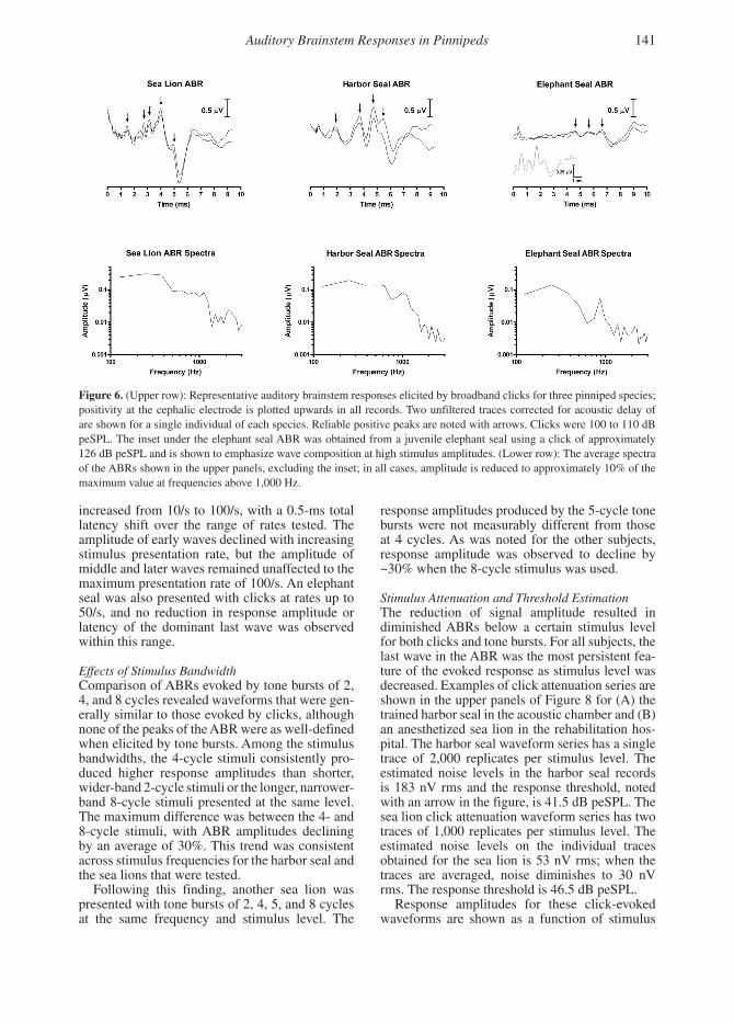
Features of the Click-Evoked ABRUnfiltered click-evoked ABRs to stimuli between 100 and 110 dB peSPL were obtained from four sea lions, two harbor seals, and two northern ele-phant seals. Examples of these responses are pre-sented in Figure 6 in both the time and frequency domains. The shape, amplitude, and latency of the ABR waveform varied slightly between indi-viduals tested but were repeatable within subjects. Across species, all ABR waveforms showed a series of four to five positive peaks followed by a deep negative trough, although the early posi-tive peaks for the elephant seal were only obvious when the stimulus level was raised by 10 to 20 dB (see Figure 6 inset). For the sea lions and elephant seals, the most obvious feature of the response was the last positive peak and the following trough. In contrast, the harbor seals showed relatively high response amplitudes of early, middle, and late positive peaks without a marked trough follow-ing the last wave. Frequency analysis of the ABRs showed the majority of energy below 1 kHz with about 10% of peak amplitude present above 2 kHz. The mean peak-to-peak response amplitude elicited by clicks at these levels was 1.8 µV for the sea lions, 1.4 µV for the harbor seals, and 0.3 µV for the elephant seals.
Effects of FilteringBandpass filtering of the click-evoked ABRs gen-erally reduced noise in the recordings but also affected the ABR. Figure 7 shows a series of wave-forms obtained from an anesthetized California sea lion at different filter settings; the rms noise levels of corresponding blank (stimulus absent) records were also compared to assess relative noise reduc-tion with different filter settings. The widest band-pass filter setting (65 Hz to 1,450 Hz) had little effect on the ABR and did not reduce noise levels in comparison to the unfiltered condition. Further decreasing the low-pass filter to 670 Hz resulted in decreased definition of the ABR waveform but otherwise did not alter the signal or significantly reduce the noise. Raising the high-pass filter from 65 Hz to 220 Hz diminished noise by nearly 65%; it also decreased response amplitude by more than half and distorted the ABR waveform. In particu-lar, the last positive wave (~5.1 ms) in the ABR was truncated, and a prominent positive peak at ~7 ms was introduced. Latency of the ABR waves was also slightly affected by phase distortion introduced by filtering.
Effects of Stimulus Presentation RateCharacteristics of the click-evoked ABR as a func-tion of stimulus presentation rate were investigated in the trained harbor seal. Latency of the entire ABR gradually increased as the click rate was
140 Reichmuth et al.
increased from 10/s to 100/s, with a 0.5-ms total latency shift over the range of rates tested. The amplitude of early waves declined with increasing stimulus presentation rate, but the amplitude of middle and later waves remained unaffected to the maximum presentation rate of 100/s. An elephant seal was also presented with clicks at rates up to 50/s, and no reduction in response amplitude or latency of the dominant last wave was observed within this range.
Effects of Stimulus BandwidthComparison of ABRs evoked by tone bursts of 2, 4, and 8 cycles revealed waveforms that were gen-erally similar to those evoked by clicks, although none of the peaks of the ABR were as well-defined when elicited by tone bursts. Among the stimulus bandwidths, the 4-cycle stimuli consistently pro-duced higher response amplitudes than shorter, wider-band 2-cycle stimuli or the longer, narrower-band 8-cycle stimuli presented at the same level. The maximum difference was between the 4- and 8-cycle stimuli, with ABR amplitudes declining by an average of 30%. This trend was consistent across stimulus frequencies for the harbor seal and the sea lions that were tested.
Following this finding, another sea lion was presented with tone bursts of 2, 4, 5, and 8 cycles at the same frequency and stimulus level. The
response amplitudes produced by the 5-cycle tone bursts were not measurably different from those at 4 cycles. As was noted for the other subjects, response amplitude was observed to decline by ~30% when the 8-cycle stimulus was used.
Stimulus Attenuation and Threshold EstimationThe reduction of signal amplitude resulted in diminished ABRs below a certain stimulus level for both clicks and tone bursts. For all subjects, the last wave in the ABR was the most persistent fea-ture of the evoked response as stimulus level was decreased. Examples of click attenuation series are shown in the upper panels of Figure 8 for (A) the trained harbor seal in the acoustic chamber and (B) an anesthetized sea lion in the rehabilitation hos-pital. The harbor seal waveform series has a single trace of 2,000 replicates per stimulus level. The estimated noise levels in the harbor seal records is 183 nV rms and the response threshold, noted with an arrow in the figure, is 41.5 dB peSPL. The sea lion click attenuation waveform series has two traces of 1,000 replicates per stimulus level. The estimated noise levels on the individual traces obtained for the sea lion is 53 nV rms; when the traces are averaged, noise diminishes to 30 nV rms. The response threshold is 46.5 dB peSPL.
Response amplitudes for these click-evoked waveforms are shown as a function of stimulus
Figure 6. (Upper row): Representative auditory brainstem responses elicited by broadband clicks for three pinniped species; positivity at the cephalic electrode is plotted upwards in all records. Two unfiltered traces corrected for acoustic delay of are shown for a single individual of each species. Reliable positive peaks are noted with arrows. Clicks were 100 to 110 dB peSPL. The inset under the elephant seal ABR was obtained from a juvenile elephant seal using a click of approximately 126 dB peSPL and is shown to emphasize wave composition at high stimulus amplitudes. (Lower row): The average spectra of the ABRs shown in the upper panels, excluding the inset; in all cases, amplitude is reduced to approximately 10% of the maximum value at frequencies above 1,000 Hz.
Auditory Brainstem Responses in Pinnipeds 141

level in the lower panels of Figure 8. For both subjects, amplitude decreases with decreasing stimulus level below about 70 dB peSPL. The trend is linear for a 25 dB range above the esti-mate response threshold (harbor seal: r2 = 0.84, p < 0.05; sea lion: r2 = 0.97, p < 0.01).
The tone-burst attenuation records obtained from the awake harbor seal were unfiltered and gener-ally were not of sufficient quality to visually esti-mate response thresholds with confidence. These records revealed some features of the ABR related to both bandwidth and tonal frequency, however. In terms of stimulus type, the widest bandwidth/shortest duration stimuli, the 2-cycle tone bursts, evoked reasonably large initial ABRs that did not persist as long as those evoked by 4- and 8-cycle tone bursts. The narrowest bandwidth and longest duration stimuli, the 8-cycle tone bursts, evoked a much smaller initial response, but this response persisted longer with attenuation. While responses to the 4-cycle stimuli did not persist quite as long, responses to these stimuli were easier to detect in noise than those elicited by the 8-cycle stimuli. Stimulus frequency also differentially affected the ABRs. Within a given stimulus bandwidth, the
lower frequencies tested (those < about 5.6 kHz) appeared to evoke larger initial responses from the seal (across all components of the waveform), but these responses attenuated more quickly than the smaller but more persistent responses generated by higher-frequency stimulation.
Improved record quality due to subject anes-thetization and increased averaging made visual response threshold estimation possible for the sea lions. The noise floor for all the sea lion sub-jects was similarly low (below 60 nV rms), which allowed comparisons to be made between indi-viduals. The responses for one subadult male to 5-cycle tone bursts of 2, 4, and 8 kHz are shown in Figure 9. Response thresholds are 46.5, 33.5, and 36.5 dB peSPL, respectively. Two female sea lions were also tested at these frequencies and at 16 kHz. The stimuli were comparable to those used with the male (4-cycle tone bursts). One of these females showed response thresholds lower than 50 dB peSPL for all four frequencies (a prob-lem with the attenuator prevented stimulus levels from being decreased below this level, but ABRs were still clearly detectable and of moderately high amplitude at 50 dB). The other sea lion, an older female, had response thresholds above 62 dB peSPL at all four frequencies, with the highest threshold of 79 dB obtained for the 16-kHz stimu-lus (see Figure 10). Note that while her response thresholds are indicated with arrows at high stim-ulus levels, both of the other sea lions tested still had clearly identifiable responses below the lowest stimulus levels presented to this subject.
Discussion
The primary goal of the present study was to deter-mine the stimulus and recording conditions that would result in the highest signal-to-noise ratios of ABRs measured from the skin of pinnipeds. To this end, we chose to use binaural sound presen-tation because binaural stimuli produce auditory responses of significantly higher amplitude than monaural stimuli of the same level (van Olphen et al., 1978). This technique also allowed us to examine the overall auditory response of an indi-vidual rather than relying on findings from one or two ears tested separately.
One of the most notable features of scalp-recorded ABRs is their detectability from a vari-ety of spatial locations on the head (Jewett & Williston, 1971). There are subtle differences in the amplitude, latency, and polarity of individual waves measured from spatially segregated posi-tions, however, that are related to the size, scal-ing, and orientation of the response generators in different species. These differences are also influ-enced by the volume conduction characteristics
Figure 7. Effects of filter settings on ABR waveform in an anesthetized adult male California sea lion; filter bandpass settings are shown to the left of each waveform. Positivity at the cephalic electrode is plotted upwards. Stimulus pre-sentation rate was 24.1/s. Two records averaged from 1,000 responses each are shown for each condition. Noise levels from corresponding stimulus absent trials were as follows: unfiltered: 100 nV rms, 65 to 1,450 Hz; 160 nV rms, 65 to 670 Hz; 160 nV rms, 220 to 1,450 Hz; 35 nV rms, 220 to 670 Hz; and 39 nV rms.
142 Reichmuth et al.

of the head (Martin & Moore, 1977; Starr & Squires, 1982; Merzenich et al., 1983; Holliday & Te Selle, 1985). Correspondingly, there are species-typical differences in spatial measure-ments of ABR amplitude. In the present study, we measured ABRs referenced to a noncephalic
reference electrode along two transects of the sur-face of the head. While the positions measured do not cover all possible recording locations, they do provide a general indication of spatially distrib-uted response features. In the three species tested, maximal response amplitudes for all identifiable
Figure 8. (Upper row): ABR traces in response to attenuated broadband clicks in an awake, unsedated harbor seal (A) and an anesthetized subadult male sea lion (B); stimulus levels are reported to the left of each waveform in dB peSPL. Response thresholds are noted by an arrow to the right of each trial series. Positivity at the cephalic electrode is plotted upwards. For the harbor seal, stimulus presentation rate was 18.1/s and bandpass filtering was from 65 Hz to 1,450 Hz. One trace averaged from 200 responses is shown for each level. For the sea lion, stimulus presentation rate was 24.1/s and bandpass filtering was from 220 Hz to 1,450 Hz. Two traces averaged from 1,000 responses are shown for each level. Noise on corresponding stimulus absent trials was 183 nV rms for the harbor seal and 53 nV rms on each trace for the sea lion. (Lower row): Peak-to-peak amplitude of the ABR as a function of level for the harbor seal (C) and the sea lion (D).
Auditory Brainstem Responses in Pinnipeds 143

waves were found on the anterior portion of the head with little drop-off along the lateral tran-sect. While the resultant response amplitude maps varied slightly among the pinniped species tested, spatial distribution of response amplitude was grossly similar to that reported for domestic dogs (Holliday & Te Selle, 1985).
The individuals tested in the present response mapping effort were fairly small relative to adult conspecifics. The elephant seals were the most extreme case with respect to body size, with the mass of the subjects being up to 20 times smaller than that of mature adult males. The smaller body
sizes of the individuals tested likely yielded rela-tively large responses as far-field ABR amplitude increases with decreasing body size (Yamaguchi et al., 1991). Increasing body size, however, is not reported to influence the pattern of response characteristics measured over the surface of the head. Therefore, it is reasonable to conclude that the results of the present mapping effort represent species-typical patterns of relative response ampli-tude. On the basis of these amplitude maps, the optimal cephalic recording position was identified along the dorsal midline of the head at a position 2 to 4 cm forward of the ears.
Figure 9. Effects of stimulus frequency and level on ABR waveforms in an anesthetized subadult male California sea lion; stimuli are 5-cycle tone bursts with frequencies of 2, 4, and 8 kHz. Stimulus levels are reported to the left of each waveform in dB peSPL. Response thresholds are noted by an arrow to the right of each trial series. Positivity at the cephalic electrode is plotted upwards. Stimulus presentation rate was 24.1/s, and bandpass filtering was from 220 Hz to 1,450 Hz. The waveforms shown at each stimulus level are averaged from two records of 1,000 responses each. Noise on corresponding stimulus absent trials was 30 nV rms.
Figure 10. Effects of stimulus frequency and level on ABR waveforms in an anesthetized female sea lion estimated to be > 15 years old; stimuli are 4-cycle tone bursts with frequencies of 2, 4, 8, and 16 kHz. Stimulus levels are reported to the left of each waveform in dB peSPL. Response thresholds are noted by an arrow to the right of each trial series. Activity at the cephalic electrode is plotted upwards. Stimulus presentation rate was 18.7/s and bandpass filtering was from 220 Hz to 1,450 Hz. The waveforms shown at each stimulus level are averaged from 1,500 responses. Noise on corresponding stimulus absent trials was 50 nV rms.
144 Reichmuth et al.

Clinical studies of humans and animals typi-cally employ two cephalic electrodes rather than one cephalic electrode referenced to a noncephalic site. The most commonly used configuration is placement of an electrode near the vertex that is referenced to another near the mastoid (Stapells & Oates, 1997). In our study, this configuration yielded enhanced early components and dimin-ished later components relative to the waveform of the ABRs recorded with the reference electrode in the noncephalic position. This observation is consistent with those reported for humans (Starr & Squires, 1982) and domestic dogs (Holliday & Te Selle, 1985). The results can be explained by considering how both wave amplitude and polar-ity measured independently at the two sites during mapping interact when the responses at each site are differentially compared to one another. Clinicians often use the vertex-mastoid configura-tion for diagnostic purposes, as well as for audio-metric evaluation, because it highlights features of the ABR arising from different positions along the primary auditory pathway. These components are useful in the identification of sensorineural hear-ing loss, and also can be used along with monaural stimuli and contralateral masking noise to test hear-ing in separate ears. In the present study, we were primarily interested in maximizing the amplitude of the latter portion of the ABR most commonly used in audiometry. Because ABR amplitude was significantly higher, a single cephalic electrode referenced to a noncephalic site, rather than the vertex-mastoid electrode configuration, was most appropriate.
With respect to electrode type, we found that subdermal needle electrodes yielded higher signal-to-noise ratios than did flat disc electrodes as has been demonstrated in other animal studies (see Merzenich et al., 1983). Other advantages of sub-dermal electrodes over surface electrodes include rapid placement, increased stability during testing, and consistently low and balanced inter-electrode impedance (Hall, 2007). Therefore, testing effi-ciency and response detectability were maximized with the use of subdermal electrodes in this study. In subsequent efforts with the trained harbor seal, we have observed that, in situations where initial response levels are relatively high and stimuli are not attenuated for audiometric purposes, surface disc electrodes can also reasonably be used to obtain auditory evoked responses, even in awake subjects.
The noise levels in the records obtained were affected by several variables, including the cephalic electrode position. The midline position identified as optimal for our subjects generated relatively high response amplitudes, but proximity to the eyes and muzzle in some cases resulted in
increased levels of myogenic noise. The amount of noise in the subject records decreased with a decreasing state of arousal, therefore the highest signal-to-noise ratios were obtained in the anesthe-tized subjects. Since time for data collection was limited, 1,000 to 2,000 individual responses were averaged to obtain each record; in most situations, this provided an adequate compromise between noise reduction and trial duration. Ultimately, we found that collecting two separate traces derived from 1,000 averages each offered the best com-promise for our purposes. The responses could be averaged following data collection to achieve additional noise reduction but also separately compared to aid in visual response identification.
Subject state appeared to influence signal-to-noise ratios mainly by affecting the noise pres-ent in the records rather than by altering signal strength. Although a variety of drugs are known to influence auditory evoked responses, fortunately, very few affect the ABR. This is because the influ-ence of most drugs is on cortical, rather than on brainstem, functioning. Of the drugs used in the present study, diazepam, a benzodiazepine, is reported to have minimal effect on the ABR, and isoflurane, a volatile anesthetic, has been reported to only slightly or negligibly slow ABR interwave latencies without altering waveform amplitude or morphology (see Hall, 2007). Telazol® (a mixture of tiletamine HCl and zolazepam) has been shown by Houser et al. (this issue) to have no observable influence on ABRs measured from an elephant seal, which is consistent with reports indicating that ketamine, another dissociative anesthetic, does not affect ABR latency or amplitude values. Correspondingly, it is also unlikely that medeto-midine, an alpha-2 adrenergic agonist that acts as a sedative-analgesic, alters ABR measurements because other neuroleptic drugs are known to have no significant influence on early auditory evoked responses (see Hall, 2007). In the present study, we tested subjects opportunistically under differ-ent drug regimes that varied by species, making it difficult to directly quantify how the subject’s state influenced the noise levels in our records. It was obvious, however, that the cleanest records with the highest signal-to-noise levels were obtained from the anesthetized animals, despite the con-current use of a mechanical ventilator and some electrical monitoring equipment.
The unfiltered ABRs that were obtained for each of the species in the present study had char-acteristics that were generally similar to those reported for other non-odontocete mammals (Huang, 1980; Merzenich et al., 1983), consist-ing of four to five vertex-positive peaks spaced about 1 ms apart, followed in some cases by a negative rounded trough. We did not attempt to
Auditory Brainstem Responses in Pinnipeds 145

identify the components of the ABRs accord-ing to the Roman numeral system recommended for humans (American Electroencephalographic Society, 1984), although the peaks appeared to be grossly similar, especially in the sea lions. We also found that while the responses we identified could be compared to those described for domes-tic dogs, they were not easily described in detail by the numbering system proposed for that spe-cies (Kawasaki & Inada, 1994). To identify sig-nificant ABR characteristics for the pinnipeds, we simply considered the responses with respect to early (vertex-positive waves 1 and 2), late (last vertex-positive wave and following trough), and middle (intermediate) components of the responses. For all subjects, the late components of the ABR were identifiable. While the sea lions and the elephant seals had relatively low-amplitude early and middle ABR components compared to late components, the harbor seals generally had relatively high-amplitude waves throughout the response, consistent with observations by Wolski et al. (2003). While it seems likely that the latter portion of the response observed in our subjects corresponds to wave V-V´ in humans (the most persistent feature of the ABR, commonly used in audiometric investigations), additional investiga-tion is required to definitively link the response components obtained from our subjects to their generators in the primary auditory pathway.
The effects of analog filtering of the electro-physiological records were explored in an anes-thetized sea lion in order to refine the species-appropriate data collection protocol. Filtering of the responses sometimes resulted in noise reduc-tion but also decreased response amplitude and introduced changes in latency due to phase dis-tortion (see Coats, 1983; Hall, 2007). The results observed are consistent with the spectra of the unfiltered ABRs, which show the majority of response energy between 100 Hz and 1,000 Hz for all three species (as shown in Figure 7). Low-pass filtering at 1,450 Hz had a negligible effect on response waveform and amplitude but slightly increased response latency. Dropping the func-tional low-pass filter to 670 Hz decreased the res-olution of individual waves and further increased response latency while not reducing noise in the record or significantly reducing response ampli-tude. In contrast, high-pass filtering at 65 Hz resulted in obvious loss of amplitude and distor-tion of ABR waves, especially in the latter portion of the response, without reducing noise. Further increasing the high-pass filter to 220 Hz simi-larly decreased and distorted the ABR waveform but reduced noise by 75%. The filtering effects observed are consistent with those reported for human subjects (see Hall, 2007) and suggest that
filter recommendations established for human ABR testing (see Mason et al., 2002; Stevens et al., 2002) are probably appropriate for pinni-peds. When possible, responses should be broadly filtered. The use of an analog low-pass filter set to 1,500 Hz or higher may reduce noise and will prevent aliasing of responses when the sampling rate is relatively low. High-pass filtering above 65 Hz will change the ABR spectrum but, in cer-tain situations, may be necessary to reduce noise. Stapells (1994) has noted that high-pass filtering of human ABRs differentially affects responses elicited by lower-frequency stimuli (500 Hz tone bursts), so additional care must be taken when frequency-specific responses are measured. The use of digital zero phase-shift filtering to provide some post hoc high-pass filtering may improve signal-to-noise ratios following data collection and will be worth exploring in future studies with pinnipeds.
One of the stimulus properties explored in the present study was the rate of stimulus presenta-tion. While it has been established that presenta-tion rate is a critical variable in the ABR testing of mammals (Hall, 1992), the extent to which audi-tory responses are altered by stimulus presenta-tion rates is a species-specific property related to temporal processing capabilities. In the case of our harbor seal, response latency gradually increased and the amplitude of the early ABR component gradually decreased as click presentation rates were raised. Since the amplitude of the late ABR component remained stable with increasing click rates to at least 100/s in the harbor seal, and to at least 50/s in the elephant seal, it is likely that stimulus presentation rates as high as 50/s, and probably as high as 100/s, can be used to improve recording efficiency in situations where late wave amplitude is the primary response metric. Given the time constraints on electrophysiological testing of wild subjects, this savings is not insignificant.
Broadband clicks stimulate a wide portion of the basilar membrane and therefore do not provide a great deal of information about the frequency response of the auditory system (see Stapells & Oates, 1997). Clicks windowed by masking noise or acoustic stimulation with short duration tone bursts provide a better indication of frequency sen-sitivity. The observations obtained for the harbor seal and sea lions exposed to tone bursts of varying bandwidths (number of cycles) indicate that 4- to 5-cycle tone bursts provide the best compromise between stimulus bandwidth and ABR ampli-tude. The 2-, 4-, 5-, and 8-cycle tone bursts used in the present study had measured bandwidths of 0.9, 0.5, 0.4, and 0.2 oct, respectively. The 4- and 5-cycle tone bursts yielded the highest response amplitudes. When bandwidth was widened using
146 Reichmuth et al.
2-cycle stimuli, a larger portion of the basilar membrane was excited, but the shorter stimuli had less overall energy. When bandwidth was nar-rowed using 8-cycle stimuli, a smaller portion of the basilar membrane was excited, and the longer stimuli had correspondingly longer rise and fall times, resulting in a less synchronous response from hair cells in the cochlea and a smaller over-all response amplitude. As a result, the best result was achieved with the 5-cycle stimuli, which provided reasonable frequency specificity (less than half an octave) and relatively high-response amplitudes. This result is consistent with recom-mended tone burst testing parameters for human subjects (Mason et al., 2002).
The observable reduction in response ampli-tude with decreasing stimulus levels within a broad range shows that the ABRs obtained from pinnipeds using clicks and tone bursts can be used to provide an indication of stimulus sensitiv-ity. The primary challenge, as it is for all types of audiometry, is response detection in noise or close to threshold where signal-to-noise ratios are poorest. In addition to some of the other subject and measurement factors discussed, the use of multiple traces at each stimulus level that could be compared to one another as well as averaged (e.g., Stapells, 1994) aided visual response detec-tion. Our preliminary results also suggest that reliability of response thresholds’ measurement is likely to be affected by stimulus frequency as well as by record quality. We had difficulty elic-iting ABRs with low-frequency stimuli (below 2 kHz) despite presumably good hearing in this range (e.g., Reichmuth Kastak et al., 2004). Initial responses to low-frequency stimuli were high, probably due to spectrum splatter at high stimulus intensities (Stapells & Oates, 1997), but responses dropped out quickly and likely did not reflect the real sensitivity of the auditory system. While it appears that reasonable sensitivity estimates can be obtained from pinnipeds at higher stimulus fre-quencies, it remains to be seen whether reliable response detection can be conducted using evoked potential techniques at low frequencies.
We were conservative in estimating ABR thresh-olds based on the last visually detectable response. This method of threshold estimation typically results in threshold estimates that are 10 to 30 dB higher than those obtained using standard psycho-physical procedures (e.g., Stapells & Oates, 1997; Brittan-Powell et al., 2005). Even so, we were able to estimate response thresholds as low as 33.5 dB peSPL in the non-acoustically controlled environ-ment of the rehabilitation facility. Thresholds this low are in the region of the ambient noise mea-sured in the testing environment and highlight the fact that absolute (unmasked) hearing thresholds
are not likely to be measurable in nonlaboratory conditions using current testing paradigms. Recent psychophysical measurements of aerial hear-ing sensitivity support the claim that most pub-lished reports of aerial hearing in pinnipeds have been limited by ambient noise (Holt et al., 2004). Electrophysiological response measurements are not immune to effects of masking noise, and inves-tigators will need to consider this limitation in studies of aerial hearing using evoked potential techniques in typical ambient noise settings.
Despite the limitations of ambient noise, it is clear that ABR-derived estimates of hearing sensi-tivity can reveal significant differences in the hear-ing of individuals. The oldest subject we tested, a California sea lion estimated to be at least 15 y of age, had hearing thresholds that were elevated by at least 20 dB relative to those of two younger animals tested under nearly identical conditions. The hearing loss was most significant at the high-est frequency tested (16 kHz). While the origin of this hearing impairment is unknown, the findings are comparable to those obtained with a captive sea lion who had documented age-related hear-ing loss (Schusterman et al., 2002). Thus, despite certain limitations, it is reasonable to state that evoked potential methods show promise for the study of auditory physiology and auditory sensi-tivity in pinnipeds, and that these methods may provide much needed insight into population-level variability in hearing.
The findings and observations reported in this paper are merely a first step towards developing electrophysiological measurement techniques for use with pinnipeds. More sophisticated analysis procedures, such as those relying on objective response detection, should improve threshold esti-mates obtained using click and tone burst stimuli. These include statistical procedures such as cross-correlation measures and automated analyses of signal-to-noise ratios. Another procedure com-monly used with marine mammals is thresh-old extrapolation from the linear portion of the response amplitude-stimulus level plot. We were conservative in avoiding this analysis technique in the present study; the approach would have yielded lower thresholds but is valid for only a very small range of stimulus amplitudes just above the actual threshold (see Hall, 2007). Because we knew that masking by ambient noise was an issue with much of our data, we also wanted to avoid extrapolat-ing thresholds below the level of the background noise. In the future, the use of sinusoidal amplitude modulated stimuli may provide a greater ability to objectively detect and measure early auditory evoked responses in pinnipeds as techniques using these stimuli continue to be refined for use with
Auditory Brainstem Responses in Pinnipeds 147

other marine mammals (see this issue) as well as with humans (see Stapells et al., 2005).
Regardless of how ABR data are used to obtain estimates of hearing sensitivity, it is important to keep in mind that ABR procedures are not percep-tual hearing tests; as noted by Hall (2007), these responses provide no information on auditory func-tion above the level of the brainstem. Even for human clinical testing, where a great deal of effort has been invested in developing ABR techniques, behavioral thresholds can only be accurately estimated using empirically derived relationships between evoked potential and behavioral data (Stapells et al., 1995). The difference in ABR and behavioral thresholds can be partly attributed to the difference in stimulus durations used in the two procedures: the relatively long tones used in behavioral audiometry (> 500 ms) are above the upper limit of auditory tempo-ral summation in mammals while the brief tone bursts used in ABR studies are well below this limit (< 10 ms). While it may be tempting to normalize the energy content of the two signals with differ-ent durations in order to generate equivalent thresh-olds that can be directly compared (e.g., see Wolski et al., 2003), it is not appropriate to do so since this transformation fails to account for time constants associated with temporal summation.
A rough comparison of tone burst ABRs and pure tone behavioral thresholds expressed in dB SPL for the trained harbor seal in this study show ABR thresholds at least 30 dB higher than behav-ioral thresholds at frequencies below 10 kHz; this correspondence improved to within 10 dB at the few frequencies tested above 10 kHz. These results are promising, but at this point, cannot substitute for behavioral audiometric measurements. This contrasts with recent findings with odontocete cetaceans (see Finneran & Houser, 2006; Yuen et al., 2005) that reveal remarkably similar behav-iorally derived measurements of hearing sensitiv-ity and evoked potential measurements obtained using sinusoidally amplitude-modulated stimuli.
A great deal remains to be learned about the audi-tory physiology of pinnipeds and the ways in which electrophysiological measurements can be applied to improving our understanding of their auditory function and sensitivity. One of the main challenges for testing these amphibious animals will be to determine if and how estimates of underwater hear-ing can be obtained using evoked potential meth-ods. With respect to the continued development of species-appropriate methods for obtaining physi-ologic hearing data, future efforts should attempt to expand these measurements using individual ABRs to those relying on auditory steady-state responses, also known as envelope-following responses. The findings of the present study should provide a foundation for continued investigation.
Acknowledgments
The animal protocols used in this study were approved by the Institutional Animal Care and Use Committees at University of California–Santa Cruz (UCSC) and The Marine Mammal Center (TMMC). Research was conducted under National Marine Fisheries Service (NMFS) marine mammal permits 932-1489-07 and 1072-1771-00. Funding for this study was provided in part by the National Oceanographic Partnership Program under award N00014-1-7070, the NMFS Ocean Acoustics Program and the Office of Naval Research under award N00014-05-1-0911, and the Science and Technology Engagement Program at ONR Global under award N00014-05-1-4022. We gratefully acknowledge invaluable support from Martin Haulena, Frances Gulland, Sophie Dennison, and Delphine Sarran at TMMC. We also thank the hard-working team at the Pinniped Cognition and Sensory Systems Lab at UCSC for their help with this project, especially Kristy Lindemann, who trained the harbor seal, and Asila Ghoul, who pro-vided artwork for Figure 4. David Stapells at the University of British Columbia generously answered many of our questions as we analyzed the data, and two anonymous reviews provided helpful comments that improved this manuscript. Finally, we wish to thank Paul Nachtigall for organizing the 2002 Pauley summer workshop that gave rise to this cooperative project.
Literature Cited
American Electroencephalographical Society. (1984). Guidelines for clinical evoked potential studies. Journal of Clinical Neurophysiology, 1, 3-53.
Brittan-Powell, E. F., Lohr, B., Hahn, D. C., & Dooling, R. J. (2005). Auditory brainstem responses in the Eastern Screech Owl: An estimate of auditory thresh-olds. Journal of the Acoustical Society of America, 118(1), 314-321.
Bullock, T. H., Ridgway, S. H., & Suga, N. (1971). Acoustically evoked potentials in midbrain auditory structures in sea lions (Pinnipedia). Zeitschrift für Vergleichende Physiologie, 74, 372-387.
Bullock, T. H., Grinnell, A. D., Ikezono, F., Kameda, K., Katsuki, Y., Nomoto, M., et al. (1968). Electrophysiological studies of the central auditory mechanisms in cetaceans. Zeitschrift für Vergleichende Physiologie, 59, 117-156.
Coats, A. C. (1983). Instrumentation. In E. J. Moore (Ed.), Bases of auditory brain-stem evoked responses (pp. 197-220). New York: Grune and Stratton.
Finneran, J. J., & Houser, D. S. (2006). Comparison of in-air evoked potential and underwater behavioral hearing thresholds in four bottlenose dolphins (Tursiops trunca-tus). Journal of the Acoustical Society of America, 119, 3181-3191.
148 Reichmuth et al.

Hall, J. W. (1992). Handbook of auditory evoked responses. Boston: Allyn & Bacon. 419 pp.
Hall, J. W. (2007). New handbook of auditory evoked responses. Boston: Pearson Education. 750 pp.
Hemilä, S., Nummela, S., Berta, A., & Reuter, T. (2006). High-frequency hearing in phocid and otariid pinni-peds: Inertial and cochlear constraints. Journal of the Acoustical Society of America, 120(6), 3463-3466.
Holliday, T. A., & Te Selle, M. E. (1985). Brain stem audi-tory-evoked potentials of dogs: Wave forms and effects of recording electrode positions. American Journal of Veterinary Research, 46, 845-851.
Holt, M. M., Southall, B. L., Kastak, D., Reichmuth Kastak, C., & Schusterman, R. J. (2001). Aerial hearing sensitivity in pinnipeds: A comparison of free-field and headphone thresholds. Proceedings of the 14th Biennial Conference on the Biology of Marine Mammals, p 102.
Holt, M. M., Southall, B. L., Kastak, D., Schusterman, R. J., & Reichmuth Kastak, C. (2004). Temporal integration in a sea lion and a harbor seal: Estimates of aerial auditory sensitivity as a function of signal duration. Journal of the Acoustical Society of America, 116, 2531.
Houser, D. S., Crocker, D. E., Kastak, C., Mulsow, J., & Finneran, J. J. (2007, this issue). Auditory evoked poten-tials in northern elephant seals (Mirounga angustiros-tris). Aquatic Mammals, 33(1), 110-121.
Huang, C. (1980). A comparative study of the brain stem auditory response in mammals. Brain Research, 184, 215-219.
International Electrotechnical Commission. (1994). Audiometers. Part 3: Auditory test signals of short dura-tion for audiometric and neuro-otological purposes (International Standard 60645-3). 18 pp.
Jewett, D. L., & Williston, J. S. (1971). Auditory-evoked far fields averaged from the scalp of humans. Brain, 94, 681-696.
Kawasaki, Y., & Inada, S. (1994). Peaks of brainstem audi-tory evoked potentials in dogs. Veterinary Research Communications, 18, 383-396.
Kelly, J. B., Kavanagh, G. L., & Picton, T. W. (1989). Brainstem auditory evoked response in the ferret (Mustela putorius). Hearing Research, 39, 231-240.
Martin, M. E., & Moore, E. J. (1977). Scalp distribution of early (0 to 10 msec) auditory evoked responses. Archives of Otolaryngology, 103, 326-328.
Mason, S., Elliott, C., Lightfoot, G., Parker, D., Stapells, D. R., Stevens, J., et al. (2002). Auditory brainstem response testing in babies using tone pip stimulation: A recommended test protocol. Retrieved 3 March 2005 from www.nhsp.info.
Merzenich, M. M., Gardi, J. N., & Vivion, M. C. (1983). Animals. In E. J. Moore (Ed.), Bases of auditory brain-stem evoked responses (pp. 391-412). New York: Grune and Stratton.
Moore, E. J. (1983). History. In E. J. Moore (Ed.), Bases of auditory brain-stem evoked responses (pp. 3-14). New York: Grune and Stratton.
Munro, K. J., Shiu, J. N., & Cox, C. L. (1997). The effect of head size of the auditory brainstem response for two breeds of dog. British Journal of Audiology, 31(5), 309-314.
National Research Council (NRC). (2000). Marine mam-mals and low-frequency sound: Progress since 1994. Washington, DC: National Academy Press. 146 pp.
NRC. (2003). Ocean noise and marine mammals. Washington, DC: National Academy Press. 192 pp.
NRC. (2005). Marine mammal populations and ocean noise. Washington, DC: National Academy Press. 126 pp.
Reichmuth Kastak, C., Kastak, D., Holt, M. M., Schusterman, R. J., & Southall, B. L. (2004). Aerial hearing in some pinnipeds is comparable to that of humans. Journal of the Acoustical Society of America, 115, 2406.
Ridgway, S. H., & Joyce, P. L. (1975). Studies on seal brain by radiotelemetry. Rapports et Procès-Verbaux des Réunions, 169, 81-91.
Schusterman, R. J., Southall, B. L., Kastak, D., & Reichmuth Kastak, C. (2002). Age-related hearing loss in sea lions and their scientists. Journal of the Acoustical Society of America, 111, 2342-2343.
Shiu, J. N., Munro, K. J., & Cox, C. L. (1997). Normative auditory brainstem response data for hearing threshold and neuro-otological diagnosis in the dog. Journal of Small Animal Practice, 38(3), 103-107.
Silman, S., & Silverman, C. A. (1991). Auditory diagno-sis: Principles and applications. San Diego: Academic Press. 412 pp.
Sininger, Y. S., Cone-Wesson, B., Folsom, R. C., Gorga, M. P., Vohr, B. R., Widen, J. E., et al. (2000). Identification of neonatal hearing impairment: Auditory brain stem responses in the perinatal period. Ear and Hearing, 21(5), 383-399.
Southall, B. L., Schusterman, R. J., & Kastak, D. (2000). Masking in three pinnipeds: Underwater, low-frequency critical ratios. Journal of the Acoustical Society of America, 108(3), 1322-1326.
Southall, B. L., Schusterman, R. J., & Kastak, D. (2003). Auditory masking in three pinnipeds: Aerial critical ratio and critical bandwidth measurements. Journal of the Acoustical Society of America, 114, 1660-1666.
Stapells, D. R. (1994). Low-frequency hearing and the auditory brainstem response. The American Journal of Audiology, 3(2), 11-13.
Stapells, D. R., & Oates, P. (1997). Estimation of the pure-tone audiogram by the auditory brainstem response: A review. Audiology and Neuro Otology, 2, 257-280.
Stapells, D. R., Gravel, J. S., & Martin, B. A. (1995). Thresholds for auditory brainstem responses to tones in notched noise from infants and young children with normal hearing or sensorineural hearing loss. Ear and Hearing, 16, 361-371.
Stapells, D. R., Herdman, A., Small, S. A., Dimitrijevic, A., & Hatton, J. (2005). Current status of the auditory steady-state responses for estimating an infant’s audio-gram. In R. C. Seewald & J. M. Banford (Eds.), A sound
Auditory Brainstem Responses in Pinnipeds 149

foundation through early amplification 2004 (pp. 43-59). Basel, Switzerland: Phonak AG.
Starr, A., & Squires, K. (1982). Distribution of auditory brainstem potentials over the scalp and nasopharynx in humans. Annals of the New York Academy of Sciences, 338, 427-442.
Stevens, J., Elliott, C., Lightfoot, G., Mason, S., Parker, D., Stapells, D., et al. (2002). Click auditory brainstem response testing in babies: A recommended test proto-col. Retrieved 3 March 2005 from www.nhsp.info.
Supin, A. Ya., Popov, V. V., & Mass, A. M. (2001). The sen-sory physiology of aquatic mammals. Boston: Kluwer Academic Publishers. 332 pp.
van Olphen, A. F., Rodenburg, M., & Verwey, C. (1978). Distribution of brain stem responses to acoustic stimuli over the human scalp. Audiology, 17, 511-518.
Wartzok, D., & Ketten, D. R. (1999). Marine mammal sen-sory systems. In J. E. Reynolds & S. A. Rommel (Eds.), Biology of marine mammals (pp. 117-175). Washington, DC: Smithsonian Institution Press.
Wilson, W. J., & Mills, P. C. (2005). Brainstem auditory-evoked response in dogs. American Journal of Veterinary Research, 66, 2177-2187.
Wolski, L. F., Anderson, R. C., Bowles, A. E., & Yochem, P. K. (2003). Measuring hearing in the harbor seal (Phoca vitulina): Comparison of behavioral and audi-tory brainstem response techniques. Journal of the Acoustical Society of America, 113, 629-637.
Yamaguchi, J., Yagi, T., Baba, S., Aoki, H., & Yamanobe, S. (1991). Relationship between auditory brainstem response waveform and head size. ORL – Journal for Oto-Rhino-Laryngology and Its Related Specialties, 53, 94-99.
Yuen, M. M. L., Nachtigall, P. E., Breese, M., & Supin, A. Ya. (2005). Behavioral and auditory evoked poten-tial audiograms of a false killer whale (Pseudorca crassidens). Journal of the Acoustical Society of America, 118, 2688-2695.
150 Reichmuth et al.


![1 ECE Department [I/O] ³ Input/Output Cubed Kevin Eykholt Tom Finneran Chris Pitoniak Shamit Som.](https://static.fdocuments.us/doc/165x107/56649ce05503460f949aa47b/1-ece-department-io-inputoutput-cubed-kevin-eykholt-tom-finneran-chris.jpg)