
MEA-System Extracellular ... Cell lines or primary cell preparations are cultivated directly on the...
16
MEA-System Extracellular recording with microelectrode arrays for all applications • Well-established technology • Recording from up to 256 channels • Microelectrode arrays with various layouts • Broad range of accessories
Transcript of MEA-System Extracellular ... Cell lines or primary cell preparations are cultivated directly on the...

MEA-SystemExtracellular recording with microelectrode arrays for all applications
• Well-established technology
• Recording from up to 256 channels
• Microelectrode arrays with various layouts
• Broad range of accessories

Benefits of the MEA-Systems from MCS• Suitable for upright and inverted microscopes.
• Widest range of MEAs available on the market.
• Unlimitedandfreesoftwareupgrades:flexibledataacquisitionand analysis software MC_Rack.
• Easy adaption to our stimulus generators.
• Stimulus artifact suppression.
• Expandabletomultipleamplifiersystem.
• Software selection of stimulation electrodes.
• Real-time spike detection and feedback generation.
Microelectrode Array: A well-established technologySince its introduction 30 years ago, the technology and the related culture methods for
electrophysiological cell and tissue assays have been continually improved and have
found their way into many academic and industrial laboratories. MEA technology has
attracted increased interest because of a growing need to screen selected compounds
against ion channel targets in their native environment at organic, cellular, and sub-
cellular level.
The Microelectrode Array (MEA)-System is a compact and innovative tool for in vitro
experiments. You can place cell and tissue preparations from heart, brain, and muscle
ontheMEAandrecordtheelectrophysiologicalsignalswiththeMEAamplifier.The
signals are then analyzed with the included software. The modular principle offers
various possibilities for a set-up extension with perfusion and stimulation devices.The MEA-technology is an easy
andstraightforwardapproachtoapplyelectrophysiologicaltechniquesfordrugscreeningandbasicresearch.Over
600publicationsinscientificjournalsproveitsversatilityandreliability.
Classical MEA-Systems with 60, 120 or 240 channelsThe classical MEA-System consists of a
dataacquisitioncomputer,onetofour
MEAamplifiers,MEAs,andatemperature
controller.
ThecoreelementistheMEAamplifierwith
60 channels. Depending on your experimental
need, you can decide whether to build a
setupwithone,twoorfouramplifiers,
resulting in a system with 60, 120 or 240
channels respectively. You can run completely
independent experiments on each of the
amplifiers.
MEAamplifiersareavailableintwodifferentversions,designedspecificallyforuprightandinvertedmicroscopes.
Fordataacquisition,youcanchooseeitherthePCI-busdataacquisitioncard,whichispreinstalledinacomputer
oraUSBdataacquisition,whichcaneasilybeconnectedviaUSB2.0HighSpeedtoanyPCorlaptop.
Nomatterwhichoptionyouchoose,flexibility,thepossibilityforsetupexpansions,andourrecordingandanalysis
software MC_Rack are always included.
Data acquisition
MEA amplifiers
Temperature controllers
Perfusion canullas
Overview MEA-System

All-in-one solution for 256 channelsTheUSB-MEA256-Systemisastand-aloneplug-and-playdataacquisitionsystem
based on signal processing technology.
All necessary components are combined in one device:
• Integratedamplifierfor252+4channels:252channelsfromthemicroelectrodearray plus 4 additional channels for simultaneous patch clamp recordings or anyotheranalogsignalssuchastemperature,pH,etc.
• Integratedanalog/digitalboardforconvertinganalogsignalstodigitaldatastreamsat16bitresolutionandasamplingrateof40kHz/channel.
• Integratedtemperaturecontrol.
• Easy adaption to our stimulus generators for current and voltage driven stimulation. Each electrode can be used for stimulation.
Acute hippocampal slice recording systemThe acute hippocampal slice recording
system, MEA2100-32-System, is a stand-
alone solution for extracellular recording and
stimulation using perforated microelectrode
arrays(pMEAs).Itisdesignedspecifically
for experiments with acute hippocampal
slices, but can be used for all acute slice
preparations.
Based on the MEA2100 technology, the
system consists of a headstage and an
interfaceboard.Headstagesareavailable
for one or two microelectrode arrays (32
recording, 12 stimulation electrodes) and
containa32-or64-channelamplifierand
dataacquisition,aswellasanintegrated
three-channelstimulatorperMEA.Perfusion,
heating, and the possibility to apply suction
through the pMEAs are also included.
The system is compact and can be used on a
standard lab bench. As two headstages can
be connected to one interface board, you
can record from four microelectrode arrays
simultaneously.
MEA-Systems with integrated stimulation: MEA2100-SystemTheMEA2100-Systemiscomposedofadataacquisitioncomputerwithsoftware,
an interface board, one or two MEA-headstages with integrated stimulation,
MEAs, as well as temperature control and perfusion canulla. Due to its
small-sized design you can position the MEA-headstage
onanyinvertedoruprightmicroscope.Itis
connectedviaonlyoneMCSHighSpeed
cable to the interface board, which offers
variousdigitalandanalogin-/outputsfor
synchronization with other instruments.
ThemainadvantageoftheMEA2100-Systemisitsflexibility.
Multi Channel Systems offers various contact units for the MEA-headstage. You can
decide whether to work with one 60-electrode MEA, one 120-electrode MEA, or
even two 60-electrode MEAs.
Moreover, it has a digital signal processor for real-time signal detection and
feedback.
TheflexibilityoftheMEA-2100-Systemisalsoshowninthepossibilitytoconnect
two MEA-headstages to the interface board. This way, you can record from up to
240 channels. By e.g. using two headstages with two 60-electrode MEAs each, you
have a four-fold system and increased throughput. The headstages are controlled
completelyindependentlybyopeningthedataacquisitionsoftwareMC_Rack
multiple times.

Flexible and easy-to-use softwareThedataacquisitionandanalysisprogramMC_Rackishighlyadaptablewith
unlimitedpossibilities.ItisincludedwithallMEA-
and ME-Systems.
For routine lab work, the program is set up like
an instrument rack on a workbench:
• Combine virtual instruments (e.g. oscilloscope,filter,spikesorter,andmuchmore).
• Virtual instrument rack: Use task-oriented template racks or design your own.
• Select any permutation of data streams for displaying, analyzing, exporting, etc.
• Extract parameters like spike rates for online orofflineanalysis.
• Applyseveraldigitalfilterswithdifferentcutofffrequenciese.g.toseparatespikeactivityfromlocalfieldpotentials.
Online and offline analysis features of MC_RackOnlinefiltering,spikesorting,localfieldpotential(LFP)extraction,and
triggering allow you to monitor parameters during the experiment
andsaveofflineanalysistime.Forultimateexperimentalcontrol,
youcanintegratetheprogramcontrols(DLL)intoyourowncustom
software.
After the experiment you can review the raw data and extract
additionalparametersoffline.Adjustspikedetectionoranalyzer
settings and re-run your experiment any number of times. Take the
computer performance to the limit and extract multiple parameters in
parallel. For example, you can use signal rate, peak-peak amplitude,
slope10/90%andmore;orseparatedifferentsignalfrequenciesby
digitalfiltersandanalyzethemseparately.
Real-time signal detection and feedbackSynaptic communication between neurons and
wave propagation in cardiac tissue happen on a
millisecondtimescale.Manyapplicationsrequire
stimulatingatdefinedlocationswithinaneuronal
network as a response to activity on one or more
specificelectrodes.Thereal-timesignaldetection
and feedback allows sending out trigger pulses
within less than 100 µs and trigger a stimulus
withlessthan1msdelay.Inordertoachievethis
extraordinary performance the signal detection,
analysis, and feedback logic happens on a special signal processor unit within the USB
dataacquisition.ThisbypassesthePCaltogethertospeeduptheprocess.Withreal-time
signal detection and feedback, it is possible to enable communication between devices
acceptingTTLpulsesasatriggerwithoutdelay.
Channel 3
Log
ic
Dig Out 0
Dig Out 1
Dig Out 2
Dig Out 15
STG
Channel 2
Channel 1
Channel 64
Highpass
Filter Lowpass
Bandpass
Highpass
Filter Lowpass
Bandpass
Highpass
Filter Lowpass
Bandpass
Highpass
Filter Lowpass
Bandpass
Threshold
Signal/ Spike Detection
Threshold
Signal/ Spike Detection
Threshold
Signal/ Spike Detection
Threshold
Signal/ Spike Detection
Single Channel Analyzer
Events in a time frame
Multi Channel Analyzer
Events in a time frame
Log
ic
Software
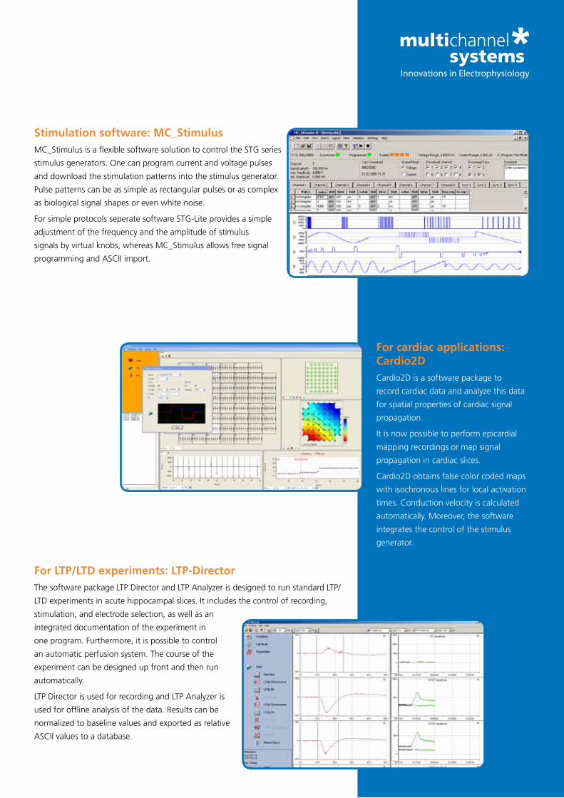
Stimulation software: MC_StimulusMC_StimulusisaflexiblesoftwaresolutiontocontroltheSTGseries
stimulusgenerators.Onecanprogramcurrentandvoltagepulses
and download the stimulation patterns into the stimulus generator.
Pulsepatternscanbeassimpleasrectangularpulsesorascomplex
as biological signal shapes or even white noise.
ForsimpleprotocolsseperatesoftwareSTG-Liteprovidesasimple
adjustmentofthefrequencyandtheamplitudeofstimulus
signals by virtual knobs, whereas MC_Stimulus allows free signal
programmingandASCIIimport.
For cardiac applications: Cardio2DCardio2D is a software package to
record cardiac data and analyze this data
for spatial properties of cardiac signal
propagation.
Itisnowpossibletoperformepicardial
mapping recordings or map signal
propagation in cardiac slices.
Cardio2D obtains false color coded maps
with isochronous lines for local activation
times. Conduction velocity is calculated
automatically. Moreover, the software
integrates the control of the stimulus
generator.
For LTP/LTD experiments: LTP-DirectorThesoftwarepackageLTPDirectorandLTPAnalyzerisdesignedtorunstandardLTP/
LTDexperimentsinacutehippocampalslices.Itincludesthecontrolofrecording,
stimulation, and electrode selection, as well as an
integrated documentation of the experiment in
one program. Furthermore, it is possible to control
an automatic perfusion system. The course of the
experiment can be designed up front and then run
automatically.
LTPDirectorisusedforrecordingandLTPAnalyzeris
usedforofflineanalysisofthedata.Resultscanbe
normalized to baseline values and exported as relative
ASCIIvaluestoadatabase.

Stimulus generatorsThe stimulus generators of the 4000 series operate in voltage or current mode.
The respective mode is selected by the included software. You can decide in favor
of 2, 4 or even 8 completely independent stimulus outputs. Every single output is
optically isolated and has the ability to provide any arbitrary analog waveform as
stimulationsignal.EverySTGcomeswithMC_Stimulussoftware.
Furthermore,foreverysinglestimulusoutputthereisoneTTLin-andoutput,
soyoucansynchronizeyourdataacquisitionortriggerotherdevices.Youcan
dynamically change the output signal and downstream pulses during stimulation.
Stimulation isolation units (one per output channel) are already included in the
stimulusgenerator.Youdonotneedanyotherdevice-justpluginyourstimulator
and start your experiment!
Temperature controllerThe general purpose temperature controller (TC) is included with
each MEA-System. Depending on the set-up, the TC
has one or two output channels.
ThePt100sensorguaranteesastableandprecise
temperature control over a wide temperature range.
Youcanadjustthetemperatureaccuratelyfrom
ambient temperature up to 105 °C using either the buttons
on the device itself or the included TCX_Control software. This
software also tracks the temperature and saves the data, so you can
review it anytime.
Peristaltic perfusion systemThe peristaltic perfusion system is the perfect addition to all microelectrode array recording
systems.TheperistalticpumpPPS2hasoneinletandoneoutletpump.
ThemainadvantageofthePPS2isitslowpulsation.Alow-pulsationcontruction,a
brushless motor with constant rotation speed and low electromagnetic emission, and
droplet isolation chambers all contribute to an overall low pulsation level.
Theflowrateofbothpumpscanbeadjustedinthefollowingways:
•Software(included,connectionviaUSB2.0)
•Touchscreenonthepumpitself
•Additionalanaloginput
TheperistalticperfusionpumpisincludedwithallMEA-Systemswithperfusioncannula.In
combination with the also included magnetic perfusion holders, you have all that is needed
to start perfusion right away.
Accessories

Extracellular recording with Microelectrode ArraysA microelectrode array (MEA) is an arrangement of typically 60 electrodes
allowing the targeting of several sites in parallel for extracellular recording
and stimulation.
Cell lines or primary cell preparations are cultivated directly on the MEA.
Freshly prepared slices can be used for acute recordings, or can be
cultivatedasorganotypiccultures(OTC)ontheMEA.
Several MEA geometries are provided for a wide variety of applications.
Almost all excitable or electrogenic cells and tissues can be used for
extracellular recording in vitro, for example, central or peripheral neurons,
cardiac myocytes, whole-heart preparations, or retina.
Widest and best choice for all applicationsThe broad range of applications is
reflectedbythevarietyofMEAswith
different geometries that have been
developed to cover as many applications
as possible.
The biological sample can be positioned
directlyontherecordingarea;the
MEA serves as a culture and perfusion
chamber. A temperature controller
controls the temperature in the culture
chamber. Various culture chambers are
available, for example, ones with leak
proof lids or with semipermeable seals.
Anincubatorisnotnecessarilyrequired,
long-term recordings in the MEA culture
chamber are possible over several weeks
or even months.
Production at highest quality standardsTheNaturalandMedicalSciencesInstitute(NMI)inReutlingen,
Germany(www.nmi.de),producesMEAsfromverypure,fine
qualityandhighlybiocompatiblematerials.TheNMIisaresearch
institute, with which Multi Channel Systems has collaborated on
manyprojectsandovermanyyears.
Quality controls and production processes have been improved
over the last years so that MEAs are always of an excellent and
consistentquality.
MEA-Introduction

Standard 8x8 layoutTheconfigurationof8by8electrodesisthe
mostversatileconfiguration.Applications
range from neuronal networks to brain slices
and from stem cell derived cardiomyocytes
to cardiac tissue preparations. The spacing of
the electrodes is available at 100 µm and 200
µm.Thisrepresentsasquareshapedrecording
area of 700 µm or 1.4 mm respectively. The
electrodes are available with diameters of
10 µm and 30 µm. The advantage of 30 µm
diameter electrodes is their low impedance and
low noise level. 10 µm electrodes enable recording from single neurons or single cardiomyocytes.
Some MEAs feature internal reference electrodes. Due to the integrated reference the culture can
be kept sterile during recording to enable repeated recordings of long-term cultures.
6x10 layoutThe 6 by 10 layout features an interelectrode distance of 500 µm.
Thiscreatesarecordingfieldof4.5mmby2.5mm.Withthese
dimensions larger tissue samples can be recorded on one array.
Each of the electrodes can be used for stimulation as well. All MEAs
with the 6x10 layout also feature internal reference electrodes. Each
of the electrodes can be selected as a stimulation electrode as well.
The electrode material is TiN. The micro-column structure of each
electrode minimizes impedance and allows low-noise recordings.
The extremely durable material allow as much as 50 re-use cycles
with acute experiments.
HighDense MEAsA 30 µm electrode spacing is the ultimate
in spatial resolution. This is possible by
arranging 60 electrodes in two recording
areas of 30 electrodes each. These two areas
arespacedat500µm.Theconfiguration
within the two distinct recording areas is 5 by
6 electrodes. This translates into two distinct
recordingfieldsof120µmx150µm.The
gap between the recording areas is used to
guide the connecting lanes to the contact
pads.
The main application of this MEA type is high resolution recording of individual
neurons in neuronal networks. The electrode diameter is 10 µm. A low noise level is
guaranteed by the use of TiN as electrode material.
MEA-Layouts

HexaMEAsThe60electrodesofHexaMEAsareavailableintwogeometries.
Either,theelectrodesarealignedinequaldistances(40µm)with
one electrode diameter (10 µm) as shown on the left or in a special
configurationwithvaryingelectrodediameters(10,20,30µm)and
interelectrode distances.
Thespecificlayoutwithvaryingdiametersanddistancesideally
resembles the regularity of the retina’s architecture. The density of
neurons is more important in the center than in the peripheral. This
is matched by the density of electrodes on the MEA, which is also
higher in the center than in the peripheral.
Multi-well MEAsTo increase the throughput in drug testing
and screening applications we offer MEAs
with multiple wells. These allow operation
of up to nine independent experiments
on one MEA. They can be used to run a
number of replicates in parallel or to obtain
a complete dose response curve within one
recording. The 256MEA technology has 28
electrodes and internal reference in each
wellofa9-wellMEAchip.
Applications include toxicology,
neurobiology, stem cell research, and safety
pharmacology. The data streams from the
different wells can be analyzed individually.
Currently MCS offers 6-well MEAs with
60electrodesand9-wellMEAswith252
electrodes.Special layoutsDifferentapplicationsdohavespecialrequirementsregardingelectrodeconfiguration,
materials, and special features. Multi Channel Systems puts customer needs as
thetoppriority.Incollaborationwithsomeofour
customers we have developed a wide range of special
electrodelayouts.Sometypicalmodificationsinclude
the addition of specially shaped stimulation electrodes
orset-uplayoutswithfourquadrantsofhighdensity
recording areas.
The material used for special layout MEAs is very
variable.ElectrodescanbemadefromGold,TiN,ITOor
evenIridium.Itispossibletointegratemicrofluidicsand
perforation. The basic material can be glass, printed
circuit boards or a polyimide foil.
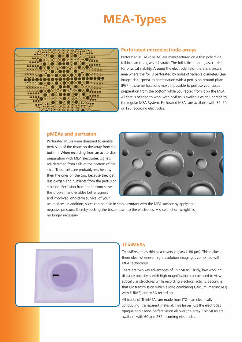
Perforated microelectrode arraysPerforatedMEAs(pMEAs)aremanufacturedonathinpolyimide
foilinsteadofaglasssubstrate.Thefoilisfixedonaglasscarrier
forphysicalstability.Aroundtheelectrodefield,thereisacircular
area where the foil is perforated by holes of variable diameters (see
image,darkspots).Incombinationwithaperfusiongroundplate
(PGP),theseperforationsmakeitpossibletoperfuseyourtissue
preparation from the bottom while you record from it on the MEA.
All that is needed to work with pMEAs is available as an upgrade to
theregularMEA-System.PerforatedMEAsareavailablewith32,60
or 120 recording electrodes.
pMEAs and perfusionPerforatedMEAsweredesignedtoenable
perfusion of the tissue on the array from the
bottom. When recording from an acute slice
preparation with MEA electrodes, signals
are detected from cells at the bottom of the
slice. These cells are probably less healthy
then the ones on the top, because they get
less oxygen and nutrients from the perfusion
solution.Perfusionfromthebottomsolves
this problem and enables better signals
and improved long-term survival of your
acuteslices.Inaddition,slicescanbeheldinstablecontactwiththeMEAsurfacebyapplyinga
negative pressure, thereby sucking the tissue down to the electrodes. A slice anchor (weight) is
no longer necessary.
ThinMEAsThinMEAs are as thin as a coverslip glass (180 µm). This makes
them ideal whenever high resolution imaging is combined with
MEA technology.
There are two key advantages of ThinMEAs: Firstly, low working
distanceobjectiveswithhighmagnificationcanbeusedtoview
subcellular structures while recording electrical activity. Second is
that UV transmission which allows combining Calcium imaging (e.g.
with FURA2) and MEA recording.
AlltracksofThinMEAsaremadefromITO–anelectrically
conducting,transparentmaterial.Thisleavesjusttheelectrodes
opaqueandallowsperfectvisionalloverthearray.ThinMEAsare
available with 60 and 252 recording electrodes.
MEA-Types

MEAs with 256 electrodesWith the introduction of the USB-MEA256-System MCS also introduced MEAs
with 256 electrodes. There are three advantages with the increased number of
electrodes:
• Higherspatialresolution
• Largerrecordingarea
• Higherthroughput
By reducing the electrode spacing it is possible to map a distinct area
withahigherspatialresolution.Ina16by16electrodearraygrid
electrode spacing of 30, 60, 100, and 200 µm are available. The electrode
diameter is 8, 10, and 30 µm. All 256MEA layouts have internal reference
electrodes.
Apartfromthe16x16layout,MultiChannelSystemsalsooffersa9-well
MEAfortheUSB-MEA256-System.Ithas26recordingelectrodes(30µm
diameter, TiN) and two large stimulation electrodes (200 µm by 50 µm) in
every well. Each well has an internal reference electrode. Every recording electrode
can be used for stimulation and even the stimulation electrodes can be used for
recording.
The researcher has all options to design the experimental setup that best suits
them. Since analysis of such a large set of data recorded with 256 channels is
tedious, MCS offers semi-automated analysis routines and easy export features
into Matlab or other analysis programs.
MEAs with 120 electrodes for the MEA2100-SystemOneofthemainadvantagesoftheMEA2100-Systemis
itsflexibility.Becauseitisavailablewithvariouscontact
units, you can choose to operate it with MEAs with 120
electrodes. They are arranged in a 12x12 grid, sparing
6 electrodes in each corner. Every single electrode is
selectableforstimulationviatheincludedsoftware.Only
click on the respective electrode and it will be used for
stimulation.
Currently, 120MEAs are available with 100µm and 200
µm electrode spacing and 30 µm electrode diameter, as
standardglassMEAsaswellasperforatedMEAs.Otherconfigurations
areunderdevelopment.Pleasecontactusifyouneedacustomlayout.
EcoMEAsThe EcoMEA is a low cost option for
routine experiments, particularly in
cardiacresearch.Astherequirements
to spatial resolution are not that
challenging here MCS opted for a
more economic manufacturing process.
Electrodes are made from gold, have a
diameter of 100 µm, and a spacing of
700µm.Wecaneitherusefloatglass
carrier or printed circuit board. The latter
allows us to design special layouts at
very reasonable costs. Both materials can
be sterilized by autoclaving, radiation or
ethanol.Goldelectrodesareveryrobust
and guarantee an extended number of
re-use cycles.

RetinaRetina whole mount preparations can be
recorded on the MEA. Special MEA layouts
are available that are adapted to the
architectureoftheretina.Itispossibleto
stimulate the tissue either with light or with
electrical stimuli with the MEA electrodes.
SpikesandµERGscanberecordedatthe
same time and the signal components can
beseparatedbyadjustableonlinefilters.
Acute slicesAcute slice preparations can be placed on the MEA, and all
electrodes can be used for simultaneous recording or stimulation.
Thelargenumberofelectrodesmakesitpossibletoacquire
information from all areas of your preparation simultaneously.
When using a MEA-System with the blanking circuit technology ,
it is very easy and fast to scan for the best stimulation site, as the
stimulation electrode can be selected by software.
PerforatedMEAswerespecificallydesignedtooptimizerecording
conditions and survival of acute tissue slices on MEAs, thereby
allowing stable long-term recordings. pMEAs are also available in
layouts adapted to the hippocampal formation.
Neuronal cell culturesItispossibletogrowprimaryneuronalcells
or cell lines directly on the MEA surface,
and record continuously or repeatedly over
extended periods of time (up to several
months). The high number of electrodes
and the large recording surface ensures that
the activity from a wide part of the network
is detected, and not only from a single
spot. Neuronal cultures on MEA are a well
established system that is used in many labs
around the world.
Applications

Organotypic culturesOrganotypicculturescanbegrownonfiltermembranes
and then be recorded on MEAs the same way as acute
tissue preparations. Alternatively, it is possible to grow the
tissue cultures directly on the MEAs. This enables repeated
recordings over an extended period of time, and makes
it possible to follow long term processes like neuronal
development or regeneration in one preparation.
Cardiac cell culturesCardiomyocytes, isolated from
embryonic chicken, neonatal rat or
mouse,orcardiaccelllinessuchasHL-1
cells can be cultured on the MEA dish.
Thecellscouplebygapjunctionsand
form a functional syncytium with one
or multiple pacemakers. The cell culture
can be paced by external or internal
stimulation electrodes.
The cardiac cell culture on MEA is a
valuable assay system in cardiac toxicity
andsafetypharmacologyresearch.It
can also be used as a model system for
arrhythmia research. The multitude of
electrodes allows measuring conduction
velocity and plot local activation time
maps.
Stem cellsEmbryonic and adult stem cells from mouse, monkey,
and man as well as induced pluripotent cells can be
differentiated into neurons or cardiomyocytes. These
cells can be cultured on the MEA and characterized
by repeated measurements over extended time scales.
Itispossibletodeterminefunctionaldetailsofthe
differentiated cells by selected pharmacological tools
(e.g. differentiate between ventricular and atrial
cardiomyocytes).
Cardiomyocytes obtained from stem cells are a
valuable screening tool in drug discovery as they fully
reflectthepropertiesofhumancardiomyocytes.

Overview MEAs
● =fixed,○ = optional, *Culturechambers/lidsavailableforallrings,exceptplasticringswithoutthread.**Otherringheightsonrequest(glassring:12mm,plastic ring without thread: 3 mm, plastic ring with thread: 15 mm). § 10 mm high. $9mmhigh.Materials:
SiN (Silicon nitride): very hard material, high strength over a broad temperature range, very high fracture toughness
Ti(Titanium):opaquetracksarevisibleandtracetocontactpadsforstimulation can be found easily
TiN (Titanium nitride): very stable material, long life, can be reused several times
ITO(Indiumtinoxide):perfectviewofthespecimenunderthemicroscope
Gold:lowspatialresolution,usefulformediumthroughputscreening,lowcost
PEDOT-CNT(carbonnanotubes):idealforstimulationandlownoiserecordings
Product name Electrode grid
Total # of elec-trodes
Electrode spacing
(µm)
Electrode diameter
Ø(µm)
ITO tracks option
Culture chamber interface options*
w/oring
glass ring 6 mm high**
plastic ring without thread 6 mm high**
plastic ring with thread
6 mm high**
macrolon ring
Standard MEAs: TiN electrodes, SiN isolator, opaque or transparent contact pads (TiN or ITO) and tracks (Ti or ITO)
60MEA100/10 8x8 60 100 10 ○ ○ ○ ○ ○
60MEA200/10 8x8 60 200 10 ○ ○ ○ ○ ○
60MEA200/30 8x8 60 200 30 ○ ○ ○ ○ ○
60MEA500/10 6x10 60 500 10 ○ ○ ○ ○
60MEA500/30 6x10 60 500 30 ○ ○ ○ ○
SquareMEAs: TiN electrodes, SiN isolator, opaque or transparent contact pads (TiN or ITO) and tracks (Ti or ITO)
60SquareMEA 8x8 60 200 50 ○ ○ ○ ○
PedotMEAs: PEDOT-CNT electrodes, gold contact pads and tracks
60PedotMEA200/30 8x8 60 200 30 ○ ○ ○ ○
High Dense MEAs: TiN electrodes, SiN isolator, transparent (ITO) contact pads and tracks
60HDMEA30/10 2x (5x6) 60 30 10 ● ○ ○ ○ ○
HexaMEAs: hexagonal layout. TiN electrodes, SiN isolator, opaque or transparent contact pads (TiN or ITO) and tracks (Ti or ITO)
60HexaMEA hexagonal 60 30,60,90 10, 20, 30 ○ ○ ○ ○ ○
60HexaMEA40/10 hexagonal 60 40 10 ● ○ ○ ○ ○
pMEAs: Polyimide foil with perforation on glass carrier, TiN electrodes, SiN isolator, opaque contact pads (TiN) and tracks (Ti)
60pMEA100/30 6x10 60 100 30 ○ ○ ○ ○
60pMEA200/30 8x8 60 200 30 ○ ○ ○ ○
120pMEA100/30 12x12 120 100 30 ○ ○ ○ ○
120pMEA200/30 12x12 120 200 30 ○ ○ ○ ○
pMEAs for acute hippocampal slices for use with USB-MEA32-STIM4-System: Polyimide foil with perforation on glass carrier, TiN electrodes, SiN isolator, opaque contact pads (TiN) and tracks (Ti), four layouts L1 to L4
pMEA32S12 10+12+ 10 or 4x8
32 (rec.) 12 (stim.)
90,100,125, 150
30 (rec.) 50 (stim.)
○ ○
StimMEAs: TiN electrodes (4 pairs of large stimulation electrodes with 70x250µm), SiN isolator, opaque contact pads (TiN) and tracks (Ti)
60StimMEA200/30 8x8 60 200 30 ○ ○
120MEAs: TiN electrodes, SiN isolator, opaque or transparent contact pads (TiN or ITO) and tracks (Ti or ITO)
120MEA30/10 12x12 120 30 10 ● ○ ○ ○ ○
120MEA100/30 12x12 120 100 30 ● ○ ○ ○ ○
120MEA200/30 12x12 120 200 30 ○ ○ ○ ○
256MEAs: TiN electrodes, SiN isolator, transparent (ITO) contact pads and tracks
256MEA30/8 16x16 252 30 8 ● ○ ○ ○ ○
256MEA60/10 16x16 252 60 10 ● ○ ○ ○ ○
256MEA100/30 16x16 252 100 30 ● ○ ○ ○ ○
256MEA200/30 16x16 252 200 30 ● ○ ○ ○ ○
EcoMEAs: Polyimide or glass base, gold electrodes, contact pads, and tracks
EcoMEA 8x8 60 700 100 ○ ○ ○ ○
ThinMEAs: 180µm glass, TiN electrodes, SiN isolator, transparent (ITO) contact pads and tracks
60ThinMEA30/10 2x(5x6) 60 30 10 ● ○ ○ ○ ○
60ThinMEA100/10 8x8 60 100 30 ● ○ ○ ○ ○
60ThinMEA200/30 8x8 60 200 30 ● ○ ○ ○ ○
256ThinMEA 16x16 252 200 30 ● ○ ○ ○ ○
6-wellMEAs: TiN electrodes, SiN isolator, opaque contact pads (TiN) and tracks (Ti)
60-6wellMEA 6x(3x3) 54 200 30 ○ ○§
256-6wellMEA 6x(7x6) 252 200 30 ● ○ ○§
9-wellMEAs: TiN electrodes, SiN isolator, opaque contact pads (TiN) and tracks (Ti)
256-9wellMEA 9x(6x5) 252 300 30 ○ ○$
4Q MEAs: 4 quadrands, TiN electrodes, SiN isolator, opaque contact pads (TiN) and tracks (Ti)
4QMEA1000 4quad-rands
60 200, 500, 1000
30 ○ ○ ○ ○

Extracellular multisite recording• Easy to set up and operate
• Simultaneous recording from many electrodes in a single experiment greatly increases information content
• Long-termstudiesofcellculturesorslicesforseveralweeksorevenmonthspossible
Compact and functional hardware• Complete plug and play system with light weight, compact, and functional
components
• CombinedMEAinterfaceandamplifier–minimizednoise
• BroadrangeofMEAlayoutsavailable–optimizedfordifferentapplications
• Modular system, can easily be upgraded and combined with various custom instruments
• System performance advances with computer performance and new technologies
Flexible software – for all applications• Variety of analysis options
• Event detector based on threshold or on waveform
• Digitalfiltering
• Multiple ways to display signals for the best presentation of your data
• Free and unlimited software upgrades
• Flexible data stream management saves disk space
• DatafileformatcompatiblewithmanyanalysistoolssuchasMatlab,NeuroExplorer,Origin
Most advanced technology on the market• Amplifierwithblankingtechnologyforsuperiorstimulusartifactsuppression
• Stimulation and recording from electrodes selected in software
• Signal-triggeredTTLpulsesforonlinefeedbackstudies
• Fourfold MEA-System for recording 240 channels from four MEAs with a singledataacquisitioncomputer
• Real-time signal detection and feedback
Market leader• Proventechnologywithmanysatisfiedcustomers
• World-widedistributionnetwork;free,fast,friendly,andqualifiedsupport
Advantages

© September 2015MultiChannelSystemsMCSGmbH
Productinformationissubjecttochangewithoutnotice.Productsthatarereferredtointhisdocumentmaybeeithertrademarksand/orregisteredtrademarks of the respective owners. The publisher and the author make no claim to these trademarks.
Multi Channel SystemsMCSGmbH
Aspenhaustraße 2172770 ReutlingenGermany
Fon+49-7121-9092525Fax +49-7121-9092511
Distributed by:
System version MEA60-SystemMEA2100-
System
USB-MEA256-
System
MEA2100-
32-System
Raw data
channels60, 120 or 240 60, 120 or 240 252 32, 64
Gain 500 - 5000software
selectable1100
software
selectable
Bandwidth 1Hz-10kHzsoftware
selectable1Hz-5kHz
software
selectable
Data resolution 16 bit 16 bit 16 bit 16 bit
Sampling rate per
channelupto50kHz upto50kHz upto40kHz upto50kHz
Integrated
stimulationno
yes (current and
voltage)no
yes (current and
voltage)
Check out our new website on MEA-technology:
www.multielectrodes.com