Laser-Induced Autofluorescence as a Possible Diagnostic Tool
PERIPAPILLARY ATROPHY IN STARGARDT DISEASE
John C. Hwang, MD, MBA*, Jana Zernant, MS*, Rando Allikmets, PhD*,†, Gaetano R. Barile,MD*, Stanley Chang, MD*, and R. Theodore Smith, MD, PhD**Department of Ophthalmology; Columbia University, New York, New York.†Department of Pathology & Cell Biology, Columbia University, New York, New York.
AbstractObjective—To demonstrate that Stargardt disease (STGD) can present with peripapillary atrophy.
Methods—Retrospective case series. The medical records of 150 consecutive patients (300 eyes)were reviewed retrospectively from a STGD database from January 1999 to May 2007 at ColumbiaUniversity’s Harkness Eye Institute. STGD patients demonstrating peripapillary atrophy wereidentified.
Results—Three of 150 cases of STGD (2.0%) demonstrated peripapillary atrophy. Case 1 revealedperipapillary and central atrophy with heterozygous ABCA4 mutations P1380L and IVS40 + 5G>A.Case 2 demonstrated atrophic fleck lesions involving the peripapillary region and central atrophywith homozygous ABCA4 mutations P1380L and P1380L. Case 3 revealed bilateral central atrophyand pisciform fleck atrophy involving the peripapillary, macular, and peripheral regions with ABCA4mutations P1380L and R2030Q. Overall, ABCA4 mutation P1380L was noted in 13 cases (8.7%),IVS40 + 5G>A in 6 cases (4.0%), and R2030Q in 1 case (0.7%). The remaining cases shared onecommon STGD mutation with Case 1, 2, and 3 (P1380L or IVS40 + 5G>A) and demonstrated classicSTGD findings of central atrophy and varying presence of peripheral flecks without peripapillarylesions.
Conclusion—STGD can present with peripapillary atrophy. This relatively uncommon phenotypemay arise from specific combinations of STGD ABCA4 mutations rather than single mutations.
Keywordsperipapillary atrophy; Stargardt disease
Stargardt disease (STGD) is the most frequent cause of inherited juvenile macular dystrophy.1 The disease is generally diagnosed before age 20 when bilateral central visual loss is firstnoted.2 Prognosis is generally poor and the majority of patients have visual acuities in the20/200 to 20/400 range.1 The gene for autosomal recessive STGD, ABCA4 (ABCR), wasisolated in 1997 providing means for definitive diagnosis in the majority of cases.2
Reported phenotypes resemble those of the atrophic form of age-related macular degenerationand demonstrate macular atrophy, generalized centrifugal expansion of atrophy with age, andvarying presence of peripheral flecks.1–4 Recent works suggest that the peripapillary region is
Copyright © by Ophthalmic Communications Society, Inc. Unauthorized reproduction of this article is prohibited.Reprint requests: R. Theodore Smith, MD, PhD, Edward S. Harkness Eye Institute, Columbia University, 635 West 165th Street, Suite314, New York, NY 10032; e-mail: [email protected] authors have no commercial, proprietary, or financial interest in any of the products or companies described in this article.
NIH Public AccessAuthor ManuscriptRetina. Author manuscript; available in PMC 2009 September 15.
Published in final edited form as:Retina. 2009 February ; 29(2): 181–186. doi:10.1097/IAE.0b013e31818a2c01.
NIH
-PA Author Manuscript
NIH
-PA Author Manuscript
NIH
-PA Author Manuscript
uniquely spared from retinal and retinal pigment epithelium degeneration and its preservationis critical for visual function in patients with large central scotomas.3,4
In this case series, we reviewed 150 cases of STGD and describe three cases with peripapillaryatrophy. We also report clinical findings from additional cases of STGD that share commonABCA4 mutations.
MethodsOne hundred fifty consecutive STGD patients were identified and reviewed retrospectively forperipapillary atrophy from a STGD database from June 1999 to May 2007 at ColumbiaUniversity Medical Center’s Harkness Eye Institute. Two STGD patients without peripapillaryatrophy who shared similar ABCA4 mutations as those with peripapillary atrophy wereincluded in our case reports for comparison purposes.
Patients were enrolled with the approval of the Institutional Review Board (protocol#AAAB6560) at Columbia University. Genotyping was performed by the ABCR400microarray5 followed by direct sequencing to confirm identified variants.
Case ReportsCase 1
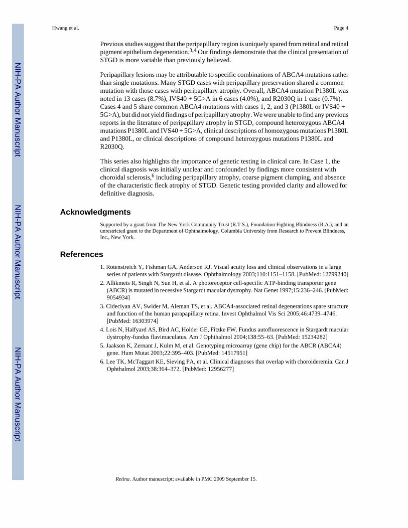
A 55-year-old male was examined for long-standing central visual impairment since age 18.Family history was not significant for ocular disease. His best-corrected visual acuity of 20/350OD and 20/200 OS was consistent with measurements over the last 20 years. Sphericalrefractive error measured −3.5 OD and −2.0 OS. Anterior segment examination wasunremarkable and applanation tonometry measured 17 mmHg OD and 14 mmHg OS. Posteriorsegment examination and autofluorescence imaging were significant for sharply demarcatedcentral and peripapillary zones of atrophy and the absence of fleck lesions (Figure 1, A–D).Humphrey visual fields demonstrated bilateral central scotomas with eccentric fixation at theinferior border. ERG examination was subnormal and similar to results obtained 22 years ago.
Genetic testing was employed for further diagnostic information and two heterozygous ABCA4mutations, P1380L and IVS40 + 5G>A, were identified and classified as disease-causingalleles, thereby confirming the diagnosis of STGD.
Case 2A 19-year-old female referred for consultation for difficulty reading, particularly in dim light.She was clinically diagnosed with STGD 3 month prior, after complaining of gradual difficultyin focusing. Family history was not significant for ocular disease. Visual acuity measured 20/40OD and 20/150 OS. Slit-lamp examination was unremarkable with normal anterior segmentsand applanation tensions of 14 mmHg OU. Funduscopic exam revealed bilateral centralatrophy, multiple fleck lesions, multiple clumps of yellowish deposits at the level of the retinalpigment epithelium in the posterior pole surrounded with some pigment clumping, and noevidence of atrophy of the retinal pigment epithelium. Autofluorescence imaging revealedmultifocal atrophic lesions involving the central macula and peripheral hyperautofluorescentflecks OU. The peripapillary regions demonstrated atrophic flecks OU without confluentatrophy (Figure 2, A and B). Genotyping revealed homozygous ABCA4 mutations, P1380Land P1380L.
Hwang et al. Page 2
Retina. Author manuscript; available in PMC 2009 September 15.
NIH
-PA Author Manuscript
NIH
-PA Author Manuscript
NIH
-PA Author Manuscript
Case 3A 15-year-old female presented with complaints of difficulty reading materials held greaterthan 6 inches from her eyes. She was clinically diagnosed with STGD 4 years prior and noteddecreased visual acuity since age seven, which stabilized over the past few years. Family historywas not significant for ocular disease. Best-corrected visual acuity was 20/150 OU. Anteriorsegment exam was unremarkable with applanation tonometry measuring 16 mmHg OU.Posterior segment exam and autofluorescence was significant for bilateral central atrophy andpisciform fleck atrophy involving the peripapillary, macular, and peripheral regions OU(Figure 3, A and B). Genotyping was significant for ABCA4 mutations P1380L and R2030Q.
Case 4A 52-year-old male was examined for declining vision OS over the past few months. He waspreviously clinically diagnosed with STGD 7 years before presentation. Family history wasnot significant for ocular disease. Best-corrected visual acuity measured 20/100 OD and 20/70OS. Spherical refractive error measured −3.00 OD and −3.25 OS. Anterior segmentexamination was unremarkable and applanation tonometry measured 17 mmHg OD and 14mmHg OS. Posterior segment examination was significant for central atrophy and classicperipheral pisciform flecks sparing the peripapillary regions OU (Figure 4, A and B).Autofluorescence imaging demonstrated inner atrophic flecks and outer hyperautofluorescentflecks. Moderate peripapillary hypoautofluorescence, but not atrophy, was present, likelysecondary to the patient’s myopia (Figure 4, C and D). Genotyping revealed two heterozygousABCA4 mutations, P1380L and S1696N.
Case 5A 46-year-old male was examined for visual acuity loss starting at age 17. His family historywas significant for visual acuity loss by his brother and father during the second decade of lifewithout known etiology. Genotype and phenotype data were not available. In addition, thepatient’s son was diagnosed with STGD with symptoms beginning at age 14. Best-correctedvisual acuity measured 20/400 bilaterally. Anterior segment examination was unremarkableand applanation tonometry measured 12 mmHg OD and 13 mmHg OS. Posterior segmentexamination and autofluorescence imaging were significant for multifocal small atrophiclesions confined to the fovea OU and normal peripapillary regions (Figure 5, A and B).Genotyping revealed a single ABCA4 mutation, IVS40 + 5G>A, confirming the diagnosis ofSTGD. The patient’s son was also examined and demonstrated classic STGD findings withperipapillary sparing. Genetic testing revealed a single ABCA4 mutation R1108C.
The pattern of inheritance between the father and son represents pseudo-dominant STGD. Bothcases demonstrated classic STGD findings and a single identifiable mutation along with anondetected ABCA4 mutation. The son received the nondetected mutation from the father andthe identified mutation from the carrier mother, thereby accounting for the difference ingenotype.
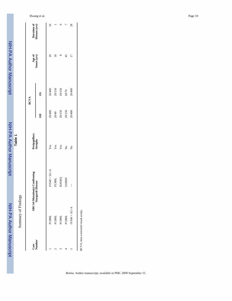
DiscussionIn this series, we reviewed 150 consecutive cases of STGD and identified three cases withperipapillary atrophy (2.0%) (Table 1). Case 1 demonstrated peripapillary and central atrophyin a mildly myopic patient with ABCA4 mutations P1380L and IVS40 + 5G>A. Case 2demonstrated atrophic fleck lesions involving the peripapillary region, central atrophy, andhomozygous ABCA4 mutations P1380L and P1380L. Case 3 revealed bilateral central atrophyand pisciform fleck atrophy involving the peripapillary, macular, and peripheral regions withABCA4 mutations P1380L and R2030Q.
Hwang et al. Page 3
Retina. Author manuscript; available in PMC 2009 September 15.
NIH
-PA Author Manuscript
NIH
-PA Author Manuscript
NIH
-PA Author Manuscript
Previous studies suggest that the peripapillary region is uniquely spared from retinal and retinalpigment epithelium degeneration.3,4 Our findings demonstrate that the clinical presentation ofSTGD is more variable than previously believed.
Peripapillary lesions may be attributable to specific combinations of ABCA4 mutations ratherthan single mutations. Many STGD cases with peripapillary preservation shared a commonmutation with those cases with peripapillary atrophy. Overall, ABCA4 mutation P1380L wasnoted in 13 cases (8.7%), IVS40 + 5G>A in 6 cases (4.0%), and R2030Q in 1 case (0.7%).Cases 4 and 5 share common ABCA4 mutations with cases 1, 2, and 3 (P1380L or IVS40 +5G>A), but did not yield findings of peripapillary atrophy. We were unable to find any previousreports in the literature of peripapillary atrophy in STGD, compound heterozygous ABCA4mutations P1380L and IVS40 + 5G>A, clinical descriptions of homozygous mutations P1380Land P1380L, or clinical descriptions of compound heterozygous mutations P1380L andR2030Q.
This series also highlights the importance of genetic testing in clinical care. In Case 1, theclinical diagnosis was initially unclear and confounded by findings more consistent withchoroidal sclerosis,6 including peripapillary atrophy, coarse pigment clumping, and absenceof the characteristic fleck atrophy of STGD. Genetic testing provided clarity and allowed fordefinitive diagnosis.
AcknowledgmentsSupported by a grant from The New York Community Trust (R.T.S.), Foundation Fighting Blindness (R.A.), and anunrestricted grant to the Department of Ophthalmology, Columbia University from Research to Prevent Blindness,Inc., New York.
References1. Rotenstreich Y, Fishman GA, Anderson RJ. Visual acuity loss and clinical observations in a large
series of patients with Stargardt disease. Ophthalmology 2003;110:1151–1158. [PubMed: 12799240]2. Allikmets R, Singh N, Sun H, et al. A photoreceptor cell-specific ATP-binding transporter gene
(ABCR) is mutated in recessive Stargardt macular dystrophy. Nat Genet 1997;15:236–246. [PubMed:9054934]
3. Cideciyan AV, Swider M, Aleman TS, et al. ABCA4-associated retinal degenerations spare structureand function of the human parapapillary retina. Invest Ophthalmol Vis Sci 2005;46:4739–4746.[PubMed: 16303974]
4. Lois N, Halfyard AS, Bird AC, Holder GE, Fitzke FW. Fundus autofluorescence in Stargardt maculardystrophy-fundus flavimaculatus. Am J Ophthalmol 2004;138:55–63. [PubMed: 15234282]
5. Jaakson K, Zernant J, Kulm M, et al. Genotyping microarray (gene chip) for the ABCR (ABCA4)gene. Hum Mutat 2003;22:395–403. [PubMed: 14517951]
6. Lee TK, McTaggart KE, Sieving PA, et al. Clinical diagnoses that overlap with choroideremia. Can JOphthalmol 2003;38:364–372. [PubMed: 12956277]
Hwang et al. Page 4
Retina. Author manuscript; available in PMC 2009 September 15.
NIH
-PA Author Manuscript
NIH
-PA Author Manuscript
NIH
-PA Author Manuscript
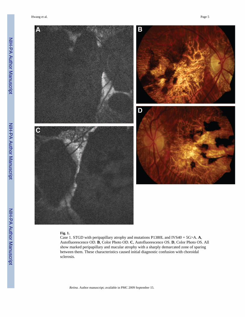
Fig. 1.Case 1. STGD with peripapillary atrophy and mutations P1380L and IVS40 + 5G>A. A,Autofluorescence OD. B, Color Photo OD. C, Autofluorescence OS. D, Color Photo OS. Allshow marked peripapillary and macular atrophy with a sharply demarcated zone of sparingbetween them. These characteristics caused initial diagnostic confusion with choroidalsclerosis.
Hwang et al. Page 5
Retina. Author manuscript; available in PMC 2009 September 15.
NIH
-PA Author Manuscript
NIH
-PA Author Manuscript
NIH
-PA Author Manuscript
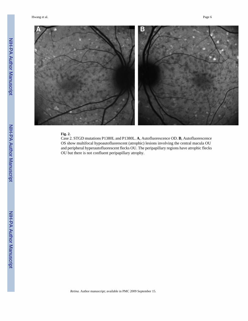
Fig. 2.Case 2. STGD mutations P1380L and P1380L. A, Autofluorescence OD. B, AutofluorescenceOS show multifocal hypoautofluorescent (atrophic) lesions involving the central macula OUand peripheral hyperautofluorescent flecks OU. The peripapillary regions have atrophic flecksOU but there is not confluent peripapillary atrophy.
Hwang et al. Page 6
Retina. Author manuscript; available in PMC 2009 September 15.
NIH
-PA Author Manuscript
NIH
-PA Author Manuscript
NIH
-PA Author Manuscript
Fig. 3.A and B, Case 3. STGD mutations P1380L and R2030Q. Autofluorescence OD and OSdemonstrate atrophic fleck lesions involving the macula and periphery with an area of confluentatrophy superotemporal to the fovea OD. Hyperautofluorescent flecks are also present in theperiphery OU. The peripapillary regions reveal atrophic flecks OU without areas of confluentatrophy.
Hwang et al. Page 7
Retina. Author manuscript; available in PMC 2009 September 15.
NIH
-PA Author Manuscript
NIH
-PA Author Manuscript
NIH
-PA Author Manuscript
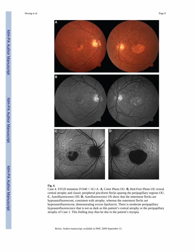
Fig. 4.Case 4. STGD mutation IVS40 + 5G>A. A, Color Photo OU. B, Red-Free Photo OU revealcentral atrophy and classic peripheral pisciform flecks sparing the peripapillary regions OU.C, Autofluorescence OD. D, Autofluorescence OS show that the innermost flecks arehypoautofluorescent, consistent with atrophy, whereas the outermost flecks arehyperautofluorescent, demonstrating excess lipofuscin. There is moderate peripapillaryhypoautofluorescence that is not as dark as this patient’s central atrophy or the peripapillaryatrophy of Case 1. This finding may thus be due to the patient’s myopia.
Hwang et al. Page 8
Retina. Author manuscript; available in PMC 2009 September 15.
NIH
-PA Author Manuscript
NIH
-PA Author Manuscript
NIH
-PA Author Manuscript
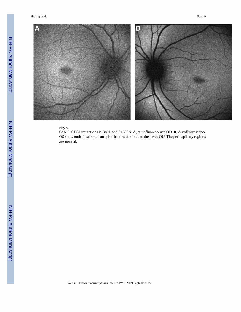
Fig. 5.Case 5. STGD mutations P1380L and S1696N. A, Autofluorescence OD. B, AutofluorescenceOS show multifocal small atrophic lesions confined to the fovea OU. The peripapillary regionsare normal.
Hwang et al. Page 9
Retina. Author manuscript; available in PMC 2009 September 15.
NIH
-PA Author Manuscript
NIH
-PA Author Manuscript
NIH
-PA Author Manuscript
NIH
-PA Author Manuscript
NIH
-PA Author Manuscript
NIH
-PA Author Manuscript
Hwang et al. Page 10Ta
ble
1Su
mm
ary
of F
indi
ngs
Cas
eN
umbe
rA
BC
A4
Mut
atio
n(s)
Con
firm
ing
Star
gard
t Dis
ease
Peri
papi
llary
Atr
ophy
BC
VA
Age
of
Ons
et (y
rs)
Dur
atio
n of
Dis
ease
(yrs
)O
DO
S
1P1
380L
IVS4
0 +
5G>A
Yes
20/4
0020
/400
2916
2P1
380L
P138
0LY
es20
/40
20/1
5018
1
3P1
380L
R20
30Q
Yes
20/1
5020
/150
86
4P1
380L
S169
6NN
o20
/150
20/7
045
7
5IV
S40
+ 5G
>A—
No
20/4
0020
/400
1728
BC
VA
, bes
t-cor
rect
ed v
isua
l acu
ity.
Retina. Author manuscript; available in PMC 2009 September 15.