MCP-1, KC-like and IL-8 as critical mediators of ... · resulted in transient low parasitemia...
21
RESEARCH ARTICLE MCP-1, KC-like and IL-8 as critical mediators of pathogenesis caused by Babesia canis Asier Gala ´n 1 *, Iva Mayer 2 , Renata Barić Rafaj 3 , Kres ˇ o Bendelja 4 , Velimir Sus ˇić 5 , Jose ´ Joaquı ´n Cero ´n 6 , Vladimir Mrljak 1,2 1 ERA Chair project ’’VetMedZg’’, Clinic for Internal Diseases, Faculty of Veterinary Medicine, University of Zagreb, Zagreb, Croatia, 2 Clinic for Internal Diseases, Faculty of Veterinary Medicine, University of Zagreb, Zagreb, Croatia, 3 Department of Chemistry and Biochemistry, Faculty of Veterinary Medicine, University of Zagreb, Zagreb, Croatia, 4 Institute of Immunology, Zagreb, Croatia, 5 Department of Animal Husbandry, Faculty of Veterinary Medicine, University of Zagreb, Zagreb, Croatia, 6 Department of Animal Medicine and Surgery, University of Murcia, Spain * [email protected] Abstract Canine babesiosis caused by the intraerythrocytic protozoan parasite Babesia canis is a tick-borne disease characterized by a host response that involves both cellular and humoral immunity. This study focuses on the secretion of cytokines Granulocyte-Macrophage Col- ony-Stimulating Factor (GM-CSF), Keratinocyte Chemotactic-like (KC-like), Interleukins (IL)-2, IL-7, IL-8, IL-10, IL-15, IL-18 and Monocyte Chemotactic Protein-1 (MCP-1) in babe- siosis caused by Babesia canis upon treatment with Imizol®. We assessed time dependent changes in cytokine levels and tested whether these changes correlate with pathogenesis of the disease. Sixteen healthy dogs and 31 dogs infected with Babesia canis, of which 18 showed complications, were treated with Imizol®. One dog died during the study (3.2%). Longitudinal study was perfomed by monitoring dogs at the first day of presentation (day 1) and 6 days later (day 7). Our results show that higher MCP-1 levels on day 1 are positively associated with the occurrence of complications, (complicated vs. uncomplicated; p = 0.00016). A similar pattern was observed for KC-like on day 1 (p = 0.0326) and day 7 (p = 0.044). Moreover, babesiosis caused by B. canis produced a steady increase in IL-8 levels with a moderate to strong negative correlation with erythrocyte counts and hematocrit in uncomplicated diseased dogs only (Spearman’s rank correlation coefficient r s = -0.582 and r s = -0.598 respectively). Like for MCP-1, KC-like levels also differed in complicated and uncomplicated diseased dogs on day 1 (p = 0.03236) and day 7 (p = 0.044). Furthermore, KC-like levels were strongly correlated with IL-8 levels (r s = 0.663–0.7) and non-segmented neutrophil counts (rs = 0.572–0.732) in both diseased groups. Analysis of ROC suggests the use of serum levels of MCP-1 and IL-7 as predictors of the occurrence of complications with an AUC of 0.906 and 0.896 respectively and linear combinations of MCP-1, KC-Like, IL-7 and GM-CSF with values up to AUC = 0.983. Cytokine cluster analysis presented in this study can contribute to a better understanding of the pathogenesis of babesiosis and serve as a prognostic tool for the early detection of cases with highest likelihood of developing complications. Overall, our studies show that infection by B. canis elicits a cytokine pattern that is distinct from that observed with B. rossi, and that some of the inflammatory mediators PLOS ONE | https://doi.org/10.1371/journal.pone.0190474 January 5, 2018 1 / 21 a1111111111 a1111111111 a1111111111 a1111111111 a1111111111 OPEN ACCESS Citation: Gala ´n A, Mayer I, Rafaj RB, Bendelja K, Sus ˇić V, Cero ´n JJ, et al. (2018) MCP-1, KC-like and IL-8 as critical mediators of pathogenesis caused by Babesia canis. PLoS ONE 13(1): e0190474. https://doi.org/10.1371/journal.pone.0190474 Editor: J. Stephen Dumler, Johns Hopkins University, UNITED STATES Received: August 16, 2016 Accepted: December 15, 2017 Published: January 5, 2018 Copyright: © 2018 Gala ´n et al. This is an open access article distributed under the terms of the Creative Commons Attribution License, which permits unrestricted use, distribution, and reproduction in any medium, provided the original author and source are credited. Data Availability Statement: All relevant data are within the paper and its Supporting Information files. Funding: This work was supported by CORDIS, 621394 (http://cordis.europa.eu/project/rcn/ 189878_en.html) and HRZZ, 4135 (http://www. hrzz.hr/default.aspx?id=78&pid=4135&rok=2013- 11). The funders had no role in study design, data collection and analysis, decision to publish, or preparation of the manuscript. Competing interests: The authors have declared that no competing interests exist. CORDIS,
Transcript of MCP-1, KC-like and IL-8 as critical mediators of ... · resulted in transient low parasitemia...

RESEARCH ARTICLE
MCP-1, KC-like and IL-8 as critical mediators of
pathogenesis caused by Babesia canis
Asier Galan1*, Iva Mayer2, Renata BarićRafaj3, Kreso Bendelja4, Velimir Susić5, Jose
Joaquın Ceron6, Vladimir Mrljak1,2
1 ERA Chair project ’’VetMedZg’’, Clinic for Internal Diseases, Faculty of Veterinary Medicine, University of
Zagreb, Zagreb, Croatia, 2 Clinic for Internal Diseases, Faculty of Veterinary Medicine, University of Zagreb,
Zagreb, Croatia, 3 Department of Chemistry and Biochemistry, Faculty of Veterinary Medicine, University of
Zagreb, Zagreb, Croatia, 4 Institute of Immunology, Zagreb, Croatia, 5 Department of Animal Husbandry,
Faculty of Veterinary Medicine, University of Zagreb, Zagreb, Croatia, 6 Department of Animal Medicine and
Surgery, University of Murcia, Spain
Abstract
Canine babesiosis caused by the intraerythrocytic protozoan parasite Babesia canis is a
tick-borne disease characterized by a host response that involves both cellular and humoral
immunity. This study focuses on the secretion of cytokines Granulocyte-Macrophage Col-
ony-Stimulating Factor (GM-CSF), Keratinocyte Chemotactic-like (KC-like), Interleukins
(IL)-2, IL-7, IL-8, IL-10, IL-15, IL-18 and Monocyte Chemotactic Protein-1 (MCP-1) in babe-
siosis caused by Babesia canis upon treatment with Imizol®. We assessed time dependent
changes in cytokine levels and tested whether these changes correlate with pathogenesis
of the disease. Sixteen healthy dogs and 31 dogs infected with Babesia canis, of which 18
showed complications, were treated with Imizol®. One dog died during the study (3.2%).
Longitudinal study was perfomed by monitoring dogs at the first day of presentation (day 1)
and 6 days later (day 7). Our results show that higher MCP-1 levels on day 1 are positively
associated with the occurrence of complications, (complicated vs. uncomplicated; p =
0.00016). A similar pattern was observed for KC-like on day 1 (p = 0.0326) and day 7 (p =
0.044). Moreover, babesiosis caused by B. canis produced a steady increase in IL-8 levels
with a moderate to strong negative correlation with erythrocyte counts and hematocrit in
uncomplicated diseased dogs only (Spearman’s rank correlation coefficient rs = -0.582 and
rs = -0.598 respectively). Like for MCP-1, KC-like levels also differed in complicated and
uncomplicated diseased dogs on day 1 (p = 0.03236) and day 7 (p = 0.044). Furthermore,
KC-like levels were strongly correlated with IL-8 levels (rs = 0.663–0.7) and non-segmented
neutrophil counts (rs = 0.572–0.732) in both diseased groups. Analysis of ROC suggests
the use of serum levels of MCP-1 and IL-7 as predictors of the occurrence of complications
with an AUC of 0.906 and 0.896 respectively and linear combinations of MCP-1, KC-Like,
IL-7 and GM-CSF with values up to AUC = 0.983. Cytokine cluster analysis presented in this
study can contribute to a better understanding of the pathogenesis of babesiosis and serve
as a prognostic tool for the early detection of cases with highest likelihood of developing
complications. Overall, our studies show that infection by B. canis elicits a cytokine pattern
that is distinct from that observed with B. rossi, and that some of the inflammatory mediators
PLOS ONE | https://doi.org/10.1371/journal.pone.0190474 January 5, 2018 1 / 21
a1111111111
a1111111111
a1111111111
a1111111111
a1111111111
OPENACCESS
Citation: Galan A, Mayer I, Rafaj RB, Bendelja K,
Susić V, Ceron JJ, et al. (2018) MCP-1, KC-like and
IL-8 as critical mediators of pathogenesis caused
by Babesia canis. PLoS ONE 13(1): e0190474.
https://doi.org/10.1371/journal.pone.0190474
Editor: J. Stephen Dumler, Johns Hopkins
University, UNITED STATES
Received: August 16, 2016
Accepted: December 15, 2017
Published: January 5, 2018
Copyright: © 2018 Galan et al. This is an open
access article distributed under the terms of the
Creative Commons Attribution License, which
permits unrestricted use, distribution, and
reproduction in any medium, provided the original
author and source are credited.
Data Availability Statement: All relevant data are
within the paper and its Supporting Information
files.
Funding: This work was supported by CORDIS,
621394 (http://cordis.europa.eu/project/rcn/
189878_en.html) and HRZZ, 4135 (http://www.
hrzz.hr/default.aspx?id=78&pid=4135&rok=2013-
11). The funders had no role in study design, data
collection and analysis, decision to publish, or
preparation of the manuscript.
Competing interests: The authors have declared
that no competing interests exist. CORDIS,

can be useful to predict complications. Our results also suggest targets for the development
of novel therapeutic strategies in babesiosis caused by B. canis.
Introduction
Acute babesiosis is a malaria-like infection, characterized by fever, hemolytic anemia and
hemoglobinuria. In dogs, it is usually caused by Babesia canis, present in North America,
Southern Europe, parts of Asia and Africa [1] whose vectors are ticks such as Dermacentor reti-culatus while other species are transmitted by Haemaphysalis leachi (B. rossi) and Rhipicephalussanguineus (B. vogeli) [2]. Babesiosis caused by B. canis usually is characterized by low parasite-
mia, which does not necessarily correlate with the severity of illness [3]. Hemoglobinuria has
been described in naturally infected dogs [4, 5] whereas experimental infection with B. canisresulted in transient low parasitemia (1–2%), thrombocytopenia, an increase in the activated
partial thromboplastin time (APTT) and hypotension [6].
In contrast, B. rossi, the main causative organism of canine babesiosis in sub-Saharan
Africa, generally elicits a virulent illness with high parasitemia [7], hypoglycemia [8] and cere-
bral, lung and renal involvement [9] associated with mortality. Interestingly, a polymorphic
phosphoprotein, B. rossi erythrocyte membrane antigen 1 (BrEMA1), expressed on the cyto-
plasmic membrane of B. rossi-infected erythrocytes, has been recently described. This protein
is suspected to be a major virulence factor in B. rossi-induced canine babesiosis [10]. B. canisand B. vogeli do not express the BrEMA1 gene.
Other babesial species cause a broad spectrum of clinical signs: B. vogeli usually causes a
subclinical to moderate clinical disease with possible severe to fatal hemolytic anemia in young
dogs and pups [11]. Immune-mediated hemolytic anemia (IMHA) [12] is common in B. vogeliinfection but inflammatory patterns are not so uniform as in B. canis infections [13]. A cur-
rently unnamed large form of Babesia was described for the first time in a dog under chemo-
therapy for lymphoma [14]. B. gibsoni causes frequently achronic disease often associated with
weight loss and fatigue [15]. B. conradae is usually more virulent than B. gibsoni resulting in
higher parasitemia, more severe anemia and higher rate of mortality [16]. B. microti-like piro-
plasm (Theileria annae) is a recently described small piroplasm causing azotemia [17].
Canine babesiosis is defined as a protozoal sepsis [18] occurring along with a generalized
uncontrolled inflammatory response of the host [19] which represents a central factor in the
development of complications. Infection initiates a mechanism of antibody-mediated cyto-
toxic destruction of erythrocytes. Autoantibodies are directed against components of the
membranes of infected and uninfected erythrocytes causing intra- and extravascular hemoly-
sis, which can evolve to anaemia. C-reactive protein (CRP) and serum amyloid A (SAA) signif-
icantly increase in canine babesiosis but their levels are not related with the occurrence of
complications neither with outcome [18, 20]. Proinflammatory cytokines and chemokines,
such as TNF-α, IFN-γ, IL-1β, IL-2, IL-6, IL-8, IL-12, IL-18 and MCP-1 are necessary to initiate
an effective inflammatory response [21] and promote the transendothelial migration of leuko-
cytes. TNF-α and IL-1β are considered the proximal or initiator cytokines of the proinflamma-
tory response as they trigger the production of other cytokines such as IL-6 and IL-8 (distal
cytokines). On the other hand, IL-4, IL-10 and transforming growth factor-beta (TGF-β), are
required to down-regulate the cell-mediated inflammatory response by inhibiting the synthesis
of pro-inflammatory cytokines [22]. In fact, the acute phase response (APR) which results in
systemic inflammatory response syndrome (SIRS), one of the complications of babesiosis, is a
Cytokines MCP-1, KC-like and IL-8 in canine babesiosis
PLOS ONE | https://doi.org/10.1371/journal.pone.0190474 January 5, 2018 2 / 21
Community Research and Development
Information Service is the European Commission’s
primary portal for results of EU-funded research
projects, As our funder, doesn’ t restrict sharing of
data and/or materials. On behalf of all authors I
declare that there are no competing interests that
interferes with, or could reasonably be perceived as
interfering with, the full and objective presentation,
peer review, editorial decision-making, or
publication of this research.
Abbreviations: APTT, Activated partial
thromboplastin time; BrEMA1, Babesia rossi
erythrocyte membrane antigen; CXC chemokines,
Cysteine-X-Cysteine motif-containing chemokine;
GM-CSF, Granulocyte-macrophage colony
stimulating factor; KC-like, Keratinocyte-derived
chemokine; IL, Interleukin; IMHA, Immune-
mediated hemolytic anemia; MCP-1, Monocyte
chemoattractant protein 1; MODS, Multiple organ
disfunction syndrome; PCR, Polymerase chain
reaction; ROC, Receiver operating characteristic;
SIRS, Systemic inflammatory response syndrome;
TGF-β, Transforming growth factor beta; PON-1,
Paraoxonase-1.

common feature of other types of sepsis [23]. Tissue hypoxia, trigger of many of the clinical
signs and studied comprehensively in B. rossi infection, is considered to be more important
than hemoglobinuria, and its consequent nephrotoxic effect, as a cause of kidney damage in
dogs with babesiosis [24]. In a similar way as in malaria, the inefficient use of oxygen by mito-
chondria triggered by the effects of inflammatory cytokines is the main cause of hypoxia-
related tissue damage [25]. Release of reactive oxygen species and harmful cytokine effects
have been linked to endothelial damage and augmented vascular permeability in canine babe-
siosis [26, 27, 28].
Uncomplicated babesiosis might be a consequence of hemolysis without excessive inflam-
matory response while in complicated canine babesiosis and in malaria caused by Plasmodiumfalciparum pathology is believed to be the result of excessive production of pro-inflammatory
cytokines [29] generating SIRS and multiple organ dysfunction syndrome (MODS) [30] with a
15% mortality rate.
In case of the most severe complications of P. falciparum malaria, cerebral malaria (CM)
and severe malarial anemia (SA), both likely occur along with a dysregulation of the immune
system [31]. Cytokines in malaria are key factors in regulating the progression of disease and
are closely related to symptoms, parasitemia, severity of pathology and outcome [32]. High
concentrations of pro-inflammatory cytokines such as TNF-α, IFN-γ, IL-6, IL-8, IL-18 and
MCP-1 have been associated with severe malaria and death [33]. Regulatory cytokines such as
IL-10 and TGF-β are important to limit that pro-inflammatory response [34].
Despite the comprehensive research on the inflammatory response caused by babesiosis, it
is not completely understood how imbalances in cytokine levels develop and determine the
course of the disease, occurrence of complications and outcome. In this study we use a multi-
plex approach to monitor simultaneously the concentrations of diverse cytokines and detect
expected imbalances, to determine which cytokines discriminate more significantly uncompli-
cated and complicated babesiosis and to establish levels of cytokines as prognostic markers.
Materials and methods
Animals
47 dogs were retrospectively included in this study and divided into 2 main groups. Group
1consisted of 31 dogs naturally infected by B. canis, admitted at the Clinic for Internal Dis-
eases, Faculty of Veterinary Medicine, University of Zagreb, Croatia, with clinical signs of
acute babesiosis. The clinical manifestations included anorexia, lethargy and fever, pale
mucous membranes, anemia, jaundice, hemoglobinuria or hematuria, splenomegaly, tachycar-
dia and vomiting. Dogs were of various breeds, between 1 and 14 years of age, 18 males and 13
females. Protocol was approved by the Ethics Committee for Animal Experimentation, Faculty
of Veterinary Medicine, University of Zagreb, Croatia (Permit No: 251–61–01/139–12–2). Per-
mission to collect blood samples was obtained from each dog owner. Owners were informed
about the use of the specimens and the aims of the research.
Diagnosis was confirmed by demonstration of the parasites within the infected erythrocytes
in Romanowsky-stained thin blood smears. One dose (6 mg/kg of body weight) of imidocarb
dipropionate (Imizol1, Schering–Plough) was administered to all the dogs subcutaneously on
the day of admission (day 1). Additional treatment consisted of various fluids (colloid and
crystalloid therapy), and whole blood transfusion when it was indicated. Subspecies were con-
firmed using PCR (polymerase chain reaction) [6]. On the basis of clinical manifestations
and laboratory data the infected dogs were classified into two subgroups: complicated (18
dogs, 8 Female, 10 Male) and uncomplicated (13 dogs, 5 Female, 8 Male).The classification of
clinical manifestations of the complicated form of babesiosis is based on the World Health
Cytokines MCP-1, KC-like and IL-8 in canine babesiosis
PLOS ONE | https://doi.org/10.1371/journal.pone.0190474 January 5, 2018 3 / 21

Organization (WHO) classification for malaria [35]. The main complications are the develop-
ment of an excessive inflammatory response named”systemic inflammatory response syn-
drome” or SIRS [36] and a multiple organ dysfunction syndrome or MODS [37].
According to the criteria for the diagnosis of SIRS used in this study [9], [38] dogs were
classified as SIRS positive if two or more of the following 4 criteria were fulfilled: body temper-
ature higher than 39.5˚C or lower than 38˚C, heart rate more than 160 beats/min, respiration
rate more than 20 breaths/min and WBC count less than 6 × 10⁹/L or more than 12 × 109/L or
with more than 10 percent band neutrophils. Dogs with obvious concurrent inflammatory
processes as trauma, wounds or infections, known cardiopathies or neoplastic diseases were
excluded from the study. Dogs treated with any anti-inflammatory medication within 3 weeks
prior to diagnosis were also excluded. All dogs showing complications were treated according
to standard procedures (infusion, anti-inflammatories and blood transfusion only in case
hematocrit was under 20%). Blood samples for cytokine quantification were obtained 6–8
hours after the treatment was administered.
An animal was classified as MODS positive if two or more of the following criteria were ful-
filled: renal dysfunction (serum creatinine concentration higher than 180 μmol/l), hepatic dys-
function (both alanine aminotransferase (ALT) greater than 176 IU/l and alkaline phosphatase
(AP) greater than 360 IU/l), respiratory system dysfunction (radiographic evidence of pulmo-
nary oedema or dyspnoea), and muscular involvement (creatine phosphokinase (CPK) more
than 600 IU/l) [9]. Cases with high serum creatinine concentration due to pre-renal causes
(e.g., dehydration due to vomiting) were excluded after examining oral mucosa, skin turgor
and ocular appearance as well as checking concentrations of serum urea, serum albumin and
total protein and determining urine specific gravity. 66% of complicated cases were MODS
positive and all of them were SIRS positive.
Group 2 (healthy controls) included two subgroups: animals treated by prophylactic single
application of imidocarb dipropionate (Imizol1, Schering-Plough) consisted of 8 dogs
(Group 2a) of different breeds and sexes, aged from 1 to 10 years. These dogs were considered
healthy based on physical examination and hematological and biochemical data, and they
attended the hospital to receive a prophylactic dose of imidocarb dipropionate (6 mg/kg) as a
preventive measure against babesiosis upon owners’ request. Another subgroup (Group 2b)
consisted of 8 dogs, different breeds and genders, aged from 1 to 14 years. These dogs attended
routine check controls and were considered healthy based on clinical examination and hema-
tological and biochemical data. Due to the fact that no significant differences in clinical hema-
tology, serum biochemistry and cytokine profile results were identified between the two
subgroups, these subgroups were merged into a single healthy control group.
All serum samples from dogs were screened for simultaneous qualitative detection of circu-
lating antibodies, both immunoglobulin G (IgG) and IgM, to B. canis, Borrelia burgdorferi,Anaplasma phagocytophilum and Dirofilaria immitis antigen using an in-clinic enzyme-linked
immunosorbent assay (ELISA) SNAP 4Dx (IDEXX Laboratories, Hoofddorp, The Nether-
lands), according to the manufacturer’s instructions.
Blood samples
Blood samples from groups 1 and 2a were collected from the cephalic vein on the day of
admission, and 6 days after the administration of imidocarb dipropionate (Imizol1). Blood
samples for analysis from group 2b were obtained during routine visits to a Clinic. Blood was
collected using a 20 gauge needle in tubes with EDTA for hematological analysis and serum
vacutainer tubes. A complete list of all samples with cytokine concentration values and hema-
tologic parameters as well as reference ranges is included in S1 Table.
Cytokines MCP-1, KC-like and IL-8 in canine babesiosis
PLOS ONE | https://doi.org/10.1371/journal.pone.0190474 January 5, 2018 4 / 21

Hematological analysis
Blood smears were prepared and PCR reactions performed using blood obtained on EDTA.
White blood cell count (WBC), platelet count, hematocrit (HCT) and other hematologic
parameters were determined using a Horiba ABX automatic hematology analyser (Diagnos-
tics, Montpellier, France). Segmented and non-segmented neutrophils were manually counted
in blood smear by experienced personnel.
Quantification of cytokines in serum
Blood collected in serum tubes was allowed to clot and tubes were centrifuged at 1200 xg, 10
minutes at room temperature. A portion of the serum was used for routine biochemical analy-
sis while the remainder was stored at −80˚C until it was used to analyse cytokines. Cytokine
profiles were determined using the MILLIPLEX MAP Canine Cytokine/ Chemokine Magnetic
Bead Panel (CCYTO-90K Millipore, Billerica, MA) with an automated analyser (Luminex 200,
Luminex Corporation, Austin, TX). The concentrations of interleukin-2 (IL-2), IL-7, IL-8, IL-
10, IL-15, IL-18, MCP-1, granulocyte-macrophage colony stimulating factor (GM-CSF) and
keratinocyte chemoattractant KC-like were analyzed. Prior to analysis, samples were thawed,
vortexed, and centrifuged at 1000x g for 10 min to separate particulates. The analytes were
measured in duplicate, according to the manufacturer’s instructions. Briefly, the plate was pre-
wet with wash buffer and standards, controls and samples were added to the appropriate wells.
After washing, the detection antibodies were added to each well. After incubation, the plate
was run on Luminex. The median fluorescent intensity (MFI) was analyzed and the concentra-
tions of analytes were derived using standard curves.
Statistical analysis
Statistical analysis was performed using STATISTICA version 10 (StatSoft. Inc., 2011). Given
that most variables were not normally distributed, statistical differences were assessed using
the non-parametric Mann-Whitney U test. p-values of<0.05 were considered to be significant.
Correlations were assessed by both Spearman’s rank correlation test and Pearson’s product-
moment correlation test.
Results
We have analysed cytokine concentrations of 31 dogs suffering from babesiosis versus 16
healthy animals (Table 1). We observed higher levels of cytokines on day 1. This trend reached
a plateau or reverted (remarkably for IL-10) on day 7 for most cytokines with the exception of
IL-8 for which an increase was noted.
All cytokine levels shown registered a significant increase upon babesiosis (Table 2) on day
1 and all except IL-7, IL-10 and MCP-1 keep significantly higher levels than healthy controls
on day 7. When comparing uncomplicated and complicated diseased dogs, all cytokines except
Table 1.
Group Gender Age (years) Average±Standard deviation
Group 1
Diseased uncomplicated (n = 13) 8M (61.5%) 5F (38.5%) 4.31±2.81
Diseased complicated (n = 18) 10M (56%) 8F (44%) 5.83±3.56
Group 2 (a+b)
Healthy (n = 16) 9M (56%) 7F (44%) 5.07±3.78
https://doi.org/10.1371/journal.pone.0190474.t001
Cytokines MCP-1, KC-like and IL-8 in canine babesiosis
PLOS ONE | https://doi.org/10.1371/journal.pone.0190474 January 5, 2018 5 / 21

IL-8, IL-10, IL-15 and IL-18 are significantly different on the day 1 but only KC-like and
MCP-1 show significantly different levels on day 7. The most significant longitudinal variation
within uncomplicated cases was for IL-10, IL-8 and GM-CSF while only IL-10 and IL-8 varied
significantly over time in complicated cases.
MCP-1 concentrations were significantly elevated for the B. canis-infected dogs when com-
paring healthy dogs and complicated diseased dogs on the day 1 (p<0.00001) and 7
(p = 0.01174). Uncomplicated and complicated cases on both day 1 and 7 (p = 0.00016 and
p = 0.01352 respectively) show significant differences in MCP-1 levels (Fig 1).
We observed a very significant increase in IL-8 concentration (Fig 2) comparing healthy
dogs with uncomplicated and complicated cases (p< 0.00001 and p< 0.0005 respectively) with
Table 2. Cytokine levels in different groups and subgroups. Cytokine concentrations (in pg/ml serum) for healthy dogs (n = 16), B. canis- infected dogs
on the day 1 (n = 31) and 7 (n = 30) and subset of uncomplicated (n = 13) and complicated groups (n = 18). 1 dog with complications died during the study. p-
values for significant differences are shown for each comparison.
CONCENTRATION (pg / ml serum)
GM-CSF KC-like IL-2 IL-7 IL-8 IL-10 IL-15 IL-18 MCP-1
Healthy (n = 16)
Min-Max 60–154 28–269 10–75 39–109 122–235 1–1 42–142 2–840 159–792
Median 87 39 17 55 137 1 77 83 193
SD 24 58 26 20 42 0 26 232 154
Diseased day 1 (n = 31)
Min-Max 130–782 104–2739 22–2514 31–1957 10–6502 1–2106 12–5436 2–2278 163–1921
Median 182 532 96 101 856 560 160 188 390
SD 176 639 525 378 1541 421 1032 487 494
p-value < 0.0001 < 0.0001 < 0.0001 0.019 < 0.0001 < 0.0001 0.007 0,044 0,001
Diseased day 7 (n = 30)
Healthy(n = 16)
Min-Max 135–2822 110–2184 12–4038 35–3633 425–8818 1–548 29–5548 2–8748 156–7667
Median 196 561 49 80 2754 1 219 207 237
SD 544 602 774 706 2185 108 1089 1900 1434
p-value < 0.0001 < 0.0001 0.014 0.1308 < 0.0001 0.321 0.007 0.029 0.09
Uncomplicated day 1 (n = 13)
Min-Max 130–220 104–969 22–187 31–119 35–5019 1–2106 12–342 2–490 163–436
Median 143 330 89 51 601 461 111 145 224
SD 29 282 44 26 1477 527 110 180 95
Complicated day 1 (n = 18)
Min-Max 131–782 162–2739 22–2514 35–1957 10–6502 180–1499 12–5436 2–2278 181–1921
Median 254 567 131 139 908 592 265 188 576
SD 198 749 665 470 1620 342 1324 610 552
p-value 0.00066 0.0322 0.025 0.00019 0.509 0.675 0.065 0.34683 0.00015
Uncomplicated day 7 (n = 13)
Min-Max 139–272 117–2009 12–276 39–183 1391–6294 1–1 29–1109 2–762 156–282
Median 154 440 28 68 2852 1 184 145 191
SD 44 474 87 48 1362 0 309 231 42
Complicated day 7 (n = 17)
Min-Max 135–2822 110–2184 12–4038 35–3633 425–8818 1–548 29–5548 2–8748 156–7667
Median 254 830 65 84 1911 1 253 225 365
SD 689 635 1005 917 2695 141 1406 2484 1844
p-value 0.059 0.04455 0.391 0.503 0.503 0.601 0.769 0.834 0.013
https://doi.org/10.1371/journal.pone.0190474.t002
Cytokines MCP-1, KC-like and IL-8 in canine babesiosis
PLOS ONE | https://doi.org/10.1371/journal.pone.0190474 January 5, 2018 6 / 21

even more significant increase on day 7 (Fig 2). IL-8 levels are not useful to discriminate
uncomplicated and complicated cases, but increased over time in both groups (p = 0.00174 for
uncomplicated; p = 0.0139 for complicated).
KC-like levels show a very significant increase in all diseased animals in both day 1 and day
7 (p < 0.00001). Additionally, KC-like levels show significant differences when comparing
uncomplicated and complicated cases both on day 1 and 7 (p = 0.0324 and p = 0.044 respec-
tively) (Fig 3) (Table 2). We registered a strong negative correlation of KC-Like levels with
erythrocyte counts (rs = -0.627) for complicated cases on day 7, and a positive correlation with
non-segmented neutrophil percent for both diseased groups on the same day (rs = 0.732 for
uncomplicated, rs = 0.736 for complicated) as well as in complicated cases on the day 7
(r = 0.572) (Table 3).
We registered a very significant IL-10 increase in all diseased animals (p<0.00001) on day 1
(Fig 4) and a decrease back to the levels registered in healthy dogs on the day 7 of treatment
(slightly more significant in complicated cases with a p<0.00001 vs. a p = 0.00008 for uncom-
plicated cases). No association with the occurrence of complications was detected (Fig 4).
Interestingly, IL-8 data obtained on the day 1 of treatment show a negative correlation with
both hematocrit (rs = -0.598) and erythrocyte count (rs = -0.582) in uncomplicated cases on the
Fig 1. MCP-1 concentration in healthy and naturally infected dogs and its relation with outcome. Box and whisker plot
with outliers showing interquartile range (median bar in intersect between two colors in box) of MCP-1 concentration values
among different groups: healthy dogs (H), uncomplicated (-C) and complicated (+C) cases for both day 1 and day 7. Mann-
whitney test p- values are shown for comparison of different groups.
https://doi.org/10.1371/journal.pone.0190474.g001
Cytokines MCP-1, KC-like and IL-8 in canine babesiosis
PLOS ONE | https://doi.org/10.1371/journal.pone.0190474 January 5, 2018 7 / 21
day 1. Additionally, IL-8 shows a very strong (r = 0.7) correlation with KC-Like among uncom-
plicated cases in measurements performed on day 1, as well as in the same group on day 7
(r = 0.692) (Table 3) confirming results obtained previously for B. canis-caused babesiosis [39].
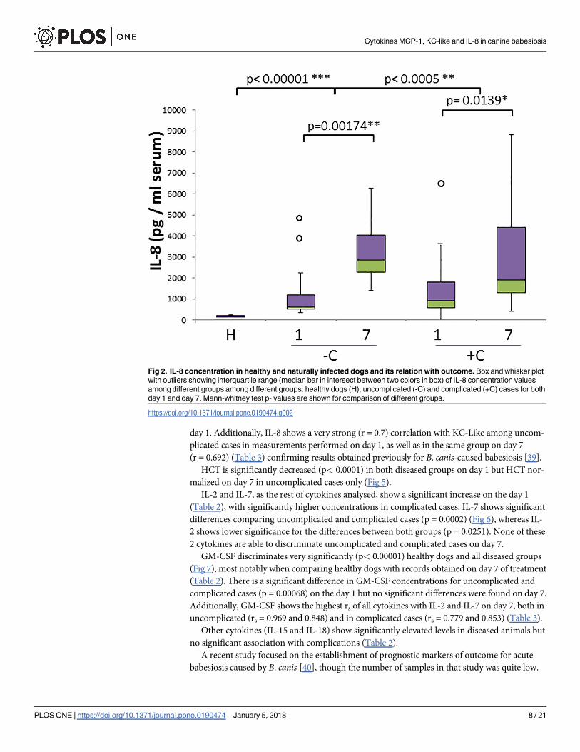
HCT is significantly decreased (p< 0.0001) in both diseased groups on day 1 but HCT nor-
malized on day 7 in uncomplicated cases only (Fig 5).
IL-2 and IL-7, as the rest of cytokines analysed, show a significant increase on the day 1
(Table 2), with significantly higher concentrations in complicated cases. IL-7 shows significant
differences comparing uncomplicated and complicated cases (p = 0.0002) (Fig 6), whereas IL-
2 shows lower significance for the differences between both groups (p = 0.0251). None of these
2 cytokines are able to discriminate uncomplicated and complicated cases on day 7.
GM-CSF discriminates very significantly (p< 0.00001) healthy dogs and all diseased groups
(Fig 7), most notably when comparing healthy dogs with records obtained on day 7 of treatment
(Table 2). There is a significant difference in GM-CSF concentrations for uncomplicated and
complicated cases (p = 0.00068) on the day 1 but no significant differences were found on day 7.
Additionally, GM-CSF shows the highest rs of all cytokines with IL-2 and IL-7 on day 7, both in
uncomplicated (rs = 0.969 and 0.848) and in complicated cases (rs = 0.779 and 0.853) (Table 3).
Other cytokines (IL-15 and IL-18) show significantly elevated levels in diseased animals but
no significant association with complications (Table 2).
A recent study focused on the establishment of prognostic markers of outcome for acute
babesiosis caused by B. canis [40], though the number of samples in that study was quite low.
Fig 2. IL-8 concentration in healthy and naturally infected dogs and its relation with outcome. Box and whisker plot
with outliers showing interquartile range (median bar in intersect between two colors in box) of IL-8 concentration values
among different groups among different groups: healthy dogs (H), uncomplicated (-C) and complicated (+C) cases for both
day 1 and day 7. Mann-whitney test p- values are shown for comparison of different groups.
https://doi.org/10.1371/journal.pone.0190474.g002
Cytokines MCP-1, KC-like and IL-8 in canine babesiosis
PLOS ONE | https://doi.org/10.1371/journal.pone.0190474 January 5, 2018 8 / 21

Since our results point out MCP-1 as the analyte with the most significant prognostic value,
we calculated the Receiver Operating Characteristic (ROC) curve (Fig 8) to discriminate
uncomplicated from complicated cases on the day 1, with an Area Under the Curve (AUC) of
0.906, indicating an excellent discrimination. ROC curves for individual values of IL-7,
GM-CSF and KC-like showed the highest AUC values among the rest of the cytokines in the
present study (0.896, 0.867 and 0.729 respectively). Linear combinations of cytokine concen-
tration values yielded higher AUC values that individual cytokines in ROC curves (Fig 8) with
highest values obtained corresponding to the combinations including MCP-1, KC-like, IL-7
and GM-CSF (AUC = 0.983for LC3). Cut-off values for every parameter in ROC curves and
their corresponding sensitivity and specificities along with AUC values are shown in Table 4.
Calculation of correlation coefficient was performed for all combinations of cytokines and
hematological parameters and the highest values (strongest positive or negative correlations)
are summarized in Table 3.
Discussion
Despite diagnostic and therapeutic advances [41], complications from babesiosis caused by B.
canis continue to be a cause of mortality. Pro-and anti-inflammatory cytokines act in a multi-
factorial manner after being released following injury, endotoxin release, complement
Fig 3. KC-like concentration in healthy and naturally infected dogs and its relation with outcome. Box and whisker plot
with outliers showing interquartile range (median bar in intersect between two colors in box) of KC-like concentration values
among different groups: healthy dogs (H), uncomplicated (-C) and complicated (+C) cases for both day 1 and day 7. Mann-
whitney test p- values are shown for comparison of different groups.
https://doi.org/10.1371/journal.pone.0190474.g003
Cytokines MCP-1, KC-like and IL-8 in canine babesiosis
PLOS ONE | https://doi.org/10.1371/journal.pone.0190474 January 5, 2018 9 / 21

activation and others. In babesiosis, when the inflammatory response becomes uncontrolled,
SIRS takes place. SIRS, if not down-regulated, can lead to MODS and, if not treated to terminal
condition.
Several efforts have been made in different inlammatory disorders to elucidate the origin of
imbalances in cytokine clusters associated to SIRS, MODS and other major complications.
TNF- α levels have been correlated with indices of renal damage and blood pressure in dogs
infected with B. canis [42]. Additionally, the injection of TNF-α into experimental animals
causes a syndrome resembling septic shock and infusion of recombinant TNF-α into humans
results in SIRS [43]. Equilibrium between TNF-α, IL-1β and anti-inflammatory IL-10 has been
proven highly dynamic and dependent on genetic heterogenity in human sepsis and SIRS. Our
Table 3. Highest Spearman’ s rank and Pearson’s product moment correlation coefficients (rs and r). Highest values for rs and r (in brackets) for cyto-
kine:cytokine and cytokine:hematologic parameter within different groups. E: Erythrocyte count, HCT:Hematocrit, Thr: Thrombocyte count, NN: Non-seg-
mented neutrophil count SN: Segmented neutrophil count, L: Leukocyte count, Eos: Eosinophil count.
Cytokine: cytokine GM-CSF IL-2 IL-7 IL-8 IL-15 IL-18 MCP-1
Healthy
IL-7 0.482(0.769) 0.716(0,782)
IL-18 0.498(0.895)
-C day 1
GM-CSF 0.634(0.722)
KC-LIKE 0.7(0.866)
+C day 1
KC-LIKE 0.663(0.654)
IL-2 0.717(0.798)
-C day 7
GM-CSF 0.969(0.827) 0.848(0.796) 0.806(0.810)
KC-LIKE 0.692(0.777)
IL-2 0.822(0.726) 0.65(0.832) 0.837(0.793)
IL-7 0.717(0.742)
+C day 7
GM-CSF 0.779(0.934) 0.853(0.973) 0.751(0.956) 0.771(0.958) 0.746(0.946)
IL-2 0.810(0.981) 0.799(0.984) 0.500(0.891)
IL-7 0.893(0.993) 0.597(0.917)
IL-15 0.796(0.914) 0.525(0.827)
Cytokine:Hematology E HCT Thr NN% SN% Eos Mono%
Healthy
GM-CSF 0.563(0.758)
MCP-1 -0.611(-0.572) 0.578(0.597)
-C day 1
KC-LIKE 0.732(0.812)
IL-8 -0.582(-0.863) -0.598(-0.542) 0.514(0.677) 0.641(0.825)
+C day 1
KC-LIKE 0.736(0.834)
+C day 7
GM-CSF 0.563(0.796)
KC-LIKE -0.627(-0.656) -0.648(-0.688) 0.572(0.610)
IL-7 0.546(0.766)
IL-15 0.513(0.746)
IL-18 0.612(0.880)
https://doi.org/10.1371/journal.pone.0190474.t003
Cytokines MCP-1, KC-like and IL-8 in canine babesiosis
PLOS ONE | https://doi.org/10.1371/journal.pone.0190474 January 5, 2018 10 / 21

present data registered a global increase of cytokine concentrations in serum of B.canis-infected dogs though some of them show higher significance, a stronger association with com-
plications and a different longitudinal behaviour. Among the analytes, MCP-1 and KC-like
showed significantly different levels in uncomplicated and complicated babesiosis cases on
both day 1 and 7. GM-CSF, IL-2 and IL-7 showed significant differences between those same
groups but only on day 1. Time-course variation was significant for IL-8 and IL-10 from day 1
to day 7 both in uncomplicated and complicated cases (though with opposite trends as IL-8
increased longitudinally while IL-10 registered a very pronounced decrease). IL-8 showed a
negative correlation with erythrocyte counts and hematocrit and a positive correlation with
non-segmented neutrophil counts in uncomplicated cases (day 1) whereas KC-like registered
as well a negative correlation with erythrocyte counts and hematocrit but in complicated cases
(day 7) and a generally strong positive correlation with non-segmented neutrophil counts in
all babesiosis groups.
Results observed for MCP-1 were the most significant in this study. MCP-1 together with
KC-like are the only cytokines in the present study discriminating complicated from uncom-
plicated cases both on day 1 and 7. MCP-1 acts in the recruitment of monocytes / macro-
phages, memory T cells, and dendritic cells to sites where either tissue injury or inflammation
occurs [44]. Our results are consistent with previously reported data [45] for human sepsis in
which patients show an increased level of MCP-1 with higher concentrations among non-sur-
vivors although with no statistically significant differences. Statistically significant differences
Fig 4. IL-10 concentration in healthy and naturally infected dogs and its relation with outcome. Box and whisker plot
with outliers showing interquartile range (median bar in intersect between two colors in box) of IL-10 concentration values
among different groups: healthy dogs (H), uncomplicated (-C) and complicated (+C) cases for both day 1 and day 7. Mann-
whitney test p- values are shown for comparison of different groups.
https://doi.org/10.1371/journal.pone.0190474.g004
Cytokines MCP-1, KC-like and IL-8 in canine babesiosis
PLOS ONE | https://doi.org/10.1371/journal.pone.0190474 January 5, 2018 11 / 21
in MCP-1 levels have been previously found between control dogs (118 pg/mL), survivors (431
pg/mL) and non-survivors (757 pg/mL) in the case of B.canis- induced babesiosis [18]. Our
present data confirm that complicated cases register a higher concentration of MCP-1 than
uncomplicated cases and additionally offer a comprehensive insight into the time-dependent
variations of this and other cytokines.
MCP-1 has been involved in pathogenesis of several autoimmune diseases characterized by
monocytic infiltrates, such as psoriasis, rheumatoid arthritis and atherosclerosis [46]. Some
observations have shown that MCP-1 secretion can be induced by erythrocytic debris and coa-
gulopathy [47] the latter being associated to mortality. Paraoxonase 1 (PON-1) has been
observed to inhibit this process decreasing endothelial MCP-1 secretion [48] and previous
studies have indicated a decrease in PON-1 concentration in acute babesiosis [28]. Moreover,
very high plasma levels of MCP-1 and IL-6 have been often observed after incompatible trans-
fusion–induced hemolysis, reinforcing the relationship of immune-mediated hemolysis and
MCP-1. In malaria, most of the evidence supports the hypothesis that cells from the mono-
cyte/macrophage lineage are more effective than neutrophils at phagocyting parasitized eryth-
rocytes [31] and this could explain the fact that MCP-1 shows much higher significance as
outcome marker than IL-8 in present study.
Fig 5. Hematocrit (HCT) values and outcome. From left to right hematocrit values for healthy dogs and values registered on day 1 and 7 of
treatment for uncomplicated (-C) and complicated cases (+C). Mann-whitney test p- values are shown for comparison of different groups.
https://doi.org/10.1371/journal.pone.0190474.g005
Cytokines MCP-1, KC-like and IL-8 in canine babesiosis
PLOS ONE | https://doi.org/10.1371/journal.pone.0190474 January 5, 2018 12 / 21

IL-8 or neutrophil chemotactic factor is secreted as a response of high IL-1 and TNF-α lev-
els, bacterial or viral products and cellular stress [49]. It induces chemotaxis, in neutrophils
and other granulocytes. Elevated IL-8 has been documented in many inflammatory conditions
for both humans and animals [50, 51] as well as in severe malaria [52] and previously in B.
canis-caused babesiosis [39]. In babesiosis caused by B.rossi, an exceptional decrease in IL-8
was previously reported [53] probably related to the higher virulence of B. rossi, pointing out
the possibility that a weaker pro-inflammatory response in acute-phase can lead to a worse
outcome. Our data show a clear longitudinal increase of IL-8 levels in contrast with data for B.
rossi-caused canine babesiosis [53].
Recent studies found that both IL-8 and MCP-1 levels increase in leptospirosis, another
protozoan disease [54]. In turn, IL-10 increased in only 38% of patients at the very early part of
diseases, and after 2 weeks it decreased at the level of healthy group. In recent studies about
malaria, levels of pro-inflammatory biomarkers, like IL-8, were higher in cerebral malaria
(CM) than in non-cerebral malaria patients. In contrast, the concentrations of anti-inflamma-
tory cytokines, like IL-10, were comparable or lower in CM patients [55]. We observed a simi-
lar pattern: higher MCP-1 levels in complicated cases and increasing levels of IL-8 in the
period studied (day 1 to 7), in remarkable contrast with the decrease observed in B. rossi-induced babesiosis, and a similar or slightly lower IL-10 concentration associated to
complications.
Recent research conducted on cell cultures has proven that the enhanced production of
CXC chemokines (IL-8 is an example) can trigger oxidative stress [56] which, besides second-
ary IMHA, has been considered the main cause of erythrocyte destruction in canine babesiosis
Fig 6. IL-7 concentration in healthy and naturally infected dogs and its relation with outcome. Box and whisker plot with
outliers showing interquartile range (median bar in intersect between two colors in box) of IL-7 concentration values among
different groups: healthy dogs (H), uncomplicated (-C) and complicated (+C) cases for both day 1 and day 7. Mann-whitney test p-
values are shown for comparison of different groups.
https://doi.org/10.1371/journal.pone.0190474.g006
Cytokines MCP-1, KC-like and IL-8 in canine babesiosis
PLOS ONE | https://doi.org/10.1371/journal.pone.0190474 January 5, 2018 13 / 21

[27]. In our data, the negative correlations observed between IL-8 and hematocrit suggests that
the sustained increase in IL-8 concentration occurs in parallel to hemolysis. Moreover, in a
recent study erythrocyte structure was found to be notably affected by IL-8 with morphological
changes resembling those typically observed in eryptosis (programmed red cell death) [57]
most probably after interaction with duffy antigen chemokine receptor (DARC) on erythro-
cyte surface [58]. Additionally, the present data show that IL-8 levels strongly correlate with
KC-Like levels among uncomplicated cases in measurements performed on the day 1, as well
as in the same group on day 7. Similar results were obtained in a study on canine experimental
LPS-induced endotoxemia [59]. Previous studies have shown that the concentration of IL-8
and KC-like are important factors in the pathogenesis of canine babesiosis [39]. A balance
between these two cytokines might be important to avoid the occurrence of complications
[11]. In relation with other hematologic parameters, we found a strong positive correlation
between IL-8 as well as KC-like with non-segmented or immature neutrophils. Shorter life
expectancy was recently found in patients with systemic inflammation who showed highest
counts of immature neutrophils [60]. The strongest correlation between non-segmented neu-
trophils and KC-like was registered in complicated cases on day 1, confirming the role of KC-
like in neutrophil proliferation. KC-like has been recently reported as a possible biomarker for
diagnosing sepsis and uterine bacterial infection in dogs [61]. We found increased levels of
KC-like in all diseased groups compared with healthy dogs and most importantly, it discrimi-
nates uncomplicated and complicated cases both on day 1 and 7.
Fig 7. GM-CSF concentration in healthy and naturally infected dogs and its relation with outcome. Box and whisker plot with
outliers showing interquartile range (median bar in intersect between two colors in box) of GM-CSF concentration values among
different groups: healthy dogs (H), uncomplicated (-C) and complicated (+C) cases for both day 1 and day 7. Mann-whitney test p-
values are shown for comparison of different groups.
https://doi.org/10.1371/journal.pone.0190474.g007
Cytokines MCP-1, KC-like and IL-8 in canine babesiosis
PLOS ONE | https://doi.org/10.1371/journal.pone.0190474 January 5, 2018 14 / 21

IL-10 displays the most significant variation among all the molecules monitored in this
analysis when comparing healthy and diseased groups on day 1 of treatment, in agreement
with previous data for B. rossi [53] in which no significant difference in IL-10 was found in
non-survivors with respect to survivors. Similarly, we observed no significant differences
related to the occurrence of complications but we found a very significant longitudinal drop of
IL-10 levels on day 7. IL-10 is an anti-inflammatory cytokine. During infection, IL-10 inhibits
Th1 cells, NK cells and macrophages. Given that these cells are required for pathogen clear-
ance, high levels of IL-10 can indirectly contribute to tissue damage. High levels of IL-10 have
been detected in mice serum after infection with B. microti [62]. In that study the frequency of
IL-10-producing regulatory B cells (Bregs) and CD4+ T cells increased during the course of B.
microti infection. Transfer of IL-10-producing B cells induced by B. microti infection led to
Fig 8. Receiver operating characteristic (ROC) curve as a tool for prognosis of complications in B.canis-
caused babesiosis and cut-off values. ROC plot showing curves corresponding to individual cytokine values and
linear combinations of different cytokine clusters for uncomplicated vs. complicated cases. MCP-1 (black), IL-7
(green), LC1 = KC-Like+IL-7+2MCP-1(red), LC2 = KC-Like+2IL-7+2MCP-1 (orange), LC3 = GM-CSF+KC-Like+2IL-7
+2MCP-1(violet).
https://doi.org/10.1371/journal.pone.0190474.g008
Table 4. Cut-off values (with corresponding True Positive Rate, TPR or sensitivity and False Positive Rate, FPR or 1-specificity) and AUC for cyto-
kines and hematologic parameters. All values correspond to day 1 except HCT. Curve Sensitivity and specificity are indicated for each cut-off value. AUC:
Area Under the Curve, SE: Standard Error.
Cut-off value (pg/ml serum) Sensitivity (%) Specificity (%) AUC SE
MCP-1 360 83.3 84.6 0.906 0.02
IL-7 85 83.3 92.3 0.896 0.07
KC-LIKE 360 83.3 69.2 0.729 0.11
GM-CSF 225 66.7 100 0.867 0.05
HCT day 7 42 82.3 69.2 0.757 0.14
LC1 1445 83.3 100 0.94 0.07
LC2 1520 88.9 100 0.957 0.06
LC3 1650 94.4 100 0.983 0.04
https://doi.org/10.1371/journal.pone.0190474.t004
Cytokines MCP-1, KC-like and IL-8 in canine babesiosis
PLOS ONE | https://doi.org/10.1371/journal.pone.0190474 January 5, 2018 15 / 21

increased susceptibility of recipient mice to infection with B. microti. In humans with sepsis
and early-stage systemic inflammatory response, high levels of IL-10 and IL-10/ TNF-α ratio
respectively are linked with poor outcome [63]. IL-10 was observed to be increased in Plasmo-dium falciparum-infected patients in agreement with our study [64] although we observed that
IL-10 values reverted to values seen in healthy dogs within 6 days post-diagnosis both in
uncomplicated and complicated cases indicating that babesiosis could disrupt the IL-10-pro-
ducing regulatory B cell mechanisms and the fate of the disease would in part be determined
by the initial levels of inflammatory cytokines rather that by the sustained inhibition of the
immune reponse.
Our data show a very significant increase in GM-CSF in all diseased groups and higher lev-
els in complicated versus uncomplicated groups on day 1. GM-CSF-deficient mice have been
reported to display impaired resistance to blood-stage malaria reflecting the importance of
hematopoietic cytokines to fight sepsis-inducing parasites [65]. GM-CSF stimulates stem cells
to produce granulocytes (neutrophils, eosinophils, and basophils) and monocytes. Increase in
GM-CSF has been reported in B.rossi infection [53] related to more severe cases.
We observed significant differences between complicated and uncomplicated cases in both
IL-2 and IL-7 only on day 1. In the case of B. rossi- caused babesiosis a similar trend was
observed, as well as in human babesiosis caused by B. microti [66].
Our study shows in summary a general trend in the establisment of a prevalent cytokine
cluster led by MCP-1and KC-Like as well as IL-2, IL-7 and GM-CSF and a longitudinal behav-
ior characterised by a steady increase in IL-8 levels together with a decreased concentration of
the anti-inflammatory cytokine IL-10. Nevertheless, data must be read cautiously due to the
low case numbers and thus the possibility of type 1 error. Another limitation is the heterogeni-
city in the stage of the disease at the moment of the presentation in clinics as well as the differ-
ential treatment in the presence or absence of complications (blood tranfusions for hematocrit
values under 20%) and the potential effect on cytokine measurements.
Despite limitations, results obtained in longitudinal study reinforce the trends observed as
it confirms the evolution of the inflammatory response in two time points for both uncompli-
cated and complicated cases. These data suggest the possibility of using cytokine levels as pre-
dictors of the occurrence of complications in babesiosis. Individual cytokines and groups of
cytokines that might represent outcome-correlated cytokine clusters are receiving much atten-
tion in clinical research [64, 67]. The linear combination of 4 cytokines of the present study
was found to be the best predictor for the occurrence of complications. Cytokines involved in
tissue infiltration might be of major importance when considering the progression from sim-
ple hemolysis to SIRS and MODS.
Our data suggest the use of some of the cytokines analysed as prognostic markers. ROC
curves for MCP-1 and GM-CSF show a high AUC (area under the curve) for discriminating
uncomplicated from complicated cases. Hematocrit values on the day 7 are an interesting
parameter to determine outcome and IL-7 shows a similar discriminating power for uncom-
plicated and complicated cases.
Due to the significance of MCP-1 levels observed in this analysis, a therapeutic approach
can be suggested that would inhibit endothelial MCP-1 secretion. Directly affecting MCP-1
expression (using inhitors as thiazolidinedione or anti-inflammatory antibiotics like doxycy-
cline) to inhibit monocyte chemotaxis and migration into organs might result in benefitial
effects as observed in studies performed with human lung epithelial cell lines [68] and liver
[69]. An easier and more specific possibility is to use antibodies to block MCP-1 activity.
Cytokines MCP-1, KC-like and IL-8 in canine babesiosis
PLOS ONE | https://doi.org/10.1371/journal.pone.0190474 January 5, 2018 16 / 21

Conclusion
Complications derived from babesiosis as SIRS and MODS are product of a complex balance
of inflammatory and anti-inflammatory signals with an outcome often dependent on genetics,
immune status, type of pathogen etc. Thus, clinical practice would greatly benefit from the dis-
covery of tools to predict the onset of complications and therapeutic targets to limit the effects
of excessive inflammatory response
Our data show that the hallmark of canine babesiosis caused by Babesia canis is a systemic
inflammatory response which shares some features with hemolytic disorders and sepsis, thus,
both clinical presentation and outcome of disease depend on the balance between pro- and
anti-inflammatory response (cytokines). MCP-1 and KC-like discriminate complicated and
uncomplicated cases on day 1, representing an important marker for pathology and outcome.
IL-8 was found to show a different secretion pattern when compared to published data for
babesiosis caused by B. rossi. This remarkable difference suggests a specific response for less
virulent B. canis, characterised by a clearly increased level of IL-8, this cytokine was found to
decrease its levels in B.rossi-infection, and MCP-1 together with a pronounced longitudinal
drop in the level of regulatory IL-10. Consequently, our data suggest a sustained inflammatory
response associated to hemolysis that might help in understanding the occurrence of compli-
cations and final death of dogs with babesiosis. Present data, as observed in ROC curves, for
individual cytokine levels and even clearer for linear combinations of these values, can be use-
ful as a foundation to develop a prognostic tool and explore its performance against a high
number of individual samples. Finally, our data suggest as well the possibility of inhibiting the
expression and/or secretion of MCP-1 as a way to keep the balance of pro- and anti-iflamma-
tory responses to improve the outcome of canine babesiosis. An easier and more specific possi-
bility is to use antibodies to block MCP-1 activity.
Supporting information
S1 Table. Cytokine levels, hematologic data for all samples and reference ranges used in
this study.
(XLSX)
Acknowledgments
Cytokine level determination was performed at Fideltia biotech company’s facilities in Zagreb,
a company belonging to Galapagos international group. We would like to thank Kristijan
Grdan for his assistance during data collection.
Author Contributions
Conceptualization: Asier Galan, Vladimir Mrljak.
Data curation: Asier Galan, Kreso Bendelja, Vladimir Mrljak.
Formal analysis: Iva Mayer.
Funding acquisition: Renata Barić Rafaj, Vladimir Mrljak.
Investigation: Asier Galan, Iva Mayer, Kreso Bendelja, Velimir Susić.
Methodology: Iva Mayer, Velimir Susić, Jose Joaquın Ceron.
Project administration: Renata Barić Rafaj.
Supervision: Asier Galan.
Cytokines MCP-1, KC-like and IL-8 in canine babesiosis
PLOS ONE | https://doi.org/10.1371/journal.pone.0190474 January 5, 2018 17 / 21
Validation: Velimir Susić, Vladimir Mrljak.
Writing – original draft: Asier Galan, Iva Mayer, Renata Barić Rafaj, Kreso Bendelja, Jose Joa-
quın Ceron, Vladimir Mrljak.
Writing – review & editing: Asier Galan, Vladimir Mrljak.
References
1. Brown WC, Norimine J, Knowles DP, Goff WL. Immune control of Babesia bovis infection. Veterinary
Parasitology. 2006; 138(1–2):75–87. https://doi.org/10.1016/j.vetpar.2006.01.041 PMID: 16510249
2. Schoeman JP. Canine babesiosis. Onderstepoort Journal of Veterinary Research. 2009; 76(1):59–66.
PMID: 19967929
3. Uilenberg G. Babesia—A historical overview. Veterinary Parasitology. 2006; 138(1–2):3–10. https://doi.
org/10.1016/j.vetpar.2006.01.035 PMID: 16513280
4. Cardoso L, Yisaschar-Mekuzas Y, Rodrigues FT, Costa A, Machado J, Diz-Lopes D, et al. Canine
babesiosis in northern Portugal and molecular characterization of vector-borne co-infections. Parasites
& Vectors. 2010;3–10
5. BarićRafaj R, Kules J, Selanec J, VrkićN, Zovko V, ZupančičM, et al. Markers of coagulation activa-
tion, endothelial stimulation, and inflammation in dogs with babesiosis. J Vet Intern Med. 2013; 27
(5):1172–8. https://doi.org/10.1111/jvim.12146 PMID: 23875771
6. Beck R VL, Mrljak V, Marinculić A, Beck A, Zivicnjak T, Cacciò SM. Diversity of Babesia and Theileria
species in symptomatic and asymptomatic dogs in Croatia. Int J Parasitol. 2009; 39(7):843–8. PMID:
19367832
7. Bohm M, Leisewitz AL, Thompson PN, Schoeman JP. Capillary and venous Babesia canis rossi parasi-
taemias and their association with outcome of infection and circulatory compromise. Veterinary Parasi-
tology. 2006; 141(1–2):18–29. https://doi.org/10.1016/j.vetpar.2006.05.002 PMID: 16806713
8. Keller N, Jacobson LS, Nel M, de Clerq M, Thompson PN, Schoeman JP. Prevalence and risk factors
of hypoglycemia in virulent canine babesiosis. Journal of Veterinary Internal Medicine. 2004; 18
(3):265–70. PMID: 15188810
9. Welzl C, Leisewitz AL, Jacobson LS, Vaughan-Scott T, Myburgh E. Systemic inflammatory response
syndrome and multiple-organ damage/dysfunction in complicated canine babesiosis. Journal of the
South African Veterinary Association-Tydskrif Van Die Suid-Afrikaanse Veterinere Vereniging. 2001; 72
(3):158–62.
10. Matjila PT, Carcy B, Leisewitz AL, Schetters T, Jongejan F, Gorenflot A, et al. Preliminary Evaluation of
the BrEMA1 Gene as a Tool for Associating Babesia rossi Genotypes and Clinical Manifestation of
Canine Babesiosis. Journal of Clinical Microbiology. 2009; 47(11):3586–92. https://doi.org/10.1128/
JCM.01110-08 PMID: 19741079
11. Solano-Gallego L, Baneth G. Babesiosis in dogs and cats-Expanding parasitological and clinical spec-
tra. Veterinary Parasitology. 2011; 181(1):48–60. https://doi.org/10.1016/j.vetpar.2011.04.023 PMID:
21571435
12. Carli E, Tasca S, Trotta M, Furlanello T, Caldin M, Solano-Gallego L. Detection of erythrocyte binding
IgM and IgG by flow cytometry in sick dogs with Babesia canis canis or Babesia canis vogeli infection.
Veterinary Parasitology. 2009; 162(1–2):51–7. https://doi.org/10.1016/j.vetpar.2009.02.002 PMID:
19269745
13. Solano-Gallego L, Trotta M, Carli E, Carcy B, Caldin M, Furlanello T. Babesia canis canis and Babesia
canis vogeli clinicopathological findings and DNA detection by means of PCR-RFLP in blood from Italian
dogs suspected of tick-borne disease. Veterinary Parasitology. 2008; 157(3–4):211–21. https://doi.org/
10.1016/j.vetpar.2008.07.024 PMID: 18789581
14. Birkenheuer AJ, Levy MG, Breitschwerdt EB. Efficacy of combined atovaquone and azithromycin for
therapy of chronic Babesia gibsoni (Asian genotype) infections in dogs. Journal of Veterinary Internal
Medicine. 2004; 18(4):494–8. PMID: 15320586
15. Birkenheuer AJ, Correa MT, Levy MG, Breitschwerdt EB. Geographic distribution of babesiosis among
dogs in the United States and association with dog bites: 150 cases (2000–2003). Javma-Journal of the
American Veterinary Medical Association. 2005; 227(6):942–7.
16. Kjemtrup AM, Wainwright K, Miller M, Penzhorn BL, Carreno RA. Babesia conradae, sp Nov., a small
canine Babesia identified in California. Veterinary Parasitology. 2006; 138(1–2):103–11. https://doi.org/
10.1016/j.vetpar.2006.01.044 PMID: 16524663
Cytokines MCP-1, KC-like and IL-8 in canine babesiosis
PLOS ONE | https://doi.org/10.1371/journal.pone.0190474 January 5, 2018 18 / 21

17. Camacho AT, Guitian EJ, Pallas E, Gestal JJ, Olmeda AS, Goethert HK, et al. Azotemia and mortality
among Babesia microti-like infected dogs. Journal of Veterinary Internal Medicine. 2004; 18(2):141–6.
PMID: 15058762
18. Matijatko V, Mrljak V, Kis I, Kucer N, Forsek J, Zivicnjak T, et al. Evidence of an acute phase response
in dogs naturally infected with Babesia canis. Veterinary Parasitology. 2007; 144(3–4):242–50. https://
doi.org/10.1016/j.vetpar.2006.10.004 PMID: 17116368
19. Boozer AL, Macintire DK. Canine babesiosis. Veterinary Clinics of North America-Small Animal Prac-
tice. 2003; 33(4):885–904.
20. Brown AL, Shiel RE, Irwin PJ. Clinical, haematological, cytokine and acute phase protein changes dur-
ing experimental Babesia gibsoni infection of beagle puppies. Experimental Parasitology. 2015;
157:185–96. https://doi.org/10.1016/j.exppara.2015.08.002 PMID: 26297954
21. Morais Fernandes AA, de Moura Carvalho LJ, Zanini GM, Revoredo da Silva Ventura AM, Souza JM,
Cotias PM, et al. Similar cytokine responses and degrees of anemia in patients with Plasmodium falcip-
arum and Plasmodium vivax infections in the Brazilian Amazon region. Clinical and Vaccine Immunol-
ogy. 2008; 15(4):650–8. https://doi.org/10.1128/CVI.00475-07 PMID: 18256207
22. Loisa P, Rinne T, Laine S, Hurme M, Kaukinen S. Anti-inflammatory cytokine response and the devel-
opment of multiple organ failure in severe sepsis. Acta Anaesthesiologica Scandinavica. 2003; 47
(3):319–25. PMID: 12648199
23. Uhle F, Chousterman BG, Grutzmann R, Brenner T, Weber GF. Pathogenic, immunologic, and clinical
aspects of sepsis—update 2016. Expert Review of Anti-Infective Therapy. 2016; 14(10):917–27.
https://doi.org/10.1080/14787210.2016.1224971 PMID: 27530423
24. Ayoob AL, Hackner SG, Prittie J. Clinical management of canine babesiosis. Journal of Veterinary
Emergency and Critical Care. 2010; 20(1):77–89. https://doi.org/10.1111/j.1476-4431.2009.00489.x
PMID: 20230437
25. Clark IA, Budd AC, Alleva LM, Cowden WB. Human malarial disease: a consequence of inflammatory
cytokine release. Malaria Journal. 2006; 5:85 https://doi.org/10.1186/1475-2875-5-85 PMID: 17029647
26. Leisewitz AL JL, de Morais HS, Reyers F. The mixed acid-base disturbances of severe canine babesio-
sis. J Vet Intern Med. 2001; 15(5):445–52. PMID: 11596731
27. Crnogaj M, Petlevski R, Mrljak V, Kis I, Torti M, Kucer N, et al. Malondialdehyde levels in serum of dogs
infected with Babesia canis. Veterinarni Medicina. 2010; 55(4):163–71.
28. Rossi G, Kules J, Rafaj RB, Mrljak V, Lauzi S, Giordano A, et al. Relationship between paraoxonase 1
activity and high density lipoprotein concentration during naturally occurring babesiosis in dogs.
Research in Veterinary Science. 2014; 97(2):318–24. https://doi.org/10.1016/j.rvsc.2014.07.010 PMID:
25104322
29. Krause PJ, Daily J, Telford SR, Vannier E, Lantos P, Spielman A. Shared features in the pathobiology
of babesiosis and malaria. Trends in Parasitology. 2007; 23(12):605–10. https://doi.org/10.1016/j.pt.
2007.09.005 PMID: 17988944
30. Matijatko V, Kis I, Torti M, Brkljacic M, Rafaj RB, Zvorc Z, et al. Systemic inflammatory response syn-
drome and multiple organ dysfunction syndrome in canine babesiosis. Veterinarski Arhiv. 2010; 80
(5):611–26.
31. Angulo I, Fresno M. Cytokines in the pathogenesis of and protection against malaria. Clinical and Diag-
nostic Laboratory Immunology. 2002; 9(6):1145–52. https://doi.org/10.1128/CDLI.9.6.1145-1152.2002
PMID: 12414742
32. Lourembam SD, Sawian CE, Baruah S. Dysregulation of cytokines expression in complicated falcipa-
rum malaria with increased TGF-beta and IFN-gamma and decreased IL-2 and IL-12. Cytokine. 2013;
64(2):503–8. https://doi.org/10.1016/j.cyto.2013.08.007 PMID: 24012048
33. Deroost K, Tyberghein A, Lays N, Noppen S, Schwarzer E, Vanstreels E, et al. Hemozoin Induces Lung
Inflammation and Correlates with Malaria-Associated Acute Respiratory Distress Syndrome. American
Journal of Respiratory Cell and Molecular Biology. 2013; 48(5):589–600. https://doi.org/10.1165/rcmb.
2012-0450OC PMID: 23328641
34. Noone C, Parkinson M, Dowling DJ, Aldridge A, Kirwan P, Molloy SF, et al. Plasma cytokines, chemo-
kines and cellular immune responses in pre-school Nigerian children infected with Plasmodium falcipa-
rum. Malaria Journal. 2013;12–20.
35. Jacobson LS, Clark IA. The pathophysiology of canine babesiosis—new approaches to an old puzzle.
Journal of the South African Veterinary Association-Tydskrif Van Die Suid-Afrikaanse Veterinere Vere-
niging. 1994; 65(3):134–45.
36. Bone RC. Toward an epidemiology and natural-history of sirs (systemic inflammatory response syn-
drome). Jama-Journal of the American Medical Association. 1992; 268(24):3452–5.
Cytokines MCP-1, KC-like and IL-8 in canine babesiosis
PLOS ONE | https://doi.org/10.1371/journal.pone.0190474 January 5, 2018 19 / 21

37. Mrljak V, Kucer N, Kules J, Tvarijonaviciute A, Brkljacic M, Crnogaj M, et al. Serum concentrations of
eicosanoids and lipids in dogs naturally infected with Babesia canis. Veterinary Parasitology. 2014; 201
(1–2):24–30. https://doi.org/10.1016/j.vetpar.2014.01.002 PMID: 24468427
38. Purvis D, Kirby R. Systemic inflammatory response syndrome—septic shock. Veterinary Clinics of
North America-Small Animal Practice. 1994; 24(6):1225–47.
39. Mayer I, Bendelja K, Brkljacic M, Crnogaj M, Smit I, Torti M, et al. Serum levels of the chemokines kera-
tinocyte chemoattractant and interleukin-8 in dogs naturally infected with Babesia canis canis. Veteri-
narski Arhiv. 2015; 85(4):369–83.
40. Eichenberger RM, Riond B, Willi B, Hofmann-Lehmann R, Deplazes P. Prognostic Markers in Acute
Babesia canis Infections. Journal of Veterinary Internal Medicine. 2016; 30(1):174–82. https://doi.org/
10.1111/jvim.13822 PMID: 26727465
41. Mosqueda J, Olvera-Ramirez A, Aguilar-Tipacamu G, Canto GJ. Current Advances in Detection and
Treatment of Babesiosis. Current Medicinal Chemistry. 2012; 19(10):1504–18. https://doi.org/10.2174/
092986712799828355 PMID: 22360483
42. Zygner W, Gojska-Zygner O, Baska P, Dlugosz E. Increased concentration of serum TNF alpha and its
correlations with arterial blood pressure and indices of renal damage in dogs infected with Babesia
canis. Parasitology Research. 2014; 113(4):1499–503. https://doi.org/10.1007/s00436-014-3792-1
PMID: 24553975
43. Schulte W, Bernhagen J, Bucala R. Cytokines in Sepsis: Potent Immunoregulators and Potential Thera-
peutic Targets-An Updated View. Mediators of Inflammation. 2013. 2013:165974 https://doi.org/10.
1155/2013/165974 PMID: 23853427
44. Deshmane SL, Kremlev S, Amini S, Sawaya BE. Monocyte Chemoattractant Protein-1 (MCP-1): An
Overview. Journal of Interferon and Cytokine Research. 2009; 29(6):313–26. https://doi.org/10.1089/jir.
2008.0027 PMID: 19441883
45. Bossink AWJ, Paemen L, Jansen PM, Hack CE, Thijs LG, Vandamme J. Plasma levels of the chemo-
kines monocyte chemotactic protein-1 and protein-2 are elevated in human sepsis. Blood. 1995; 86
(10):3841–7. PMID: 7579352
46. Ohman MK1 WA, Wickenheiser KJ, Luo W, Russo HM, Eitzman DT. Monocyte chemoattractant pro-
tein-1 deficiency protects against visceral fat-induced atherosclerosis. Arterioscler Thromb Vasc Biol.
2010; 30(6):1151–8. https://doi.org/10.1161/ATVBAHA.110.205914 PMID: 20299683
47. Goddard A, Wiinberg B, Schoeman JP, Kristensen AT, Kjelgaard-Hansen M. Mortality in virulent canine
babesiosis is associated with a consumptive coagulopathy. Veterinary Journal. 2013; 196(2):213–7.
48. Mackness B, Hine D, Liu YF, Mastorikou M, Mackness M. Paraoxonase-1 inhibits oxidised LDL-induced
MCP-1 production by endothelial cells. Biochemical and Biophysical Research Communications. 2004;
318(3):680–3. https://doi.org/10.1016/j.bbrc.2004.04.056 PMID: 15144891
49. Hoffmann E D-BO, Holtmann H, Kracht M. Multiple control of interleukin-8 gene expression. J Leukoc
Biol. 2002; 72(5):847–55. PMID: 12429706
50. de Andres PJ, Illera JC, Caceres S, Diez L, Perez-Alenza MD, Pena L. Increased levels of interleukins
8 and 10 as findings of canine inflammatory mammary cancer. Veterinary Immunology and Immunopa-
thology. 2013; 152(3–4):245–51. https://doi.org/10.1016/j.vetimm.2012.12.010 PMID: 23351639
51. Kim CS, Park HS, Kawada T, Kim JH, Lim D, Hubbard NE, et al. Circulating levels of MCP-1 and IL-8
are elevated in human obese subjects and associated with obesity-related parameters. International
Journal of Obesity. 2006; 30(9):1347–55. https://doi.org/10.1038/sj.ijo.0803259 PMID: 16534530
52. Friedland JS, Ho M, Remick DG, Bunnag D, White NJ, Griffin GE. Interleukin-8 and Plasmodium falcip-
arum malaria in Thailand. Transactions of the Royal Society of Tropical Medicine and Hygiene. 1993;
87(1):54–5. PMID: 8465395
53. Goddard A, Leisewitz AL, Kjelgaard-Hansen M, Kristensen AT, Schoeman JP. Excessive Pro-Inflam-
matory Serum Cytokine Concentrations in Virulent Canine Babesiosis. Plos One. 2016; 11(3):
e0150113. https://doi.org/10.1371/journal.pone.0150113 PMID: 26953797
54. Chirathaworn C, Kongpan S. Immune responses to Leptospira infection: roles as biomarkers for dis-
ease severity. Brazilian Journal of Infectious Diseases. 2014; 18(1):77–81. https://doi.org/10.1016/j.
bjid.2013.08.002 PMID: 24275371
55. Dieye Y, Mbengue B, Dagamajalu S, Fall MM, Loke MF, Nguer CM, et al. Cytokine response during
non-cerebral and cerebral malaria: evidence of a failure to control inflammation as a cause of death in
African adults. PeerJ. 2016; 4:e1965-e. https://doi.org/10.7717/peerj.1965 PMID: 27168977
56. Faleiros RR, Leise BB, Westerman T, Yin C, Nuovo GJ, Belknap JK. In Vivo and In Vitro Evidence of
the Involvement of CXCL1, a Keratinocyte-Derived Chemokine, in Equine Laminitis. Journal of Veteri-
nary Internal Medicine. 2009; 23(5):1086–96. https://doi.org/10.1111/j.1939-1676.2009.0349.x PMID:
19572911
Cytokines MCP-1, KC-like and IL-8 in canine babesiosis
PLOS ONE | https://doi.org/10.1371/journal.pone.0190474 January 5, 2018 20 / 21

57. Bester J, Pretorius E. Effects of IL-1 beta, IL-6 and IL-8 on erythrocytes, platelets and clot viscoelastic-
ity. Scientific Reports. 2016; 6:32188 https://doi.org/10.1038/srep32188 PMID: 27561337
58. Nebor D, Durpes MC, Mougenel D, Mukisi-Mukaza M, Elion J, Hardy-Dessources MD, et al. Associa-
tion between Duffy antigen receptor for chemokines expression and levels of inflammation markers in
sickle cell anemia patients. Clinical Immunology. 2010; 136(1):116–22. https://doi.org/10.1016/j.clim.
2010.02.023 PMID: 20347396
59. Floras ANK, Holowaychuk MK, Bienzle D, Bersenas AME, Sharif S, Harvey T, et al. N-Terminal Pro-C-
Natriuretic Peptide and Cytokine Kinetics in Dogs with Endotoxemia. Journal of Veterinary Internal Med-
icine. 2014; 28(5):1447–53. https://doi.org/10.1111/jvim.12409 PMID: 25056958
60. Mare TA, Treacher DF, Shankar-Hari M, Beale R, Lewis SM, Chambers DJ, et al. The diagnostic and
prognostic significance of monitoring blood levels of immature neutrophils in patients with systemic
inflammation. Critical care (London, England). 2015; 19:57.
61. Karlsson I, Hagman R, Johannisson A, Wang LY, Sodersten F, Wernersson S. Multiplex cytokine analy-
ses in dogs with pyometra suggest involvement of KC-like chemokine in canine bacterial sepsis. Veteri-
nary Immunology and Immunopathology. 2016; 170:41–6. https://doi.org/10.1016/j.vetimm.2016.01.
005 PMID: 26837616
62. Jeong Y-I, Hong S-H, Cho S-H, Lee W-J, Lee S-E. Induction of IL-10-Producing CD1d(high)CD(5+)
Regulatory B Cells following Babesia microti-Infection. Plos One. 2012; 7(10): e46553. https://doi.org/
10.1371/journal.pone.0046553 PMID: 23071588
63. Gogos CA, Drosou E, Bassaris HP, Skoutelis A. Pro- versus anti-inflammatory cytokine profile in
patients with severe sepsis: A marker for prognosis and future therapeutic options. Journal of Infectious
Diseases. 2000; 181(1):176–80. https://doi.org/10.1086/315214 PMID: 10608764
64. Prakash D, Fesel C, Jain R, Cazenave PA, Mishra GC, Pied S. Clusters of cytokines determine malaria
severity in Plasmodium falciparum—Infected patients from endemic areas of central India. Journal of
Infectious Diseases. 2006; 194(2):198–207. https://doi.org/10.1086/504720 PMID: 16779726
65. Riopel J, Tam M, Mohan K, Marino MW, Stevenson MM. Granulocyte-macrophage colony-stimulating
factor-deficient mice have impaired resistance to blood-stage malaria. Infection and Immunity. 2001; 69
(1):129–36. https://doi.org/10.1128/IAI.69.1.129-136.2001 PMID: 11119498
66. Shaio MF, Lin PR. A case study of cytokine profiles in acute human babesiosis. American Journal of
Tropical Medicine and Hygiene. 1998; 58(3):335–7. PMID: 9546414
67. Yan XJ, Dozmorov I, Li WT, Yancopoulos S, Sison C, Centola M, et al. Identification of outcome-corre-
lated cytokine clusters in chronic lymphocytic leukemia. Blood. 2011; 118(19):5201–10. https://doi.org/
10.1182/blood-2011-03-342436 PMID: 21911837
68. Momoi A, Murao K, Imachi H, Ishida T, Cao WM, Sato M, et al. Inhibition of monocyte chemoattractant
protein-1 expression in cytokine-treated human lung epithelial cells by thiazolidinedione. Chest. 2001;
120(4):1293–300. PMID: 11591574
69. Baeck C WA, Karlmark KR, Heymann F, Vucur M, Gassler N, Huss S, et al. Pharmacological inhibition
of the chemokine CCL2 (MCP-1) diminishes liver macrophage infiltration and steatohepatitis in chronic
hepatic injury. Gut. 2012 61(3):416–26. https://doi.org/10.1136/gutjnl-2011-300304 PMID: 21813474
Cytokines MCP-1, KC-like and IL-8 in canine babesiosis
PLOS ONE | https://doi.org/10.1371/journal.pone.0190474 January 5, 2018 21 / 21