Chapter 19: The Ankle and Lower Leg © 2011 McGraw-Hill Higher Education. All rights reserved.
Upload
shona-mckenzieCategory
view
214download
0
McGraw-Hill/Irwin © 2013 McGraw-Hill Companies. All Rights Reserved.
Chapter 15: The Ankle and Lower Leg

15-2

15-3

15-4
Preventing Injury in the Lower Leg and Ankle
• Achilles Tendon Stretching– Tight heel cord may
limit dorsiflexion– Should routinely
stretch before and after practice
– Performed with knee extended and flexed 15-30 degrees
15-5
• Strength Training– Static and dynamic joint stability is important in
preventing injury– Develop a balance in strength throughout the range
15-6
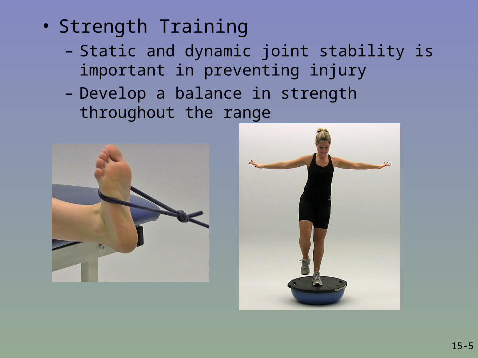
• Neuromuscular Control Training– Can be enhanced
by training in controlled activities on uneven surfaces or a balance board
15-7
• Footwear– Can be an important factor in reducing injury– Shoes should not be used in activities they were not
made for
• Preventive Taping and Orthoses– Tape can provide some prophylactic protection– Improperly applied tape can disrupt normal
biomechanical function and cause injury– Lace-up braces have even been found to be
effective in controlling ankle motion
15-8
Assessing the Lower Leg and Ankle
• History– Past history– Mechanism of injury– When does it hurt?– Type of, quality of, duration of pain?– Sounds or feelings?– How long were you disabled?– Swelling?– Previous treatments?
15-9
• Observations– Postural deviations?– Genu valgum or varum?– Is there difficulty with walking?– Deformities, asymmetries or swelling?– Color and texture of skin, heat, redness?– Patient in obvious pain?– Is range of motion normal?
• Palpation– Begin with bony landmarks and progress to soft tissue– Attempt to locate areas of deformity, swelling and localized
tenderness

15-10
• Special Tests - Lower Leg – Percussion/bump and Compression tests
• Used when fracture is suspected• Percussion test is a blow to the tibia, fibula or
heel to create vibratory force that resonates w/in fracture causing pain
• Compression test involves compression of tibia and fibula either above or below site of concern
15-11
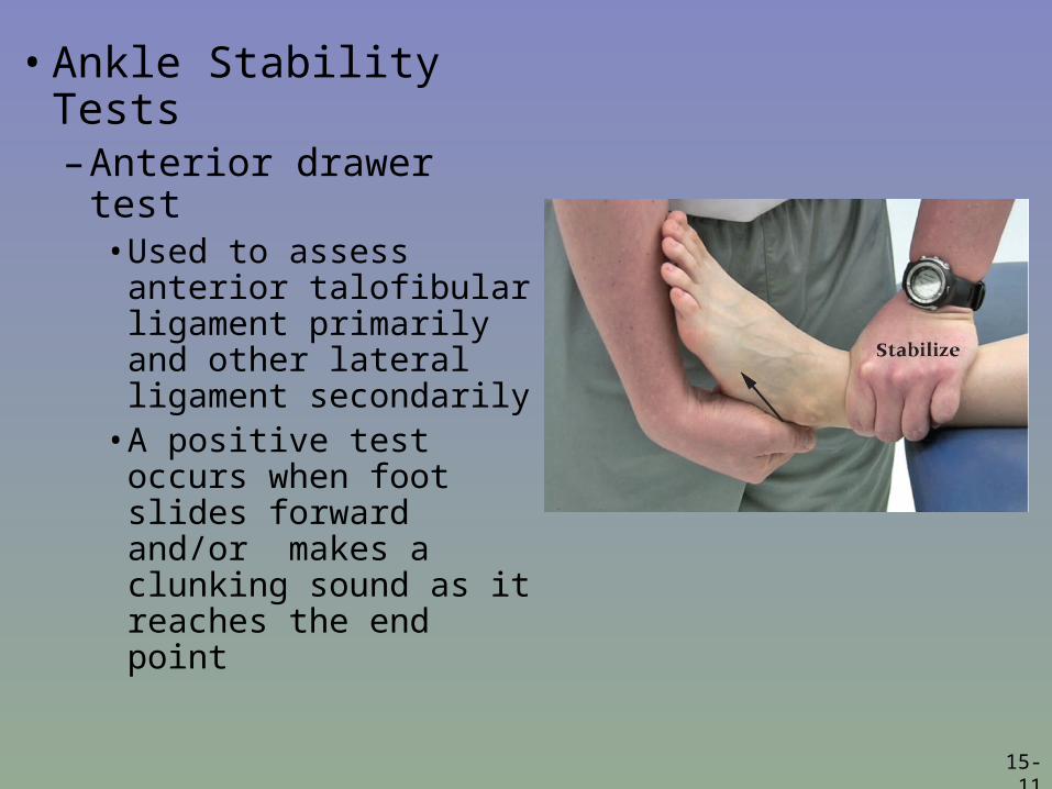
• Ankle Stability Tests– Anterior drawer test
• Used to assess anterior talofibular ligament primarily and other lateral ligament secondarily
• A positive test occurs when foot slides forward and/or makes a clunking sound as it reaches the end point

15-12
– Talar tilt test• Performed to
determine extent of inversion or eversion injuries
• Calcaneus is inverted and excessive motion indicates injury to calcaneofibular ligament and possibly the anterior and posterior talofibular ligaments
• If the calcaneus is everted, the deltoid ligament is tested
15-13
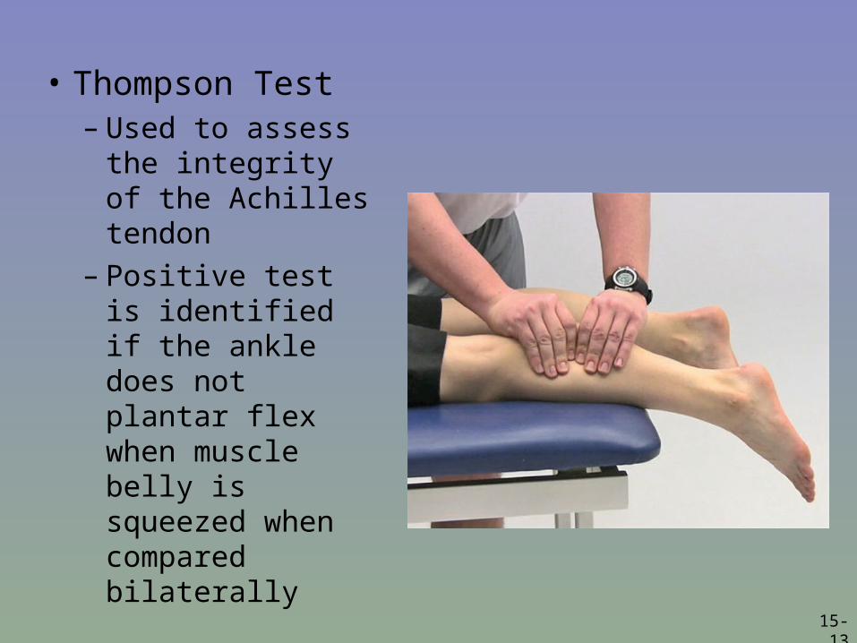
• Thompson Test– Used to assess
the integrity of the Achilles tendon
– Positive test is identified if the ankle does not plantar flex when muscle belly is squeezed when compared bilaterally

15-14
• Functional Tests
– While weight bearing the following should be performed
• Walk on toes (plantar flexion)• Walk on heels (dorsiflexion)• Hops on injured ankle• Start and stop running• Change direction rapidly• Run figure eights

15-15
Recognition and Management of Injuries to the Ankle
• Ankle Injuries: Sprains– Single most common injury in athletics caused by sudden
inversion or eversion moments
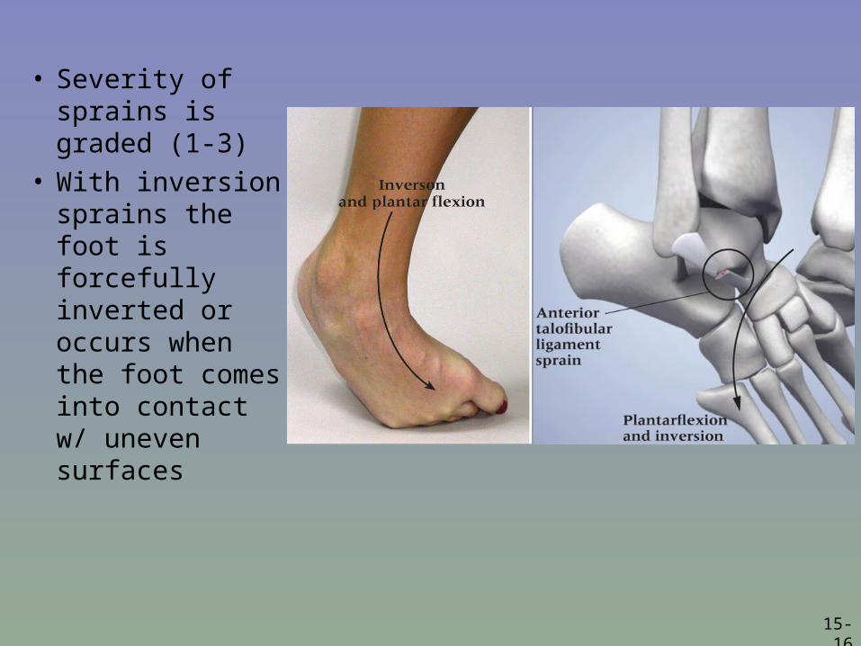
• Inversion Sprains– Most common and results in injury to the lateral ligaments– Anterior talofibular ligament is injured with inversion,
plantar flexion and internal rotation– Occasionally the force is great enough for an avulsion
fracture to occur w/ the lateral malleolus
15-16
• Severity of sprains is graded (1-3)
• With inversion sprains the foot is forcefully inverted or occurs when the foot comes into contact w/ uneven surfaces

15-17

15-18
Eversion Ankle Sprains
• Etiology – Bony protection and
ligament strength decreases likelihood of injury
– Eversion force resulting in damage to deltoid and possibly fx of the fibula
– Deltoid can also be impinged and contused with inversion sprains
15-19
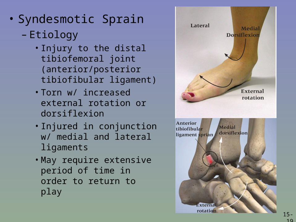
• Syndesmotic Sprain– Etiology
• Injury to the distal tibiofemoral joint (anterior/posterior tibiofibular ligament)
• Torn w/ increased external rotation or dorsiflexion
• Injured in conjunction w/ medial and lateral ligaments
• May require extensive period of time in order to return to play
15-20
• Graded Ankle Sprains – Signs of Injury
• Grade 1– Mild pain and disability; weight bearing is minimally impaired; point tenderness
over ligaments and no laxity
• Grade 2– Feel or hear pop or snap; moderate pain w/ difficulty bearing weight; tenderness
and edema– Positive talar tilt and anterior drawer tests– Possible tearing of the anterior talofibular and calcaneofibular ligaments
• Grade 3– Severe pain, swelling, hemarthrosis, discoloration– Unable to bear weight– Positive talar tilt and anterior drawer– Instability due to complete ligamentous rupture

15-21
– Care• Must manage pain and swelling• Apply horseshoe-shaped foam pad for focal compression • Apply wet compression wrap to facilitate passage of cold from
ice packs surrounding ankle• Apply ice for 20 minutes and repeat every hour for 24 hours• Continue to apply ice over the course of the next 3 days• Keep foot elevated as much as possible• Avoid weight bearing for at least 24 hours • Begin weight bearing as soon as tolerated• Return to participation should be gradual and dictated by healing
process
15-22
• Ankle Fractures/Dislocations– Cause of Injury
• Number of mechanisms – often similar to those seen in ankle sprains
– Signs of Injury• Swelling and pain may be extreme with possible deformity
– Care• Splint and refer to physician for X-ray and examination• RICE to control hemorrhaging and swelling• Once swelling is reduced, a walking cast or brace may be applied, w/
immobilization lasting 6-8 weeks• Rehabilitation is similar to that of ankle sprains once range of motion is
normal
15-23
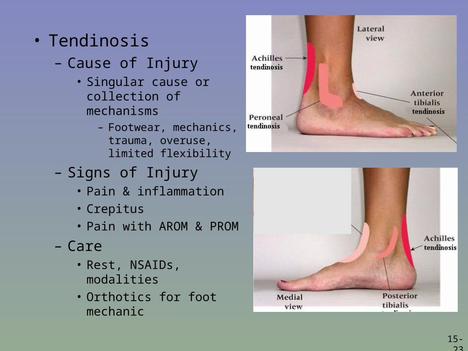
• Tendinosis– Cause of Injury
• Singular cause or collection of mechanisms
– Footwear, mechanics, trauma, overuse, limited flexibility
– Signs of Injury• Pain & inflammation• Crepitus• Pain with AROM & PROM
– Care• Rest, NSAIDs, modalities• Orthotics for foot
mechanic

15-24
• Tibial and Fibular Fractures– Cause of Injury
• Result of direct blow or indirect trauma• Fibular fractures seen with tibial fractures or as the result of direct
trauma
– Signs of Injury• Pain, swelling, soft tissue insult• Leg will appear hard and swollen (Volkman’s contracture)• Deformity – may be open or closed
– Care• Immediate treatment should include splinting to immobilize and ice,
followed by medical referral• Restricted weight bearing for weeks/months depending on severity

15-25

15-26
• Stress Fracture of Tibia or Fibula– Cause of Injury
• Common overuse condition, particularly in those with structural and biomechanical insufficiencies
• Result of repetitive loading during training and conditioning
– Signs of Injury• Pain with activity
• Pain more intense after exercise than before
• Point tenderness; difficult to discern bone and soft tissue pain
• Bone scan results (stress fracture vs. periostitis)
15-27
• Care– Eliminate offending activity– Discontinue stress inducing activity 14 days– Use crutch for walking– Weight bearing may return when pain subsides– After pain free for 2 weeks athlete can gradually
return to activity– Biomechanics must be addressed

15-28
• Medial Tibial Stress Syndrome (Shin Splints)– Cause of Injury
• Pain in anterior portion of shin• Stress fractures, muscle strains, chronic anterior
compartment syndrome, periosteum irritation• Caused by repetitive microtrauma• Weak muscles, improper footwear, training errors, varus
foot, tight heel cord, hypermobile or pronated feet and even forefoot supination can contribute to MTSS
• May also involve, stress fractures or exertional compartment syndrome

15-29
• Shin Splints (continued)– Signs of Injury
• Diffuse pain about disto-medial aspect of lower leg• As condition worsens ambulation may be painful, morning pain and
stiffness may also increase
• Can progress to stress fracture if not treated
– Care• Physician referral for X-rays and bone scan• Activity modification
• Correction of abnormal biomechanics• Ice massage to reduce pain and inflammation
• Flexibility program for gastroc-soleus complex
• Arch taping and orthotics
15-30
• Shin Contusion– Cause of Injury
• Direct blow to lower leg (impacting periosteum anteriorly)
– Signs of Injury• Intense pain, rapidly forming hematoma w/ jelly like consistency
• Increased warmth
– Care• RICE, NSAID’s and analgesics as needed
• Maintaining compression for hematoma (which may need to aspirated)
• Fit with doughnut pad and orthoplast shell for protection

15-31
15-32
• Compartment Syndrome– Cause of Injury
• Rare acute traumatic syndrome due to direct blow or excessive exercise
• May be classified as acute, acute exertional or chronic
– Signs of Injury • Excessive swelling compresses muscles, blood supply
and nerves• Deep aching pain and tightness is experienced• Weakness with foot and toe extension and occasionally
numbness in dorsal region of foot

15-33
– Care• If severe acute or chronic case, may present as medical
emergency that requires surgery to reduce pressure or release fascia
• RICE, NSAID’s and analgesics as needed – Avoid use of compression wrap = increased pressure
• Surgical release is generally used in recurrent conditions
– May require 2-4 month recovery (post surgery)
• Conservative management requires activity modification, icing and stretching
– Surgery is required if conservative management fails
15-34
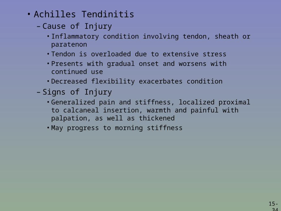
• Achilles Tendinitis– Cause of Injury
• Inflammatory condition involving tendon, sheath or paratenon
• Tendon is overloaded due to extensive stress
• Presents with gradual onset and worsens with continued use
• Decreased flexibility exacerbates condition
– Signs of Injury• Generalized pain and stiffness, localized proximal to
calcaneal insertion, warmth and painful with palpation, as well as thickened
• May progress to morning stiffness
15-35
– Care• Resistant to quick resolution due to slow healing
nature of tendon• Must reduce stress on tendon, address structural
faults (orthotics, mechanics, flexibility)• Aggressive stretching and use of heel lift may be
beneficial• Use of anti-inflammatory medications is suggested
15-36
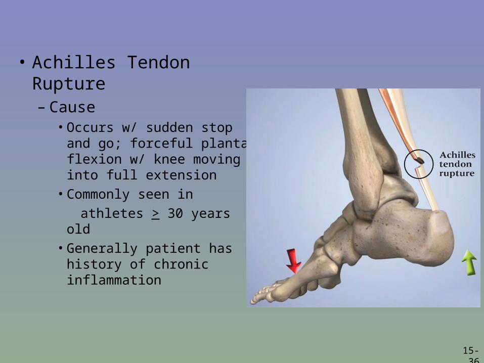
• Achilles Tendon Rupture– Cause
• Occurs w/ sudden stop and go; forceful plantar flexion w/ knee moving into full extension
• Commonly seen in
athletes > 30 years old
• Generally patient has history of chronic inflammation
15-37
– Signs of Injury• Sudden snap (kick in the leg) w/ immediate pain which rapidly
subsides
• Point tenderness, swelling, discoloration; decreased ROM
• Obvious indentation and positive Thompson test
– Care• Usual management involves surgical repair for serious injuries
• Non-operative treatment consists of RICE, NSAID’s, analgesics, and a non-weight bearing cast for 6 weeks to allow for proper tendon healing
• Must work to regain normal range of motion followed by gradual and progressive strengthening program