McFarlane et al. 2011 Replication & Recombination …e.bangor.ac.uk/5108/2/Phd-Fainal for print...
26
1| Page Chapter Number Eukaryote DNA replication and recombination: an intimate association. Ramsay J. McFarlane 1 , Khalid Al-Zeer 1 & Jacob Z. Dalgaard 2 1 – North West Cancer Research Fund Institute, College of Natural Sciences, Brambell Building, Bangor University, Deiniol Road, Bangor, Gwynedd, LL57 2UW, United Kingdom 2 – Warwick Medical School, Clinical Science Research Institute, Gibbert Hill Campus, University of Warwick, Coventry, CV4 7AL, United Kingdom 1. Introduction When the anti-parallel duplex structure of DNA was first postulated it paved a logical pathway to the proposal of semi- conservative DNA replication in which the two parental strands are unwound to provide a template for de novo polynucleotide chain synthesis (Crick & Watson, 1953a; 1953b). Whilst the structure of DNA makes for a relatively simple model for the DNA replicative processes this simplicity is counter balanced by the array of complex molecular pathways which must orchestrate the high fidelity duplication of chromosomes. These mechanisms need to cope with a variety of types of damage to the parental duplex, which must be processed in a relatively error free fashion. Failures to correct DNA damage associated with the progression of the DNA replicative machinery, the replisome, can render a cell unviable or diseased. Conversely, alterations to genomes, be they single base pair changes or more substantial structural rearrangements, are required to instil genetic change, which is essential for the evolution of living systems. One repair process associated with replication is genetic recombination, which serves to repair breakages in DNA strands and failed forks. Failures in recombinogenic processing of replication-associated lesions is associated with significant genome rearrangements and in humans these have been linked directly to genetic disease states, including cancers (Abeysinghe et al., 2006; Admire et al., 2006; Aguilera & Gómez-González, 2008; Bartek et al., 2007; Chen et al., 2010; Halazonetis et al., 2008; Lambert et al., 2005; Lemoine et al., 2005; Putnam et al., 2009; Weinstock et al., 2006; Weinert et al., 2009; Moynahan & Jasin, 2010). Whilst recombination mechanisms serve other functions in genome dynamics, such as the programmed conjoining of homologous chromosomes during meiosis, it could be argued that the primary role of recombination is to mediate genome repair in close association with the replisome. Here we will explore current understanding of the types of DNA replication-associated lesions which require recombination processing and the current models proposed for the various recombination mechanisms which ultimately repair and maintain genetic integrity during cellular duplication events. Replication-associated recombination also plays distinct functional roles in regulation of specific genomic events, such as mating type switching in fission yeast (Dalgaard & Klar, 1999), and it regulates some genomic regions via distinct mechanisms, such as the highly repetitive rDNA locus (Dalgaard et al., 2011; Chapter X, this book). Whilst we will not consider these specific regulatory mechanisms here, this chapter will provide an understanding of the mechanistic basis of the role of recombination in preserving genome integrity in response to aberrations in the replicative process and will thus provide an enlightened platform for further reading relating to regulation of specific genomic events/regions. 2. The generation of replication-associated recombinogenic lesion The process of DNA replication can result in unscheduled generation of recombinogenic DNA lesions which can take the form of single-stranded gaps within a duplex, one-sided DNA double-strand breaks (DSBs), two-sided DSBs or collapsed fork structures. Breaks in the DNA arise due to the replisome encountering an array of distinct problems. In this section we outline models which have been proposed for the generation of distinct classes of lesion . 2. 1. The conversion of single-stranded nicks into DSBs Nicks or short gaps can be generated in one strand of the duplex during normal cellular metabolism. They can be generated by a single broken bond in the sugar-phosphate backbone of one strand or by the excision of a nucleotides during excision repair processes, such as nucleotide excision repair (NER) (for example, see Moriel-Carretero & Aguilera, 2010a; 2010b); moreover, nicks in the duplex can be generated by internally orexternally generated DNA damaging agents, such as highly reactive superoxide radicals or radiation.
Transcript of McFarlane et al. 2011 Replication & Recombination …e.bangor.ac.uk/5108/2/Phd-Fainal for print...

1 | P a g e
Chapter Number
Eukaryote DNA replication and recombination: an intimate association.
Ramsay J. McFarlane1, Khalid Al-Zeer1 & Jacob Z. Dalgaard2
1 – North West Cancer Research Fund Institute, College of Natural Sciences, Brambell Building, Bangor University, Deiniol Road, Bangor, Gwynedd, LL57 2UW, United Kingdom
2 – Warwick Medical School, Clinical Science Research Institute, Gibbert Hill Campus, University of Warwick, Coventry, CV4 7AL, United Kingdom
1. Introduction
When the anti-parallel duplex structure of DNA was first postulated it paved a logical pathway to the proposal of semi-conservative DNA replication in which the two parental strands are unwound to provide a template for de novo polynucleotide chain synthesis (Crick & Watson, 1953a; 1953b). Whilst the structure of DNA makes for a relatively simple model for the DNA replicative processes this simplicity is counter balanced by the array of complex molecular pathways which must orchestrate the high fidelity duplication of chromosomes. These mechanisms need to cope with a variety of types of damage to the parental duplex, which must be processed in a relatively error free fashion. Failures to correct DNA damage associated with the progression of the DNA replicative machinery, the replisome, can render a cell unviable or diseased. Conversely, alterations to genomes, be they single base pair changes or more substantial structural rearrangements, are required to instil genetic change, which is essential for the evolution of living systems. One repair process associated with replication is genetic recombination, which serves to repair breakages in DNA strands and failed forks. Failures in recombinogenic processing of replication-associated lesions is associated with significant genome rearrangements and in humans these have been linked directly to genetic disease states, including cancers (Abeysinghe et al., 2006; Admire et al., 2006; Aguilera & Gómez-González, 2008; Bartek et al., 2007; Chen et al., 2010; Halazonetis et al., 2008; Lambert et al., 2005; Lemoine et al., 2005; Putnam et al., 2009; Weinstock et al., 2006; Weinert et al., 2009; Moynahan & Jasin, 2010). Whilst recombination mechanisms serve other functions in genome dynamics, such as the programmed conjoining of homologous chromosomes during meiosis, it could be argued that the primary role of recombination is to mediate genome repair in close association with the replisome. Here we will explore current understanding of the types of DNA replication-associated lesions which require recombination processing and the current models proposed for the various recombination mechanisms which ultimately repair and maintain genetic integrity during cellular duplication events. Replication-associated recombination also plays distinct functional roles in regulation of specific genomic events, such as mating type switching in fission yeast (Dalgaard & Klar, 1999), and it regulates some genomic regions via distinct mechanisms, such as the highly repetitive rDNA locus (Dalgaard et al., 2011; Chapter X, this book). Whilst we will not consider these specific regulatory mechanisms here, this chapter will provide an understanding of the mechanistic basis of the role of recombination in preserving genome integrity in response to aberrations in the replicative process and will thus provide an enlightened platform for further reading relating to regulation of specific genomic events/regions.
2. The generation of replication-associated recombinogenic lesion
The process of DNA replication can result in unscheduled generation of recombinogenic DNA lesions which can take the form of single-stranded gaps within a duplex, one-sided DNA double-strand breaks (DSBs), two-sided DSBs or collapsed fork structures. Breaks in the DNA arise due to the replisome encountering an array of distinct problems. In this section we outline models which have been proposed for the generation of distinct classes of lesion .
2. 1. The conversion of single-stranded nicks into DSBs
Nicks or short gaps can be generated in one strand of the duplex during normal cellular metabolism. They can be generated by a single broken bond in the sugar-phosphate backbone of one strand or by the excision of a nucleotides during excision repair processes, such as nucleotide excision repair (NER) (for example, see Moriel-Carretero & Aguilera, 2010a; 2010b); moreover, nicks in the duplex can be generated by internally orexternally generated DNA damaging agents, such as highly reactive superoxide radicals or radiation.

2 | P a g e
Figure 1. Replication-associated recombinogenic lesions. (A) Generation of a one-sided DSB. A unidirectional replication fork approaches a nick (discontinuity in one of the parental duplexes), and a one-sided break is generated. (B) Generation of a two-sided DSB. Opposing forks converge on the side of a parental strand nick to generate a two-sided DSB. (C) Generation of a DSB from a inter-strand cross link (ICL). Replication forks converge on an ICL. This generates a structure which is cleaved by structure-specific nuclease activity, which in turn creates a two-sided DSB. The cross linked abduct remains associated with the unbroken duplex and translesion polymerase synthesis past the abduct generating a complete duplex with an abduct associated with one strand which can now undergo an excision repair (vertical black arrows). (D) single-stranded gap generated by lesion skipping. The replisome ‘skips’ a DNA damage abduct (purple oval) leaving a ssDNA gap behind the fork. (E) Replication fork collapse at a replication barrier. The replisome encounters a barrier to progression and fork stability becomes compromised. The replisome dissociates from the DNA leaving a fork-like branched structure which requires processing for repair / fork recovery. (F) Replication fork collapse due to uncoupling of the replicative helicase and the polymerase. The uncoupling of the helicase (red oval with white arrow) from the polymerases (purple oval) results in the helicases running ahead of new strand synthesis generating extensive regions of ssDNA and ultimately causing fork demise.
When a nick is encountered in either the leading or the lagging strand template by a unidirectional DNA replication fork then a one-sided DSB can result (Figure 1A) (for example, see Harper et al., 2010). The generation of this break can be concomitant with the ‘fill in’ of the nick in the unbroken duplex, or the nick can remain in the duplex associated with the other parental strand. If a nick should be proximal to the point at which replication forks converge, or it stalls the progression of a unidirectional fork permitting time for the arrival of a converging fork to replicate from the opposite direction, then there is a chance that both forks will now encounter a template strand discontinuity, potentially resulting in a two-sided DSB (Figure 1B). The one-sided and two-sided DSBs differ considerably in that a one-sided DSB needs to undergo a repair mechanism which will ideally re-establish a functional DNA replication fork to permit the replicative process to continue; whereas a two-sided DSB needs to undergo post replication repair to produce a continuous duplex.
2. 2 The generation of two-sided DSBs as a result of inter-strand cross linking
The Crick and Watson strands of a DNA duplex are held together by hydrogen bonds between the organic bases. Such hydrogen bonding is readily broken by the helicases associated with the replisome to expose the parental strands of a duplex to the template-dependent actions of the DNA polymerases. However, when covalent bonds are formed between two strands of a duplex helicases can no longer dissociate the template strands and replication fork progression is halted. The prevalence of such covalent linkage in a cell not exposed to specific DNA damaging agents is unclear, although a

3 | P a g e
number of anti-cancer drugs, such as mitomycin C and cisplatin act through the generation of covalent inter-strand cross links (Vasquez, 2010). The proposed model for DSB generation in response to covalent cross links (and subsequent repair) is dependent upon additional factors mediating breakage in response to the stalling of the replication fork (Figure 1C) (Nakanishi et al., 2011). To generate a two-sided DSB it is proposed that forks travelling in opposing directions converge upon the site of the cross linkage. This triggers unknown nucleases, possibly the structure-specific endonuclease Mus81-Eme1(Mms4) (see below) to cleave one of the parental strands either side of the site of the cross link. This, in combination with the fact that leading and lagging strand synthesis for that template strand were not complete (due to the cross link) results in a two-sided DSB (Figure 1C). This results in a cross linked nucleotide (or nucleotides, dependent upon the position of the cleavage) being associated with the other, uncleaved template strand (Figure 1C, bottom). It is proposed that this cross linked entity has the ability to ‘swing’ out of the way of the DNA polymerases and that translesion polymerases replicate past the cross linked adduct; at this stage an inappropriate base may be inserted, and so this mechanism is potentially highly mutagenic. The cross linked adduct in the new daughter duplex is proposed to be recognised by the NER pathway (Wood, 2010), which will remove the adduct and use the strand which was newly synthesised via by translesion synthesis (TLS) as the template for repair. TLS is mediated by a number of distinct DNA polymerases (Prakash et al., 2005; Loeb & Monnat, 2008; Ho & Schärer, 2010), which are thought to be recruited to the sites of lesions via activity of proliferating-cell nuclear antigen (PCNA), a homotrimer which plays multiple roles at the replication fork during both normal unperturbed replication (Navadgi-Patil & Burgers, 2009; Stoimenov & Helleday, 2009). The repaired daughter template now becomes the homologous substrate for recombination-mediated DSB repair and so if a mutated DNA sequence is generated by a TLS it is transferred to the other duplex in a stable fashion, ensuring that this process results in two new duplexes, both with stably inherit cross linking agent-induced mutation.
2.3 The generation of a recombinogenic single-stranded DNA gaps via lesion bypass
Whilst DSBs are one of the most potentially dangerous lesions in DNA due to the fact that they represent an un-tethering of covalent linkages, single-stranded DNA (ssDNA) gaps in DNA also represent significant biological problems, not only because they have the potential to mediate gross chromosomal rearrangements, but also because they represent the loss of one of the transcriptional templates (Lehmann & Fuchs, 2006). Also, if left unrepaired, gaps can be converted into DSBs in subsequent rounds of DNA replication (see above). Whilst ssDNA gaps can be generated by other means (for example, incomplete NER), they can be generated when the replisome encounters an adduct on the duplex (for example, see Lopes et al., 2006; Figure 1D). If the replisome can ‘skip’ the adduct, replication will progress leaving a ssDNA gap in the wake of the fork (Figure 1D). The nature of adducts capable of being ‘skipped’ and the prevalence of this route of gap generation is difficult to discern, but when generated, such lesions can trigger distinct repair pathways (see below).
2. 4 The generation of dysfunctional fork structures
DNA replication forks can encounter blocks to their progression as a result of normal chromosome dynamics such as collisions between the replisome and RNA polymerases (Aguilera, 2002; Prado & Aguilera, 2005; Poveda et al., 2010; Tuduri et al., 2010) / nascent transcripts (Mischo et al., 2011), encounters with adducts on the DNA (for example, see Cordeiro-Stone et al., 1999) such as those which might be caused by DNA damaging agents (Mirkin & Mirkin, 2007) or encounters with unusual DNA structures (for example, see Narayanan et al., 2006). In some biological systems barriers to the progression of the DNA replication fork have evolved to play a programmed role in specific chromosomal regulatory pathways, such as the RTS1 replication fork barrier which is required for efficient mating type switching in the fission yeast (Dalgaard & Klar, 2001; Dalgaard et al., 2009; Vengrova et al., 2002). Stalling of a fork can result in what is often referred to as ‘fork collapse’ where some or all of the proteins of the replisome dissociate from the fork leaving non-replicative fork structures (Figure 1E) (for example, see Weinert et al., 2009). Moreover, the replicative helicases, which unwind the DNA duplex to provide single-stranded template, can become uncoupled from the replicative polymerases (for example, see Pacek & Walter, 2004; Pages & Fuchs, 2003; Lopes et al., 2006), this can result in fork collapse and more extensive regions of ssDNA become associated with the failed fork structure (Figure 1F). Nucleotide depletion is also thought to result in replication fork collapse; the ribonucleotide reductase inhibitor hydroxyl urea is frequently used to deplete nucleotide pools and so disrupt the progression of the S-phase. In these cases the fork failure does not result in strand breakage, but a structure is generated which is thought to require recombination-associated mechanisms to re-establish a functional fork structure. The replisome is associated with functions which serve to prevent fork collapse and these functions have intimate mechanistic links to the checkpoint signal transduction pathways which can be triggered in the event of a replication-associated lesion generating a significant initiator signal (most likely ssDNA). Moreover, there are factors associated with the repolisome known as the replication fork progression complex which serves to monitor the ‘traffic’ ahead of the DNA replication fork and mediate a stable and appropriate delay in the fork progression (presumably until the barrier is removed) and the ‘so called ‘sweepase’ (for example, see Ivessa et al., 2003) which functions to remove barriers, such as RNA polymerases, to prevent them triggering a barrier response in the replisome thereby minimises the chance of a potential fork collapse scenario. This chapter will not review the function of these anti-collapse and fork stabilisation mechanisms, or their link to the cellular checkpoint systems and the reader is

4 | P a g e
directed to a number of other excellent reviews which cover these subjects in more detail (Bartek et al., 2004; Branzei & Foiani, 2007a; 2007b; 2009; 2010; Grallert & Boye, 2008; Harrison & Haber, 2006; Lambert et al., 2007; Labib, 2008; Labib & Hodgson, 2007; McFarlane et al., 2010; Paulsen & Cimprich, 2007; Putnam et al., 2009; Yao & O’Donnell, 2009).
3. Models for recombination-mediated recovery from replication-associated lesion damage
Homologous recombination requires the presence of a homologous duplex molecule which can be employed as a surrogate template for synthesis-dependent repair of breaks and gaps in duplex DNA molecules. During the repair of replication-generated two-sided DSBs or gaps the aim of recombination is to repair the lesions post replicatively. The aim of the repair process for other replication induced recombinogenic lesions is to re-engage a functional replication fork to permit the completion of genomic replication prior to cell division. Here we will consider the various models proposed for the repair of the recombinogenic lesions described above. For simplicity of understanding we will firstly outline the proposed models at the level of the DNA strands and then in subsequent sections we will consider the proteins which might mediate these repair mechanisms (see section 4).
Figure 2. Model for recombination mediated generation of a DNA replication fork from a one-sided DSB. (a) The one-sided break is processed to generate a single-stranded overhang with a free 3' end (arrow head). (b) The 3' single-stranded end invades the nascent sister chromatid to form a D-loop. (c) The D-loop undergoes nucleolytic processing and strand ligation to re-generate a functional DNA replication fork.
3.1. The processing of a one-sided DSB to re-establish a functional replication fork
On generation of a one-sided DNA break, it is likely that the broken nascent duplex remains associated with the other nascent duplex molecule. This association is primarily facilitated by cohesin, a complex of conserved proteins which serve to maintain inter-sister associations prior to sister chromatid segregation at the metaphase to anaphase transition (for reviews, see Merkenschlager, 2010; Nasmyth & Haering, 2009; Sherwood et al., 2010; Wood et al., 2010; Xiong & Gerton, 2010). The broken end will be exposed permitting association with recombination mediators and will undergo initial end processing. Given that homologous recombination repair of breaks is dependent upon one of the participating strands providing a substrate for new strand synthesis, a free 3’ ssDNA end must be generated during end processing. This processing is often referred to as end resection [Figure 2 step (a)] (see section 4.1). Following this, the free 3’ end of the ssDNA invades a homologous duplex, in this case the sister chromatid, to establish a so called D-loop (displacement loop) structure [Figure 2, step (b)]. This generates a structure which can be recognised by structure-specific endonucleases, such as the Mus81-Eme1 (Mms4) heterodimer (see section 4.), which cleaves the anti-parallel strand to which the invading strand is annealed; thus, following a one step strand ligation, a structure resembling a DNA replication fork is re-established and replication can proceed [Figure 2 step (c)]. This model is appealing as it is a relatively simple pathway to fork re-establishment and it is in essence similar to break-induced replication (for reviews, see McEachen & Haber, 2006; Llorente et al., 2008). It is dependent upon there being an intimate association between the recombination mediators and the factors required to re-build a functional replication fork, although little is known about this relationship at this proposed step.
3.2. Two-sided DSB repair
Two-sided DSBs provide a particular challenge for the cell as gaps in a DNA duplex must be ‘filled’ whilst maintaining the original DNA sequence, although, in the case of cross linking agent-induced DSBs, this can be mutagenic (see above). One of the major factors governing how a DSB is processed is the stage within the cell division cycle that the break is generated (Branzei & Foiani, 2008; Heyer et al., 2010). In G1 of the cell division cycle homologous recombination repair of
5 | P a g e
chromosomal breakage could have detrimental outcomes, such as a loss of heterozygocity through inter-homologue recombination events. Two-sided DSBs generated in G1 are more likely to be processed by a non-homologous DNA end joining mechanism (for reviews, see Lieber, 2010; Mladenov & Iliakis, 2011). The lesions generated in G1 are not likely to have a causal association with the DNA replication machinery and so will not be covered in this chapter.
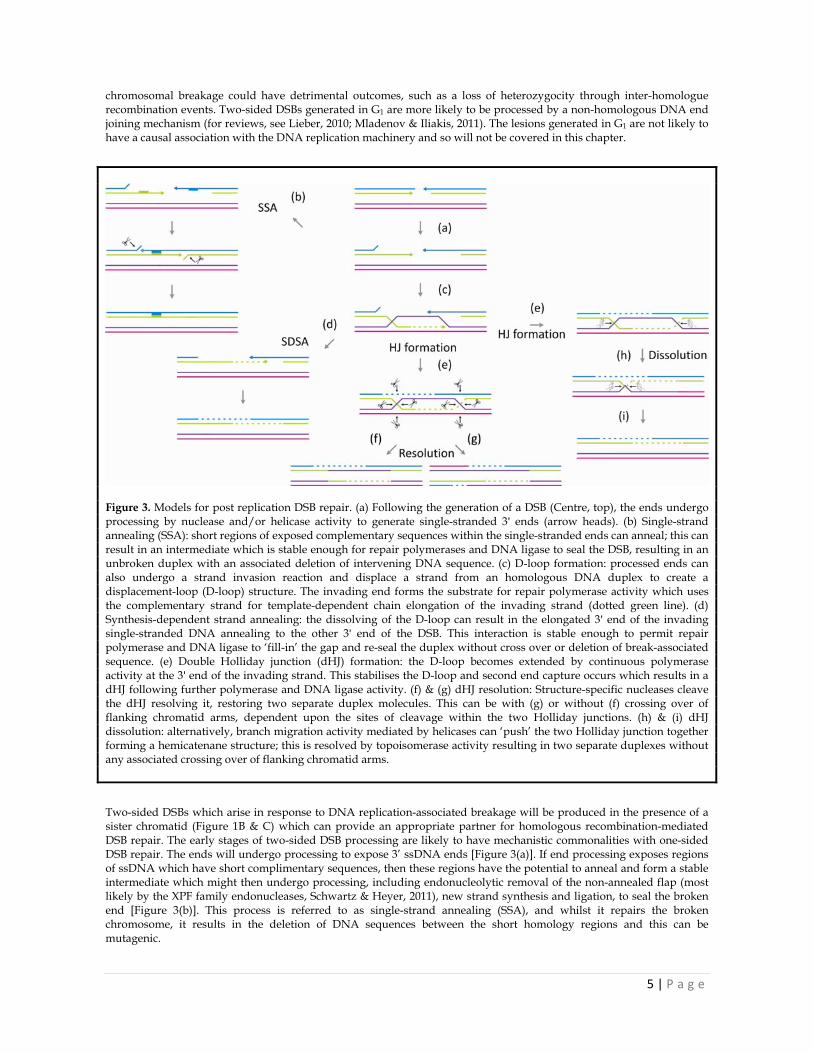
Figure 3. Models for post replication DSB repair. (a) Following the generation of a DSB (Centre, top), the ends undergo processing by nuclease and/or helicase activity to generate single-stranded 3' ends (arrow heads). (b) Single-strand annealing (SSA): short regions of exposed complementary sequences within the single-stranded ends can anneal; this can result in an intermediate which is stable enough for repair polymerases and DNA ligase to seal the DSB, resulting in an unbroken duplex with an associated deletion of intervening DNA sequence. (c) D-loop formation: processed ends can also undergo a strand invasion reaction and displace a strand from an homologous DNA duplex to create a displacement-loop (D-loop) structure. The invading end forms the substrate for repair polymerase activity which uses the complementary strand for template-dependent chain elongation of the invading strand (dotted green line). (d) Synthesis-dependent strand annealing: the dissolving of the D-loop can result in the elongated 3' end of the invading single-stranded DNA annealing to the other 3' end of the DSB. This interaction is stable enough to permit repair polymerase and DNA ligase to ‘fill-in’ the gap and re-seal the duplex without cross over or deletion of break-associated sequence. (e) Double Holliday junction (dHJ) formation: the D-loop becomes extended by continuous polymerase activity at the 3' end of the invading strand. This stabilises the D-loop and second end capture occurs which results in a dHJ following further polymerase and DNA ligase activity. (f) & (g) dHJ resolution: Structure-specific nucleases cleave the dHJ resolving it, restoring two separate duplex molecules. This can be with (g) or without (f) crossing over of flanking chromatid arms, dependent upon the sites of cleavage within the two Holliday junctions. (h) & (i) dHJ dissolution: alternatively, branch migration activity mediated by helicases can ‘push’ the two Holliday junction together forming a hemicatenane structure; this is resolved by topoisomerase activity resulting in two separate duplexes without any associated crossing over of flanking chromatid arms.
Two-sided DSBs which arise in response to DNA replication-associated breakage will be produced in the presence of a sister chromatid (Figure 1B & C) which can provide an appropriate partner for homologous recombination-mediated DSB repair. The early stages of two-sided DSB processing are likely to have mechanistic commonalities with one-sided DSB repair. The ends will undergo processing to expose 3’ ssDNA ends [Figure 3(a)]. If end processing exposes regions of ssDNA which have short complimentary sequences, then these regions have the potential to anneal and form a stable intermediate which might then undergo processing, including endonucleolytic removal of the non-annealed flap (most likely by the XPF family endonucleases, Schwartz & Heyer, 2011), new strand synthesis and ligation, to seal the broken end [Figure 3(b)]. This process is referred to as single-strand annealing (SSA), and whilst it repairs the broken chromosome, it results in the deletion of DNA sequences between the short homology regions and this can be mutagenic.

6 | P a g e
Alternatively, processed breaks with 3’ single-stranded free ends can undergo recombination repair which entails the initial invasion of one of the free 3’ ends into an homologous sister duplex molecule, [Figure 3(c)]. Following strand invasion by the free 3’ end, repair DNA polymerases catalyse chain extension of the invading 3’ end using the anti-parallel strand of the invaded duplex as the template. This process generates new DNA which spans the position of the original break, and thus this repair pathway is replication-dependent. The structure at this point can be referred to as an extended D-loop, as the replicative extension of the invading strand has displaced the opposing strand in the invaded duplex to a greater extent [Figure 3(c)]. At this point one of two key pathways can ensue. Firstly, the extended invading strand can dissociate from the homologous duplex and dissolve the D-loop structure, with the invaded duplex remaining intact (see section 4.3). The dissociated broken end, and the now extended 3’ tail can anneal with the ssDNA of the 3’ tail of the other side of the DSB resulting in a hydrogen bond-dependent, end-to-end reconnecting of the DSB [Figure 3(d)]. Further DNA polymerase ‘fill-in’ and ligation result in a full repair of the DSB with no deletion of any DNA flanking the DSB site and provided the DNA sequence of the participating homologue was identical to that of the broken chromosome (which might not be the case for inter-strand crosslink-induced breaks), this is a non-mutagenic process, with the original sequence being faithfully restored [Figure 3(d)]. Moreover, this process does not involve the cross over exchange of duplexes between participating homologues and so, provided the polymerase steps of the process were faithful, no genetic change should arise from this pathway. This is referred to as synthesis-dependent strand annealing (SDSA) (for recent reviews of DSB repair see San Filippo et al., 2008; Heyer et al., 2010; Moynahan & Jasin, 2010).
Figure 4. Models for post replicative, recombination-mediated gap repair. Gaps may be generated by the replisome ‘skipping’ a lesion in one strand of the DNA (orange oval). (a & b) The 3' side of the gap (magenta arrow head) is converted to single-stranded DNA via helicase action. The single-stranded end invades the adjacent nascent sister chromatid to form a D-loop. The 3' end of the invading strand provides a substrate for DNA polymerase which extends the invading strand (dotted magenta line). The D-loop then either dissolves (a) or stabilises permitting second strand capture by the displaced D-loop (b). (c & d) If the D-loop is dissolved the newly elongated single-strand re-anneals with the single-stranded region filling the gap. Further processing by nucleases and ligase repair the gap; lesions associated with the gap remain in the new duplex and these can be acted on by other excision repair pathways (black vertical arrows flanking the lesion). (e) D-loop stabilisation associated with D-loop extension by continued DNA polymerase activity on the invading strand and second strand capture can result in a dHJ. The dHJ can be resolved by structure-specific nuclease digestion (f) or by dissolution (g). Resolution can potentially result in crossing over of the daughter duplexes, dependent upon the strand specificity of the dHJ cleavage reactions (f). Dissolution of the dHJ does not result in crossing over (g). (h-j) Strand invasion of the fully replicated duplex by the lesion-containing ssDNA gap results in a structure which provides a substrate for structure-specific nuclease attack (i). This results in a duplex at the site of the lesion and the gap is transferred to the undamaged daughter duplex and is subsequently filled by repair polymerases (j).

7 | P a g e
At the D-loop stage [Figure 3(c)] a more extensive expansion of the D-loop (via polymerase activity or continued strand invasion) is thought to stabilise this structure, making its dissolution, and ultimately the SDSA pathway less likely. D-loop stabilisation results in the capture of the second 3’ end of the processed DSB; second end capture is mediated by the displaced strand of the D-loop which will have anti-parallel complementarity with the second end of the DSB (Nimonkar & Kowalczykowski, 2009). Second end capture and subsequent continued activity of repair polymerases and DNA ligase results in the formation of two adjacent Holliday junctions, often referred to as a double Holliday junction (dHJ) [Figure 3(e)]. dHJs are a covalent linkage between homologous duplexes, most likely formed between sister chromatids and have only recently been demonstrated to be recombination intermediates during DSB repair in mitotically dividing cells (Bzymek et al., 2010). For sister chromatids joined in this way to be properly segregated at anaphase the dHJ must be processed to restore two independent duplex molecules. This separation of conjoined duplexes can be mediated by one of two mechanisms. Firstly, the resolution of the dHJ by structure-specific endonucleases (see section 4.5), which can result in crossing over [Figure 3(g)] or non-cross over [Figure 3(f)] outcomes, dependent upon the position within the junction the resolving enzyme cleaves. Alternatively, the dHJ can be dissolved by helicase/translocase-like activities which branch migrates the two Holliday junctions toward each other, resulting in a structure known as a hemicatenane [Figure 3(h)]; this can then be resolved by topoisomerase activity to disconnect the duplexes [Figure 3(i)] (see Section 4.5).
3.3. Post replicative recombination-mediated repair of a single-stranded gap
The generation of gaps due to, for example, lesion skipping by the replisome, has the advantage that the replication process can largely continue without delay, leaving the gap to be repaired after the fork has passed [Figure 1D]. Such lesions are not as potentially harmful as DSBs as the covalent continuity of at least one of the strands ensures that the DNA molecule remains a continuous thread; this also has the advantage that the replication process will have produced a cohesin-dependent associated sister duplex; this ensures that there is an accessible homologous partner permitting a recombination-mediated mechanism to mediate the gap repair. Recombination mediated gap repair has some features in common with DSB repair, but also presents distinct challenges to the repair machineries of the cell. One commonality is that it needs to be initiated with a strand invasion reaction. In DSB repair this occurs after the broken ends have been processed to generate free 3' ends. In gap repair the substrates available to the recombination mediators provide some distinct options [Figure 4]. Firstly, the 3' end of the gapped strand can invade the intact replicated sister, generating a D-loop structure, thus providing a 3' ended substrate for polymerase-mediated strand extension, using the anti-parallel strand of the homologous duplex as a template [Figure 4 (a & b)]. As for the initial step in DSB repair, the D-loop can either be dissolved [Figure 4(c)] or can stabilise, permitting second strand capture, which in this case is the unreplicated strand from the parental duplex which the replisome skipped to form a dHJ [Figure 4(c)]. Dissolution of the D-loop following extension of the invading strand, results in a template-directed repair of the gap. This strand now re-anneals with the anti-parallel strand from the duplex from which it originated and following a final strand processing and re-ligation, the gap is filled [Figure 4(d)]. This processes results in any gap causing lesions to be transmitted to one of the new daughter duplexes, which can then be repaired (say by NER) [Figure 4(d)]. Alternatively, the second strand capture of by the displaced ssDNA of the stable D-loop can result in the formation of a dHJ, which can be resolved, resulting in either cross over or non-cross over products [Figure 4(f)], or dissolved [Figure 4(g)] in a similar fashion to the clearance of dHJs generated in DSB repair (see above) (Figure 3). An alternative possibility involves strand invasion of the intact sister duplex by the ssDNA within the gap [Figure 4(h)]. Processing of this intermediate structure will result in a new duplex being generated at the previously gapped lesion site and a new lesion-free gap in the opposite duplex, which can now be filled by repair polymerase activity [Figure 4 (i & j)] (for recent reviews of DSB repair see San Filippo et al., 2008; Heyer et al., 2010; Moynahan & Jasin, 2010).
3.4. recombination-mediated repair of collapsed DNA replication forks
When replication fork stabilisation mechanisms fail and forks collapse, there is a partial or full dissociation of the trans-acting factors required to continue the progression of DNA replication. This results in a fork structure which is stalled and incapable of re-associating with the factors needed for continued replication. The distinct events which can create collapsed forks (see above) are likely to provide failed replicative structures which present subtly distinct substrates to the recombination and replication re-start programs, however, the ultimate requirement will be to re-establish a functional DNA replication fork in all cases. One way in which a failed fork can be processed is to convert it to a one-sided DSB [Figure 5(a)]. This could be via the action of structure-specific nucleases such as Mus81-Eme1 (see below). Such DSBs would then be acted upon by recombination factors to generate a new DNA replication fork (Section 3.1) [Figure 5(a)]. Alternatively, the failed fork can undergo a fork regression and the nascent strands of the daughter duplexes can anneal with one another to form a region of duplex DNA which protrudes away from the template duplex [Figure 5(b & c)] (Higgins et al., 1976; Sogo et al., 2000). This regressed fork structure is often referred to as a ‘chicken foot’ structure due to its passing resemblance to a three toed chicken’s foot [Figure 5(b & c)]. The chicken foot structure can itself potentially be cleaved by nuclease activity

8 | P a g e
Figure 5. Models for the recombinogenic repair of collapsed DNA replication forks. See the main text for full details (Section 3.4). The figure shows two distinct types of collapsed forks; these are schematically represented as the top two structures in the image. The top structure is a fork which has collapsed without encountering a DNA damage lesion (for example, another replication barrier or a helicase-polymerase uncoupling). The lower structure represents a fork which has collapsed due to a DNA damage abduct on one strand of the DNA (small red oval). The large grey arrows indicate the possible routes to repair / fork recovery. Holliday junction resolution is indicated by black arrows with a scissor symbol; Holliday junction dissolution is represented by black arrows with a pushing hand symbol.

9 | P a g e
to generate a one-sided DSB [Figure 5(d)], or it can undergo a number of distinct routes of processing; the processing of the structure can be dependent upon the structural configuration of the duplex generated by the re-annealing of the nascent daughter strands (the middle toe of the foot). This can have either a 5’ [Figure 5(b)] or 3’ [Figure 5(c)] ssDNA tail, depending upon how it was formed; for example, if lagging strand synthesis extends beyond the position reached by the leading strand, then a 5’ single-stranded tail can be generated on fork reversion [Figure 5(b)]; however, if the leading strand extended beyond the lagging strand at fork collapse the reversion will result in a tail with protruding single-stranded DNA with a free 3’ end [Figure 5(c)]. Duplex regions of nascent daughter strands which result in flush ends (no significant overhang of single-stranded DNA) can potentially be recognised as a DSB and processed to generate single-stranded 3' DNA ends [Figure 5(g)]. A reversed fork with a 5’ single-stranded overhang on the protruding nascent strand duplex [Figure 5(b)] can provide a substrate for polymerase mediated extension of the free 3’ end using the 5’ terminating strand as a template (sometimes referred to as template switching); this will result in a flush or near flush end to the nascent strand duplex [Figure 5(e)]. If the original fork collapse was caused by a lesion in leading strand template [Figure 5(b)], then re-reversal of the chicken foot following polymerase activity will result in the re-annealing of the leading strand with the 3' end now positioned beyond the lesion on the original leading strand template [Figure 5(f)]; thus the generation of a chicken foot intermediate provided a means to generate a new template (the nascent lagging strand) from which the leading strand could be extended to ultimately permit lesion bypass upon reversion of the chicken foot [Figure 5(f)]. Alternatively, if the flush ended duplex generated from annealed / filled in nascent strands is recognised by end processing factors, the 5' end can be resected exposing a 3' single-stranded tail in the nascent strands duplex [figure 5(g)]. This free 3' end now acts as a substrate for recombinases which mediate the invasion of this strand into the template duplex at a position ahead of the position at which the fork failed [Figure 5(h)]. Processing by DNA polymerase and ligase activities results in the formation of a dHJ structure behind the position of the strand invasion, which now becomes a newly established DNA replication fork [Figure 5(i &j)]. As for dHJ structures generated in gap repair or following two-sided DSB repair, the dHJ can be processed via a resolution (cross over or non-cross over) route [Figure 5(i)] or dissolution route [Figure 5(j)]. Either of these routes provides the fork the ability to bypass lesions on the original parental template which may have been an impediment to the progression of the fork. If the original fork failed leaving a leading strand extended relative to the lagging strand, then reversion of the fork will directly generate a single-stranded tail to the duplex of nascent strand with a free 3' end [Figure 5(c)]. This provides a substrate directly for a recombinase-mediated strand invasion to ultimately re-establish a fork following dHJ processing. In this case, there is no template for further extension of the nascent leading strand prior to strand invasion from the chicken foot state, and so this is not a lesion bypass mechanism, rather a mechanism for re-establishment of a collapsed fork. Again, the dHJ structure can be resolved or dissolved, with the former potentially generating cross over products [Figure 5(k)] (for recent reviews of recombination-mediated fork recovery see Aguilera & Gómez-González, 2008; Allen et al., 2011; Branzei & Foiani, 2010; Petermann & Helleday, 2010).
4. Mediators of recombinational repair of failed DNA replication forks
The collapse of replication forks or the generation of recombinogenic lesions such as strand breakages can arise for a number of reasons and can generate a range of distinct substrates for subsequent recombinognic repair (Aguilera & Gómez-González, 2008; Budzowska & Kanaar, 2009; Branzei & Foiani, 2010; Peterman & Helleday, 2010; Allen et al., 2011). There are an array of factors which recognise these lesions, often with a strong substrate specificity, and there are also a number of distinct factors which can act upon processed substrates and the intermediates from which they form. Extensive studies have now started to shed light on this complexity and clear pictures are emerging for the role some of these factors may play in the proposed models proposed for recombination-mediated repair of lesions generated during DNA replication (Section 3). Here we will overview some of the key factors which have been associated with these proposed repair models.
4.1 DSBs: the first response
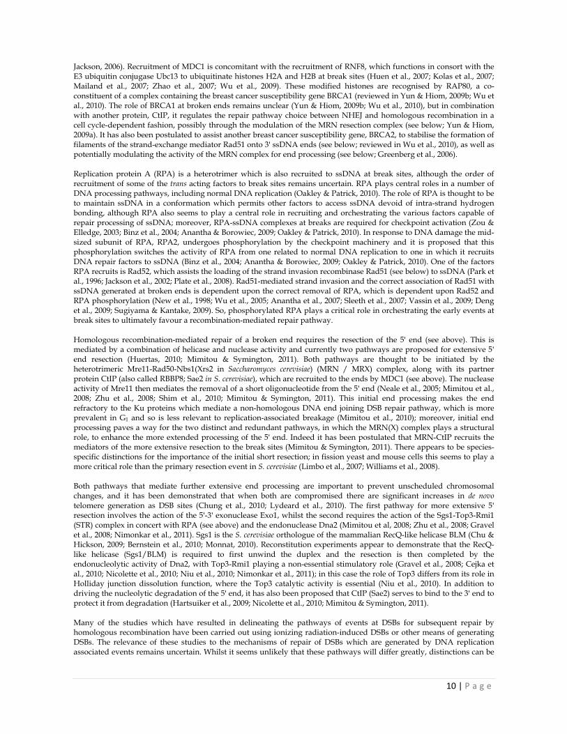
When DNA damage occurs, it is recognised by factors which signal to checkpoint pathways prompting the recruitment of other proteins which, in turn, initiate the repair process. In the case of DSBs one of the first responses is the alteration of the modification status of a histone variant known as H2AX (Fernandez-Capetillo et al., 2004; Ismail & Hendzel, 2008; Lukas & Bartek, 2009). This histone has a tyrosine residue (Y142) in the carboxy tail which, in the absence of DNA damage is phosphorylated by subunits of the H2AX interacting chromatin remodelling complex WICH (Poot et al., 2004; Bozhenok et al., 2002). On DNA damage this residue is de-phosphorylated by Eya1 and Eya3 phosphatases (Cook et al., 2009), permitting the phosphorylation of H2AX serine 139 (S139) by the ATM checkpoint kinase (Rogakou et al., 1998; Burma et al., 2001). This ATM-dependent modification is required for the binding of MDC1 a protein which recruits the MRN complex, one of the key mediators of DSB end resection (see below) (Stucki et al., 2005; Lukas et al., 2004; Stucki &
10 | P a g e
Jackson, 2006). Recruitment of MDC1 is concomitant with the recruitment of RNF8, which functions in consort with the E3 ubiquitin conjugase Ubc13 to ubiquitinate histones H2A and H2B at break sites (Huen et al., 2007; Kolas et al., 2007; Mailand et al., 2007; Zhao et al., 2007; Wu et al., 2009). These modified histones are recognised by RAP80, a co-constituent of a complex containing the breast cancer susceptibility gene BRCA1 (reviewed in Yun & Hiom, 2009b; Wu et al., 2010). The role of BRCA1 at broken ends remains unclear (Yun & Hiom, 2009b; Wu et al., 2010), but in combination with another protein, CtIP, it regulates the repair pathway choice between NHEJ and homologous recombination in a cell cycle-dependent fashion, possibly through the modulation of the MRN resection complex (see below; Yun & Hiom, 2009a). It has also been postulated to assist another breast cancer susceptibility gene, BRCA2, to stabilise the formation of filaments of the strand-exchange mediator Rad51 onto 3' ssDNA ends (see below; reviewed in Wu et al., 2010), as well as potentially modulating the activity of the MRN complex for end processing (see below; Greenberg et al., 2006). Replication protein A (RPA) is a heterotrimer which is also recruited to ssDNA at break sites, although the order of recruitment of some of the trans acting factors to break sites remains uncertain. RPA plays central roles in a number of DNA processing pathways, including normal DNA replication (Oakley & Patrick, 2010). The role of RPA is thought to be to maintain ssDNA in a conformation which permits other factors to access ssDNA devoid of intra-strand hydrogen bonding, although RPA also seems to play a central role in recruiting and orchestrating the various factors capable of repair processing of ssDNA; moreover, RPA-ssDNA complexes at breaks are required for checkpoint activation (Zou & Elledge, 2003; Binz et al., 2004; Anantha & Borowiec, 2009; Oakley & Patrick, 2010). In response to DNA damage the mid-sized subunit of RPA, RPA2, undergoes phosphorylation by the checkpoint machinery and it is proposed that this phosphorylation switches the activity of RPA from one related to normal DNA replication to one in which it recruits DNA repair factors to ssDNA (Binz et al., 2004; Anantha & Borowiec, 2009; Oakley & Patrick, 2010). One of the factors RPA recruits is Rad52, which assists the loading of the strand invasion recombinase Rad51 (see below) to ssDNA (Park et al., 1996; Jackson et al., 2002; Plate et al., 2008). Rad51-mediated strand invasion and the correct association of Rad51 with ssDNA generated at broken ends is dependent upon the correct removal of RPA, which is dependent upon Rad52 and RPA phosphorylation (New et al., 1998; Wu et al., 2005; Anantha et al., 2007; Sleeth et al., 2007; Vassin et al., 2009; Deng et al., 2009; Sugiyama & Kantake, 2009). So, phosphorylated RPA plays a critical role in orchestrating the early events at break sites to ultimately favour a recombination-mediated repair pathway. Homologous recombination-mediated repair of a broken end requires the resection of the 5' end (see above). This is mediated by a combination of helicase and nuclease activity and currently two pathways are proposed for extensive 5' end resection (Huertas, 2010; Mimitou & Symington, 2011). Both pathways are thought to be initiated by the heterotrimeric Mre11-Rad50-Nbs1(Xrs2 in Saccharomyces cerevisiae) (MRN / MRX) complex, along with its partner protein CtIP (also called RBBP8; Sae2 in S. cerevisiae), which are recruited to the ends by MDC1 (see above). The nuclease activity of Mre11 then mediates the removal of a short oligonucleotide from the 5' end (Neale et al., 2005; Mimitou et al., 2008; Zhu et al., 2008; Shim et al., 2010; Mimitou & Symington, 2011). This initial end processing makes the end refractory to the Ku proteins which mediate a non-homologous DNA end joining DSB repair pathway, which is more prevalent in G1 and so is less relevant to replication-associated breakage (Mimitou et al., 2010); moreover, initial end processing paves a way for the two distinct and redundant pathways, in which the MRN(X) complex plays a structural role, to enhance the more extended processing of the 5' end. Indeed it has been postulated that MRN-CtIP recruits the mediators of the more extensive resection to the break sites (Mimitou & Symington, 2011). There appears to be species-specific distinctions for the importance of the initial short resection; in fission yeast and mouse cells this seems to play a more critical role than the primary resection event in S. cerevisiae (Limbo et al., 2007; Williams et al., 2008). Both pathways that mediate further extensive end processing are important to prevent unscheduled chromosomal changes, and it has been demonstrated that when both are compromised there are significant increases in de novo telomere generation as DSB sites (Chung et al., 2010; Lydeard et al., 2010). The first pathway for more extensive 5' resection involves the action of the 5'-3' exonuclease Exo1, whilst the second requires the action of the Sgs1-Top3-Rmi1 (STR) complex in concert with RPA (see above) and the endonuclease Dna2 (Mimitou et al, 2008; Zhu et al., 2008; Gravel et al., 2008; Nimonkar et al., 2011). Sgs1 is the S. cerevisiae orthologue of the mammalian RecQ-like helicase BLM (Chu & Hickson, 2009; Bernstein et al., 2010; Monnat, 2010). Reconstitution experiments appear to demonstrate that the RecQ-like helicase (Sgs1/BLM) is required to first unwind the duplex and the resection is then completed by the endonucleolytic activity of Dna2, with Top3-Rmi1 playing a non-essential stimulatory role (Gravel et al., 2008; Cejka et al., 2010; Nicolette et al., 2010; Niu et al., 2010; Nimonkar et al., 2011); in this case the role of Top3 differs from its role in Holliday junction dissolution function, where the Top3 catalytic activity is essential (Niu et al., 2010). In addition to driving the nucleolytic degradation of the 5' end, it has also been proposed that CtIP (Sae2) serves to bind to the 3' end to protect it from degradation (Hartsuiker et al., 2009; Nicolette et al., 2010; Mimitou & Symington, 2011). Many of the studies which have resulted in delineating the pathways of events at DSBs for subsequent repair by homologous recombination have been carried out using ionizing radiation-induced DSBs or other means of generating DSBs. The relevance of these studies to the mechanisms of repair of DSBs which are generated by DNA replication associated events remains uncertain. Whilst it seems unlikely that these pathways will differ greatly, distinctions can be
11 | P a g e
envisaged; for example, DSBs generated in non-replicating chromatin might present a chromatin substrate which is distinct from the chromatin in proximity to the replisome. Given the fact that many of the early signalling and recruitment events are linked directly to histone modifications (see above), then there may be unique replication-associated mechanisms which require elucidation.
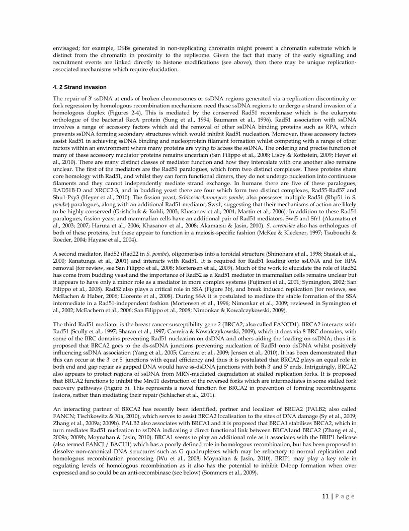
4. 2 Strand invasion
The repair of 3' ssDNA at ends of broken chromosomes or ssDNA regions generated via a replication discontinuity or fork regression by homologous recombination mechanisms need these ssDNA regions to undergo a strand invasion of a homologous duplex (Figures 2-4). This is mediated by the conserved Rad51 recombinase which is the eukaryote orthologue of the bacterial RecA protein (Sung et al., 1994; Baumann et al., 1996). Rad51 association with ssDNA involves a range of accessory factors which aid the removal of other ssDNA binding proteins such as RPA, which prevents ssDNA forming secondary structures which would inhibit Rad51 nucleation. Moreover, these accessory factors assist Rad51 in achieving ssDNA binding and nucleoprotein filament formation whilst competing with a range of other factors within an environment where many proteins are vying to access the ssDNA. The ordering and precise function of many of these accessory mediator proteins remains uncertain (San Filippo et al., 2008; Lisby & Rothstein, 2009; Heyer et al., 2010). There are many distinct classes of mediator function and how they intercalate with one another also remains unclear. The first of the mediators are the Rad51 paralogues, which form two distinct complexes. These proteins share core homology with Rad51, and whilst they can form functional dimers, they do not undergo nucleation into continuous filaments and they cannot independently mediate strand exchange. In humans there are five of these paralogues, RAD51B-D and XRCC2-3, and in budding yeast there are four which form two distinct complexes, Rad55-Rad57 and Shu1-Psy3 (Heyer et al., 2010). The fission yeast, Schizosaccharomyces pombe, also possesses multiple Rad51 (Rhp51 in S. pombe) paralogues, along with an additional Rad51 mediator, Sws1, suggesting that their mechanisms of action are likely to be highly conserved (Grishchuk & Kohli, 2003; Khasanov et al., 2004; Martin et al., 2006). In addition to these Rad51 paralogues, fission yeast and mammalian cells have an additional pair of Rad51 mediators, Swi5 and Sfr1 (Akamatsu et al., 2003; 2007; Haruta et al., 2006; Khasanov et al., 2008; Akamatsu & Jasin, 2010). S. cerevisiae also has orthologues of both of these proteins, but these appear to function in a meiosis-specific fashion (McKee & Kleckner, 1997; Tsubouchi & Roeder, 2004; Hayase et al., 2004). A second mediator, Rad52 (Rad22 in S. pombe), oligomerises into a toroidal structure (Shinohara et al., 1998; Stasiak et al., 2000; Ranatunga et al., 2001) and interacts with Rad51. It is required for Rad51 loading onto ssDNA and for RPA removal (for review, see San Filippo et al., 2008; Mortensen et al., 2009). Much of the work to elucidate the role of Rad52 has come from budding yeast and the importance of Rad52 as a Rad51 mediator in mammalian cells remains unclear but it appears to have only a minor role as a mediator in more complex systems (Fujimori et al., 2001; Symington, 2002; San Filippo et al., 2008). Rad52 also plays a critical role in SSA (Figure 3b), and break induced replication (for reviews, see McEachen & Haber, 2006; Llorente et al., 2008). During SSA it is postulated to mediate the stable formation of the SSA intermediate in a Rad51-independent fashion (Mortensen et al., 1996; Nimonkar et al., 2009; reviewed in Symington et al., 2002; McEachern et al., 2006; San Filippo et al., 2008; Nimonkar & Kowalczykowski, 2009). The third Rad51 mediator is the breast cancer susceptibility gene 2 (BRCA2; also called FANCD1). BRCA2 interacts with Rad51 (Scully et al., 1997; Sharan et al., 1997; Carreira & Kowalczykowski, 2009), which it does via 8 BRC domains, with some of the BRC domains preventing Rad51 nucleation on dsDNA and others aiding the loading on ssDNA; thus it is proposed that BRCA2 goes to the ds-ssDNA junctions preventing nucleation of Rad51 onto dsDNA whilst positively influencing ssDNA association (Yang et al., 2005; Carreira et al., 2009; Jensen et al., 2010). It has been demonstrated that this can occur at the 3' or 5' junctions with equal efficiency and thus it is postulated that BRCA2 plays an equal role in both end and gap repair as gapped DNA would have ss-dsDNA junctions with both 3' and 5' ends. Intriguingly, BRCA2 also appears to protect regions of ssDNA from MRN-mediated degradation at stalled replication forks. It is proposed that BRCA2 functions to inhibit the Mre11 destruction of the reversed forks which are intermediates in some stalled fork recovery pathways (Figure 5). This represents a novel function for BRCA2 in prevention of forming recombinogenic lesions, rather than mediating their repair (Schlacher et al., 2011). An interacting partner of BRCA2 has recently been identified, partner and localizer of BRCA2 (PALB2; also called FANCN; Tischkowitz & Xia, 2010), which serves to assist BRCA2 localisation to the sites of DNA damage (Sy et al., 2009; Zhang et al., 2009a; 2009b). PALB2 also associates with BRCA1 and it is proposed that BRCA1 stabilises BRCA2, which in turn mediates Rad51 nucleation to ssDNA indicating a direct functional link between BRCA1and BRCA2 (Zhang et al., 2009a; 2009b; Moynahan & Jasin, 2010). BRCA1 seems to play an additional role as it associates with the BRIP1 helicase (also termed FANCJ / BACH1) which has a poorly defined role in homologous recombination, but has been proposed to dissolve non-canonical DNA structures such as G quadruplexes which may be refractory to normal replication and homologous recombination processing (Wu et al., 2008; Moynahan & Jasin, 2010). BRIP1 may play a key role in regulating levels of homologous recombination as it also has the potential to inhibit D-loop formation when over expressed and so could be an anti-recombinase (see below) (Sommers et al., 2009).

12 | P a g e
In addition to assisting BRCA2 in mediating Rad51 nucleation, PALB2 has also been demonstrated to enhance D-loop formation by Rad51 in conjunction with another Rad51 interacting protein, Rad51AP1 (Dray et al., 2010; Buisson et al., 2010). Whilst RAD51AP1 was first identified as a Rad51 interacting protein over ten years ago (Kovalenko et al, 1997; Mizuta et al., 1997), evidence has only more recently been put forward to demonstrate its function in enhancing Rad51-mediated strand invasion activity (Wiese et al., 2007; Modesti et al., 2007). PALB2 also interacts with MRG15, a component of a histone acteyltransferase-deacetylase complex implicated in both transcriptional regulation and DNA repair processes providing further clues as to how these proteins may interact with DNA in the context of chromatin (Hayakawa et al., 2010 and references therein). The Rad54 motor protein is another conserved protein which assists in Rad51 function. However it plays a “Jack of all trades” role and seems to have other distinct functions in recombination (reviewed in Tan et al., 2003; Heyer et al., 2006; Symington & Heyer, 2006; Mazin et al., 2010). It has an ATP-dependent DNA translocase activity (with no measurable helicase activity) (Thomä et al., 2005; Amitani et al., 2006), and functionally interacts with Rad51 to stimulate Rad51-mediated strand exchange and heteroduplex extension (Clever et al., 1997; Petukhova et al., 1998; Raschle et al., 1998; Solinger et al., 2001). Rad54 is a Snf2 family member and it has ATP-dependent chromatin remodelling activity and this is thought to function to assist the Rad51 recombinase by either clearing recombinogenic lesions of histones, or remodelling the histones in the intact homologous duplex to be invaded and thereby assisting strand invasion / homology searching (Aleviadis et al., 2002; Alexeev & Kadonaga, 2003; Jaskelioff et al., 2003; Wolner & Peterson, 2005; Kwon et al., 2007; Zhang et al., 2007). Among its other activities Rad54 also has the ability to disrupt Rad51-generated D-loops via a structure branch migration activity (Burgreev et al., 2007), although this apparent paradox might simply relate to a requirement to switch from a D-loop intermediate to a preferred SDSA pathway to avoid crossover outcomes [Figure 3(d)] (Heyer et al., 2010). The switch between pro- to anti-recombinase may be regulated in a temporal fashion and may be regulated by distinct modifications / interactions associated with Rad54 (Mazin et al., 2010). More recently, two new factors have been found to be essential for the proper loading of Rad51 to the sites of replication stress induced ssDNA in human cells. These factors are MMS22L (Mms22 in S. cerevisiae) and TONSL (NFKBIL2) (O’Donnell et al., 2010; Duro et al., 2010). Their depletion results in a failure to load Rad51 in response to replicative stress, despite end processing occurring, indicating that this dimeric factor works at the stage of regulating Rad51 activity, although the exact mechanism of their action remains unclear (O’Donnell et al., 2010; Duro et al., 2010). Consistent with a central role in delineating the homologous recombination pathway choice following replication fork stalling, both Mms22 and its binding partner Mms1 are required for recovery from genotoxic agents which perturb DNA replication in both fission and budding yeasts (Hryciw et al., 2002; Duro et al., 2008; Dovey et al., 2009; Vejrup-Hansen et al., 2011). Rad51 forms a filament on ssDNA and this structure mediates the invasion of the homologous duplex in an ATP-dependent reaction (Holthausen et al. 2010). Stable nucleation progression requires an initial 4-5 Rad51 monomers to bind to the ssDNA; this process does not require the hydrolysis of ATP, but ATP binding does influence the reaction (van der Heijden et al., 2007). The exact mode of homologue searching remains unclear (for example, see Holthausen et al., 2010). Once D-loops are formed, they provide intermediates which can drive the synthesis-dependent repair of gapped or broken regions, paving the way for subsequent processing of the various intermediates that this key, initial event may have formed (Figures 2-5). Products of these events can be re-established forks, SDSA products, dHJs and break-induced replication initiating structures.
4.3 Anti-recombinase activities
Given the complexity of eukaryote genomes and the highly repetitive nature of some, there are instances where it is beneficial to the organism to avoid recombination pathways which might have potentially detrimental outcomes if it is permitted to proceed, or to proceed down a crossover proficient pathway. This is illustrated by Bloom’s Syndrome patients who have a mutation in an anti-recombinase activity (BLM helicase), giving rise to measurably elevated levels of inter-sister chromatid exchange events and higher levels of genome instability (German et al., 1965). The recombination-mediated repair of breakage associated with replication is more likely to follow a non-crossover route via non-crossover resolution/dissolution of more complex recombination intermediates, such as dHJs and hemi-catanane-like structures, or via a SDSA pathway. A group of so called “anti-recombinase” proteins have been identified which can serve to prevent unwanted and ectopic recombination events and to direct recombination down specific, non-crossover lineages, such as SDSA [for example, see Figure3(d)]. Anti-recombinases are proposed to function at two key stages. Firstly they can disrupt the Rad51 presynaptic filament, thus preventing the generation of D-loop intermediates. Alternatively, they can serve to dissolve D-loop structures prior to them stabilising and generating more complex recombination intermediates capable of driving crossover events. These activities are largely mediated by a group of helicases and the study of their roles as anti-recombinases is made more complex by the fact that there is a high degree of functional redundancy and that they possess both pro- and anti-recombination activities, most likely linked to substrate and temporal specificity. The known anti-recombinase helicases are the Srs2 family (budding yeast and fission yeast), the
13 | P a g e
RecQ family (conserved, but five family members have been identified in humans: BLM, WRN, RECQL1, RECQL4 and RECQL5), Fbh1 family (fission yeast and humans), FANCM family [fission yeast (Fml1/2), budding yeast (Mph1) and humans] and RTEL and BRIP1 (FANCJ) (humans; XPD family helicases) (for reviews, see Branzei & Foiani, 2007c; Chu & Hickson, 2009; Whitby, 2009; White, 2009; Wu et al., 2009; Bernstein et al., 2010;Marini & Krejci, 2010; Monnat, 2010; Yusufzai & Kadonaga, 2011). In addition to the helicases, the Rad54 translocase also has the ability to dissolve recombination intermediates and has potential anti-recombinase activity (see above; Bugreev et al., 2007). How these multiple factors are co-ordinated /uniquely specified to distinct damage sites is poorly understood; however, SUMOylation of the replisome component PCNA is known to be required for the recruitment of at least one anti-recombinase, Srs2, indicating an intimate link between residual replication mediators and regulators of replication recovery pathways (Stelter & Ultrich, 2003; Papouli et al., 2005; Pfander et al., 2005)
4.4 Regression of stalled / damaged forks
A number of the pathways postulated for the recovery of a DNA replication fork from a terminal breakdown or for lesion bypass require the regression of the replication fork to make a four way structure, the chicken foot, which has structural similarities to a Holliday junction (Figure 5; see above). As for anti-recombination activities a number of potential players have been posited to mediate fork regression. Firstly, the human RecQ orthologue, BLM, which is also proposed to function in Holliday junction dissolution (see below), has been demonstrated to possess fork regression activity (Ralf et al., 2006), although the physiological relevance of this is difficult to discern. Secondly, extensive studies have indicated that the FANCM helicase/translocase has the ability to regress stalled forks into the four way structure (Gari et al, 2008a; 2008b; Sun et al., 2008). Intriguingly, FANCM has been demonstrated to form a functional bridging role between the BLM pathway and FANC pathways, suggesting that distinct potential fork reversion activities have a close association in response to stalled replication forks (Deans & West, 2009). The histone-fold protein dimer MHF1-MHF2 has recently been identified as a co-factor for FANCM is (Thompson & Jones, 2010; Singh et al., 2010; Yan et al., 2010); this factor has been implicated in centromere kinetochore function (Amano et al., 2009) which has lead to the suggestion that FANMC activity is required to prevent functional genomic regions, made up of repeat sequences which may be highly refractory to DNA replication, from becoming highly unstable (Yan et al., 2010). However, a recent model has been proposed in which recombination triggered by modulation of the progression of a DNA replication fork may play a functional role in centromere dynamics and may also account for the specific requirement for the FANCM co-factors to associate with centromereic regions (McFarlane & Humphreys, 2010). The third pathway proposed to play a role in fork regression is mediated by another Snf2 family helicase/translocase of S. cerevisiae, Rad5, which has previously been implicated in translesion synthesis (for review, see Unk et al., 2010). Rad5 has E3 ubiquitin ligase activity, which is mediated through a RING finger domain (Ulrich & Jentsch, 2000). Rad5 can mediate fork regression (Blastyák et al., 2007) and four way structures observed in wild-type S. cerevisae cells, in response to fork stalling agents, do not accumulate in Rad5-deficient cells (Minca & Kowalski, 2010). It has been proposed that Rad5 may regress a stalled fork and that this provides a substrate for recombination-mediated processing to re-establish a functional fork (Figure 5) (Yusufzai & Kadonaga, 2011). Mammalian cells have two putative Rad5 orthologues, the helices-like transcription factor (HLTF) and SNF2 histone linker PHD RING helicase (SHPRH) (Unk et al., 2010). To date fork regression activity has been demonstrated for HLTF, but not SHRPH (Blastyák et al., 2010). Both HLTF and SHRPH possess E3 ubiquitin ligase activity and they mediate the polyubiquitination of PCNA at stalled forks, indicating a role for these proteins in modulation of replisome factors in response to stalled forks (Motegi et al., 2008), although another, independent PCNA ubiquitination pathway has also been demonstrated indicating possible functional redundancy (Krijger et al., 2011). Additionally, this paralogue pair has been demonstrated to have distinct ubiquitination activities to regulate unique mutation avoidance mechanisms (Lin et al., 2011). Recently, the ATP-dependent annealing helicase SMARCAL1 (SWI/SNF-related, matrix-associated, actin-dependent regulator of chromatin, sub-family a-like 1) / HARP (HepA-related protein) (Yusufzai & Kadonaga, 2008), which is associated with Human Schimke Immune-osseous Dysplasia (SIOD) (Boerkoel et al., 2002), has been implicated as a potential fork regression helicase (Bansbach et al., 2009; Ciccia et al., 2009; Driscoll & Cimprich, 2009; Yuan et al., 2009; Yusufzai et al., 2009). It is proposed that SMARCAL1/HARP is recruited to stalled forks via an active direct recruitment interaction with RPA where it serves to reduce the levels of potentially deleterious ssDNA via a strand annealing mechanism (Driscoll & Cimprich, 2009; Yusufzai & Kadonaga, 2011). A second strand annealing helicase activity has recently been identified, AH2 (annealing helicase 2) suggesting there could be a family of annealing helicases which play a role in genome stability regulation, although no direct link between AH2 and replication fork protection / regression has yet been demonstrated (Yusufzai & Kadonaga, 2010). In addition to the above factors the Rad54 translocase can mediate branch migration of Holliday junction-like structures (Mazin et al., 2010). It has been proposed that, in combination with Rad51, it is capable of mediating a fork regression

14 | P a g e
(Bugreev et al., 2010). Interestingly, Rad54 also exhibits interactions with the structure-specific nuclease Mus81 (reviewed in Mazin et al., 2010), which has been postulated to cleave regressed forks (see below), this might point to failed forks being highly dynamic and promiscuous in which pathway is ultimately ‘chosen’ for fork repair (Figure 5). Rad54 is likely to lie at the centre of such fluidity, as it can serve at multiple stages of the recombination processes associated with stalled fork recovery possessing both pro- and anti-recombination activities (Tan et al., 2003; Heyer et al., 2006; Mazin et al, 2010). Whilst these various helicase/translocase activities have been associated with the regression of forks, to date no activity has been proposed to revert the regressed four way structure to reform the replication fork, although this role could be mediated by the Rad54 protein (Bugreev et al., 2010). It is assumed that such activity is required, but it is also postulated that the regressed fork, the chicken foot, can provide a substrate for structure-specific nucleases. Such nucleases could cleave the regressed fork forming a substrate for further recombination-mediated fork restoration / repair pathways (Figures 5). One conserved candidate for this cleavage is the Mus81-Eme1(Mms4) dimer, which has nucleolytic activity on a range of structures which could be generated by fork regression (Osman & Whitby, 2007). However, there are other structure-specific nucleases, namely the Slx1-Slx4 (SLX4-BTBD12) and Gen1 (Yen1) nucleases which have been demonstrated to have the ability to cleave structures which may be generated by stalled or regressed forks (reviewed in Svendsen & Harper, 2010; Schwartz & Heyer, 2011).
4.5 Holliday junction resolution / dissolution Whilst Holliday junction resolution in bacteria is mediated by a relatively simple single protein mechanism, identifying the activities which are responsible for the processing of Holliday junctions in eukaryotes has revealed a significantly more complex picture. However, from this complexity key players are starting to emerge which are capable of mediating Holliday junction resolution or dissolution (Mankouri & Hickson, 2007; Schwartz & Heyer, 2011; Svendsen & Harper, 2011). As for other mechanisms in the repair of replication-associated damage, it is clear there is a degree of functional redundancy between these pathways (for example, see Weschsler et al., 2011). dHJs can undergo classical endonucleolytic resolution, or they can alternatively undergo dissolution (Figures 3-5); however, single Holliday junctions, are incapable of being processed down the dissolution route. Unlike resolution, dissolution cannot result in crossing over and dissolution does not require a classical Holliday junction resolvase activity. The first of the potential Holliday junction resolution activities is provided by the Mus81 structure-specific endonuclease. This works in concert with a partner protein Eme1 (Mms4) (Osman & Whitby, 2007; Ciccia et al., 2008). It is required for the recovery from replicative stress and Mus81-deficient cells are sensitive to agents which cause replication-associated DNA damage (for examples, see Boddy et al., 2001; Roseeaulin et al., 2008; Svendsen et al., 2009) and exhibit high levels of chromosomal re-arrangements during normal mitotic proliferation (for example, see Dendouga et al., 2005). Mus81 has been demonstrated to have Holliday junction resolution activity, possibly via a nick and counter nick mechanism (Boddy et al., 2000; Gaillard et al., 2003), but conclusive evidence that this activity is responsible for its role in maintaining genome stability in response to replication fork failures is difficult to discern for two reasons. Firstly, Mus81 has the ability to cleave other, non- Holliday junction structures during the processing of substrates generated by replication-associated DNA damage (Ciccia et al., 2003; Osman et al., 2003; Whitby et al., 2003; Fricke et al., 2005). Secondly, Mus81 does not show a particularly strong preference for Holliday junctions with continuous strands and favours structures which resemble nicked Holliday junctions (for example, see Fricke et al., 2005), although Mus81 modifications / interaction might favour Holliday junction specificity in vivo (Osman & Whitby, 2007; Schwartz & Heyer, 2011). Interestingly, Mus81 has been demonstrated to interact with Rad54, which has Holliday junction branch migrating capabilities; this might serve to indicate that targeting of Mus81 specifically to Holliday structures can be linked to early events within the repair process via the central regulator Rad54 (Interthal & Heyer, 2000). The second protein with Holliday junction resolvase activity is the Slx1-Slx4 complex (Fekairi et al., 2009; Svendsen et al,. 2009), which has been demonstrated to mediate the repair of failed replication forks (Frickle & Brill, 2003; Deng et al., 2005) and, as for many replciation repair factor genes, is implicated in the cancer predisposition disorder Fanconia anemia (Crossan et al., 2011). As for Mus81, Slx1-4 has structure-specific endonucelase activity which does not favour fixed Holliday junctions, leading to early suggestions that they played no role in Holliday junction resolution (Fricke & Brill, 2003). Later work has now demonstrated the ability of Slx1-4 complex to cleave Holliday junctions (Fekairi et al., 2009; Munoz et al., 2009; Svendsen et al,. 2009), although the physiological significance of these studies remains controversial (Schwartz & Heyer, 2011). Thirdly, Gen1 (Yen1) was identified as a bona fide Holliday junction resolvease (Ip et al., 2008; Rass et al., 2010), although it too cleaves model replication fork intermediates (Ip et al., 2008; Rass et al., 2010). Human and yeast cells dysfunctional for Gen1(yen1) do not have measurable phenotypes indicating a role in genome maintenance (for example, see Svendsen et al., 2009), but further analysis demonstrates a degree of redundancy with other proposed Holliday junction processing
15 | P a g e
pathways (Blanco et al., 2010; Tay & Wu, 2010; Ho et al., 2010; Weschsler et al., 2011). The failure of Gen1(Yen1)-deficient cells to exhibit any significant defect in genome stability pathways, and the complete absence of a Gen1 orthologue in the fission yeast, has lead to the suggestion that Holliday junctions do not play a major role in DNA damage processing pathways, including the responses to failed replication forks (Schwartz & Heyer, 2011). This argument is further supported by the findings that other Holliday junction resolving nucleases have a very low preference for bona fide Holliday junctions (see above). However, the presence of measurable dHJs in mitotic cells at least demonstrates that these structures are present and so play some role in at least one DNA damage recovery pathway during mitotic proliferation (Bzymek et al., 2010). Finally, many of the models described above which involve a dHJ as a recombination intermediate indicate that these intermediates can be dissolved to form hemicatanene structures (see above; Figures 3-5). In Section 4.1 we describe the STR complex (Sgs1-Top3-Rmi1) which plays an enhancer role during extensive end resection of DSBs (see above). This complex also has the ability to serve as a “dissolvasome” for dHJs (Mankouri & Hickson, 2007). Dissolvasome activity involves Sgs1 (BLM) helicase mediating the convergent migration of the two Holiday junctions to form the hemincatanane and Top3 activity then resolves the catanated strands (Chang et al., 2005; Mullen et al., 2005; Yin et al., 2005; Raynard et al., 2006; Wu et al., 2006; Bussen et al., 2007; Yang et al., 2010). The role of Rmi1 is to stimulate the final Top3-mediated de-catenation reaction (Cejka et al., 2010). Mammalian cells have an additional factor, Rmi2, which also serves as an essential component of the dissolvasome complex (BLM-Top3α-Rmi1-Rmi2) (Singh et al., 2008). Given that sister chromatid exchanges are rare events in cells with a fully functional dissolvasome, and that these become elevated when dissolvasome components are perturbed (for example, German et al., 1965), it is likely that this is a major route for dHJ processing, although there is considerable overlap and redundancy with other dHJ resolution factors (see above; for example, Weschler et al., 2011), although Holliday junction containing structures do persist in S. cerevisiae cells following DNA damage in the absence of Sgs1 and Top3 function (Mankouri et al., 2011).
5. Closing remarks DNA replication is at the very center of the regulation of life on earth. Perturbation of replicative processes can generate an array of highly distinct lesions which require processing to ensure that the biological requirements associated with genome duplication are met in full. This has resulted in the evolution of multiple complexes and competing pathways, each capable of acting on specific substrates. These pathways share common players and parsimony has driven the development of distinct and in some cases opposing roles for central regulators. Moreover, the complexity of distinct pathways requires the temporal modification of specific regulators, which must be co-ordinately timed to allow step-wise progression of a given process. Here we have presented some of the models proposed for the repair and processing of replication-associated lesions. We have demonstrated the many possible routes a specific substrate can follow and we have provided a basic overview of the key trans factors and their functional capabilities. It is clear that many of these factors have overlapping roles and that the many pathways in which they serve make elucidation of the exact mechanisms difficult and many key questions remain open to experimental scrutiny.
6. Acknowledgements We would like to thank Dr. Jane Wakeman for helpful comments on the manuscript. We would like to apologise to those people whose work we have not had space to cite.

16 | P a g e
7. References Abeysinghe, S.S.; Chuzhanova, N. & Cooper, D.N. (2006). Gross deletions and translocations in human genetic disease.
Genome Dynamics Vol.1, (2006), pp. 17-34. ISSN 1660-9263 Admire, A.; Shanks, L.; Danzl, N.; Wang, M.; Weier, U.; Stevens, W.; Hunt, E. & Weinert, T. (2006). Cycles of
chromosome instability are associated with a fragile site and are increased by defects in DNA replication and checkpoint controls in yeast. Genes and Development Vol.20, No.2, (January 2006), pp.159-173 ISSN 0890-9369
Aguilera, A. (2002). The connection between transcription and genomic instability. EMBO Journal Vol.21, No.3, (February 2002), pp.195-201 ISSN 0261-4189
Aguilera, A. & Gómez-González, B. (2008). Genome instability: a mechanistic view of its causes and consequences. Nature Reviews Genetics Vol.9, No.3, (March 2008), pp.204-217 ISSN 1471-0056
Akamatsu, Y; Dziadkowiec, D.; Ikequchi, M.; Shinagawa, H. & Iwasaki, H. (2003). Two different Swi5-containing protein complexes are involved in mating-type switching and recombination repair in fission yeast. Proceedings of the National Academy of Science of the United States of America Vol.100, No.26, (December 2003), pp.15770-15775 ISSN 0027-8424
Akamatsu, Y.; Tsutsui, Y.; Morishita, T.; Siddique, M.S.; Kurokawa, Y.; Ikequchi, M.; Yamao, F.; Arcangioli, B. & Iwasaki, H. (2007). Fission yeast Swi5/Sfr1 and Rhp55/Rhp57 differentially regulate Rhp51-dependent recombination outcomes. EMBO Journal Vol.26, No.5, (March 2007), pp.1352-1362 ISSN 0261-4189
Akamatsu, Y. & Jasin, M. (2010). Role for the mammalian Swi5-Sfr1 complex in DNA strand break repair through homologous recombination. PLoS Genetics Vol.6, No.10, (October 2010), pp.e1001160 ISSN 1553-7390
Alexeev, A.; Mazin, A. & Kowalczykowski, S.C. (2003). Rad54 protein possesses chromatin-remodeling activity stimulated by the Rad51-ssDNA nucleoprotein filament. Nature Structural Biology Vol.10, No.3, (March 2003), pp.182-186 ISSN 1072-8368
Alexiadis, V. & Kadonaga, J.T. (2002). Strand pairing by Rad54 and Rad51 is enhanced by chromatin. Genes and Development Vol.16, No.21, (November 2002), pp.2767-2771 ISSN 0890-9369
Allen, C.; Ashley, A.K.; Hromas, R. & Nickoloff, J.A. (2011). More forks on the road to replication stress recovery. Journal of Molecular Cell Biology Vol.3, No.1, (February 2011), pp.4-12 ISSN 1759-4685
Amano, M.; Suzuki, A.; Hori, T.; Backer, C.; Okawa, K.; Cheeseman, I.M. & Fukagawa, T. (2009). The CENP-S complex is essential for the stable assembly of outer kinetochore structure. Journal of Cell Biology Vol.186, No.2, (July 2009), pp.173-182 ISSN 0021-9525
Amitani, I.; Baskin, R.J. & Kowalczykowski, S.C. (2006). Visualization of Rad54, a chromatin remodelling protein, translocating on single DNA molecules. Molecular Cell Vol.23, No.1, (July 2006), pp.143-148 ISSN 1097-2765
Anantha, R.W. & Borowiec, J.A. (2009). Mitotic crisis: the unmasking of a novel role for RPA. Cell Cycle Vol.8,No.3, (February 2009), pp.357-361 ISSN 1551-4004
Anantha, R.W.; Vassin, V.M. & Borowiec, J.A. (2007). Sequential nand synergistic modification of human RPA stimulates chromosomal DNA repair. Journal of Biological Chemistry Vol.282, No.49, (December 2007), pp.35910-35923 ISSN 0021-9258
Bansbach, C.E.; Bétous, R.; Lovejoy, C.A.; Glick, G.G. & Cortez, D. (2009). The annealing helicase SMARCAL1 maintains genome integrity at stalled replication forks. Genes and Development Vol.23, No.20, (September 2009), pp.2405-2414 ISSN 0890-9369
Bartek, J.; Lukas, C. & Lukas, J. (2004). Checking on DNA damage in S phase. Nature Reviews in Cell Biology Vol.5, No.10, (October 2004), pp.792-804 ISSN 1471-0072
Bartek, J.; Bartkova, J. & Lukas, J. (2007). DNA damage signalling guards against activated oncogenes and tumour progression. Oncogene Vol.26, No.56, (December 2007), pp.7773-7779 ISSN 0950-9232
Baumann, P.; Benson, F.E. & West, S.C. (1996). Human Rad51 protein promotes ATP-dependent homologous pairing and strand transfer reactions in vitro. Cell Vol.87, No.4, (November 1996), pp.757-766 ISSN 0092-8674
Bernstein, K.A., Gangloff, S. & Rothstein, R. (2010). The RecQ DNA helicases in DNA repair. Annual Reviews of Genetics Vol.44, (2010), pp.393-417 ISSN 0066-4197
Binz, S.K.; Sheehan, A.M. & Wold, M.S. (2004). Replication protein A phosphorylation and the cellular response to DNA damage. DNA Repair Vol.3, No.2-9, (August-September 2004), pp.1015-1024 ISSN 1568-7856
Blanco, M.G.; Matos, J.; Rass, U.; Ip, S.C. & West, S.C. (2010). Functional overlap between the structure-specific nucleases Yen1 and Mus81-Mms4 for DNA damage repair in S. cerevisiae. DNA Repair Vol.9, No.4, (April 2010), pp.394-402 ISSN 1568-7856
Blastyák, A.; Pintér, L.; Unk, I.; Prakash, L.; Prakash, S. & Haracska, L. (2007). Yeast Rad5 protein required for postreplication repair has a DNA helicase activity specific for replication fork regression. Molecular Cell Vol.28, No.2, (October 2007), pp.181-183 ISSN 1097-2765
Boddy, M.N.; Gaillard, P.H.; McDonald, W.H.; Shanahan, P.; Yates, J.R. 3rd. & Russell, P. (2001). Mus81-Eme1 are essential components of a Holliday junction resolvase. Cell Vol.107, No.4, (November 2001), pp.537-548 ISSN 0092-8674
Boerkoel, C.F.; Takashima, H.; John, J.; Stankiewicz, P.; Rosenbarker, L.; André, J.L.; Bogdanovic, R.; Burquet, A.; Cockfield, S.; Cordeiro, I.; Fründ, S.; Illies, F.; Joseph, M.; Kaitila, I.; Lama, G.; Loirat, C.; McLeod, D.R.; Milford,

17 | P a g e
D.V.; Petty, E.M.; Rodrigo, F.; Saraiva, J.M.; Schmidt, B.; Smith, G.C.; Spranger, J.; Stein, A.; Thiele, H.; Tizard, J.; Weksberg, R.; Lupski, J.R. & Stockton, D.W. (2002). Mutant chromatin remodelling protein SMARCAL1 causes Schimke immune-osseous dysplasia. Nature Genetics Vol.30, No.2, (February 2002), pp.215-220 ISSN 1061-4036
Bozhenok, L.; Wade, P.A. & Varga-Weisz, P. (2002). WSTF-ISWI chromatin remodelling complex targets heterochromatic replication foci. EMBO Journal Vol.21, No.9, (May 2002), pp.2231-2241 ISSN 0261-4189
Branzei, D. & Foiani, M. (2007a). Interplay of replication checkpoints and repair proteins at stalled replication forks. DNA Repair Vol.6, No.7, (July 2007), pp.994-1003 ISSN 1568-7856
Branzei, D. & Foiani, M. (2007b). Template switching: from replication fork repair to genome rearrangements. Cell Vol.131, No.7, (December 2007), pp.1228-1230 ISSN 0092-8674
Branzei, D. & Foiani, M. (2007c). RecQ helicases queuing with Srs2 to disrupt Rad51 filament and suppress recombination. Genes and Development Vol. 21, No.23, (December 2007), pp.3019-3026 ISSN 0890-9369
Branzei, D. & Foiani, M. (2008). Regulation of DNA repair throughout the cell cycle. Nature Reviews in Molecular Cell Biology Vol.9, no.4, (April 2008), pp.297-308 ISSN 1471-0072
Branzei, D. & Foiani, M. (2009). The checkpoint response to replication stress. DNA Repair Vol. 8. No.9, (September 2009), pp.1038-1046 ISSN 1568-7856
Branzei, D. & Foiani, M. (2010). Maintaining genome stability at the replication fork. Nature Reviews in Molecular Cell Biology Vol.11, No.3, (March 2010), pp.208-219 ISSN 1471-0072
Budzowska, M. & Kanaar, R. (2009). Mechanisms of dealing with DNA damage-induced replication problems. Cell Biochemistry and Biophysics Vol.53, No.1, (November 2009), pp.17-31 ISSN 1085-9195
Bugreev, D.V.; Hanaoka, F. & Mazin, A.V. (2007). Rad54 dissociates homologous recombination intermediates by branch migration. Nature Structural and Molecular Biology Vol.14, No.8, (July 2007), pp.746-753 ISSN 1545-9985
Bugreev, D.V.; Rossi, M.J. & Mazin, A.V. (2011). Cooperation of RAD54 and RAD54 in regression of a model replication fork. Nucleic Acids Research Vol.39, No.6, (March 2011), pp.2153-2164 ISSN 0305-1048
Burma, S.; Chen, B.P.; Murphy, M.; Kurimasa, A. & Chen, D.J. (2001). ATM phosphorylates histone H2AX in response to DNA double-strand breaks. Journal of Biological Chemistry Vol.276, No.45, (November 2001), pp.42462-42467 ISSN 0021-9258
Bussen, W.; Raynard, S.; Busygina, V.; Singh, A.K. & Sung, P. (2007). Holliday junction processing activity of the BLM-Topo IIIα-BLAP75 complex. Journal of Biological Chemistry Vol.282, No.43, (October 2007), pp.31484-31892 ISSN 0021-9258
Bzymek, M.; Thayer, N.H.; Oh, S.D.; Kleckner, N. & Hunter, N. (2010). Double Holliday junctions are intermediates of DNA break repair. Nature Vol.464, No.7290, (April 2010), pp.937-941 ISSN 0028-0836
Carreira, A.; Hilario, J.; Amitani, I.; Baskin, R.J.; Shivji, M.K.; Venkitaraman, A.R. & Kowalczykowski, S.C. (2009). The BRC repeats of BRCA2 modualte the DNA-binding selectivity of RAD51. Cell Vol.136, No.6, (March 2009), pp.1032-1043 ISSN 0092-8674
Carreira, A. & Kowalczykowski, S.C. (2009). BRCA2: shining light on the regulation of DNA-binding selectivity by RAD51. Cell Cycle Vol.8, No.21, (November 2009), pp.3445-3447 ISSN 1551-4005
Cejka, P.; Plank, J.L.; Bachrati, C.Z.; Hickson, I.D. & Kowalczykowski, S.C. (2010). Rmi1 stimulates decatenation of double Holliday junctions during dissolution by Sgs1-Top3. Nature Structural and Molecular Biology Vol.17, No.11, (November 2010), pp.1377-1382 ISSN 1545-9985
Cejka, P.; Cannavo, E.; Polaczek, P.; Masuda-Sasa, T.; Pokharel, S.; Campbell, J.L. & Kowalczykowski, S.C. (2010). DNA end resection by Dna2-Sgs1-RPA and its stimulation by Top3-Rmi1 and Mre11-Rad50-Xrs2. Nature Vol.467, No.7311, (September 2010), pp.112-116 ISSN 0028-0836
Chang, M.; Bellaoui, M.; Zhang, C.; Desai, R.; Morozov, P.; Delgado-Cruzata, L.; Rothstein, R.; Freyer, G.A.; Boone, C. & Brown, G.W. (2005). RMI1/NCE4, a suppressor of genome instability, encodes a member of the RecQ helicase/Topo III complex. EMBO Journal Vol.24, No.11, (June 2005), pp.2024-2033 ISSN 0261-4189
Chen, J.M.; Cooper, D.N.; Férec, C.; Kehrer-Sawatzki, H. & Patrinos, G.P. (2010). Genomic rearrangements in inherited disease and cancer. Seminars in Cancer Biology Vol.20, No.4, (August 2010), pp. 222-233 ISSN 1044-579X
Chung, W.H.; Zhu, Z.; Papusha, A.; Malkova, A. & Ira, G. (2010). Defective resection at DNA double-strand breaks leads to de novo telomere formation and enhances gene targeting. PLoS Genetics Vol.6, No.5, (May 2010), pp.e1000948 ISSN 1553-7390
Chu, W.K. & Hickson, I.D. (2009). RecQ helicases: multifunctional genome caretakers. Nature Reviews Cancer Vol.9, No.9, (September 2009), pp.644-654 ISSN 1474-175X
Ciccia, A.; Constantinou, A. & West, S.C. (2003). Identification and characterisation of the human mus81-eme1 endonuclease. Journal of Biological Chemistry Vol.278, no.27, (July 2003), pp.25172-25178 ISSN 0021-9258
Ciccia, A.; Bredemeyer, A.L.; Sowa, M.E.; Terret, M.E.; Jallepalli, P.V.; Harper, J.W. & Elledge, S.J. (2009). The SIOD disorder protein SMARCAL1 is an RPA-interacting protein involved in replication fork restart. Genes and Development Vol.23, No.20, (September 2009), pp.2415-2425 ISSN 0890-9369
Ciccia, A.; McDonald, N. & West, S.C. (2008). Structural and functional relationships of the XPF/MUS81 family of proteins. Annual Review of Biochemistry Vol.77, (2008), pp.259-287 ISSN 006-4154

18 | P a g e
Clever, B.; Interthal, H.; Schmuckli-Maurer, J.; King, J.; Sigrist, M. & Heyer, W.D. (1997). Recombinational repair in yeast: functional interactions between Rad51 and Rad54 proteins. EMBO Journal Vol.16, No.9, (May 1997), pp.2535-2544 ISSN 0261-4189
Cook, P.J.; Ju, B.G.; Telese, F.; Wang, X.; Glass, C.K. & Rosenfeld, M.G. (2009). Tyrosine dephosphorylation of H2AX modulates apoptosis and survival decisions. Nature Vol.458, No.7238, (April 2009), pp.591-596 ISSN 0028-0836
Cordeiro-Stone, M.; Makhov, A.M.; Zaritskaya, L.S. & Griffith, J.D. (1999). Analysis of DNA replication forks encountering a pyrimidine dimer in the template to the leading strand. Journal of Molecular Biology Vol.289, No.5, (June 1999), pp.1207-1218 ISSN 022-2836
Crossan, G.P.; van de Weyden, L.; Rosado, I.V.; Langevin, F.; Gaillard, P.H.; McIntyre, R.E.; Sanger mouse Genetics Project; Gallagher, F.; Kettunen, M.I.; Lewis, D.Y.; Brindle, K.; Arends, M.J.; Adams, D.J. & Patel, K.J. (2011). Disruption of mouse Slx4, a regulator of structure-specific nucelases, phenocopies Fanconia anemia. Nature Genetics Vol.43, no.2, (February 2011), pp.147-152 ISSN 1061-4036
Dalgaard, J.Z.; Eydmann, T.; Koulintchenko, M.; Sayrac, S.; Vengrova, S. & Yamada-Inagawa, T. (2009). Random and site-specific replication termination. Methods in Molecular Biology Vol.521, (2009), pp.35-53 ISSN 1064-3745
Dalgaard, J.Z. & Klar, A.J. (1999). Orientation of DNA replication establishes mating-type switching pattern in S. pombe. Nature Vol.400, No.6740, (July 1999), pp.181-184 ISSN 0028-0836
Dalgaard, J.Z. & Klar, A.J. (2001). Does S. pombe exploit the intrinsic asymmetry of DNA synthesis to imprint daughter cells for mating-type switching? Trends in Genetics Vol.17, No.3, (March 2001), pp.153-157 ISSN 0168-9525
Deans, A.J. & West, S.C. (2009). FANCM connects the genome instability disorder Bloom’s Syndrome and Fanconia Anemia. Molecular Cell Vol.36, No.6, (December 2009), pp.943-953 ISSN 1097-2765
Dendouga, N.; Gao, H.; Moechars, D.; Janicot, M.; Vialard, J. & McGowan, C.H. (2005). Disruption of murine Mus81 increases genomic instability and DNA damage sensitivity but does not promote tumorigenesis. Molecular and Cellular Biology Vol.25, No.17, (September 2005), pp.7569-7579 ISSN 0270-7306
Deng, X.; Prakash, A.; Dhar, K.; Baia, G.S.; Kolar, C.; Oakley, G.G. & Borqstahl, G.E. (2009). Human replication protein A-Rad52-single-stranded DNA complex: stoichiometry and evidence for strand transfer regulation by phosphorylation. Biochemistry Vol.48, No.28, (July 2009), pp.6633-6643 ISSN 0006-2960
Dovey, C.L.; Aslanian, A.; Sofueva, S.; Yates, J.R. 3rd & Russell, P. (2009). Mms1-Mms22 complex protects genome integrity in Schizosaccharomyces pombe. DNA Repair Vol.8, No.13, (December 2009), pp.1390-1399 ISSN 1568-7856
Driscoll, R. & Cimprich, K.A. (2009). HARPing on about the DNA damage response during replication. Genes and Development Vol.23, No.20, (September 2009), pp.2359-2365 ISSN 0890-9369
Duro, E.; Lundin, C.; Ask, K.; Sanchez-Pulido, L.; MacArtney, T.J.; Toth, R.; Ponting, C.P.; Groth, A.; Helleday, T. & Rouse, J. (2010). Identification of the MMS22L-TONSL complex that promotes homologous recombination. Molecular Cell Vol.40, No.4, (November 2010), pp.632-644 ISSN 1097-2765
Duro, E.; Vaisica, J.A.; Brown, G.W. & Rouse, J. (2008). Budding yeast Mms22 and Mms1 regulate homologous recombination induced by replisome blockage. DNA Repair Vol.7, No.5, (May 2008), pp.811-818 ISSN 1568-7856
Fekairi, S.; Scaglione, S.; Chahwan, C.; Taylor, E.R.; Tissier, A.; Coulson, S.; Dong, M.Q.; Ruse, C.; Yates, J.R. 3rd.; Russell, P.; Fuchs, R.P.; McGowan, C.H. & Gaillard, P.H. (2009). Human SLX4 is a Holliday junction resolvase subunit that binds multiple DNA repair/recombination endonucleases. Cell Vol.138, No.1, (July 2009), pp.78-89 ISSN 0092-8674
Fernandez-Capetillo, O.; Lee, A.; Nussenweig, M. & Nussenzweig, A. (2004). H2AX: the histone guardian of the genome. DNA Repair Vol.3, No.8-9, (August-September 2004), pp.959-967 ISSN 1568-7856
Fricke, W.M. & Brill, S.J. (2003). Slx1-Slx4 is a second structure-specific endonuclease functionally redundant with Sgs1-Top3. Genes and Development Vol.17, No.14, (July 2003), pp.1768-1778 ISSN 0890-9369
Fricke, W.M.; Bastin-Shanower, S.A. & Brill, S.J. (2005). Substrate specificity of the Saccharomyces cerevisiae Mus81-Mms4 endonuclease. DNA Repair Vol.4, No.2, (February 2005), pp.243-251 ISSN 1568-7856
Fujimori, A.; Tachiiri, S.; Sonoda, E.; Thompson, L.H.; Dhar, P.K.; Hiraoka, M.; Takeda, S.; Zhang, Y.; Reth, M. & Takata, M. (2001). Rad52 partially substitutes for the Rad51 paralog XRCC3 in maintaining chromosomal integrity in vertebrate cells. EMBO Journal Vol.20, No.19, (October 2001), pp.5513-5520 ISSN 0261-4189
Gaillard, P.H.; Noguchi, E.; Shanahan, P. & Russell, P. (2003). The endogenous Mus81-Eme1 complex resolves Holliday junctions by a nick and counternick mechanism. Molecular Cell Vol.12, No.3, (September 2003), pp.747-759 ISSN 1097-2765
Gari, K.; Décaillet, C.; Delannoy, M.; Wu, L. & Constantinou, A. (2008a). Remodeling of DNA replication structures by the branch point translocase FANCM. Proceedings of the National Academy of Science or the United States of America Vol.105, No.42, (October 2008), pp.16107-16112 ISSN 0027-8424
Gari, K.; Décaillet, C.; Stasiak, A.Z.; Stasiak, A. & Constantinou, A. (2008b). The Fanconia anemia protein FANCM can promote branch migration of Holliday junctions and replication forks. Molecular Cell Vol.29, No.1, (January 2008), pp.141-148 ISSN 1097-2765
German, J.; Archibald, R. & Bloom, D. (1965). Chromosomal breakage in a rare and probably genetically determined syndrome of man. Science Vol.148, (April 1965), pp.506-507 ISSN 0036-8075
19 | P a g e
Grallert, B. & Boye, E. (2008). The multiple facets of the intra-S checkpoint. Cell Cycle Vol.7, No.15, (August 2008), pp.2315-2320 ISSN 1551-4005
Gravel, S.; Chapman, J.R.; Magill, C. & Jackson, S.P. (2008). DNA helicases Sgs1 and BLM promote DNA double-strand break resection. Genes and Development Vol.22, No.20, (October 2002), pp.2767-2772 ISSN 0890-9369
Greenberg, R.A.; Sobhian, B.; Pathania, S.; Cantor, S.B.; Nakatani, Y. & Livingston, D.M. (2006). Multifactorial contributions to an acute DNA damage response by BRCA1/BARD1-containing complexes. Genes and Development Vol.20, No.1, (January 2006), pp.34-46 ISSN 0890-9369
Grishchuk, A.L. & Kohli, J. (2003). Five RecA-like proteins of Schizosaccharomyces pombe are involved in meiotic recombination. Genetics Vol.165, No.3, (November 2003), pp.1031-1043 ISSN 0016-6731
Halazonetis, T.D.; Gorgoulis, V.G. & Bartek, J. (2008) An oncogene-induced DNA damage model for cancer development. Science Vol.319, No.5868, (March 2008), pp.1352-1355 ISSN 0036-8075
Harper, J.V.; Anderson, J.A. & O’Neill, P. (2010). Radiation induced DNA DSBs: contribution from stalled replication forks? DNA Repair Vol.8, No.8, (August 2010), pp.907-913 ISSN 1568-7856
Harrison, J.C. & Haber, J.E. (2006). Surviving the breakup: the DNA damage checkpoint. Annual Reviews in Genetics Vol.40, (2006), pp. 209-235, ISSN 0066-4197
Hartsuiker, E.; Neale, M.J. & Carr, A.M. (2009). Distinct requirements for the Rad32(Mre11) nuclease and Ctp1(CtIP) in the removal of covalently bound topoisomerase I and II from DNA. Molecular Cell Vol.33, No.1, (January 2009), pp.117-123 ISSN 1097-2765
Haryta, N.; Kurokawa, Y.; Murayama, Y.; Akamatsu, Y.; Unzai, S.; Tsutsui, Y. & Iwasaki, H. (2006). The Swi5-Sfr1 complex stimulates Rhp51/Rad51- and Dmc1-mediated DNA strand exchange in vitro. Nature Structural and Molecular Biology Vol.13, No.9, (September 2006), pp.823-830 ISSN 1545-9985
Hayakawa, T.; Zhang, F.; Hayakawa, Y.; Ohtani, Y.; Shinmyozu, K.; Nakayama, J. & Andreassen, P.R. (2010). MRG15 binds directly to PALB2 and stimulates homology-directed repair of chromosomal breaks. Journal of Cell Science Vol.123, No.7, (April 2010), pp.1124-1130 ISSN 0021-9533
Hayase, A.; Takaqi, M.; Miyazaki, T.; Oshiumi, H.; Shinohara, M. & Shinohara, A. (2004). A protein complex containing Mei5 and Sae3 promotes the assembly of the meiosis-specific RecA homolog Dmc1. Cell Vol.119, No.7, (December 2004), pp.927-940 ISSN 0092-8674
Heyer, W.D.; Ehmsen, K.T. & Liu, J. (2010) Regulation of homologous recombination. Annual Reviews of Genetics Vol.44, (2010), pp.113-139 ISSN 0066-4197
Heyer, W.D.; Li, X.; Rolfsmeier, M. & Zhang, X.P. (2006). Rad54: the Swiss Army knife of homologous recombination? Nucleic Acids Research Vol. 34, No.15, (August 2006), pp.4115-4125 ISSN 0305-1048
Higgins, N.P.; Kato, K. & Strauss, B. (1976). A model for replication repair in mammalian cells. Journal of Molecular Biology Vol.101, No.3, (March 1976), pp.417-425 ISSN 0022-2836
Ho, C.K.; Mazón, G.; Lam, A.F. & Symington, L.S. (2010). Mus81 and Yen1 promote reciprocal exchange during mitotic recombination to maintain genome integrity in budding yeast. Molecular Cell Vol.40, No.6, (December 2010), pp.988-1000 ISSN 1097-2765
Ho, T.V. & Schärer, O.D. (2010). Translesion DNA synthesis polymerases in DNA interstrand crosslink repair. Environmental and Molecular Mutagenesis Vol.51, No.6, (July 2010), pp.552-566 ISSN 0893-6692
Holthausen, J.T.; Wyman, C. & Kanaar, R. (2010). Regulation of DNA strand exchange in homologous recombination. DNA Repair Vol.9, no.12, (December 2010), pp.1264-1274 ISSN 1568-7856
Hryciw, T.; Tang, M.; Fontanie, T. & Xiao, W. (2002). MMS1 protects against replication-dependent DNA damage in Saccharomyces cerevisiae. Molecular Genetics and Genomics Vol.266, No.5, (January 2002), pp.848-857 ISSN 1617-4623
Huen, M.S.; Grant, R.; Manke, I.; Minn, K.; Yu, X.; Yaffe, M.B. & Chen, J. (2007). RNF8 transduces the DNA-damage signal via histone ubiquitylation and checkpoint assembly. Cell Vol.131, No.5, (November 2007), pp.901-914 ISSN 0092-8674
Huertas, P. (2010). DNA resection in eukaryotes: deciding how to fix the break. Nature Structural and Molecular Biology Vol.17, No.1, (January 2010), pp.11-16 ISSN 1545-9985
Interthal, H. & Heyer, W.D. (2000). MUS81 encodes a novel helix-hairpin-helix protein involved in the response to UV-and methylation-induced DNA damage in Saccharomyces cerevisiae. Molecular and General Genetics Vol.263, No.5, (Jun 2000), pp.812-827 ISSN 0026-8925
Ip, S.C.; Rass, U.; Blanco, M.G.; Flynn, H.R.; Skehel, J.M. & West, S.C. (2008). Identification of Holliday junction resolvases from humans and yeast. Nature Vol.456, No.7220, (November 2008), 357-361 ISSN 0028-0836
Ismail, I.H. & Hendzel, M.J. (2008). The γH2AX: is it just a surrogate marker of double-strand breaks or much more? Environmental and Molecular Mutagenesis Vol.49, no.1, (January 2008), pp.73-82 ISSN 0893-6692
Ivessa, A.S.; Lenzmeier, B.A.; Bessler, J.B.; Goudsouzian, L.K.; Schnakenberg, S.L. & Zakian, V.A. (2003). The Saccharomyces cerevisiae helicase Rrm3p facilitates replication past non-histone protein-DNA complexes. Molecular Cell Vol.12, No.6, (December 2003), pp.1525-1536 ISSN 1097-2765
Jackson, D.; Dhar, K.; Wahl, J.K.; Wold, M.S. & Borgstahl, G.E. (2002). Analysis of the human replication protein A:Rad52 complex: evidence for crosstalk between RPA32, RPA70, Rad52 and DNA. Journal of Molecular Biology Vol.32, No.1, (August 2002), pp.133-148 ISSN 0022-2836

20 | P a g e
Jaskelioff, M.; van Komen, S.; Krebs, J.E.; Sung, P. & Peterson, C.L. (2003). Rad54p is a chromatin remodelling enzyme required for heteroduplex DNA joint formation with chromatin. Journal of Biological Chemistry Vol.278, No.11, (January 2003), pp.9212-9218 ISSN 0021-9258
Jensen, R.B.; Carreira, A. & Kowalczykowski, S.C. (2010). Purified human BRCA2 stimulates RAD51-mediated recombination. Nature Vol.467, No.7316, (October 2010), pp.678-683 ISSN 0028-0836
Khasanov, F.K.; Salakhova, A.F.; Chepurnaja, O.V.; Korolev, V.G. & Bashkirov, V.I. (2004). Identification and characterization of the rlp1+, the novel Rad51 paralog in the fission yeast Schizosaccharomyces pombe. DNA Repair Vol.3, No.10, (October 2004), pp.1363-1374 ISSN 1568-7856
Khasanov, F.K.; Salakhova, A.F.; Khasanova, O.S.; Grishchuk, A.L.; Chepurnaja, O.V.; Korolev, V.G.; Kohli, J. & Bashkirov, V.I. (2008). Genetic analysis reveals different roles of Schizosaccharomyces pombe sfr1/dds20 in meiotic and mitotic DNA recombination and repair. Current Genetics Vol.54, No.4, (October 2008), pp.197-221 ISSN 0172-8083
Kolas, N.K.; Chapman, J.R.; Nakada, S.; Ylanko, J.; Chahwan, R.; Sweeney, F.D.; Panier, S.; Mendez, M.; Wildenhain, J.; Thomson, T.M.; Pelletier, L.; Jackson, S.P. & Durocher, D. (2007). Orchestration of the DNA-damage response by the RNF8 ubiquitin ligase. Science Vol.318, No.5856, (December 2007), pp.1637-1640 ISSN 0036-8075
Kovalenko, O.V.; Golub, E.I.; Bray-Ward, P.; Ward, D.C. & Radding, C.M. (1997). A novel nucleic acid-binding protein that interacts with human Rad51 recombinase. Nucleic Acids Research Vol.25, No.24, (December 1997), pp.4946-4953 ISN 0305-1048
Krijger, P.H.; Lee, K.Y.; Wit, N.; van der Berk, P.C.; Wu, X.; Roest, H.P.; Maas, A.; Ding, H.; Hoeijmaker, J.H.; Myung, K. & Jacobs, H. (2011). HLTF and SHPRH are not essential for PCNA polyubiquitination, survival and somatic hypermutation: existence of an alternative E3 ligase. DNA Repair Vol. 10, No.4, (January 2011), pp.438-444 ISSN 1568-7856
Kwon, Y.; Chi, P., Roh, D.H.; Klein, H. & Sung, P. (2007). Synergistic action of the Saccharomyces cerevisae homologous recombination factors Rad54 and Rad51 in chromatin remodeling. DNA Repair Vol.6, No.10, (June 2007), pp.1496-1506 ISSN 1568-7856
Labib, K. (2008). Making connections at DNA replication forks: Mrc1 takes the lead. Molecular Cell Vol.32, No.2, (October 2008), pp.116-168 ISSN 1097-2765
Labib, K. & Hodgson, B. (2007) Replication fork barriers: pause for a break or stalling for time? EMBO Reports Vol.8, No.4, (April 2007), pp.346-353 ISSN 1469-221X
Lambert, S.; Watson, A.; Sheedy, D.M.; Martin, B. & Carr, A.M. (2005). Gross chromosomal rearrangements and elevated recombination at an inducible site-specific replication fork barrier. Cell Vol.121, No.5, (June 2005), pp.689-702 ISSN 0092-8674
Lambert, S.; Froget, B. & Carr, A.M. (2007). Arrested replication fork processing: interplay between checkpoints and recombination. DNA Repair Vol.6, No.7, (July 2007), pp.1042-1061 ISSN 1568-7856
Lehmann, A.R. & Fcuhs, R.P. (2006). Gaps and forks in DNA replication: rediscovering old models. DNA Repair Vol.5, No.12, (December 2006), pp.1495-1498 ISSN 1568-7856
Lemoine, F.J.; Degtyareva, N.P.; Lobachev, K. & Petes, T.D. (2005). Chromosomal translocations in yeast induced by low levels of DNA polymerase: a model for chromosome fragile sites. Cell Vol.120, No.5, (March 2005), pp.587-598 ISSN 0092-8674
Lieber, M.R. (2010). The mechanisms of double-strand break repair by the nonhomologous DNA end-joining pathway. Annual Reviews of Biochemistry Vol.79, (2010), pp.181-211 ISSN 0066-4154
Limbo, O.; Chahwan, C.; Yamada, Y.; de Bruin, R.A.; Wittenberg, C. & Russell, P. (2007). Ctp1 is a cell-cycle-regulated protein that functions with Mre11 complex to control double-strand break repair by homologous recombination. Molecular Cell Vol.28, No.1, (October 2007), pp.134-146 ISSN 1097-2765
Lin, J.R.; Zeman, M.K.; Chen, J.Y.; Yee, M.C. & Cimprich, K.A. (2011). SHPRH and HLTF act in a damage-specific manner to coordinate different forms of postreplication repair and prevent mutagenesis. Molecular Cell Vol.42, No.2, (April 2011), pp.237-249 ISSN 1097-2765
Lisby, M. & Rothstein, R. (2009). Choreography of recombination proteins during the DNA damage response. DNA Repair Vol.8, No.9, (September 2009), pp.1068-1076 ISSN 1568-7856
Loeb, L.A. & Monnat, R.J. Jr. (2008). DNA polymerases and human disease. Nature Reviews Genetics Vol.9, No.8, (August 2008), pp.594-604 ISSN 1471-0056
Lopes, M.; Foiani, M., & Sogo, J.M. (2006). Multiple mechanisms control chromosome integrity after replication fork uncoupling and restart at irreparable UV lesions. Molecular Cell Vol.21, No.1, (January 2006), pp.15-27 ISSN 1097-2765
Llorente, B.; Smith, C.E. & Symington, L.S. (2008). Break-induced replication: what is it and what is it for? Cell Cycle Vol.7, No.7. (January 2008), 859-864 ISSN 1551-4005
Lukas, J. & Bartek, J. (2009). DNA repair: new tales of an old tail. Nature Vol.458, No.7238, (April 2009), pp.581-583 ISSN 0028-0836
Lukas, C.; Melander, F.; Stucji, M.; Falck, J.; Bekker-Jensen, S.; Goldberg, M.; Lerenthal, Y.; Jackson, S.P.; Bartek, J. & Lukas, J. (2004). Mdc1 couples DNA double-strand break recognition by Nbs1 with its H2AX-dependent chromatin retention. EMBO Journal Vol.23, No.13, (July 2004), pp.2674-2683 ISSN 0261-4189

21 | P a g e
Lydeard, J.R.; Lipkin-Moore, Z.; Jain, S.; Eapen, W. & Haber, J.E. (2010). Sgs1 and Exo1 redundantly inhibit break-induced replication and de novo telomere addition at broken chromosome ends. PLoS Genetics Vol.6, No.5, (May 2010), pp.e1000973
Mailand, N.; Bekker-Jensen, S.; Faustrup, H.; Melander, F.; Bartek, J.; Lukas, C. & Lukas, J. (2007). RNF8 ubiquitylates histones at DNA double-strand breaks and promotes assembly of repair proteins. Cell Vol.131, No.5, (November 2007), pp.887-900 ISSN 0092-8674
Mankouri, H.W.; Ashton, T.M. & Hickson, I.D. (2011). Holliday junction-containing structures persist in cells lacking Sgs1 or Top3 following exposure to DNA damage. Proceedings of the National Academy of Science of the United States of America Vol.108, No.12, (March 2011), pp.4944-4949 ISSN 0027-8424
Mankouri, H.W. & Hickson, I.D. (2007). The RecQ helicase-topoisomerase III-Rmi1 complex: a DNA structure-specific ‘dissolvasome’? Trends in Biochemical Sciences Vol.32, no.12, (December 2007), pp.538-546 ISSN 0968-0004
Marini, V. & Krejci, L. (2010). Srs2: The “odd-job man” in DNA repair. DNA Repair Vol.9, No.3, (February 2010), pp.268-275 ISSN 1568-7856
Martín, V.; Chahwan, C.; Gao, H.; Blais, V.; Wohlschlegel, J.; Yates, J.R. 3rd.; McGowan, C.H. & Russell, P. (2006). Sws1 is a conserved regulator of homologous recombination in eukaryotic cells. EMBO Journal Vol.25, No.11, (May 2006), pp.2564-2574 ISSN 0261-4189
Mazin, A.V.; Mazina, O.M.; Bugreev, D.V. & Rossi, M.J. (2010). Rad54, the motor of homologous recombination. DNA Repair Vol.9, No.3, (January 2010), pp.286-302 ISSN 1568-7856
McEachern, M.J. & Haber, J.E. (2006). Break-induced replication and recombinational telomere elongation in yeast. Annual Reviews of Biochemistry Vol.75, (2006), pp.111-135 ISSN 0066-4154
McFarlane, R.J. & Humphrey, T.C. (2010). A role for recombination in centromere function. Trends in Genetics Vol.26, No.5, (May 2010), pp.209-213 ISSN 0168-9525
McFarlane, R.J.; Mian, S. & Dalgaard, J.Z. (2010). The many facets of the Tim-Tipin protein families’ roles in chromosome biology. Cell Cycle Vol.9, No.4, (February 2010), pp.700-705 ISSN 1551-4005
McKee, A.H. & Kleckner, N. (1997). Mutations in Saccharomyces cerevisiae that block meiotic prophase chromosome metabolism and confer cell cycle arrest at pachytene indentify two new meiosis-specific genes SAE1 and SAE3. Genetics Vol.146, No.3, (July 1997), pp.817-834 ISSN 0016-6731
Merkenschlager, M. (2010). Cohesin: a global player in chromosome biology with local ties to the gene regulation. Current Opinion in Genetics and Development Vol20, No.5, (October 2010), pp.555-561 ISSN 0959-437X
Mimitou, E.P. & Symington, L.S. (2008). Sae2, Exo1 and Sgs1 collaborate in DNA double-strand break processing. Nature Vol.455, No.7214, (October 2008), pp.770-774 ISSN 0028-0836
Mimitou, E.P. & Symington, L.S. (2010). Ku prevents Exo1 and Sgs1-dependent resection of DNA ends in the absence of a functional MRX complex or Sae2. EMBO Journal Vol.29, No.6, (October 2010), pp.3358-3369 ISSN 0261-4189
Mimitou, E.P. & Symington, L.S. (2011). DNA end resection-unraveling the tail. DNA Repair Vol.10, No.3, (March 2011), pp.344-348 ISSN 1568-7856
Minca, E.C. & Kowalski, D. (2010). Multiple Rad5 activities mediate sister chromatid recombination to bypass DNA damage at stalled replication forks. Molecular Cell Vol.38, No.5, (June 2010), pp.649-661 ISSN 1097-2765
Mirkin, E.V. & Mirkin, S.M. (2007). Replication fork stalling at natural impediments. Microbiology and Molecular Biology Reviews Vol.71, No.1, (March 2007), pp.13-35 ISSN 1092-2172
Mischo, H.E.; Gómez-González, B.; Grechnik, P.; Rondón, A.G.; Wei, W.; Steinmetz, L.; Aguilera, A. & Proudfoot, N. (2011). Yeast Sen1 helicase protects the genome from transcription-associated instability. Molecular Cell, Vol.41, No.1, (January 2011), pp. 21-32, ISSN 1097-276
Mizuta, R.; La Salle, J.M.; Cheng, H.L.; Shinohara, A.; Ogawa, H.; Copeland, N.; Jenkins, N.A., Lalande, M. & Alt, F.W. (2997). RAB22 and RAB163/mouse BRCA2: proteins that specifically interact with RAD51 protein. Proceedings of the National Academy of Science of the United States of America Vol.94, No.13, (June 1997), pp.6927-6932 ISSN 0027-8424
Mladenov, E. & Iliakis, G. (2011). Induction and repair of DNA double-strand breaks: the increasing spectrum of non-homologous end joining pathways. Mutation Research In press ISSN 00275107
Modesti, M.; Budzowska, M.; Baldeyron, C.; Demmers, J.A.; Ghirlando, R. & Kanaar, R. (2007). RAD51AP1 is a structure-specific DNA binding protein that stimulates joint molecule formation during RAD51-mediated homologous recombination. Molecular Cell Vol.28, No.3, (November 2007), pp.468-481 ISSN 1097-2765
Monnat, R.J. Jr. (2010). Human RecQ helicases: roles in DNA metabolism, mutagenesis and cancer biology. Seminars in Cancer Biology Vol.20, No.5, (October 2010), pp.329-339 ISSN 1044-579X
Moriel-Carratero, M. & Aguilera, A. (2010a). A postincision-deficient TFIIH causes replication fork breakage and uncovers alternative Rad51- or Pol32-mediated restart mechanisms. Molecular Cell, Vol.37, No.5, (March 2010), pp. 690-701, ISSN 1097-2765
Moriel-Carratero, M. & Aguilera, A. (2010b). Replication fork breakage and re-start: new insight into Rad3/XPD-associated deficiencies. Cell Cycle, Vol.9, No.15, (August 2010), pp.2958-2962, ISSN 1551-4005
Mortensen, U.H.; Bendixen, C.; Sunjevaric, I. & Rothstein, R. (1996). DNA strand annealing is promoted by the yeast Rad52 protein. Proceedings of the National Academy of Science of the United States of America Vol.93, No.20, (October 1996), pp.10729-10734 ISSN 0027-8424

22 | P a g e
Mortensen, U.H.; Lisby, M. & Rothstein, R. (2009). Rad52. Current Biology Vol.19, No.16, (August 2009), pp.R676-R677 ISSN 0960-9822
Motegi, A.; Liaw, H.J.; Lee, K.Y.; Roest, H.P.; Maas, A.; Wu, X.; Moinova, H.; Markowitz, S.D.; Ding, H.; Hoeijmaker, J.H. & Myung, K. (2008). Polyubiquitination of proliferating cell nuclear antigen by HLTF and SHPRH prevents genomic instability from stalled replication forks. Proceedings of the National Academy of Science of the United States of America Vol.105, No.34, (August 2008), pp.12411-12416 ISSN 0027-8424
Moynahan, M.E. & Jasin, M. (2010). Mitotic homologous recombination maintains genomic stability and suppresses tumorigenesis. Nature Reviews Molecular Cell Biology Vol.11, No.3, (March 2010), pp.196-207 ISSN 1471-0072
Mullen, J.R.; Nallaseth, F.S.; Lan, Y.Q.; Slagle, C.E. & Brill, S.J. (2005). Yeast Rmi1/Nce4 controls genome stability as a subunit of the Sgs1-Top3 complex. Molecular and Cellular Biology Vol.25, No.11, (June 2005), pp.4476-4487 ISSN 0270-7306
Munoz, I.M.; Hain, K.; Déclais, A.C.; Gardiner, M.; Toh, G.W.; Sanchez-Pulido, L.; Heuckmann, J.M.; Toth, R.; Macartney, T.; Eppink, B.; Kanaar, R.; Ponting, C.P.; Lilley, D.M. & Rouse, J. (2009). Coordination of structure-specific nucleases by human SLX4/BTBD12 is required for DNA repair. Molecular Cell Vol.35, No.1, (July 2009), pp.116-127 ISSN 1097-2765
Nakanishi, K.; Cavallo, F.; Perrouault, L.; Giovannangeli, C.; Moynahan, M.E., Barchi, M.; Brunet, E. & Jasin, M. (2011). Homology-directed Fanconi anemia pathway cross-link repair is dependent on DNA replication. Nature Structural and Molecular Biology Vol.18, No.4, (April 2011), pp.500-503 ISSN 1545-9985
Narayanan, V.; Mieczkowski, P.A.; Kim, H.M.; Petes, T.D. & Lobachev, K.S. (2006). The pattern of gene amplification is determined by the chromosomal location of hairpin-capped breaks. Cell Vol.125, No.7, (June 2006), pp.1283-1296 ISSN 0092-8674
Nasmyth, K. & Haering, C.H. (2009). Cohesin: its roles and mechanisms. Annual Reviews of Genetics Vol.43, (2009), pp.525-558 ISSN 0066-4197
Navadgi-Patil, V.M. & Burgers, P.M. (2009). A tale of two tails: activation of DNA damage checkpoint kinase Mec1/ATR by the 9-1-1 clamp and by Dpb11/TopBP1. DNA Repair Vol.8, No.9, (September 2009), pp.996-1003 ISSN 1568-7856
New, J.H.; Sugiyama, T.; Zaitseva, E. & Kowalczykowski, S.C. (1998). Rad52 protein stimulates DNA strand exchange by Rad51 and replication protein A. Nature Vol.391, No.6665, (January 1998), pp.407-410 ISSN 0028-0836
Nicolette, M.L.; Lee, K.; Guo, Z.; Rani, M.; Chow, J.M.; Lee, S.E. & Paull, T.T. (2010). Mre11-Rad50-Xrs2 and Sae2 promote 5’ strand resection of DNA double-strand breaks. Nature Structural and Molecular Biology Vol.17, No.12, (December 2010), pp.1478-1485 ISSN 1545-9985
Nimonkar, A.V.; Genschel, J.; Kinoshita, E.; Polaczek, P.; Campbell, J.L.; Wyman, C.; Modrich, P. & Kowalczykowski, S.C. (2011). BLM-DNA2-RPA-MRN and EXO1-BLM-RPA-MRN constitute two DNA end resection machineries for human DNA break repair. Genes and Development Vol.25, No.4, (February 2011), pp.350-362 ISSN 0890-9369
Nimonkar, A.V. & Kowalczykowski, S.C. (2009). Second-end capture in double-strand break repair: how to catch a DNA by its tail. Cell Cycle Vol.8, No.12, (June 2009), pp.1816-1817 ISSN 1551-4005
Nimonkar, A.V.; Sica, R.A. & Kowalczykowski, S.C. (2009). Rad52 promotes second-end capture in double strand break repair to form complement-stabilized joint molecules. Proceedings of the National Academy of Science of the United States of America Vol.106, No.9, (March 2009), pp.3077-3082 ISSN 0027-8424
Niu, H.; Chung, W.H.; Zhu, Z.; Kwon, Y.; Zhao, W.; Chi, P.; Prakash, R.; Seong, C.; Liu, L.; Ira, G. & Sung, P. (2010). Mechanism of the ATP-dependent DNA end-resection machinery from Saccharomyces cerevisiase. Nature Vol.467, No.7311, (September 2010), pp.108-111 ISSN 0028-0836
Oakley, G.G. & Patrick, S.M. (2010). Replication protein A: directing traffic at the intersection of replication and repair. Frontiers in Bioscience Vol.15, No.1, (June 2010), pp.883-900 ISSN 1093-4715
O’Donnell, L.; Panier, S.; Wildenhain, J.; Tkach, J.M.; Al-Hakim, A.; Landry, M.C.; Escribano-Diaz, C.; Szilard, R.K.; Young, J.T.; Munro, M.; Canny, M.D.; Kolas, N.K.; Zhang, W.; Harding, S.M.; Ylanko, J.; Mendez, M.; Mullin, M.; Sun, T.; Habermann, B.; Datti, A.; Bristow, R.G., Gingras, A.C.; Tyers, M.D.; Brown, G.W. & Durocher, D. (2010). The MMS22L-TONSL complex mediates recovery from replication stress and homologous recombination. Molecular Cell Vol.40, No.4, (November 2010), pp.619-631 ISSN 1097-2765
Osman, F.; Dixon, J.; Doe, C.L. & Whitby, M.C. (2003). Generating crossovers by resolution of nicked Holliday junctions: a role for Mus81-Eme1 in meiosis. Molecular Cell Vol.12, No.3, (September 2003), pp.761-774 ISSN 1097-2765
Osman, F. & Whitby, M.C. (2007). Exploring the roles of Mus81-Eme1/Mms4 at perturbed replication forks. DNA Repair Vol.6, No.7, (July 2007), pp.1004-1017 ISSN 1568-7856
Pacek, M. & Walter, J.C. (2004). A requirement for MCM7 and Cdc45 in chromosome unwinding during eukaryotic DNA replication. EMBO Journal Vol.23, No.18, (August 2004), pp.3667-3676 ISSN 0261-4189
Pagés, V. & Fuchs, R.P. (2003). Uncoupling of leading- and lagging-strand DNA replication during lesion bypass in vivo. Science Vol.300, No.5623, (May 2003), pp.1300-1303 ISSN 1095-8075
Papouli, E.; Chen, S.; Davies, A.A.; Huttner, D.; Krejci, L.; Sung, P. & Ulrich, H.D. (2005). Crosstalk between SUMO and ubiquitin on PCNA is mediated by recruitment of the helicase Srs2. Molecular Cell Vol.19, No.1, (July 2005), pp.123-133 ISSN 1097-2765

23 | P a g e
Park, M.S.; Ludwig, D.L.; Stigger, E. & Lee, S.H. (1996). Physical interaction between human RAD52 and RPA is required for homologous recombination in mammalian cells. Journal of Biological Chemistry Vol.271, No.31, (August 1996), pp.18996-19000 ISSN 0021-9258
Paulsen, R.D. & Cimprich, K.A. (2007). The ATR pathway: fine-tuning the fork. DNA Repair Vol.6, No.7, (July 2007), pp.953-966 ISSN 1568-7856
Petermann, E. & Helleday, T. (2010). Pathways of mammalian replication fork restart. Nature Reviews Molecular Cell Biology Vol.11, No.10, (October 2010), pp.683-687 ISSN 1471-0072
Pfander, B.; Moldovan, G.L.; Sacher, M.; Hoege, C. & Jentsch, S. (2005). SUMO-modified PCNA recruits Srs2 to prevent recombination during S phase. Nature Vol.436, No.7049, (June 2005), pp.428-433 ISSN 0028-0836
Plate, I.; Hallwyl, S.C.; Shi, I.; Krejci, L.; Müller, C.; Albertsen, L.; Sung, P. & Mortensen, U.H. (2008). Interaction with RPA is necessary for Rad52 repair center formation and for its mediator activity. Journal of Biological Chemistry Vol.283, No.43, (October 2008), pp.29077-29085 ISSN 0021-9258
Poot, R.A.; Bozhenok, L.; van den Berg, D.L.; Steffensen, S.; Ferreira, F.; Grimaldi, M.; Gilbert, N.; Ferreira, J. & Varga-Weisz, P.D. (2004). The Williams syndrome transcription factor interacts with PCNA to target chromatin by ISWI to replication foci. Nature Cell Biology Vol.6, No.12, (December 2004), pp.1236-1244 ISSN 1465-7392
Poveda, A.M.; Le Clech, M. & Pasero, P. (2010). Transcription and replication: breaking the rules of the road causes genomic instability. Transcription Vol.1, No.2, (September 2010), pp.99-102 ISSN 2154-1272
Prado, F. & Aguilera, A. (2005). Impairment of replication fork progression mediates RNA polII transcription-associated recombination. EMBO Journal Vol.24, No.6, (March 2005), pp.1267-1276 ISSN 0261-4189
Prakash, S.; Johnson, R.E. & Prakash, L. (2005). Eukaryotic translesion synthesis DNA polymerases: specificity of structure and function. Annual Reviews in Biochemistry Vol. 74, (2005), pp.317-353 ISSN 0066-4154
Putnam, C.D.; Hayes, T.K. & Kolodner, R.D. (2009). Specific pathways prevent duplication-mediated genome rearrangements. Nature Vol.460, No.7258, (August 2009), pp.984-989 ISSN 0028-0836
Putnam, C.D.; Jaehnig, E.J. & Kolodner, R.D. (2009). Perspectives on the DNA damage and replication checkpoint response in Saccharomyces cerevisiae. DNA Repair Vol.8, No.9, (September 2009), pp.974-982 ISSN 1568-7856
Ranatunga, W.; Jackson, D.; Lloyd, J.A.; Forget, A.L.; Knight, K.L. & Borgstahl, G.E. (2001). Human Rad52 exhibits two modes of self-association. Journal of Biological Chemistry Vol.276, No.19, (February 2001), pp.15876-15880 ISSN 0021-9258
Raschle, M.; van Komen, S.; Chi, P.; Ellenberger, T. & Sung, P. (2004). Multiple interactions with Rad51 recombinase govern the homologous recombination function of Rad54. Journal of Biological Chemistry Vol.279, No.50, (September 2004), pp.1973-1980 ISSN 0021-9258
Rass, U.; Compton, S.A.; Matos, J.; Singleton, M.R.; Ip, S.C.; Blanco, M.G.; Griffith, J.D. & West, S.C. (2010). Mechanism of Holliday junction resolution by the human GEN1 protein. Genes and Development Vol.24, No.14, (July 2010), pp.1559-1569 ISSN 1549-5477
Raynard, S.; Bussen, W. & Sung, P. (2006). A double Holliday junction dissolvasome compromising BLM, topoisomerase IIIα, and BLAP75. Journal of Biological Chemistry Vol.281, No.20, (May 2006), pp.13861-13864 ISSN 0021-9258
Rogakou, E.P.; Pilch, D.R.; Orr, A.H.; Ivanova, V.S. & Bonner, W.M. (1998). DNA double-strand breaks induce histone H2AX phosphorylation on serine 139. Journal of Biological Chemistry Vol.6, No.273, (March 1998), pp.5858-5868 ISSN 0021-9258
Roseaulin, L.; Yamada, Y.; Tsutsui, Y.; Russell, P.; Iwasaki, H. & Arcangioli, B. (2008). Mus81 is essential for sister chromatid recombination at broken replication forks. EMBO Journal Vol.27, No.9, (April 2008), pp.1378-1387 ISSN 0261-4189
San Filippo, J.; Sung, P. & Klein, H. (2008). Mechanism of eukaryotic homologous recombination. Annual Reviews of Biochemistry Vol.77, (2008), pp.229-257 ISSN 0066-4154
Schlacher, K.; Christ, N.; Siaud, N.; Egashira, A.; Wu, H. & Jasin, M. (2011). Homologous recombination-independent role for BRCA2 in blocking stalled replication fork degradation by MRE11. Cell Vol.144, No.10, (May 2011), pp. ISSN 0092-8674
Schwartz, E.K. & Heyer, W.D. (2011). Processing of joint molecule intermediates by structure-selective endonucleases during homologous recombination in eukaryotes. Chromosoma Vol.120, No.2, (April 2011), pp.109-127 ISSN 0009-5915
Scully, R.; Chen, J.; Ochs, R.L.; Keegan, K.; Hoekstra, M.; Feunteun, J. & Livingston, D.M. (1997). Dynamic changes of BRCA1 subnuclear location and phosphorylation state are initiated by DNA damage. Cell Vol.90, No.3, (August 1997), pp.425-435 ISSN 0092-8674
Sharan, S.K.; Morimatsu, M.; Albrecht, U.; Lim, D.S.; Reqel, E.; Dinh, C.; Sands, A.; Eichele, G.; Hasty, P. & Bradley, A. (1997). Embryonic lethality and radiation hypersensitivity mediated by Rad51 in mice lacking BRCA2. Nature Vol.386, No.6627, (April 1997), pp.804-810 ISSN 0028-0836
Sherwood, R.; Takahashi, T.S. & Jallepalli, P.V. (2010). Sister acts: coordinating DNA replication and cohesion establishment. Genes and Development Vol24, No.24, (December 2010), pp.2723-2731 ISSN0890-9369
Shim, E.Y.; Chung, W.H.; Nicolette, M.L.; Zhang, Y.; Davis, M.; Zhu, Z.; Paull, T.T.; Ira, G. & Lee, S.E. (2010). Saccharomyces cerevisiae Mre11/Rad50/Xrs2 and Ku proteins regulate association of Exo1 and Dna2 with DNA breaks. EMBO Journal Vol.29, No.19, (October 2010), pp.3370-3380 ISSN 0261-4189

24 | P a g e
Shinohara, A.; Shinohara, M.; Ohta, T.; Matsuda, S. & Ogawa, T. (1998). Rad52 forms ring structures and co-operates with RPA in single-strand annealing. Genes to Cells Vol.3, No.3, (March 1998), pp.145-156 ISSN 1356-9597
Singh, T.R.; Ali, A.M.; Busygina, V.; Raynard, S.; Fan, Q.; Du, C.H.; Andreassen, P.R.; Sung, P. & Meetei, A.R. (2008). BLAP18/RMI2, a novel OB-fold –containing protein, is an essential component of the Bloom helicase-double Holliday junction dissolvasome. Genes and Development Vol.22, No.20, (October 2008), pp.2856-2868 ISSN 0890-9369
Singh, T.R.; Saro, D.; Ali, A.M.; Zheng, X.F.; Du, C.H.; Killen, M.W.; Sachpatzidis, A.; Wahengbam, K.; Pierce, A.J.; Xiong, Y.; Sung, P. & Meetei, A.R. (2010). MHF1-MHF2, a histone-fold-containing protein complex, participates in the Fanconia anemia pathway via FANCM. Molecular Cell Vol.37, No.6, (March 2010), pp.879-886 ISSN 1097-2765
Sleeth, K.M.; Sørensen, C.S.; Issaeva, N.; Dziegielewski, J.; Bartek, J. & Helleday, T. (2007). RPA mediates recombination repair during replication stress and is displaced from DNA by checkpoint signalling in human cells. Journal of Molecular Biology Vol.373, No.1, (October 2007), pp.38-47 ISSN 0022-2836
Sogo, J.M.; Lopes, M. & Foiani, M. (2000). Fork reversal and ssDNA accumulation at stalled replication forks owing to checkpoint defects. Science Vol.297, No.5581, (July 2000), pp.599-602 ISSN 0036-8075
Solinger, J.A.; Lutz, G.; Sugiyama, T.; Kowalczykowski, S.C. & Heyer, W.D. (2001). Rad54 protein stimulates heteroduplex DNA formation in the synaptic phase of DNA strand exchenge via specific interactions with the presynaptic Rad51 nucleoprotein filament. Journal of Molecular Biology Vol.307, No.5, (April 2001), 1207-1221 ISSN 0022-2836
Sommers, J.A.; Rawtani, N.; Gupta, R.; Bugreev, D.V.; Mazin, A.V.; Cantor, S.B. & Brosh, R.M. Jr. (2009). FANCJ uses its motor ATPase to destabilize protein-DNA complexes, unwind triplexes, and inhibit RAD51 strand exchange. Journal of Biological Chemistry Vol.284, No.12, (March 2009), pp.7505-7517 ISSN 0021-9258
Stasiak, A.Z.; Larquet, E.; Stasiak, A.; Müller, S.; Engel, A.; van Dyck, E.; West, S.C. & Egelman, E.H. (2000). The human Rad52 protein exists as a heptameric ring. Current Biology Vol.10, No.6, (March 2000), pp.337-340 ISSN 0960-9822
Stelter, P. & Ulrich, H.D. (2003). Control of spontaneous and damage-induced mutagenesis by SUMO and ubiquitin conjugation. Nature Vol.425, No.6954, (September 2003), pp.188-191 ISSN 0028-0836
Stoimenov, I. & Helleday, T. (2009). PCNA on the crossroad of cancer. Biochemical Society Transactions Vol.37, No.3, (June 2009), pp.605-613 ISSN 0300-5127
Stucki, M.; Clapperton, J.A.; Mohammad, D.; Yaffe, M.B.; Smerdon, S.J. & Jackson, S.P. (2005). MCD1 directly binds phosphorylated histone H2AX to regulate cellular responses to DNA double-strand breaks. Cell Vol.123, No.7, (December 2005), pp.1213-1226 ISSN 0092-8674
Stucki, M. & Jackson, S.P. (2006). γH2AX and MDC1: anchoring the DNA-damage-response machinery to broken chromosomes. DNA Repair Vol.5, No.5, (May 2006), pp.534-543 ISSN 1568-7856
Sun, W.; Nandi, S.; Osman, F.; Ahn, J.S.; Jakovleska, J.; Lorenz, A. & Whitby, M.C. (2008). The FANCM ortholog Fml1 promotes recombination at stalled replication forks and limits crossing over during DNA doiuble-strand break repair. Molecular Cell Vol.32, No.1, (October 2008), pp.118-128 ISSN 1097-2765
Sung, P. (1994). Catalysis of ATP-dependent homologous DNA pairing and strand exchange by yeast RAD51 protein. Science Vol.265, No.5176, (August 1994), pp.1241-1243 ISSN 0036-8075
Svendsen, J.M & Harper, J.W. (2010). GEN1/Yen1 and the SLX4 complex: solutions to the problem of Holliday junction resolution. Genes and Development Vol.24, No.6, (March 2010), pp.521-536 ISSN 0890-9369
Svendsen, J.M.; Smogorzewska, A.; Sowa, M.E.; O’connell, B.C.; Gygi, S.P.; Elledge, S.J. & Harper, J.W. (2009). Mammalian BTBD12/SLX4 assembles a Holliday junction resolvase and is required for DNA repair. Cell Vol.138, No.1, (July 2009), pp.63-77 ISSN 0092-8674
Sy, S.M.; Huen, M.S. & Chen, J. (2009). PALB2 is an integral component of the BRCA complex required for homologous recombination repair. Proceedings of the National Academy of Science of the United States of America Vol.106, No.17, (April 2009), pp.7155-7160 ISSN 0027-8424
Symington, L.S. (2002). Role of Rad52 epistasis group genes in homologous recombination and double-strand break repair. Microbiology and Molecular Biology Reviews Vol.66, No.4, (December 2002), pp.630-670 ISSN 1092-2172
Symington, L.S. & Heyer, W.D. (2006). Some disassembly required: role of DNA translocases in the disruption of recombination intermediates and dead-end complexes. Genes and Development Vol.20, No.18, (September 2006), pp.2479-2486 ISSN 0890-9369
Tan, T.L.; Kanaar, R. & Wyman, C. (2003). Rad54, a Jack of all trades in homologous recombination. DNA Repair Vol.2, No.7, (July 2003), pp.787-794 ISSN 1568-7856
Tay, Y.D. & Wu, L. (2010). Overlapping roles for Yen1 and Mus81 in cellular Holliday junction processing. Journal of Biological Chemistry Vol285, No.15, (April 2010), pp11427-11432 ISSN 0021-9258
Thomä, N.H.; Czyzewski, B.K.; Alexeev, A.A.; Mazin, A.V.; Kowalczykowski, S.C. & Pavletich, N.P. (2005). Structure of teh SWI2/SNF2 chromatin-remodeling domain of eukaryotic Rad54. Nature Structural and Molecular Biology Vol.12, No.4, (April 2005), pp.350-356 ISSN 1545-9985
Thompson, L.H. & Jones, N.J. (2010). Stabilizing and remodeling the blocked DNA replication fork: anchoring FANCM and the Fanconia anemia damage response. Molecular Cell Vol.37, No.6, (March 2010), pp.749-751 ISSN 1097-2765

25 | P a g e
Tischlowitz, M. & Xia, B. (2010). PALB2/FANCN: recombining cancer and Fanconia anemia. Cancer Research Vol.70, No.19, (September 2010), pp.7353-7359 ISSN 0008-5472
Tsubouchi, H. & Roeder, G.S. (2004). The budding yeast mei5 and sae3 proteins act together with dmc1 during meiotic recombination. Genetics Vol.168, No.3, (November 2004), pp.1219-1230 ISSN 0016-6731
Tuduri, S.; Crabbe, L.; Tourrière, H.; Coquelle, A. & Pasero, P. (2010). Does interference between replication and transcription contribute to genomic instability in cancer cells? Cell Cycle Vol.9, No.10, (May 2010), pp.1886-1892 ISSN 1551-4005
Unk, I.; Hajdú, I.; Blastyák, A. & Haracska, L. (2010). Role of yeast Rad5 and its human orthologs, HLTF and SHPRH in DNA damage tolerance. DNA Repair Vol.9, No.3, (March 2010), pp.257-267 ISSN 1568-7856
Van der Heijden, T.; Seidel, R.; Modesti, M.; Kanaar, R.; Wyman, C. & Dekker, C. (2007). Real-time assembly and disassembly of human RAD51 filaments on individual DNA molecules. Nucleic Acids Research Vol.35, No.17, (August 2007), pp.5646-5657 ISSN 0305-1048
Vasquez, K.M. (2010). Targeting and processing of site-specific DNA interstrand crosslinks. Environmental and Molecular Mutagenesis Vol.51, No.6, (July 2010), pp.527-539 ISSN 0893-6692
Vassin, V.M.; Anantha, R.W.; Sokolova, E.; Kanner, S. & Borowies, J.A. (2009). Human RPA phosphorylation by ATR stimulates DNA synthesis and prevents ssDNA accumulation during DNA-replication stress. Journal of Cell Science Vol.122, No.22, (November 2009), pp.4070-4080 ISSN 0021-9533
Vejrup-Hansen, R.; Mizuno, K.; Miyabe, I.; Fleck, O.; Holmberg, C.; Murray, J.M.; Carr, A.M. & Nielsen, O. (2011). Schizosaccharomyces pombe Mms1 channels repair of perturbed replication into Rhp51 independent homologous recombination. DNA Repair Vol.10, No.3, (March 2011), pp.283-295 ISSN 1568-7856
Vengrova, S.; Codlin, S. & Dalgaard, J.Z. (2002). RTS1-an eukaryotic terminator or replication. The International Journal of Biochemistry and Cell Biology Vol.34, No.9, (September 2002), pp.1031-1034 ISSN 1357-2725
Watson, J.D. & Crick, F.H. (1953a). Molecular structure of nucleic acids; a structure for deoxyribose nucleic acid. Nature Vol.171, No.4356, (April 1953), pp.737-738 ISSN 0028-0836
Watson, J.D. & Crick, F.H. (1953b). Genetic implications of the structure of deoxyribonucleic acid. Nature Vol.171, No.4361, (May 1953), pp.964-967 ISSN 0028-0836
Weschsler, T.; Newman, S. & West, S.C. (2011). Aberrant chromosome morphology in human cells defective for Holliday junction resolution. Nature Vol.471, No.7340, (March 2011), pp.642-646 ISSN 0028-0836
Weinert, T.; Kaochar, S.; Jones, H.; Paek, A. & Clarke, A.J. (2009). The replication fork’s five degrees of freedom, their failure and genome rearrangements. Current Opinions in Cell Biology Vol.21, No.6, (December 2009), pp.778-784 ISSN 0955-0674
Weinstock, D.M.; Richardson, C.A.; Elliot, B. & Jasin, M. (2006) Modeling oncogenic translocations: distinct roles for double-strand break repair pathways in translocation formation in mammalian cells. DNA Repair Vol.5, No.9-10, (September 2006), pp.1065-1074 ISSN 1568-7856
Whitby, M.C. (2010). The FANCM family of DNA helicases/translocases. DNA Repair Vol.9, No.3, (February 2010), pp.224-236 ISSN 1568-7856
Whitby, M.C.; Osman, F. & Dixon, J. (2003). Cleavage of model replication forks by fission yeast Mus81-Eme1 and budding yeast Mus81-Mms4. Journal of Biological Chemistry Vol.278, No.9, (February 2003), pp.6928-6935 ISSN 0021-9258
White, M.F. (2009). Structure, function and evolution of the XPD family of iron-sulfur-containing 5' to 3' DNA helicases. Biochemical Society Transaction Vol.37, No.3, (June 2009), pp.547-551 ISSN 0300-5127
Wiese, C.; Dray, E.; Groesser, T.; San Filippo, J.; Shi, I.; Collins, D.W.; Tsai, M.S.; Williams, G.J.; Rydberg, B.; Sung, P. & Schild, D. (2007). Promotion of homologous recombination and genomic stability by RAD51AP1 via RAD51 recombinase enhancement. Molecular Cell Vol.28, No.3, (November 2007), pp.482-490 ISSN 1097-2765
Williams, R.S.; Moncalian, G.; Williams, J.S.; Yamada, Y.; Limbo, O.; Shin, D.S.; Groocock, L.M.; Cahill, D.; Hitomi, C.; Guenther, G.; Moiani, D.; Carney, J.P.; Russell, P. & Tainer, J.A. (2008). Mre11 dimers coordinate DNA bridging and nuclease processing in double-strand break repair. Cell Vol.135, No.1, (October 2008), pp.97-109 ISSN 0092-8674
Wolner, B. & Peterson, C.L. (2005). ATP-dependent and ATP-independent roles for the Rad54 chromatin remodelling enzyme during recombination repair of a DNA double strand break. Journal of Biological Chemistry Vol.280, No.11, (January 2005), pp.10855-10860 ISSN 0021-9258
Wood, R.D. (2010) Mammalian nucleotide excision repair proteins and interstrand crosslink repair. Environmental Molecular Mutagenesis Vol.51, No.6, (July 2010) pp.520-526 ISSN 0893-6692
Wood, A.J.; Severson, A.F. & Meyer, B.J. (2010). Condensin and cohesion complexity: the expanding repertoire of functions. Nature Reviews Genetics Vol.11, No.6; (May 2010), pp.391-404 ISSN 1471-0056
Wu, L.; Barchrati, C.Z.; Ou, J.; Xu, C.; Yin, J.; Chang, M.; Wang, W.; Li, L.; Brown, G.W. & Hickson, I.D. (2006). BLAP75/RMI1 promotes the BLM-dependent dissolution of homologous recombination intermediates. Proceedings of the National Academy of Science of the United States of America Vol.103, No.11, (March 2006), pp.4068-4073 ISSN 0027-8424
26 | P a g e
Wu, J.; Huen, M.S.; Lu, L.Y.; Dou, Y.; Ljungman, M.; Chen, J. & Yu, X. (2009). Histone ubiquitination associates with BRCA1-dependent DNA damage response. Molecular and Cellular Biology Vol.29, No.3, (February 2009), pp.849-860 ISSN 0270-7306
Wu, J.; Lu, L.Y. & Yu, X. (2010). The role of BRCA1 in DNA damage response. Protein and Cell Vol.1, No.2, (February 2010), pp.117-123 ISSN 1674-8018
Wu, Y.; Suhasini, A.N. & Brosh, R.M. Jr. (2009). Welcome the family of FANCJ-like helicases to the block of genome stability maintenance proteins. Cellular and Molecular Life Sciences Vol.66, No.7, (April 2009), pp.1209-1222 ISSN 1420-682X
Wu, X.; Yang, Z.; Liu, Y. & Zou, Y. (2005). Preferential localization of hyperphosphorylation replication protein A to double-strand break repair and checkpoint complexes upon DNA damage. Biochemical Journal Vol.391, No.3, (November 2005), pp.473-480 ISSN 0264-6021
Wu, Y.; Shin-ya, K. & Brosh, R.M. Jr. (2008). FANCJ helicase defective in Fanconia anemia and breast cancer unwinds G-quadruplex DNA to defend genomic stability. Molecular and Cellular Biology Vol.28, No.12, (June 2008), 4116-4128 ISSN 0270-7306
Xiong, B. & Gerton, J.L. (2010). Regulators of the cohesion network. Annual Reviews of Biochemistry Vol.79, (2020), pp.131-153 ISSN 0066-4154
Yao, N.Y. & O’Donnell, M. (2009). Replisome structure and conformational dynamics underlie fork progression past obstacles. Current Opinions in Cell Biology Vol.21, No.3, (June 2009), pp.336-343 ISSN 0955-0674
Yan, Z.; Delannoy, M.; Ling, C.; Daee, D.; Osman, F.; Muniandy, P.A.; Shen, X.; Oostra, A.B.; Du, H.; Steltenpool, J.; Lin, T.; Schuster, B.; Décaillet, C.; Stasiak, A.; Stasiak, A.Z.; Stone, S.; Hoatlin, M.E.; Schindler, D.; Woodcock, C.L.; Joenje, H.; Sen, R.; de Winter, J.P.; Li, L.; Seidman, M.M.; Whitby, M.C.; Myung, K.; Constantinou, A. & Wang, W. (2010). A histone-fold complex and FANCM form a conserved DNA-remodeling complex to maintain genome stability. Molecular Cell Vol.37, No.6, (March 2010), pp.865-878 ISSN 1097-2765
Yang, H.; Li, Q.; Fan, J.; Holloman, W.K. & Pavletich, N.P. (2005). The BRCA2 homologue Brh2 nucelates RAD51 filament formation at a dsDNA-ssDNA junction. Nature Vol.433, No.7026, (February 2005), pp.653-657 ISSN 0028-0836
Yang, J.; Bachrati, C.Z.; Ou, J.; Hickson, I.D. & Brown, G.W. (2010). Human topoisomerase IIIα is a single-stranded DNA decatenase that is stimulated by BLM and RMI1. Journal of Biological Chemistry Vol.285, No.28, (July 2010), pp.21426-21436 ISSN 0021-9258
Yin, J.; Sobeck, A.; Xu, C.; Meetei, A.R.; Hoatlin, M.; Li, L. & Wang, W. (2005). BLAP75, an essential component of Bloom’s syndrome protein complexes that maintain genome integrity. EMBO Journal Vol.24, No.7, (April 2005), pp.1465-1476 ISSN 0261-4189
Yuan, J.; Ghosal, G. & Chen, J. (2009). The annealing helicase HARP protects stalled replication forks. Genes and Development Vol.23, No.20, (September 2009), pp.2394-2399 ISSN 0890-9369
Yun, M.H. & Hiom, K. (2009a). CtIP-BRCA1 modulates the choice of DNA double-strand break repair pathway throughout the cell cycle. Nature Vol.459, No.7245, (May 2009), pp.460-463 ISSN 0028-0836
Yun, M.H. & Hiom, K. (2009b). Understanding the functions of BRCA1 in the DNA-damage response. Biochemical Society Transactions Vol.37, No.3, (June 2009), pp.597-604 ISSN 0300-5127
Yusufzai, T. & Kadonaga, J.T. (2008). HARP is an ATP-driven annealing helicase. Science Vol.322, No.5902, (October 2008), pp.748-750 ISSN 0036-8075
Yusufzai, T. & Kadonaga, J.T. (2010). Annealing helicase 2 (AH2), a DNA-rewinding motor with an HNH motif. Proceedings of the National Academy of Science of the United States of America Vol.107, No.49, (November 2010), pp.20970-20973 ISSN 0027-8424
Yusufzai, T. & Kadonaga, J.T. (2011). Branching out with DNA helicases. Current Opinions in Genetics and Development Vol.21, No.2, (February 2011), pp.214-218 ISSN 0959-437X
Yusufzai, T.; Kong, X.; Yokomori, K. & Kadonaga, J.T. (2009). The annealing helicase HARP is recruited to DNA repair sites via an interaction with RPA. Genes and Development Vol.23, No.20, (September 2009), pp.2400-2404 ISSN 0890-9369
Zhang, Z.; Fan, H.Y.; Goldman, J.A. & Kingston, R.E. (2007). Homology-driven chromatin remodeling by human RAD54. Nature Structural and Molecular Biology Vol.14, No.5, (May 2007), pp.397-405 ISSN 1545-9985
Zhang, F.; Fan, Q., Ren, K. & Andreassen, P.R. (2009a). PALB2 functionally connects the breast cancer susceptibility proteins BRCA1 and BRCA2. Molecular Cancer Research Vol.7, No.7, (July 2009), pp.1110-1118 ISSN 1541-7786
Zhang, F.; Ma, J.; Wu, J.; Ye, L.; Cai, H.; Xia, B. & Yu, X. (2009b). PALB2 links BRCA1 and BRCA2 in the DNA-damage response. Current Biology Vol.19, No.6, (March 2009), pp.524-529 ISSN 0960-9822
Zhao, G.Y.; Sonada, E.; Barber, L.J.; Oka, H.; Murakawa, Y.; Yamada, K.; Ikura, T.; Wang, X.; Kobayashi, M.; Yamamoto, K.; Boulton, S.J. & Takeda, S. (2007). A critical role for the ubiquitin-conjugating enzyme Ubc13 in initiating homologous recombination. Molecular Cell Vol.25, No.5, (March 2007), pp.663-675 ISSN 1097-2765
Zhu, Z.; Chung, W.H.; Shim, E.Y.; Lee, S.E. & Ira, G. (2008). Sgs1 helicase and two nucelases Dna2 and Exo1 resect DNA double-strand break ends. Cell Vol.134, No.6, (September 2008), pp.981-994 ISSN 0092-8674
Zou, L. & Elledge, S.J (2003). Sensing DNA damage through ATRIP recognition of RPA-ssDNA complexes. Science Vol.300, No.5625, (June 2003), pp.1542-1548 ISSN 0036-8075