MCB 317 Genetics and Genomics MCB 317 Topic 10, part 4 A Story of Transcription.
Upload
miya-stickneyCategory
view
214download
0
MCB 317Genetics and Genomics
Topic 9Overview of Eukaryotic Gene Expression

Gene Regulation in Eukaryotes

Readings
Chromatin: Hartwell Chapter 12, pages 405-410Heterochromatin: Hartwell Chapter 12, section
12.3

Concept: Every step in a biological process is a potential site of regulation
Gene Expression v. Transcription
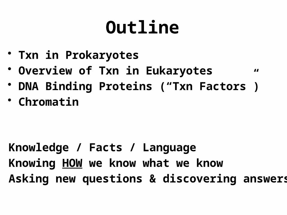
Outline• Txn in Prokaryotes• Overview of Txn in Eukaryotes• DNA Binding Proteins (“Txn Factors”)• Chromatin
1. Knowledge / Facts / Language2. Knowing HOW we know what we know3. Asking new questions & discovering answers

Expectations and Review
1. Prokaryotes: Basic process and nomenclature
• Process of txn• Start and stop signals for txn• Gene orientation• One RNAP

Expectations and Review
Nomenclature: ORF, promoter, codon, Start/stop codons, mRNA, untranslated region, tRNA, consensus sequence, homolog, coding and noncoding strands, activator (proteins), repressor, etc…
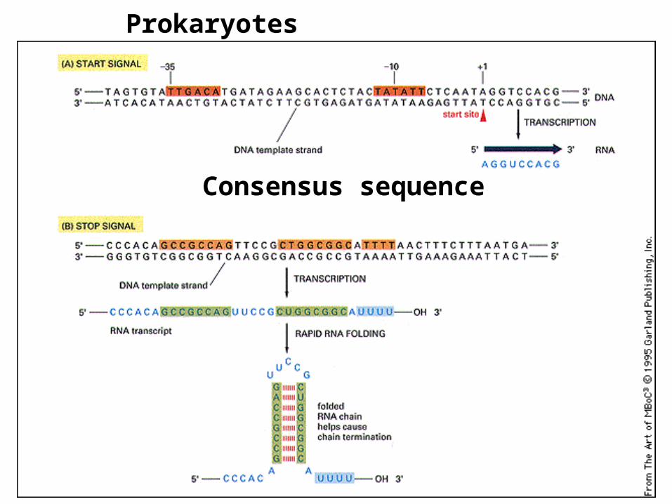
Consensus sequence
Prokaryotes

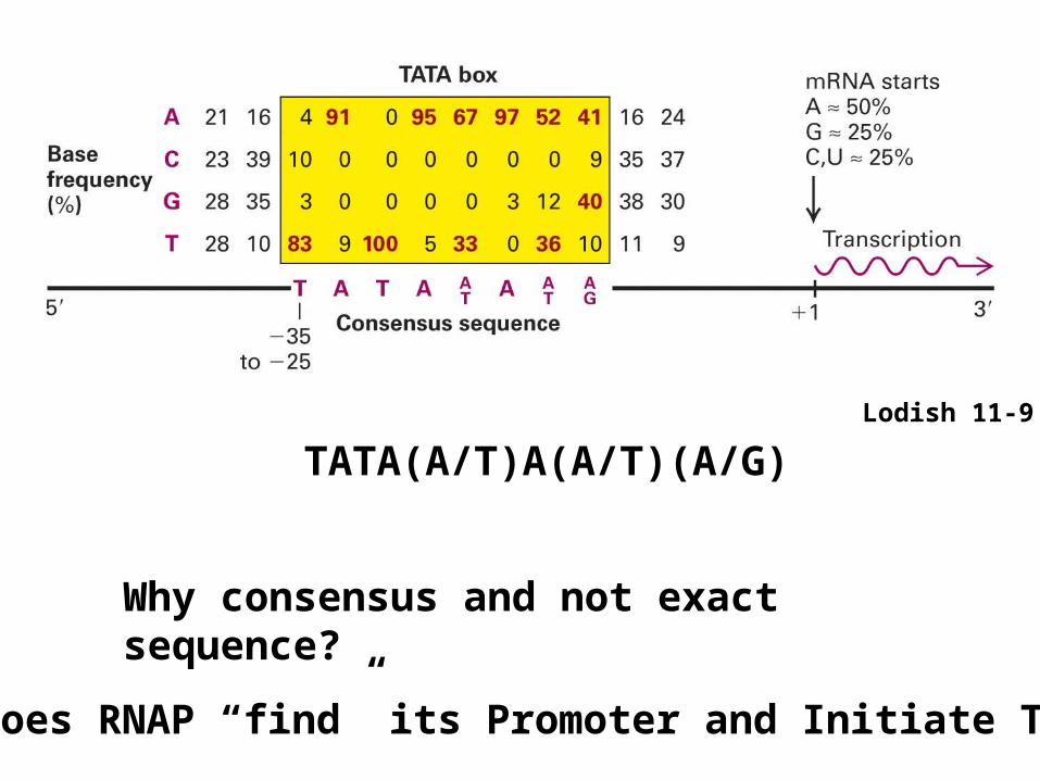
Lodish 11-9
Why consensus and not exact sequence?
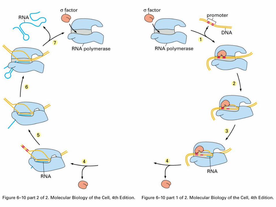
How Does RNAP “find” its Promoter and Initiate Txn?
TATA(A/T)A(A/T)(A/G)

Consensus sequences provide for binding to specific DNA sites over a range of affinities
Concept:Biological Reactions are often Optimized, not Maximized

Synthesis/polymerization is in the 5’ to 3’ direction

Coding v. non-coding strand,Directionality
Coding looks like mRNA
Non-coding can base-pairWith mRNA

Promoters are Directional

Activators

Repressors

Concept:Turning Genes ON and OFF
ON -> ActivatedOFF -> RepressedOFF -> Not Activated
In General: Repressors Win
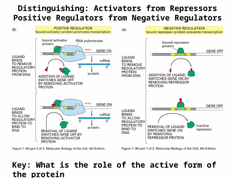
Distinguishing: Activators from RepressorsPositive Regulators from Negative Regulators
Key: What is the role of the active form of the protein

Regulation: Activation/Repression in Response toParticular Conditions

Repressors
Activators and Repressors vs. Inducers

Outline
• Txn in Prokaryotes (Review)• Overview of Txn in Eukaryotes• Chromatin

Thinking About Prokaryotic v. Eukaryotic Txn
1. Dynamic Range of Regulation: Prokaryotes v. Eukaryotes
A. E.coli ON:OFF = 200-1000:1 max. Most “OFF” genes about 100 x below ON
B. Most Eukaryotes ON:OFF = 108:1

Thinking About Eukaryotic Txn
2. Genome sizeHow do Regulatory proteins find their targets in the face of 1000 fold increase in “non-specific” DNA?
3. Chromatin and Higher order DNA packaging
Concept:Euk genomes are more complex; therefore, the txn machinery is more complex
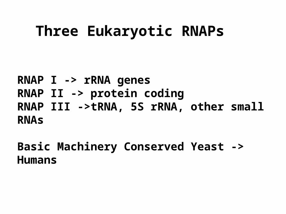
Three Eukaryotic RNAPs
RNAP I -> rRNA genesRNAP II -> protein codingRNAP III ->tRNA, 5S rRNA, other small RNAs
Basic Machinery Conserved Yeast -> Humans

DNA Sequence Elements and DNA Binding Proteins
DNA sequence elements that regulate transcription typically bind specific regulatory proteins or protein
complexes
“cis” sequence elements
“trans” factors = proteins or complexes

Eukaryotes:Tighter regulationLarger range of regulationLarger genomeMulticellularChromatin
More ComplexRegulation

Enhancers,Activators
Promoter,Basal Factors =General Factors
Language Caution: Genetic Activator vs. a Txn Activator protein = Activator

Watson 9-6 and 9-8
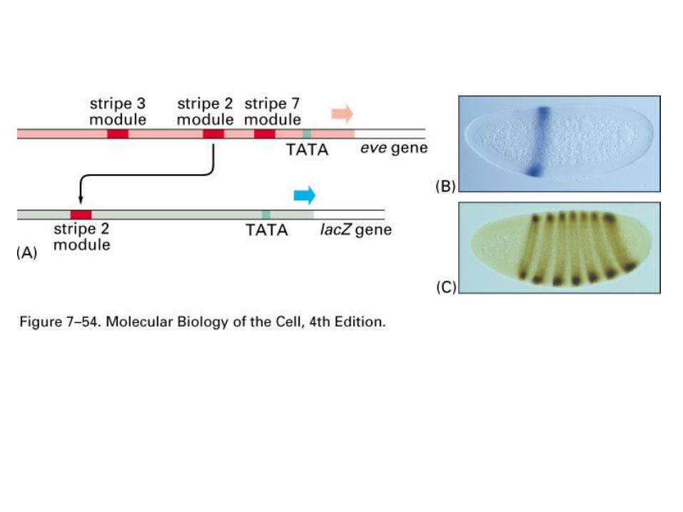
Enhancers= short regions (typically ~ 200 bp)of densely packed consensus elements
Some elements found in both promoters and enhancers

Lodish 11-35
Eukaryotic txn = large protein complexes
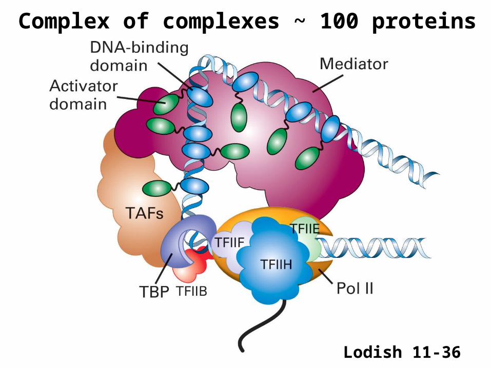
Lodish 11-36
Complex of complexes ~ 100 proteins

Lodish 11-37
Txn in the face of chromatin and higher order packing

Enhancers act independently and cumulatively
Reporter Genes

Reporter Genes


Reporter Genes
E1 E1 Pr Coding Region
E1 E1 Pr Reporter Cod. Reg.
Reporter Genes typically code for easily visualized protiens:lacZ = enzyme: colorless precursor -> blue productGFP = Green flourescent protein (Jellyfish)

Reporter Genes


E1 Pr Coding Region GFP
For Sub-cellularLocalization
For Expression Pattern (and subcellular local): txn and translation
E1 Pr GFP
For Txn Pattern:

E1 Pr Myo2 GFP
For Expression Pattern: txn and translation

Sub-cellular localization of splicing factors
Splicing Factor-GFP fusion

Ab
Protein
ExpressionPattern
Gene
Gene (Organism 2)
Mutant Gene
Biochemistry
Genetics
Mutant Organism
1
2
3
4
78
56 9
10
11
12

Molecular Genetics Summary1. Column Chromatograpy (ion exchange, gel filtration)2. A. Make Polyclonal Ab; B. Make Monoclonal Ab3. Western blot, in situ immuno-fluorescence (subcellular, tissue)4. Screen expression library (with an Ab)5. Screen library with degenerate probe6. Protein expression (E. coli)7. A. Differential hybridization8. A. Northern blot, in situ hybridization, GFP reporter, GFP Fusion9. A. low stringency hybridization; B. computer search/clone by phone; C.
computer search PCR10. Clone by complementation (yeast, E. coli)11. A. Genetic screen; B. genetic selection12. RNAi “knockdown”
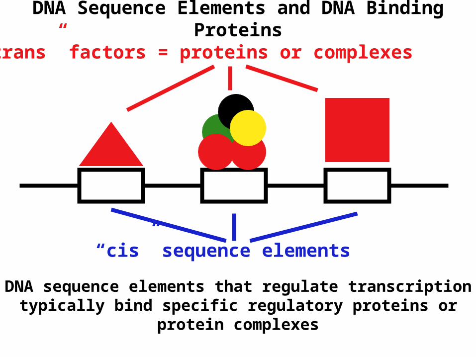
DNA Sequence Elements and DNA Binding Proteins
DNA sequence elements that regulate transcription typically bind specific regulatory proteins or protein complexes
“cis” sequence elements
“trans” factors = proteins or complexes

Regulation: Activation/Repression in Response toParticular Conditions

DNA elements (sequence elements) act by binding proteins
The proteins do the work

Outline
• Txn in Prokaryotes (Review)• Overview of Txn in Eukaryotes• Chromatin

Fig. 12.1


Fig. 12.5

Interphase/prophase
Mitosis

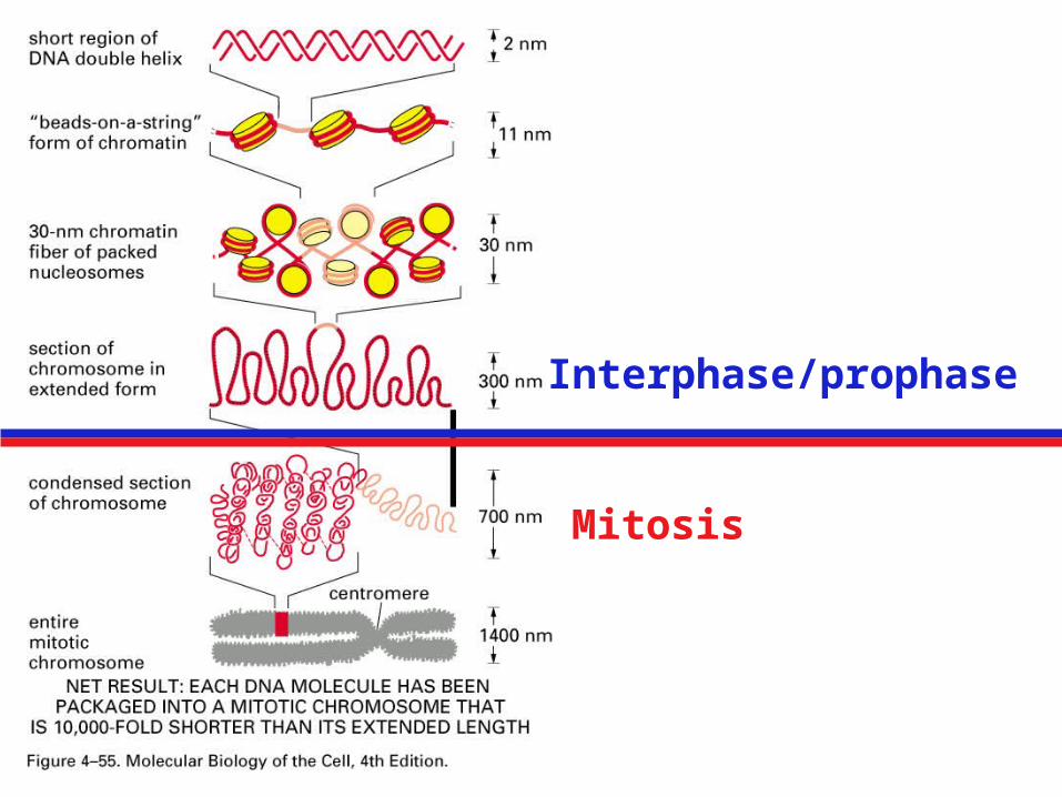
DNA Compaction: Spaghetti in a Sailboat
2 x 3 x 109 bp x .34 nm/bp = 2 meters DNA/nucleusScale by 1,000,000:
Nucleus 10m 10 meter sailboatDNA diameter 2 nm 2 millimeterDNA length 2 meters 2000 kilometers (1200
miles)
DNA persistence length (rigid rod): 50 nm 5 cm
UIUC to Orlando, Florida = 1066 miles
Interphase/prophase
Mitosis


Four Core Histones

X-Ray Crystallography

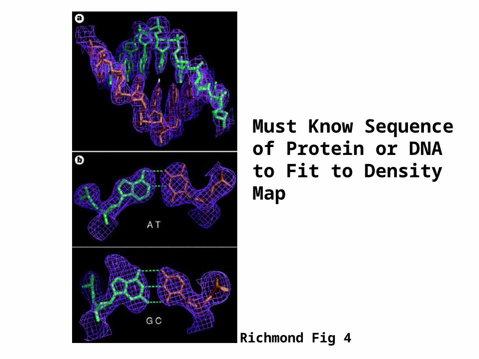
Richmond Fig 4
Must Know Sequence of Protein or DNA to Fit to Density Map

DNA = Two Turns/Nucleosome

Histone Globular Domains and Tails
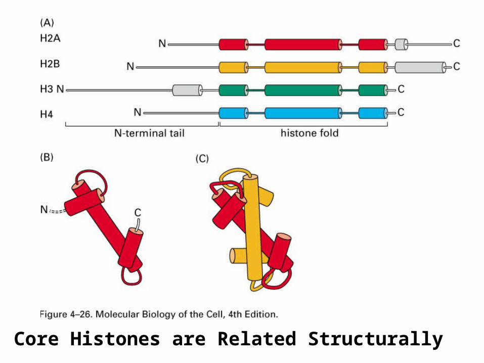
Core Histones are Related Structurally


What are Tails Doing?- Basic = Positively Charged Amino Acids.
Tails don’t appear In Crystal Structure =Flexible/Unstructured

Tails = Site of Post-Translational Modification

Histone Code

Histone Code

Histone Code

Global v. Local Histone Modification
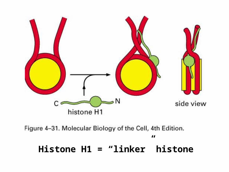
Histone H1 = “linker” histone

Fig. 12.3

Readings
Chromatin: Hartwell 465-470 Heterochromatin: Hartwell 479-481

Outline
• Txn in Prokaryotes (Review)• Overview of Txn in Eukaryotes• Chromatin
– Chromosome compaction– Chromatin structure– Heterochromatin and it’s effect on
transcription

Euchromatin and Heterochromatin
• Euchromatin = “active” and “decondensed”• Constitutive Heterochromatin = always
condensed• Facultative Heterochromatin = condensed in
some cells but not others
Barr Body
• X chromosome inactivation• A mechanism of Dosage Compensation

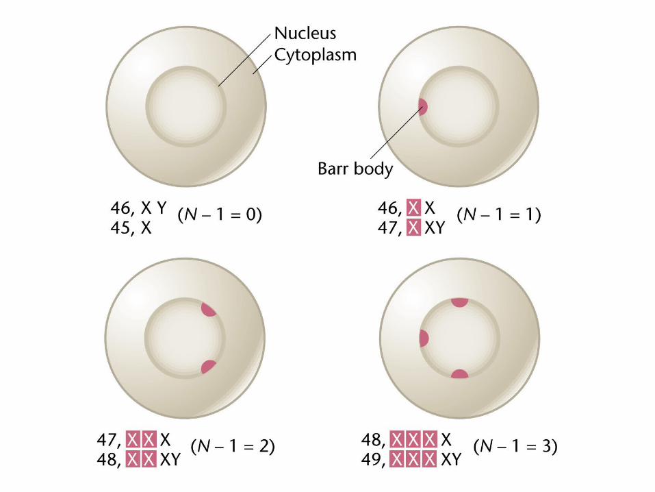

X Chromosome Inactivation
Choice of which X is inactivate is random
Once inactivated early in development remains inactive throughout cell division and the life of the organism (except in eggs)
We can infer two properties:
1. There must be a mechanism(s) of initial inactivation.
2. There must be a mechanism(s) of “duplication” of the inactive state

Calico Cats
Males = Only Orange or Only BlackFemales = Orange and Black Mosaics

Calico CatsO Gene is on the X chromosomeO+ = Black (converts orange pigment to black)o- = Orange
Males:XY O+Y = all black, o-Y = all orange (in the regions that show color)
FemalesO+o- =
-> black in cells in which o- is inactivated-> orange in cells in which O+ is inactivated

Ectodermal Dysplasia
X-linked recessive disorder
Lack Hair, teeth, sweat glands
Mosaic Expression PatternClonal = Heritable (Mitosis)

The initial inactivation of the X-chromosomeand subsequent “maintenance” of inactivation or “duplication” of the inactive state was inferred based on the mosaic nature of the associated phenotypes

X Chromosome inactivation is an example of epigenetics
Two genes with same promoters and enhancers in the same cell- one is on, the other off. Therefore whether or not a gene is on or off is independent of it’s “normal” genetic regulation.
Epigenetic regulation of txn often results from the formation of stable states of chromatin
Epigenetic regulation of txn often invovles persitant patterns of histone modification (histone code)

X Chromosome inactivation and “non-mosaic phenotypes”
Thinking about Hemophelia, diffusable factors and “cells”

Outline
• Txn in Prokaryotes (Review)• Overview of Txn in Eukaryotes• Chromatin
– Chromosome compaction– Chromatin structure– Heterochromatin and it’s effect on
transcription• X Chromosome inactivation• Autosomal heterochromatin and position effect
variegation (PEV)

Heterochromatin formation and properties
Some regions of chromosomes (autosomes) are heterochromatic- genes in these regions are shut off
Some regions are euchromatic- genes in these regions are available to be turned on
Heterochromatin assembles by a spreading mechanism; assembly starts at a particular site
Boundary elements = DNA elements that stop the spreading and define the ends of the heterochromatic regions
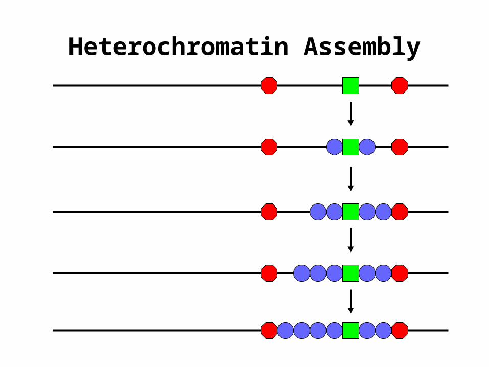
Heterochromatin Assembly

Heterochromatin DuplicationHeterochromatin is initially assembled by
spreading during development
Once it is formed it is copied/duplicated without having to be assembled by spreading de novo each cell division
Don’t need boundary elements to keep heterochromatin from spreading: Once a region of heterochromatin is formed it stays the same size through subsequent rounds of mitotic cell division
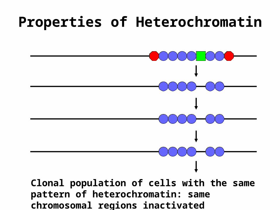
Properties of Heterochromatin
Clonal population of cells with the same pattern of heterochromatin: same chromosomal regions inactivated
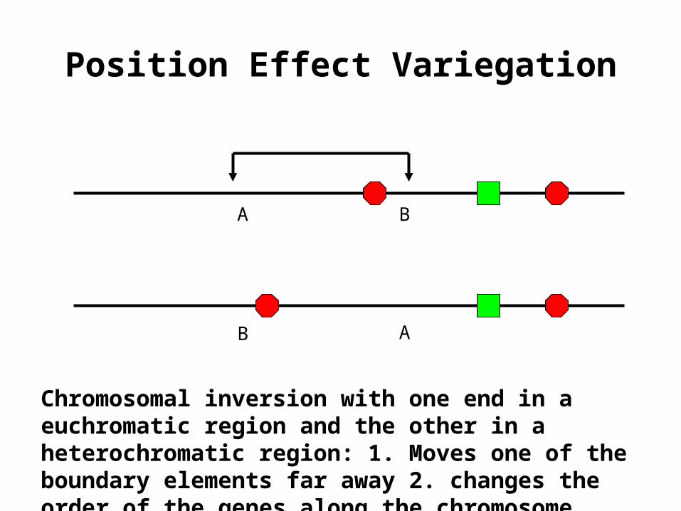
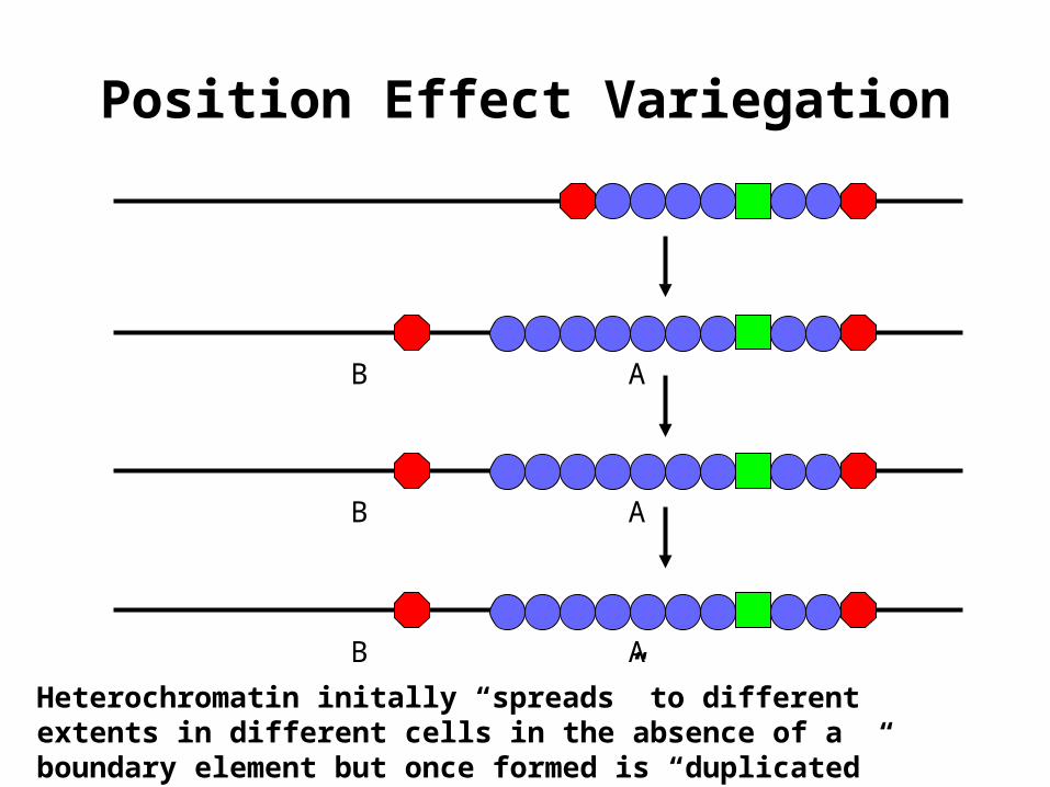
Position Effect Variegation
A B
B A
Chromosomal inversion with one end in a euchromatic region and the other in a heterochromatic region: 1. Moves one of the boundary elements far away 2. changes the order of the genes along the chromosome

Y
Position Effect Variegation
A B
B A
Heterochromatin initally “spreads” to different extents in different cells in the absence of a boundary element
Y
Position Effect Variegation
B A
Heterochromatin initally “spreads” to different extents in different cells in the absence of a boundary element but once formed is “duplicated” during cell division
B A
B A

Y
Position Effect Variegation
A B
B A
Heterochromatin initally “spreads” to different extents in different cells in the absence of a boundary element
Y
YB A
In Cell 1 and its progeny Y is Transcribed:
In Cell 2 and its progeny Y is Not Transcribed:

Fig. 12.14a
Position effect Variegation