May -Thurner, Nutcracker and Paget -SchroetterSyndromes · May -Thurner, Nutcracker and Paget...
65
May - Thurner, Nutcracker and Paget - Schroetter Syndromes Robert M Schainfeld, DO Section of Vascular Medicine Massachusetts General Hospital Boston, MA
Transcript of May -Thurner, Nutcracker and Paget -SchroetterSyndromes · May -Thurner, Nutcracker and Paget...

May - Thurner, Nutcracker and Paget - Schroetter Syndromes
Robert M Schainfeld, DOSection of Vascular Medicine
Massachusetts General HospitalBoston, MA

Disclosure Statement of Financial Interest
I, Robert Schainfeld, DO NOT have a financial interest / arrangement or affiliation with one or more organizations that could be perceived as a real or apparent conflict of interest in the context of the subject of this presentation.

Massachusetts General HospitalVascular Interventional Suite # 5

May - Thurner (MTS) or Cockett’s Syndrome
• 1851 - Virchow sinistral (left-sided) DVT• Dx in 2% - 5% of pts w/ venous dz• Right CIA/compresses the left CIV against
the lumbar vertebrae• 25% of asymptomatic pts > 50% stenosis• 3rd – 5th decade of life / women

May - Thurner Syndrome

Magenetic Resonance Venography

MRV of Abdomen / Pelvis

8
MRV of Abdomen / Pelvis

“Palacios - Schainfeld Syndrome?”

Diagnosis of May - Thurner Syndrome
• MRV (>95%)• Venography (66%)• IVUS (>95%)• CTV
Raju S, Neglen P. J Vasc Surg 2006;44:136-44

May - Thurner Syndrome Clinical Presentation
Unilateral – L >> R• Pain• Edema• Chronic venous insufficiency• Venous ulceration• Ilio - femoral DVT• Phlegmasia cerulea dolens
Cryptogenic stroke ???

“Team Palacios”

Wallstents
• Stainless steel• Self expanding• High radial force• Flexible• Wide range of sizes

Endovascular Treatment (EVT) of Chronic Iliac Vein Obstruction
Objectives
Compare stent - related and clinical outcomes, results and complications of EVT in limbs with non - thrombotic iliac vein lesions and post -thrombotic syndrome
NIVL = Non - thrombotic iliac vein lesion (NIVL / MTS)PTS = Post - thrombotic syndrome
Neglen et al., J Vasc Surg, 2007: 979-990

EVT of Chronic Iliac Vein ObstructionMaterials/Methods
• 870 patients / 982 limbs• Chronic symptoms (mean = 60 mos)• NIVL = 518, PTS = 464 limbs• Follow-up (mean = 22 mos), range 1-107
mos• IVUS• PTA / stenting
Neglen et al., J Vasc Surg, 2007: 979-990

EVT of Chronic Iliac Vein Obstruction Technical Results
• Technical success = 97%• Post-op stent thrombosis (<30 d) = 1.5%• Primary, primary-assisted and secondary
patency rates (6-years) 79%, 100%, 100% (NIVL) 57%, 80%, 86% (PTS) ISR (>50%) = 5%
- 10% (PTS), 1% (NIVL)
Neglen et al., J Vasc Surg, 2007: 979-990

EVT of Chronic Iliac Vein Obstruction Clinical Results
• Pain - free @ (5 - yrs)• 6% - 60% (PTS)• 26% - 59% (NIVL)
• Absence of limb swelling • 3% - 42% (PTS)• 5% - 43% (NIVL)
• Ulcer healing = 58%Neglan et al., J Vasc Surg, 2007:979-990

49 ♀ with:• 2 day history of left thigh / calf swelling• associated mild dyspnea on exertion• 4 days prior had arthroscopic surgery of right knee
Past Medical History- Gronblad – Strandberg syndrome- subretinal hemorrhages of left eye- PAD - intermittent claudication bilateral calves
Medications- OCP
CD

Physical Examination
Vital signs:BP: 110 / 80 mmHg, P= 104,SaO2 = 94% (RA), T= 98.6 F
• Cardiorespiratory exam:- normal
• Extremities:- Left thigh and calf 2+
edema, non - tender, pedal pulses palpable bilaterally

Diagnostic DataDuplex US MRV

Post power pulse spray
DVX catheter: 10mg tPA
22
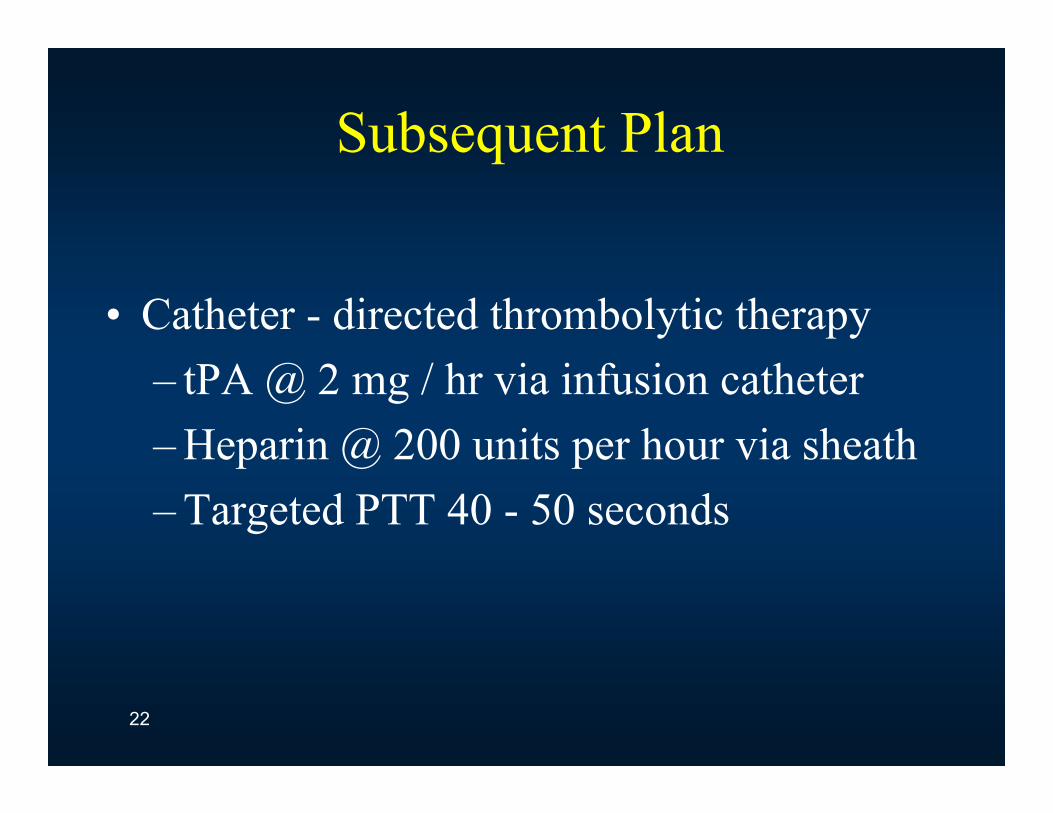
Subsequent Plan
• Catheter - directed thrombolytic therapy – tPA @ 2 mg / hr via infusion catheter– Heparin @ 200 units per hour via sheath– Targeted PTT 40 - 50 seconds
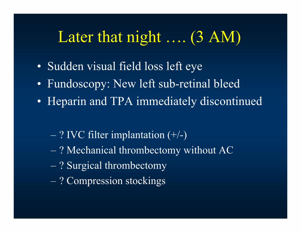
Later that night …. (3 AM)
• Sudden visual field loss left eye• Fundoscopy: New left sub-retinal bleed • Heparin and TPA immediately discontinued
– ? IVC filter implantation (+/-)– ? Mechanical thrombectomy without AC – ? Surgical thrombectomy– ? Compression stockings

Diagnosis ???

Pseudoxanthoma Elasticum• Rare (1:70,000 - 160,000)• Involves skin, eye and cardiovascular• Retina – involvement in 85% of cases. Severe
visual loss in ~ 5%• Xanthomas (neck, groin, popliteal fossa)• Abnormal GI bleeding and ? IC aneurysms• Symptoms (CAD / ACS or PAD)• Diffuse arterial disease with prominent
collaterals


Angiography

Venogram @ 3 - months (IVC retrieval)
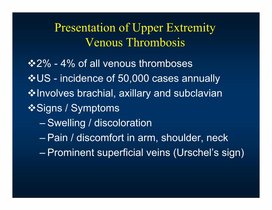
Presentation of Upper Extremity Venous Thrombosis
2% - 4% of all venous thrombosesUS - incidence of 50,000 cases annuallyInvolves brachial, axillary and subclavianSigns / Symptoms
– Swelling / discoloration– Pain / discomfort in arm, shoulder, neck– Prominent superficial veins (Urschel’s sign)

Clinical Sequelae
• Pulmonary embolus ~ 12% symptomatic and up to ~ 36% may remain asymptomatic
• Venous hypertension PTS (severe 13%)• Loss of future vascular access or SVC syndrome• Mortality 15% - 50% (underlying etiology)• Recurrence after Tx ~ 2% - 8%

Etiologies of Upper Extremity Venous Thrombosis
• Primary axillo - SCV thrombosis (idiopathic or Paget - Schroetter syndrome)
– No associated disease or trauma– Exertion - related
• Secondary axillo – SCV thrombosis– Recognized cause– 2o to central venous catheters (CVC), ICD,
pacemakers– Systemic due to malignancy, thrombophilia, trauma

Paget - Schroëtter Syndrome
Leopold-von-Schroëtter, Vienna (1837-1908)
Sir James Paget, London
(1814-1898)
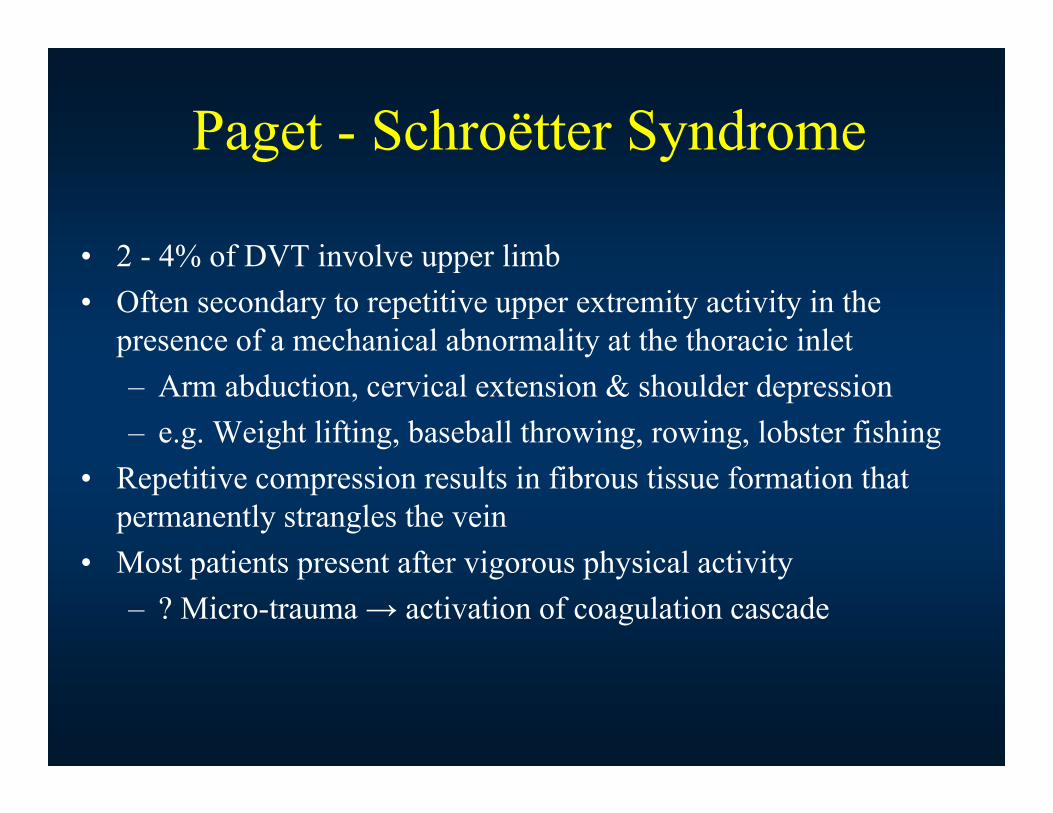
Paget - Schroëtter Syndrome
• 2 - 4% of DVT involve upper limb• Often secondary to repetitive upper extremity activity in the
presence of a mechanical abnormality at the thoracic inlet– Arm abduction, cervical extension & shoulder depression – e.g. Weight lifting, baseball throwing, rowing, lobster fishing
• Repetitive compression results in fibrous tissue formation that permanently strangles the vein
• Most patients present after vigorous physical activity– ? Micro-trauma → activation of coagulation cascade

• Common anomalies- Young athlete with hypertrophied muscle- First or clavicular rib- Musculofascial bands- Cervical ribs

Ms SM • 48 yo F presented with right upper extremity
swelling• HPI
– Previously well & active– Woke - up with pain & gross swelling of arm– Heavy lifting & mammogram few days prior– Multiple presentations to OSH
• Rx as cellulitis• PMH
– HTN– Smoker– No VTE (DVT or PE)


Duplex Ultrasound

Orders to Dr. Charles Dotter !!!

Dr. Dotter’s “Rebuttal”

Venogram Right Axillary - Subclavian Vein

EKOS Catheter

Ultrasound Accelerated Catheter-Directed Lysis
5F EKOS Peripheral System
Multi Side Port Infusion Catheter
Ultrasound Core Wire • tPA @ 2mg/hr x 4 hrs
• Heparin (PTT 40-50)

Venogram (Post - tPA @ 4 hours)

Balloon Angioplasty (PTA)
• PTA 12 x 40 mm balloon

Final Venogram

Venogram @ 3 - weeks(Post -1st rib resection)

Venogram @ 3 - weeks(Post -1st rib resection)

Venogram 6 Month Follow - up
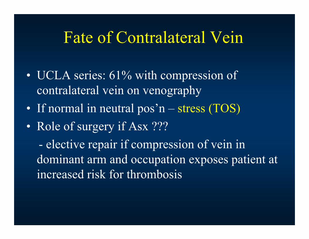
Fate of Contralateral Vein
• UCLA series: 61% with compression of contralateral vein on venography
• If normal in neutral pos’n – stress (TOS)• Role of surgery if Asx ???
- elective repair if compression of vein in dominant arm and occupation exposes patient at increased risk for thrombosis

Treatment of Primary ASDVTResults
Largest retrospective series (50 - years)– 626 limbs / 608 patients– Best results in 511 / 548 patients < 6 weeks &
prompt surgery– 24 / 42 limbs > 6 weeks all remained sx – 36 patients, no lysis
• 10 - ASX• 25 (PTS) - despite first rib resection
Urschel et al., Ann Thorac Surg, 2008:254-260

Algorithm for the Management of 1o ASDVT
Symptomatic
PTA +/- Stent
Residual Stenosis Normal Vein
1st Rib Resection(early vs. delayed?)
AsymptomaticAbnormality
AsymptomaticNo Abnormality
Evaluation
Anticoagulation
Lysis
Thrombosis Compression/Stricture
Venography
Urschel et al., Ann Thorac Surg, 2002:69


CT Abdomen


Nutcracker Syndrome

Nutcracker Syndrome (NCS)
• AKA, renal vein entrapment or mesoaorticcompression of the left renal vein
• Results most commonly from compression of the left renal vein between the aorta and superior mesenteric artery, although other variants exist

Signs and Symptoms
• Hematuria• Anemia• Left flank and/or pelvic pain• Orthostatic proteinuria and intolerance• Left testicular pain (males)• LLQ pain (females)• Nausea and vomiting• Varicocele and varicose veins

Demographic Features
• Childhood to 7th decade of life• Most symptomatic pts 2nd to 3rd decade • Second peak in middle-aged women• Not a hereditary phenomenon

Diagnosis of NCS
• Left renal venography• CT• MRI/MRA• Abdominal ultrasonography• DSA

Differential Diagnosis of NCS
• Pelvic Congestion Syndrome• Renal stones• Genitourinary malignancy• Loin pain hematuria syndrome

Treatment of NCS
• Conservative Tx age < 18 YO• ACE-I for orthostatic proteinuria• Endovascular stenting
- balloon-expandable or self-expanding- Cxs include migration, thrombosis, ISR, deformities and erosions

Surgical Treatment of NCS• Medial nephropexy (excision of renal varices)• Left renal vein (LRV) bypass• LRV transposition between SMA and aorta • Renal-to-IVC shunt• Renal autotransplant• Gonadocaval bypass• Nephrectomy• Coil embolization of ovarian vein with PCS

Compressive Disorders: Diagnosis or Bust