Maxillary Expansion in the Management of Sleep …...Maxillary Expansion in the Management of Sleep...
21
Maxillary Expansion in the Management of Sleep Apnea Michael B. Melugin , DDS, MS Orthodontics and Oral and Maxillofacial Surgery Department of Dentistry Children’s Hospital of Wisconsin Private Practice, New Berlin and Brookfield/Waukesha
Transcript of Maxillary Expansion in the Management of Sleep …...Maxillary Expansion in the Management of Sleep...

Maxillary Expansion in the Management of Sleep Apnea
Michael B. Melugin, DDS, MSOrthodontics and Oral and Maxillofacial Surgery
Department of Dentistry
Children’s Hospital of Wisconsin
Private Practice, New Berlin and Brookfield/Waukesha

Who needs to be expanded?
• Significant incisor crowding
• Incisors blocked out from eruption
• Individuals with sleep disordered breathing with known or suspected obstruction due to restricted nasal air flow
• Typically, maxillary constriction significant enough to limit nasal air flow will also demonstrate incisor crowding

Who needs to be expanded?
• Maxillary intermolar width less than 43 mm (mesiolingual cusp tip to mesiolingual cusp tip) in association with other signs of maxillary constriction
43 mm

What happens in Mx expansion?

Changes in snoring,hypersomnolence, and OSA
• Gray LP. Results of 310 cases of rapid maxillary expansion selected for medical reasons. Journal of Laryngology & Otology. ;89(6):601-14, 1975 Jun.
• White BC. Woodside DG. Cole P. The effect of rapid maxillary expansion on nasal airway resistance. Journal of Otolaryngology. 18(4):137-43, 1989 Jun.– Average reduction in NAR 48.7%. Pts w/ higher initial
nasal airway resistance saw greatest reduction• Hartgerink DV. Vig PS. Lower anterior face height and lip
incompetence do not predict nasal airway obstruction. Angle Orthodontist. 59(1):17-23, 1989 – Decreased NAR w/ RME, but no correlation between
amt of expansion & quantity of change in NAR
• Cistulli PA. Palmisano RG. Poole MD. Treatment of obstructive sleep apnea syndrome by rapid maxillary expansion. Sleep. 21(8):831-5, 1998 Dec 15.

Maxillary Expansionfor the management of OSA
• Cistulli PA. Palmisano RG. Poole MD. Treatment of obstructive sleep apnea syndrome by rapid maxillary expansion. Sleep. 21(8):831-5, 1998 Dec 15.
– 10 young adults 27+/-2yrs of age, mild-moderate OSA – Maxillary transverse deficiency with diminished nasal air
flow– All RME, 6 SARPE (Surgically Assisted Rapid Max
Expansion)– Results:
• 9 pts reported improvement in snoring & hypersomnolence
• AHI 19+/-4 to 7+/-4 for the group, 7 had AHI <5– Their conclusion: This preliminary data suggests
that RME may be a useful treatment alternative for selected patients with OSA

Changes in Nasal Airway Resistance (NAR) 2o to Maxillary Expansion• Hershey HG. Stevart BL. Warren DW. Changes in nasal airway resistance
associated with rapid maxillary expansion. American Journal of Orthodontics. 69(3):274-84,1976 Mar.
– RME sig reduced NAR, stable after 3 months, low correlation btwnchange in NAR & amt of Mx expansion at the first molar, or amt of nasal cavity widening
• Hartgerink DV. Vig PS. Abbott DW.The effect of rapid maxillary expansion on nasal airway resistance. American Journal of Orthodontics & DentofacialOrthopedics. 92(5):381-9, 1987 Nov.
– Significant median reduction in NAR following RME, but high individual response & variability means some unpredictability
• Timms DJ. The reduction of nasal airway resistance by rapid maxillary expansion and its effect on respiratory disease. Journal of Laryngology & Otology. 98(4):357-62, 1984 Apr.
• Timms DJ. The effect of rapid maxillary expansion on nasal airway resistance. British Journal of Orthodontics. 13(4):221-8, 1986 Oct.
– NAR decreased average of 36.2%. Correlation btwn NAR decrease & magnitude of expansion was weak. Nasal manometry used.
Maxillary Expansion for the Management of OSA
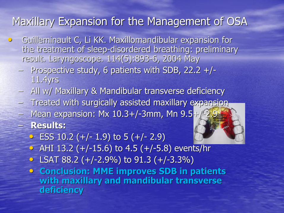
• Guilleminault C, Li KK. Maxillomandibular expansion for the treatment of sleep-disordered breathing: preliminary result. Laryngoscope. 114(5):893-6, 2004 May
– Prospective study, 6 patients with SDB, 22.2 +/-11.4yrs
– All w/ Maxillary & Mandibular transverse deficiency
– Treated with surgically assisted maxillary expansion
– Mean expansion: Mx 10.3+/-3mm, Mn 9.5+/-2.9
– Results:
• ESS 10.2 (+/- 1.9) to 5 (+/- 2.9)
• AHI 13.2 (+/-15.6) to 4.5 (+/-5.8) events/hr
• LSAT 88.2 (+/-2.9%) to 91.3 (+/-3.3%)
• Conclusion: MME improves SDB in patients with maxillary and mandibular transverse deficiency
Maxillary Expansion for the Management of OSA
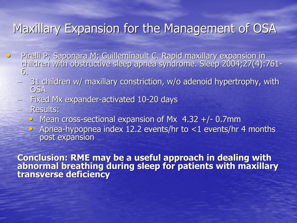
• Pirelli P; Saponara M; Guilleminault C. Rapid maxillary expansion in children with obstructive sleep apnea syndrome. Sleep 2004;27(4):761-6.
– 31 children w/ maxillary constriction, w/o adenoid hypertrophy, with OSA
– Fixed Mx expander-activated 10-20 days– Results:
• Mean cross-sectional expansion of Mx 4.32 +/- 0.7mm• Apnea-hypopnea index 12.2 events/hr to <1 events/hr 4 months
post expansion
Conclusion: RME may be a useful approach in dealing with abnormal breathing during sleep for patients with maxillary transverse deficiency

Changes in Nasal Airway Resistance (NAR) 2o to Maxillary Expansion
• Doruk C., Sokucu O, Canbay EI. Evaluation of nasal airway resistance during rapid maxillary expansion using acoustic rhinometry. European Journal of Orthodontics. 26(4):397-401, 2004 Aug. – NAR significantly reduced w/ the use of RME

Maxillary Expansion and OSA Management• Villa MP, Rizzoli A, Miano S, Malagola C. Efficacy of
rapid maxillary expansion in children with obstructive sleep apnea syndrome: 36 months of follow-up. Sleep Breath. 2011 May;15(2):179-84. (Italy)
• 10 children, mean age 6.6 ± 2.1 years at entry and 9.7 ± 1.6
years at 36 month follow-up. O/W healthy kids who’s parents
refused adenotonsillectomy and had crossbite, maxillary
constriction and/or high arch narrow palate
• Brouillette symptom questionnaire, AHI, HOI (Hypopnea
Obstruction Index), and AI (Arousal Index) compared
• AHI went from 5.8 +/-6.8 down to 1.5 +/- 1.6
• Habitual snoring went from 78.5% down to 35.7%
• Sleepiness went from 50% down to 7.1%
• Oral breathing went from 92.9% down to 14.3%

Maxillary Expansion and OSA Management• Buccheri A, Chine F, Fratto G, Manzon L: Rapid
Maxillary Expansion in Obstructive Sleep Apnea in Young Patients: Cardio-Respiratory Monitoring. J Clin Pediatr Dent. 2017;41(4):312-316.
• 11 OSAS young subjects (mean age 6.9±1.04 years)
• Polysomnography and Cardiovascular monitoring
• Pre expansion AHI=6.09±3.47; SAO2=93.09%±1.60.
• 12 months post expansion, AHI=2.36 ± 2.24;SAO2=96.81%
±1.60.
• These changes were associated with an improvement in
clinical symptoms, such as reduction of snoring and sleep
apnea.

Maxillary and Mandibular Expansion

Maxillary incisors blocked out from eruption as noted by failed or delayed eruption after deciduous teeth have exfoliated

Upper primary lateral incisors are over-retained due to maxillary constriction, lower incisors with marked crowding

Maxillary lateral incisor eruption delayed, lower incisors with marked crowding, decreased intermolar width

ZM
Pre-treatment Progress

Pre-treatment to phase I retention
Phase I retention

What Does Maxillomandibular Expansion Do?
• Increases dental arch size to provide space for tooth eruption
• Improves stability of future orthodontic treatment by allowing teeth to erupt into more stable positions relative to surrounding bone
• Encourages normal mandibular AP growth
• Decreases nasal airway resistance, improving nasal air flow
• May be beneficial to individuals with OSA that are know or believed to have obstruction of nasal air flow
• Expansion improves oral cavity width, likely allowing a more forward tongue position

Who is a candidate for maxillomandibular expansion?
• Any individual with dental arch width deficiency that has inadequate space for eruption of permanent teeth (dental indication)
• Individuals with polysomnographic evidence of OSA, who have undergone evaluation by members of the sleep team, and for whom other sources of airway obstruction have been ruled out (soft palate, tongue base, tonsils/adenoids, etc) or have failed medical intervention, and for whom increased nasal airway resistance is suspected or confirmed - with or without significant dental crowding.
• Often, individuals with suspected increased nasal airway resistance also have the dental crowding indication for expansion as well

Thank You!• ESS – Epworth Sleepiness Scale (self-reported 8 item
symptom based questionnaire)
• LSAT – Lowest O2 Saturation recorded during sleep
• AHI – Apnea Hypopnia Index, events per hour. <5 normal. Apnea must be 10 sec in length with associated hypoxia
(5-15 mild apnea, 15-30 moderate, >30 severe)
• Peak Nasal Inspiratory Airflow (PNIA) uses spirometry. Requires patient effort – maximum inspiration – can vary
• Acoustic Rhinometry – uses sound waves to measure nasal/upper airway volume. 35 y.o. technique. Does not require patient maximum effort, less potential variation
• Nasal Monometry (Rhinomanometry) – measure airflow in the pharynx and at the nares and use Ohm’s law for gases to calculate nasal airflow, pressure, and resistance