Maternal Thyroid hormone: a strong repressor of neuronal ... · forth nNOS as a novel target of...
21
1 Maternal Thyroid hormone: a strong repressor of neuronal nitric oxide synthase (nNOS) in rat embryonic neocortex Short Title: Maternal thyroxine and nNOS expression Rohit Anthony Sinha ¶ , Amrita Pathak ¶ , Vishwa Mohan, Sanghamitra Bandyopadhyay § , Leena Rastogi and Madan M Godbole *. Department of Endocrinology, Sanjay Gandhi Postgraduate Institute of Medical Sciences, Lucknow 226014, India §Division of Developmental Toxicology, Indian Institute of Toxicology Research, Lucknow ¶ Equal Contribution *To whom correspondence should be addressed Professor Madan M. Godbole, Department of Endocrinology, SGPGIMS, Raebareli Road, Lucknow 226 014 India. Phone: +91-522-2668700, Extension: 2368 Fax: +91-522-2668017, E-mail: [email protected] Keywords: Maternal thyroxine, Developing cerebral cortex, nNOS Disclosure Statement: The authors have nothing to declare. This work was supported by grant from Department of Science & Technology (DST), Govt. of India through F.I.S.T. SR/SO/HS/17/2003 and SR/SO/HS/95/2007 to MMG and Senior Research Fellowship from Council of Scientific and Industrial Research to RAS [10-2(5)/2003(II)-E.U.II] and University Grants Commission to AP [2-89/98(SA-I)] from Government of India, New Delhi. Endocrinology. First published ahead of print May 8, 2008 as doi:10.1210/en.2007-1617 Copyright (C) 2008 by The Endocrine Society
Transcript of Maternal Thyroid hormone: a strong repressor of neuronal ... · forth nNOS as a novel target of...

1
Maternal Thyroid hormone: a strong repressor of neuronal nitric oxide synthase (nNOS) in rat embryonic neocortex
Short Title: Maternal thyroxine and nNOS expression
Rohit Anthony Sinha¶, Amrita Pathak¶, Vishwa Mohan, Sanghamitra Bandyopadhyay§, Leena Rastogi and Madan M Godbole *.
Department of Endocrinology, Sanjay Gandhi Postgraduate Institute of Medical Sciences, Lucknow 226014, India
§Division of Developmental Toxicology, Indian Institute of Toxicology Research, Lucknow ¶ Equal Contribution
*To whom correspondence should be addressed Professor Madan M. Godbole, Department of Endocrinology, SGPGIMS, Raebareli Road, Lucknow 226 014 India. Phone: +91-522-2668700, Extension: 2368 Fax: +91-522-2668017, E-mail: [email protected]
Keywords: Maternal thyroxine, Developing cerebral cortex, nNOS
Disclosure Statement: The authors have nothing to declare.
This work was supported by grant from Department of Science & Technology (DST), Govt. of India through F.I.S.T. SR/SO/HS/17/2003 and SR/SO/HS/95/2007 to MMG and Senior Research Fellowship from Council of Scientific and Industrial Research to RAS [10-2(5)/2003(II)-E.U.II] and University Grants Commission to AP [2-89/98(SA-I)] from Government of India, New Delhi.
Endocrinology. First published ahead of print May 8, 2008 as doi:10.1210/en.2007-1617
Copyright (C) 2008 by The Endocrine Society

2
Abstract
Understanding of how maternal thyroid inadequacy during early gestation poses a risk for developmental outcomes is still a challenge for the neuro-endocrine community. Early neocortical neurogenesis is accompanied by maternal thyroid hormone transfer to fetal brain, appearance of thyroid hormone receptors and absence of anti neurogenesis signals, followed by optimization of neuronal numbers through apoptosis. However, the effects of thyroid hormone (TH) deprivation on neurogenesis and neuronal cell death before the onset of fetal thyroid are still not clear. We show that maternal TH deficiency during early gestational period causes massive premature elevation in the expression of neuronal nitric oxide synthase (nNOS) with an associated neuronal death in embryonic rat neocortex. Maternal hypothyroidism was induced by feeding methimazole (0.025% w/v) in the drinking water to pregnant Sprague Dawley rats from embryonic day (ED) 6. Cerebral cortices from fetuses were harvested at different embryonic stages (ED14, ED16 and ED18) of hypothyroid and euthyroid group. Immunoblotting and Real-Time PCR results showed that both protein and RNA levels of nNOS were pre-maturely increased under maternal hypothyroidism and showed reversibility upon thyroxine administration. Immunohistochemistry revealed an increased nNOS immuno-reactivity in both the cortical plate and proliferative zone of neo-cortex along with a corroborative decrease in the Microtubule associated protein-2 (MAP-2) positive neurons under maternal TH insufficiency. Results combined, put forth nNOS as a novel target of maternal thyroid hormone action in embryonic neocortex and underscore the importance of prenatal screening and timely rectification of maternal thyroid hormone insufficiency even of a moderate degree.
3
Introduction Endocrinologists are still equivocal about thyroid screening in early pregnancy and treatment in spite of associated IQ loss seen in offspring’s of pregnant women with hypothyroidism (1-3). The need for prevention of maternal thyroid hormone (TH) insufficiency before mid-pregnancy, however moderate, and whatever the underlying cause has been advocated (4). Unfortunately, in spite of increasing evidences (5-11) that TH action in brain occurs much before the onset of fetal thyroid function, we know very little about the commencement of its molecular action during brain development. Whether and how TH deprivation affects the developmental process such as cortical neurogenesis and neuronal apoptosis which coincides with TH receptors (TRs) appearance (12) in brain is still not clear.Although TH insufficiency is known to be associated with defective expression of neuronal markers (8), the reasons for the same are still not clear. Both neurogenesis and neuronal apoptotic mechanisms are tightly regulated to ensure optimization of neuronal number during the course of development (13). One of the major players in this process is a neurotransmitter known as nitric oxide produced by an enzyme neuronal nitric oxide synthase (nNOS) expressed in newly generated neurons (13). Though nNOS expression is modulated by steroid and thyroid hormones during postnatal development (14, 15) but it’s modulation by TH during embryonic period lacks definite evidences. The present study has therefore been taken up to understand the effect of maternal thyroid hormone deficiency on the expression of nNOS and its consequent impact on generation and survival of neocortical neurons. We here uncover a novel role of maternal TH in repressing nNOS expression (an anti-neurogenic signal) during fetal neo-cortical development and proposes that its premature up-regulation even under moderate TH deficiency could have a detrimental bearing on fetal neurogenesis and neuronal survival during early gestation.
Materials and Methods
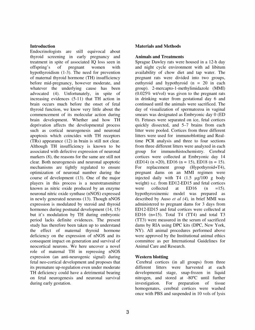
Animals and Treatments Sprague Dawley rats were housed in a 12-h day and night cycle environment with ad libitum availability of chow diet and tap water. The pregnant rats were divided into two groups, euthyroid and hypothyroid (n = 20 in each group). 2-mercapto-1-methylimidazole (MMI) (0.025% wt/vol) was given to the pregnant rats in drinking water from gestational day 6 and continued until the animals were sacrificed. The day of visualization of spermatozoa in vaginal smears was designated as Embryonic day 0 (ED 0). Fetuses were separated on ice, fetal cortices quickly dissected, and 5–7 brains from each litter were pooled. Cortices from three different litters were used for immunoblotting and Real-time PCR analysis and three to four sections from three different litters were analyzed in each group for immunohistochemistry. Cerebral cortices were collected at Embryonic day 14 (ED14) (n =20), ED16 (n = 15), ED18 (n = 15). For replacement group (Hypothyroid+T4), pregnant dams on an MMI regimen were injected daily with T4 (1.5 μg/100 g body weight) s.c. from ED12-ED15 and fetal cortices were collected at ED16 (n =15). hypothyroxinemic model was prepared as described by Auso et al (4), in brief MMI was administered to pregnant dams for 3 days from ED12-ED15 and fetal cortices were collected at ED16 (n=15). Total T4 (TT4) and total T3 (TT3) were measured in the serum of sacrificed dams by RIA using DPC kits (DPC, New York, NY). All animal procedures performed above were approved by the Institutional animal ethics committee as per International Guidelines for Animal Care and Research.
Western blotting Cerebral cortices (in all groups) from three different litters were harvested at each developmental stage, snap-frozen in liquid nitrogen, and stored at -80ºC until further investigation. For preparation of tissue homogenates, cerebral cortices were washed once with PBS and suspended in 10 vols of lysis

4
buffer [10 mM Tris-Cl (pH 7.5), 50 mM sodium chloride and 1% Triton-X-100 containing phenylmethylsulfonyl fluoride (1 mM) and protease inhibitor cocktail (a mixture of 4-(2-aminoethyl) benzenesulfonyl fluoride, pepstatinA, E-64, bestatin, leupeptin, and aprotinin (Sigma, St. Louis, MO)] and kept on ice for 10 min. Then the tissues were homogenized using a Teflon homogenizer and centrifuged at 12,000Xg for 15 min at 40C, and the supernatant was collected. Protein concentration was determined in the above samples, in the tissue homogenate, using a protein assay kit (Bio-Rad, Hercules, CA). Tissue homogenate proteins (50–100 μg) were subjected to 6-12% SDS-PAGE and electro-transferred onto nitrocellulose membrane. The membranes were incubated with either anti-nNOS, anti-PCNA (Abcam, Denmark) or PARP (Cell signaling technology, MA) followed by incubation with horseradish peroxidase-conjugated secondary antibodies (Santa Cruz biotechnology, CA). The signals were detected using an enhanced chemiluminescence detection system (Amersham Biosciences, Little Chalfont, and UK). Relative expression of each protein was determined by densitometric analysis using LabWorks 4.0 software (UVP Ltd., Cambridge, UK).
RNA extraction and Real time PCR Total RNA was isolated from the neocortex at ED16 (from both euthyroid and hypothyroid groups) from three different litters following single step RNA isolation method using TRIZOL reagent (MRC Inc., Cincinnati, OH). Total RNA (2 μg) was reverse transcribed to cDNA using oligo-(dT)16 primers with Thermoscript RT-PCR kit (Invitrogen) following manufacturer’s instructions. Real time analysis for nNOS, TR-α and normalizing gene, GAPDH was performed using specific Taqman® UNIVERSAL PCR MASTER mix assays [Rn00583793_m1(nNOS), Rn00579692_m1(TR-α), Rn00576699_m1 (GAPDH)] as per the manufacturer instruction (Applied Biosystems, CA) on ABI Prism 7500 Sequence Detection System and fold changes in gene expression was calculated using 2-ΔΔC
T
method (16).
Immunohistochemistry 4%PFA fixed paraffin embedded (5μm) brain sections were used. After de-paraffinization and rehydration steps, the sections were boiled in microwave oven using 10 mM citrate buffer (pH 6.0) for antigen retrieval. Sections were blocked with 10 % normal sheep serum for 20 min and were stained with polyclonal antibodies against nNOS, TRα and MAP-2 using Quick Universal ABC KIT [Vector Laboratories, Burlingame, USA (PK-8800)] according to manufacturer’s instructions followed by peroxidase staining reaction with DAB/H2O2 as substrate. In situ detection of apoptosis was performed by terminal deoxynucleotide-transferase (TdT)-mediated dUTP nick end labeling (TUNEL) as per manufacture’s instruction (Roche Diagnostics, USA). TUNEL-positive cells were counted in five randomly selected fields, spanning the cortical plate. At least 10,000 cells in each section were scored. Relative TUNEL positivity was expressed as number of TUNEL-positive cells/100 nuclei (Hoechst stained). Image-Pro Plus 5.1 software (Media Cybernetics Inc., Silver Spring, MD) was used for image capturing and cell counting. Three to four sections from three different litters were analyzed in each group.
Statistical analysis Statistical analysis was performed by using SPSS software version 11 (SPSS, Inc., Chicago, IL). The data are presented as a mean ± SE. Significant differences between groups were compared by two-way ANOVA, factors being developmental stages and experimental groups (Euthyroid vs. Hypothyroid). One-way ANOVA was then used to identify developmental stages affected by the hypothyroidism, followed by Turkey’s or Duncan post hoc test. P value <0.05 was considered statistically Significant.
Results
Maternal TH insufficiency during early embryonic periods results in premature and increased expression of nNOS in developing cerebral cortex Pregnant Sprague-Dawley rats were rendered hypothyroid by administering MMI and their

5
thyroidal status was assessed by RIA. Total circulating T3 and T4 concentrations in MMI-treated dams, at ED14, ED16 and ED18, were markedly reduced to less than 10 % of those of the euthyroid animals. This model was further validated by western blot analysis of Glial Fibrillary acidic protein (GFAP). As reported earlier (8) it is found to be down regulated under TH deficiency (Fig. 1A). In fetal neocortices obtained from euthyroid dams, nNOS expression is prominently detectable on western blots only by embryonic day 18 (ED18) in agreement with earlier reports (17) (Fig. 1B). However, its massive premature expression under hypothyroidism is seen as early as ED14 which remained elevated thereafter (Fig. 1B). This increase in the protein level under hypothyroidism follows 37.3 x 10³
fold increase in its RNA level (p<0.001) measured by sensitive real-time PCR analysis at ED16 . Since TH are known to regulate gene expression through nuclear receptors, we therefore looked into the status of their receptors under maternal TH deficiency. Interestingly, the increase in levels of nNOS correlated with 2.2 x 10³ fold increase seen in the level of TR-α(p<0.001) that is known to be expressed early during development (12). Relative spatial distribution of nNOS within the developing neocortex through immuno-histochemical staining at ED16 revealed its weak staining in the cortical plate (CP) and near absence in ventricular zone under euthyroid condition (17,18) (Fig. 2A). In contrast, we observed a significant (p<0.001) increase in the levels of nNOS positive cells both in the proliferative zone and prominently in the cortical plate of hypothyroid brain (Fig. 2B-D). Similarly TR-αlevels were also increased both in the proliferative zone and cortical plate under hypothyroidism (Fig. 2E,F).
Increased nNOS expression correlates with decreased MAP-2 staining and increased cell death in the cortical plate under hypothyroidism Since nNOS is a negative regulator of neurogenesis (18, 19) and was up-regulated in the proliferative ventricular zone under maternal hypothyroidism, we next looked whether its
increased expression associated with a defective neurogenesis under hypothyroidism. For this, we did immunohistochemistry with MAP-2 antibody (marker of differentiated neurons). Results showed a significant reduction in the number of MAP-2 positive cells in the cortical plate indicating reduced neurogenesis under hypothyroidism (Fig. 3A-C). Moreover the reversibility of MAP-2 staining on T4 supplementation demonstrated a correspondence with nNOS expression (Fig. 3A-C & Fig. 4A). Nitric oxide inhibitory action on neurogenesis could involve either reduction in the proliferative potential of neural precursor cells (NPCs) (17) or a compromised survival of newly generated neurons after differentiation (13). To resolve this, we looked at the levels of PCNA (a proliferation marker) (Fig. 3C) and cleaved PARP (an apoptotic marker) (Fig. 3D). Results showed that maternal hypothyroidism does not alter proliferation of NPCs as indicated by PCNA levels. However, the cleavage of PARP was significantly increased under hypothyroidism. Since PARP cleavage is associated with increased neuronal death during development (20) these results provide evidence for increased cell death under TH deficiency which was also confirmed by TUNEL labeling (Fig. 3E,F). However, the direct relationship suggested by strong association between nNOS and poor survival of newly born neurons under TH deprivation still need to be proved.
Decreased Maternal Thyroxine (T4) and not Tri-iodothyronine (T3) is associated with the up-regulated nNOS levels in developing cerebral cortex Recent studies by Morreale de Escobar et al (3) have shown that most of the T3 available to the fetal brain before the onset of fetal thyroid function (around ED17.5) is derived from deiodination of maternal T4. Therefore to investigate the role of maternal T4/T3 alteration in regulating nNOS expression we employed a model of moderate and transient maternal TH deficiency or hypothyroxinemia as described by Auso et al (4). Results revealed that even a moderate and transient decrease in maternal T4 (with T3 levels still within the normal range) increased nNOS expression significantly and this was reversible by T4 administration to

6
hypothyroid pregnant dams (Fig. 4A). Thereforeour results support that the alterations in fetal brain are responsive to circulating maternal T4 and not T3.
Discussion
During early pregnancy, the fetus is totally dependent on maternal thyroid hormone for normal brain development. Adequate dietary intake of iodine during pregnancy is essential for maternal TH production and later for thyroid function in the fetus (1-3). Several clinical studies in various countries have consistently documented a relationship between maternal thyroid deficiency during pregnancy and problems with neuropsychological development of the offspring (1, 2, 21-23). However molecular explanations for these clinical findings have only recently been explored (5-11). In line with these attempts we here report nNOS as a crucial target gene of maternal TH in developing fetal cerebral cortex. nNOS catalyze the conversion of L-Arginine to L-Citrulline with production of nitric oxide (NO). Besides its role in regulating synaptic plasticity in the adult brain (24-26), nitric oxide is known to regulate neurogenesis in both developing and adult brain (18, 19). The anti-neurogenic action of nNOS is perhaps justified by its ontogenic appearance observed during brain development which marks the end of neurogenesis and a period of programmed cell death of postmitotic neurons (27). The precocious upregulation of nNOS under maternal hypothyroidism and its association with decreased MAP-2 staining indicates defective neurogenesis in developing neo-cortex. Although nitric oxide inhibition of neurogenesis involves either mitotic arrest of NPC’s (18) or increased death of the newly generated neurons (13,19) the latter seems to be a more probable reason of decreased MAP-2 positivity under TH insufficiency as shown by increased PARP cleavage and TUNEL positivity. However, whether this loss of MAP-2 immunoreactivity is also a contribution of a maturation defects in cortical neurons still needs to be investigated. Unfortunately, inspite of the huge increase in the nNOS expression, we still don’t know the mechanism by which maternal hypothyroidism bring about this increase. A
strong possibility exist that the unliganded TR-αregulates nNOS levels at certain neuro-developmental stages (ED14-18) under hypothyroidism. Regulation of brain development by unliganded TH receptor are now well known (29, 30) and may have some implication in the premature onset of nNOS expression during neocortical development. However, whether such regulation of nNOS is purely genomic or also involves a non-genomic TH signaling as described for other NOS variants (31) will need further investigation. Moreover, whether TRs directly regulate nNOS expression or indirectly through repression of a putative nNOS transcriptional silencer also needs to be resolved. In this respect the appearance of nNOS at later stages under euthyroid condition could be due to following possible reasons (a) loss of this nNOS silencing mechanism during the progressive phase of neo-cortical development, (b) control of nNOS expression by other regulatory factor like neurotrophins and (c) alteration in TRs and their gene regulatory potential through changes in the levels of heterodimerization partners and transacting modulators of transcription. Understanding and answering these question will perhaps help us to resolve the paradox of whether TH deficiency per se or their unliganded receptors are detrimental for proper brain development (28-30) and whether the unliganded TRs act as “Dependence receptors” creating a state of dependence during developmental processes which may otherwise be independent of thyroid hormones (32). Recent studies (4, 23, 33, 34) show that even a modest maternal T4 insufficiency during neo-corticogenesis may result in serious anatomical and cognitive insults even if the maternal T3 and TSH are well maintained within the normal range. We extend support to these assertions and show that gene alteration in fetal brain from a hypothyroxinemic mother is as sensitive as that of a hypothyroid mother. Since maternal hypothyroxinemia is much more prevalent than hypothyroidism per se and is usually left undiagnosed in various societies (23, 35, 36) it warrants immediate attention and our study adds to this view with a molecular evidence showing a highly sensitive regulation of fetal nNOS by circulating maternal thyroxine. To conclude, this

7
work complements the growing evidence for the indispensable role of maternal thyroxine in fetal brain development and supplements a molecular insight into its action in part through repressing
the ontogenic induction of nNOS during embryonic neocortical development.
Acknowledgement We express thanks to Dr. PJ Hanson (Aston University, UK) & Dr. K.P. Campbell (University of Iowa, IA) for providing specific antibodies against nNOS, Dr. PM Yen(John Hopkins, MD) for antibodies against TRα and to Dr. B. Pettmann (Developmental Biology Institute of Marseille, France) for MAP-2 antibody.
Abbreviation: ED; Embryoonic day, PCNA; PCNA; Proliferating cell nuclear antigen, PARP; Poly ADP Ribose Polymerase,E-64; (2S,3S)-3-(N-{(S)-1-[N-(4-guanidinobutyl)carbamoyl]3-methylbutyl}carbamoyl)oxirane-2-carboxylic acid,CP;Corticalplate.

8
Reference
1. Haddow JE, Palomaki GE, Allan WC, Williams JR, Knight GJ, Gagnon J, O’Heir CE, Mitchell ML, Hernos RJ, Waisbren SE, Faix JD, Klein RZ 1999 Maternal thyroid deficiency during pregnancy and subsequent neuropsychological development of the child. N Eng J Med 341:549-555
2. Davies, TF. 2007 Time for the American thyroid association to lead on thyroid screening in pregnancy.Thyroid. 17:697-698
3. Morreale de Escobar G, Obregon, MJ, Escobar del Rey F 2004 Role of thyroid hormone during early brain development. Eur J Endocrinol 151 Suppl 3, U25-37
4. Auso E, Lavado-Autric R, Cuevas E, del Rey FE, Morreale de Escobar G, Berbel, P 2004 A moderate and transient deficiency of maternal thyroid function at the beginning of fetal neocorticogenesis alters neuronal migration. Endocrinology 145:4037-4047
5. Zoeller RT, Rovet J 2004 Timing of thyroid hormone action in the developing brain: clinical observations and experimental findings. J Neuroendocrinol 16:809-818
6. Dowling AL, Martz GU, Leonard JL, Zoeller RT 2000 Acute changes in maternal thyroid hormone induce rapid and transient changes in gene expression in fetal rat brain. J Neurosci 20:2255-2265
7. Dowling AL, Iannacone EA, Zoeller RT 2001 Maternal hypothyroidism selectively affects the expression of neuroendocrine-specific protein A messenger ribonucleic acid in the proliferative zone of the fetal rat brain cortex. Endocrinology. 142(1):390-399
8. Sampson D, Pickard MR, Sinha AK, Evans IM, Leonard AJ, Ekins RP 2000 Maternal thyroid status regulates the expression of neuronal and astrocytic cytoskeletal proteins in the fetal brain. J Endocrinol.167(3):439-445
9. Dowling AL, Zoeller RT 2000 Thyroid hormone of maternal origin regulates the expression of RC3/neurogranin mRNA in the fetal rat brain. Brain Res Mol Brain Res. 82:126-132
10. Pickard MR, Sinha AK, Ogilvie LM, Leonard AJ, Edwards PR, Ekins RP 1999 Maternal hypothyroxinemia influences glucose transporter expression in fetal brain and placenta. J Endocrinol. 163:385-394
11. Bansal R, You SH, Herzig CT, Zoeller RT 2005 Maternal thyroid hormone increases HES expression in the fetal rat brain: an effect mimicked by exposure to a mixture of polychlorinated biphenyls (PCBs). Brain Res Dev Brain Res.156(1):13-22
12. Bradley DJ, Towle HC, Young WS 1992 Spatial and temporal expression of α- and β-thyroid hormone receptor mRNAs, including the β2-subtype, in the developing mammalian nervous system. J Neurosci 12:2288–2302
13. Samdani AF, Newcamp C, Resink A, Facchinetti F, Hoffman BE, Dawson VL, Dawson TM.1997 Differential susceptibility to neurotoxicity mediated by neurotrophins and neuronal nitric oxide synthase. J. Neurosci. 17:4633-4641
14. Serfozo Z, Kiss PB, Kukor Z, Lontay B, Palatka K, Varga V, Erdodi F, Elekes K. 2008 Thyroid Hormones affect the Level and Activity of Nitric Oxide Synthase in Rat Cerebral Cortex during Postnatal Development. Neurochem Res. 33(3):569-578
15. Gingerich S, Krukoff TL. 2005 Estrogen modulates endothelial and neuronal nitric oxide synthase expression via an estrogen receptor beta-dependent mechanism in hypothalamic slice cultures.Endocrinology. 146(7):2933-2941
16. Livak KJ and Schmittgen 2001 Analysis of Relative Gene Expression Data Using Real-Time Quantitative PCR and the 2-Δ ΔC
T Method. Methods 25: 402-408 17. Terada H, Nagai T, Kimura H, Kitahama K, Okada S 1996 Distribution of nitric oxide
synthase-immunoreactive neurons in fetal rat brains at embryonic day 15 and day 19. J Chem Neuroanat.10:273-278

9
18. Cheng A,Wang S, Cai J, Rao MS, Mattson MP 2003 Nitric oxide acts in a positive feedback loop with BDNF to regulate neural progenitor cell proliferation and differentiation in the mammalian brain. Dev Biol. 258:319-333
19. Fritzen S, Schmitt A, Köth K, Sommer C, Lesch KP, Reif A 2007 Neuronal nitric oxide synthase (NOS-I) knockout increases the survival rate of neural cells in the hippocampus independently of BDNF. Mol Cell Neurosci. 35:261-271
20. Bonfoco E, Zhivotovsky B, Rossi AD, Aguilar-Santelises M, Orrenius S, Lipton SA, Nicotera P. 1996 BCL-2 delay apoptosis and PARP cleavage induced by NO donors in GT1-7 cells. Neuroreport. 8(1):273-276.
21. Tiwari BD, Godbole MM, Chattopadhyay N, Mandal A and Mittal A 1996 Learning disabilities and poor motivation to achieve due to prolonged iodine deficiency. Am J Clin Nutr. 63: 782-786
22. Becker DV, Braverman LE, Delange F, Dunn JT, Franklyn JA, Hollowell JG, Lamm SH, Mitchell ML, Pearce E, Robbins J, Rovet JF 2006 Iodine supplementation for pregnancy and lactation-United States and Canada: recommendations of the american thyroid association. Thyroid 16(10):949-951
23. Vermiglio F, Lo Presti VP, Moleti M, Sidoti M, Tortorella G, Scaffidi G, Castagna MG, Mattina F, Violi MA, Crisà A, Artemisia A, Trimarchi F 2004 Attention deficit and hyperactivity disorders in the offspring of mothers exposed to mild-moderate iodine deficiency: a possible novel iodine deficiency disorder in developed countries. J Clin Endocrinol Metab. 89:6054-6060
24. Esplugues JV 2002 NO as a signaling molecule in the nervous system. British J Pharmacology 135:1079-1095
25. Petzold GC, Scheibea F, Brauna JS, Freyera D, Prillera J, Dirnagla U, and Dreiera JP 2005 Nitric oxide modulates calcium entry through P/Q-type calcium channels and N-methyl-d-aspartate receptors in rat cortical neurons. Brain Research 1063:9-14
26. Contestabile A 2000 Roles of NMDA receptor activity and nitric oxide production in brain development. Brain Res Brain Res Rev.32(2-3):476-509
27. Bredt DS and Snyder SH. 1994 Transient nitric oxide synthase neurons in embryonic cerebral plate, sensory ganglia, and olfactory epithelium. Neuron. 13: 301-313
28. Gothe S, Wang Z, Ng L, Kindblom JM, Barros AC, Ohlsson C, Vennstrom B, Forrest D. 1999. Mice devoid of all known thyroid hormone receptors are viable but exhibit disorders of pituitary-thyroid axis,growth,and bone matuaration. Genes Dev. 13: 1329-1341
29. Morte B, Manzano J, Scanlan T, Vennström B, Bernal J. 2002 Deletion of thyroid hormone receptor α 1 prevents the structural alterations of the cerebellum induced by hypothyroidism. Proc Natl Acad Sci USA. 99:3985-3989
30. Wallis K, Sjögren M, van Hogerlinden M, Silberberg G, Fisahn A, Nordström K, Larsson L, Westerblad H, Morreale de Escobar G, Shupliakov O, Vennström B. 2008 Locomotor deficiencies and aberrant development of subtype-specific GABAergic interneurons caused by an unliganded thyroid hormone receptor alpha1. J Neurosci. 28(8):1904-1915
31. Hiroi Y, Kim HH, Ying H, Furuya F, Huang Z, Simoncini T, Noma K, Ueki K, Nguyen NH, Scanlan TS, Moskowitz MA, Cheng SY, Liao JK. 2006 Rapid nongenomic actions of thyroid hormone. Proc Natl Acad Sci USA. 103:14104-14109
32. Munoz A, Wrighton C, Seliger B, Bernal, J, Beug H. 1993. Thyroid Hormone Receptor/c-erbA: Control of Commitment and Differentiation in the Neuronal/Chromatfin Progenitor Line PC12. J. cell. Biol. 121, 423-438
33. Lavado-Autric R, Auso E, Garcia-Velasco JV, Arufe Mdel C, del Rey FE, Berbel P, Morreale de Escobar, G 2003 Early maternal hypothyroxinemia alters histogenesis and cerebral cortex cytoarchitecture of the progeny. J Clin Invest 111:1073-1082

10
34. Goodman JH, Gilbert ME 2007 Modest thyroid hormone insufficiency during development induces a cellular malformation in the corpus callosum: a model of cortical dysplasia. Endocrinology. 148(6):2593-2597
35. Morreale de Escobar G, Obregon MJ, Escobar del Rey, F 2000 Is Neuropsychological development related to maternal hypothyroidism or to maternal hypothyroxinemia? J Clin Endocrinol Metab 85:3975-3987
36. Berbel P, Obregón MJ, Bernal J, Rey FE, Escobar GM. 2007 Iodine supplementation during pregnancy: a public health challenge. Trends Endocrinol Metab. 18: 338-343

11
Figure legends
Figure 1. Maternal thyroid hormones regulate nNOS expression in developing neo-cortex. (A) Immunoblot showing the levels of GFAP in euthyroid and hypothyroid cerebral cortex at ED16. (B) Immunoblot showing 160KDa immuno-reactive band of nNOS. Relative density of nNOS in euthyroid and hypothyroid groups. Each bar represents the mean of the respective individual ratios ± SE (n =3 rats at each developmental stage). Significant differences compared with age-matched euthyroid counterpart are indicated (*p<0.05, ANOVA).
Figure 2. Hypothyroidism induced nNOS and TRα up-regulation within both proliferative zone and cortical plate of developing neo-cortex. nNOS immunoreactivity in euthyroid (A) and hypothyroid (B) neo-cortex at ED16 (40X magnification). Note the increase in the levels of nNOS in both the ventricular zone (white arrows) and cortical plate (black arrows) under hypothyroidism, both membrane and cytosolic staining is seen. (C) An enlarged (100X magnification) view showing nNOS immunoreativity in the proliferative zone of developing cerebral cortex at ED16 under hypothyroidism. Arrows denote nNOS positive cells. (D) The number of nNOS labeled cells present in cortical plate and ventricular zone at ED16 were determined in horizontal strips (400 μm length) spanning their entire thickness. Data (expressed as means of cell count ± SEM) were compared by ANOVA and post hoc t tests. (E) TRαimmunoreactivity in euthyroid and hypothyroid neocortex at ED16 (40X magnification). (F) An immunoblot showing TRα levels in euthyroid and hypothyroid neocortex at ED16.
Figure 3. nNOS upregulation is associated with increased cell death in developing cerebral cortex under hypothyroidism. (A,B) MAP-2 immunoreactivity in euthyroid, hypothyroid and T4 supplemented groups at ED16 in developing neocortex (40X magnification). The number of MAP-2 labeled cells present in cortical plate were determined in horizontal strips (400 μm length) spanning their entire thickness. Data (expressed as means of cell count ± SEM) were compared by ANOVA and post hoc t tests. (C, D) Immunoblots showing PCNA and cleaved PARP levels at ED16. Significant differences compared with age-matched euthyroid counterpart are indicated (*p<0.05, ANOVA). (E) Representative photomicrographs of coronal sections of developing cerebral cortex at ED16 showing TUNEL-positive cells in the cortical plate of the euthyroid and hypothyroid rat fetus. Note the increase in TUNEL positive cells in the hypothyroid group compared with the euthyroid group (60X magnification). (F) Relative increase in TUNEL positive cells in the cortical plate in the euthyroid and hypothyroid rats. TUNEL positive cells were counted in five different areas spanning the cortical plate and expressed as number of relative TUNEL-positive cells per 100 nuclei (Hoechst stained). Significant differences compared with age-matched euthyroid pups are indicated (*p<0.01).
Figure 4. Maternal T4 and not T3 regulate nNOS expression in fetal cerebral cortex (A) Immunoblot showing nNOS expression in euthyroid (TT4=35.0±2.4nM/L & TT3=0.65±0.065nM/L), hypothyroid (TT4=0.0±0.5 nM/L & TT3=0.05±0.02nM/L), hypothyroxinemic (TT4=3.75±1.5nM/L & TT3=0.6±0.06nM/L) and T4 replacement group (TT4=30.0 ± 2.0nM/L & TT3=0.8±0.05 nM/L) at ED16. Each bar represents the mean of the respective individual ratios ± SE (n=3 rats at each developmental stage). Significant differences compared with age-matched euthyroid at ED16 are indicated (*p<0.001, ANOVA).

GFAP
α-Tubulin
AEuthyroid
Hypothyroid
Figure 1

0
5
10
15
20
25
30
E14 E16 E18
Embryonic stages
Rel
ativ
e de
nsity
of n
NO
S Euthyroid
Hypothyroid
* **
Euthyroid Hypothyroid
ED14 ED16 ED18 ED14 ED16 ED18
B
160KDa (nNOS)
α-Tubulin
Figure 1


0
5
10
15
20
25
30
35
VZ CP
Rel
ativ
e in
crea
se in
nN
OS
pos
itive
cel
ls* *
D
Figure 2

Figure 2

Figure 3

Hyp
othy
roid
Eut
hyro
id
Hyp
o+T4
PCNA (29KDa)
0
10
20
30
40
50
Euthyroid Hypothyroid Hypo+T4
Rel
ativ
e d
ensi
ty o
f P
CN
A
0
10
20
30
40
50
60
Euthyroid Hypothyroid Hypo+T4Rel
ativ
e d
ensi
ty o
f cl
eave
d P
AR
P
*
PARP(116 KDa)
Cleaved PARP(80 KDa)
<α-Tubulin
C
D
<α-Tubulin
Figure 3

Figure 3

0
1
2
3
4
5
6
Euthyroid HypothyroidRel
ativ
e in
crea
se i
n T
UN
EL
rea
ctiv
ity
at
ED
16
*
F
Figure 3

0
10
20
30
40
50
60
Euthy
roid
Hypoth
yroid
Hypoth
yrox
inemic
Hypoth
yroid
+T4
Rel
ativ
e d
ensi
ty o
f n
NO
S **
AEuth
yroid
Hypoth
yroid
Hypoth
yroxin
emic
Hypo+T4
160KDa (nNOS)
<α-Tubulin
Figure 4