MATERIALS AND METHODS - Shodhgangashodhganga.inflibnet.ac.in/bitstream/10603/35090/9/09_chapter...
42
Chapter 2 MATERIALS AND METHODS
Transcript of MATERIALS AND METHODS - Shodhgangashodhganga.inflibnet.ac.in/bitstream/10603/35090/9/09_chapter...

Chapter 2
MATERIALS
AND METHODS

31
MATERIALS AND METHODS
2.1 CHEMICALS
Agarose, ethidium bromide, 5-bromo-4-chloro-3-indolyl-p-D-galactoside (X-Gal),
isopropyl P-D-thiogalactoside (IPTG), sodium dodecyl sulphate (SDS), bovine serum
albumin (BSA), acrylamide, bis-acrylamide, p-mercaptoethanol, ammonium persulphate,
N, N, N', N'- Tetramethyl ethylene diamine (TEMED), sodium citrate, gaunosine-5'-
triphosphate, gaunosine-5' -diphosphate, gaunosine-5' -monophosphate, kanamycin,
chloramphenicol and ascorbic acid were purchased from the Sigma Chemical Company
(St. Louis, MO, USA). Tryptone, peptone, yeast extract, sodium chloride and agar were
obtained from Hi-Media Pvt. Ltd. (Mumbai, India). All restriction enzymes, Klenow
polymerase, T 4 DNA polynucleotide kinase and DNA molecular weight markers were
purchased from New England Biolabs Inc. (Beverly, USA). Taq DNA polymerase and
deoxy nucleotide triphosphate mix were purchased from Applied Biosystems (California,
USA). Lysozyme, RNAaseA, Proteinase K, Moloney Murine Leukemia Virus Reverse
Transcriptase (M-MuLV-RT) were purchased from Roche Diagnostics GmbH
(Mannheim, Germany). Protein molecular weight markers, pMOS Blue blunt ended
cloning kit, nitrocellulose and Hybond-N+ membranes were purchased from Amersham
Life Sciences (Buckinghamshire, UK). QIAquicko gel extraction kit for DNA elution from
Agarose gel, Ni-NTA agarose for protein purification, Anti-his taq antibody, QIAfilter,
plasmid midi and maxi Kit for plasmid isolation were purchased from QIAGEN (Hilden,
Germany). The DNA random primer kit for labeling DNA and radio labeled nucleotides
were procured from BRIT (Hyderabad, India). Sephadex G-25 was purchased from
Pharmacia Biotech (Uppsala, Sweden). Custom made oligonucleotides were obtained
from the in-house oligonucleotide synthesis facility. fmol DNA cycle sequencing system,
T4 DNA ligase, RNAase free DNAase1 and pGEM-T easy vector system I for PCR

32
product cloning were obtained from Promega Corporation (Madison, USA). Whatman
filter paper was purchased from Whatman International Ltd., (Maidstone, UK). All other
analytical grade chemicals were purchased from local manufacturers.
2.2 BACTERIAL STRAINS AND PLASMIDS
Bacterial strains and plasmids used in this study are given in Table 2.1
Table 2.1. Bacterial strains and plasm ids used in this study
Strain Characteristics Sources Escherichia coli DH5a supE, 44~lcaU169(080 lacZM15), Laboratory stock,
,hsdR17, recA, endA1, gyrA96 , thi- Hanahan (1983). 1relA1
Escherichia coli DH5a Escherichia coli DH5a harboring This study (pET28b: :trmE) pET28b:: trmE
Escherichia coli DH5a Escherichia coli DH5a harboring This study (pGL 10::Histag-trmE) pGL 10::Histag-trmE
Escherichia coli DH5a Escherichia coli DH5a harboring This study (pGL 10::promoter-trmE) pGL 10::promoter-trmE
Escherichia coli DH5a Escherichia coli DH5a harboring This study (pKZ27) transcriptional fusion promoter probe
vector (pKZ27) Escherichia coli DH5a Escherichia coli DH5a harboring This study (pKZ27:: promoter) pKZ27::promoter Escherichia coli F- ompT, [Ion] hsdSB, (rB-mB; an Studier, (1990).
BL21 (DE3) Escherichia coli B strain) with DE3, a lambda prophage carrying the T7 RNA polymerase gene
Escherichia coli BL21 Escherichia coli BL21 (DE3) harboring This study (DE3) (pET28b::trmE) pET28b:: trmE
Escherichia coli S 17-1 RP4-2Tc::Mu-Kn::Tn7 pro hsdR, recA Simon et al. (1983) Escherichia coli S 17-1 Escherichia coli S 17 -1, harboring This study
(pGL 10::promoter-trmE) (pGL 10::promoter-trmE) Escherichia coli S 17-1 Escherichia coli S 17 -1, harboring This study
(pGL 10) (pGL 10) Pseudomonas syringae Wild type, Antarctic isolate Shivaji et al. (1989)
(Lz4W) Pseudomonas syringae Te( trmE:: Tn5 This study
(Lz4W) (CSM1) Pseudomonas syringae Tef, trmE:: Tn5, pGL 10 This study
(Lz4W) (CSM1) +pGL 10 Pseudomonas syringae Tetr
, trmE:: Tn5, (pGL 10::promoter-trmE) This study (Lz4W) (CSM1)+

33
(pGL 10:: promoter-trmE)
Pseudomonas syringae Wild type harboring pKZ27 This study (Lz4W)
Pseudomonas syringae Wild type harboring pKZ27::promoter This study (Lz4W)
Pseudomonas syringae Wild type harboring pKZ27::promoter This study (Lz4W1 (different promoter deletion constructs)
Plasmid pOT182 Suicide plasmid for Tn5 mutagenesis, Merriman and
Tetr, Ampr, Chlr, CoIE1 replicon Lamont, (1993). pET28b Expression vector, Kanr Novagen pET28b + trmE pET28b containing trmE This study
pGL10 Broad host cloning vector, IncP replicon, Bidle and Bartlett, mob+, Kad (1999).
pGL 10 + Histag-trmE pGL 10 containing trmE along with Histag This study GTPase
pGL 10 + promoter-trmE pGL 10 containing trmE along with This study promoter region
pMOSBlue Cloning vector, Ampr Amersham Life Science (Buck-inghamshire, UK)
pKZ27 LacZ transcriptional fusion vector, Kanr Wu et al. (2000) pKZ27 + promoter fragment pKZ27 containing promoter fragment This study pKZ27 + promoter fragment pKZ27 containing promoter fragment with This study
with -10 region deleted -10 region deleted
pKZ27 + promoter fragment pKZ27 containing promoter fragment with This study with -35 region deleted -35 region deleted
pKZ27 + promoter fragment pKZ27 containing promoter fragment with This study with UP element deleted UP element deleted
pKZ27 + promoter fragment pKZ27 containing promoter fragment with This study with 5'-UTR deleted 5'-UTR deleted
pKZ27 + promoter fragment pKZ27 containing promoter fragment with This study with cold-box deleted cold-box deleted
pKZ27 + promoter fragment pKZ27 containing promoter fragment with This study with DEAD-box deleted DEAD-box deleted
pKZ27 + promoter fragment pKZ27 containing promoter fragment with This study with cold-box and cold-box and DEAD-box deleted DEAD-box deleted
pKZ27 + promoter fragment pKZ27 containing promoter fragment with This study with a conserved region a conserved region deleted deleted

34
2.3 PRIMERS
The primers used in the present study are given in Table 2.2
Table 2.2. Primers used in this study
Primer name Oligonucleotide sequence Ash 1 5'CTG CAA GGC GAT TAA GTT GGG 3' Ash 2 5'CCA TGT TAG GAG GTC ACA TGG 3' Ash 3 5'CCT GAT GCA GTA ATC CTA CGG 3' Smit 1 5'ACA TGG AAG TCA GAT CCT TGG 3' Ash 5 5'CAA TCG CAG CTT CGA CGT AG 3' Ash 8 5'TCC AGT TCC GTT TAT CCG GG 3' Ash 9 5'TAC CTT GTG GAG CGA CAT 3' Ash 13 5'TTC TTC CTG TGG TTC CCG GC 3' Ash 14 5'TCG TCT GCA GGT CTG TGA CG 3' Ash 44 5' CCG GAA TTC ATG AGT GTT GCT GCT GAA 3' Ash 45 5' CCC AAG CTT CTA TTT ACC GAT ACA GAA 3' Ash 46 5'CCC AAG CTT AA T GAG TGT TGC TGC TGA 3' Ash 47 5'CCG GAA TTC CTA TTT ACC GAT ACA GA 3' Ash 56 5'GCTGCA GCA GGC CTG GAA CTG ACT GTC 3' Ash 57 5'GCT GCAGCA AGC TAC AAG TCG ATG GCC C 3' Ash 59 5'GGG GTA CCC CAC ACC ACC TCG Gce TTG 3' Ash 60 5'ATC AAG GAC CCG TTC TTG CTT TCC ATC ACT 3' Ash 61 5'AGT GAT GGA AAG CAA GAA CGG GTC CTT GAT 3' Ash 62 5'ACT TCT GGA AAG TGT GCG CCA GGC GCC GTT 3' Ash 63 5'AAC GGC GCC TGG CGC ACA CTT TCC AGA AGT 3' Ash 64 5'GCG CCG TTC A TG CTC ACC GAC CTG TCG A TC 3' Ash 65 5'GAT CGA CAG GTC GGT GAG CAT GAA CGG CGC 3' Ash 66 5'GCC TGT CCA TCG CCC GGT CGA AGC TGC TAC 3' Ash 67 5'GTA GCA GCT TCG ACC GGG CGA TGG ACA GGC 3' Ash 70 5' CCC CGG GAG CTA CAA GTC GAT GGC CC 3' Ash 71 5' ACC GAC TAC ACA AA T CAG CGA T 3' Ash 72 5' TTC ATT CCC CAG CGA CCA GAT 3' Ash 73 5'ACACGCTGTGCGACCGCTACG~
Ash 74 5' GGG GTA CCA TCT GGC CCA GGC CGA AGC C 3' Ash 75 5' GGG GTA CCA GAC CTG CAG ACG AA T A TC C 3' Ash 76 5' AGC CTT GTT GCG GGA TCA AGG 3' Ash 77 5' GGT CGA AGC TGC TAC GCA GCC GAG T AA CTT 3' Ash 78 5' AAG TTA CTC GGC TGC GTA GCA GCT TCG ACC 3' Ash 105 5' AAG TGA TGC CCT GAA GCA CG 3' Ash 106 5' GCT TTC CAT CAC TTT TGT TTT G 3' Ash 107 5' CAA AAC AM AGT GAT GGA AAG C 3' Ash 108 5' CCC AAG CTT CCA GGC CTG GAA CTG ACT GTC 3' Ash 109 5' TGT CCT TGT ACT GGG GTT GAA ATG CGC CAG 3' Ash 110 5' CTG GCG CAT TTC AAC CCC AGT ACA AGG ACA 3' Ash 111 5' TGC TGT ACT GGG TTG CTG TCC ATC GCC CAA 3' Ash 112 5' TTG GGC GAT GGA CAG CAA CCC AGT ACA GCA 3' Ash 113 5' GCC TGT CCA TCG CCC CAT CAC ACG T AA GGT 3' Ash 114 5' . ACC TTA CGT GTG A TG GGG CGA TGG ACA GGC 3'

35
M131 pUC 5' CGC CAG GGT TTT CCC AGT CAC GAC 3' forward primer M131 pUC 5' AGC GGA T M CM TIT CAC ACA GGA 3' reverse primer T7 forward 5' TM TAC GAC TCA CTA TAG GG 3' T7 reverse 5' TAT GCT AGT TAT TGC TCA G 3'
2.4 GROWTH CONDITIONS
2.4.1 Culture conditions for Pseudomonas syringae (Lz4W)
The wild type psychrophilic Pseudomonas syringae (Lz4W) was isolated from
Lake Zub, Antarctica (Shivaji et aI., 1989). This strain was routinely grown in Antarctic
Bacterial Medium (ABM) at 22°C either in ABM broth or ABM agar. The ABM broth
contained peptone (0.5% w/v) and yeast extract (0.2% w/v) and the ABM agar plates
were prepared by the addition of 1.5% (w/v) agar to ABM broth.
2.4.2 Culture conditions for Escherichia coli
All Escherichia coli strains were grown in Luria-Bertani (LB) medium at 3rC. LB
contained tryptone (1% w/v), yeast extract (0.5% w/v) and sodium chloride (1% w/v) and
the pH of LB was adjusted to 7.0. LB agar plates were prepared by the addition of 1.5%
(w/v) agar to LB broth.
2.5 REAGENTS AND BUFFERS
2.5.1 Antibiotics
The antibiotics used in this study are listed in Table 2.3.

36
Table 2.3. The antibiotics used in this study
Antibiotic Escherichia coli Pseudomonas Solvent syringae (Lz4W)
Ampicillin (Jlg/ml) 100 Ampicillin Water Kanamycin (Jlg/ml) 50 or 25 50 or 25 Water Tetracycline (J,.I.Q/ml) 20 20 Methanol Rifampicin (Jlg/ml) 100 100 Methanol and
ethanol mixture Chloramphenicol (Jlg/ml) 12.5 - Ethanol
2.5.2 Solutions for molecular biology experiments
2.5.2.1 Solutions for plasmid isolation by the alkaline lysis method
Solution I : 50 mM glucose; 25 mM Tris-CI (pH 8.0) and 10 mM EOTA (pH 8.0)
Solution II : 0.2 N NaOH and 1 % SOS (freshly prepared)
Solution III : 60 ml potassium acetate (5 M); 11.5 ml glacial acetic acid and 28.5 ml
water. The resulting solution was 3 M with respect to potassium and 5 M with respect to
acetate. The pH of the solution was approximately 4.8.
2.5.2.2 Buffer for electrophoresis of nucleic acids
TAE : This solution was prepared as a 50 X concentrated stock solution by dissolving
242 g Tris base in 800 ml distilled water, followed by addition of 57.1 ml of glacial acetic
acid and 100 ml of 0.5 M EOTA (pH 8.0). Finally, the volume was adjusted to one liter
with distilled water. This stock solution was diluted to 1 X at the time of use.
TBE : This solution was prepared as a 5 X concentrated stock solution by dissolving 54
g of Tris base in 800 ml of distilled water, followed by addition of 27.5 g of boric acid and
20 ml of 0.5 M EOTA (pH 8.0). Finally, the volume was adjusted to one liter with distilled
water. This stock solution was diluted to 1 X at the time of use.

37
2.5.2.3 Hybridization solution for Southern Analysis
The hybridization solution contained 0.5 M Na2HP04 (pH 7.2) and 7% SDS. This
solution was freshly prepared by mixing equal volumes of 1 M Na2HP04 (pH 7.2) and
14% SDS.
2.5.2.4 Transformation buffer (TB) for preparation of ultra competent cells
This buffer contained PIPES (10 mM, pH 6.7), CaCI2.2H20 (15 mM), KCI (250
mM) and MnCI2 (55 mM).
2.5.2.5 DNA loading dye (10 X)
This buffer contained 0.25% bromophenol blue, 0.25% xylene cyanol FF and
30% glycerol in distilled water.
2.5.2.6 Sequencing gel loading dye (1 X)
This buffer contained 98% deionized formamide, 10 mM EDTA (pH 8.0), 0.025%
xylene cyanol FF and 0.025% bromophenol blue in distilled water.
2.5.2.7 Tris-EDTA buffer (TE)
Tris was dissolved in distilled water to a concentration of 10 mM, the pH adjusted
to 8.0 with HCI and EDTA was added to a concentration of 1 mM.
2.5.2.8 Tris-saturated phenol
Distilled phenol (500 ml) was saturated with 500 ml of 1 M tris-HCI (pH 8.0)
followed by 500 ml of 0.1 M Tris-HCI (pH 8.0). To this solution 8-hydroxy quinine was
added to a final concentration of 0.01 %.

38
2.5.2.9 Hot-phenol
Distilled phenol 250 ml was saturated with sodium acetate of pH 5.5.
2.5.2.10 sse (20 X)
SSC was prep~rE!d by dissolving 173.5 9 NaCI and 88.2 9 sodium citrate in BOO
ml of distilled water. After adjusting the pH to 7.0 the final volume was made up to 1000
ml with distilled water and the solution was sterilized by autoclaving.
2.5.2.11 STE
This solution contains 10 mM Tris, 50 mM NaCI and 1 mM EDTA. Finally, the pH
of the solution was adjusted to B.D.
2.5.2.12 Sodium acetate (3 M of pH 5.2)
This solution was prepared by dissolving 40.B1 g sodium acetate (CH3COONa.
3H20) in BO ml distilled water and the pH was adjusted to 5.2 with glacial acetic acid.
Finally the volume was made to 100 ml with distilled water.
2.5.2.13 SOS
A 10% stock solution of SDS was prepared by dissolving 100 g of sodium
dodecyl sulphate (SDS) in 900 ml of distilled water by heating to 6BoC. The pH was then
adjusted to 7.2 with concentrated HCI and the volume was made upto 1 liter with distilled
water.
2.5.2.14 Ethidium bromide (10 mg/ml)
This solution was made by dissolving 0.1 g of ethidium bromide in 10 ml of water
and stored in a dark container.

39
2.5.2.15 IPTG (1 M)
This solution was made by dissolving 2.383 g of isopropylthio-f1-D galactoside
(IPTG) in 4 ml distilled water after which the solution was sterilized by filtration through
0.22 micron disposable filter.
2.5.2.16 X-gal solution (20 mg/ml)
This solution was made by dissolving 20 mg of 5-bromo-4-chloro-3-indolyl- f1-D
galactoside (X-gal) in one ml of N, N-dimethylformamide.
2.5.2.17 RNAase solution (10 mg/ml)
This solution was made by dissolving 10 mg of pancreatic RNAase A in one ml of
solution containing 10 mM Tris-CI (pH 7.5) and 15 mM NaC!. The solution was heated to
100°C for 15 min and allowed to cool slowly at room temperature.
2.5.2.18 Lysozyme solution (20 mg/ml)
This solution was made by dissolving 20 mg lysozyme in 1 ml of 10 mM Tris-CI
(pH 8.0).
2.5.2.19 Proteinase K solution (20 mg/ml)
The stock solution was made by dissolving 20 mg of proteinase K in one ml of
autoclaved Milli-Q® water.
2.5.2.20 Acrylamide (30%)
This solution was prepared by dissolving 29.2 g of acrylamide and 0.8 g of N, N'-
methylenebisacrylamide in 60 ml of Milli-Q® water and the volume was then adjusted to
100 ml with Milli-Q® water

40
2.5.2.21 EDT A (0.5 M, pH 8.0)
This solution was prepared by dissolving 186.1 9 of dihydrated ethylene diamine
tetra acetate in 800 ml of water by vigorous stirring on a magnetic stirrer. The pH of the
solution was adjusted to 8.0 with 10 N NaOH. Finally the volume of solution was
adjusted to one liter with distilled water and sterilized by autoclaving.
2.5.2.22 Z buffer
This solution was prepared by dissolving 0.80 9 Na2HP04.7H20 (0.06 M), 0.28 9
NaH2P04.H20 (0.04 M), 0.5ml 1 M KCI (0.01 M), 0.05 ml 1 M MgS04 (0.001 M) and
0.135 ml of ~-mercaptoethanol (BME) (0.05 M) in approximately 40 ml of Milli-Q® water.
The pH was adjusted to 7.0 and finally the volume was made to 50 ml with autoclaved
Milli-Q® water and stored at 4°C.
2.5.2.23 ONPG
ONPG (Ortho-nitrophenyl-p-galactoside) (400 mg) was dissolved in 90 ml of Z
buffer and then the volume was made up to 100 ml with the help of Z buffer. The ONPG
solution was stored in a dark bottle at 4°C.

41
2.6 METHODS
2.6.1 Preparation of genomic DNA
Genomic DNA was isolated as described earlier (Shivaji et aI., 1989). Briefly, a
single colony of the bacterium was inoculated into 5 ml of ABM broth and cultured at
22°C with continuous shaking (250 rpm) till the culture reached the stationary phase.
This culture (2.5 ml) was then inoculated into 250 ml ABM broth and grown again till it
reached 00600 -2. The cells were harvested at 5000 rpm for 10 minutes at 4°C and the
cell pellet was suspended in 50 ml 1 X STE buffer containing 10 IJg/ml of lysozyme and
incubated at 37°C for 30 min. 10% SDS was added drop by drop to a final concentration
of 1 %. Sodium chloride (5 M) was then added to a final concentration of 1 M and equal
volume of chloroform and isoamylalcohol (24: 1) was added and gently mixed. The
content of the tube was then centrifuged at 6000 rpm for 10 min and the supernatant
was recovered. DNA was spooled on to a glass rod from the supernatant by adding
sodium acetate to a final concentration of 2% and then dissolved in 5 ml of 0.1 X SSC.
RNA and protein contamination were then removed by treating the DNA with RNAase
and proteinase K. For this purpose RNAase was added to a final concentration of 20
IJg/ml and incubated at 37°C for 30 min. Subsequently, proteinase K (10 IJg/ml) was
added and the contents incubated at 37°C for another 30 min. DNA was then extracted
twice with phenol, once with phenol: chloroform: isoamylalcohol (25:24: 1, v/v/v) and
once with chloroform: isoamylalcohol (24:1, v/v). Pure DNA was spooled from the
supernatant after adding 1/10 volume of 3 M sodium acetate and isopropanol. The
spooled DNA was dissolved in TE or Milli-Q® water and stored at 4°C.
2.6.2 Quantification of nucleic acids
The concentration of nucleic acids was estimated by measuring the 00 at 260
nm (Sam brook et aI., 1989). The following empirical equations were used to calculate

42
. the concentrations. 00260 of 1 corresponds to approximately 50 !-Ig/ml of double-
stranded DNA, 40 !-Ig/ml of RNA and 33 !-Ig/ml of single-stranded oligonuceotide. The
purity of nucleic acids was estimated by calculating the 00260/00280 ratio. The
00260100280 ratio of pure DNA was observed to be -1.8.
2.6.3 Polymerase Chain Reaction (PCR)
The PCR reaction mix consisted of genomic DNA (0.5 to 1 ng/J.l1) , forward and
reverse primers (5 p mol each), dNTPs mix (0.2 mM), MgCI2 (1.5 !-1M) and 2 units of Taq
DNA polymerase in a 50 !-II of appropriate reaction buffer. PCR amplification was carried
out for 35 cycles under the following conditions: initial denaturation at 94°C for 4 min,
denaturation at 94°C for 1min, annealing at 50-65°C for 1 min and extension at 72°C for
1-2 min. Final extension was carried out at 72°C for 5 min. PCR was carried out in a MJ
research's DNA Engine™ thermal cycler (MJ Research Inc., Waltham, USA) and the
amplified PCR products were stored at 4°C.
2.6.4 Agarose gel electrophoresis
DNA samples were analyzed by agarose gel electrophoresis. DNA samples were
mixed with the required amount of 10. X DNA loading dye (0.25% bromophenol blue,
0.25% xylene cyanol and 30% glycerol) to a final concentration of 1 X and the samples
were then loaded on to a 0.8% to 1.5% agarose gel that was made in 1 X TAE
containing 0.5 I-Ig/ml ethidium bromide. Electrophoresis was carried out at a constant
voltage of 5 V/cm of the gel. DNA size markers were also run alongside for estimation of
DNA fragment sizes. The ethidium bromide stained DNA samples were visualized on a
UV transilluminator and photographed using Syngene gel . documentation system
(Frederick, USA).

43
2.6.5 Extraction of DNA from agarose gel
DNA fragments resolved by agarose gel electrophoresis were excised from the
gel with a scalpel and eluted from gel as recommended by Qiaquick gel extraction kit
(Hilden, Germany). Three volumes of the buffer QG was added to the excised gel
(based on weight) and incubated at 55°C for 10 min in order to dissolve the gel.
Subsequently, equal volume of isopropanol was added to the gel and mixed. The entire
mixture was transferred onto a QIAquick spin column and centrifuged at 6,000 rpm for 1
min. The flow through was discarded and the column containing DNA was washed with
buffer PE twice by centrifugation at 6,000 rpm for 1 min followed by centrifugation at
14,000 rpm for 2 min at room temperature in order to remove residual PE buffer. The
DNA bound to the column was then eluted with 30-50 1-11 of warm Milli-Q® water by
centrifugation at 14,000 rpm for 1 min.
2.6.6 Automated DNA sequencing
Automated DNA sequencing was carried out using 50-100 ng of template DNA
with 2.5 pmol of the specific primer using Big Dye Terminator Sequencing kit (PE
Applied Biosystems, California, USA). Sequencing PCR was carried out in a total
volume of 6 1-11 in GeneAmp 9600 PCR machine (Perkin Elmer, California, USA) for 30
cycles. The reaction conditions were as follows: denaturation step (96°C for 10 s),
annealing step (50°C for 5 s) and finally extension step (60°C for 4 min). Sequencing
PCR products were precipitated on ice for 10 min using 2 1-11 of sodium acetate (3 M, pH
5.2) and 125 1-11 of absolute ethanol. The pellet was then recovered by centrifugation at
5000 rpm for 20 min at 4°C, washed with 70% ethanol, dried under vacuum and
dissolved in 10 1-11 of loading buffer (50% formamide). The sequencing gel run was
carried out in an automated DNA Analyzer (ABI Prism model 3700 version, Applied
Biosystem, California, USA) according to the manufacturer's instructions.
44
2.6.7 Sequence analyses
Nucleotide and protein sequences were analyzed and compared with BLAST N
and BLAST X programs respectively (NCBI, http://www.ncbLnlm.nih.gov/BLAST). The
'Translate' program in the Expasy site wa·s used to translate the nucleotide sequences
into amino acid sequences (http://www.expasy.ch.ltools/dna.html) and ProtParam
(http://ca.expasy.ch/tools/protparam.html) was used to arrive at the amino acid
composition. The restriction pattern analysis for DNA sequences, single stranded DNA
to double stranded DNA conversion, designing of PCR primers and several nucleic acid
sequence analyses were carried out using SeWeR
(http://iubio.bio.indiana.edu/webapps/SeWeRxx). The multiple alignment of TrmE from
different micro-organisms was carried out using ClustalW
(http://www.chembnet.org/software/clustals.html). Prokaryotic promoter prediction was
carried out by neural network software (NNPP/Prokaryotic) available in BCM launcher
(http://www.fruitfly.org/segtools/promoter.html).
2.6.8 Digestion of DNA with restriction endonucleases
About 500 ng to 1 I-Ig of DNA was digested with 5 units of a restriction enzyme
using the specific buffer recommended by New England Biolabs Inc. (Beverly, USA) in a
final volume of 30 to 40 1-11. Digestion was carried out for 3 h for plasmid DNA and over
night for genomic DNA at temperature as specified by the manufacturers. The digested
DNA fragments were analyzed by agarose gel electrophoresis and specific fragments
were purified by using Qiagen's gel purification kit (Hilden, Germany). Double digestion
of DNA was carried in compatible buffer as recommended in New England Biolabs
catalogue.
45
2.6.9 Ligation of DNA fragments
DNA fragments (50-100 ng) of interest were ligated into a suitable vector using
T4 DNA ligase in 1 X ligase buffer (50 mM Tris.HCI pH 7.5, 10 mM OTT, 10 mM MgCI2,
1 mM ATP and 25 mg/mIBSA). The ligation reaction was carried out in 1:3 molar ratio of
vector to insert DNA at 16°C for 16 h or at 22°C for 3 to 4 h in a 10 III of reaction volume.
2.6.10 Preparation of competent cells
2.6.10.1 Ultra competent cells of Escherichia coli DH5a and 517-1 \
Ultra competent cells of Escherichia coli DH5a and S17-1 were prepared
according to the method described by Inoue et al. (1990). A single colony of Escherichia
coli DH5a maintained on a fresh LB agar plate was inoculated into 5 ml of LB broth and
incubated at 37°C at 250 rpm for 12 h. 2.5 ml of this overnight grown culture was
inoculated into 250 ml of LB medium and incubated at 18°C at 250 rpm, till the 00600
reached -0.6. The culture was incubated on ice for 30 min and bacterial cells were
harvested at 4000 rpm for 10 min at 4°C in a GSA rotor in a Sorvall RC5B PLUS
centrifuge (DuPont Company, Wilmington, USA). The cells were then resuspended in 80
ml of ice-cold transformation buffer (10 mM PIPES pH 6.7, 15 mM CaCI2, 250 mM KCI
and 55 mM MnCI2) and incubated on ice for 10 min. Centrifugation was repeated and the
cells were resuspended in 20 ml of transformation buffer containing 7% OM SO and kept
on ice for 10 min. Aliquots 100 IJl/vial were flash frozen in liquid nitrogen and stored at -
70°C. These ultra competent cells yielded a transformation efficiency of 1 x 10⁸ coloniesl
IJg of pBSKS DNA Ultra competent cells were stable for six months at -70°C.
2.6.10.2 Electrocompetent cells of Pseudomonas syringae (Lz4W)
A single colony of Pseudomonas syringae (Lz4W) was inoculated into 10 ml of
ABM broth and grown to stationary phase in an incubator shaker at 22°C and 250 rpm.
46
This culture was used to inoculate a three liter conical flask containing 500 ml of ABM
broth. The culture was grown to 00600 -1.0 and incubated on ice for one hour. Then, the
culture was pelleted at 5,000 rpm for 15 min at 4°C. The pellet was washed once with
sterile Milli-Q® water and three times with 15% glycerol. All these steps were performed
at 4°C. Finally, the pellet was suspended in 2 ml of Milli-Q® water containing 15 %
glycerol and 100 jJl aliquots were stored at -70°C till further use.
2.6.11 Transformation
2.6.11.1 Transformation by heat shock
Competent cells obtained from the -70°C freezer were thawed on ice to which
ligated DNA sample or purified plasmid was added, mixed gently and incubated on ice
for 10-15 min and then subjected to heat shock at 42°C for 90 sec. After the heat shock,
900 jJl of LB was added to the cells and incubated at 37°C for 1 h. The cells were then
spread on LB agar plates containing appropriate antibiotics. In cases where the plasmid
vector displayed the property of a-complementation, the cells were plated on LB agar
• plates containing appropriate antibiotics and 40 jJg/ml final concentration of X-gal.
Plates were incubated at 37°C for 12-15 h to enable the transformants to grow.
2.6.11.2 Transformation by electroporation
The plasmid DNA (500 ng) to be introduced into Pseudomonas syringae (Lz4W)
was mixed with 100 )ll of competent cells and incubated on ice for 10 min. Further, the
entire mixture was transferred into a 2 mm Bio-rad electroporation cuvette and subjected
to electroporation using a Bio-Rad electroporator at 25 )IF capacitance, 12.5 kV/cm and
resistance of 172 Q. Immediately after electroporation, 1 ml of ABM medium was added
to the cuvette and the content was transferred to a sterile 1.5 ml microfuge tube and
incubated at 22°C for 3-5 h and spread on ABM plates with the appropriate antibiotic.

47
2.6.12 Plasmid isolation
2.6.12.1 Small-scale plasmid isolation
Small-scale preparation of plasmid DNA was carried out using the alkaline lysis
method (Birnboim et aI., 1979) as described by Sam brook et al. (1989). LB medium (5
ml) containing the appropriate antibiotic was inoculated with a single colony of
Escherichia coli harboring the appropriate plasmid and incubated overnight at 37°C with .
shaking at 250 rpm. Bacterial cells were harvested from 5 ml of the overnight grown
culture by centrifugation at 14,000 rpm for 5 min at room temperature. The pellet was
then resuspended in 100 J.l1 of ice-cold Solution I (50 mM glucose, 25 mM Tris.HCI pH
8.0 and 10 mM EDTA pH 8.0) by vortexing and to this 200 J.l1 of freshly prepared solution
. II (0.2 N NaOH and 1 % SDS) was added and the contents of the tube were mixed gently
by inverting the tube several times and then incubated for 5 min. This was followed by
the addition of 150 J.l1 of ice-cold solution III (3 M potassium and 5 M acetate, pH 4.8)
and gentle mixing. The tube was then incubated on ice for 5 min and centrifuged at
14,000 rpm for 10 min at 4°C. The supernatant was transferred to another centrifuge
tube and plasmid DNA was precipitated by the addition of 0.7 volumes of isopropanol
and recovered by centrifugation at 14,000 rpm for 20 min at room temperature. The
. pellet was washed with 70% ethanol, vacuum-dried and dissolved in 50 J.l1 of water
containing 20 J.lg/ml of DNAase free pancreatic RNAase and incubated at 37°C for 30
min. The reaction mixture was extracted twice with phenol: chloroform: isoamylalcohol
(25: 24: 1, v/v/v) and precipitated by adding 2.5 volumes of absolute ethanol and 1/10
volume of 3M sodium acetate. The precipitated plasmid DNA was washed with 70%
ethanol, vaccum dried and finally dissolved in 50 J.l1 of Milli-Q® water.

48
2.6.12.2 Plasmid isolation using QIAfilter Plasmid Midi Kit
Plasmids were isolated using the QIAGEN Plasmid Midi Kit according to the
protocol of the manufacturer (Qiagen, Hilden, Germany). A single colony from a freshly
streaked plate was inoculated in to 5 ml of LB medium containing the appropriate
antibiotic and incubated at 37°C incubator shaker with continuous shaking (250 rpm).
This fully grown culture was again inoculated (1%) into 25 ml fresh medium for high copy
number plasmid and 100 ml fresh medium in case of low copy number plasmid and the
culture was grown as above. Bacterial cells were harvested at 6000 rpm for 15 min at
4°C in a Sorvall-GSA centrifuge and the bacterial pellet recovered and suspended in 4
ml of P1 buffer containing RNAase A. After that 4 ml of P2 buffer was added and mixed
gently but thoroughly by inverting the tubes 4-6 times and incubated at room
temperature for 5 min. Again 4 ml of chilled P3 buffer was added to the lysate and the
contents mixed gently by inverting the tubes 4-6 times. The lysate was then transferred
into the barrel of screw capped QIAfilter Midi Cartridge and incubated at room
temperature for 10 min. Meanwhile, the QIAGEN-tip 100 column was equilibrated by
applying 4 ml of QBT buffer and the column was allowed to empty by gravity. The cap
from the QIAfilter Cartridge outlet nozzle was removed and the cell lysate was then
filtered through the equilibrated QIAGEN-tip with the help of a plunger. The QIAGEN-tip
was washed twice with 10 ml of QC buffer and plasmid DNA was eluted in 4 ml of QF
buffer. Finally, the plasmid DNA was precipitated with 0.7 volume of isopropanol by
centrifugation at 14,000 rpm for 30 min at 4°C. Pellet was washed with 70% of alcohol;
air dried for 10 min and dissolved in 50 IJI of autoclave MiIIi-Q® water and stored at 4°C.
2.6.12.3 Plasmid isolation using QIAfilter Plasmid Maxi Kit
Plasm ids were isolated using the QIAGEN Plasmid Maxi Kit according to the
protocol of the manufacturer (Qiagen, Hilden, Germany). This method is very similar to
the method above described for plasmid isolation using QIAfilter Plasmid Midi Kit. For

49
the Maxi kit bacterial pellets were grown as above and harvested from 100- 250 ml of
culture and extracted with P1, P2 and P3 as above and the clear lysate was then
transferred into screw capped QIAfilter Maxi Cartridge and incubated at room
temperature for 10 min. All the other steps for purification of the plasmid are as above.
Plasmid DNA pellet was washed with 70% of alcohol, air dried for 10 min and dissolved
in 100 IJI of autoclaved Milli-Q® water and stored at 4°C.
2.6.13 Random primer labeling of DNA
DNA was radioactive labeled using random primer labeling kit. About 100 ng (20
IJI) of double-stranded DNA was denatured by boiling for 5 min and then quickly chilled
on ice. This was followed by the sequential addition of 5 IJI of a solution of random
primers (10 pmoles), 5 IJI of 10 X reaction buffer, 4 IJI each of dCTP, dGTP and dTTP
(10 mM each), 40 IJCi of [a_32p] dATP and 2 units of Klenow polymerase. The reaction
volume was made up to 50 IJI and the reaction was carried out at 37°C for 1 h. After 1 h
the enzyme was inactivated at 75°C for 10 min and the probe was separated from the
unincorporated nucleotides by Sephadex G-50 column chromatography. Prior to use, the
purified probe was denatured in a boiling water bath for 5-10 min followed by quick
chilling on ice.
2.6.14 Sephadex G-251 G-50 column chromatography
Sephadex gel filtration column chromatography was used to separate
unincorporated nucleotides from the labeled DNA. Sephadex G-25 was used for the
purification of oligonucleotides of length 15 to 150 bp and Sephadex G-50 was used for
purification of oligonucleotides of length greater than 150 bp. Sephadex columns were
prepared using 1 ml sterile disposable plastic syringes. The outlet of the syringe was
plugged with sterile glass wool and then filled with Sephadex G-25 or G-50 slurry and

50
packed by centrifugation at 3,SOO rpm for S min in a Sorvall HB-4 rotor at room
temperature. To purify the labeled DNA (100 Ill), the solution containing the free
nucleotides and DNA was loaded on to the column and centrifuged at 3,SOO rpm for S
min at room temperature. DNA was collected from the bottom of the tube as a flow
through from the column.
2.6.15 Southern hybridization
The DNA samples to be hybridized were digested with the appropriate restriction
enzymes and resolved on 1 % agarose gels along with DNA markers. The gel was
stained with ethidium bromide, photographed and transferred to Hybond N+ membrane
by upward capillary transfer using 0.4 M NaOH. The blot was then rinsed in with 6 X-
SSC, air-dried and incubated inea prehybridization buffer (containing O.S M sodium
phosphate and 7% SDS) at 6SoC for 2 h. The filters were then hybridized at 6SoC for 16
h by the addition of denatured radio labeled probe. Subsequently, the filters were
washed in 100 ml of wash solution I (2 X SSC containing O.S% SDS) for 20 min at room
temperature, followed by two changes of wash solution II (0.1 X SSC containing O.S%
SDS) for 20 min at 6SoC. The washed filters were wrapped in saran wrap (without
drying) and exposed to the phosphor imager screen for 10-12 h and scanned in a Fuji
phosphor imager machine.
2.6.16 Selection of recombinant clones by colony hybridization
Colonies were lifted on to Hybond N+ membrane (Amersham Life Science,
Buckinghamshire, UK) and the membrane with the colony side up was placed on a pad
of adsorbent filter paper soaked in a denaturing solution (0.4 N NaOH) for 7 min and
subsequently neutralized by transferring the membrane on to a pad of adsorbent filter
paper soaked in a neutralizing solution (1.0 M Tris-CI, pH 7.S) for 3 min. Neutralization

51
was repeated once again prior to washing with 2X sse (0.3 M Nael and 0.03 M Sodium
citrate). The membrane was then air dried with the colony side up and hybridization was
done for 3 h at 650e in a buffer containing 0.5 M NaH2P04, 7% SOS and the trmE gene
which was random primer labeled with [a_32p] dATP. Subsequently, the membrane was
washed with Solution I (2X sse containing 0.5% SOS) at room temperature followed by
another stringent wash with wash Soultion II (0.1 X sse containing 0.5% SOS). The blot
was then covered with saran wrap (without drying) and exposed to the phosphor imager
screen for 4-5 h and scanned in a Fuji phosphor imager machine ..
2.6.17 Conjugation
Biparental mating was carried out by the filter mating protocol (Roine et aI., 1996)
with minor modifications. The donor and the recipient strains were grown in 5 ml of the
appropriate medium supplemented with appropriate antibiotics to the mid logarithmic
phase (0.0600 -0.6-0.9). The cells were harvested at 6000 rpm for 5 minutes at 4°e,
washed with 1.5 ml of sterile ABM or LB and resuspended in 300 III of ABM. Donor and
recipient cells were then mixed in the ratio of 1: 1 (v/v) and 50 III of the mixture was
spotted onto N+ membrane (Amersham Life Sciences, Buckinghamshire, UK) placed on
ABM agar. Following incubation for 24-72 h at 22°e, the cells were scraped off from the
N+ Nylon membrane with sterile toothpicks, resuspended in ABM and appropriate
dilutions were plated on ABM with appropriate antibiotics. The plates were incubated at
22°e for 48 h to obtain trans-conjugants.
2.6.18 Generation of cold sensitive mutants by transposon
mutagenesis
Mutagenesis of Pseudomonas syringae (Lz4W) was carried out with a Tn5-
transposon based suicide plasmid vector (pOT182) as described earlier (Merriman et aI.,
52
1993). Briefly, a rifampicin-resistant strain of Pseudomonas syringae (Lz4W) was used
as a recipient for conjugation with the donor strain Escherichia coli S 17-1 (harboring
pOT182 and the tetracycline selectable marker). The trans-conjugants were selected on
ABM-agar plates supplemented with rifampicin (100 (.Jg/ml) and tetracycline (20 (.Jg/ml)
(Kannan et aI., 1998). For isolation of the cold-sensitive phenotype, the trans-conjugants
were patched in duplicate on ABM-agar plates supplemented with rifampicin and
tetracycline and one set was incubated at 22°C and the other at 4°C. Cold-sensitive
mutants were identified by their inability to grow or exhibit delayed growth on plates
incubated at 4°C for one week compared to the wild type cells. Wild type cells normally
grow within 4-5 days on ABM-agar plates at 4°C.
2.6.19 Identification of transposon insertion
Transposon insertion in the genome of the cold sensitive mutant (CSM1) of
Pseudomonas syringae (Lz4W) was confirmed by Southern analysis. The genomic DNA
of cold sensitive mutant (CSM1) and wild type Pseudomonas syringae (Lz4W) were
digested with EcoR I and resolved using a 1 % agarose gel. The gel was stained with
ethidium bromide, photographed and the DNA was transferred to Hybond N+ membrane
for Southern hybridization as described in section 2.6.14. Denatured radio labeled
p0T182 was used as the probe.
2.6.20 Growth analysis
A single colony of Pseudomonas syringae (Lz4W) was inoculated into a flask
containing 5 ml ABM, incubated at 22°C and grown to stationary phase. This stationary
phase culture served as the pre-inoculum (1%) for a 100 ml culture, which was used for
growth studies at 5°C, 22°C and 28°C. At regular time intervals, 1 ml of the culture was

53
removed and the OD600 was measured as a function of time using a Shimadzu 1601 UV
visible spectrophotometer (Kyoto, Japan).
2.6.21 Inverse PCR for the identification of the mutated gene
This technique involves the restriction digestion of genomic DNA, ligation of the
restriction digested DNA fragment and amplification of the ligated product by PCR using
transposon specific outward going primers. The PCR conditions were as follows: initial
denaturation at 94°C for 4 min, denaturation at 94°C for 1 min, annealing at 62°C for 1
min), extension at 72°C for 2 min for 35 cycles and final extension at 72°C for 5 min. The
upstream region of the transposon was amplified as a PCR product of -300 bp using
Xho I restriction enzyme and Ash 1 and Ash 3 outward going primers where as the
downstream region of transposon was amplified as a PCR product of 1.2 kb using
BamH1 restriction enzyme and Ash 1 and Ash 2 outward going primers. The PCR
amplified products were gel eluted using QIAquick gel extraction kit (Hilden, Germany).
Finally, the PCR products were sequenced using transposon specific outward going
primers (Ash 1 and Ash 3).
2.6.22 Construction of genomic library
Genomic library of Pseudomonas syringae (Lz4W) was constructed in pCC 1 FOS
using the Copy Control Fosmid Library Production Kit as recommended by Epicentre
Technologies Pvt. Ltd. (Madison, Wisponsin, USA). Genomic DNA was isolated from
Pseudomonas syringae (Lz4W) as in Section 2.6.1 and highly random DNA fragments
were generated, by passing the genomic DNA through a 26 G needle. The sheared
genomic DNA fragments were separated on a 0.8% agarose gel along with a 40 kb
marker. Genomic DNA fragments of 35-40 kb length were gel eluted using QIAquick gel
extraction kit (See section 2.6.5) and the eluted DNA fragments were 'end-repaired' to

54
generate blunt end fragments. The 35-40 kb blunt ends fragments were then ligated to
the linear, blunt-ended and dephosphorylated Copy Control pCC1 FOS vector. The
ligated products were then packaged using lambda phage packaging extract and
EPI300T as the host cells which were transfected with the packaged phage. The
transformants were selected on chloramphenicol (12.5 IJg/ml) containing LB agar plates.
Approximately 1000 clones represented the complete library of Pseudomonas syringae
(Lz4W) and it was stored at -70°C.
2.6.23 Total RNA isolation
Total RNA was isolated from Pseudomonas syringae (Lz4W) cells using the hot
phenol method (Kiran et aI., 2004). Cells were harvested from 3 ml of the culture grown
under experimental conditions and the cell pellet suspended in 500 IJI of TES buffer (50
mM Tris-CI, 5 mM EDTA and 0.5% SDS) of pH 8.0. Further, 500 IJI of acid phenol
(distilled phenol equilibrated with sodium acetate, pH 5.5) was added and the centrifuge
tube was incubated at 65°C for 10 min with gentle mixing (after every 2 min interval).
The mixture was centrifuged at 14,000 rpm for 10 min at 4°C and the aqueous phase
was gently pipetted out into another fresh tube and precipitated at -30°C for 15 min with
1/10 volume of 3 M sodium acetate (pH 5.2) and 2.5 volumes of absolute ethanol. The
precipate of RNA was then collected by centrifugation at 14,000 rpm for 15 min at 4°C.
The supernatant was decanted and the RNA pellet was washed with 70% alcohol. The
pellet was dried, resuspended in 50 IJI RNAase-free Milli-a® water and then treated with
DNase I reaction mixture at 37°C for 1 h. The DNase I reaction mixture contained 1 X
DNase I buffer and 10 Units of DNase I (RNAase free) in 150 IJI of reaction volume. The
reaction mixture was extracted with phenol: chloroform: isoamylalcohol (25: 24: 1) and
subsequently precipitated at -30°C for 15 min with 1/10 volume of 3 M sodium acetate
(pH 5.2) and 2.5 volumes of absolute ethanol. The precipitated RNA was recovered by
centrifugation at 14,000 rpm for 15 min at 4°C. RNA pellet was washed with 70%

55
alcohol, dried and dissolved in 50 1-11 of RNAase free autoclaved Milli-Q® water. RNA was
stored in -70°C freezer until required.
2.6.24 Reverse transcription-polymerase chain reaction (RT -peR)
For the synthesis of the first strand of l1-galactosidase cDNA, 2 I-Ig of total RNA of
Pseudomonas syringae (Lz4W) (harboring promoter construct) was taken in a RNAase
free micro-fuge tube containing 4 1-11 of 5 X buffer, 0.8 1-11 of 25 mM dNTP mix, 1 1-11 of 10
pmoles/l-ll of primer Ash 72, 2 1-11 of 100 mM OTT and the volume was made to 19 1-11 with
RNAase free Milli-Q® water. The tube was incubated at 65°C for 10 minutes and rapidly
cooled on ice. Subsequently, 1 1-11 of 50 unitll-ll of Moloney Murine Leukemia Virus
reverse transcriptase (M-MuLV-RT) was added and the tube was incubated at 45°C for
50 min. Finally, the reaction mixture was heated at 70°C for 10 min to denature the
enzyme and the synthesized first strand cDNA was stored at -20°C.
The PCR reaction mix contained 1 1-11 of cDNA, 2 1-11 of 10X PCR buffer, 1.5 1-11 of
25 mM MgClz, 2 1-11 of 2 mM dNTP mix, 2 1-11 of each of the primers (Ash 71 and Ash 72),
1.5 units of Taq DNA polymerase and finally the volume was adjusted to 20 1-11 with Milli-
Q® water. PCR was carried out for total 21 cycles and the conditions were as follows:
initial denaturation at 94°C for 5 min, denaturation at 94°C for 1 min, annealing at 55°C
for 1 min, extension at 72°C for 1 min and the final extension at 72°C for 5 min.
The RT-PCR for trmE was carried out as above using Ash 5 as the reverse and
Ash 44 as forward primers respectively.

56
2.6.25 Cloning of trmE in pET28b
The complete open reading frame of trmE was PCR amplified from
Pseudomonas syringae (Lz4W) using primers Ash 44 and Ash 45 under the following
conditions : initial denaturation at 94°C for 4 min, denaturation at 94°C for 1 min ,
annealing at 60°C for 1 min , extension at 72°C for 1.5 min for 35 cycles and fina l
extension at 72°C for 5 min . The PCR amplified fragment was gel eluted using QIAquick
gel extraction kit (Hilden , Germany), restriction digested with EcoR I and Hind III and
cloned into expression vector pET28b in frame with Histag . For the purpose of TrmE
over expression and purification , construct pET28b :: trmE was transformed in to
Escherichia coli strain BL21 (ADE3) and transformants were selected on kanamycin (50
!-I9/ml) containing LB plate.
Xhol A Hot I Hind III Sail Sac I
RI
pBR3220ri pET28b
ori
pBR322 ori
pET28btrmE
lael
Xhol Not I Hind III
t~de I t~co I
57
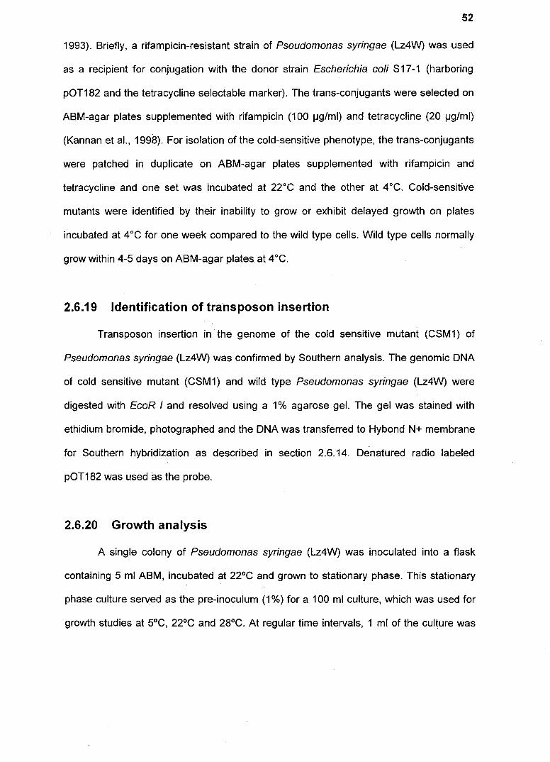
B
Fig 2.1. (A) Schematic diagram of plasm id pET28b. The plasmid contains pBR322 and
F1 origin of replication , Kanamycin resistance gene (Kad) , Lac! gene (lacl) , multiple
cloning site and T7 promoter. (8) Schematic diagram of the plasmid pET28b-trmE
containing the gene trmE in pET28b. The DNA fragment is cloned in the multiple clon ing
site .
2.6.26 Over expression and purification of TrmE
TnnE was over expressed in Escherichia coli BL21 cells which were transformed
with pET28b :: trmE. About 5 ml of overnight culture of the transformant was used to
inoculate 500 ml of fresh LB medium and the culture was incubated at 37°C in a shaker
at 250 rpm. The culture was induced with 1 mM IPTG when its optical density (00600 )
reached - 0.6 and the cells were harvested after 4 h. Protein purification was ca rried out
as recommended by Qiagen (Hilden , Germany). The pellet was resuspended in 5 ml of
lysis buffer B (100 mM NaH2P04 , 10 mM TriS .CI and 8 M Urea , pH 8.0), stirred for 30
min at room temperature and after complete lysis (when the solution became

58
translucent) the lysate was centrifuged at 14,000 rpm for 25 min at room temperature.
Supernatant was mixed with 1 ml of 50% Ni-NTA Agarose slurry (Qiagen) and incubated
at 200 rpm in a rotary shaker for 45 min at room temperature. The Iysate-Ni-NTA
Agarose mixture was loaded on to a column and the column was washed with buffer C
(100 mM NaH2P04, 10 mM Tris.CI and 8 M Urea, pH 6.3) followed with buffer D (100
mM NaH2P04 , 10 mM Tris.CI and 8 M Urea, pH 5.9) to remove the other proteins.
Finally, GTPase was eluted in 5 fractions of 0.5 ml each using buffer E (100 mM
NaH2P04 , 10 mM Tris.CI and 8 M Urea, pH 4.5) and the eluted fractions were analyzed
on SDS-PAGE for its purity.
2.6.27 Cloning of Histag-trmE in pGL 10
Histag-trmE was PCR amplified using pET28b::trmE construct as the template
using T7 F and T7 R primers under the following conditions: initial denaturation at 94°C
for 4 min, denaturation at 94°C for 1 min, annealing at 50°C for 1 min, extension at 72°C
for 1.5 min for 35 cycles and the final extension was at 72°C for 5 min. The resulting
PCR product was then cloned in pMOS as recommended by Amersham Life Science
(8uckinghamshire, UK). The complete open reading frame along with the ribosome
binding site and Histag of pET28b was excised with the restriction enzyme Xba I from
pMOS::Histag-trmE construct. This fragment was cloned in pGL 10 (broad host range
vector) at Xba I site. The resulting construct (pGL 1 0:: Histag-trmE) was selected by
colony PCR for correct orientation. Plasmids were isolated from positive clones as
describe previously (see section 2.6.11) and about 4-5 I-Ig of the pure plasmid was
transformed in to electrocompetent cells of psychrophilic Pseudomonas syringae (Lz4W)
as describe earlier (Kiran et aI., 2004). The transformants were subsequently used for
isolation of TrmE in the native form.

l
Bglll
BspHI
BglII
BspHI
parA
stul
stul
pGLIO (8.5 kb)
kan Clal
pGLIO-Histag-trmE
Clal BspHI
A
Xhol Sphl Pstl Hindlll Xbal BamHI Smal Kpnl Sac I EcoRi
term
BspHI
Xhol
Histag-trmE
Sphl Pstl Hindlll Xbal BamHI Smal Kpnl Sac! :coRi
B
59
Fig.2.3. (A) Schematic diagram of the plasmid pGL 10. This vector contains origin of replication (oriV and
oriT) , trfA (encodes TrfA which controls plasmid copy number) , Kanamycin resistance gene (Kanr), parA
(encodes for ParA which controls cell division and chromosome segregation), term (termination sequence)
and lacZ with multiple cloning site . (8) Schematic diagram of the plasmid pGL 1 O-Histag-trmE containing the
gene Histag-trmE in pGL 10. The DNA fragment is cloned at the Xbal site in multiple cloning sites

60
2.6.28 Expression and purification of TrmE in native form
TrmE was expressed and purified in its native form from psychrophilic
Pseudomonas syringae (Lz4W). Experimentally, the construct pGL 1 0:: Histag-trmE was
transformed in to Pseudomonas syringae (Lz4W) and transformants were selected on
kanamycin containing ABM agar plate. One of the transformants was then pre-
inoculated in 10 ml of ABM broth and finally inoculated in to 1000 ml of ABM broth and
the culture was grown at 22°C in an incubator shaker at 250 rpm. When the culture
reached an OD600 -1.5, it was pelleted down at 10,000 rpm at 4°C and resuspended in
10 ml of buffer A (50mM NaH2P04 .H20, 300 mM NaCI and 10 mM imidazole, pH 8.0).
Cells were lysed by sonication and cell debris was removed by centrifugation at 14,000
rpm for 20 min. Supernatant was applied to the Ni-nitrilotriacetic acid agarose resin
(Qiagen, Hilden, Germany) column equilibrated with buffer A. Column was washed with
buffer B (50 mM NaH2P04 .H20, 300 mM NaCI and 20 mM Imidazole, pH 8.0) followed
by buffer C (50 mM NaH2P04 .H20, 300 mM NaCI and 50 mM Imidazole, pH 8.0) to
remove other proteins. Finally, TrmE was eluted using buffer D (50 mM NaH2P04 .H20,
300 mM NaCI and 250 mM Imidazole, pH 8.0) and the eluted fractions were dialyzed
against 0.1 M Tris (pH 9.0) buffer containing 20% glycerol.
2.6.29 50S-Polyacrylamide Gel Electrophoresis (50S-PAGE)
SDS-PAGE was essentially carried out by following the method of Laemmli
(Laemmli, 1970). A Hoefer mighty small apparatus (San Francisco, CA, USA) was used
to cast and run gels. A stock of the following solutions were made: (a) acrylamide:
bisacrylamide (29.2: 0.8), (b) 1.5 M Tris buffer pH 8.8, (c) 1 M Tris buffer pH6.8, (d) 10%
SDS and (e) 10% APS. A 10% resolving gel and a 5% stacking gel were made by mixing
the stock solutions according to Sambrook et al. (1989). The bacterial pellet was
resuspended in Laemmli buffer (50 mM Tris, pH 6.8, 2% SDS, 10% glycerol, 5% /1-
61
mercaptoethanol and 0.01% Bromophenol blue) and boiled for 10 minutes in a water
bath. The cellular debris was discarded as a pellet after centrifugation at 14,000 rpm for
15 minutes. The supernatant was loaded into the wells of the stacking gel. Molecular
weight standards were also loaded when required. The 1 X running buffer TGS (25 mM
Tris, 250 mM Glycine and 1 % SOS) was made fresh by diluting from a 10 X stock and
the gel was run for 20 mA for stacking and 30 mA for resolving the sample.
Electrophoresis was terminated when the bromophenol blue dye of the samples reached
the bottom edge of the resolving gel and was further processed for staining or for
immunoblotting as required. For staining, the gel was dipped overnight in 0.5%
Coomassie brilliant blue solution (made in 50% methanol and 10% acetic acid).
Oestaining was further done with the staining solvent, till background stain was· removed.
2.6.30 Cross linking of TrmE
Pure TrmE (50-100 I-lg) was covalently cross linked in its native forms using 0.5%
glutaraldehyde (Sigma). The cross linking was carried out in a buffer containing 50 mM
Tris.HCI (pH 8.8), 100 mM NaCI and 20% glycerol to which 0.5% glutaraldehyde was
added and the contents incubated for 30 min at 15°C. The cross linked product was
mixed with 2X SOS-Ioading buffer (50 mM Tris.CI, pH 6.8, 2% SOS, 10% glycerol,
0.01% bromophenol blue and 5% p-mercaptoethanol) and boiled for 10 min. The
resulting samples were then analyzed on 10% SOS-PAGE and stained with 0.25%
Coomassie Brilliant Blue R-250 as described earlier (Sambrook et aI., 1989).
2.6.31 Western Blotting
2.6.31.1 Protein transfer
The electro-transfer of proteins from the polyacrylamide gel to nitrocellulose
membrane (Hybond C) was essentially done following the semi-dry method of Towbin

62
(Towbin et aI., 1979). The gel and the size membrane were soaked in the transfer buffer
(composition) for 10 minutes and then stacked with the membrane towards the anode
sandwiched between three Whatman blotting sheets which were priorly soaked in
transfer buffer. Current applied for electro-transfer was 0.8 rnA per cm2 for1.5 h. After
electro-transfer of proteins the membrane was washed thoroughly with distilled water
and put in Ponceau S stain (0.1 % in 1 % acetic acid) for few seconds, to stain the
proteins bands. The stained protein bands on a Hybond C membrane were distinctly
visible when the membrane was given a quick rinse in methanol after staining. The stain
was again washed away from the membrane by rinsing in distilled water.
2.6.31.2 Processing of the blot with transferred proteins
The blot with transferred protein(s} was blocked with 3% BSA for 2 h. Next, the
blot was incubated with primary antibody made in 1% BSA in TBS-T. The incubation
time was 2 h for all primary antibodies. Next, the blot was incubated with the appropriate
secondary antibody also made in 1% BSA in TBS-T (1 h). All the incubations were
carried out at room temperature and were interspersed with washes with TBS-T (10
minutes each) on a shaker. Finally the blot with alkaline phosphatase conjugated
secondary antibody was developed using NBT and BCIP as substrates. The blot was
briefly incubated with alkaline phosphatase buffer (100 mM NaCl, 5 mM MgCb and 100
mM Tris, pH 9.5) at room temperature to which was added 66 IJI NBT (5% stock) and 33
IJI of HCIP (5% stock) per 10 ml of the buffer. The blot was incubated in dark till a distinct
band appeared. The blot was then washed with distilled water.
2.6.32 GTPase activity assay
GTPase activity was assayed at 15°C and for 30 minutes using 10 IJg of the
purified protein in a 50 IJI reaction mixture of 50 mM Tris-HCI (pH 9.0) containing 200

63
mM KCI, 5 mM MgCb, 1mM DTT and 10 jJM GTP. Reaction was stopped by addition of
200 jJl of 12% SDS and then 100 jJl of H20 was added. Hydrolyzed GTP was quantitated
by monitoring the release of P04-3 using a spectrophotometer. For this purpose, 400 jJl
of buffer BC [containing an equal mixture of buffer B (6% ascorbic acid in 1 N HCI) and
buffer C (1% ammonium molybdate)] was added, mixed and incubated at room
temperature for 10 min. Finally, 600 jJl of buffer D (2% sodium citrate, 2% sodium meta
arsenite and 2% acetic acid) was added, mixed and incubated at 37°C for 10 min and
the absorbance was read at 850 nm (Doerrler and Raetz, 2002; Gonzalez-Romo et aI.,
1992). Na2HP04 was used to obtain a standard curve for phosphate estimation.
2.6.33 5' end labeling of oligonucleotides
Oligonucleotides were end labeled using T 4 DNA polynucleotide kinase.
Approximately, 50 ng of oligonucleotides was mixed in MiIIi-Q® water containing 1 X T4
PNK (polynucleotide kinase) buffer, 2 Jd of [y_32p] ATP and 5-10 units of T4 PNK in 50 ~I
reaction volume and incubated at 37°C for 1 h. Probes were purified by passing through
a sephadex G-25 spin column (as described in section 2.6.13).
2.6.34 Primer extension analysis
Transcription start site mapping was carried out using Murine Leukaemia. Virus
Reverse Transcriptase (M-MuLV-RT). Approximately 50 p moles of primer Ash 59 (5'
GGG GTA CCC CAC ACC ACC TCG GCC TTG 3') corresponding to the
complementary sequence of the putative N-terminal end of the GTPase was labeled at
the 5' end using T 4 DNA polynucleotide kinase in a 50 jJl reaction volume containing 1 X
PNK buffer. The reaction mixture was incubated at 37°C for 1 h and the end labeled
primer was then separated from the unincorporated labeled [y-32p] A TP using a
Sephadex G-25 column (as described in section 2.6.13).

64
For primer extension analysis the equivalent of 106 cpm of the labeled primer
was mixed with 5 I-Ig of RNA and reverse transcription reaction was carried out by
addition of 5 mM MgCb, 1 mM dNTPs mix, 1 X RT buffer and 10 unit of M-MuLV-RT in
25 1-11 reaction volume. The RT reaction mix was incubated at 42°C for 1 hand
subsequently the stop solution (sequencing gel loading dye) was added to 1 X final
concentration. DNA sequencing reactions were also carried out with the same labeled
primer Ash 59 on the double stranded DNA template with fmol sequencing kit (Promega
Corporation, Madison, USA). The sequencing loading dye was mixed with the DNA
sequencing reactions. After heat denaturation at 94°C for 4 min followed by rapid cooling
on ice, the DNA sequencing reactions were run parallel with the primer extended
products on an 8 M urea-6% polyacrylamide gel and electrophoresed using 1 X TBE
buffer. The gel was then dried and the bands were visualized by autoradiography.
2.6.35 Construction of site-specific deletion promoter constructs
In order to construct trmE promoters with a specific deletion, overlap extension
PCR was carried out (Urban et aI., 1997). In the first step both the upstream (fragment 1)
and downstream regions (fragment 2) of the stretch to be deleted (blue box marked with
an X) were amplified separately using two sets of primers [namely Ash 57 (forward
primer) and the mutagenic reverse primer C and mutagenic forward primer B and Ash 59
as the reverse primer] to generate two fragments in which 'X' is deleted. The mutagenic
oligonucleotide primers (forward and reverse) are synthesized with the deletion and are
complementary to each other. PCR was carried out with AccuPrime Pfx DNA
polymerase in the reaction mixture as recommended by the manufacturer (Invitrogen,
Carlsbad, California) and the conditions were as follows: initial denaturation at 94°C for
3 min, denaturation at 94°C for 30 s, annealing at 55°C for 45 s, extension at 72°C for 1

65
min for 35 cycles and the final extension at 75°C for 5 min. PCR products were gel
eluted with Qiaquick gel extraction kit (Hilden, Germany). In the second step, overlap
PCR was carried out with the above two PCR products for 10 cycles under the same
conditions as above without any primers. Subsequently PCR was continued for another
25 cycles with both the flanking primers (Ash 57 and Ash 59). The flanking primers (Ash
57 and Ash 59) are same but the pairs of mutagenic primers are different for different
constructs. Using this strategy constructs with deletion of the -10 region (the region -5 to
-10 was deleted using the primers Ash 57, Ash 65 and Ash 64, Ash 59), -35 region (the
region from -33 to -38 is deleted using the primers Ash 57, Ash 63 and Ash 62, Ash 59),
UP element (the region from -41 to -54 is deleted using primers Ash 57, Ash 110 and
Ash 109, Ash 59), cold-box (the region from +184 to +194 is deleted using the primers
Ash 57, Ash 112 and Ash 112, Ash 59), DEAD-box (the region from +208 to +217 is
deleted using the primers Ash 57, Ash 114 and Ash 113, Ash 59), 5'-UTR (the region
from +9 to +322 is deleted using the primers Ash 57, Ash 61 and Ash 60, Ash 59) and a
specific sequence from position +246 to +252 were deleted using the primers (Ash 57,
Ash 78 and Ash 77, Ash 59). PCR products were restriction digested with Pst! and Kpnl
and cloned in transcriptional fusion promoter probe vector pKZ27. In the constructs, site-
specific deletion of the specific region in the putative promoter was confirmed by DNA
sequence analysis.

Step 1
Step 2
B Ash 57 Fragment 1
5' -------------------________ 3'
3' ------------------- 5'
Ash 57
5' -. ____ ...... _ _ ______ ~-5 '
.-Ash 59 t Overlap PCR
Ash 57 B 5 ' ___________________ H" ~ (Productive)
(Productive) ~ ..... H. C ---------------""!Al""s'l"'h~59 5'
+ B
(Non productive) • .....• ----------------- 3' Fragment with deletion
3' _______________ ...... ~ ... H. (Non productive)
Ash 57 -. t 5' _______ ~_ . 5' ~ C -------------.-~A-sh~5~9
Ash 57 -. 5' _____________________________________ __ t Amplification
_____________________________________ ~~~ 5·
.- Ash 59
66
Fig. 2.4. Overlap extension PCR to generate site-specific deletions in the promoter
represented by the blue and brown strands . X represents the region to be deleted . In
step 1 two fragments 1 and 2 were amplified using two sets of primers [namely Ash 57
(forward primer) and the mutagenic reverse primer C and mutagenic forward primer B
and Ash 59 as the reverse primer] to generate two fragments in which X is deleted . In
step 2 overlap PCR was carried out using the above two fragments for ten cycles without
using any primers so as to generate a fragment with a specific deletion. This fragment
was then amplified using Ash 57 and Ash 59 .
2.6.36 Construction of trmE promoter::/acZ reporter constructs
A low copy number, promoter less, transcriptional fusion promoter probe plasmid
vector (pKZ27) was used to construct various trmE promoter::lacZ fusions constructs .
The full length trmE promoter was PCR amplified using primers Ash 57 (Pst I) and Ash

67
59 (Kpn I) with intrinsic restriction sites using the following conditions: initial denaturation
at 94°e for 4 min, denaturation at 94°e for 1 min , annealing at 62°e for 1 min , extension
at 72°e for 1 min for 35 cycles and the final extension was at 75°e for 5 min. The peR
product was cloned in pMOS as recommended by the manufacturer (Amersham
biosciences, Buckinghamshire, UK). Subsequently, the promoter fragment was excised
using restriction enzyme, Pst1 and Kpn1 from pMOS::promoter as recommended by
New England Biolabs Inc . (Beverly, USA). The promoter fragment was eluted from
agarose gel using QIAqu ick gel extraction kit (Hilden, Germany) and cloned in pKZ27 at
Pst I and Kpn I site. Promoter fusion constructs were electroporated into wild type
Pseudomonas syringae (Lz4W) to measure the 0-galactosidase activity.
Hindlll
pKZ27 10000 bp
XlIol D,.,
P*ll Stul Xt,01 0\1111 S .. Xb.J ~1tI
Sm.1 Kpnl Sm,1 Xb., tHndlil
A
pKZ27- tmlE promoter 10655 bp
Xh~1 0,.1
B
Pol'

pKZ27 -trmE promoter (UP element deletion)
10641 bp
Hindlll
Hlndlll
Xhol Dnl
trm E prom oter
pKZ27 -tnnE promoter (-35 deletion)
10649 bp
Xhol Oral
pKZ27.lrmE promoter (DEAD bqx deletion
10645 bp
.... "
c
PsU
Pstl
Kpnl Smol
leb. 1 Hin~1II
lpnl Smal Xbol Hindlll
(OEAO b.oM "-:Ic C'I ... J ...... -n. .. .......
E
G
IIn",
pKZ27-tnnE promoter (-1 0 deletion)
10649 bp
IIhai Onl
( I pKZ27 -trmE promoter
(cold box deletion) 10644 bp
Hindi.
Xho' Dr "
pKZ27 ·tmlE promo er (cold and DEAD box delelton)
0634 bp
- u..., 0 ...
68
PsI,
P ... ,
D
M .... 5 ..... , ~bol
HhdJI
F
H

pKZ27 -tm7E promoter (conserved region deletion)
10648 bp
Hinclll
x"'"
Pst'
Hlfldlll
pKZ27-trmE promoter (5'UTR deletion)
10342 bp
Xhol Dr ..
69
Pst,
(5'UTR de'eHon)
Kon' Smal XbO' Hindi.
Fig, 2.5. A. Schematic diagram of the plasmid pKZ27. The vector contains Kanamycin
resistance gene (Kad) , Trrn , lacZ and multiple cloning sites upstream to lacZ. In B trmE
promoter is cloned upstream to lacZ where as in C to J trmE promoter with various
specific regions deleted such as UP element deletion (C), -10 region deletion (0) , -35
region deletion (E), cold-box deletion (F), DEAD-box deletion (G), cold-box and DEAD-
box deletion (H), conserved region deletion (I) and 5'-UTR deletion (J) , The different
promoter fragments were cloned at the Kpn I and Pst I site in multiple cloning sites .
2.6.37 B-galactosidase assay
All promoter constructs were transformed into wild type Pseudomonas syringae (Lz4W)
by electroporation. A single colony of each construct was then inoculated into 10 ml of
ABM medium containing 25 )1g/ml of kanamycin and incubated at 22°C overnight. Then
1 % of the over night grown culture was inoculated into 25 ml of fresh ABM containing 25
)1g/ml of kanamycin , incubated at 22°C and then 1 ml of the culture was removed at
different time intervals and 00600 was determined . To measure the ~-galactosidase
activity, 100 )11 of the culture was taken in a 1.5 ml centrifuge tube and 900 IJI of Z-buffer
(see section 2.5.2 .22), 10 )11 of 10% SDS and 50 )11 of chloroform was added to the tube.
Sample was vortexed for 15 s to break the cells . ONPG (200 pi) was added to the
J

70
sample and incubated at room temperature till yellow colour appeared. The reaction was
stopped by adding 500 IJI of 1 M sodium carbonate. The time interval that lapsed
between the start and the end of reaction was noted down and p-galactosidase activity
(Miller units) was calculated by using the following formula of Miller (1972) as shown
below.
P.,-galactosidase activity (Miller units) = 1000 (00420-1.75 x 00550)1 VxTx00600
where 'V' is the volume of the sample in ml and 'T' is the time of incubation in minutes.
2.6.38 Generation of complementation vector
The complete open reading frame of trmE along with its promoter was PCR
amplified from Pseudomonas syringae (Lz4W) using primers Ash 57 and Ash 47 under
the following conditions: initial denaturation at 94°C for 4 min, denaturation at 94°C for 1
min, annealing at 60°C for 1 min, extension at 72°C for 2 min for 35 cycles and final
extension at 72°C for 5 min. The PCR amplified fragment was gel eluted using QIAquick
gel extraction kit (Hilden, Germany), ligated to pMOS the blunt end closing vector
(Amersham Life Science, Buckinghamshire, UK) and transformed in Escherichia coli,
OH5u. The complete open reading frame along with the promoter was then excised with
Hind 11/ and EcoR I from pMOS::promoter-trmE construct. Ligated to pGL 10 (see
section, 2.6.27) in the Hind 11/ and EcoR I site and used for the transformation of
Escherichia coli OH5u. The resulting construct (pGL 10::promoter-trmE) was selected by
colony PCR and the resulting recombinant plasmid was transformed to Escherichia coli
S17-1 and mobilized in to CSM1 mutant by biparental mating as described earlier.

BglII
BspHI
parA
stul
pGL 10::promoter-trmE
CIa I BspHI
Xhol
Sphl Pstl Hindlll EcoRi
Promoter-trmE
Fig 2_6. Schematic diagram of the pGL 1 O-promoter-trmE construct
71