Mata Kornea
37
Prof, DR, dr Rukiah Sawal SpM (K)
description
Mata Kornea
Transcript of Mata Kornea
Prof, DR, dr Rukiah Sawal SpM (K)

Anatomy and PhysiologyAnt part of the eye
• Avascular
• Transparant
• Refracting and Protective “ window” of
Route light rays
(MEDIA REFRACTA)
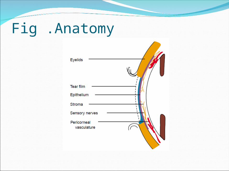
Fig .Anatomy


Fig. Histology
TransparancyUniform Structure• Avascular
• Deturgescence

NutritionPerilimbal capillaries• Air + tear film
• Aqueous humor
Innervation : N V1

Desease of the corneaExtremely serious
Permanent visual impairment
Blindness
Prompt diagnosis and prompt treatment
PathologyCongenital
Inflamation : Keratitis
Tumor
Trauma
Degeneration

KeratitisKeratitis : - Superficial - Profunda (interstitial) - Non ulceration - Cornea Ulcer
Superficial : epithel and superficial stroma
Cornea ulcer: defect / discontinuity

Superficial KeratitisFig histology
Ethiology :
- Infective
- Degenerative
- Allergic
- Toxic


Classification Cornea Ulcer1. Bacterial2. Viral3. Fungal4. Hypersensitivity reaction5. Neurothropic6. Exposure7. Idiopathic
Clinical PresentationPainPhotophobiaLacrimationBlepharo spasmeBlurred visionPericorneal / ciliary injectionInfiltrate, edem, defect cornea

Bacterial Corneal UlcerSight – threatening- Progressive stromal in flurocen- Progresive tissue destruction- Cornea perforation- Infection to adjacent tissue

Risk FactorsContact lens wearTrauma Contaminated ocular medicationImpaired defense mechanismAltered structure of corneal surface

Clinical PersentationPain, photophobia, blepharospasme lacrimation, decreased visionPericorneal injection – red eyeSharply demarcated epithelial defect Stromal edema Suppurative Stromal inflamationAnt chamber reaction : KP. Hypopyon


Viral Corneal ulcerClinical presentationHSK Foreignbody sensation, photophobia Lacrimation, blurred visionPericornea injection / ciliary flushRose bangal, fluoroscein Staining (+)Reduced corneal sensation
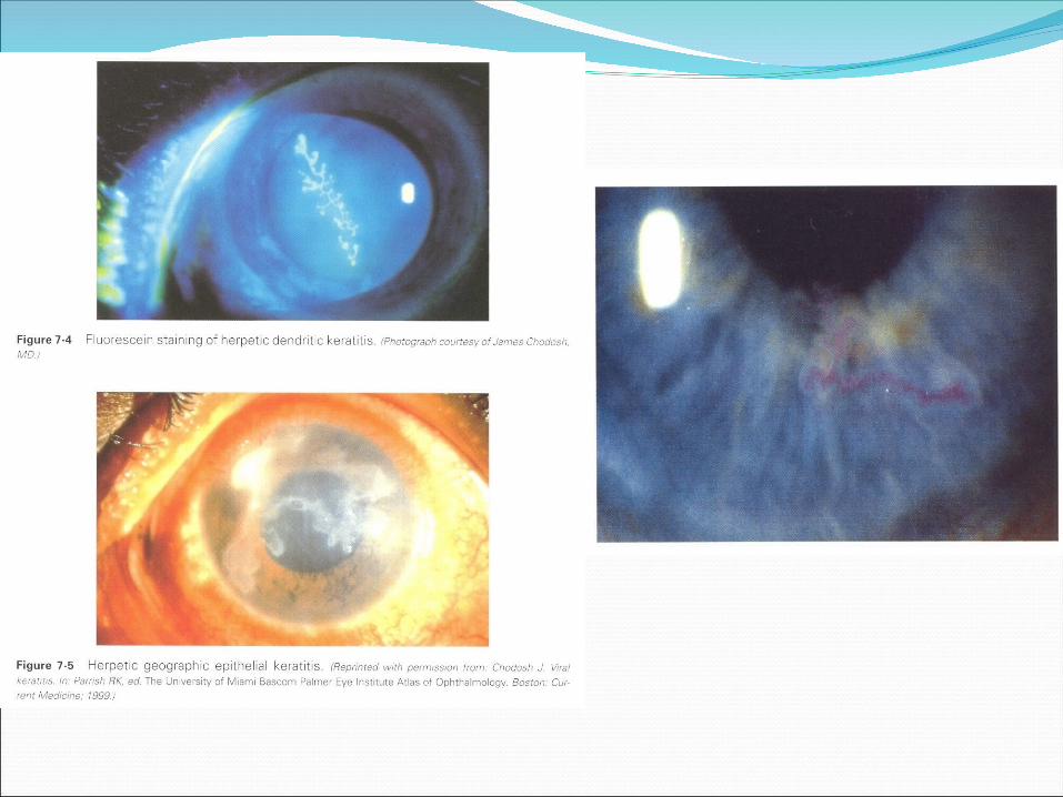

Keratitis herpes simpleks

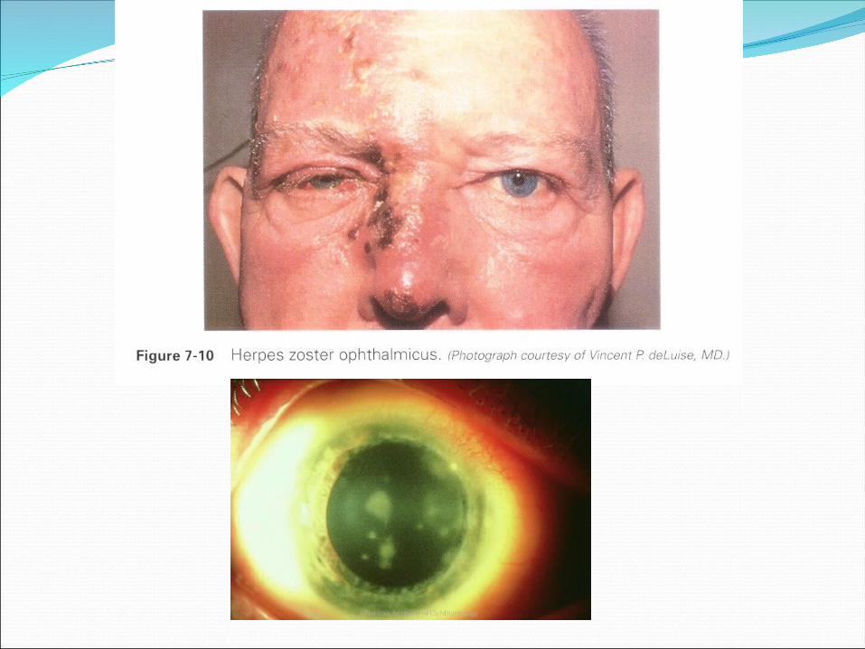
HZ0Zoster dermatitis affected N V1Punctate or dendritic epithelial keratitis50% decreared corneal sensationIntestitial keratitis and anterior uveitis >
HSK

Fungal Corneal ulcerRish Factor :Gardener : preplant or vegetableContact lens wearCorticosteroid treatment topical / systemic

Clinical PresentationResemble with batecterial ulcerGray-white infiltrate, irreguler and filament
marginsSatelite infiltrateAnterior chamber reaction, hypopion


Hypersensitivity reaction1. Atopic keratoconjunctivitis2. Steven Johnson syndrome3. Ocular cicatrical Pemphigoid4. Mooren Ulcer
1
2 34

Treatment cornea ulcerEtiology / causePredisposing factorsPotentially sight threateming
Local - cycloplegic : atropin 0.5% - Specific : antibiotic, anti viral, anti fungal, anti inflamation / immunosuppresive eye drop / ointment
• Systemic : oral : IV
Subconjunctiva, subtenon
- Surgical ( complication )

Complications1. Corneal scar : nebula, macula, leucoma2. Iridocyclitis : Synechia, complited cataract,
secondary glaucoma3. Perforation4. Endofthalmitis5. Panophthalmitis6. Atrophia bulbi
Visual impairment – visual loss


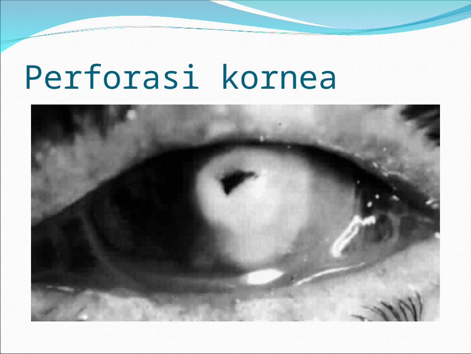
Perforasi kornea