
Mast Cell-Orchestrated Immunity
13
Mast cells are a key cell type of the haematopoietic lineage that has evolutionarily conserved functions in pathogen surveillance. They are dispersed through- out most tissues but are crucially located at the host’s interfaces with the environment, such as the skin and mucosae, supporting a role in the recognition of patho- gens or other signs of infection (FIG. 1). Mast cells and many of their products are best known for their associa- tion with pathological conditions such as asthma, allergy and anaphylaxis, in which aberrant, chronic or systemic activation of mast cells promotes harmful inflammatory sequellae and damage to host tissues. However, despite the potential detrimental effects that mast cells can have on immune homeostasis, these cells are indispensable to the host, as suggested by the observations that they are evolutionary preserved across many species and that humans that lack mast cells have never been described 1 . The first strong evidence that mast cells function in a protective capacity against infectious disease came from studies of host–parasite interactions 2,3 , and an increas- ing amount of work supports their essential contribu- tion to controlling a wide range of pathogenic infections, including those by parasites, bacteria and probably viruses. We now understand that mast cells function not only as sentinels but also as modulators of innate and adaptive immune responses, ultimately influencing disease outcomes. In this Review, we discuss recent advances in our understanding of mast cell responses to pathogens. We first discuss the potential mechanisms by which mast cells can be activated by pathogens. We then describe the responses of mast cells, particularly with regard to the timing of the responses and the various roles they have in host defence, as sensors of pathogens, as effectors of adaptive immune responses and as modulators of local inflammation. Finally, we examine the evidence indicat- ing that mast cells make meaningful contributions to controlling infectious challenges and discuss how mast cells might be harnessed during vaccination. Cell biology of mast cells Mast cells arise from bone marrow-derived precursors that circulate in the blood and become differentiated after entering tissues. They are long-lived cells, able to survive for months or years and, despite being termi- nally differentiated, they can proliferate in response to appropriate signals 4 . All mature mast cells reside in the body’s tissues and have a common fundamental morphology with prominent electron dense granules in their cytoplasm. At the earliest stages of infection, mast cells are important for communicating the pres- ence of a pathogen to many cell types located nearby in the site of infection and distally in draining lymph nodes (FIG. 2). To facilitate these interactions, mast cells *Departments of Immunology, ‡ Pathology and Molecular Genetics and Microbiology, Duke University Medical Center, Durham, North Carolina 27710, USA. § Program in Emerging Infectious Diseases, DUKE- National University of Singapore Graduate Medical School, Singapore 169857. Correspondence to S.N.A. e-mail: soman.abraham@ duke.edu doi:10.1038/nri2782 Mast cell-orchestrated immunity to pathogens Soman N. Abraham* ‡§ and Ashley L. St. John* Abstract | Although mast cells were discovered more than a century ago, their functions beyond their role in allergic responses remained elusive until recently. However, there is a growing appreciation that an important physiological function of these cells is the recognition of pathogens and modulation of appropriate immune responses. Because of their ability to instantly release several pro-inflammatory mediators from intracellular stores and their location at the host–environment interface, mast cells have been shown to be crucial for optimal immune responses during infection. Mast cells seem to exert these effects by altering the inflammatory environment after detection of a pathogen and by mobilizing various immune cells to the site of infection and to draining lymph nodes. Interestingly, the character and timing of these responses can vary depending on the type of pathogen stimulus, location of pathogen recognition and sensitization state of the responding mast cells. Recent studies using mast cell activators as effective vaccine adjuvants show the potential of harnessing these cells to confer protective immunity against microbial pathogens. REVIEWS 440 | JUNE 2010 | VOLUME 10 www.nature.com/reviews/immunol © 20 Macmillan Publishers Limited. All rights reserved 10
-
Upload
paulo-fernandes-marcusso -
Category
Documents
-
view
230 -
download
1
Transcript of Mast Cell-Orchestrated Immunity
Mast cells are a key cell type of the haematopoietic lineage that has evolutionarily conserved functions in pathogen surveillance. They are dispersed through-out most tissues but are crucially located at the host’s interfaces with the environment, such as the skin and mucosae, supporting a role in the recognition of patho-gens or other signs of infection (FIG. 1). Mast cells and many of their products are best known for their associa-tion with pathological conditions such as asthma, allergy and anaphylaxis, in which aberrant, chronic or systemic activation of mast cells promotes harmful inflammatory sequellae and damage to host tissues. However, despite the potential detrimental effects that mast cells can have on immune homeostasis, these cells are indispensable to the host, as suggested by the observations that they are evolutionary preserved across many species and that humans that lack mast cells have never been described1. The first strong evidence that mast cells function in a protective capacity against infectious disease came from studies of host–parasite interactions2,3, and an increas-ing amount of work supports their essential contribu-tion to controlling a wide range of pathogenic infections, including those by parasites, bacteria and probably viruses. We now understand that mast cells function not only as sentinels but also as modulators of innate and adaptive immune responses, ultimately influencing disease outcomes.
In this Review, we discuss recent advances in our understanding of mast cell responses to pathogens. We first discuss the potential mechanisms by which mast cells can be activated by pathogens. We then describe the responses of mast cells, particularly with regard to the timing of the responses and the various roles they have in host defence, as sensors of pathogens, as effectors of adaptive immune responses and as modulators of local inflammation. Finally, we examine the evidence indicat-ing that mast cells make meaningful contributions to controlling infectious challenges and discuss how mast cells might be harnessed during vaccination.
Cell biology of mast cellsMast cells arise from bone marrow-derived precursors that circulate in the blood and become differentiated after entering tissues. They are long-lived cells, able to survive for months or years and, despite being termi-nally differentiated, they can proliferate in response to appropriate signals4. All mature mast cells reside in the body’s tissues and have a common fundamental morph ology with prominent electron dense granules in their cytoplasm. At the earliest stages of infection, mast cells are important for communicating the pres-ence of a pathogen to many cell types located nearby in the site of infection and distally in draining lymph nodes (FIG. 2). To facilitate these interactions, mast cells
*Departments of Immunology, ‡Pathology and Molecular Genetics and Microbiology, Duke University Medical Center, Durham, North Carolina 27710, USA.§Program in Emerging Infectious Diseases, DUKE-National University of Singapore Graduate Medical School, Singapore 169857.Correspondence to S.N.A.e-mail: [email protected]:10.1038/nri2782
Mast cell-orchestrated immunity to pathogensSoman N. Abraham*ठand Ashley L. St. John*
Abstract | Although mast cells were discovered more than a century ago, their functions beyond their role in allergic responses remained elusive until recently. However, there is a growing appreciation that an important physiological function of these cells is the recognition of pathogens and modulation of appropriate immune responses. Because of their ability to instantly release several pro-inflammatory mediators from intracellular stores and their location at the host–environment interface, mast cells have been shown to be crucial for optimal immune responses during infection. Mast cells seem to exert these effects by altering the inflammatory environment after detection of a pathogen and by mobilizing various immune cells to the site of infection and to draining lymph nodes. Interestingly, the character and timing of these responses can vary depending on the type of pathogen stimulus, location of pathogen recognition and sensitization state of the responding mast cells. Recent studies using mast cell activators as effective vaccine adjuvants show the potential of harnessing these cells to confer protective immunity against microbial pathogens.
R E V I E W S
440 | june 2010 | VoluMe 10 www.nature.com/reviews/immunol
© 20 Macmillan Publishers Limited. All rights reserved10
Nature Reviews | Immunology
a
c
b
d e
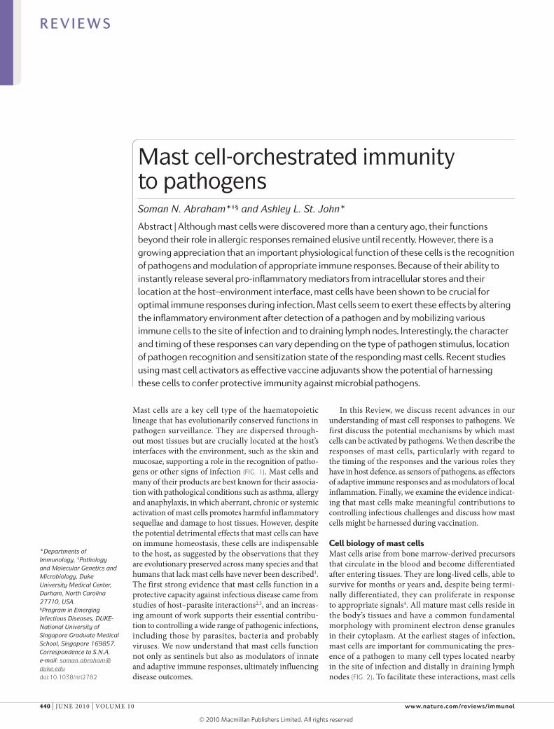
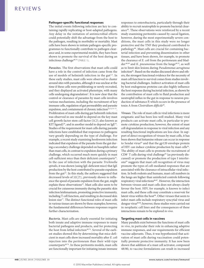
are strategically located at the host–environment inter-face, proximal to both blood vessels (FIG. 1a,b) and lym-phatic vessels (FIG. 1c), as well as to nerve fibres (FIG. 1d) and tissue-resident immune cells, including dendritic cells (DCs) (FIG. 1e).
Despite having a common lineage, granulated mor-phology and functions, mast cells are highly heterogene-ous and phenotypically malleable cells5,6, the intricacies of which have only begun to be defined, with little known about their distinct functionality. However, it is likely that this heterogeneity is shaped by the require-ments of residing in a particular tissue or encountering unique pathogen challenges. on the basis of distinct staining properties, it was quickly recognized that rodent mast cells fall into two broad categories: mucosal and connective tissue mast cell types. These distinct mast cell types can now be further distinguished by sev-eral features, including granule composition, differing degranulation responses to pharmacological stimula-tion and the ability to proliferate in response to para-sitic challenge7. This suggests that responses to other stimuli, including pathogens, might differ depending on the mast cell type.
The main protein components of mast cell granules are proteases. In mice, the granules of connective tissue mast cells contain two types of protease — tryptases and chymases — that are bound to heparin, whereas mucosal mast cells contain only chymases, which are bound to chondroitin sulphate7. Human mast cells also show hetero geneity with regard to these two main protease types, although with less stringent tissue-type specificity8. The storage of proteases varies not only between mast cell subtypes but also within an individual mast cell depend-ing on the stimuli it receives. For example, in mouse mast cells, the expression of these proteases has been shown to be modulated at a transcriptional level by interleukin-10 (Il-10)9, and treatment of human mast cells in vitro with Il-4 increased the relative amount of chymase incorporated into granules10. Two types of human mast cell, defined by relative tryptase and chymase content, also vary with respect to their expression of the recep-tor for complement component C5a (C5aR)11. Although other inflammatory mediators and surface receptors might also have tissue-type or activation-specific spe-cificity, the varied composition of granules (particularly well characterized for proteases) shows the heterogeneity of mast cells.
Armed with granules containing preformed media-tors, mast cells have the potential to be the first respond-ers (within seconds to minutes) following recognition of an invading pathogen. The findings that mast cells can respond to their environment — not only by produc-ing appropriate mediators for the pathogen they have encountered, such as selective cytokine production12, but also by altering the transcription and storage of pre-formed mediators9,10 — suggest that they can modulate their phenotype during the course of infection. They also have the ability to replenish their granules dur-ing an infection or after its resolution7,13. Altering the production of preformed mediators and, thereby, the composition of their granules, if shown to be beneficial
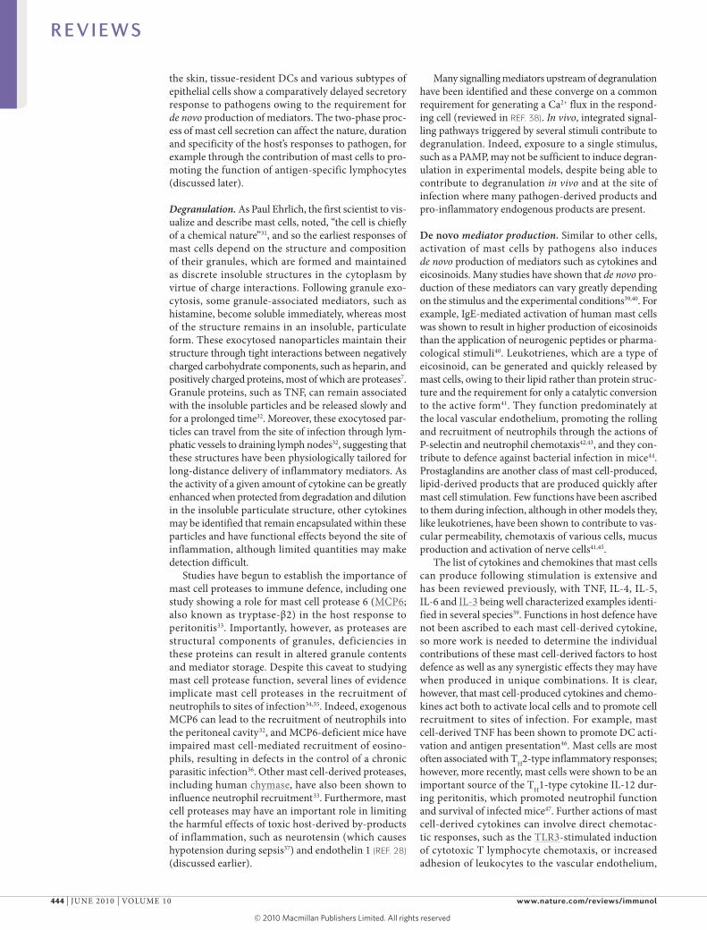
Figure 1 | Mast cells are strategically located in host peripheral tissues. a | A partially degranulated mast cell is visualized by electron microscopy, releasing granules near a blood vessel in mouse ear tissue activated by topical application of phorbol 12-myristate 13-acetate (PMA). The arrow indicates a granule that seems to be in the process of being released. b | Mouse ear tissue was stained in whole mount for CD31-expressing blood vessels (red) and mast cell granules, using a mast cell-specific fluorescent conjugated probe (green), after topical PMA treatment to activate mast cells. c | PMA-treated mouse ear tissue is stained to visualize LYVE1-expressing lymphatic vessels (blue) and mast cells (green). d | This image depicts tyrosine hydroxylase-expressing neurons (red) in close proximity to activated mast cells in compound 48/80-treated whole mounted bladder. e | Mouse ears were treated with PMA and prepared in whole mount for imaging by staining mast cells (green), CD31-expressing blood vessels (red) and CD11c-expressing dendritic cells (blue). For images in b–d, tissue was imaged at x10 magnification. Owing to whole mount preparation and large depth of field, overlap of stained elements can be seen.
R E V I E W S
nATuRe ReVIeWs | IMMunology VoluMe 10 | june 2010 | 441
© 20 Macmillan Publishers Limited. All rights reserved10
Nature Reviews | Immunology
Endothelial cell
Adhesion moleculeupregulation andvascular permeability
Chemotaxis andbacterial killing
T cell chemotaxis and activation throughantigen presentation
Antigen presentation, recruitment into tissue and mobilization todraining lymph nodes
Activation andintracellular killing
Cytokine andmucus production
Contraction
Pain
Lymphocyte retention indraining lymph nodes andenhanced antibody production
NeutrophilMast cell
T cell
TCR BCR
TNFEicosinoids(such as LTB4)
Eicosinoids (such as LTC4 and LTD4)
Eicosinoids (such as PGI2 and PGE2)
Antibody(IgG ad IgE)
TNF Proteases (such as MCP1)IL-6
TNF Histamine
For example,IL-4
For example,substance P
For example,CCL5
B cell
Dendritic cellMacrophage Epithelial cells
Smooth muscle cell
Neuron
Fc receptor
in preventing or controlling reinfection, could be con-sidered a form of immunological memory. This would allow the responses of pathogen-experienced mast cells to be refined by the infectious challenges they have previously encountered.
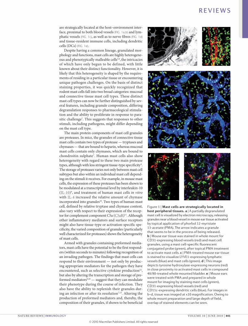
Mast cells as sentinels of infectionDirect recognition of microorganisms. At the initiation of infection, the first responsibility of mast cells is to rec-ognize that pathogen invasion has occurred. Many cells, including DCs, epithelial and endothelial cells, can alert the host immune system to the presence of pathogens, whether in the skin, gut or other sites in the body that are exposed to the environment or susceptible to pathogen encounter. This can be achieved by directly recognizing pathogens through pattern recognition receptors (PRRs), such as Toll-like receptors (TlRs), which are activated in response to conserved pathogen-associated molecular patterns (PAMPs)14. Mast cells are well equipped for this task as, in addition to being able to recognize PAMPs, they can detect a range of products through the expres-sion of other receptors that sense pathogens (for exam-ple, Fc receptors (FcRs), which bind pathogen-specific
antibodies) and receptors for inflammatory factors pro-duced at the site of infection. Direct pathogen recognition by mast cells occurs both in response to factors that are common to classes of pathogens (such as through TlRs) and those that are specific to only a certain infectious challenge (such as through binding of antibodies specific for pathogen-associated epitopes). Interestingly, mast cell responses to TlR triggering alone can vary depend-ing on the PAMP stimulus. For example, lipopolysac-charide (lPs) stimulation of rodent mast cells through TlR4 promoted cytokine production in the absence of degranulation, whereas stimulation through TlR2 by peptidoglycan induced both degranulation and cytokine production15. In this study, responses to individual PAMP stimulation overlapped for some cytokines (stimula-tion of either TlR4 or TlR2 promoted the production of tumour necrosis factor (TnF), Il-6 and Il-13), but diverged for other cytokines (TlR4 stimulation resulted in Il-1β production but not Il-4 or Il-5 production, whereas TlR2 stimulation resulted in Il-4 and Il-5 pro-duction but not Il-1β production)15. similarly, for mast cells derived from human cord blood, both peptidoglycan and lPs were shown to induce a T helper 2 (TH2)-type
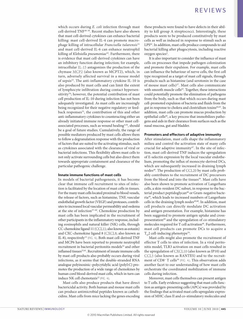
Figure 2 | Cellular communication by mast cells promotes host defence. Mast cells ‘communicate’ with various cell types, including immune cells (such as lymphocytes50,67,71, macrophages51, dendritic cells41,46,49,61,68–70 and neutrophils34,35,43,44,48,52), epithelial cells66, smooth muscle cells45,63 and endothelial cells49,57–60. These interactions contribute to pathogen surveillance, antipathogen immunity and other mechanisms of eliminating microorganisms from the host. These cellular targets of mast cells are located both in the site of infection and in distant draining lymph nodes. Examples of functional consequences of mast cell communication are shown, as are examples of mast cell mediators that have been shown to contribute to the target cell response. BCR, B cell receptor; CCL5, CC-chemokine ligand 5; IL, interleukin; LT, leukotriene; MCP1, mast cell protease 1; PG, prostaglandin; TCR, T cell receptor; TNF, tumour necrosis factor.
R E V I E W S
442 | june 2010 | VoluMe 10 www.nature.com/reviews/immunol
© 20 Macmillan Publishers Limited. All rights reserved10
Nature Reviews | Immunology
Mast cell
Degranulation
De novo mediator production
Preformed mediators:ProteasesTNFHistamine
EicosinoidsProstaglandinsLeukotrienes
Cytokines such asTNF and IL-4
Granules replenished Survival, possible proliferation
Seconds Minutes Hours Days Weeks
Granule New granule
cytokine response, however only peptidoglycan resulted in histamine release from intracellular stores16. In addi-tion to conventional PRRs, other receptors on the surface of mast cells can be activated in response to pathogens; for example, CD48, which can detect the presence of fim-briated Escherichia coli, Mycobacterium tuberculosis and Staphylococcus aureus17–19.
Activation by FcRs. owing to the expression of multiple FcRs, including FcγRII receptors and the high-affinity receptor for Ige, FcεRI, mast cells can bind both IgG and Ige and become sensitized to antigens that have been previously encountered by the host. subsequently, mast cells can become activated, resulting in degranu-lation following receptor cross-linking by polyvalent antigen20,21. Antibody-mediated mast cell recognition of specific antigens and the signalling events downstream of receptor cross-linking have been most thoroughly characterized in models of asthma and allergy, but are also likely to be relevant in the context of infection as suggested by one study examining parasite clearance22. Interestingly, it has been shown that FcεRI activation and TlR stimulation can have synergistic effects on cytokine production by mast cells, enhancing cytokine transcription through the cumulative increase in activ-ity of mitogen-activated protein kinases (MAPKs)23. Mast cells can also be activated by FcR signalling triggered by bacterial superantigens, such as S. aureus protein A, which can to bind certain classes of antibodies, independent of antigen specificity24.
Activation by pathogen-associated substances. Mast cells can also undergo degranulation in response to some exogenous stimuli that accompany pathogen injection into the skin or breaching of the skin bar-rier, such as components of wasp venom25 or mosquito
saliva26. Mastoparan, for example, is a 14-amino acid peptide found in wasp venom that efficiently induces mast cell degranulation25. Degranulation in response to mosquito saliva could have implications for immune defence against arboviruses or vector-transmitted para-sitic diseases such as malaria, although this remains to be investigated.
Activation by endogenous inflammatory factors. several host endogenous peptides, including neuro-tensin, substance P27 and endothelin 1 (ReF. 28), and by-products of inflammation, such as complement components, can also activate mast cells. Activation of mast cells through complement receptors, particularly C5aR, can result in degranulation29. This receptor was also identified as the main receptor responsible for mast cell detection of, and degranulation in response to, the yeast product zymosan, rather than TlR2 (which is gen-erally ascribed the function of recognizing zymosan)30. The inflammation marker endothelin 1, which can have toxic side-effects during inflammation, is degraded by mast cell-derived proteases, showing an important feedback mechanism of mast cells in limiting potential detrimental effects of inflammatory processes through granule exocytosis28.
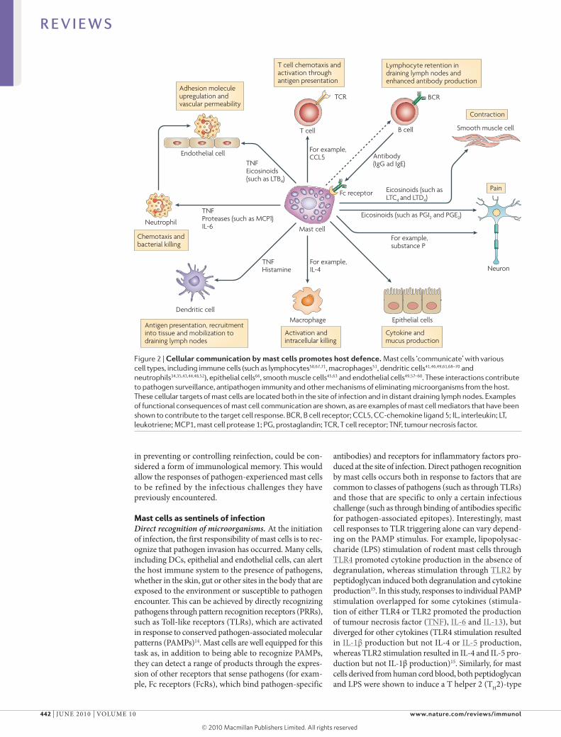
Two waves of mediator releaseAs mentioned, activation of mast cells by pathogens can result in both degranulation and de novo cytokine synthesis. Degranulation involves the rapid (begin-ning within seconds to minutes following stimula-tion) release of pre-packaged, insoluble mediators into the surrounding tissue, a strategy that gives mast cell-derived products a temporal advantage over those produced by other immune surveillance cells (FIG. 3). other sentinel cells, for example, langerhans cells in
Figure 3 | Timing of mast cell responses to pathogens. Mast cells can respond quickly to pathogen challenges owing to the presence of preformed mediators in cytoplasmic granules that can be quickly released at a site of infection through the process of degranulation. Mast cells also begin to produce lipid-derived eicosinoid mediators in the initial minutes of activation, as transcription is not required for these mediators to be converted to an active form. In a second wave of the response, consistent with the processes that are initiated by other cells involved in pathogen surveillance, mast cells begin to release de novo synthesized mediators, including a large number of cytokines — such as tumour necrosis factor (TNF) and interleukin-4 (IL-4) — that are transcribed and translated in response to pathogens. They can also replenish their granules, possibly with altered contents, in response to inflammatory signals. Mast cells are unique in their ability to survive for prolonged periods after activation compared with other innate immune cell types that may begin to die during the contraction of the innate response. Finally, mast cells can survive in the tissues and might proliferate in response to appropriate stimuli.
R E V I E W S
nATuRe ReVIeWs | IMMunology VoluMe 10 | june 2010 | 443
© 20 Macmillan Publishers Limited. All rights reserved10
the skin, tissue-resident DCs and various subtypes of epithelial cells show a comparatively delayed secretory response to pathogens owing to the requirement for de novo production of mediators. The two-phase proc-ess of mast cell secretion can affect the nature, duration and specificity of the host’s responses to pathogen, for example through the contribution of mast cells to pro-moting the function of antigen-specific lymphocytes (discussed later).
Degranulation. As Paul ehrlich, the first scientist to vis-ualize and describe mast cells, noted, “the cell is chiefly of a chemical nature”31, and so the earliest responses of mast cells depend on the structure and composition of their granules, which are formed and maintained as discrete insoluble structures in the cytoplasm by virtue of charge interactions. Following granule exo-cytosis, some granule-associated mediators, such as histamine, become soluble immediately, whereas most of the structure remains in an insoluble, particulate form. These exocytosed nanoparticles maintain their structure through tight interactions between negatively charged carbohydrate components, such as heparin, and positively charged proteins, most of which are proteases7. Granule proteins, such as TnF, can remain associated with the insoluble particles and be released slowly and for a prolonged time32. Moreover, these exocytosed par-ticles can travel from the site of infection through lym-phatic vessels to draining lymph nodes32, suggesting that these structures have been physiologically tailored for long-distance delivery of inflammatory mediators. As the activity of a given amount of cytokine can be greatly enhanced when protected from degradation and dilution in the insoluble particulate structure, other cytokines may be identified that remain encapsulated within these particles and have functional effects beyond the site of inflammation, although limited quantities may make detection difficult.
studies have begun to establish the importance of mast cell proteases to immune defence, including one study showing a role for mast cell protease 6 (MCP6; also known as tryptase-β2) in the host response to peritonitis33. Importantly, however, as proteases are structural components of granules, deficiencies in these proteins can result in altered granule contents and mediator storage. Despite this caveat to studying mast cell protease function, several lines of evidence implicate mast cell proteases in the recruitment of neutrophils to sites of infection34,35. Indeed, exogenous MCP6 can lead to the recruitment of neutrophils into the peritoneal cavity32, and MCP6-deficient mice have impaired mast cell-mediated recruitment of eosino-phils, resulting in defects in the control of a chronic parasitic infection36. other mast cell-derived proteases, including human chymase, have also been shown to influence neutrophil recruitment33. Furthermore, mast cell proteases may have an important role in limiting the harmful effects of toxic host-derived by-products of inflammation, such as neurotensin (which causes hypotension during sepsis37) and endothelin 1 (ReF. 28) (discussed earlier).
Many signalling mediators upstream of degranulation have been identified and these converge on a common requirement for generating a Ca2+ flux in the respond-ing cell (reviewed in ReF. 38). In vivo, integrated signal-ling pathways triggered by several stimuli contribute to degranulation. Indeed, exposure to a single stimulus, such as a PAMP, may not be sufficient to induce degran-ulation in experimental models, despite being able to contribute to degranulation in vivo and at the site of infection where many pathogen-derived products and pro-inflammatory endogenous products are present.
De novo mediator production. similar to other cells, activation of mast cells by pathogens also induces de novo production of mediators such as cytokines and eicosinoids. Many studies have shown that de novo pro-duction of these mediators can vary greatly depending on the stimulus and the experimental conditions39,40. For example, Ige-mediated activation of human mast cells was shown to result in higher production of eicosinoids than the application of neurogenic peptides or pharma-cological stimuli40. leukotrienes, which are a type of eicosinoid, can be generated and quickly released by mast cells, owing to their lipid rather than protein struc-ture and the requirement for only a catalytic conversion to the active form41. They function predominately at the local vascular endothelium, promoting the rolling and recruitment of neutrophils through the actions of P-selectin and neutrophil chemotaxis42,43, and they con-tribute to defence against bacterial infection in mice44. Prostaglandins are another class of mast cell-produced, lipid-derived products that are produced quickly after mast cell stimulation. Few functions have been ascribed to them during infection, although in other models they, like leukotrienes, have been shown to contribute to vas-cular permeability, chemotaxis of various cells, mucus production and activation of nerve cells41,45.
The list of cytokines and chemokines that mast cells can produce following stimulation is extensive and has been reviewed previously, with TnF, Il-4, Il-5, Il-6 and Il-3 being well characterized examples identi-fied in several species39. Functions in host defence have not been ascribed to each mast cell-derived cytokine, so more work is needed to determine the individual contributions of these mast cell-derived factors to host defence as well as any synergistic effects they may have when produced in unique combinations. It is clear, however, that mast cell-produced cytokines and chemo-kines act both to activate local cells and to promote cell recruitment to sites of infection. For example, mast cell-derived TnF has been shown to promote DC acti-vation and antigen presentation46. Mast cells are most often associated with TH2-type inflammatory responses; however, more recently, mast cells were shown to be an important source of the TH1-type cytokine Il-12 dur-ing peritonitis, which promoted neutrophil function and survival of infected mice47. Further actions of mast cell-derived cytokines can involve direct chemotac-tic responses, such as the TlR3-stimulated induction of cytotoxic T lymphocyte chemotaxis, or increased adhesion of leukocytes to the vascular endothelium,
R E V I E W S
444 | june 2010 | VoluMe 10 www.nature.com/reviews/immunol
© 20 Macmillan Publishers Limited. All rights reserved10
which occurs during E. coli infection through mast cell-derived TnF48–50. Recent studies have also shown that mast cell-derived cytokines can enhance bacterial killing: mast cell-derived Il-4 can promote macro-phage killing of intracellular Francisella tularensis51 and mast cell-derived Il-6 can enhance neutrophil killing of Klebsiella pneumoniae52. Furthermore, there is evidence that mast cell-derived cytokines can have an inhibitory function during infection; for example, intracellular Il-15 antagonizes the production of the chymase MCP2 (also known as MCPT2), which, in turn, adversely affected survival in a mouse model of sepsis53. The anti-inflammatory cytokine Il-10 is also produced by mast cells and can limit the extent of lymphocyte infiltration during contact hypersen-sitivity54; however, the potential contribution of mast cell production of Il-10 during infection has not been adequately investigated. As mast cells are increasingly being recognized for their negative regulatory or feed-back responses55, the contribution of this and other anti-inflammatory cytokines to counteracting either an already initiated immune response or other mast cell-associated processes, such as wound healing4,56, should be a goal of future studies. Cumulatively, the range of possible mediators produced by mast cells allows them to follow a degranulation response with the production of factors that are suited to the activating stimulus, such as cytokines associated with the clearance of viral or bacterial infections. This flexibility allows mast cells to not only activate surrounding cells but also direct them towards appropriate containment and clearance of the particular pathogenic challenge.
Innate immune functions of mast cellsIn models of bacterial pathogenesis, it has become clear that immune cell recruitment to sites of infec-tion is facilitated by the location of mast cells in tissues. For the many mast cells located proximal to blood vessels, the release of factors, such as histamine, TnF, vascular endothelial growth factor (VeGF) and proteases, contrib-utes to increased local vascular permeability and oedema at the site of infection57–60. Chemokine production by mast cells has been implicated in the recruitment of other participants in the inflammatory response, includ-ing eosinophils and natural killer (nK) cells, through CC-chemokine ligand 11 (CCl11; also known as eotaxin) and CXC-chemokine ligand 8 (CXCl8; also known as Il-8), respectively12 (FIG. 4). Both mast cell-derived TnF and MCP6 have been reported to promote neutrophil recruitment in bacterial peritonitis models34 and other inflamed tissues52,61. Recruitment of innate immune cells by mast cell products also probably occurs during viral infections, as it seems that the double-stranded RnA analogue polyinosinic–polycytidylic acid (polyI:C) pro-motes the production of a wide range of chemokines by human cord blood-derived mast cells, which in turn can induce nK cell chemotaxis12 (FIG. 4).
Mast cells also produce products that have direct bactericidal activity. Both human and mouse mast cells can produce antimicrobial peptides known as catheli-cidins. Mast cells from mice lacking the genes encoding
these products were found to have defects in their abil-ity to kill group A streptococci. Interestingly, these products seem to be produced constitutively by mast cells as well as induced in response to stimuli such as lPs62. In addition, mast cells produce compounds to aid bacterial killing after phagocytosis, including reactive oxygen species4.
It is also important to consider the influence of mast cells on processes that impede pathogen colonization and promote their expulsion. For example, mast cells can influence the behaviour of nerve cells, the first cell type recognized as a target of mast cell signals, through products such as histamine (and serotonin in the case of mouse mast cells)27. Mast cells also communicate with smooth muscle cells63. Together, these interactions could potentially promote the elimination of pathogens from the body, such as that which occurs during mast cell-promoted expulsion of bacteria and fluids from the gut in response to cholera and clostridium toxins64,65. In addition, mast cells can promote mucus production by epithelial cells66, a key process that immobilizes patho-gens and aids in their clearance from surfaces such as the nasal mucosa, gut and bladder.
Promoters and effectors of adaptive immunityAfter stimulation, mast cells shape the inflammatory milieu and control the activation state of many cells crucial for adaptive immunity67. In the site of infec-tion, mast cell-derived TnF induces the upregulation of e-selectin expression by the local vascular endothe-lium, promoting the influx of monocyte-derived DCs, which are subsequently increased in draining lymph nodes49. The production of CCl20 by mast cells prob-ably contributes to the recruitment of DC precursors from the blood and into the tissues39. Mast cells have also been shown to promote activation of langerhans cells, a skin-resident DC subset, in response to the bac-terial product peptidoglycan68 or Gram-negative bacte-ria49, which leads to increased numbers of langerhans cells in the draining lymph nodes49,68. In addition, mast cell products can directly modulate DC activation and antigen presentation. For example, histamine has been suggested to promote antigen uptake and cross-presentation69 and the upregulation of co-stimulatory molecules required for T cell activation46. Furthermore, mast cell products can promote DCs to acquire a TH2 cell-inducing phenotype70.
Mast cells might also promote the recruitment of effector T cells to sites of infection. In a viral perito-nitis model, TlR3 activation on mast cells resulted in the upregulation of CXCl10 (also known as IP10) and CCl5 (also known as RAnTes) and to the recruit-ment of CD8+ T cells50 (FIG. 4). This observation adds another facet to our understanding of how mast cells orchestrate the coordinated mobilization of immune cells during infection.
Moreover, mast cells themselves can present antigen to T cells. early evidence suggesting that mast cells func-tion as antigen-presenting cells (APCs) was provided by the findings that activated mast cells upregulate expres-sion of MHC class II and co-stimulatory molecules and
R E V I E W S
nATuRe ReVIeWs | IMMunology VoluMe 10 | june 2010 | 445
© 20 Macmillan Publishers Limited. All rights reserved10
a Bacterial infection
b Parasitic infection
Infected tissue
Bacteria
DC
Mast cell
Mast cellprecursor
Mast cell
Lymphatics
Extracellulargranule
Blood vessel
Gut lumen
Intestinalepithelial cell
Lymphocyte
DC precursor
Neutrophil
BasophilEosinophil
Parasite
Villus
Draining lymph node
Lymphocyteretention
Lymphocyte
Nature Reviews | Immunology
Mast cell
NK cell
Virus
Peritoneal cavity
CD8+
T cell
Blood vessel
Cytokine andchemokinerelease
c Viral infection
Proliferation
Blood vessel
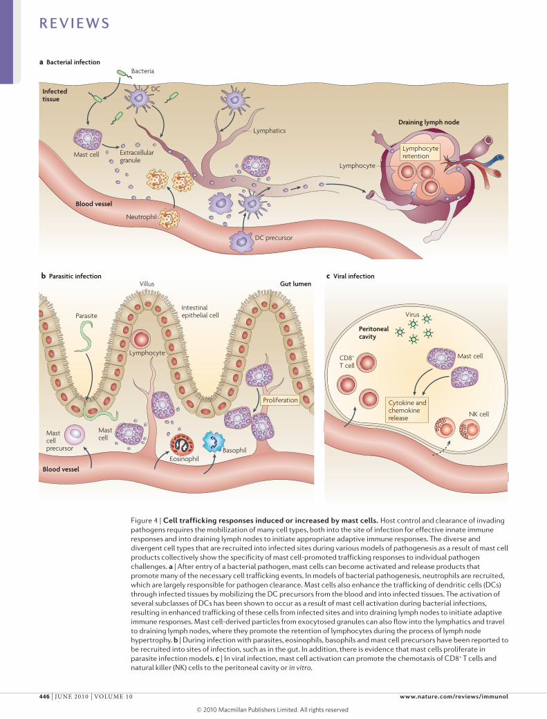
Figure 4 | Cell trafficking responses induced or increased by mast cells. Host control and clearance of invading pathogens requires the mobilization of many cell types, both into the site of infection for effective innate immune responses and into draining lymph nodes to initiate appropriate adaptive immune responses. The diverse and divergent cell types that are recruited into infected sites during various models of pathogenesis as a result of mast cell products collectively show the specificity of mast cell-promoted trafficking responses to individual pathogen challenges. a | After entry of a bacterial pathogen, mast cells can become activated and release products that promote many of the necessary cell trafficking events. In models of bacterial pathogenesis, neutrophils are recruited, which are largely responsible for pathogen clearance. Mast cells also enhance the trafficking of dendritic cells (DCs) through infected tissues by mobilizing the DC precursors from the blood and into infected tissues. The activation of several subclasses of DCs has been shown to occur as a result of mast cell activation during bacterial infections, resulting in enhanced trafficking of these cells from infected sites and into draining lymph nodes to initiate adaptive immune responses. Mast cell-derived particles from exocytosed granules can also flow into the lymphatics and travel to draining lymph nodes, where they promote the retention of lymphocytes during the process of lymph node hypertrophy. b | During infection with parasites, eosinophils, basophils and mast cell precursors have been reported to be recruited into sites of infection, such as in the gut. In addition, there is evidence that mast cells proliferate in parasite infection models. c | In viral infection, mast cell activation can promote the chemotaxis of CD8+ T cells and natural killer (NK) cells to the peritoneal cavity or in vitro.
R E V I E W S
446 | june 2010 | VoluMe 10 www.nature.com/reviews/immunol
© 20 Macmillan Publishers Limited. All rights reserved10
Nature Reviews | Immunology
Mast cell
Proliferation of mast cell (in response to IgE or SCF)
Adaptive immunityTriggering of innate receptors and degranulation
Sensitization of mast cell with pathogen-specificIgG or IgE
Local inflammatorycytokines such asIL-10 or IL-4 can alterpreformed mediators
• Released preformed mediators may vary from those in naive tissue• Antigen-specific Fc-mediated responses• Total mast cell numbers are increased during a secondary or chronic challenge
High-affinityantibodies
Granule
New granule
T cell B cell Plasma cell
Pathogen
FcεRI
FcγR
IgE
IgG
Pathogen-specific immunity with increased specificity, kinetics and/or magnitude
that they have been visualized in vivo physically inter-acting with T cells7. However, the functional require-ment of MHC class II-dependent antigen presentation by mast cells has yet to be fully evaluated in vivo. By contrast, there is now evidence that mast cells function efficiently as APCs for MHC class I-restricted CD8+ T cells in vivo71. In this recent study, antigen-pulsed mast cells were shown to promote CD8+ T cell activa-tion, proliferation and production of T cell products such as Il-2 and granzyme B. This is the first report describing an important functional role for mast cells in
antigen presentation71 and, although shown in a model of autoimmunity, it raises the possibility that mast cells, acting as APCs, directly promote and modulate CD8+ T cell function during infection.
Together with local responses to pathogens, mast cells have long-distance and long-term effects in the host by modulating draining lymph nodes and promot-ing the development of adaptive immunity to pathogens. As mentioned, mast cells can also influence cell traffick-ing to draining lymph nodes (FIG. 4). In an E. coli infec-tion model, we showed that mast cell-derived TnF is required for normal lymph node hypertrophy, a crucial event in which draining lymph nodes double in size dur-ing the first 24 hours of infection owing to the retention of lymphocytes49. This process increases the probability that rare antigen-specific lymphocytes will be present in draining lymph nodes during the induction of adaptive immunity, probably improving the specificity of adap-tive responses. Together, these two processes of DC trafficking from or through infected tissues to lymph nodes and lymphocyte sequestration in draining lymph nodes should increase the magnitude and specificity of the adaptive immune response by ensuring that appreci-able peripheral antigen is shuttled to the draining lymph nodes and that, simultaneously, rare antigen-specific lymphocytes are retained there. We observed func-tional consequences of mast cell-promoted responses in enhanced humoral immunity to E. coli in wild-type mice compared with mast cell-deficient mice, including increased E. coli-specific antibody titres and protection after passive immunization49, although it is currently unclear if mast cells have direct effects on B cells or antibody production.
As discussed previously, and best characterized in models of allergy, mast cells can function as effectors of adaptive immunity through their ability to become sensitized by binding antibodies through FcRs7. After a primary response, in which high-affinity pathogen-specific antibodies are present, antibody cross-linking of mast cell FcRs can result in faster responses of greater magnitude, allowing mast cells to participate in the enhanced immunity that results after immunological memory formation (FIG. 5). This may be particularly important during infection with some viral pathogens against which degranulation may not occur or may not be an immediate response. At least during chronic para-site infections, this sensitization can be important for ongoing control of latent infections7,22,72, indicating that mast cells can translate a functional antibody response into protection against a pathogenic challenge.
Conversely, T cells can modulate mast cells, par-ticularly through the production of chemokines such as CCl3 (also known as MIP1α) and CCl2, which probably contribute to mast cell degranulation, as well as through physical contact between mast cells and T cells73. In vitro, mast cell production of TnF and release of histamine can be enhanced by contact with T cells73, suggesting that another feedback mechanism exists, by which the adaptive immune system might regulate mast cell function during an ongoing inflammatory process or infection.
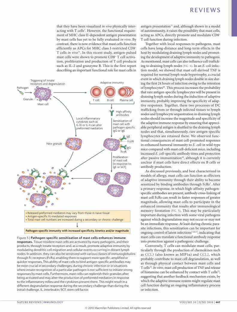
Figure 5 | Pathogen-specific sensitization of mast cells enhances immune responses. Tissue-resident mast cells are activated by many pathogens, and their products, through innate receptors and, as a result, promote adaptive immunity by modulating dendritic cell migration and cellular events occurring in distant lymph nodes. In addition, they can also be sensitized with various classes of immunoglobulins through Fc receptors (FcRs), enabling them to support more specific, amplified or quicker responses. This ability of mast cells to bind antigen-specific antibodies may be most crucial in secondary challenges, during chronic infection or in situations where innate recognition of a particular pathogen is not sufficient to initiate strong responses by mast cells. Furthermore, mast cells can replenish their granules after being activated and may alter the production of preformed mediators in response to the inflammatory milieu and the cytokines present there. This might result in a different degranulation response during the secondary challenge than during the initial challenge. IL, interleukin; SCF, stem cell factor.
R E V I E W S
nATuRe ReVIeWs | IMMunology VoluMe 10 | june 2010 | 447
© 20 Macmillan Publishers Limited. All rights reserved10
Pathogen-specific functional responsesThe initial events following infection are key to con-taining rapidly replicating or host-adapted pathogens. Any delay in the initiation of antimicrobial efforts could potentially shift the advantage from the host to the patho gen, resulting in morbidity or mortality. Mast cells have been shown to initiate pathogen-specific pro-grammes to functionally contribute to pathogen clear-ance and, in some experimental models, they have been shown to promote the survival of the host during an infectious challenge48,74 (TABLe 1).
Parasites. The first observations that mast cells could have a role in the control of infection came from the use of models of helminth infection in the gut2,3. In these early studies, mast cells were observed to cluster around sites with parasites, although it was unclear at the time if these cells were proliferating or newly recruited, and they displayed an activated phenotype, with many cells undergoing degranulation2. It is now clear that the control or clearance of parasites by mast cells involves various mechanisms, including the recruitment of key immune cells, regulation of gut permeability and parasite expulsion, and containment of chronic infection22,72,75–77. Proliferation of mast cells during gut helminth infection was observed in one model to depend on the key mast cell growth factor stem cell factor (sCF; also known as KIT ligand)78, and in another model to depend on Ige79. studies of the requirement for mast cells during parasite infections have established that responses to pathogens vary greatly depending on the type of challenge. For example, a recent study examining hookworm infection indicated that expulsion of the parasite from the gut dur-ing a secondary challenge depended on basophils rather than mast cells, in contrast to expulsion during a primary challenge, which occurred much more quickly in mast cell-sufficient mice than their deficient counterparts76. In the case of infection with the parasite Trichinella spiralis, it was shown (using Ige-deficient mice) that Ige production by the host contributed to parasite expulsion from the gut22. In this study, the authors suggested that decreased levels of MCP1, previously shown to influ-ence the speed of parasite expulsion from the gut, might explain these observations22. Mast cells also seem to be crucial for cutaneous immunity during the parasitic skin infection leishmaniasis, promoting protective immunity, including T cell function, and resulting in decreased skin lesion size77. The distinct functional roles of mast cells in various tissues are shown by these examples; however, the fundamental differences between responses require further characterization.
Bacteria. Mast cells are clearly essential for initiating both innate and adaptive immune responses to many bacterial pathogens and products, and for protecting the host from lethal infection48,74. several of the earli-est studies showed this by determining that mice defi-cient in mast cells show increased mortality after E. coli injection into the peritoneum than their wild-type counterparts48,74. In these peritonitis models, mast cells are functionally important for initiating innate immune
responses to enterobacteria, particularly through their ability to recruit neutrophils to promote bacterial clear-ance48. These observations were reinforced by a recent study examining peritonitis caused by caecal ligation, however, during the most experimentally severe con-ditions, the mast cells in this study were no longer protective and the TnF they produced contributed to pathology80. Mast cells are crucial for containing bac-terial infection and preventing dissemination in other tissues, and have been shown, for example, to promote the clearance of E. coli from the peritoneum and blad-der48,49 and K. pneumoniae from the lungs48,52, as well as to limit skin lesions during Pseudomonas aeruginosa infection81. Based on the studies discussed above and oth-ers, the strongest functional evidence for the necessity of mast cell function to survival comes from studies involv-ing bacterial challenges. Indirect activation of mast cells by host endogenous proteins can also highly influence the host response during bacterial infection, as shown by the contribution of mast cells to fluid production and neutrophil influx in the gut in response to neuron pro-duction of substance P, which occurs in the presence of toxin A from Clostridium difficile82.
Viruses. The role of mast cells in viral infections is more enigmatic and has been less well studied. Many viral products can activate mast cells, in particular to pro-mote cytokine production; however, the extent of mast cell degranulation in response to viral infection and any resulting functional implications are less clear. In sup-port of direct recognition of viruses by mast cells, it has been shown that histamine release can occur in response to sendai virus83 and that the gp120 envelope protein of HIV can induce cytokine production by mast cells84. The ability of mast cells to promote the recruitment of CD8+ T cells during viral challenge50 (as previously dis-cussed) or promote the production of type I interfer-ons85 suggests that mast cell recognition of virus may promote the types of cell-mediated responses that are associated with the clearance of intracellular viral infec-tion. In both rodents and humans, mast cell numbers in the lungs are higher than uninfected controls following respiratory viral infections86,87. However, the interaction between viruses and mast cells does not always clearly favour the host. HIV, for example, is known to infect mast cells, and these cells may constitute a reservoir for latent virus within the host88. other viruses that seem to infect mast cells include respiratory syncytial virus and dengue virus89,90; however, these studies were carried out in neoplastic cell lines and the consequences of these interactions remain to be explored in vivo.
Targeting mast cells in vaccinesMany parallels exist between the functions of mast cells in vivo, in particular their role in enhancing adaptive immune responses, and our requirements for efficient vaccine adjuvants. Thus, it was hypothesized that acti-vation of mast cells during vaccination could poten-tially promote protective immunity. It has now been shown that addition of a mast cell activator, compound 48/80, to vaccine formulations can result in increased
R E V I E W S
448 | june 2010 | VoluMe 10 www.nature.com/reviews/immunol
© 20 Macmillan Publishers Limited. All rights reserved10
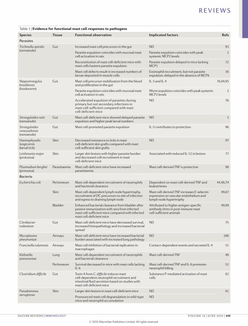
Table 1 | Evidence for functional mast cell responses to pathogens
Species Tissue Functional observation Implicated factors Refs
Parasites
Trichinella spiralis (nematode)
Gut Increased mast cell precursors in the gut ND 93
Parasite expulsion coincides with mucosal mast cell activation in rats
Parasite expulsion coincides with peak systemic MCP2 levels
2
Reconstitution of mast cell-deficient mice with mast cells hastens parasite expulsion
Parasite expulsion delayed in mice lacking MCP1
72
Mast cell defects result in increased numbers of larvae deposited in muscle cells
Eosinophil recruitment, but not parasite expulsion, delayed in the absence of MCP6
36
Nippostrongylus brasiliensis (hookworm)
Gut Mast cell precursor mobilization from the blood and proliferation in the gut
IL-3 and IL-4 76,94,95
Parasite expulsion coincides with mucosal mast cell activation in rats
Worm expulsion coincides with peak systemic MCP2 levels
2
Accelerated expulsion of parasites during primary but not secondary infections in mast cell-sufficient compared with mast cell-deficient mice
ND 76
Strongyloides ratti (nematode)
Gut Mast cell-deficient mice showed delayed parasite expulsion and higher peak larval numbers
ND 3
Strongyloides venezuelensis (nematode)
Gut Mast cell-promoted parasite expulsion IL-3 contributes to protection 96
Haemaphysalis longicornis (larval tick)
Skin Decreased resistance to ticks in mast cell-deficient skin grafts compared with mast cell-sufficient skin grafts
ND 97
Leishmania major (protozoa)
Skin Larger skin lesions with higher parasite burden and decreased cell recruitment in mast cell-deficient mice
Associated with reduced IL-12 in lesions 77
Plasmodium berghei (protozoa)
Parasitaemia Mast cell-deficient mice have increased parasitaemia
Mast cell-derived TNF is protective 98
Bacteria
Escherichia coli Peritoneum Mast cell-dependent recruitment of neutrophils and bacterial clearance
Dependent on mast cell-derived TNF and leukotrienes
44,48,74
Skin Mast cell-dependent lymph node hypertrophy, recruitment of DC precursors to site of infection and egress to draining lymph node
Mast cell-derived TNF increases E-selectin expression on vascular endothelium and lymph node hypertrophy
49,67
Bladder Enhanced bacterial clearance from bladder after passive immunization with sera from infected mast cell-sufficient mice compared with infected mast cell-deficient mice
Attributed to higher antigen-specific antibody titres in post-immune mast cell-sufficient animals
49,99
Citrobacter rodentium
Gut Mast cell-deficient mice have decreased survival, increased histopathology and increased bacterial spread
ND 75
Mycoplasma pneumoniae
Airways Mast cell-deficient mice have increased bacterial burden associated with increased lung pathology
ND 100
Francisella tularensis Airways Mast cell inhibition of bacterial replication in macrophages
Contact-dependent events and secreted IL-4 51
Klebsiella pneumoniae
Lung Mast cell-dependent recruitment of neutrophils and bacterial clearance
Mast cell-derived TNF 48
Peritoneum Survival decreased in mice with mast cells lacking IL-6
Mast cell-derived TNF and IL-6 promotes neutrophil killing
52
Clostridium difficile Gut Toxin A from C. difficile induces mast cell-dependent neutrophil recruitment and intestinal fluid secretion based on studies with mast cell-deficient mice
Substance P-mediated activation of mast cells
82
Pseudomonas aeruginosa
Skin Larger skin lesions in mast cell-deficient mice ND 81
Pronounced mast cell degranulation in wild-type mice and neutrophil accumulation
ND
R E V I E W S
nATuRe ReVIeWs | IMMunology VoluMe 10 | june 2010 | 449
© 20 Macmillan Publishers Limited. All rights reserved10
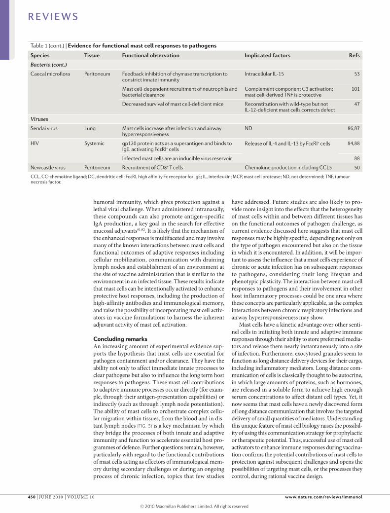
Table 1 (cont.) | Evidence for functional mast cell responses to pathogens
Species Tissue Functional observation Implicated factors Refs
Bacteria (cont.)
Caecal microflora Peritoneum Feedback inhibition of chymase transcription to constrict innate immunity
Intracellular IL-15 53
Mast cell-dependent recruitment of neutrophils and bacterial clearance
Complement component C3 activation; mast cell-derived TNF is protective
101
Decreased survival of mast cell-deficient mice Reconstitution with wild-type but not IL-12-deficient mast cells corrects defect
47
Viruses
Sendai virus Lung Mast cells increase after infection and airway hyperresponsiveness
ND 86,87
HIV Systemic gp120 protein acts as a superantigen and binds to IgE, activating FcεRI+ cells
Release of IL-4 and IL-13 by FcεRI+ cells 84,88
Infected mast cells are an inducible virus reservoir 88
Newcastle virus Peritoneum Recruitment of CD8+ T cells Chemokine production including CCL5 50
CCL, CC-chemokine ligand; DC, dendritic cell; FcεRI, high affinity Fc receptor for IgE; IL, interleukin; MCP, mast cell protease; ND, not determined; TNF, tumour necrosis factor.
humoral immunity, which gives protection against a lethal viral challenge. When administered intranasally, these compounds can also promote antigen-specific IgA production, a key goal in the search for effective mucosal adjuvants91,92. It is likely that the mechanism of the enhanced responses is multifaceted and may involve many of the known interactions between mast cells and functional outcomes of adaptive responses including cell ular mobilization, communication with draining lymph nodes and establishment of an environment at the site of vaccine administration that is similar to the environment in an infected tissue. These results indicate that mast cells can be intentionally activated to enhance protective host responses, including the production of high-affinity antibodies and immunological memory, and raise the possibility of incorporating mast cell activ-ators in vaccine formulations to harness the inherent adjuvant activity of mast cell activation.
Concluding remarks An increasing amount of experimental evidence sup-ports the hypothesis that mast cells are essential for pathogen containment and/or clearance. They have the ability not only to affect immediate innate processes to clear pathogens but also to influence the long term host responses to pathogens. These mast cell contributions to adaptive immune processes occur directly (for exam-ple, through their antigen-presentation capabilities) or indirectly (such as through lymph node potentiation). The ability of mast cells to orchestrate complex cellu-lar migration within tissues, from the blood and in dis-tant lymph nodes (FIG. 3) is a key mechanism by which they bridge the processes of both innate and adaptive immunity and function to accelerate essential host pro-grammes of defence. Further questions remain, however, particularly with regard to the functional contributions of mast cells acting as effectors of immunological mem-ory during secondary challenges or during an ongoing process of chronic infection, topics that few studies
have addressed. Future studies are also likely to pro-vide more insight into the effects that the hetero geneity of mast cells within and between different tissues has on the functional outcomes of pathogen challenge, as current evidence discussed here suggests that mast cell responses may be highly specific, depending not only on the type of pathogen encountered but also on the tissue in which it is encountered. In addition, it will be impor-tant to assess the influence that a mast cell’s experience of chronic or acute infection has on subsequent responses to pathogens, considering their long lifespan and pheno typic plasticity. The interaction between mast cell responses to pathogens and their involvement in other host inflammatory processes could be one area where these concepts are particularly applicable, as the complex interactions between chronic respiratory infections and airway hyperresponsiveness may show.
Mast cells have a kinetic advantage over other senti-nel cells in initiating both innate and adaptive immune responses through their ability to store preformed media-tors and release them nearly instantaneously into a site of infection. Furthermore, exocytosed granules seem to function as long distance delivery devices for their cargo, including inflammatory mediators. long distance com-munication of cells is classically thought to be autocrine, in which large amounts of proteins, such as hormones, are released in a soluble form to achieve high enough serum concentrations to affect distant cell types. Yet, it now seems that mast cells have a newly discovered form of long distance communication that involves the targeted delivery of small quantities of mediators. understanding this unique feature of mast cell biology raises the possibil-ity of using this communication strategy for prophylactic or therapeutic potential. Thus, successful use of mast cell activators to enhance immune responses during vaccina-tion confirms the potential contributions of mast cells to protection against subsequent challenges and opens the possibilities of targeting mast cells, or the processes they control, during rational vaccine design.
R E V I E W S
450 | june 2010 | VoluMe 10 www.nature.com/reviews/immunol
© 20 Macmillan Publishers Limited. All rights reserved10
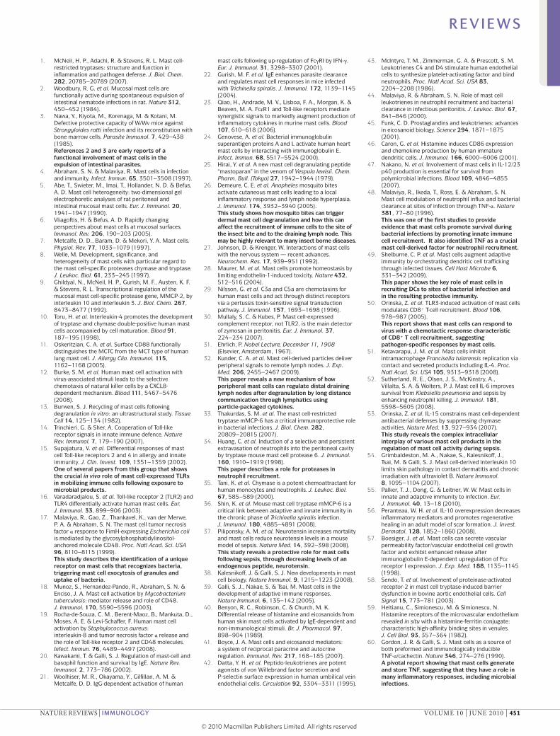
1. McNeil, H. P., Adachi, R. & Stevens, R. L. Mast cell-restricted tryptases: structure and function in inflammation and pathogen defense. J. Biol. Chem. 282, 20785–20789 (2007).
2. Woodbury, R. G. et al. Mucosal mast cells are functionally active during spontaneous expulsion of intestinal nematode infections in rat. Nature 312, 450–452 (1984).
3. Nawa, Y., Kiyota, M., Korenaga, M. & Kotani, M. Defective protective capacity of W/Wv mice against Strongyloides ratti infection and its reconstitution with bone marrow cells. Parasite Immunol. 7, 429–438 (1985).References 2 and 3 are early reports of a functional involvement of mast cells in the expulsion of intestinal parasites.
4. Abraham, S. N. & Malaviya, R. Mast cells in infection and immunity. Infect. Immun. 65, 3501–3508 (1997).
5. Abe, T., Swieter, M., Imai, T., Hollander, N. D. & Befus, A. D. Mast cell heterogeneity: two-dimensional gel electrophoretic analyses of rat peritoneal and intestinal mucosal mast cells. Eur. J. Immunol. 20, 1941–1947 (1990).
6. Vliagoftis, H. & Befus, A. D. Rapidly changing perspectives about mast cells at mucosal surfaces. Immunol. Rev. 206, 190–203 (2005).
7. Metcalfe, D. D., Baram, D. & Mekori, Y. A. Mast cells. Physiol. Rev. 77, 1033–1079 (1997).
8. Welle, M. Development, significance, and heterogeneity of mast cells with particular regard to the mast cell-specific proteases chymase and tryptase. J. Leukoc. Biol. 61, 233–245 (1997).
9. Ghildyal, N., McNeil, H. P., Gurish, M. F., Austen, K. F. & Stevens, R. L. Transcriptional regulation of the mucosal mast cell-specific protease gene, MMCP-2, by interleukin 10 and interleukin 3. J. Biol. Chem. 267, 8473–8477 (1992).
10. Toru, H. et al. Interleukin-4 promotes the development of tryptase and chymase double-positive human mast cells accompanied by cell maturation. Blood 91, 187–195 (1998).
11. Oskeritzian, C. A. et al. Surface CD88 functionally distinguishes the MCTC from the MCT type of human lung mast cell. J. Allergy Clin. Immunol. 115, 1162–1168 (2005).
12. Burke, S. M. et al. Human mast cell activation with virus-associated stimuli leads to the selective chemotaxis of natural killer cells by a CXCL8-dependent mechanism. Blood 111, 5467–5476 (2008).
13. Burwen, S. J. Recycling of mast cells following degranulation in vitro: an ultrastructural study. Tissue Cell 14, 125–134 (1982).
14. Trinchieri, G. & Sher, A. Cooperation of Toll-like receptor signals in innate immune defence. Nature Rev. Immunol. 7, 179–190 (2007).
15. Supajatura, V. et al. Differential responses of mast cell Toll-like receptors 2 and 4 in allergy and innate immunity. J. Clin. Invest. 109, 1351–1359 (2002).One of several papers from this group that shows the crucial in vivo role of mast cell-expressed TLRs in mobilizing immune cells following exposure to microbial products.
16. Varadaradjalou, S. et al. Toll-like receptor 2 (TLR2) and TLR4 differentially activate human mast cells. Eur. J. Immunol. 33, 899–906 (2003).
17. Malaviya, R., Gao, Z., Thankavel, K., van der Merwe, P. A. & Abraham, S. N. The mast cell tumor necrosis factor α response to FimH-expressing Escherichia coli is mediated by the glycosylphosphatidylinositol-anchored molecule CD48. Proc. Natl Acad. Sci. USA 96, 8110–8115 (1999).This study describes the identification of a unique receptor on mast cells that recognizes bacteria, triggering mast cell exocytosis of granules and uptake of bacteria.
18. Munoz, S., Hernandez-Pando, R., Abraham, S. N. & Enciso, J. A. Mast cell activation by Mycobacterium tuberculosis: mediator release and role of CD48. J. Immunol. 170, 5590–5596 (2003).
19. Rocha-de-Souza, C. M., Berent-Maoz, B., Mankuta, D., Moses, A. E. & Levi-Schaffer, F. Human mast cell activation by Staphylococcus aureus: interleukin-8 and tumor necrosis factor α release and the role of Toll-like receptor 2 and CD48 molecules. Infect. Immun. 76, 4489–4497 (2008).
20. Kawakami, T. & Galli, S. J. Regulation of mast-cell and basophil function and survival by IgE. Nature Rev. Immunol. 2, 773–786 (2002).
21. Woolhiser, M. R., Okayama, Y., Gilfillan, A. M. & Metcalfe, D. D. IgG-dependent activation of human
mast cells following up-regulation of FcγRI by IFN-γ. Eur. J. Immunol. 31, 3298–3307 (2001).
22. Gurish, M. F. et al. IgE enhances parasite clearance and regulates mast cell responses in mice infected with Trichinella spiralis. J. Immunol. 172, 1139–1145 (2004).
23. Qiao, H., Andrade, M. V., Lisboa, F. A., Morgan, K. & Beaven, M. A. FcεR1 and Toll-like receptors mediate synergistic signals to markedly augment production of inflammatory cytokines in murine mast cells. Blood 107, 610–618 (2006).
24. Genovese, A. et al. Bacterial immunoglobulin superantigen proteins A and L activate human heart mast cells by interacting with immunoglobulin E. Infect. Immun. 68, 5517–5524 (2000).
25. Hirai, Y. et al. A new mast cell degranulating peptide “mastoparan” in the venom of Vespula lewisii. Chem. Pharm. Bull. (Tokyo) 27, 1942–1944 (1979).
26. Demeure, C. E. et al. Anopheles mosquito bites activate cutaneous mast cells leading to a local inflammatory response and lymph node hyperplasia. J. Immunol. 174, 3932–3940 (2005).This study shows how mosquito bites can trigger dermal mast cell degranulation and how this can affect the recruitment of immune cells to the site of the insect bite and to the draining lymph node. This may be highly relevant to many insect borne diseases.
27. Johnson, D. & Krenger, W. Interactions of mast cells with the nervous system — recent advances. Neurochem. Res. 17, 939–951 (1992).
28. Maurer, M. et al. Mast cells promote homeostasis by limiting endothelin-1-induced toxicity. Nature 432, 512–516 (2004).
29. Nilsson, G. et al. C3a and C5a are chemotaxins for human mast cells and act through distinct receptors via a pertussis toxin-sensitive signal transduction pathway. J. Immunol. 157, 1693–1698 (1996).
30. Mullaly, S. C. & Kubes, P. Mast cell-expressed complement receptor, not TLR2, is the main detector of zymosan in peritonitis. Eur. J. Immunol. 37, 224–234 (2007).
31. Ehrlich, P. Nobel Lecture, December 11, 1908 (Elsevier, Amsterdam, 1967).
32. Kunder, C. A. et al. Mast cell-derived particles deliver peripheral signals to remote lymph nodes. J. Exp. Med. 206, 2455–2467 (2009).This paper reveals a new mechanism of how peripheral mast cells can regulate distal draining lymph nodes after degranulation by long distance communication through lymphatics using particle-packaged cytokines.
33. Thakurdas, S. M. et al. The mast cell-restricted tryptase mMCP-6 has a critical immunoprotective role in bacterial infections. J. Biol. Chem. 282, 20809–20815 (2007).
34. Huang, C. et al. Induction of a selective and persistent extravasation of neutrophils into the peritoneal cavity by tryptase mouse mast cell protease 6. J. Immunol. 160, 1910–1919 (1998).This paper describes a role for proteases in neutrophil recruitment.
35. Tani, K. et al. Chymase is a potent chemoattractant for human monocytes and neutrophils. J. Leukoc. Biol. 67, 585–589 (2000).
36. Shin, K. et al. Mouse mast cell tryptase mMCP-6 is a critical link between adaptive and innate immunity in the chronic phase of Trichinella spiralis infection. J. Immunol. 180, 4885–4891 (2008).
37. Piliponsky, A. M. et al. Neurotensin increases mortality and mast cells reduce neurotensin levels in a mouse model of sepsis. Nature Med. 14, 392–398 (2008).This study reveals a protective role for mast cells following sepsis, through decreasing levels of an endogenous peptide, neurotensin.
38. Kalesnikoff, J. & Galli, S. J. New developments in mast cell biology. Nature Immunol. 9, 1215–1223 (2008).
39. Galli, S. J., Nakae, S. & Tsai, M. Mast cells in the development of adaptive immune responses. Nature Immunol. 6, 135–142 (2005).
40. Benyon, R. C., Robinson, C. & Church, M. K. Differential release of histamine and eicosanoids from human skin mast cells activated by IgE-dependent and non-immunological stimuli. Br. J. Pharmacol. 97, 898–904 (1989).
41. Boyce, J. A. Mast cells and eicosanoid mediators: a system of reciprocal paracrine and autocrine regulation. Immunol. Rev. 217, 168–185 (2007).
42. Datta, Y. H. et al. Peptido-leukotrienes are potent agonists of von Willebrand factor secretion and P-selectin surface expression in human umbilical vein endothelial cells. Circulation 92, 3304–3311 (1995).
43. McIntyre, T. M., Zimmerman, G. A. & Prescott, S. M. Leukotrienes C4 and D4 stimulate human endothelial cells to synthesize platelet-activating factor and bind neutrophils. Proc. Natl Acad. Sci. USA 83, 2204–2208 (1986).
44. Malaviya, R. & Abraham, S. N. Role of mast cell leukotrienes in neutrophil recruitment and bacterial clearance in infectious peritonitis. J. Leukoc. Biol. 67, 841–846 (2000).
45. Funk, C. D. Prostaglandins and leukotrienes: advances in eicosanoid biology. Science 294, 1871–1875 (2001).
46. Caron, G. et al. Histamine induces CD86 expression and chemokine production by human immature dendritic cells. J. Immunol. 166, 6000–6006 (2001).
47. Nakano, N. et al. Involvement of mast cells in IL-12/23 p40 production is essential for survival from polymicrobial infections. Blood 109, 4846–4855 (2007).
48. Malaviya, R., Ikeda, T., Ross, E. & Abraham, S. N. Mast cell modulation of neutrophil influx and bacterial clearance at sites of infection through TNF-α. Nature 381, 77–80 (1996).This was one of the first studies to provide evidence that mast cells promote survival during bacterial infections by promoting innate immune cell recruitment. It also identified TNF as a crucial mast cell-derived factor for neutrophil recruitment.
49. Shelburne, C. P. et al. Mast cells augment adaptive immunity by orchestrating dendritic cell trafficking through infected tissues. Cell Host Microbe 6, 331–342 (2009).This paper shows the key role of mast cells in recruiting DCs to sites of bacterial infection and in the resulting protective immunity.
50. Orinska, Z. et al. TLR3-induced activation of mast cells modulates CD8+ T-cell recruitment. Blood 106, 978–987 (2005).This report shows that mast cells can respond to virus with a chemotactic response characteristic of CD8+ T cell recruitment, suggesting pathogen-specific responses by mast cells.
51. Ketavarapu, J. M. et al. Mast cells inhibit intramacrophage Francisella tularensis replication via contact and secreted products including IL-4. Proc. Natl Acad. Sci. USA 105, 9313–9318 (2008).
52. Sutherland, R. E., Olsen, J. S., McKinstry, A., Villalta, S. A. & Wolters, P. J. Mast cell IL-6 improves survival from Klebsiella pneumonia and sepsis by enhancing neutrophil killing. J. Immunol. 181, 5598–5605 (2008).
53. Orinska, Z. et al. IL-15 constrains mast cell-dependent antibacterial defenses by suppressing chymase activities. Nature Med. 13, 927–934 (2007).This study reveals the complex intracellular interplay of various mast cell products in the regulation of mast cell activity during sepsis.
54. Grimbaldeston, M. A., Nakae, S., Kalesnikoff, J., Tsai, M. & Galli, S. J. Mast cell-derived interleukin 10 limits skin pathology in contact dermatitis and chronic irradiation with ultraviolet B. Nature Immunol. 8, 1095–1104 (2007).
55. Palker, T. J., Dong, G. & Leitner, W. W. Mast cells in innate and adaptive immunity to infection. Eur. J. Immunol. 40, 13–18 (2010).
56. Peranteau, W. H. et al. IL-10 overexpression decreases inflammatory mediators and promotes regenerative healing in an adult model of scar formation. J. Invest. Dermatol. 128, 1852–1860 (2008).
57. Boesiger, J. et al. Mast cells can secrete vascular permeability factor/vascular endothelial cell growth factor and exhibit enhanced release after immunoglobulin E-dependent upregulation of Fcε receptor I expression. J. Exp. Med. 188, 1135–1145 (1998).
58. Sendo, T. et al. Involvement of proteinase-activated receptor-2 in mast cell tryptase-induced barrier dysfunction in bovine aortic endothelial cells. Cell Signal 15, 773–781 (2003).
59. Heltianu, C., Simionescu, M. & Simionescu, N. Histamine receptors of the microvascular endothelium revealed in situ with a histamine-ferritin conjugate: characteristic high-affinity binding sites in venules. J. Cell Biol. 93, 357–364 (1982).
60. Gordon, J. R. & Galli, S. J. Mast cells as a source of both preformed and immunologically inducible TNF-α/cachectin. Nature 346, 274–276 (1990).A pivotal report showing that mast cells generate and store TNF, suggesting that they have a role in many inflammatory responses, including microbial infections.
R E V I E W S
nATuRe ReVIeWs | IMMunology VoluMe 10 | june 2010 | 451
© 20 Macmillan Publishers Limited. All rights reserved10
61. Biedermann, T. et al. Mast cells control neutrophil recruitment during T cell-mediated delayed-type hypersensitivity reactions through tumor necrosis factor and macrophage inflammatory protein 2. J. Exp. Med. 192, 1441–1452 (2000).
62. Di Nardo, A., Vitiello, A. & Gallo, R. L. Cutting edge: mast cell antimicrobial activity is mediated by expression of cathelicidin antimicrobial peptide. J. Immunol. 170, 2274–2278 (2003).This study shows for the first time that antimicrobial actions of mast cells could also be attributable to their expression of antimicrobial peptides.
63. Margulis, A. et al. Mast cell-dependent contraction of human airway smooth muscle cell-containing collagen gels: influence of cytokines, matrix metalloproteases, and serine proteases. J. Immunol. 183, 1739–1750 (2009).
64. Klimpel, G. R. et al. A role for stem cell factor and c-kit in the murine intestinal tract secretory response to cholera toxin. J. Exp. Med. 182, 1931–1942 (1995).
65. Pothoulakis, C., Castagliuolo, I. & LaMont, J. T. Nerves and intestinal mast cells modulate responses to enterotoxins. News Physiol. Sci. 13, 58–63 (1998).
66. Bischoff, S. C. Physiological and pathophysiological functions of intestinal mast cells. Semin. Immunopathol. 31, 185–205 (2009).
67. McLachlan, J. B. et al. Mast cell-derived tumor necrosis factor induces hypertrophy of draining lymph nodes during infection. Nature Immunol. 4, 1199–1205 (2003).This paper indicates that peripheral mast cells, and specifically their product TNF, activate distal draining lymph nodes promoting hypertrophy in response to infection.
68. Jawdat, D. M., Rowden, G. & Marshall, J. S. Mast cells have a pivotal role in TNF-independent lymph node hypertrophy and the mobilization of Langerhans cells in response to bacterial peptidoglycan. J. Immunol. 177, 1755–1762 (2006).This is the first demonstration that mast cells can mobilize a subset of DCs to draining lymph nodes in response to bacterial products.
69. Amaral, M. M. et al. Histamine improves antigen uptake and cross-presentation by dendritic cells. J. Immunol. 179, 3425–3433 (2007).
70. Mazzoni, A., Siraganian, R. P., Leifer, C. A. & Segal, D. M. Dendritic cell modulation by mast cells controls the Th1/Th2 balance in responding T cells. J. Immunol. 177, 3577–3581 (2006).
71. Stelekati, E. et al. Mast cell-mediated antigen presentation regulates CD8+ T cell effector functions. Immunity 31, 665–676 (2009).
72. Knight, P. A., Wright, S. H., Lawrence, C. E., Paterson, Y. Y. & Miller, H. R. Delayed expulsion of the nematode Trichinella spiralis in mice lacking the mucosal mast cell-specific granule chymase, mouse mast cell protease-1. J. Exp. Med. 192, 1849–1856 (2000).
73. Mekori, Y. A. & Metcalfe, D. D. Mast cell-T cell interactions. J. Allergy Clin. Immunol. 104, 517–523 (1999).
74. Echtenacher, B., Mannel, D. N. & Hultner, L. Critical protective role of mast cells in a model of acute septic peritonitis. Nature 381, 75–77 (1996).This study was one of the first to provide in vivo evidence of the crucial role of mast cells and TNF in promoting survival of the host during bacterial infections.
75. Wei, O. L., Hilliard, A., Kalman, D. & Sherman, M. Mast cells limit systemic bacterial dissemination but not colitis in response to Citrobacter rodentium. Infect. Immun. 73, 1978–1985 (2005).
76. Ohnmacht, C. & Voehringer, D. Basophils protect against reinfection with hookworms independently of mast cells and memory Th2 cells. J. Immunol. 184, 344–350 (2010).
77. Maurer, M. et al. Skin mast cells control T cell-dependent host defense in Leishmania major infections. FASEB J. 20, 2460–2467 (2006).
78. Newlands, G. F., Coulson, P. S. & Wilson, R. A. Stem cell factor dependent hyperplasia of mucosal-type mast cells but not eosinophils in Schistosoma mansoni-infected rats. Parasite Immunol. 17, 595–598 (1995).
79. Asai, K. et al. Regulation of mast cell survival by IgE. Immunity 14, 791–800 (2001).
80. Piliponsky, A. M. et al. Mast cell-derived TNF can exacerbate mortality during severe bacterial infections in C57BL/6–KitW-sh/W-sh mice. Am. J. Pathol. 176, 926–938 (2010).
81. Siebenhaar, F. et al. Control of Pseudomonas aeruginosa skin infections in mice is mast cell-dependent. Am. J. Pathol. 170, 1910–1916 (2007).
82. Wershil, B. K., Castagliuolo, I. & Pothoulakis, C. Direct evidence of mast cell involvement in Clostridium difficile toxin A-induced enteritis in mice. Gastroenterology 114, 956–964 (1998).
83. Sugiyama, K. Histamine release from rat mast cells induced by Sendai virus. Nature 270, 614–615 (1977).One of the early suggestions of a possible role for mast cells in modulating immune responses to viruses.
84. Patella, V., Florio, G., Petraroli, A. & Marone, G. HIV-1 gp120 induces IL-4 and IL-13 release from human FcεRI+ cells through interaction with the VH3 region of IgE. J. Immunol. 164, 589–595 (2000).
85. Kulka, M., Alexopoulou, L., Flavell, R. A. & Metcalfe, D. D. Activation of mast cells by double-stranded RNA: evidence for activation through Toll-like receptor 3. J. Allergy Clin. Immunol. 114, 174–182 (2004).
86. Castleman, W. L., Sorkness, R. L., Lemanske, R. F. Jr & McAllister, P. K. Viral bronchiolitis during early life induces increased numbers of bronchiolar mast cells and airway hyperresponsiveness. Am. J. Pathol. 137, 821–831 (1990).
87. Sorden, S. D. & Castleman, W. L. Virus-induced increases in bronchiolar mast cells in Brown Norway rats are associated with both local mast cell proliferation and increases in blood mast cell precursors. Lab. Invest. 73, 197–204 (1995).
88. Sundstrom, J. B. et al. Human tissue mast cells are an inducible reservoir of persistent HIV infection. Blood 109, 5293–5300 (2007).
89. Shirato, K. & Taguchi, F. Mast cell degranulation is induced by A549 airway epithelial cell infected with respiratory syncytial virus. Virology 386, 88–93 (2009).
90. Legrand, L. F., Hotta, H., Hotta, S. & Homma, M. Antibody-mediated enhancement of infection by dengue virus of the P815 murine mastocytoma cell line. Biken J. 29, 51–55 (1986).
91. McLachlan, J. B. et al. Mast cell activators: a new class of highly effective vaccine adjuvants. Nature Med. 14, 536–541 (2008).
The first demonstration of small molecular activators of mast cells as highly effective vaccine adjuvants.
92. McGowen, A. L., Hale, L. P., Shelburne, C. P., Abraham, S. N. & Staats, H. F. The mast cell activator compound 48/80 is safe and effective when used as an adjuvant for intradermal immunization with Bacillus anthracis protective antigen. Vaccine 27, 3544–3552 (2009).
93. Dillon, S. B. & MacDonald, T. T. Limit dilution analysis of mast cell precursor frequency in the gut epithelium of normal and Trichinella spiralis infected mice. Parasite Immunol. 8, 503–511 (1986).
94. Kasugai, T. et al. Infection with Nippostrongylus brasiliensis induces invasion of mast cell precursors from peripheral blood to small intestine. Blood 85, 1334–1340 (1995).
95. Madden, K. B. et al. Antibodies to IL-3 and IL-4 suppress helminth-induced intestinal mastocytosis. J. Immunol. 147, 1387–1391 (1991).
96. Lantz, C. S. et al. Role for interleukin-3 in mast-cell and basophil development and in immunity to parasites. Nature 392, 90–93 (1998).
97. Matsuda, H. et al. Necessity of IgE antibodies and mast cells for manifestation of resistance against larval Haemaphysalis longicornis ticks in mice. J. Immunol. 144, 259–262 (1990).
98. Furuta, T., Kikuchi, T., Iwakura, Y. & Watanabe, N. Protective roles of mast cells and mast cell-derived TNF in murine malaria. J. Immunol. 177, 3294–3302 (2006).This study provides the first in vivo evidence of a protective role for mast cells against a blood-borne parasite.
99. Malaviya, R., Ikeda, T. & Abraham, S. N. Contribution of mast cells to bacterial clearance and their proliferation during experimental cystitis induced by type 1 fimbriated E. coli. Immunol. Lett. 91, 103–111 (2004).
100. Xu, X. et al. Mast cells protect mice from Mycoplasma pneumonia. Am. J. Respir. Crit. Care Med. 173, 219–225 (2006).
AcknowledgementsThe authors’ work is supported by the US National Institutes of Health grants R01 AI35678, R01 DK077159, R01 AI50021, R37 DK50814 and R21 AI056101. We thank M. M. Ng and H. Yap of the National University of Singapore for their help in acquiring the image in FIG. 1a and Z. Swan for reviewing the manuscript.
Competing interests statement.The authors declare no competing financial interests.
DATABASESUniProtKB: http://www.uniprot.orgC5aR | CCL2 | CCL3 | CCL5 | CCL11 | CCL20 | CD48 | chymase | CXCL8 | CXCL10 | endothelin 1 | FcεRI | IL-1β | IL-3 | IL-4 | IL-5 | IL-6 | IL-10 | IL-13 | IL-15 | MCP1 | MCP2 | MCP6 | neurotensin | SCF | substance P | TLR2 | TLR3 | TLR4 | TNF
FURTHER INFORMATIONSoman N. Abraham’s homepage: http://pathology.mc.duke.edu/website/webform.aspx?id=GradAbraham
All lInkS ARe ACTIve In The onlIne PdF
R E V I E W S
452 | june 2010 | VoluMe 10 www.nature.com/reviews/immunol
© 20 Macmillan Publishers Limited. All rights reserved10