Massive’Obstetric’Hemorrhage ’’ The’Team’Approach’...
83
Massive Obstetric Hemorrhage The Team Approach RECC Update in Obstetric Anesthesia Wednesday June 15, 2011
Transcript of Massive’Obstetric’Hemorrhage ’’ The’Team’Approach’...

Massive Obstetric Hemorrhage The Team Approach
RECC -‐ Update in Obstetric Anesthesia
Wednesday June 15, 2011

DeclaraGons
• Dr. Kallie Honeywood – Kootenay Boundary Regional Hospital (Interior Health)
• Dr. Wendy MacLeod – Royal Columbian Hospital (Fraser Health)
• Dr. Louis Prinsloo – GPA, Northern Health Authority
• No disclosures or conflicts

Massive Obstetric Hemorrhage Goals
• DefiniGons • EGology, Epidemiology and Risk factors of OB Hemorrhage
• Team approach to Massive Hemorrhage – PrevenGon – Medical / Surgical/ IntervenGonal – Point of Care / Rapid Turnover TesGng
• Massive Transfusion Protocols
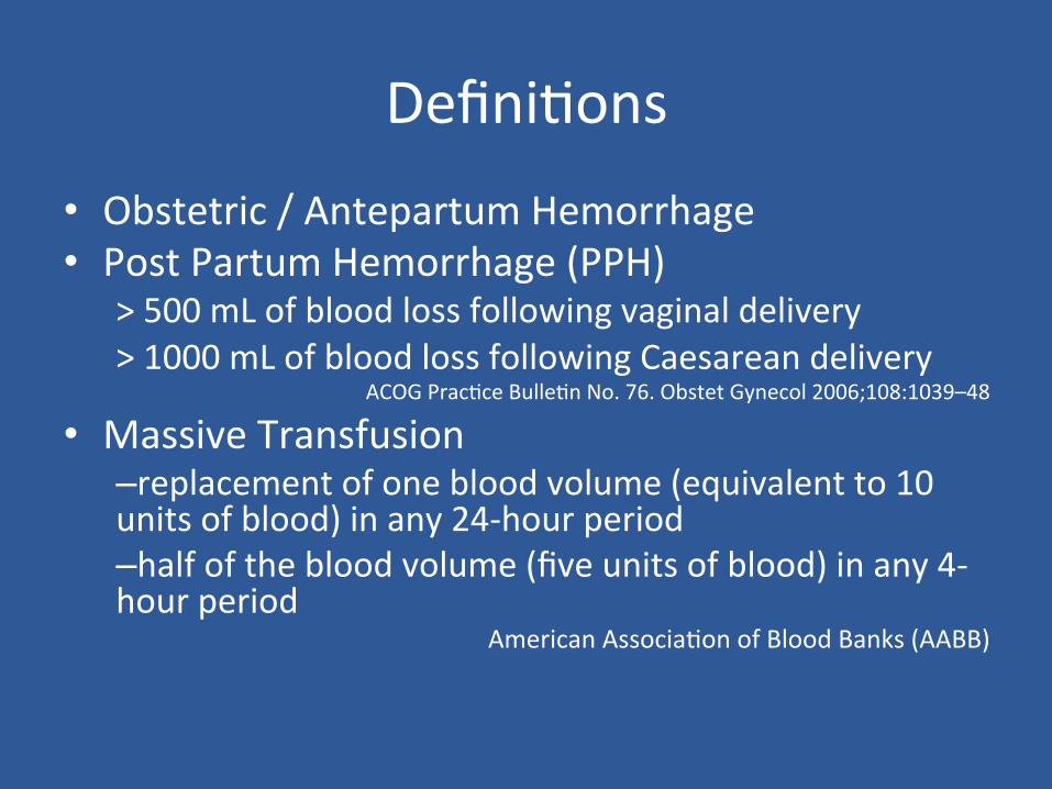
DefiniGons • Obstetric / Antepartum Hemorrhage • Post Partum Hemorrhage (PPH)
> 500 mL of blood loss following vaginal delivery > 1000 mL of blood loss following Caesarean delivery
ACOG PracGce BulleGn No. 76. Obstet Gynecol 2006;108:1039–48
• Massive Transfusion – replacement of one blood volume (equivalent to 10 units of blood) in any 24-‐hour period – half of the blood volume (five units of blood) in any 4-‐hour period
American AssociaGon of Blood Banks (AABB)

Antepartum Hemorrhage
• Incidence 2 – 5% of all pregnancies > 24 weeks – Unknown origin (~ 50%) – Placental AbrupGon – Placenta Previa – Abnormal PlacentaGon (Morbidly adherent) – Uterine Rupture
BJA 2009; 103(suppl): i47–i56

Placental AbrupGon
• SeparaGon of the placental bed prior to delivery of fetus
• Vaginal blood loss, uterine tenderness, increased uterine acGvity
• Coagulopathy in 10% • Coagulopathy increases to 50% when associated fetal demise
Anesthesiology Clin 2008;26:53-‐66

Placenta Previa
• Placenta implants in advance of the fetal presenGng part
• Painless vaginal bleeding, fetal compromise • Occurs in 0.5% of pregnancies • Usually associated with a previous uterine scar
Anesthesiology Clin 2008;26:53-‐66

AmnioGc Fluid Embolus
• Incidence 1:10,000 pregnancies • Rapid and severe coagulopathy • Coagulopathy worsened with fetal demise
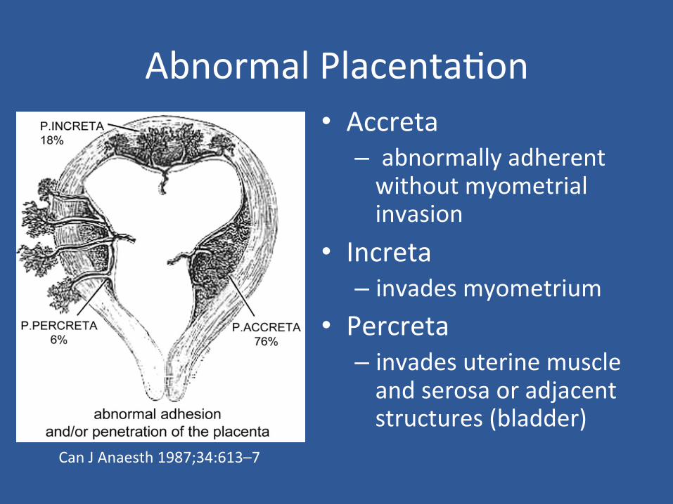
Abnormal PlacentaGon • Accreta – abnormally adherent without myometrial invasion
• Increta – invades myometrium
• Percreta – invades uterine muscle and serosa or adjacent structures (bladder)
Can J Anaesth 1987;34:613–7

Abnormal PlacentaGon • Incidence increasing with increasing C/S rates • ≥ 1 Risk Factor is present in 94% of cases – Placenta Previa – Previous Cesarean SecGon(s) – Previous Uterine Surgery
Eur J Obstet Gynecol Reprod Biol 1993;52:151–6
• > 90% require hysterectomy due to massive bleeding Obstet Gynecol 2010;115:65–9

Abnormal PlacentaGon
• Risk of Adherent Placenta increases when associated with Placenta Previa:
Unscarred uterus: 5% incidence Previous C/S: 10% incidence >1 C/S: > 50% incidence
• Ultrasound and MRI can be used for screening but they are poorly sensiGve
J Soc Gynecol InvesGg 2002;9:37–40.
Uterine Rupture
• Most significant risk factor is previous Cesarean SecGon
• Incidence 0.2% with previous uterine scar • Pain and bleeding are more common and severe with rupture of an unscarred uterus
• Scarred vs unscarred uterine ruptures have equivalent maternal-‐fetal morbidity
Am J Obstet Gynecol 2004;191:425–9

Postpartum Hemorrhage
• Uterine Atony • Retained Products of ConcepGon • Genital Tract Trauma • CoagulaGon AbnormaliGes

Epidemiology
• 2004 incidence of PPH (all cause) 2.93 per 100 deliveries in the USA
• Incidence of PPH (all cause) requiring transfusion 0.26 per 100 deliveries
• Uterine atony accounted for 79% of cases of PPH
• Uterine atony accounted for 0.19% of transfusions
Anesth Analg 2010;110:1368–73

Epidemiology
Figure 1. Trends in the rate of overall postpartum hemorrhage and hemorrhage by underlying eGology: 1995 to 2004.
Anesth Analg 2010;110:1368–73

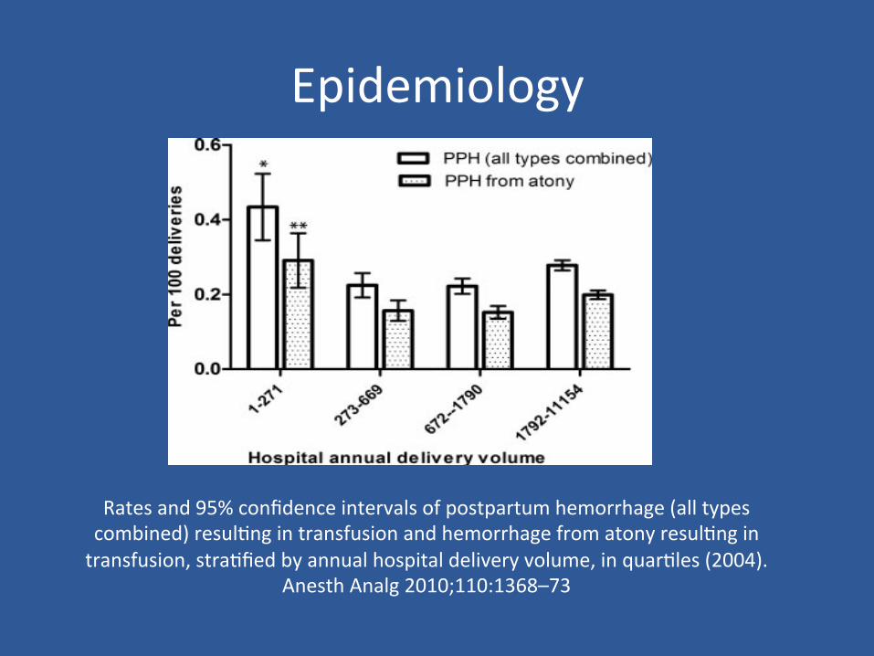
Epidemiology
Rates and 95% confidence intervals of postpartum hemorrhage (all types combined) and hemorrhage from atony, straGfied by annual hospital delivery volume, in quarGles (2004).
Anesth Analg 2010;110:1368–73
Epidemiology
Rates and 95% confidence intervals of postpartum hemorrhage (all types combined) resulGng in transfusion and hemorrhage from atony resulGng in transfusion, straGfied by annual hospital delivery volume, in quarGles (2004).
Anesth Analg 2010;110:1368–73

PPH Risk Factors
• Maternal age < 20 or >/= 40 • Caesarean delivery (+/-‐ labour) • Hypertensive disorders of pregnancy • Polyhydramnios • ChorioamnioniGs • MulGple GestaGon • Retained Placenta • Antepartum Hemorrhage

ComplicaGons of PPH
Outcome Odds Ra-o
Mortality 7.8
Renal Failure 13.8
Acute Respiratory Failure 10.9
Prolonged VenGlaGon 6.5
Coagulopathy 4.7
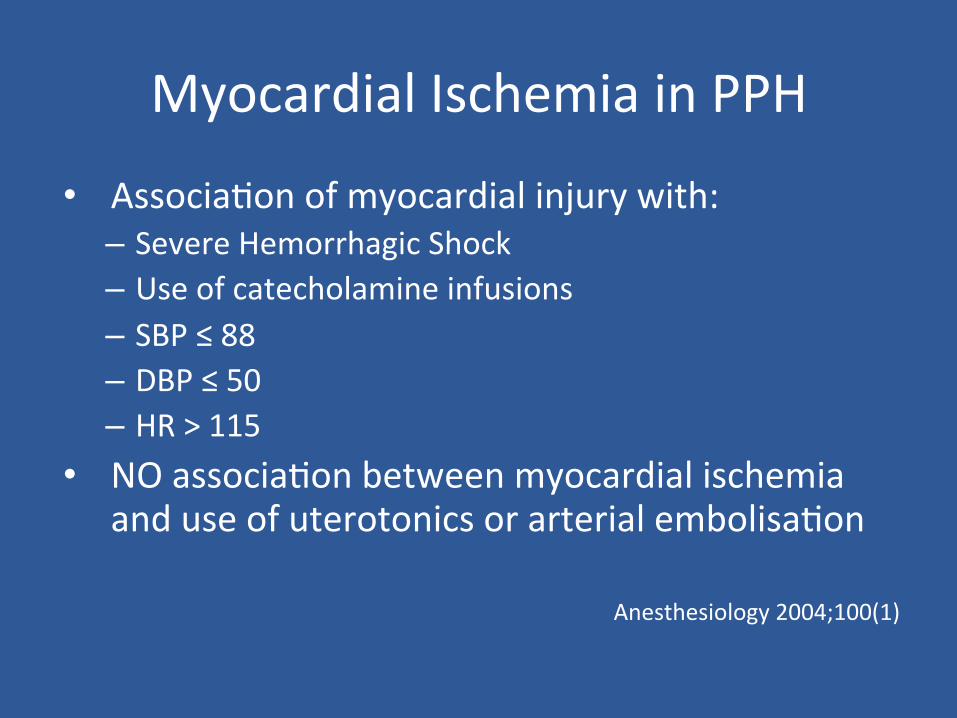
Myocardial Ischemia in PPH
• AssociaGon of myocardial injury with: – Severe Hemorrhagic Shock – Use of catecholamine infusions – SBP ≤ 88 – DBP ≤ 50 – HR > 115
• NO associaGon between myocardial ischemia and use of uterotonics or arterial embolisaGon
Anesthesiology 2004;100(1)

Summary-‐ PrevenGon
• There are many cases of OB Hemorrhage that we can try to idenGfy – Placenta Previa in the senng of a previous scar – Morbidly adherent placenta and screening – Known RF’s for PPH
• Good communicaGon between Primary Care, OB, Anesthesia, Surgeon locally
• Determine best plan and locaGon for delivery
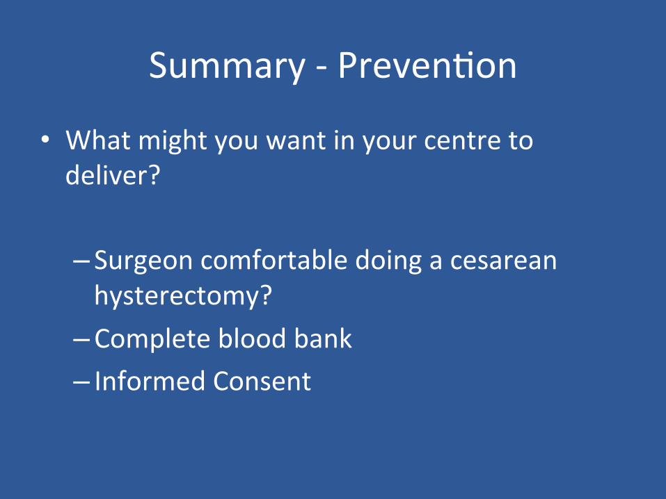
Summary -‐ PrevenGon
• What might you want in your centre to deliver?
– Surgeon comfortable doing a cesarean hysterectomy? – Complete blood bank – Informed Consent

Summary -‐ Management
• Many cases of OB Hemorrhage that can not be predicted
• Maternal morbidity and mortality associated with Massive Hemorrhage
• Treatment must be iniGated locally

How to plan for a Major Obstetrical Hemorrhage?
• Pre-‐determined, pracGcal definiGons to trigger the prompt diagnosis
• Simultaneous and mulGdisciplinary management
• Pre-‐planned step-‐ management protocol for management of massive hemorrhage
Anesthesiology Clin 2008;26:53-‐66

Massive Obstetric Hemorrhage The Team Approach
RECC -‐ Update in Obstetric Anesthesia

Team Approach to the Management of Massive Obstetric Hemorrhage
• Goals • PrevenGon and Team approach
• Management – Medical – Surgical/Invasive/IntervenGonal – ResuscitaGon and Transfusion Treatment
• Coagulopathy • Factor VIIa • Rapid turnover TesGng/Point of care tesGng

Goals of Management
Minimize morbidity and mortality • Developing a coordinated, mulGdisciplinary approach
• EffecGve Guidelines • Maternal Hemorrhage protocol • Massive Transfusion protocol
• Prompt recogniGon and response to Hemorrhage
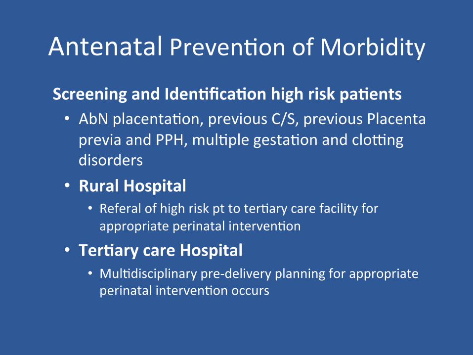
Antenatal PrevenGon of Morbidity
Screening and Iden-fica-on high risk pa-ents • AbN placentaGon, previous C/S, previous Placenta previa and PPH, mulGple gestaGon and clonng disorders
• Rural Hospital • Referal of high risk pt to terGary care facility for appropriate perinatal intervenGon
• Ter-ary care Hospital • MulGdisciplinary pre-‐delivery planning for appropriate perinatal intervenGon occurs

Perinatal PrevenGon of PPH AcGve management of the third stage of labor be offered to all women during childbirth, to ê risk of PPH
Ac-ve management should include: 1. ProphylacGc uterotonic drugs (oxytocin) ater delivery 2. Early clamping of the cord once uterine contracGon noted 3. Delivery of the placenta by controlled cord tracGon, followed by uterine massage.

PrevenGon of Morbidity & Mortality
Co-‐ordinated MulGdisciplinary approach – Primary care, Obstetrics, Anesthesia, Blood Bank, Hematology/Lab as well as Referral centers
– Referral centers – Radiology, Urology and ICU
WHO Guidelines for management of PPH 2009
States New York, California and Illinois developed MHP

PrevenGon of Morbidity
EffecGve Guidelines: Maternal Hemorrhage Protocols
– predetermined,pracGcal definiGons designed – trigger prompt diagnosis + clear acGons – mulGdisciplinary management plan – avoids treatment delays
An Update on Maternal PaGent Safety Jill M. Mhyre, MDAnesth Analg 2010,vol X
Massive Transfusion protocol

Maternal Hemorrhage Protocols
Emphasizing the importance of: • effecGve, prompt communicaGon • ability to rapidly mobilize resources • prioriGze and delegate tasks • simultaneously assess and treat paGents • predetermined pracGcal referral guidelines for smaller centers
• prevenGon secondary injury

Prompt recogniGon and response
to Hemorrhage Most maternal morbidity in Massive Hemorrhage
• UnderesGmaGon of blood loss • Delay in intervenGon in face clinical signs • Inadequate volume replacement Major Obstetric Hemorrhage Mercier et al, Anesthesiology Clinics 2008
• DuraGon of down-‐&me directly related to poor outcome.

Management of Massive Maternal Hemorrhage
• Medical • Surgical/Invasive • IntervenGonal • ResuscitaGon and Transfusion Treatment

WHO Guidelines for Management of PPH
Evidence based stepwise algorithm
Medical 1.Uterine massage 2.Uterotonic drugs
-‐ Oxytocin -‐ Ergometrine -‐ Prostaglandin -‐ Tranexamic acid (if trauma)
Non-‐operaGve/OperaGve Mx 1.Bimanual uterine
compression 2.Intrauterine balloon
tamponade 3.Uterine artery embolizaGon 4.Uterine compression suture 5.Arterial ligaGon 6.Subtotal Hysterectomy
WHO guidelines for the management of postpartum haemorrhage; 2009
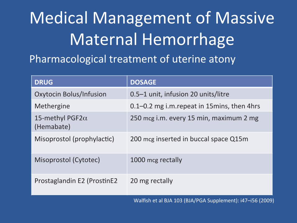
Medical Management of Massive Maternal Hemorrhage
Walfish et al BJA 103 (BJA/PGA Supplement): i47–i56 (2009)
DRUG DOSAGE
Oxytocin Bolus/Infusion 0.5–1 unit, infusion 20 units/litre
Methergine 0.1–0.2 mg i.m.repeat in 15mins, then 4hrs
15-‐methyl PGF2α (Hemabate)
250 mcg i.m. every 15 min, maximum 2 mg
Misoprostol (prophylacGc) 200 mcg inserted in buccal space Q15m
Misoprostol (Cytotec) 1000 mcg rectally
Prostaglandin E2 (ProsGnE2
20 mg rectally
Pharmacological treatment of uterine atony

Side Effects & Controversies • Oxytocin
• WHO guidelines -‐ no bolus • Bolus -‐ 10u significant hypotension (Svanstrom et al 2008) • Bolus -‐ 1u 100% response (Carvalho et al 2004
• Ergot alkaloids • α adrenergic receptors agonist cauGon
– Pre-‐eclampsia – hypertension – heart disease
• PGF2α • Hypertension, diarrhea, ébronchial tone • CauGon in asthma • Not for IV use
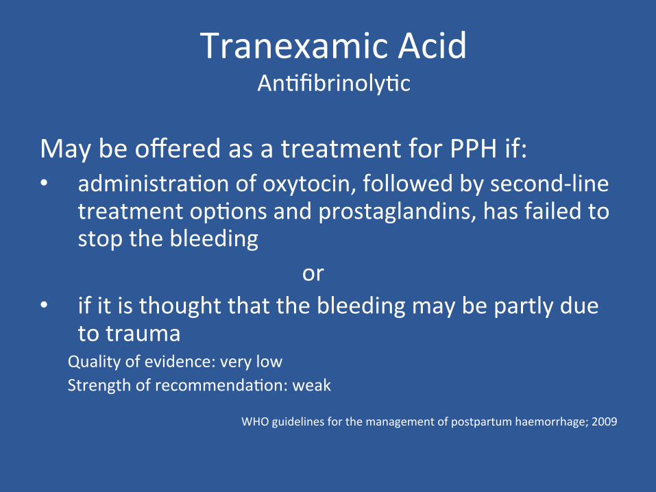
Tranexamic Acid AnGfibrinolyGc
May be offered as a treatment for PPH if: • administraGon of oxytocin, followed by second-‐line
treatment opGons and prostaglandins, has failed to stop the bleeding or
• if it is thought that the bleeding may be partly due to trauma
Quality of evidence: very low Strength of recommendaGon: weak
WHO guidelines for the management of postpartum haemorrhage; 2009

Invasive/Surgical Management
Success Rates of Invasive OpGons
• Intrauterine Balloon Tamponade 84% • Uterine Compression Sutures 92% • Arterial LigaGon 85% • Angiographic EmbolisaGon 91% • Hysterectomy
M. Walfish BriGsh Journal of Anaesthesia 103 (BJA/PGA Supplement): i47–i56 (2009)

Arterial LigaGon Uterine, Internal Iliac Arteries
• Useful when other measures failed • Requires laparotomy • Technically challenging • Time consuming • Significant complicaGons
• Incorrect vessel ligaGon • Ureteric injury
• Study of 103 paGents, found uterine devascularizaGon in face of uncontrolled hemorrhage, was 100% effecGve and hysterectomy was not needed in any case
AbdRabbo SA. Stepwise uterine devascularizaGon: a novel technique for management of uncontrollable
postpartum haemorrhage with preservaGon of the uterus. Am J Obstet Gynecol 1994;171: 694–700

Hysterectomy • EsGmated 0.8 per 1000 deliveries • DefiniGve treatment for PPH once alternaGve intervenGons failed – Subtotal hysterectomy recommended – Life saving – Offered early to those who refuse blood transfusions – Technically difficult risks include:
• Bladder and ureteric injury • Possibility é bleeding
Glaze S, Ekwalanga P, Roberts G, et al. Peripartum hysterectomy: 1999 to 2006. Obstet Gynecol 2008; 111: 732–8

IntervenGonal Management Requires fluoroscopic guidance and intervenGonal experGse
• Angiographic Balloon occlusion – ProphylacGc use in elecGve/semi urgent CS for abnormal placentaGon
• Angiographic EmbolisaGon – Uterine arteries are branches of Internal iliac arteries, (ovarian
arteries) – Requires stabilizaGon of paGent prior to transfer to facility with
intervenGonal radiology – Several studies have noted that clonng disorders improve rapidly
ater embolizaGon possibly due to the facilitaGon of uterine contracGon that leads to a secondary liberaGon of procoagulant factors into circulaGon
Deux JF,, et al. Is selecGve embolisaGon of uterine arteries a safe alternaGve to hysterectomy in paGents with postpartum haemorrhage? Am J Roentgenol 2001; 177: 145–9

Transfusion / ResuscitaGon Therapy
Caveats for the Pregnant PaGent • Blood loss almost always underesGmated • Masking of hemodynamic signs due to physiological adaptaGons of Pregnancy
• Pregnant paGent can lose up to 40% blood volume before showing any signs of hemodynamic instabilty (c/t 25% in non pregnant females)
• DO NOT DELAY TRANSFUSION WHILE AWAITING
• HEMODYNAMIC INSTABILTY • LAB RESULTS
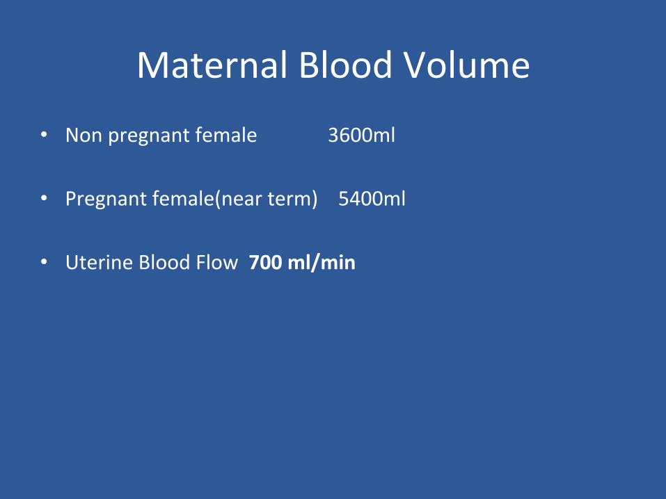
Maternal Blood Volume • Non pregnant female 3600ml
• Pregnant female(near term) 5400ml
• Uterine Blood Flow 700 ml/min
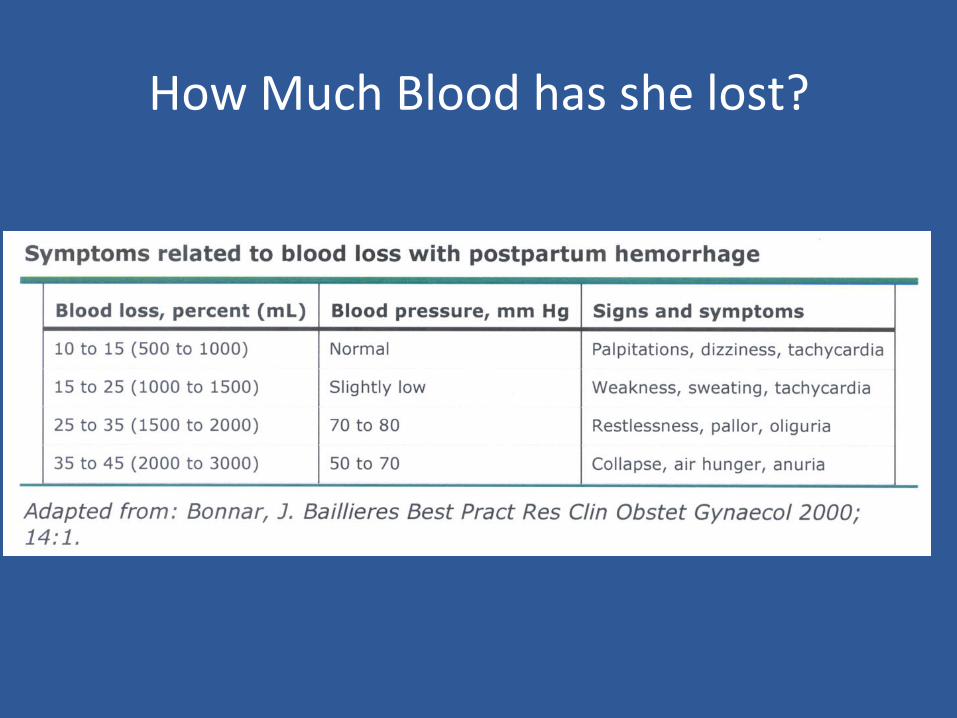
How Much Blood has she lost?
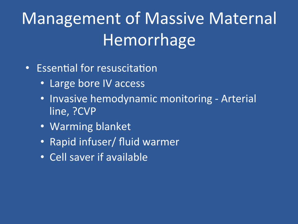
Management of Massive Maternal Hemorrhage
• EssenGal for resuscitaGon • Large bore IV access • Invasive hemodynamic monitoring -‐ Arterial line, ?CVP
• Warming blanket • Rapid infuser/ fluid warmer • Cell saver if available

Management of Massive Maternal Hemorrhage
• EssenGal for resuscitaGon • Warm crystalloids (+/-‐ colloids) unGl blood and blood products
available • Inotropic support if needed (should not replace adequate volume
resuscitaGon) • OpGmize medical and surgical Mx of bleeding • Prevent / correct coagulopathy • Targets
• Hb >80 – RBCs • Plts > 50 -‐ Platelets • INR < 1,8 -‐ Plasma • Fibrinogen > 1 -‐ Cryoprecipitate.
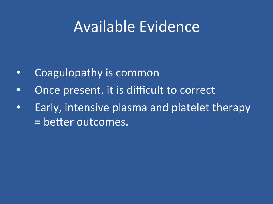
Available Evidence
• Coagulopathy is common • Once present, it is difficult to correct • Early, intensive plasma and platelet therapy
= bezer outcomes.

Lethal Triad of Death
Coagulopathy
Hypothermia Acidosis

PrevenGon of Secondary Injury by avoiding:
• Coagulopathy: early intensive resuscitaGon • Hypothermia: ++ contributes to coagulpathy • Acidosis: maintain Gssue perfusion (BE, Lactate) • Hypocalcemia • Hypofibrinogenemia • Transfusion reac-ons: clerical errors • Awareness: check anesthesia depth • Surgical misadventures:
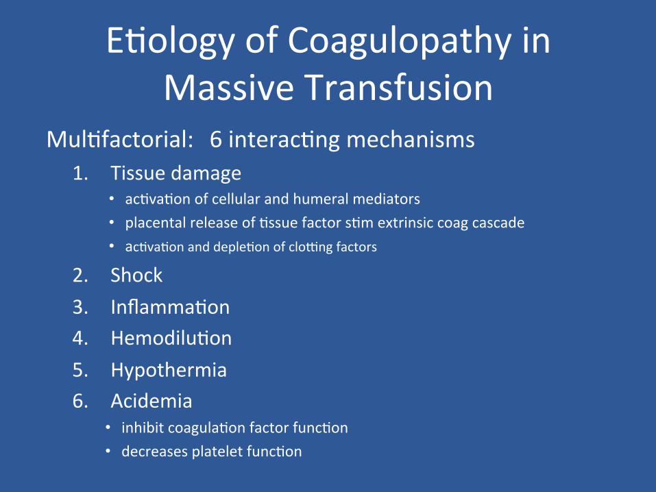
EGology of Coagulopathy in Massive Transfusion
MulGfactorial: 6 interacGng mechanisms 1. Tissue damage
• acGvaGon of cellular and humeral mediators • placental release of Gssue factor sGm extrinsic coag cascade • acGvaGon and depleGon of clonng factors
2. Shock 3. InflammaGon 4. HemodiluGon 5. Hypothermia 6. Acidemia
• inhibit coagulaGon factor funcGon • decreases platelet funcGon

Transfusion Therapy Therapy at present is guided by: – Clinical judgement -‐
• Oten underesGmates blood loss (esp vag delivery) • Causes delay in insGtuGng appropriate treatment • End up behind and playing catch up
– Lab results • Slow Turn around Time (TAT) • not useful for guiding therapy in Massive Hx
– More recently by Formula driven MTP • Advocate empirical early use of plasma and platelets with RBCs
– 1:1:1 RBC:plasma:platelets – 1:1 RBC:plasma

Formula Driven Massive Transfusion Protocol Debate
• DefiniGve data is not yet out Pro’s
– ê overall blood usage – ê mortality rate from exsanguinaGon, improved survival – ê dependency on lab during acute resuscitaGon phase – Prevent coagulopathy rather than use reacGve strategy to treat it Con’s
– é risk of TRALI by é exposure to unnecessary plasma and platelets. – é use of inappropriately triggered Formula driven MTP where RBC’s alone
would have been sufficient. – é wastage of plasma – Trauma literature thought to have a survivor bias – RetrospecGve studies with inherent limitaGons
“Formula-‐Driven” Versus “Lab-‐Driven” Massive Transfusion Protocols: At a State of Clinical Equipoise Jeannie L. Calluma,Transfusion Medicine Reviews, Vol 23, No 4
(October), 2009: pp 247-‐254
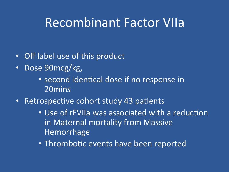
Recombinant Factor VIIa
• Off label use of this product • Dose 90mcg/kg,
• second idenGcal dose if no response in 20mins
• RetrospecGve cohort study 43 paGents • Use of rFVIIa was associated with a reducGon in Maternal mortality from Massive Hemorrhage • ThromboGc events have been reported
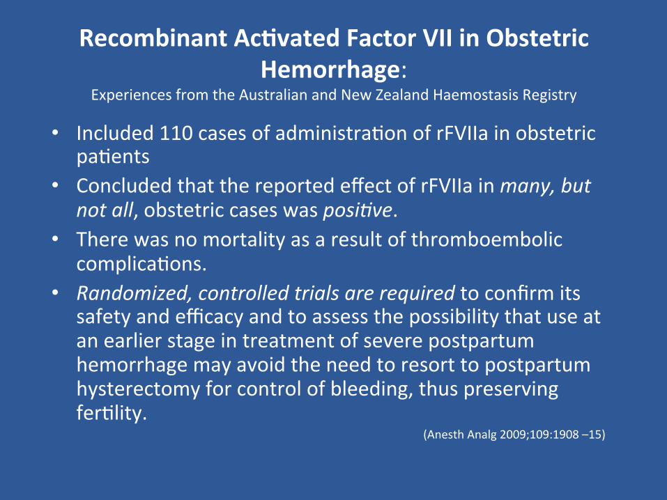
Recombinant Ac-vated Factor VII in Obstetric Hemorrhage:
Experiences from the Australian and New Zealand Haemostasis Registry
• Included 110 cases of administraGon of rFVIIa in obstetric paGents
• Concluded that the reported effect of rFVIIa in many, but not all, obstetric cases was posi&ve.
• There was no mortality as a result of thromboembolic complicaGons.
• Randomized, controlled trials are required to confirm its safety and efficacy and to assess the possibility that use at an earlier stage in treatment of severe postpartum hemorrhage may avoid the need to resort to postpartum hysterectomy for control of bleeding, thus preserving ferGlity.
(Anesth Analg 2009;109:1908 –15)

WHO Guidelines rFVIIa A high rate of thrombo-c events (185 events in 165 treated paGents)has been reported in paGents receiving rFVIIa for off-‐label use
O’Connell KA et al. Thromboembolic adverse events ater use of recombinant human coagulaGon factor VIIa. JAMA, 2006, 295(3):293–298.
Recommenda-on
Not enough evidence to make any recommenda-on regarding the use of recombinant factor VIIa for the treatment of PPH. Recombinant factor VIIa for the treatment of PPH should be limited to women with specific haematological indicaGons.
Remark
Use of rFVIIa could be life-‐saving Side-‐effects may be significant FVIIa is expensive
WHO guidelines for the management of postpartum haemorrhage; 2009

Lab Results and TAT Blood transfusion labs • Consider unmatched or group specific RBCs
– Unmatched: 10 min – Group specific: 15 min (if specimen in TML) – Crossmatched: 45 min
Rapid laboratory tesGng for trauma paGents: where a perfect result may not be in the best interests of the paGent Jeannie L. Callum, TRANSFUSION 2010;50:2529-‐2532

Lab Results “Where a perfect result may not be in the best interests of the paGent “
• CoagulaGon Labs • Turn around Gmes (TAT) sGll too slow • Emergency Hemorrhage Panel
– Standard coagulaGon test 30-‐60 min – ModificaGons to CoagulaGon Tests to ê TAT 20 min
» shortening centrifuge Gme for INR » not checking for clots » criGcal results are reported before being repeated
• Develop Rapid Metabolic profile – ABG, Hb, Na, K, Ca+,lactate (from single blood gas syringe)
Rapid laboratory tesGng for trauma paGents: where a perfect result may not be in the best interests of the paGent Jeannie L. Callum, TRANSFUSION 2010;50:2529-‐2532
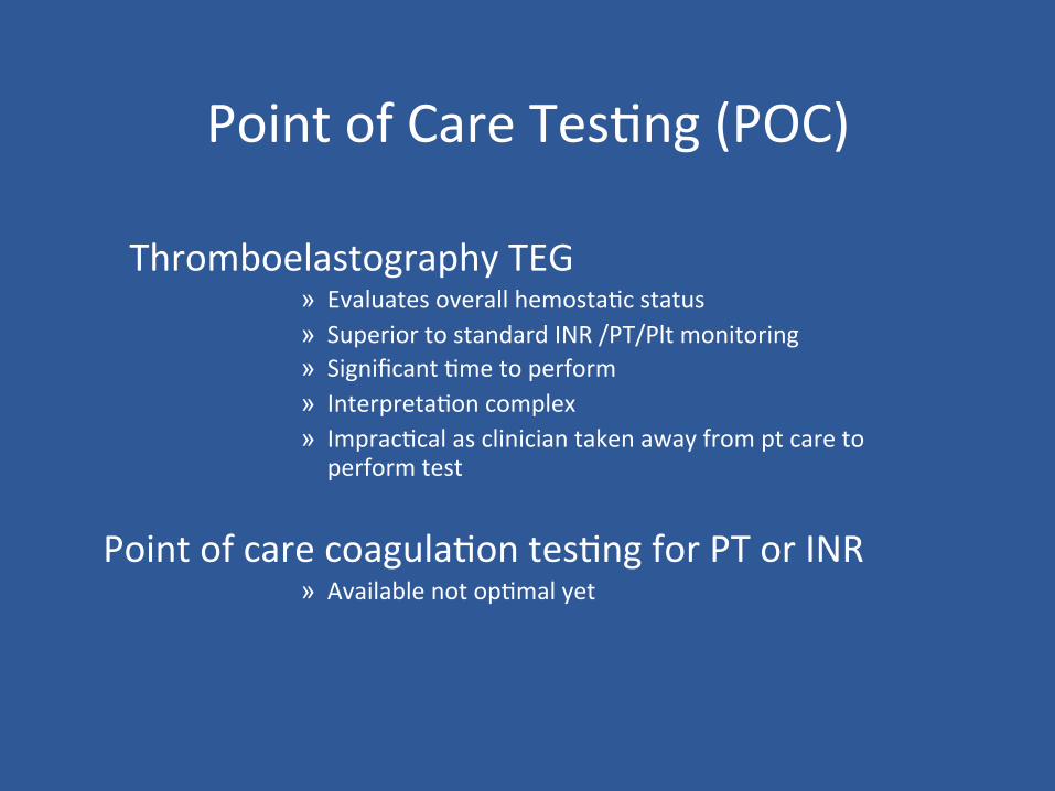
Point of Care TesGng (POC)
Thromboelastography TEG » Evaluates overall hemostaGc status » Superior to standard INR /PT/Plt monitoring » Significant Gme to perform » InterpretaGon complex » ImpracGcal as clinician taken away from pt care to perform test
Point of care coagulaGon tesGng for PT or INR » Available not opGmal yet

Hemocue
Hemocue -‐POC Hb tesGng -‐very useful -‐easy to use -‐accurate
Hemocue

Summary • Facility specific plan (MHP & MTP) • Appropriate risk assessment, referral • Early recogniGon and aggressive treatment • OpGmise medical and surgical Mx • Early blood products (RBC, FFP, Platelets, cryoppt) and transfer if needed
• Adequate resuscitaGon avoiding hypothermia, acidosis and coagulopathy
• ? rFVIIa

Massive Obstetric Hemorrhage The Team Approach
RECC -‐ Update in Obstetric Anesthesia
Massive Transfusion Protocol
• Case study, what went well, what didn’t • ImplicaGons of a MTP • Components of a MTP • Examples of MTP

Case MF
• I’m in ER, busy…. • 22 yr female • G1P1, 2h post delivery • Call from floor: Pt in far bed in room • “Bleeding to death”
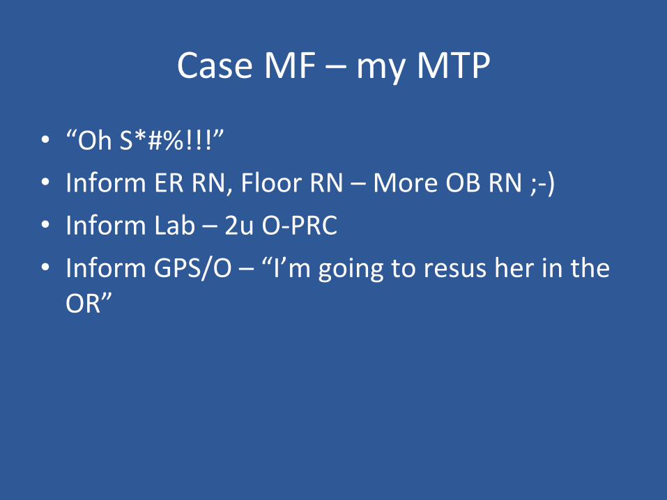
Case MF – my MTP
• “Oh S*#%!!!” • Inform ER RN, Floor RN – More OB RN ;-‐) • Inform Lab – 2u O-‐PRC • Inform GPS/O – “I’m going to resus her in the OR”

Case MF – Outcome
• More OB RN -‐ Hemabate • 2u PRC, numerous Saline • Manual compression • No GPS/O • Bleeding stopped

Case MF
• What went well? • What didn’t go well (where can we improve)?
– Could we do bezer by having a MTP?

ImplicaGons of a MTP
• Reduce chaos • Reduce blood bank delays • Reduced mortality • ReducGon in organ failure, sepsis & Abdominal Compartment Syndrome
• Increased blood products early, less total • Less use of crystalloids
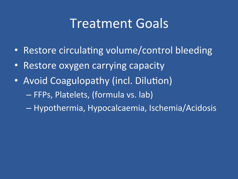
Treatment Goals
• Restore circulaGng volume/control bleeding • Restore oxygen carrying capacity • Avoid Coagulopathy (incl. DiluGon) – FFPs, Platelets, (formula vs. lab) – Hypothermia, Hypocalcaemia, Ischemia/Acidosis

Purpose of a MTP
• MobilizaGon of resources • PrioriGzaGon, delegaGon • Simultaneous assessment and treatment • EffecGve communicaGon

Components of a MTP
• InformaGon – IniGaGon Trigger – Who – What: PRBC, FFP, Platelets, Cryo, labs – Reminders: Hypoxia, Acidosis, Hypothermia, Hypocalcaemia
• Paperwork – Guide, Consent, Lab recs, recording documentaGon, (work sheet, criGque form, InformaGon booklet for paGents)

Components of a MTP – The Who
• MDs, RNs, Clerk, Porter • Lab, blood bank, Hematologist • Ambulance/OR

Components of a MTP – The Who (BCWH)
Declare Massive Hemorrhage -‐ BCWH OR 1. Hematology lab 2. Transfusion Lab 3. 2nd call Anesthesiologist 4. LDR charge Nurse 5. 2nd call Obstetrician; General Surgeon 6. Anesthesia Assistant 7. Perfusionist 8. IntervenGonal Radiologist 9. CriGcal Care / Internal Medicine Physician 10. Hematologist on call 11. Ward Clerk


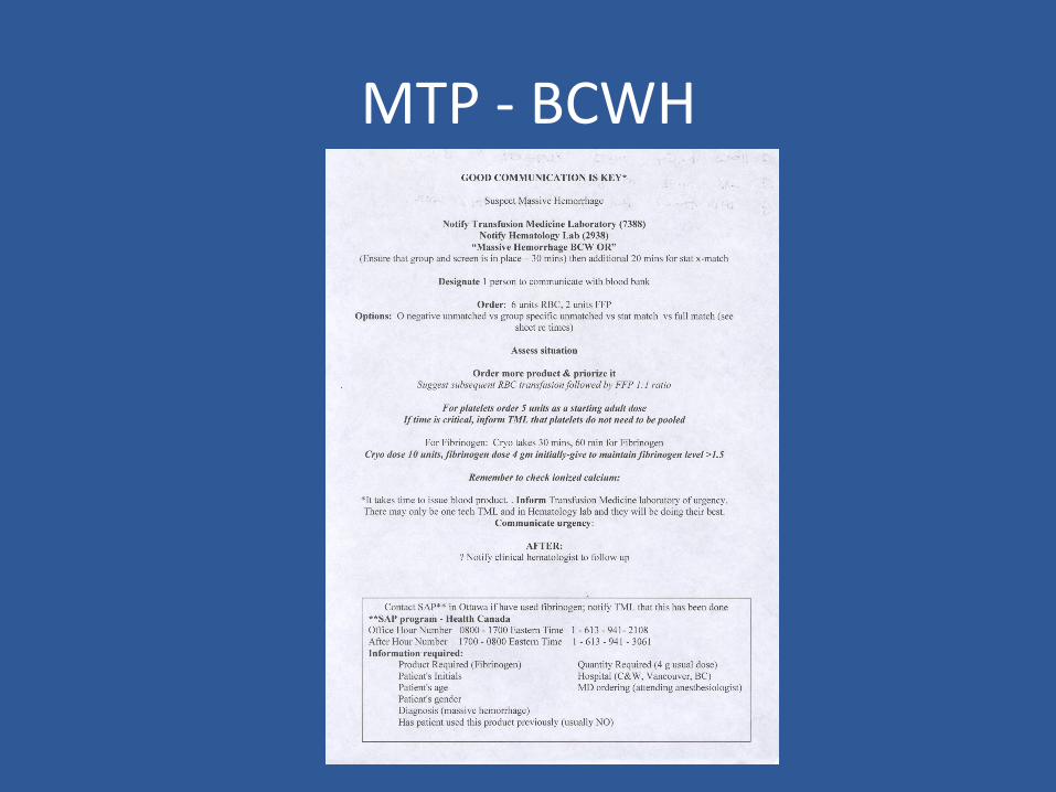
MTP -‐ BCWH
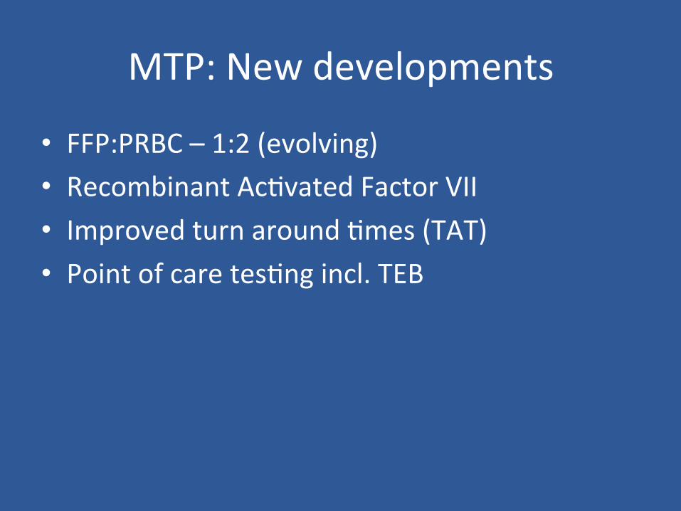
MTP: New developments
• FFP:PRBC – 1:2 (evolving) • Recombinant AcGvated Factor VII • Improved turn around Gmes (TAT) • Point of care tesGng incl. TEB

Massive Transfusion Protocol
• Rural quesGons:
– FFPs and Platelets; send PaGent or send Products?
– Call a Hematopathologist / Transfusionist

MTP: Take home
• Risk assessment, avoidance • Early recogniGon • Facility specific plan (MTP) • Early blood products (PRBC, FFP, Platelets) or transfer
• Avoiding hypothermia & cellular hypoxia and acidosis