Feline Immunodeficiency Virus (FIV) Neutralization: A - MDPI.com

Mar. Drugs 2011, 9, 1056-1100; doi:10.3390/md9061056
Marine Drugs
ISSN 1660-3397
www.mdpi.com/journal/marinedrugs
Review
Marine Bioactives as Functional Food Ingredients: Potential to
Reduce the Incidence of Chronic Diseases
Sinéad Lordan, R. Paul Ross and Catherine Stanton *
Teagasc Food Research Centre Moorepark, Fermoy, Co. Cork, Ireland;
E-Mails: [email protected] (S.L.); [email protected] (R.P.R.)
* Author to whom correspondence should be addressed; E-Mail: [email protected];
Tel.: +353-25-42-606; Fax: +353-25-42-340.
Received: 2 April 2011; in revised form: 2 June 2011 / Accepted: 8 June 2011 /
Published: 14 June 2011
Abstract: The marine environment represents a relatively untapped source of functional
ingredients that can be applied to various aspects of food processing, storage, and
fortification. Moreover, numerous marine-based compounds have been identified as having
diverse biological activities, with some reported to interfere with the pathogenesis of
diseases. Bioactive peptides isolated from fish protein hydrolysates as well as algal fucans,
galactans and alginates have been shown to possess anticoagulant, anticancer and
hypocholesterolemic activities. Additionally, fish oils and marine bacteria are excellent
sources of omega-3 fatty acids, while crustaceans and seaweeds contain powerful
antioxidants such as carotenoids and phenolic compounds. On the basis of their bioactive
properties, this review focuses on the potential use of marine-derived compounds as
functional food ingredients for health maintenance and the prevention of chronic diseases.
Keywords: disease; functional food ingredients; marine; polyunsaturated fatty acids
1. Introduction
Increasing knowledge regarding the impact of diet on human health along with state-of-the-art
technologies has led to significant nutritional discoveries, product innovations, and mass production on
an unprecedented scale [1]. In particular, naturally occurring bioactive extracts or single compounds
thereof, that are believed to benefit human health, have spawned an important and dynamic new area
of research resulting in substantial advances in nutritional knowledge. There is also growing awareness
OPEN ACCESS
Mar. Drugs 2011, 9
1057
that dietary source and form of food may affect overall health. Suitably, the role of food as an agent for
improving health has been recognised, initiating the development of new classes of food, known as
functional foods [2].
The concept of functional foods is to improve the general conditions of the body and decrease the
risk of illness and disease [3]. That is to say, bioactive compounds present as natural constituents or as
fortificants in food having the potential to provide health benefits beyond the basic nutritional value of
the product. Marine-derived nutrients and other marine bioactive components have excellent potential
as functional food ingredients as they possess advantageous physiological effects, with medicinal
characteristics and added health benefits such as anticancer or anti-inflammatory activity.
The marine world, due to its phenomenal biodiversity, is a rich natural resource of many
biologically active compounds such as polyunsaturated fatty acids (PUFAs), sterols, proteins,
polysaccharides, antioxidants and pigments. Many marine organisms live in complex habitats exposed
to extreme conditions and, in adapting to new environmental surroundings, they produce a wide
variety of secondary (biologically active) metabolites which cannot be found in other organisms.
Moreover, considering its great taxonomic diversity, investigations related to the search of new
bioactive compounds from the marine environment can be seen as an almost unlimited field [4,5].
Marine-based bioactive food ingredients can be derived from a vast array of sources, including
marine plants, microorganisms, and sponges, all of which contain their own unique set of biomolecules
[4]. However, proving that these naturally occurring bioactive substances have a defined health benefit
poses a dilemma in nutritional research as investigating preventive activity can be difficult when effect
is only moderate. This means that the effect of the compounds on the human body may be very small
over relatively short periods but could contribute significantly to health when they are consumed
throughout life as part of the daily diet [1]. Therefore, to facilitate discussion of this issue, the
following review examines the existing scientific knowledge which demonstrates the suitability of
marine-derived bioactive compounds as functional food ingredients for the prevention and treatment of
chronic diseases.
2. Sources of Marine Functional Food Ingredients
2.1. Macroalgae
Marine algae are simple chlorophyll containing organisms composed of one cell or grouped
together in colonies or as organisms with many cells, sometimes collaborating together as simple
tissues. These unicellular or multicellular vegetative organisms do not have true roots or stems and
vary greatly in size and morphology—from organisms 3–10 m in length to giant kelps up to 70 m
long and growing up to 50 cm per day. Correspondingly, algae can be classified into two major groups
according to their size: macroalgae or microalgae [6,7].
Macroalgae are more commonly known as seaweeds and several characteristics are used to classify
them including the nature of their chlorophyll, their cell wall chemistry, and the presence or absence of
flagella. However, the feature most commonly employed in algal classification is the presence of
specific pigments, other than chlorophyll, which clearly identify macroalgae as belonging to one of
three algal divisions. In accordance with this criterion, macroalgae can be classified as brown algae

Mar. Drugs 2011, 9
1058
(Phaeophyceae), red algae (Rhodophyceae), or green algae (Chlorophyceae). The presence of these
different phytopigments in algae is related to their sea habitat because not all macroalgae need the
same light intensity to perform photosynthesis. Thus, green macroalgae, which are able to absorb large
amounts of light energy, abound in coastal waters, while red and brown macroalgae prevail at greater
depths where penetration of sunlight is limited [6].
Macroalgae are a source of biologically active phytochemicals, which include carotenoids,
phycobilins, fatty acids, polysaccharides, vitamins, sterols, tocopherol and phycocyanins among
others. Many of these compounds are known to possess biological activity and hence have potential
beneficial use in healthcare [8]. However, the chemical and nutritional composition of seaweeds
depends on many factors, including species, geographical origin or area of cultivation, seasonal,
environmental, and physiological variations, time of harvest, water temperature, and processing
methods [6,9–11]. For example, a seasonal variation of protein content of Palmaria palmata was
observed, with maximum values (approximately 21%) occurring during the winter-spring period and
lower levels (12%) during the summer-early autumn period [12].
2.1.1. Proteins, Peptides and Amino Acids
The protein content of macroalgae varies greatly from phylum to phylum [9]. Generally, the protein
fraction of brown seaweeds is low (3–15% of dry weight) compared with that of the green or red
seaweeds (10–47% of dry weight) [10]. The protein in macroalgae contains all essential amino acids,
however, variations in their concentrations are known to occur [12]. Leucine, valine, and methionine
are abundant essential amino acids of Palmaria palmata and their average levels are close to those
generally reported for ovalbumin. On the other hand, isoleucine and threonine concentrations are
similar to those recorded for legume proteins [6]. Leucine, phenylalanine and valine are the major
essential amino acids of Ulva rigida, while levels of histidine, which is an essential amino acid in
children, are similar to those found in legumes and eggs [13].
Recently, much attention has been paid to unraveling the structural, compositional and sequential
properties of bioactive peptides. Marine bioactive peptides may be produced by one of three methods;
solvent extraction, enzymatic hydrolysis or microbial fermentation of marine proteins. However,
particularly in food and pharmaceutical industries, the enzymatic hydrolysis method is preferred on
account of lack of residual organic solvents or toxic chemicals in the products. Bioactive peptides
usually contain 3–20 amino acid residues and their activities are based on their amino acid composition
and sequence [14,15]. These peptides are reported to be involved in various biological functions such
as antihypertension, immunomodulatory, antithrombotic, antioxidant, anticancer and antimicrobial
activities, in addition to nutrient utilisation [15,16].
Among the algal proteins, it is worth noting the occurrence of protein-pigment complexes called
phycobiliproteins, some of which are currently used as fluorescent markers in the fields of clinical
diagnosis and biotechnological applications [17,18]. Recent studies have shown that phycobiliproteins,
which generally make up 1–10% of dry weight of algal biomass, impart antioxidant properties which
could be beneficial in the prevention or treatment of several diseases [17]. Moreover, in some
countries, phycobiliproteins are utilised as natural food colourings in products such as chewing gums,
dairy products, jellies and ice sherbets [4].

Mar. Drugs 2011, 9
1059
Of note, the in vivo digestibility of algal proteins is poorly described, and available studies about
their assimilation by humans have not provided conclusive results. Nonetheless, several studies have
described a high rate of algal protein degradation in vitro by proteolytic enzymes such as pepsin,
pancreatin and pronase. For instance, the in vitro digestibility of proteins from the red seaweed
Porphyra tenera is approximately 70%. There is a possibility, however, that the high phenolic content
of some algae may limit protein availability in vivo [9].
2.1.2. Fatty Acids
The lipid content of macroalgae represents only 1–5%, thus its contribution as a food energy source
appears to be low [17]. However, PUFAs account for almost half of this lipid fraction, with much of it
occurring in the form of omega-3 (n-3) and omega-6 (n-6) fatty acids such as eicosapentanoic acid
(EPA) and arachidonic acid (AA) [19]. PUFAs regulate a wide range of functions in the body
including blood pressure, blood clotting, and correct development and functioning of the brain and
nervous systems [20]. Furthermore, PUFAs have a role in regulating inflammatory responses through
the production of inflammatory mediators termed eicosanoids [21].
The n-3 to n-6 ratio of macroalgae is closely matched which may add to their efficacy as a dietary
supplement or as part of a balanced diet [22]. Moreover, they contain many essential fatty acids. Red
and brown algae, for instance, are particularly rich in the n-3 fatty acids, EPA and -linolenic acid, and
the n-6 fatty acids, AA and linoleic acid, along with relatively high levels of oleic and palmitic acids
[11]. In contrast, green seaweeds, like Ulva pertusa, are characterised by the presence of
hexadecatetraenoic (n-3), oleic and palmitic acids [23]. The n-3 fatty acid, octadecatetraenoic acid, is
abundant in Laminaria sp. and Undaria pinnatifida while hexadecatetraenoic acid is prominent in
Ulva sp. [6,24].
In addition to fatty acids, the unsaponifiable fraction of macroalgae contains carotenoids (such as
-carotene, lutein and violaxanthin in red and green seaweeds, fucoxanthin in brown seaweeds),
tocopherols, sterols (such as fucosterol in brown seaweeds) and terpenoids [25–28].
2.1.3. Polysaccharides
Although algal carbohydrate content is relatively high, macroalgae cannot be considered a potential
energy rich food as digestibility of these carbohydrates is low [6]. Moreover, the carbohydrate type
varies greatly between algae species. Typical polysaccharides in red algae varieties consist of floridean
starch, cellulose, xylan and mannan, and the water soluble fibre fraction is formed by sulfur containing
galactans such as agar and carrageenan. Standard polysaccharides in brown algae are fucoidan,
laminaran, cellulose, alginates and mannitol whilst the fibres are mainly cellulose and insoluble
alginates. Most of these polysaccharides are not digestible by the human gastrointestinal tract and,
therefore, can be regarded as dietary fibres [11]. The total dietary fibre content of seaweeds ranges
from 29.3–62.3 g/100 g [11,19,29], and so is higher than the fibre content of most fruits and
vegetables. Human consumption of algal fibre has been proven to be health promoting and its benefits
are well documented [13,30,31].
Storage polysaccharides, such as agar, carrageenans and alginates, are the most commercially
exploited components in seaweeds. These storage polysaccharides exhibit textural and stabilizing

Mar. Drugs 2011, 9
1060
properties [19]; thus they are used in food applications such as thickening aqueous solutions, forming
gels, forming water soluble films and stabilizing products such as ice-cream [4].
Fucoidans are a complex series of sulfated polysaccharides found widely in the cell walls of brown
macroalgae. Fucoidans are reported to display numerous physiological and biological properties,
including anticoagulant, antiviral, antithrombotic, antitumor and antioxidant activities, as well as
having an effect on the inflammatory and immune systems [32,33]. In addition, the therapeutic
potential of fucoidans increases with their degree of sulfation and they can be easily extracted using
either hot water or an acid solution [32]. Another sulfated polysaccharide, porphyran, makes up the
main components of the red macroalga, Porphyra [30]. This polysaccharide has reported uses as a
gelling agent, a nutritional supplement and as an antioxidant [34]. Alternatively, laminarin, the second
major storage glucan in brown algae, has been identified as a modulator of intestinal metabolism
through its effects on mucus composition, intestinal pH and short chain fatty acid production [34–36].
Another group of carbohydrate derivatives, oligosaccharides, are commonly defined as
carbohydrate molecules with a low degree of polymerisation. Oligosaccharides can be produced
naturally or may be derived from algal polysaccharides after chemical, physical or biochemical
degradations. To date, numerous oligosaccharides with immunostimulation activities as well as
antioxidant and antitumor properties have been characterised. Moreover, oligosaccharides can be
beneficial to health when they are added to the diet to enhance the growth of prebiotic bacteria. In this
case, oligomers that resist the digestive process are used as a specific substrate for the growth of health
beneficial bacteria [37]. For instance, xylo-oligosaccharides and fructo-oligosaccharides are
non-digestible oligomers that cannot be absorbed in the gastrointestinal tract. Hence, they are intact in
the large bowel and are used as a preferential substrate by anaerobic bacteria such as bifidobacteria and
lactobacilli [38,39]. Interestingly, no specific conformation is correlated to the non-digestible
oligosaccharide’s biological activity, whereas the immunostimulating, antioxidant, antiangiogenic
and antithrombotic activities of poly/oligosaccharides molecules are determined by glycan
conformation [37].
While algal polysaccharides have yet to be exploited in the food industry, the fact that they are easy
to isolate and have numerous health benefits gives them the potential to serve as valuable bioactive
ingredients in functional foods [4].
2.1.4. Vitamins, Minerals and Antioxidants
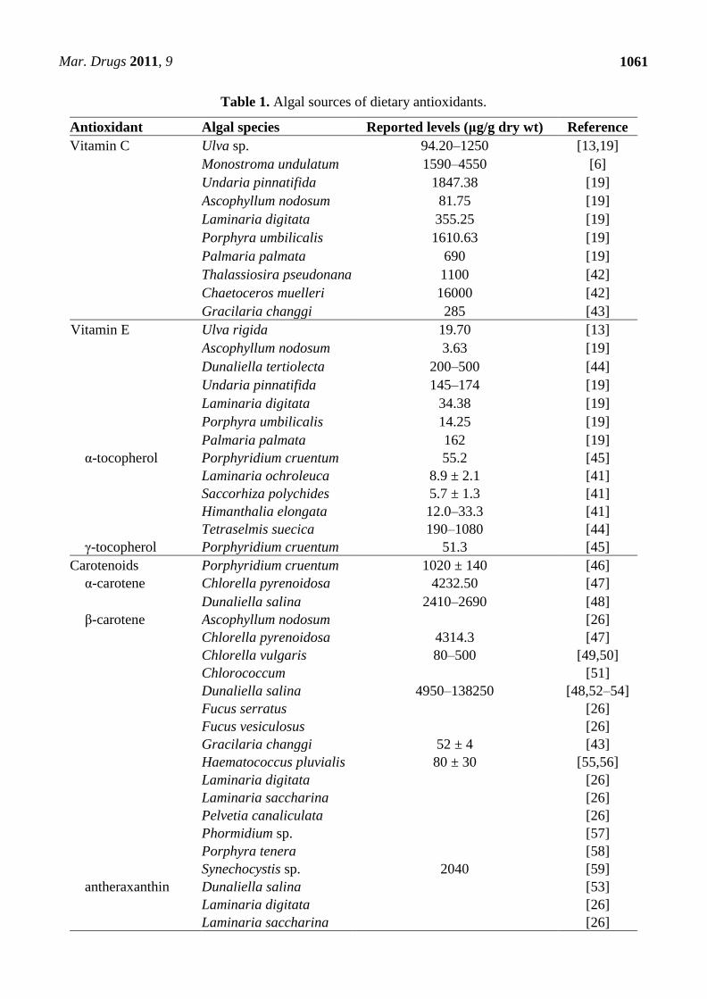
One of the principal nutritive characteristics of seaweeds is their high antioxidant content (Table 1).
In addition, vitamin B12 is found in red macroalgae (e.g., Palmaria longat and Porphyra tenera) and
in certain green seaweeds [9]. Red and brown algae contain high levels of folic acid and folate
derivatives including 5-metil-tetrahydro-folate, 5-formyl-tetrahydro-folate and tetrahydro-folate.
Indeed, amounts as high as 150 g folic acid per 100 g of dry Undaria pinnatifida algae have been
detected [40]. As well as seasonal, environmental and physiological variations, vitamin content also
depends on the type of seaweed processing. For example, the content of -tocopherol in Himanthalia
longate dehydrated (33.3 g/g dry weight) was considerably higher than in canned Himanthalia
longate (12 g/g dry weight) [41].
Mar. Drugs 2011, 9
1061
Table 1. Algal sources of dietary antioxidants.
Antioxidant Algal species Reported levels (μg/g dry wt) Reference
Vitamin C Ulva sp. 94.20–1250 [13,19]
Monostroma undulatum 1590–4550 [6]
Undaria pinnatifida 1847.38 [19]
Ascophyllum nodosum 81.75 [19]
Laminaria digitata 355.25 [19]
Porphyra umbilicalis 1610.63 [19]
Palmaria palmata 690 [19]
Thalassiosira pseudonana 1100 [42]
Chaetoceros muelleri 16000 [42]
Gracilaria changgi 285 [43]
Vitamin E Ulva rigida 19.70 [13]
Ascophyllum nodosum 3.63 [19]
Dunaliella tertiolecta 200–500 [44]
Undaria pinnatifida 145–174 [19]
Laminaria digitata 34.38 [19]
Porphyra umbilicalis 14.25 [19]
Palmaria palmata 162 [19]
α-tocopherol Porphyridium cruentum 55.2 [45]
Laminaria ochroleuca 8.9 ± 2.1 [41]
Saccorhiza polychides 5.7 ± 1.3 [41]
Himanthalia elongata 12.0–33.3 [41]
Tetraselmis suecica 190–1080 [44]
γ-tocopherol Porphyridium cruentum 51.3 [45]
Carotenoids Porphyridium cruentum 1020 ± 140 [46]
α-carotene Chlorella pyrenoidosa 4232.50 [47]
Dunaliella salina 2410–2690 [48]
β-carotene Ascophyllum nodosum [26]
Chlorella pyrenoidosa 4314.3 [47]
Chlorella vulgaris 80–500 [49,50]
Chlorococcum [51]
Dunaliella salina 4950–138250 [48,52–54]
Fucus serratus [26]
Fucus vesiculosus [26]
Gracilaria changgi 52 ± 4 [43]
Haematococcus pluvialis 80 ± 30 [55,56]
Laminaria digitata [26]
Laminaria saccharina [26]
Pelvetia canaliculata [26]
Phormidium sp. [57]
Porphyra tenera [58]
Synechocystis sp. 2040 [59]
antheraxanthin Dunaliella salina [53]
Laminaria digitata [26]
Laminaria saccharina [26]

Mar. Drugs 2011, 9
1062
Table 1. Cont.
astaxanthin Chlorella vulgaris [60]
Chlorococcum sp. [51,61]
Haematococcus pluvialis up to 3% [52,56,62]
β-cryptoxanthin Chlorella pyrenoidosa 334.9 [47]
cantaxanthin Chlorella vulgaris [60]
Chlorococcum [51]
echinenone Phormidium sp. [57]
Synechocystis sp. 240 [59]
fucoxanthin Ascophyllum nodosum [26]
Fucus serratus [26]
Fucus vesiculosus [26]
Hijikia fusiformis [27]
Himanthalia elongata 820 [59]
Laminaria digitata [26]
Laminaria saccharina [26]
Pelvetia canaliculata [26]
loroxanthin Chlorella pyrenoidosa [63]
lutein Chlorella protothecoides 4600 [64]
Chlorella pyrenoidosa 1153009.70 [47,63]
Chlorella vulgaris 2970–3830 [49,50]
Chlorella zofingiensis 3400 [64]
Chlorococcum [51]
Dunaliella salina 6550 ± 920 [48,53]
Haematococcus pluvialis 270 ± 60 [55,56]
Muriellopsis sp. 4300 [64]
Phormidium sp. [57]
Porphyra tenera [58]
Scenedesmus almeriensis 4500 [64]
myxoxanthophyll Synechocystis sp. 580 [59]
neoxanthin Ascophyllum nodosum [26]
Chlorella pyrenoidosa 199.7 [47]
Dunaliella salina [53]
Fucus serratus [26]
Fucus vesiculosus [26]
Haematococcus pluvialis 60 ± 20 [55,56]
Laminaria digitata [26]
Laminaria saccharina [26]
Pelvetia canaliculata [26]
Phormidium sp. [57]
violaxanthin Ascophyllum nodosum [26]
Chlorella pyrenoidosa 38.1 [47,63]
Fucus serratus [26]
Fucus vesiculosus [26]
Haematococcus pluvialis 40 ± 20 [55]
Himanthalia elongata 50 [59]
Laminaria digitata [26]

Mar. Drugs 2011, 9
1063
Table 1. Cont.
Laminaria saccharina [26]
Pelvetia canaliculata [26]
Phormidium sp. [57]
zeaxanthin Ascophyllum nodosum [26]
Chlorella pyrenoidosa 2170.3 [47]
Dunaliella salina 11270 ± 1580 [48,53]
Fucus serratus [26]
Fucus vesiculosus [26]
Haematococcus pluvialis 30 ± 10 [55]
Himanthalia elongata 130 [59]
Laminaria digitata [26]
Laminaria saccharina [26]
Pelvetia canaliculata [26]
Synechocystis sp. 1640 [59]
Chlorophylls Dunaliella salina 26–3100 [53,65]
Himanthalia elongata [59]
chlorophyll a Chlorella pyrenoidosa [63]
Chlorella vulgaris 3320–9630 [49,50]
Chlorococcum [51]
Phormidium sp. [57]
Porphyra tenera [58]
Porphyridium cruentum 2130 ± 1200 [46]
Tetraselmis suecica 6040–27530 [44]
chlorophyll b Chlorella pyrenoidosa [63]
Chlorella vulgaris 2580–5770 [49,50]
Chlorococcum [51]
Haematococcus pluvialis [56]
Porphyridium cruentum 380 ± 340 [46]
pheophytin a Chlorella vulgaris [50]
Porphyridium cruentum 3310 ± 1110 [46]
pheophytin b Chlorella vulgaris 2310–5640 [49,50]
Porphyridium cruentum 30 ± 90 [46]
Polyphenols Fucus sp. 41400 ± 400 [6]
Haematococcus pluvialis [66]
Laminaria sp. 7300 ± 100 [6]
Porphyra sp. 5700 ± 100 [6]
Spongiochloris spongiosa 5.65 [67]
Undaria sp. 6600 ± 100 [6]
Seaweeds also contain an incomparable wealth of minerals and trace elements which are attributed
to their capacity to retain inorganic marine substances due to the characteristics of their cell surface
polysaccharides [6,19,68]. The mineral fraction of some seaweeds accounts for up to 36% of dry
matter [17]. Many of these essential minerals accumulate in seaweeds at much higher levels than in
terrestrial foodstuffs. For example, there is more iron in an 8 g serving of dry Palmaria palmata than
Mar. Drugs 2011, 9
1064
in 100 g of raw sirloin steak [19]. All of the essential minerals and trace elements needed for human
nutrition are present in seaweeds [68], and so it should be regarded as a valuable functional food. For
instance, the brown algae, Undaria pinnatifida and Sargassum, and the red algae, Chondrus crispus
and Gracilariopsis, can be used as food supplements to help meet the recommended daily intake of
some minerals (Na, K, Ca, Mg) and trace elements (Fe, Zn, Mn, Cu) [68,69]. Moreover, analysis of the
mineral composition of Ulva rigida revealed balanced contents of Na and K (15.9 and 15.6 g/kg
respectively, ratio near to 1), which, from a nutritional point of view, is of interest as intake of diets
with a high Na/K ratio have been related to incidence of hypertension [13]. Additionally, seaweeds are
one of the most important vegetable sources of calcium. Calcium content may be as high as 7% of the
dry weight in macroalgae and up to 25–34% in the chalky seaweed, lithotamne. Thus, seaweed
consumption may also be useful to those at risk of calcium deficiency, namely expectant mothers,
adolescents and the elderly [17].
Other bioactive compounds are the photosynthetic pigments used by autotrophs to capture solar
energy for photosynthesis [4]. As regards macroalgae, the main pigments are carotenoids and
chlorophylls (Table 1). The carotenoid fucoxanthin has potential commercial value as it has been
reported to be of use in treating obesity and reducing the risk of certain diseases, such as type 2
diabetes through its ability to promote the expression of the uncoupling protein, UCP1 [70]. In the
food industry, chlorophylls are mainly used as natural colorants in foods and beverages [4]. However,
chlorophylls and their derivatives have been shown to possess some biological activity whereby they
exhibit anticancer properties in their ability to bind carcinogenic hydrophobic compounds such as
polycyclic aromatic hydrocarbons, heterocyclic amines and aflatoxin [71,72]. Phlorotannins, a group
of polyphenolic compounds which have also been identified in several brown algal families, have been
reported to possess strong antioxidant activity. However, at present, the extractable polyphenol levels
from algae are lower than that of other phytochemicals [6,73,74].
The nutritional value ascribed to macroalgae along with their non-animal nature makes them
particularly appropriate for use in the food industry. Seaweeds have enormous potential as components
of fertilizers, in animal feed supplements, and as additives for human food. Hence, biotechnological
advances regarding macroalgae cultivation has stimulated the development of seaweed aquaculture. At
present, three genera, Laminaria, Undaria and Porphyra, constitute 93% of the algal mass cultivated
for nutritional purposes [6].
2.2. Microalgae
Microalgae are the most primitive and simply organised members of the plant kingdom, with the
majority existing as small cells of about 3–20 m [4]. These algae are ubiquitous in nature and aquatic
microalgae have been isolated in areas ranging from hot springs to glacial ice flows [75]. Microalgae
are found in both benthic and littoral habitats and also throughout the ocean waters as phytoplankton.
Phytoplankton comprises organisms such as diatoms (bacillariophyta), dinoflagellates (dinophyta),
green and yellow-brown flagellates (chlorophyta; prasinophyta; prymnesiophyta, cryptophyta,
chrysophyta and rhaphidiophyta) and blue-green algae (cyanophyta). As photosynthetic organisms,
this group plays a key role in the productivity of oceans and constitutes the basis of the marine food
chain [7].

Mar. Drugs 2011, 9
1065
There are over 50,000 different species of microalgae of which only a few have been characterised
[75]. This group of microorganisms is extremely diverse and represents a major untapped resource of
valuable bioactive compounds and biochemicals such as pigments, antioxidants, polysaccharides,
sterols, fatty acids and vitamins [76].
2.2.1. Proteins, Peptides and Amino Acids
The high protein content of various microalgal species and their amino acid pattern, which
compares favourably with that of other food proteins, is a good endorsement of microalgae as an
alternative protein source [77,78]. Spirulina, for instance, is high in protein (60–70% depending on the
strain) and, not only does this protein possess all of the essential amino acids, but these amino acids
have excellent bioavailability [52]. Furthermore, the industrial scale growth of the microalga,
Dunaliella, can turn out protein extract at about 100 times greater productivity than that reported in
agriculture and 50 fold greater than in fish farming [4].
Proteins from marine sources show promise as functional ingredients in foods because they possess
numerous important and unique properties such as film and foaming capacity, gel forming ability and
antimicrobial activity [4]. In addition, purified peptides from Chlorella vulgaris have demonstrated
significant protective effects against cellular damage [79]. With regard to one of the major proteins in
Spirulina platensis and Porphyridium, phycobiliprotein, several therapeutic bioactivities have been
described, namely, hepatoprotective, anti-inflammatory, immunomodulating, antioxidant and
anticancer effects [52].
2.2.2. Fatty Acids
The average lipid content of algal cells varies between 1 and 70% but can reach 90% of dry weight
under certain conditions [80]. Algal lipids are composed of glycerol, sugars or bases esterified to
saturated or unsaturated fatty acids. Among all the fatty acids in microalgae, some fatty acids of the
n-3 and n-6 families are of particular interest [42]. According to Mendes et al. [60], the main
constituents of the lipidic fractions of Chlorella vulgaris are oleic, palmitic and linolenic acids,
accounting for 41, 22 and 9% of the total amount, respectively. Additionally, palmitic, linolenic and
oleic acids account for more than 85% of the total fatty acid content of Dunaliella salina [81], while
the green microalga, Haematococcus, has been shown to contain short chain fatty acids with
antimicrobial activity [66].
Higher plants and animals lack the requisite enzymes to synthesize PUFAs of more than 18 carbons
and so have to obtain them from their food. Fish and fish oil are the common sources of long chain
PUFAs but safety issues have been raised because of the possible accumulation of toxins in fish.
Moreover, the application of fish oil as a food additive is limited due to problems associated with its
typical fishy smell, unpleasant taste and poor oxidative stability [78]. Consequently, long chain PUFAs
are commercially produced via microalgae cultivation for incorporation into infant milk formulations
and for use as dietary supplements and food additives [78]. Maximum n-3 fatty acid production can
also be induced by altering the growth conditions of microalgae. For instance, under optimal culture
conditions, Chlorella minutissima can produce an EPA content of up to 45% of its total fatty acid
content [4].

Mar. Drugs 2011, 9
1066
Microalgae such as Porphyridium, which shows a relatively low lipid content, contains significant
amounts of several major fatty acids such as palmitic acid, AA, EPA and linoleic acid [46,52].
Spirulina provides an interesting source of γ-linolenic acid (20–25% of the total lipid fraction), which
is a precursor of prostaglandins, leukotrienes and thromboxans involved in the modulation of
immunological, inflammatory and cardiovascular responses [17]. This microalga is also a natural
source of active fatty acids such as lauric, palmitic and oleic acids [82], with the n-3 fatty acid,
docosahexaenoic acid (DHA), accounting for up to 9.1% of the total fatty acids content. Spirulina has
been found to contain sterols, including clionasterol which has been shown to increase the production
of plaminogen-activating factor in vascular endothelial cells [76].
2.2.3. Polysaccharides
Carbohydrates in microalgae can be found in the form of starch, glucose, sugars and other
polysaccharides. Their overall digestibility is high, which is why there is no limitation to using dried
whole microalgae in foods or feeds [78]. Moreover, the biological activities of some microalgal
species have been associated with polysaccharides. Polysaccharide complexes from Chlorella
pyrenoidosa, and possibly Chlorella ellipsoidea, contain glucose and any combination of galactose,
rhamnose, mannose, arabinose, N-acetylglucosamide and N-acetylgalactosamine. These complexes are
believed to have immunostimulating properties, specifically immune stimulatory activity and can
inhibit the proliferation of Listeria monocytogenes and Candida albicans [76]. The most important
substance in Chlorella is -1,3-glucan, which is an active immunostimulator, a free radical scavenger
and a reducer of blood lipids. Chlorella can also be used as a food additive owing to the taste and
flavour adjusting actions of its colouring agent [78]. Also, novel polysaccharides isolated from
Porphyridium and Nostac flegelliforme microalgae exhibited potent antiviral activity against herpes
simplex virus (HSV-1 and 2) both in vitro and in vivo [83,84].
2.2.4. Antioxidants
The nutritional and therapeutic relevance of dietary carotenoids is attributed to their ability to act as
provitamin A; that is, they can be converted into vitamin A by the human body. Moreover, carotenoids
play a protective role by preventing the formation of reactive oxygen species [85]. Microalgal
production of carotenoids, such as -carotene and astaxanthin, is an attractive area of research as they
are valuable bioactive ingredients that can present at relatively high concentrations in algal cells
(Table 1). Moreover, cultivated algae can be induced to produce even larger quantities of carotenoids
by controlling certain environmental growth conditions. The strains of microalgae that are currently
being investigated for use as natural producers of commercial carotenoids include Dunaliella salina,
Sarcina maxima, Chlorella protothecoides, Chlorella vulgaris and Haematococcus pluvialis [4].
Dunaliella salina is the most suitable organism for the mass production of -carotene as it can
produce-carotene up to 14% of its dry weight [80]. This microalga can also be cultivated easily and
quickly when compared to plants and produces both cis and trans isomers of carotenoids for high
bioavailability and bioefficacy [85,86]. -carotene is one of the leading food colorants in the world and
has been applied to a range of food and beverage products to improve their appearance to consumers
[87]. In addition, -carotene has strong antioxidant properties which help to mediate the harmful

Mar. Drugs 2011, 9
1067
effects of free radicals implicated in numerous life-threatening diseases, including various forms of
cancer, coronary heart disease (CHD), premature aging and arthritis. The antioxidant qualities of
-carotene can also assist the body in suppressing the effects of premature aging caused by
UV rays [28,86].
Microalgal-derived -carotene has been reported to be more biologically active than synthetically
produced -carotene and can be marked as a ―natural‖ food additive [4]. Natural -carotene also
contains numerous carotenoids and essential nutrients that are not present in the synthetic form and can
be consumed in larger quantities as the body tissues regulate its use [88]. Additionally it has been
observed that, under irradiance stress, Dunaliella salina can accumulate significant amounts of
xanthophylls, particularly zeaxanthin, which possess unique biological properties with potential for
disease prevention [53].
Haematococcus is another unicellular alga that can be used in both open and closed culture systems
for the production of antioxidants, namely chlorophylls and carotenoids [4]. Under stress conditions,
Haematococcus pluvialis has the ability to accumulate large quantities (1.5–3% of dry weight) of the
high value carotenoid, astaxanthin [62]. Besides, the United States Food and Drug Administration for
marketing has cleared Haematococcus pluvialis as a dietary supplement and it has also been approved
in several European countries for human consumption [76]. With an antioxidant activity up to 10 times
stronger than other carotenoids, astaxanthin provides protective activity against cancer, inflammation
and UV light. The health benefits of astaxanthin along with its strong colouring properties make it a
potential ingredient for use in the nutraceutical, cosmetics, food and feed industries [89].
Microalgae also represent a valuable source of nearly all essential vitamins (A, B1, B2, B6, B12, C, E,
nicotinate, biotin, folic acid and pantothenic acid) and are generally rich in chlorophylls (Table 1) [78].
It has clearly been established that microalgae are a rich source of nutritious and biologically active
compounds, namely carotenoids, phycobilins, fatty acids, polysaccharides, vitamins and sterols.
Nevertheless, not only is it their huge diversity that makes these microorganisms interesting, but also
the possibility of growing them at different conditions and using them as natural reactors, leading to an
enrichment of some bioactive compounds. However, prior to this, algal material must be analysed for
the presence of toxic compounds.
Despite the growing promise of microalgae as a source of food ingredients, the industry has
developed with only varying amounts of success and its biotechnological potential remains to be fully
exploited [4]. One such future application could be in the production of special lipids. The n-3 fatty
acids found in the oils of certain cold water marine fish, which are believed to be capable of reducing
the incidence of CHD, are likely to originate from the phytoplankton in food chain. Many of these
phytoplankton species are found to be rich in reserves of oils containing various amounts of EPA and
DHA [75]. Indeed, the oil obtained from the microalga Schizochytium sp. has been authorized by the
United States to be used as a new food ingredient because of its high DHA (n-3) content and because it
contains higher levels of squalene and phytosterols but three times less cholesterol than fish oil [6,78].
2.3. Byproducts of Processing
Byproducts of processing are generated when the fish/shellfish is gutted, headed and further
processed either onboard fishing vessels or in processing plants on shore [90]. Production of

Mar. Drugs 2011, 9
1068
marine-based food ingredients from these byproducts is a growing area of interest as it could help to
reduce processing waste, thereby catering to ethical and environmental concerns over discards, and
primarily, it could result in the development of valuable nutraceutical or functional food formulations
[4]. According to Kelleher [91], discards from the world’s fisheries in 2005 exceeded 7 million tons,
with only 50% of total catch being used for actual human consumption [92]. Fish heads, viscera, skin,
tails, offal and blood, as well as seafood shells possess several compounds suitable for human health
applications [93]. Studies have identified compounds from remaining fish muscle proteins, collagen
and gelatin, fish oil, fish bone, internal organs, and shellfish and crustacean shells [94,95].
These bioactive compounds can be extracted and purified with various technologies leading to the
preparation and isolation of bioactive peptides, oligosaccharides, fatty acids, enzymes, water soluble
minerals and biopolymers for biotechnological and pharmaceutical applications [96].
2.3.1. Proteins, Peptides and Amino Acids
Fish muscle proteins derived from processing byproducts can be hydrolysed enzymatically to
recover protein biomass otherwise discarded as processing waste. Bioactive peptides isolated from
various fish protein hydrolysates have shown numerous bioactivities such as antihypertensive,
antithrombotic [96–98], anticoagulant [99], immunomodulatory and antioxidative activities [99,100].
Moreover, Jung et al. [101] reported that fish peptides are also capable of accelerating calcium
absorption.
Some of the most prevalent marine proteins used in foods are collagen, gelatin and albumin, all of
which can be extracted from fish and seafood byproducts [4]. Collagen and gelatin are unique proteins
as they are rich in non-polar amino acids (above 80%) such as glycine, alanine, valine and proline [96].
Collagen is a connective tissue protein found in skin, bones, cartilage, and ligaments which can be
extracted from fish processing byproducts [4]. Collagen derived from species living in warmer
environments (e.g., tuna) have higher contents of proline and hydroxyproline, so they present a higher
melting point and superior thermal stability than those from fish that live in cooler environments (e.g.,
cod) [93]. Gelatin is a protein product formed by the partial hydrolysis of collagen. It has a unique gel
forming ability [4] and is used as a food additive to increase the texture, the water holding capacity and
stability of several food products [92]. Traditionally, gelatin has been derived from beef or pork;
however, marine gelatin can also be extracted from the skins of flatfish, cold water fish species or
alternative sources such as squid and octopus [4,102]. Gelatin possesses a characteristic melt-in-the-
mouth property, which makes it suitable to a wide range of applications in food and pharmaceutical
industries; in particular, fish gelatin has a better release of aroma and shows a higher digestibility than
animal gelatin [103].
Other bioactive proteins that can be obtained from marine processing include albumin and
protamine. Albumin, has exhibited several properties that make it beneficial to human health, such as
antioxidant and anticoagulatory activities and the ability to maintain microvascular integrity [104].
While it is typically derived from egg whites, albumin can also be isolated from mollusks, crustaceans
and low fat fish [4]. Protamine is a simple peptide consisting largely of arginine residues that is found
in the testicles of more than 50 fish species. Protamine is a promising antibacterial agent in food
processing and preservation as it has the ability to prevent growth of Bacillus spores [92]. Also, major

Mar. Drugs 2011, 9
1069
marine enzymes are produced as a result of fish and shellfish processing. These enzymes are valuable
as food ingredients and in food processing due to their specificity, diverse properties, salt tolerance,
and high activity at mild pH [4]. However, different opinions exist as to the cost and economy in
extracting these enzymes as opposed to having them produced by microorganisms.
2.3.2. Fatty Acids
Better utilization of marine fish processing byproducts could be achieved by converting these
materials into fish oil [96]. The liver of lean white fish such as cod species, the muscle of fatty fish
(herring, mackerel, salmon) and offal generated from processing are all good sources of marine lipids
[92,96]. The fat content of fish varies from 2–30% and is mainly composed of two types of PUFAs,
EPA and DHA [96]. Compared to saturated fats, PUFAs in fish oil are readily digested for energy
production [96] and are believed to be the main protective components of fish oil that act against
certain types of diseases.
Cod liver oil has long been used as a fish oil supplement as it contains high amounts of PUFAs,
much of which is the n-3 fatty acid, EPA [105]. Although supplements are popular in Europe and
Japan, a more attractive option for many in the food industry is to enrich everyday products like bread,
egg, margarine etc. with n-3 long chain PUFAs [4]. However, the main factor limiting the application
of these PUFAs in food products is their susceptibility to lipid oxidation, which can result in strong
fishy odours and flavours [4,92].
2.3.3. Polysaccharides
Chitin is ubiquitous in marine polysaccharides; it is one of the major structural components of
crustacean shells and shellfish wastes with a structure similar to that of cellulose, and built from
n-acetyl-glucosamine monomers [106]. On a dry weight basis, shrimp, crab, lobster, prawn and
crayfish have been reported to contain between 14 and 35% chitin, while deproteinized dry shell waste
of Antarctic krill contains approximately 40% crude chitin. As the insolubility of chitin hampers most
of its applications, once isolated, chitin can be deacetylated to create chitosan, a large cationic polymer
with numerous commercial applications in the food, pharmaceutical and waste treatment industries [4].
In practice, chitin is used almost exclusively as raw material for production of chitosan,
oligosaccharides and glucosamine [93]. There are a variety of food applications for chitin, chitosan and
their derivatives, including use as antimicrobial agents, edible films, additives, nutraceuticals (e.g.,
increasing dietary fibre, reducing lipid absorption) and water purifiers [107].
Chito-oligosaccharides are chitosan derivatives that can be generated via chemical or enzymatic
hydrolysis of chitosan. Recently, these oligosaccharides have been the subject of increased attention in
terms of their pharmaceutical and medicinal applications, due to lack of toxicity, high solubility and
their positive physiological effects such as angiotensin-I-converting enzyme (ACE) inhibition,
antioxidant, antimicrobial, anticancer, immunostimulant, hypocholesterolemic, hypoglycemic and
anticoagulant properties [31].

Mar. Drugs 2011, 9
1070
2.3.4. Calcium and Astaxanthin
Fish bone, which is separated after removal of muscle proteins on the frame, is a valuable source of
calcium, which is an essential element for human health. As calcium is deficient in most regular diets,
demand for calcium fortified products is growing continuously, and fish bone material is a useful
source [108]. However, in order to incorporate fish bone into calcium fortified food it needs to be
converted into an edible form by softening its structure [96].
Astaxanthin represents between 74 and 98% of the total pigments in shellfish. Due to these high
contents, crustacean shells can not only be used for recovery of chitin but also for recovery of
astaxanthin. Owing to its useful properties, astaxanthin from natural sources is increasingly being
marketed as a functional food ingredient with prices ranging between $3,000 and $12,000 per kg. The
methods currently available for the extraction of astaxanthin from shell matrices employ different
elements such as edible oils, hydrochloric acid and organic solvents. Also, a feasible technique for
partial concentration of astaxanthin from crustacean shells is via lactic acid fermentation, which also
has the advantage of protecting the biomass from bacterial decomposition. The silage formed contains
insoluble chitin, a protein rich fraction, and a lipid rich fraction composed of astaxanthin, sterols, and
vitamins A and E [93].
2.4. Other Benthic Species
The majority of bioactive marine molecules have been isolated from benthic species such as
sponges, bryozoans, echinoderms, polychaetes, ascidians, mollusks and cnidarians [109]. These
molecules have recognized applications against cancer, inflammation, HIV-AIDS, thrombotic
disorders and infectious diseases [110]. In fact, more ascidian- and sponge-derived compounds are in
clinical and preclinical trials than compounds from any other marine taxa [109].
An emerging source of new bioactive ingredients may result from the microbial diversity in the
marine environment, particularly those microbes associated with marine plants and animals. Several
studies have demonstrated that ―living surfaces‖ represent an environment rich in epibiotic
microorganisms that produce bioactives [111]. Many of these marine microorganisms can be easily
cultured and manipulated in bioreactors and, therefore, represent an excellent renewable source of
biologically active compounds. Some deep sea bacteria have been found to contain large amounts of
EPA and DHA, presumably to allow their membranes to be fluid and adaptive to extreme temperatures
and pressures [4]. For example, Mortierella alpina can produce EPA as 15% of total extractable fatty
acid at 12 °C [112]. Moreover, extremophiles contain polysaccharides with a wide variety of chemical
and physical properties that are often not present in or are variations of the more traditional, terrestrial
plant-derived polysaccharides. One strain of Alteromonas has been found to produce an anionic
exopolysaccharide with potential use as a thickening agent, while other Altermonas strains produced
polymers with qualities such as unusual gelling properties, significant thickening ability, and high
metal binding capacity [113]. In addition, halophiles such as Halobacterium mediterranei have been
reported to contain exopolysaccharides with highly favourable rheological properties and resistance to
high salinities, temperatures and pH [4]. Other promising sources of functional food ingredients
include a red coloured bacterium obtained from Puerto Rico which was found to excrete vitamin B and
Mar. Drugs 2011, 9
1071
antibacterial substances into the sea water [75], while Dharmaraj et al. [114] confirmed the production
of food grade carotenoids by Streptomyces microbes isolated from the marine sponge
Callyspongia diffusa.
Lower invertebrates, such as sponges, represent a great diversity of lipid components, such as fatty
acids, sterols, and other unsaponifiable compounds, as well as compounds such as bioactive terpenes,
cyclic peptides, alkaloids, peroxides, and amino acid derivatives [115]. However, sponge mariculture
has not yet proven to be very lucrative as little is known about how to replicate the sponge’s natural
environment and life cycle. Also, the bioactive compounds of interest are often only produced in trace
amounts [4,115].
Another prospective source of n-3 long chain PUFAs is the class of algae-like fungi called
phycomycetes. These marine fungi have been reported to produce significant levels of γ-linolenic acid,
AA, EPA and DHA [4].
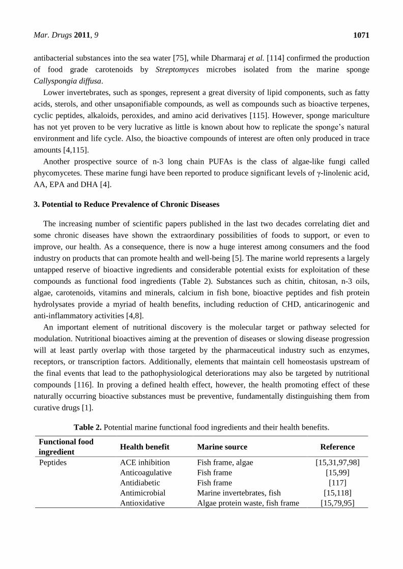
3. Potential to Reduce Prevalence of Chronic Diseases
The increasing number of scientific papers published in the last two decades correlating diet and
some chronic diseases have shown the extraordinary possibilities of foods to support, or even to
improve, our health. As a consequence, there is now a huge interest among consumers and the food
industry on products that can promote health and well-being [5]. The marine world represents a largely
untapped reserve of bioactive ingredients and considerable potential exists for exploitation of these
compounds as functional food ingredients (Table 2). Substances such as chitin, chitosan, n-3 oils,
algae, carotenoids, vitamins and minerals, calcium in fish bone, bioactive peptides and fish protein
hydrolysates provide a myriad of health benefits, including reduction of CHD, anticarinogenic and
anti-inflammatory activities [4,8].
An important element of nutritional discovery is the molecular target or pathway selected for
modulation. Nutritional bioactives aiming at the prevention of diseases or slowing disease progression
will at least partly overlap with those targeted by the pharmaceutical industry such as enzymes,
receptors, or transcription factors. Additionally, elements that maintain cell homeostasis upstream of
the final events that lead to the pathophysiological deteriorations may also be targeted by nutritional
compounds [116]. In proving a defined health effect, however, the health promoting effect of these
naturally occurring bioactive substances must be preventive, fundamentally distinguishing them from
curative drugs [1].
Table 2. Potential marine functional food ingredients and their health benefits.
Functional food
ingredient Health benefit Marine source Reference
Peptides ACE inhibition Fish frame, algae [15,31,97,98]
Anticoagulative Fish frame [15,99]
Antidiabetic Fish frame [117]
Antimicrobial Marine invertebrates, fish [15,118]
Antioxidative Algae protein waste, fish frame [15,79,95]

Mar. Drugs 2011, 9
1072
Table 2. Cont.
n-3 fatty acids Anticarcinogenic Fish [119–122]
Anti-inflammatory Fish, mussels [20,123]
Cardioprotective Fish [124,125]
Cognitive function Fish [126,127]
Polysaccharides Anticarcinogenic Algae, crustaceans
(chito-oligosaccharides)
[94,128,129]
Antioxidative Algae, crustaceans
(chito-oligosaccharides)
[129,130]
Antiviral Algae [83,129]
Cardioprotective Algae [131–133]
Carotenoids Anticarcinogenic Algae [58,134]
Antioxidative Algae [27,48]
Anti-obesity Algae [70]
Antidiabetic Algae [135]
Chlorophyll Anticarcinogenic Algae [58,71]
Polyphenols Antidiabetic Algae [136–138]
Antioxidative Brown algae [73]
3.1. Cancer
Many potent natural products which display effective anticancer activities have been discovered in
the marine environment. Indeed, since the early 1990s, there has been a dramatic increase in the
number of preclinical anticancer lead compounds from marine sources that have entered into human
clinical trials [139,140]. One of these compounds, trabectidin (Yondelis®), originally isolated from the
Caribbean marine tunicate Ecteinascidia turbinate, has been approved for use as an anticancer agent in
Europe [141,142]. In addition, dehydrodidemnin B (aplidine), a compound extracted from the
Mediterranean marine tunicate, Aplidium albicans, has been shown to be a powerful antitumor agent
with possible applications in treating prostate, gastric, breast and colon cancers [140,143].
Marine-derived anticancer molecules have varying modes of action. A cell culture study by Russo
et al. [144] reported that two lichen metabolites, sphaerophorin and pannarin, prevented UV light and
nitric oxide mediated plasmid DNA damage, and attenuated the growth of melanoma cells by, at least
in part, triggering an apoptotic process. Moreover, a number of isolated marine sponge compounds are
inhibitors of protein kinase C (PKC) e.g., BRS1, isoaaptamine, debromohymenialdisine. PKC
inhibitors have attracted much attention as there is evidence that too high levels of PKC enzyme are
involved in the pathogenesis of arthritis and psoriasis, and in tumor development. PKC is believed to
be the receptor protein of tumor promoting phorbol esters, and PKC inhibitors prevent binding of
carcinosarcoma cells to the endothelium [145]. The cytoskeleton is also an interesting target for cancer
therapy, as the microtubules and microfilaments are involved in cellular organisation during cell
division. A number of extracts of marine sponges and ascidians are believed to inhibit the protein by
binding to the microtubule binding site, ―locking up‖ the protein’s motor function, and thereby
blocking cell division [145–147]. Other metabolites can inhibit cell division by disrupting the
polymerisation of actin [148,149], inhibition of cyclin-dependent kinase 4 [150,151], inhibition of

Mar. Drugs 2011, 9
1073
protein synthesis [152,153] or by blocking topoisomerase II [154–156], the nuclear enzyme which
makes transient DNA breaks that are required for replication [157].
In contrast, incubation with kahalalide F, a cyclic depsipeptide from the herbivorous marine
mollusk, Elysia rufescens, quickly induced loss of mitochondrial membrane potential and lysosomal
integrity, severe cytoplasmic swelling and vacuolisation, irregular clumping of chromatin within the
cell nucleus, and finally, cell death in human cancer cells. These effects were independent of caspase
activation and were not associated with DNA degradation or cell cycle block. Kahalalide F has shown
to be effective against cell lines with strong multidrug resistance and against cell lines resistant to
topoisomerase II inhibitors. In vivo models have also confirmed activity in various solid tumor models
[158,159]. To date, copious numbers of compounds with reported anticancer activities have been
extracted from marine organisms, however, for some, their exact effects are still
unclear [140,142,145].
3.1.1. Algal Polysaccharides
Wijesekara et al. [129] recently reported that algal sulfated polysaccharides have potent capacities
for new anticancer product developments in the pharmaceutical as well as the food industries. Indeed,
several in vivo mouse studies have demonstrated the antitumor activity of marine-based
polysaccharides [94,160–164]. Moreover, non-digestible oligosaccharides, which are found in
abundance in macroalgae, are known to improve gut microecology and in doing so may reduce the risk
of colon cancer. In a review by Mussatto and Mancilha [38], the authors state that intake of
transgalactosylated disaccharides reduces the faecal pH as well as ammonia, p-cresol and indole
concentrations, with an increase in bifidobacteria and lactobacilli and a decrease in Bacteroidaceae
populations. As these changes in faecal physiological parameters are believed to reduce the risk of
cancer development, macroalgal non-digestible oligosaccharides could be considered as potential
anticarcinogenic food ingredients [34,38].
3.1.2. n-3 Polyunsaturated Fatty Acids
While the cardioprotective effects of fish oil n-3 PUFAs are well established, their antitumoral
effects are not widely acknowledged. However, promising data from experimental studies carried out
in animals show that elevated supplies of EPA, DHA and/or fish oil diet supplementation generally
inhibit tumor growth and metastases occurrence [121,122,165–167]. Using a mouse model of
MDA-MB-231 human breast cancer cell metastasis to bone, research by Mandal et al. [120] found that
a fish oil diet enriched in DHA and EPA prevented the formation of osteolytic lesions in bone,
indicating suppression of cancer cell metastasis to bone. The study also revealed markedly reduced
levels of CD44 mRNA and protein (associated with generation of cancer stem cells) in the tumors of
mice fed fish oil diet compared to those fed the control diet. Furthermore, Brown and colleagues [168]
provided in vitro evidence supporting epidemiological data that the dietary ratio of n-3:n-6 is crucial in
determining the risk of metastatic disease in prostate cancer. Increasing the ratio of n-3 to n-6 PUFAs,
in particular increasing the amount of EPA in the diet, can inhibit the metastatic process by blocking
the production of prostaglandin E2 and thereby reducing the risk of aggressive disease.
Mar. Drugs 2011, 9
1074
3.1.3. Carotenoids and Chlorophylls
Being lipid soluble, carotenoids are absorbed with fats and circulate bound to different lipoproteins.
The principal biological effects of carotenoids relate to their antioxidant properties, which form the
basis of potential protection against lipid peroxidation, atherogenesis, DNA oxidation, and cancer
[169]. Carotenoids have been implicated in the inhibition of cancer cells in vitro [170–172], in animal
models [173,174] and in humans, as important dietary phytonutrients having cancer preventive activity
for lung, colon, breast and prostate cancer [175,176]. As regards marine-sourced carotenoids, Cha
et al. [134] found that the carotenoid extract of Chlorella ellipsoidea exerted strong antiproliferative
effects on human colon cancer cells, including induction of apoptosis. The authors suggest that
bioactive xanthophylls of C. ellipsoidea could be potential therapeutic agents in the prevention of
human cancers. Several studies have also demonstrated the anticancer activity of astaxanthin in
mammals [177–179].
The cancer preventative effect of chlorophyll derivatives have been extensively studied, with
particular emphasis on their in vitro antimutagenic activity against numerous dietary and
environmental mutagens [180–184]. In a study presented by Chernomorsky et al. [71], the authors
conclude that food sources that yield chlorophyll derivatives may play a significant role in cancer
prevention. They found that dietary chlorophyll derivatives exhibit antimutagenic effects and reduce
tumor cell growth. Indeed, epidemiological evidence has linked diets high in chlorophyll with a
reduced risk of colon cancer in humans [185]. Antioxidant activity, mutagen trapping, modulation of
detoxification pathways, and induction of apoptosis in cancer cells have been highlighted as possible
modes of actions responsible for chlorophyll’s protective effects in vivo [186].
Neither carotenoids nor chlorophylls can be synthesized by animal tissues [187]. Thus, these
molecules must be obtained from food and, as previously illustrated, the marine environment
represents an endless resource. Carotenoid and chlorophyll molecules can be extracted and used as
natural colorants and antioxidants to restore the natural level of these molecules in food or to prepare
fortified products. They can also be chemically modified before being incorporated into food products
[187]. Overall, positive data from in vitro and animal studies have prompted an increased interest in
the potential usefulness of carotenoids and chlorophylls as preventative agents for human populations
at elevated risk of development of specific cancers [186].
3.2. Cardiovascular Disease
Cardiovascular disease (CVD) is a class of diseases that affect the heart, blood vessels (arteries and
veins) and blood circulation, and is one of the leading causes of mortality and morbidity worldwide.
Examples of CVD include atherosclerosis, CHD, stroke, heart failure, deep vein thrombosis and
peripheral arterial disease. In relation to marine bioactives, there is considerable evidence that these
compounds can help to reduce the risk factors associated with CVD. Low density lipoprotein (LDL)
cholesterol was significantly lower in rats fed a diet containing dried Ulva rigida [13], while Oben and
colleagues [188] found that individuals given a freshwater algae infusion displayed lower total
cholesterol, LDL cholesterol and triglyceride levels, and higher high density lipoprotein (HDL)
cholesterol values than those given a water placebo.

Mar. Drugs 2011, 9
1075
3.2.1. Polysaccharides
Numerous epidemiological studies have shown a strong correlation between high fibre diets and a
lower incidence of chronic disorders such as CVD [189–193]. Soluble fibre forms a viscous
indigestible mass in the gut and helps trap digestive enzymes, cholesterol, starch, glucose, and toxins
which are then expelled through the faeces. The soluble fraction of fibres has a hypocholesterolemic
effect, possibly due to augmented gastrointestinal content interfering with micelle formation and lipid
absorption, or an increase and excretion of neutral sterols and biliary acids. Given that seaweed contain
a large amount of soluble polysaccharides, they therefore have potential function as dietary fibre [194].
Additionally, several investigators have reported that water soluble fractions of seaweeds or isolated
algal polysaccharides induce hypocholesterolemic and antihypertensive effects in experimental
animals [131,195,196]. Indeed, sodium alginate and fucoidan were found to decrease serum
cholesterol levels in vivo [132,133,197].
3.2.2. n-3 Polyunsaturated Fatty Acids
The amount of fat in the diet and the type of fatty acids consumed can influence the likelihood of
CVD and its risk factors [22]. The first recognition of the beneficial effect of fatty acids on CVD came
from the observations on the longevity of Eskimos, which was later attributed to the high contents of
fish-derived n-3 long chain fatty acids (e.g., EPA and DHA) in their diets [198–201]. Since then, the
cardioprotective effects of fish oil n-3 PUFAs appear well determined. Numerous epidemiological and
experimental studies have conclusively shown that diets rich in fish and fish oils are associated with a
reduced risk of CVD [202–211]. The omega-3 index, a proposed biomarker for CVD risk, is the level
of EPA and DHA in red blood cell membranes (expressed as a percent of total fatty acids), and an
index of 8% or greater has been posed as a target for reducing CVD [212]. Research carried out by
Lerman et al. [213] found that provision of 2–3 g/day of EPA and DHA for 12 weeks increased the
omega-3 index by 3.7 to 4.1 percentage points to cardioprotective levels. Moreover, fish oil
supplementation has been shown to be beneficial in controlling high blood pressure [214–216].
3.2.3. ACE-Inhibitory Peptides
High blood pressure (hypertension) is one of the major independent risk factors for CVD [217].
ACE plays a crucial role in the regulation of blood pressure [15], and so ACE inhibition is considered
to be a useful therapeutic approach in the treatment of hypertension. Numerous investigations of
marine-derived peptides have revealed potent antihypertensive and ACE inhibitory activities in
hypertensive rats [218–221]. According to Lee et al. [222], a single oral administration of a peptide
derived from tuna frame protein hydrolysate showed a strong suppressive effect on systolic blood
pressure of spontaneously hypertensive rats and this antihypertensive activity was similar to that of
captopril, a commercial antihypertensive drug. Therefore, due to their effectiveness in both prevention
and treatment of hypertension, marine-derived bioactive peptides have prospective use as functional
ingredients [15].

Mar. Drugs 2011, 9
1076
3.2.4. Astaxanthin
Due to their antioxidant properties, carotenoids are believed to have therapeutic benefit in treating
CVD [223,224]. In addition, astaxanthin, a carotenoid ubiquitous in the marine environment, exhibits
anti-atherogenic effects. In a study involving hyperlipidemic rabbits, astaxanthin significantly reduced
macrophage infiltration in lesions and lowered the occurrence of macrophage apoptosis and plaque
ruptures [225]. Indeed, results of human intervention trials indicate that consumption of natural
astaxanthin could contribute to prevention of atherosclerosis. Iwamoto et al. [226] reported a dose
response relationship between astaxanthin and LDL oxidation time, while Yoshida and colleagues
[227] recently demonstrated that astaxanthin intake ameliorates triglyceride and HDL cholesterol in
correlation with increased adiponectin. Antihypertensive effects were also revealed when oral
administration of astaxanthin for 14 days induced a significant reduction in arterial blood pressure in
spontaneously hypertensive rats [228]. Furthermore, in a follow-up study, the authors suggest that
astaxanthin could modulate the oxidative condition and may improve vascular elastin and arterial wall
thickness in hypertension [229].
As diet is now recognised as an important modifiable risk factor for CVD, the incorporation of
marine bioactives in food could benefit heart health by modifying blood levels of HDL and LDL
cholesterol, as well as reducing hypertension (Figure 1). In particular, n-3 PUFAs and astaxanthin have
use as dietary supplements for the prevention or the alleviation of certain CVD as they also reduce
inflammation often associated with the development of CHD. Besides, several comprehensive reviews
examine the literature on pharmacological studies of marine natural compounds that affect the
cardiovascular system [145,230,231].
Figure 1. Beneficial effects of marine bioactives on the human cardiovascular system.

Mar. Drugs 2011, 9
1077
3.3. Inflammatory Conditions
Inflammation is a normal protective response to tissue damage or infection. However, if the
response is exaggerated, misdirected or long term, inflammation can adversely affect health and give
rise to many conditions such as inflammatory bowel disease, arthritis and asthma [22,86]. Interestingly
however, owing to the involvement of inflammatory mediators called eicosanoids, a number of
inflammatory conditions could potentially be alleviated by dietary modification [22].
As well as the various benefits accrued from the consumption of n-3 PUFAs as discussed
previously, the eicosanoids derived from n-3 fatty acids are considered to be less inflammatory or even
anti-inflammatory compared to eicosanoids derived from n-6 fatty acids [20]. Research has shown that
increasing the balance of n-3 fatty acids in the diet, and consequently favouring the production of EPA
in the body, or by increasing the dietary intake of EPA and DHA through consumption, leads the body
to a more anti-inflammatory environment [232]. As a result, the incidence or severity of many chronic
inflammatory diseases may be reduced [20,22,233]. In Crohn’s disease (a chronic inflammatory
disease of the alimentary tract) for example, relapse rates reduced substantially over a 12 month period
in patients receiving a fish oil supplement [234].
Another anti-inflammatory compound found extensively in the marine environment is the
carotenoid astaxanthin. The antioxidative properties of astaxanthin are believed to be linked to its
ability to relieve inflammation [89,224]. Bennedsen et al. [235] found that treatment with a cell extract
from the microalgae Haematococcus pluvialis containing astaxanthin reduced bacterial load and
gastric inflammation in Helicobacter pylori infected mice. It has also been reported that astaxanthin
significantly reduces the production of pro-inflammatory mediators and cytokines, namely nuclear
factor-κB (NF-κB), tumour necrosis factor-α (TNF-α) and interleukin-6 (IL-6) [236,237], and
suppresses T lymphocyte activation in asthma patients [238]. However, further studies are needed to
fully elucidate the anti-inflammatory effects of astaxanthin.
3.3.1. Arthritis
Arthritis describes a condition involving inflammation of the joints and is a disease affecting mostly
the aged population. Preventing inflammation with its associated pain and reduced mobility symptoms
is a primary requirement in arthritis treatment [194]. Due to the involvement of inflammatory
eicosanoids in the aetiology of this disorder, diet may be a potential therapeutic agent [22], and so the
importance of dietary PUFAs in the treatment of arthritis has been greatly investigated [239–241].
Meta-analysis of 17 randomised, controlled trials was conducted by Goldberg and Katz [233] to assess
the analgesic effects of n-3 PUFAs in patients with rheumatoid arthritis or joint pain. Favourable
outcomes such as reduced patient reported joint pain intensity, minutes of morning stiffness, number of
painful and/or tender joints and reduced non-steroidal anti-inflammatory drugs consumption were
reported following 3 to 4 months supplementation with n-3 PUFAs. In fact, the authors conclude that
n-3 PUFA supplementation is an attractive adjunctive treatment for joint pain.
In addition to n-3 PUFAs, clinical investigations suggest that ingestion of collagen hydrolysates,
which can be isolated from fish waste, reduces pain in patients suffering from osteoarthritis [242].

Mar. Drugs 2011, 9
1078
3.3.2. Asthma
Fish oil or fish containing more than 2% fat has been found to have a reduced risk of airway
hyperresponsiveness, and children who regularly eat fresh, oily fresh have a reduced risk of developing
asthma than children who rarely eat fish [243,244]. Supplementation of diet with n-3 fatty acids also
confirmed their benefit in the reduction of breathing difficulties and other symptoms, along with
reduced drug doses required by asthma patients. During treatment, the increase in the content of n-3
fatty acids in cell membranes was reported to take place at the expense of AA resulting in the
competitive inhibition of pro-inflammatory eicosanoid production and production of anti-inflammatory
eicosanoids [194,245,246]. Moreover, a number of interventional studies have demonstrated
improvement in asthmatic status following n-3 PUFA supplementation [247–249]. Emelyanov et al.
[123] assessed the effect of n-3 PUFA rich lipid extract of New Zealand green lipped mussel on
symptoms, peak expiratory flow and exhaled hydrogen peroxide (a marker of airway inflammation) in
patients with atopic asthma. This double blind randomised trial revealed a significant decrease in
daytime wheeze, reduction in the concentration of exhaled hydrogen peroxide and an increase in
morning peak expiratory flow compared to the placebo group.
3.3.3. Neuroinflammation
Recently, there has been recognition of an inflammatory component to the pathology of
neurodegeneration, most notably in Alzheimer’s disease but also in Parkinson’s disease and motor
neuron disease [250]. As anti-inflammatory n-3 PUFAs are preferentially incorporated in the brain, a
diet rich in EPA and DHA could keep neuroinflammation at a minimum [251]. In fact, elderly people
who eat fish or seafood, that are highly enriched in n-3 PUFAs, at least once a week, have been shown
to be at lower risk of developing dementia including Alzheimer’s disease [252,253].
Evidence is also emerging which suggests that marine algae could possess therapeutic activities for
combating neurodegenerative diseases associated with neuroinflammation. Jin and colleagues [254]
found that Ulva conglobata extract almost completely suppressed the expression of the
pro-inflammatory enzyme cyclooxygenase-2 (COX-2) and inducible nitric oxide (iNOS) in murine
BV2 microglia. Similarly, the brown alga, Ecklonia cava, was reported to induce significant inhibition
of NF-κB dependent cytokines as well as iNOS and COX-2, thus reducing inflammation [255]. In
addition, red alga Neorhodomela aculeate could be considered as a potential neuroprotective and
anti-inflammatory agent to treat aging related neurological disorders [256]. However, as highlighted by
the authors, further studies are needed to determine which components of each of the algae contribute
to the observed anti-inflammatory activities.
As well as the nutritional bioactives discussed, numerous anti-inflammatory compounds with
potential pharmacological applications have been isolated from marine sources
[7,145,230,231,257,258]. Nevertheless, n-3 PUFAs appear to be the most prominent and most
promising anti-inflammatory agents. In spite of this, there have not been sufficient studies to warrant a
dietary recommendation regarding the use of n-3 PUFAs in the management of inflammatory
conditions, and so there is a need for more carefully designed and controlled clinical trials in the
therapeutic applications of n-3 fatty acids [22]. There is, however, a potential complementary role

Mar. Drugs 2011, 9
1079
between drug therapy and a diet rich in n-3 PUFAs. An increased intake of n-3 fatty acids may
increase the efficacy of anti-inflammatory medications, and perhaps reduce the need for conventional
drugs. This dietary choice could be achieved through the use of marine-sourced PUFAs as fortificants
in margarine, dairy products (milk, yoghurt, cheese), breads and baked goods, meat-based and
fish-based meals, for example.
3.4. Cognitive Decline and Depression
Cognitive impairment and dementia are frequent disorders among elderly persons and influence the
individual’s ability to function independently. Due to the aging of the population, the prevalence of
cognitive impairment and dementia are expected to increase [259]. Drugs currently used in the
treatment of cognitive decline and dementia have a very limited therapeutic value, suggesting the
necessity to potentially individualise new strategies able to prevent and to slow down the progression
of predementia and dementia syndromes [260]. Numerous epidemiological studies on the association
between diet and cognitive decline suggest a role of fatty acids intake in maintaining adequate
cognitive functioning and possibly in preventing or delaying the onset of dementia, both of
degenerative or vascular origin [261–263]. In particular, fatty fish and marine n-3 PUFA consumption
was associated with a reduced risk [264]. Moreover, a diet enriched with the algae, Chlorella, reduced
oxidative stress and significantly prevented the decline of cognitive ability in an age dependent
dementia mouse model. The authors suggest that the prolonged consumption of Chlorella has the
potential to prevent the progression of cognitive impairment [265].
Several hypotheses could explain the association between dietary unsaturated fatty acids and
cognitive functioning, including mechanisms through the co-presence of antioxidant compounds in
food groups rich in fatty acids, via atherosclerosis and thrombosis, inflammation, accumulation of
amyloid β-peptide, or via an effect in maintaining the structural integrity of neuronal membranes
[260]. It is also believed that DHA is of particular importance for brain function as it maintains an
optimal state of neural membranes, enabling membrane fluidity and thickness, which in turn affects
cell signalling [266,267].
In general, PUFAs of marine origin appear to be suitable candidates for functional food ingredients
to relieve memory deficits associated with aging. Promising results have already been reported in
young population samples. Supplementation with a fish-flour bread spread containing n-3 PUFAs,
embedded in a natural food matrix, had a beneficial effect on learning and memory of children [267].
In addition, a study involving marine collagen peptide (MCP) derived from Chum Salmon skin
demonstrated that MCP facilitates learning and memory in aged mice by reducing oxidative damage in
the brain and increasing brain derived neurotrophic factor and PSD95 protein expression [268].
Several epidemiological- and dietary-based studies also suggest that the consumption of n-3 fatty
acids is inversely correlated to the prevalence and severity of depression, while clinical studies have
presented evidence relating to the benefits of n-3 compounds in the treatment of depressive disorders
[269,270]. In some trials, supplementation with long chain n-3 PUFAs has been shown to improve
mood in patients with major depression and bipolar disorder [271–274]. More recently, Venna et al.
[275] revealed that chronic supplementation with n-3 PUFAs induced antidepressant-like effects in

Mar. Drugs 2011, 9
1080
mice, while in a similar mouse study, natural products isolated from marine sponges were found to
possess antidepressant properties[276].
3.5. Diabetes
Dietary management of diabetes involves maintaining both blood glucose and blood lipid
concentrations at as near normal levels as possible to reduce the possibility of associated complications
developing, e.g., CVD, diabetic neuropathy, retinopathy and nephropathy [22]. As well as offering
protection against heart disease and cancer, some marine extracts have been associated with
improvements in glycaemic control (outlined in Table 3). Consequently, these compounds could be
exploited as potential functional food ingredients in an effort to prevent or diminish insulin resistance
and diabetes.
4. Conclusions
With increased life expectancy, our diet will play a key role in sustaining human health. This is a
challenge for the food industry as consumers not only demand tasty and convenient food, but also
healthy, nutritious food [116]. Marine nutraceuticals are both a coherent and attractive option for the
food industry as there are a multitude of functional food ingredients that can be derived from marine
sources. It is also believed that, as people become increasingly aware of the association between diet
and good health, the consumption of fishery products will most likely increase [8]. Moreover, in
relation to marine algae, their content in proteins, carbohydrates, lipids, fibre, metabolites, etc. can be
influenced by their growing parameters (water temperature, salinity, light and nutrients) [5]. This
means that, from a biotechnological perspective, algae can be considered as natural bioreactors, able to
provide different types of compounds at different quantities—an appealing attribute to the functional
food industry.
Despite the vast possibilities for use of marine bioactives in food, more multidisciplinary research is
needed. All aspects, including chemical composition, biotechnology, extraction, bioactivity, and
toxicity should be considered. Also, to effectively transfer the research into a practical field in which
marine bioactives can be used as functional food ingredients, the exact functional activity of the
extracts/compounds should be determined [52]. This primary objective should be based on
investigations in vitro or ex vivo in cellular lines or culture tissues, later in animal models and then
corroborated in studies of observation or intervention in human clinical trials [5]. Finally, to be
employed as ingredients in food products, different studies should be carried out to determine if their
activity is maintained after manufacturing and cooking processes [52].
In conclusion, marine bioactives appear to fit the criteria for development as functional food
ingredients. Firstly, they are widely available, with a guaranteed supply. Secondly, marine bioactives
are naturally occurring compounds, and their isolation/extraction is relatively cost effective. Lastly,
and probably most importantly, they are functional—their biological activities affect the pathogenesis
of several diseases. Consequently, ongoing efforts should be made into the research and development
of marine functional foods with prospect that, in the future, their consumption could lead to a reduction
in the prevalence and severity of chronic diseases.
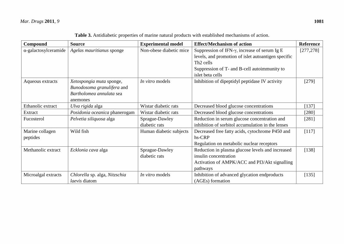
Mar. Drugs 2011, 9
1081
Table 3. Antidiabetic properties of marine natural products with established mechanisms of action.
Compound Source Experimental model Effect/Mechanism of action Reference
α-galactosylceramide Agelas mauritianus sponge Non-obese diabetic mice Suppression of IFN-γ, increase of serum Ig E
levels, and promotion of islet autoantigen specific
Th2 cells
Suppression of T- and B-cell autoimmunity to
islet beta cells
[277,278]
Aqueous extracts Xetospongia muta sponge,
Bunodosoma granulifera and
Bartholomea annulata sea
anemones
In vitro models Inhibition of dipeptidyl peptidase IV activity [279]
Ethanolic extract Ulva rigida alga Wistar diabetic rats Decreased blood glucose concentrations [137]
Extract Posidonia oceanica phanerogam Wistar diabetic rats Decreased blood glucose concentrations [280]
Fucosterol Pelvetia siliquosa alga Sprague-Dawley
diabetic rats
Reduction in serum glucose concentration and
inhibition of sorbitol accumulation in the lenses
[281]
Marine collagen
peptides
Wild fish Human diabetic subjects Decreased free fatty acids, cytochrome P450 and
hs-CRP
Regulation on metabolic nuclear receptors
[117]
Methanolic extract Ecklonia cava alga Sprague-Dawley
diabetic rats
Reduction in plasma glucose levels and increased
insulin concentration
Activation of AMPK/ACC and PI3/Akt signalling
pathways
[138]
Microalgal extracts Chlorella sp. alga, Nitzschia
laevis diatom
In vitro models Inhibition of advanced glycation endproducts
(AGEs) formation
[135]

Mar. Drugs 2011, 9
1082
Table 3. Cont.
n-3 PUFAs Fish oil Wistar rats Restoration of insulin receptor and insulin
receptor substrate-1 tyrosine phosphorylation
Maintenance of phosphatidylinositol-3’ kinase
activity and GLUT-4 content in muscle
[282]
Fish oil Healthy human subjects Reduction in glucose oxidation, increased fat
oxidation and glycogen storage
[283]
Phenylmethylene
hydantoins
Hemimycale arabica sponge In vitro model
Sprague-Dawley rats
Inhibition of glycogen synthase kinase-3β activity
Increased liver glycogen
[284]
Phlorotannin
components
Ascophyllum nodosum alga In vitro models Inhibition of α-amylase and α-glucosidase
activities
[136]
Sodium alginate Laminaria angustata alga Wistar rats Inhibition of rising blood glucose and insulin
levels
[197]
Mar. Drugs 2011, 9
1083
Acknowledgements
This work is supported through the NutraMara programme, funded by the Marine Institute and the
Department of Agriculture, Fisheries and Food.
References
1. Biesalski, H.-K.; Dragsted, L.O.; Elmadfa, I.; Grossklaus, R.; Müller, M.; Schrenk, D.;
Walter, P.; Weber, P. Bioactive compounds: Definition and assessment of activity. Nutrition
2009, 25, 1202–1205.
2. Honkanen, P. Consumer acceptance of (marine) functional food. In Marine Functional Food, 1st
ed.; Luten, J., Ed.; Wageningen Academic Publishers: Wageningen, The Netherlands, 2009;
Volume 1, pp. 141–154.
3. Siró, I.; Kápolna, E.; Kápolna, B.; Lugasi, A. Functional food. Product development, marketing
and consumer acceptance-a review. Appetite 2008, 51, 456–467.
4. Rasmussen, R.S.; Morrissey, M.T. Marine biotechnology for production of food ingredients. Adv.
Food Nutr. Res. 2007, 52, 237–292.
5. Plaza, M.; Cifuentes, A.; Ibáñez, E. In the search of new functional food ingredients from algae.
Trends Food Sci. Technol. 2008, 19, 31–39.
6. Bocanegra, A.; Bastida, S.; Benedí, J.; Ródenas, S.; Sánchez-Muniz, F.J. Characteristics and
nutritional and cardiovascular-health properties of seaweeds. J. Med. Food 2009, 12, 236–258.
7. El Gamal, A.A. Biological importance of marine algae. Saudi Pharm. J. 2010, 18, 1–25.
8. Kadam, S.; Prabhasankar, P. Marine foods as functional ingredients in bakery and pasta products.
Food Res. Int. 2010, 43, 1975–1980.
9. Mabeau, S.; Fleurence, J. Seaweed in food products: Biochemical and nutritional aspects. Trends
Food Sci. Technol. 1993, 4, 103–107.
10. Fleurence, J. Seaweed proteins: Biochemical, nutritional aspects and potential uses. Trends Food
Sci. Technol. 1999, 10, 25–28.
11. Dawczynski, C.; Schubert, R.; Jahreis, G. Amino acids, fatty acids, and dietary fibre in edible
seaweed products. Food Chem. 2007, 103, 891–899.
12. Galland-Irmouli, A.-V.; Fleurence, J.; Lamghari, R.; Luçon, M.; Rouxel, C.; Barbaroux, O.;
Bronowicki, J.-P.; Villaume, C.; Guéant, J.-L. Nutritional value of proteins from edible seaweed
palmaria palmata (dulse). J. Nutr. Biochem. 1999, 10, 353–359.
13. Taboada, C.; Millán, R.; Míguez, I. Composition, nutritional aspects and effect on serum
parameters of marine algae ulva rigida. J. Sci. Food Agric. 2010, 90, 445–449.
14. Pihlanto-Leppälä, A. Bioactive peptides derived from bovine whey proteins: Opioid and ace-
inhibitory peptides. Trends Food Sci. Technol. 2000, 11, 347–356.
15. Kim, S.-K.; Wijesekara, I. Development and biological activities of marine-derived bioactive
peptides: A review. J. Funct. Foods 2010, 2, 1–9.
16. Elias, R.J.; Kellerby, S.S.; Decker, E.A. Antioxidant activity of proteins and peptides. Crit. Rev.
Food Sci. Nutr. 2008, 48, 430–441.
17. Burtin, P. Nutritional value of seaweeds. EJEAFChe 2003, 2, 498–503.

Mar. Drugs 2011, 9
1084
18. Aneiros, A.; Garateix, A. Bioactive peptides from marine sources: Pharmacological properties
and isolation procedures. J. Chromatogr. B 2004, 803, 41–53.
19. MacArtain, P.; Gill, C.I.R.; Brooks, M.; Campbell, R.; Rowland, I.R. Nutritional value of edible
seaweeds. Nutr. Rev. 2007, 65, 535–543.
20. Wall, R.; Ross, R.P.; Fitzgerald, G.F.; Stanton, C. Fatty acids from fish: The anti-inflammatory
potential of long-chain omega-3 fatty acids. Nutr. Rev. 2010, 68, 280–289.
21. Calder, P.C. n-3 Polyunsaturated fatty acids, inflammation, and inflammatory diseases. Am. J.
Clin. Nutr. 2006, 83, 1505–1519.
22. Lunn, J.; Theobald, H. The health effects of dietary unsaturated fatty acids. Nutr. Bull. 2006, 31,
178–224.
23. Dembitsky, V.M.; Pechenkina-Shubina, E.E.; Rozentsvet, O.A. Glycolipids and fatty acids of
some seaweeds and marine grasses from the black sea. Phytochemistry 1991, 30, 2279–2283.
24. Sánchez-Machado, D.; López-Cervantes, J.; López-Hernández, J.; Paseiro-Losada, P. Fatty acids,
total lipid, protein and ash contents of processed edible seaweeds. Food Chem. 2004, 85,
439–444.
25. Piovetti, L.; Deffo, P.; Valls, R.; Peiffer, G. Determination of sterols and diterpenoids from
brown algae (cystoseiraceae). J. Chromatogr. A 1991, 588, 99–105.
26. Haugan, J.A.; Liaaen-Jensen, S. Algal carotenoids 54. Carotenoids of brown algae
(phaeophyceae). Biochem. Syst. Ecol. 1994, 22, 31–41.
27. Yan, X.; Chuda, Y.; Suzuki, M.; Nagata, T. Fucoxanthin as the major antioxidant in hijikia
fusiformis, a common edible seaweed. Biosci. Biotechnol. Biochem. 1999, 63, 605–607.
28. Dembitsky, V.M.; Maoka, T. Allenic and cumulenic lipids. Prog. Lipid Res. 2007, 46, 328–375.
29. Gómez-Ordóñez, E.; Jiménez-Escrig, A.; Rupérez, P. Dietary fibre and physicochemical
properties of several edible seaweeds from the northwestern spanish coast. Food Res. Int. 2010,
43, 2289–2294.
30. Jiménez-Escrig, A.; Sánchez-Muniz, F. Dietary fibre from edible seaweeds: Chemical structure,
physicochemical properties and effects on cholesterol metabolism. Nutr. Res. 2000, 20, 585–598.
31. Wijesekara, I.; Kim, S.K. Angiotension-i-converting enzyme (ace) inhibitors from marine
resources: Prospects in the pharmaceutical industry. Mar. Drugs 2010, 8, 1080–1093.
32. Berteau, O.; Mulloy, B. Sulfated fucans, fresh perspectives: Structures, functions, and biological
properties of sulfated fucans and an overview of enzymes active towards this class of
polysaccharide. Glycobiology 2003, 13, 29R–40R.
33. Pomin, V.H.; Mourão, P.A.S. Structure, biology, evolution, and medical importance of sulfated
fucans and galactans. Glycobiology 2008, 18, 1016–1027.
34. O’Sullivan, L.; Murphy, B.; McLoughlin, P.; Duggan, P.; Lawlor, P.G.; Hughes, H.; Gardiner,
G.E. Prebiotics from marine macroalgae for human and animal health applications. Mar. Drugs
2010, 8, 2038–2064.
35. Devillé, C.; Damas, J.; Forget, P.; Dandrifosse, G.; Peulen, O. Laminarin in the dietary fibre
concept. J. Sci. Food Agric. 2004, 84, 1030–1038.
36. Devillé, C.; Gharbi, M.; Dandrifosse, G.; Peulen, O. Study on the effects of laminarin, a
polysaccharide from seaweed, on gut characteristics. J. Sci. Food Agric. 2007, 87, 1717–1725.

Mar. Drugs 2011, 9
1085
37. Courtois, J. Oligosaccharides from land plants and algae: Production and applications in
therapeutics and biotechnology. Curr. Opin. Microbiol. 2009, 12, 261–273.
38. Mussatto, S.I.; Mancilha, I.M. Non-digestible oligosaccharides: A review. Carbohydr. Polym.
2007, 68, 587–597.
39. Wang, Y. Prebiotics: Present and future in food science and technology. Food Res. Int. 2009, 42,
8–12.
40. Rodríguez-Bernaldo de Quirós, A.; Castro de Ron, C.; López-Hernández, J.; Lage-Yusty, M.
Determination of folates in seaweeds by high-performance liquid chromatography. J.
Chromatogr. A 2004, 1032, 135–139.
41. Sánchez-Machado, D.; López-Hernández, J.; Paseiro-Losada, P. High-performance liquid
chromatographic determination of [alpha]-tocopherol in macroalgae. J. Chromatogr. A 2002,
976, 277–284.
42. Brown, M.R.; Jeffrey, S.W.; Volkman, J.K.; Dunstan, G.A. Nutritional properties of microalgae
for mariculture. Aquaculture 1997, 151, 315–331.
43. Norziah, M.H.; Ching, C.Y. Nutritional composition of edible seaweed gracilaria changgi. Food
Chem. 2000, 68, 69–76.
44. Carballo-Cárdenas, E.C.; Tuan, P.M.; Janssen, M.; Wijffels, R.H. Vitamin e (α-tocopherol)
production by the marine microalgae dunaliella tertiolecta and tetraselmis suecica in batch
cultivation. Biomol. Eng. 2003, 20, 139–147.
45. Durmaz, Y.; Monteiro, M.; Bandarra, N.; Gökpinar, Ş.; Işik, O. The effect of low temperature on
fatty acid composition and tocopherols of the red microalga, porphyridium cruentum. J. Appl.
Phycol. 2007, 19, 223–227.
46. Rebolloso Fuentes, M.M.; Acién Fernández, G.; Sánchez Pérez, J.; Guil Guerrero, J. Biomass
nutrient profiles of the microalga porphyridium cruentum. Food Chem. 2000, 70, 345–353.
47. Inbaraj, B.S.; Chien, J.T.; Chen, B.H. Improved high performance liquid chromatographic
method for determination of carotenoids in the microalga chlorella pyrenoidosa. J. Chromatogr.
A 2006, 1102, 193–199.
48. Hu, C.-C.; Lin, J.-T.; Lu, F.-J.; Chou, F.-P.; Yang, D.-J. Determination of carotenoids in
dunaliella salina cultivated in taiwan and antioxidant capacity of the algal carotenoid extract.
Food Chem. 2008, 109, 439–446.
49. Cha, K.H.; Lee, H.J.; Koo, S.Y.; Song, D.G.; Lee, D.U.; Pan, C.H. Optimization of pressurized
liquid extraction of carotenoids and chlorophylls from chlorella vulgaris. J. Agric. Food Chem.
2010, 58, 793–797.
50. Cha, K.H.; Kang, S.W.; Kim, C.Y.; Um, B.H.; Na, Y.R.; Pan, C.H. Effect of pressurized liquids
on extraction of antioxidants from chlorella vulgaris. J. Agric. Food Chem. 2010, 58,
4756–4761.
51. Yuan, J.-P.; Chen, F.; Liu, X.; Li, X.-Z. Carotenoid composition in the green microalga
chlorococcum. Food Chem. 2002, 76, 319–325.
52. Plaza, M.; Herrero, M.; Cifuentes, A.; Ibáñez, E. Innovative natural functional ingredients from
microalgae. J. Agric. Food Chem. 2009, 57, 7159–7170.

Mar. Drugs 2011, 9
1086
53. Yokthongwattana, K.; Savchenko, T.; Polle, J.E.W.; Melis, A. Isolation and characterization of a
xanthophyll-rich fraction from the thylakoid membrane of dunaliella salina (green algae).
Photochem. Photobiol. Sci. 2005, 4, 1028–1034.
54. Herrero, M.; Jaime, L.; Martín-Alvarez, P.J.; Cifuentes, A.; Ibáñez, E. Optimization of the
extraction of antioxidants from dunaliella salina microalga by pressurized liquids. J. Agric. Food
Chem. 2006, 54, 5597–5603.
55. Grewe, C.; Griehl, C. Time- and media-dependent secondary carotenoid accumulation in
haematococcus pluvialis. Biotechnol. J. 2008, 3, 1232–1244.
56. Jaime, L.; Rodríguez-Meizoso, I.; Cifuentes, A.; Santoyo, S.; Suarez, S.; Ibáñez, E.; Señorans,
F.J. Pressurized liquids as an alternative process to antioxidant carotenoids' extraction from
haematococcus pluvialis microalgae. LWT-Food Sci. Technol. 2010, 43, 105–112.
57. Rodríguez-Meizoso, I.; Jaime, L.; Santoyo, S.; Cifuentes, A.; Garcia-Blairsy Reina, G.;
Señoráns, F.; Ibáñez, E. Pressurized fluid extraction of bioactive compounds from phormidium
species. J. Agric. Food Chem. 2008, 56, 3517–3523.
58. Okai, Y.; Higashi-Okai, K.; Yano, Y.; Otani, S. Identification of antimutagenic substances in an
extract of edible red alga, porphyra tenera (asadusa-nori). Cancer Lett. 1996, 100, 235–240.
59. Plaza, M.; Santoyo, S.; Jaime, L.; García-Blairsy Reina, G.; Herrero, M.; Señoráns, F.J.; Ibáñez,
E. Screening for bioactive compounds from algae. J. Pharm. Biomed. Anal. 2010, 51, 450–455.
60. Mendes, R.L.; Fernandes, H.L.; Coelho, J.P.; Reis, E.C.; Cabral, J.M.; Novais, J.M.; Palavra,
A.F. Supercritical co2 extraction of carotenoids and other lipids from chlorella vulgaris. Food
Chem. 1995, 53, 99–103.
61. Li, H.-B.; Chen, F. Preparative isolation and purification of astaxanthin from the microalga
chlorococcum sp. by high-speed counter-current chromatography. J. Chromatogr. A 2001, 925,
133–137.
62. Tripathi, U.; Sarada, R.; Rao, S.R.; Ravishankar, G.A. Production of astaxanthin in
haematococcus pluvialis cultured in various media. Bioresour. Technol. 1999, 68, 197–199.
63. Wu, Z.; Wu, S.; Shi, X. Supercritical fluid extraction and determination of lutein in
heterotrophically cultivated chlorella pyrenoidosa. J. Food Process Eng. 2007, 30, 174–185.
64. Del Campo, J.A.; García-González, M.; Guerrero, M.G. Outdoor cultivation of microalgae for
carotenoid production: Current state and perspectives. Appl. Microbiol. Biotechnol. 2007, 74,
1163–1174.
65. Macías-Sánchez, M.D.; Mantell, C.; Rodríguez, M.; Martínez de la Ossa, E.; Lubián, L.M.;
Montero, O. Comparison of supercritical fluid and ultrasound-assisted extraction of carotenoids
and chlorophyll a from dunaliella salina. Talanta 2009, 77, 948–952.
66. Rodríguez-Meizoso, I.; Jaime, L.; Santoyo, S.; Señoráns, F.; Cifuentes, A.; Ibáñez, E. Subcritical
water extraction and characterization of bioactive compounds from haematococcus pluvialis
microalga. J. Pharm. Biomed. Anal. 2010, 51, 456–463.
67. Klejdus, B.; Kopecký, J.; Benesová, L.; Vacek, J. Solid-phase/supercritical-fluid extraction for
liquid chromatography of phenolic compounds in freshwater microalgae and selected
cyanobacterial species. J. Chromatogr. A 2009, 1216, 763–771.
68. Rupérez, P. Mineral content of edible marine seaweeds. Food Chem. 2002, 79, 23–26.

Mar. Drugs 2011, 9
1087
69. García-Casal, M.N.; Pereira, A.C.; Leets, I.; Ramírez, J.; Quiroga, M.F. High iron content and
bioavailability in humans from four species of marine algae. J. Nutr. 2007, 137, 2691–2695.
70. Maeda, H.; Hosokawa, M.; Sashima, T.; Funayama, K.; Miyashita, K. Fucoxanthin from edible
seaweed, undaria pinnatifida, shows antiobesity effect through ucp1 expression in white adipose
tissues. Biochem. Biophys. Res. Commun. 2005, 332, 392–397.
71. Chernomorsky, S.; Segelman, A.; Poretz, R.D. Effect of dietary chlorophyll derivatives on
mutagenesis and tumor cell growth. Teratog. Carcinog. Mutagen. 1999, 19, 313–322.
72. Donaldson, M.S. Nutrition and cancer: A review of the evidence for an anti-cancer diet. Nutr. J.
2004, 3, 19.
73. Li, Y.; Qian, Z.-J.; Ryu, B.; Lee, S.-H.; Kim, M.-M.; Kim, S.-K. Chemical components and its
antioxidant properties in vitro: An edible marine brown alga, ecklonia cava. Bioorg. Med. Chem.
2009, 17, 1963–1973.
74. Wang, T.; Jónsdóttir, R.; Ólafsdóttir, G. Total phenolic compounds, radical scavenging and metal
chelation of extracts from icelandic seaweeds. Food Chem. 2009, 116, 240–248.
75. Bhakuni, D.S.; Rawat, D.S. Bioactive Marine Natural Products, 1st ed.; Anamaya Publishers:
New Delhi, India, 2005.
76. Mata, T.M.; Martins, A.A.; Caetano, N.S. Microalgae for biodiesel production and other
applications: A review. Renew. Sust. Energ. Rev. 2010, 14, 217–232.
77. Guil-Guerrero, J.; Navarro-Juárez, R.; López-Martínez, J.; Campra-Madrid, P.;
Rebolloso-Fuentes, M. Functional properties of the biomass of three microalgal species.
J. Food Eng. 2004, 65, 511–517.
78. Spolaore, P.; Joannis-Cassan, C.; Duran, E.; Isambert, A. Commercial applications of
microalgae. J. Biosci. Bioeng. 2006, 101, 87–96.
79. Sheih, I.C.; Wu, T.-K.; Fang, T.J. Antioxidant properties of a new antioxidative peptide from
algae protein waste hydrolysate in different oxidation systems. Bioresour. Technol. 2009, 100,
3419–3425.
80. Metting, F.B. Biodiversity and application of microalgae. J. Ind. Microbiol. 1996, 17, 477–489.
81. Herrero, M.; Ibáñez, E.; Cifuentes, A.; Reglero, G.; Santoyo, S. Dunaliella salina microalga
pressurized liquid extracts as potential antimicrobials. J. Food Prot. 2006, 69, 2471–2477.
82. Mendiola, J.A.; Jaime, L.; Santoyo, S.; Reglero, G.; Cifuentes, A.; Ibañez, E.; Señoráns, F.
Screening of functional compounds in supercritical fluid extracts from spirulina platensis. Food
Chem. 2007, 102, 1357–1367.
83. Huheihel, M.; Ishanu, V.; Tal, J.; Arad, S. Activity of porphyridium sp. Polysaccharide against
herpes simplex viruses in vitro and in vivo. J. Biochem. Biophys. Methods 2002, 50, 189–200.
84. Kanekiyo, K.; Lee, J.-B.; Hayashi, K.; Takenaka, H.; Hayakawa, Y.; Endo, S.; Hayashi, T.
Isolation of an antiviral polysaccharide, nostoflan, from a terrestrial cyanobacterium, nostoc
flagelliforme. J. Nat. Prod. 2005, 68, 1037–1041.
85. Yeum, K.-J.; Russell, R.M. Carotenoid bioavailability and bioconversion. Annu. Rev. Nutr. 2002,
22, 483–504.
86. Miyashita, K. Function of marine carotenoids. Forum Nutr. 2009, 61, 136–146.

Mar. Drugs 2011, 9
1088
87. Dufossé, L.; Galaup, P.; Yaron, A.; Arad, S.M.; Blanc, P.; Chidambara Murthy, K.N.;
Ravishankar, G.A. Microorganisms and microalgae as sources of pigments for food use: A
scientific oddity or an industrial reality? Trends Food Sci. Technol. 2005, 16, 389–406.
88. Olson, J.A.; Krinsky, N.I. Introduction: The colourful, fascinating world of the carotenoids:
Important physiologic modulators. FASEB J. 1995, 9, 1547–1550.
89. Guerin, M.; Huntley, M.E.; Olaizola, M. Haematococcus astaxanthin: Applications for human
health and nutrition. Trends Biotechnol. 2003, 21, 210–216.
90. Undeland, I.; Lindqvust, H.; Chen-Yun, Y.; Falch, E.; Ramel, A.; Cooper, M.; Gildberg, A.;
Luten, J.; Stenberg, E.; Nielsen, H.H.; Elvevoll, E. Seafood and health: What is the full story? In
Marine Functional Food, 1st ed.; Luten, J., Ed.; Wageningen Academic Publishers: Wageningen,
The Netherlands, 2009; Volume 1, pp. 17–87.
91. Kelleher, K. Discards in the World’s Marine Fisheries. An Update; Food and Agriculture
Organization of the United Nations: Rome, Italy, 2005.
92. Rustad, T. Utilisation of marine by-products. EJEAFChe 2003, 2, 458–463.
93. Ferraro, V.; Cruz, I.B.; Jorge, R.F.; Malcata, F.X.; Pintado, M.E.; Castro, P.M.L. Valorisation of
natural extracts from marine source focused on marine by-products: A review. Food Res. Int.
2010, 43, 2221–2233.
94. Jeon, Y.-J.; Kim, S.-K. Antitumor activity of chitosan oligosaccharides produced in ultrafiltration
membrane reactor system. J. Microbiol. Biotechnol. 2002, 12, 503–507.
95. Je, J.-Y.; Park, P.-J.; Kim, S.-K. Antioxidant activity of a peptide isolated from alaska pollack
(theragra chalcogramma) frame protein hydrolysate. Food Res. Int. 2005, 38, 45–50.
96. Kim, S.-K.; Mendis, E. Bioactive compounds from marine processing byproducts-a review. Food
Res. Int. 2006, 39, 383–393.
97. Fujita, H.; Yoshikawa, M. Lkpnm: A prodrug-type ace-inhibitory peptide derived from fish
protein. Immunopharmacology 1999, 44, 123–127.
98. Je, J.-Y.; Park, P.-J.; Kwon, J.Y.; Kim, S.-K. A novel angiotensin I converting enzyme inhibitory
peptide from alaska pollack (theragra chalcogramma) frame protein hydrolysate. J. Agric. Food
Chem. 2004, 52, 7842–7845.
99. Rajapakse, N.; Jung, W.-K.; Mendis, E.; Moon, S.-H.; Kim, S.-K. A novel anticoagulant purified
from fish protein hydrolysate inhibits factor xiia and platelet aggregation. Life Sci. 2005, 76,
2607–2619.
100. Jun, S.-Y.; Park, P.-J.; Jung, W.-K.; Kim, S.-K. Purification and characterization of an
antioxidative peptide from enzymatic hydrolysate of yellowfin sole (limanda aspera) frame
protein. Eur. Food Res. Technol. 2004, 219, 20–26.
101. Jung, W.-K.; Park, P.-J.; Byun, H.-G.; Moon, S.-H.; Kim, S.-K. Preparation of hoki (johnius
belengerii) bone oligophosphopeptide with a high affinity to calcium by carnivorous intestine
crude proteinase. Food Chem. 2005, 91, 333–340.
102. Choi, S.S.; Regenstein, J.M. Physicochemical and sensory characteristics of fish gelatin. J. Food
Sci. 2000, 65, 194–199.
103. Gómez-Guillén, M.C.; Turnay, J.; Fernández-Diaz, M.D.; Ulmo, N.; Lizarbe, M.A.; Montero, P.
Structural and physical properties of gelatin extracted from different marine species: A
comparative study. Food Hydrocolloids 2002, 16, 25–34.

Mar. Drugs 2011, 9
1089
104. Nicholson, J.; Wolmarans, M.; Park, G. The role of albumin in critical illness. Br. J. Anaesth.
2000, 85, 599–610.
105. Falch, E.; Rustad, T.; Aursand, M. By-products from gadiform species as raw material for
production of marine lipids as ingredients in food or feed. Process Biochem. 2006, 41, 666–674.
106. Meyers, M.A.; Chen, P.-Y.; Lin, A.Y.-M.; Seki, Y. Biological materials: Structure and
mechanical properties. Prog. Mater. Sci. 2008, 53, 1–206.
107. Shahidi, F.; Arachchi, J.K.V.; Jeon, Y.-J. Food applications of chitin and chitosans. Trends Food
Sci. Technol. 1999, 10, 37–51.
108. Martínez-Valverde, I.; Jesús Periago, M.; Santaella, M.; Ros, G. The content and nutritional
significance of minerals on fish flesh in the presence and absence of bone. Food Chem. 2000, 71,
503–509.
109. Lloret, J. Human health benefits supplied by mediterranean marine biodiversity. Mar. Pollut.
Bull. 2010, 60, 1640–1646.
110. Leary, D.; Vierros, M.; Hamon, G.; Arico, S.; Monagle, C. Marine genetic resources: A review
of scientific and commercial interest. Mar. Policy 2009, 33, 183–194.
111. Penesyan, A.; Kjelleberg, S.; Egan, S. Development of novel drugs from marine surface
associated microorganisms. Mar. Drugs 2010, 8, 438–459.
112. Bajpai, P.; Bajpai, P.K. Eicosapentaenoic acid (epa) production from microorganisms: A review.
J. Biotechnol. 1993, 30, 161–183.
113. Guezennec, J. Deep-sea hydrothermal vents: A new source of innovative bacterial
exopolysaccharides of biotechnological interest? J. Ind. Microbiol. Biotechnol. 2002, 29,
204–208.
114. Dharmaraj, S.; Ashokkumar, B.; Dhevendaran, K. Food-grade pigments from streptomyces sp.
Isolated from the marine sponge callyspongia diffusa. Food Res. Int. 2009, 42, 487–492.
115. Luiten, E.E.M.; Akkerman, I.; Koulman, A.; Kamermans, P.; Reith, H.; Barbosa, M.J.; Sipkema,
D.; Wijffels, R.H. Realizing the promises of marine biotechnology. Biomol. Eng. 2003, 20,
429–439.
116. Schwager, J.; Mohajeri, M.H.; Fowler, A.; Weber, P. Challenges in discovering bioactives for the
food industry. Curr. Opin. Biotechnol. 2008, 19, 66–72.
117. Zhu, C.-F.; Li, G.-Z.; Peng, H.-B.; Zhang, F.; Chen, Y.; Li, Y. Effect of marine collagen peptides
on markers of metabolic nuclear receptors in type 2 diabetic patients with/without hypertension.
Biomed. Environ. Sci. 2010, 23, 113–120.
118. Rajanbabu, V.; Chen, J.-Y. Applications of antimicrobial peptides from fish and perspectives for
the future. Peptides 2011, 32, 415–420.
119. Manna, S.; Janarthan, M.; Ghosh, B.; Rana, B.; Rana, A.; Chatterjee, M. Fish oil regulates cell
proliferation, protect DNA damages and decrease her-2/neu and c-myc protein expression in rat
mammary carcinogenesis. Clin. Nutr. 2010, 29, 531–537.
120. Mandal, C.C.; Ghosh-Choudhury, T.; Yoneda, T.; Choudhury, G.G.; Ghosh-Choudhury, N. Fish
oil prevents breast cancer cell metastasis to bone. Biochem. Biophys. Res. Commun. 2010, 402,
602–607.
121. Hubbard, N.E.; Lim, D.; Erickson, K.L. Alteration of murine mammary tumorigenesis by dietary
enrichment with n-3 fatty acids in fish oil. Cancer Lett. 1998, 124, 1–7.

Mar. Drugs 2011, 9
1090
122. Karmali, R.A.; Adams, L.; Trout, J.R. Plant and marine n-3 fatty acids inhibit experimental
metastasis of rat mammary adenocarcinoma cells. Prostaglandins Leukot. Essent. Fatty Acids
1993, 48, 309–314.
123. Emelyanov, A.; Fedoseev, G.; Krasnoschekova, O.; Abulimity, A.; Trendeleva, T.; Barnes, P.
Treatment of asthma with lipid extract of new zealand green-lipped mussel: A randomised
clinical trial. Eur. Respir. J. 2002, 20, 596–600.
124. Judé, S.; Roger, S.; Martel, E.; Besson, P.; Richard, S.; Bougnoux, P.; Champeroux, P.; Le
Guennec, J.-Y. Dietary long-chain omega-3 fatty acids of marine origin: A comparison of their
protective effects on coronary heart disease and breast cancers. Prog. Biophys. Mol. Biol. 2006,
90, 299–325.
125. He, K. Fish, long-chain omega-3 polyunsaturated fatty acids and prevention of cardiovascular
disease--eat fish or take fish oil supplement? Prog. Cardiovasc. Dis. 2009, 52, 95–114.
126. Bouldrault, C.; Bazinet, R.P.; Ma, D.W.L. Experimental models and mechanisms underlying the
protective effects of n-3 polyunsaturated fatty acids in alzheimer's disease. J. Nutr. Biochem.
2009, 20, 1–10.
127. Cunnane, S.C.; Plourde, M.; Pifferi, F.; Bégin, M.; Féart, C.; Barberger-Gateau, P. Fish,
docosahexaenoic acid and alzheimer's disease. Prog. Lipid Res. 2009, 48, 239–256.
128. Itoh, H.; Noda, H.; Amano, H.; Zhuaug, C.; Mizuno, T.; Ito, H. Antitumor activity and
immunological properties of marine algal polysaccharides, especially fucoidan, prepared from
sargassum thunbergii of phaeophyceae. Anticancer Res. 1993, 13, 2045–2052.
129. Wijesekara, I.; Pangestuti, R.; Kim, S.-K. Biological activities and potential health benefits of
sulfated polysaccharides derived from marine algae. Carbohydr. Polym. 2011, 84, 14–21.
130. Mendis, E.; Kim, M.-M.; Rajapakse, N.; Kim, S.-K. An in vitro cellular analysis of the radical
scavenging efficacy of chitooligosaccharides. Life Sci. 2007, 80, 2118–2127.
131. Godard, M.; Décordé, K.; Ventura, E.; Soteras, G.; Baccou, J.-C.; Cristol, J.-P.; Rouanet, J.-M.
Polysaccharides from the green alga ulva rigida improve the antioxidant status and prevent fatty
streak lesions in the high cholesterol fed hamster, an animal model of nutritionally-induced
atherosclerosis. Food Chem. 2009, 115, 176–180.
132. Thomes, P.; Rajendran, M.; Pasanban, B.; Rengasamy, R. Cardioprotective activity of
cladosiphon okamuranus fucoidan against isoproterenol induced myocardial infarction in rats.
Phytomedicine 2010, 18, 52–57.
133. Huang, L.; Wen, K.; Gao, X.; Liu, Y. Hypolipidemic effect of fucoidan from laminaria japonica
in hyperlipidemic rats. Pharm. Biol. 2010, 48, 422–426.
134. Cha, K.H.; Koo, S.Y.; Lee, D.-U. Antiproliferative effects of carotenoids extracted from
chlorella ellipsoidea and chlorella vulgaris on human colon cancer cells. J. Agric. Food Chem.
2008, 56, 10521–10526.
135. Sun, Z.; Peng, X.; Liu, J.; Fan, K.-W.; Wang, M.; Chen, F. Inhibitory effects of microalgal
extracts on the formation of advanced glycation endproducts (ages). Food Chem. 2010, 120,
261–267.
136. Nwosu, F.; Morris, J.; Lund, V.A.; Stewart, D.; Ross, H.A.; McDougall, G.J. Anti-proliferative
and potential anti-diabetic effects of phenolic-rich extracts from edible marine algae. Food
Chem. 2011, 126, 1006–1012.

Mar. Drugs 2011, 9
1091
137. Celikler, S.; Tas, S.; Vatan, O.; Ziyanok-Ayvalik, S.; Yildiz, G.; Bilaloglu, R.
Anti-hyperglycemic and antigenotoxic potential of ulva rigida ethanolic extract in the
experimental diabetes mellitus. Food Chem. Toxicol. 2009, 47, 1837–1840.
138. Kang, C.; Jin, Y.B.; Lee, H.; Cha, M.; Sohn, E.-t.; Moon, J.; Park, C.; Chun, S.; Jung, E.-S.;
Hong, J.-S.; Kim, S.B.; Kim, J.-S.; Kim, E. Brown alga ecklonia cava attenuates type 1 diabetes
by activating ampk and akt signaling pathways. Food Chem. Toxicol. 2010, 48, 509–516.
139. Newman, D.; Cragg, G. Advanced preclinical and clinical trials of natural products and related
compounds from marine sources. Curr. Med. Chem. 2004, 11, 1693–1713.
140. Mayer, A.M.S.; Gustafson, K.R. Marine pharmacology in 2005–2006: Antitumour and cytotoxic
compounds. Eur. J. Cancer 2008, 44, 2357–2387.
141. Carter, N.; Keam, S. Trabectedin: A review of its use in the management of soft tissue sarcoma
and ovarian cancer. Drugs 2007, 67, 2257–2276.
142. Villa, F.A.; Gerwick, L. Marine natural product drug discovery: Leads for treatment of
inflammation, cancer, infections, and neurological disorders. Immunopharmacol. Immunotoxicol.
2010, 32, 228–237.
143. Rinehart, K. Antitumor compounds from tunicates. Med. Res. Rev. 2000, 20, 1–27.
144. Russo, A.; Piovano, M.; Lombardo, L.; Garbarino, J.; Cardile, V. Lichen metabolites prevent uv
light and nitric oxide-mediated plasmid DNA damage and induce apoptosis in human melanoma
cells. Life Sci. 2008, 83, 468–474.
145. Sipkema, D.; Franssen, M.C.R.; Osinga, R.; Tramper, J.; Wijffels, R.H. Marine sponges as
pharmacy. Mar. Biotechnol. 2005, 7, 142–162.
146. Sakowicz, R.; Beredelis, M.; Ray, K.; Blackburn, C.; Hopmann, C.; Faulkner, D.; Goldstein, L.
A marine natural product inhibitor of kinesin motors. Science 1998, 280, 292–295.
147. Prado, M.P.; Torres, Y.R.; Berlinck, R.G.S.; Desiderá, C.; Sanchez, M.A.; Craveiro, M.V.;
Hajdu, E.; da Rocha, R.M.; Machado-Santelli, G.M. Effects of marine organisms extracts on
microtubule integrity and cell cycle progression in cultured cells. J. Exp. Mar. Biol. Ecol. 2004,
313, 125–137.
148. Coué, M.; Brenner, S.L.; Spector, I.; Korn, E.D. Inhibition of actin polymerization by
latrunculin a. FEBS Lett. 1987, 213, 316–318.
149. Bubb, M.; Spector, I.; Bershadsky, A.; Korn, E.D. Swinholide a is a microfilament disrupting
marine toxin that stabilizes actin dimers and severs actin filaments. J. Biol. Chem. 1995, 270,
3463–3466.
150. Inaba, K.; Sato, H.; Tsuda, M.; Kobayashi, J. Spongiacidins a-d, new bromopyrrole alkaloids
from hymeniacidon sponge. J. Nat. Prod. 1998, 61, 693–695.
151. Soni, R.; Muller, L.; Furet, P.; Schoepfer, J.; Stephan, C.; Zumstein-Mecker, S.; Fretz, H.;
Chaudhuri, B. Inhibition of cyclin-dependent kinase 4 (cdk4) by fascaplysin, a marine natural
product. Biochem. Biophys. Res. Commun. 2000, 275, 877–884.
152. Burres, N.; Clement, J. Antitumor activity and mechanism of action of the novel marine natural
products mycalamide-a and -b and onnamide. Cancer Res. 1989, 49, 2935–2940.
153. Fukuoka, K.; Yamagishi, T.; Ichihara, T.; Nakaike, S.; Iguchi, K.; Yamada, Y.; Fukumoto, H.;
Yoneda, T.; Samata, K.; Ikeya, H.; Nanaumi, K.; Hirayama, N.; Narita, N.; Saijo, N.; Nishio, K.

Mar. Drugs 2011, 9
1092
Mechanism of action of aragusterol a (yta0040), a potent anti-tumor marine steroid targeting the
g(1) phase of the cell cycle. Int. J. Cancer 2000, 88, 810–819.
154. Marshall, K.M.; Matsumoto, S.S.; Holden, J.A.; Concepción, G.P.; Tasdemir, D.; Ireland, C.M.;
Barrows, L.R. The anti-neoplastic and novel topoisomerase ii-mediated cytotoxicity of
neoamphimedine, a marine pyridoacridine. Biochem. Pharmacol. 2003, 66, 447–458.
155. Juagdan, E.G.; Kalidindi, R.S.; Scheuer, P.J.; Kelly-Borges, M. Elenic acid, an inhibitor of
topoisomerase ii, from a sponge, plakinastrella sp. Tetrahedron Lett. 1995, 36, 2905–2908.
156. Fung, F.M.Y.; Ding, J.L. A novel antitumour compound from the mucus of a coral, galaxea
fascicularis, inhibits topoisomerase i and ii. Toxicon 1998, 36, 1053–1058.
157. Chen, A.; Liu, L. DNA topoisomerases: Essential enzymes and lethal targets. Annu. Rev.
Pharmacol. Toxicol. 1994, 34, 191–218.
158. Pardo, B.; Paz-Ares, L.; Tabernero, J.; Ciruelos, E.; García, M.; Salazar, R.; López, A.; Blanco,
M.; Nieto, A.; Jimeno, J.; Izquierdo, M.; Trigo, J. Phase i clinical and pharmacokinetic study of
kahalalide f administered weekly as a 1-hour infusion to patients with advanced solid tumors.
Clin. Cancer Res. 2008, 14, 1116–1123.
159. Provencio, M.; Sánchez, A.; Gasent, J.; Gómez, P.; Rosell, R. Cancer treatments: Can we find
treasures at the bottom of the sea? Clin. Lung Cancer 2009, 10, 295–300.
160. Haijin, M.; Xiaolu, J.; Huashi, G. A k-carrageenan derived oligosaccharide prepared by
enzymatic degradation containing anti-tumor activity. J. Appl. Phycol. 2003, 15, 297–303.
161. Yuan, H.; Song, J.; Li, X.; Li, N.; Dai, J. Immunomodulation and antitumor activity of
[kappa]-carrageenan oligosaccharides. Cancer Lett. 2006, 243, 228–234.
162. Hiroishi, S.; Sugie, K.; Yoshida, T.; Morimoto, J.; Taniguchi, Y.; Imai, S.; Kurebayashi, J.
Antitumor effects of marginisporum crassissimum (rhodophyceae), a marine red alga. Cancer
Lett. 2001, 167, 145–150.
163. Zhou, G.; Sun, Y.; Xin, H.; Zhang, Y.; Li, Z.; Xu, Z. In vivo antitumor and immunomodulation
activities of different molecular weight lambda-carrageenans from chondrus ocellatus.
Pharmacol. Res. 2004, 50, 47–53.
164. de Sousa, A.P.A.; Torres, M.R.; Pessoa, C.; deMoraes, M.O.; Filho, F.D.R.; Alves, A.P.N.N.;
Costa-Lotufo, L.V. In vivo growth-inhibition of sarcoma 180 tumor by alginates from brown
seaweed sargassum vulgare. Carbohydr. Polym. 2007, 69, 7–13.
165. Bougnoux, P. n-3 polyunsaturated fatty acids and cancer. Curr. Opin. Clin. Nutr. Metab. Care
1999, 2, 121–126.
166. Rose, D.P.; Connolly, J.M. Omega-3 fatty acids as cancer chemopreventive agents. Pharmacol.
Ther. 1999, 83, 217–244.
167. Senzaki, H.; Iwamoto, S.; Ogura, E.; Kiyozuka, Y.; Arita, S.; Kurebayashi, J.; Takada, H.; Hioki,
K.; Tsubura, A. Dietary effects of fatty acids on growth and metastasis of kpl-1 human breast
cancer cells in vivo and in vitro. Anticancer Res. 1998, 18, 1621–1627.
168. Brown, M.; Hart, C.; Gazi, E.; Bagley, S.; Clarke, N. Promotion of prostatic metastatic migration
towards human bone marrow stoma by omega 6 and its inhibition by omega 3 pufas. Br. J.
Cancer 2006, 94, 842–853.
169. Raghuveer, C.; Tandon, R. Consumption of functional foods and our health concerns. Pak. J.
Physiol 2009, 5, 76–83.
Mar. Drugs 2011, 9
1093
170. Das, S.K.; Hashimoto, T.; Kanazawa, K. Growth inhibition of human hepatic carcinoma hepg2
cells by fucoxanthin is associated with down-regulation of cyclin d. Biochim. Biophys. Acta
2008, 1780, 743–749.
171. Gunasekera, R.; Sewgobind, K.; Desai, S.; Dunn, L.; Black, H.; McKeehan, W.; Patil, B.
Lycopene and lutein inhibit proliferation in rat prostate carcinoma cells. Nutr. Cancer 2007, 58,
171–177.
172. Liu, C.-L.; Huang, Y.-S.; Hosokawa, M.; Miyashita, K.; Hu, M.-L. Inhibition of proliferation of a
hepatoma cell line by fucoxanthin in relation to cell cycle arrest and enhanced gap junctional
intercellular communication. Chem. Biol. Interact. 2009, 182, 165–172.
173. Narisawa, T.; Fukaura, Y.; Hasebe, M.; Ito, M.; Aizawa, R.; Murakoshi, M.; Uemura, S.;
Khachik, F.; Nishino, H. Inhibitory effects of natural carotenoids, [alpha]-carotene,
[beta]-carotene, lycopene and lutein, on colonic aberrant crypt foci formation in rats. Cancer
Lett. 1996, 107, 137–142.
174. Kim, J.; Araki, S.; Kim, D.; Park, C.; Takasuka, N.; Baba-Toriyama, H.; Ota, T.; Nir, Z.;
Khachik, F.; Shimidzu, N.; Tanaka, Y.; Osawa, T.; Uraji, T.; Murakoshi, M.; Nishino, H.; Tsuda,
H. Chemopreventive effects of carotenoids and curcumins on mouse colon carcinogenesis after
1,2-dimethylhydrazine initiation. Carcinogenesis 1998, 19, 81–85.
175. van Poppel, G. Carotenoids and cancer: An update with emphasis on human intervention studies.
Eur. J. Cancer 1993, 29, 1335–1344.
176. Tapiero, H.; Townsend, D.M.; Tew, K.D. The role of carotenoids in the prevention of human
pathologies. Biomed. Pharmacother. 2004, 58, 100–110.
177. Tanaka, T.; Morishita, Y.; Suzui, M.; Kojima, T.; Okumura, A.; Mori, H. Chemoprevention of
mouse urinary bladder carcinogenesis by the naturally occurring carotenoid astaxanthin.
Carcinogenesis 1994, 15, 15–19.
178. Tanaka, T.; Makita, H.; Ohnishi, M.; Mori, H.; Satoh, K.; Hara, A. Chemoprevention of rat oral
carcinogenesis by naturally occurring xanthophylls, astaxanthin and canthaxanthin. Cancer Res.
1995, 55, 4059–4064.
179. Tanaka, T.; Kawamori, T.; Ohnishi, M.; Makita, H.; Mori, H.; Satoh, K.; Hara, A. Suppression of
azoxymethane-induced rat colon carcinogenesis by dietary administration of naturally occurring
xanthophylls astaxanthin and canthaxanthin during the postinitiation phase. Carcinogenesis
1995, 16, 2957–2963.
180. Olvera, O.; Zimmering, S.; Arceo, C.; Cruces, M. The protective effects of chlorophyllin in
treatment with chromium(vi) oxide in somatic cells of drosophila. Mutat. Res. Lett. 1993, 301,
201–204.
181. Chung, W.-Y.; Lee, J.-M.; Park, M.-Y.; Yook, J.-I.; Kim, J.; Chung, A.-S.; Surh, Y.-J.;
Park, K.-K. Inhibitory effects of chlorophyllin on 7,12-dimethylbenz[a]anthracene-induced
bacterial mutagenesis and mouse skin carcinogenesis. Cancer Lett. 1999, 145, 57–64.
182. Lai, C.-N.; Butler, M.A.; Matney, T.S. Antimutagenic activities of common vegetables and their
chlorophyll content. Mutat. Res. 1980, 77, 245–250.
183. Negishi, T.; Rai, H.; Hayatsu, H. Antigenotoxic activity of natural chlorophylls. Mutat. Res.
1997, 376, 97–100.

Mar. Drugs 2011, 9
1094
184. Negishi, T.; Arimoto, S.; Nishizaki, C.; Hayatsu, H. Inhibitory effect of chlorophyll on the
genotoxicity of 3-amino-1-methyl-5h-pyrido[4,3-b]indole (trp-p-2). Carcinogenesis 1989, 10,
145–149.
185. Balder, H.; Vogel, J.; Jansen, M.; Weijenberg, M.; van den Brandt, P.; Westenbrink, S.; van der
Meer, R.; Goldbohm, R. Heme and chlorophyll intake and risk of colorectal cancer in the
netherlands cohort study. Cancer Epidemiol. Biomarkers Prev. 2006, 15, 717–725.
186. Ferruzzi, M.G.; Blakeslee, J. Digestion, absorption, and cancer preventative activity of dietary
chlorophyll derivatives. Nutr. Res. 2007, 27, 1–12.
187. Schoefs, B. Chlorophyll and carotenoid analysis in food products. Properties of the pigments and
methods of analysis. Trends Food Sci. Technol. 2002, 13, 361–371.
188. Oben, J.; Enonchong, E.; Kuate, D.; Mbanya, D.; Thomas, T.; Hildreth, D.; Ingolia, T.;
Tempesta, M. The effects of proalgazyme novel algae infusion on metabolic syndrome and
markers of cardiovascular health. Lipids Health Dis. 2007, 6, 20.
189. Erkkilä, A.T.; Herrington, D.M.; Mozaffarian, D.; Lichtenstein, A.H. Cereal fiber and whole-
grain intake are associated with reduced progression of coronary-artery atherosclerosis in
postmenopausal women with coronary artery disease. Am. Heart J. 2005, 150, 94–101.
190. Mozaffarian, D.; Kumanyika, S.; Lemaitre, R.; Olson, J.; Burke, G.; Siscovick, D. Cereal, fruit,
and vegetable fiber intake and the risk of cardiovascular disease in elderly individuals. J. Am.
Med. Assoc. 2003, 289, 1659–1666.
191. Bazzano, L.; He, J.; Ogden, L.; Loria, C.; Whelton, P. Dietary fiber intake and reduced risk of
coronary heart disease in us men and women: The national health and nutrition examination
survey i epidemiologic follow-up study. Arch. Intern. Med. 2002, 1897–1904.
192. Liu, S.; Buring, J.E.; Sesso, H.D.; Rimm, E.B.; Willett, W.C.; Manson, J.E. A prospective study
of dietary fiber intake and risk of cardiovascular disease among women. J. Am. Coll. Cardiol.
2002, 39, 49–56.
193. Wolk, A.; Manson, J.; Stampfer, M.; Colditz, G.; Hu, F.; Speizer, F.; Hennekens, C.; Willett, W.
Long-term intake of dietary fiber and decreased risk of coronary heart disease among women. J.
Am. Med. Assoc. 1999, 281, 1998–2004.
194. Venugopal, V. Marine Products for Healthcare. Functional and Bioactive Nutraceutical
Compounds from the Ocean, 1st ed.; CRC Press: Boca Raton, FL, USA, 2009; Volume 1.
195. Cherng, J.-Y.; Shih, M.-F. Preventing dyslipidemia by chlorella pyrenoidosa in rats and hamsters
after chronic high fat diet treatment. Life Sci. 2005, 76, 3001–3013.
196. Wong, K.H.; Sam, S.W.; Cheung, P.C.K.; Ang, P.O. Changes in lipid profiles of rats fed with
seaweed-based diets. Nutr. Res. 1999, 19, 1519–1527.
197. Kimura, Y.; Watanabe, K.; Okuda, H. Effects of soluble sodium alginate on cholesterol excretion
and glucose tolerance in rats. J. Ethnopharmacol. 1996, 54, 47–54.
198. Bang, H.; Dyerberg, J. Plasma lipids and lipoproteins in greenlandic west coast eskimos. Acta
Med. Scand. 1972, 192, 85–94.
199. Kromhout, D.; Bosschieter, E.; de Lezenne Coulander, C. The inverse relation between fish
consumption and 20-year mortality from coronary heart disease. N. Engl. J. Med. 1985, 312,
1205–1209.
Mar. Drugs 2011, 9
1095
200. William, H. Omega-3 fatty acids: The "Japanese" Factor? J. Am. Coll. Cardiol. 2008, 52,
425–427.
201. Lee, J.; O'Keefe, J.; Lavie, C.; Marchioli, R.; Harris, W. Omega-3 fatty acids for
cardioprotection. Mayo Clin. Proc. 2008, 83, 324–332.
202. Burr, M.L.; Gilbert, J.F.; Holliday, R.M.; Elwood, P.C.; Fehily, A.M.; Rogers, S.; Sweetnam,
P.M.; Deadman, N.M. Effects of changes in fat, fish, and fibre intakes on death and myocardial
reinfarction: Diet and reinfarction trial (dart). Lancet 1989, 334, 757–761.
203. Oomen, C.; Feskens, E.; Räsänen, L.; Fidanza, F.; Nissinen, A.; Menotti, A.; Kok, F.; Kromhout,
D. Fish consumption and coronary heart disease mortality in finland, italy, and the netherlands.
Am. J. Epidemiol. 2000, 151, 999–1006.
204. Lavie, C.J.; Milani, R.V.; Mehra, M.R.; Ventura, H.O. Omega-3 polyunsaturated fatty acids and
cardiovascular diseases. J. Am. Coll. Cardiol. 2009, 54, 585–594.
205. Lee, J.; O'Keefe, J.; Lavie, C.J.; Harris, W. Omega-3 fatty acids: Cardiovascular benefits, sources
and sustainability. Nat. Rev. Cardiol. 2009, 6, 753–758.
206. Psota, T.L.; Gebauer, S.K.; Kris-Etherton, P. Dietary omega-3 fatty acid intake and
cardiovascular risk. Am. J. Cardiol. 2006, 98, 3–18.
207. Singh, R.; Niaz, M.; Sharma, J.; Kumar, R.; Rastogi, V.; Moshiri, M. Randomized, double-blind,
placebo-controlled trial of fish oil and mustard oil in patients with suspected acute myocardial
infarction: The indian experiment of infarct survival--4. Cardiovasc. Drugs Ther. 1997, 11,
485–491.
208. GISSI-Prevenzione Investigators. Dietary supplementation with n-3 polyunsaturated fatty acids
and vitamin e after myocardial infarction: Results of the gissi-prevenzione trial. Lancet 1999,
354, 447–455.
209. McLennan, P.; Howe, P.; Abeywardena, M.; Muggli, R.; Raederstorff, D.; Mano, M.; Rayner, T.;
Head, R. The cardiovascular protective role of docosahexaenoic acid. Eur. J. Pharmacol. 1996,
300, 83–89.
210. McLennan, P.L.; Abeywardena, M.Y.; Charnock, J.S. Dietary fish oil prevents ventricular
fibrillation following coronary artery occlusion and reperfusion. Am. Heart J. 1988, 116,
709–717.
211. Charnock, J.S.; McLennan, P.L.; Sundram, K.; Abeywardena, M.Y. Omega-3 pufa's reduce the
vulnerability of the rat heart to ischaemic arrhythmia in the presence of a high intake of saturated
animal fat. Nutr. Res. 1991, 11, 1025–1034.
212. Harris, W.S.; von Schacky, C. The omega-3 index: A new risk factor for death from coronary
heart disease? Prev. Med. 2004, 39, 212–220.
213. Lerman, R.H.; Kaskel, L.; McIntosh, M.; Najm, W.; Fernandez, M.L.; Baruffi, E.; Harris, W.
Correction of the omega-3 index in women with metabolic syndrome by adding omega-3
supplements to a mediterranean style diet. J. Clin. Lipidol. 2011, 5, 224–224.
214. Knapp, H.R.; Fitzgerald, G.A. The antihypertensive effects of fish oil. A controlled study of
polyunsaturated fatty acid supplements in essential hypertension. N. Engl. J. Med. 1989, 320,
1037–1043.

Mar. Drugs 2011, 9
1096
215. Bønaa, K.H.; Bjerve, K.S.; Straume, B.; Gram, I.T.; Thelle, D. Effect of eicosapentaenoic and
docosahexaenoic acids on blood pressure in hypertension. A population-based intervention trial
from the tromsø study. N. Engl. J. Med. 1990, 322, 795–801.
216. Toft, I.; Bønaa, K.H.; Ingebretsen, O.C.; Nordøy, A.; Jenssen, T. Effects of n-3 polyunsaturated
fatty acids on glucose homeostasis and blood pressure in essential hypertension. A randomized,
controlled trial. Ann. Intern. Med. 1995, 123, 911–918.
217. WHO; ISH. World health organization (who)/international society of hypertension (ish)
statement on management of hypertension. J. Hypertens. 2003, 21, 1983–1992.
218. Zhao, Y.; Li, B.; Dong, S.; Liu, Z.; Zhao, X.; Wang, J.; Zeng, M. A novel ace inhibitory peptide
isolated from acaudina molpadioidea hydrolysate. Peptides 2009, 30, 1028–1033.
219. Wang, J.; Hu, J.; Cui, J.; Bai, X.; Du, Y.; Miyaguchi, Y.; Lin, B. Purification and identification
of a ace inhibitory peptide from oyster proteins hydrolysate and the antihypertensive effect of
hydrolysate in spontaneously hypertensive rats. Food Chem. 2008, 111, 302–308.
220. Je, J.-Y.; Park, P.-J.; Byun, H.-G.; Jung, W.-K.; Kim, S.-K. Angiotensin i converting enzyme
(ace) inhibitory peptide derived from the sauce of fermented blue mussel, mytilus edulis.
Bioresour. Technol. 2005, 96, 1624–1629.
221. Jung, W.-K.; Mendis, E.; Je, J.-Y.; Park, P.-J.; Son, B.W.; Kim, H.C.; Choi, Y.K.; Kim, S.-K.
Angiotensin i-converting enzyme inhibitory peptide from yellowfin sole (limanda aspera) frame
protein and its antihypertensive effect in spontaneously hypertensive rats. Food Chem. 2006, 94,
26–32.
222. Lee, S.-H.; Qian, Z.-J.; Kim, S.-K. A novel angiotensin i converting enzyme inhibitory peptide
from tuna frame protein hydrolysate and its antihypertensive effect in spontaneously
hypertensive rats. Food Chem. 2010, 118, 96–102.
223. McNulty, H.; Jacob, R.F.; Mason, R.P. Biologic activity of carotenoids related to distinct
membrane physicochemical interactions. Am. J. Cardiol. 2008, 101, S20-S29.
224. Yuan, J.P.; Peng, J.; Yin, K.; Wang, J.H. Potential health-promoting effects of astaxanthin: A
high-value carotenoid mostly from microalgae. Mol. Nutr. Food Res. 2010, 54, 1–16.
225. Li, W.; Hellsten, A.; Jacobsson, L.S.; Blomqvist, H.M.; Olsson, A.G.; Yuan, X.-M.
Alpha-tocopherol and astaxanthin decrease macrophage infiltration, apoptosis and vulnerability
in atheroma of hyperlipidaemic rabbits. J. Mol. Cell. Cardiol. 2004, 37, 969–978.
226. Iwamoto, T.; Hosoda, K.; Hirano, R.; Kurata, H.; Matsumoto, A.; Miki, W.; Kamiyama, M.;
Itakura, H.; Yamamoto, S.; Kondo, K. Inhibition of low-density lipoprotein oxidation by
astaxanthin. J. Atheroscler. Thromb. 2000, 7, 216–222.
227. Yoshida, H.; Yanai, H.; Ito, K.; Tomono, Y.; Koikeda, T.; Tsukahara, H.; Tada, N.
Administration of natural astaxanthin increases serum hdl-cholesterol and adiponectin in subjects
with mild hyperlipidemia. Atherosclerosis 2010, 209, 520–523.
228. Hussein, G.; Nakamura, M.; Zhao, Q.; Iguchi, T.; Goto, H.; Sankawa, U.; Watanabe, H.
Antihypertensive and neuroprotective effects of astaxanthin in experimental animals.
Biol. Pharm. Bull. 2005, 28, 47–52.
229. Hussein, G.; Goto, H.; Oda, S.; Sankawa, U.; Matsumoto, K.; Watanabe, H. Antihypertensive
potential and mechanism of action of astaxanthin: Iii. Antioxidant and histopathological effects
in spontaneously hypertensive rats. Biol. Pharm. Bull. 2006, 29, 684–688.

Mar. Drugs 2011, 9
1097
230. Mayer, A.M.S.; Rodríguez, A.D.; Berlinck, R.G.S.; Hamann, M.T. Marine pharmacology in
2003–4: Marine compounds with anthelmintic antibacterial, anticoagulant, antifungal,
anti-inflammatory, antimalarial, antiplatelet, antiprotozoal, antituberculosis, and antiviral
activities; affecting the cardiovascular, immune and nervous systems, and other miscellaneous
mechanisms of action. Comp. Biochem. Physiol. C Toxicol. Pharmacol. 2007, 145, 553–581.
231. Mayer, A.M.S.; Rodríguez, A.D.; Berlinck, R.G.S.; Hamann, M.T. Marine pharmacology in
2005–6: Marine compounds with anthelmintic, antibacterial, anticoagulant, antifungal,
anti-inflammatory, antimalarial, antiprotozoal, antituberculosis, and antiviral activities; affecting
the cardiovascular, immune and nervous systems, and other miscellaneous mechanisms of action.
Biochim. Biophys. Acta 2009, 1790, 283–308.
232. Schubert, R.; Kitz, R.; Beermann, C.; Rose, M.A.; Baer, P.C.; Zielen, S.; Boehles, H. Influence
of low-dose polyunsaturated fatty acids supplementation on the inflammatory response of healthy
adults. Nutrition 2007, 23, 724–730.
233. Goldberg, R.J.; Katz, J. A meta-analysis of the analgesic effects of omega-3 polyunsaturated
fatty acid supplementation for inflammatory joint pain. Pain 2007, 129, 210–223.
234. Belluzzi, A.; Brignola, C.; Campieri, M.; Pera, A.; Boschi, S.; Miglioli, M. Effect of an enteric-
coated fish-oil preparation on relapses in crohn's disease. N. Engl. J. Med. 1996, 334, 1557–1560.
235. Bennedsen, M.; Wang, X.; Willén, R.; Wadström, T.; Andersen, L.P. Treatment of h. Pylori
infected mice with antioxidant astaxanthin reduces gastric inflammation, bacterial load and
modulates cytokine release by splenocytes. Immunol. Lett. 1999, 70, 185–189.
236. Lee, S.; Bai, S.; Lee, K.; Namkoong, S.; Na, H.; Ha, K.; Han, J.; Yim, S.; Chang, K.; Kwon, Y.;
Lee, S.; Kim, Y. Astaxanthin inhibits nitric oxide production and inflammatory gene expression
by suppressing i(kappa)b kinase-dependent nf-kappab activation. Mol. Cells 2003, 16, 97–105.
237. Macedo, R.C.; Bolin, A.P.; Marin, D.P.; Otton, R. Astaxanthin addition improves human
neutrophils function: In vitro study. Eur. J. Nutr. 2010, 49, 447–457.
238. Mahmoud, F.F.; Haines, D.D.; Abul, H.T.; Abal, A.T.; Onadeko, B.O.; Wise, J.A. In vitro effects
of astaxanthin combined with ginkgolide b on t lymphocyte activation in peripheral blood
mononuclear cells from asthmatic subjects. J. Pharmacol. Sci. 2004, 94, 129–136.
239. James, M.J.; Cleland, L.G. Dietary n-3 fatty acids and therapy for rheumatoid arthritis. Semin.
Arthritis Rheum. 1997, 27, 85–97.
240. Hurst, S.; Zainal, Z.; Caterson, B.; Hughes, C.E.; Harwood, J.L. Dietary fatty acids and arthritis.
Prostaglandins Leukot. Essent. Fatty Acids 2010, 82, 315–318.
241. Stamp, L.K.; James, M.J.; Cleland, L.G. Diet and rheumatoid arthritis: A review of the literature.
Semin. Arthritis Rheum. 2005, 35, 77–94.
242. Moskowitz, R.W. Role of collagen hydrolysate in bone and joint disease. Semin. Arthritis
Rheum. 2000, 30, 87–99.
243. Hodge, L.; Salome, C.M.; Peat, J.K.; Haby, M.M.; Xuan, W.; Woolcock, A.J. Consumption of
oily fish and childhood asthma risk. Med. J. Aust. 1996, 164, 137–140.
244. Oddy, W.H.; de Klerk, N.H.; Kendall, G.E.; Mihrshahi, S.; Peat, J.K. Ratio of omega-6 to
omega-3 fatty acids and childhood asthma. J. Asthma 2004, 41, 319–326.
245. Masuev, K.A. The effect of polyunsaturated fatty acids of the omega-3 class on the late phase of
the allergic reaction in bronchial asthma patients. Ter. Arkh. 1997, 69, 31–33.

Mar. Drugs 2011, 9
1098
246. Masuev, K.A. The effect of polyunsaturated fatty acids on the biochemical indices of bronchial
asthma patients. Ter. Arkh. 1997, 69, 33–35.
247. Nagakura, T.; Matsuda, S.; Shichijyo, K.; Sugimoto, H.; Hata, K. Dietary supplementation with
fish oil rich in omega-3 polyunsaturated fatty acids in children with bronchial asthma. Eur.
Respir. J. 2000, 16, 861–865.
248. Broughton, K.S.; Johnson, C.S.; Pace, B.K.; Liebman, M.; Kleppinger, K.M. Reduced asthma
symptoms with n-3 fatty acid ingestion are related to 5-series leukotriene production. Am. J. Clin.
Nutr. 1997, 65, 1011–1017.
249. Villani, F.; Comazzi, R.; De Maria, P.; Galimberti, M. Effect of dietary supplementation with
polyunsaturated fatty acids on bronchial hyperreactivity in subjects with seasonal asthma.
Respiration 1998, 65, 265–269.
250. Esiri, M.M. The interplay between inflammation and neurodegeneration in cns disease. J.
Neuroimmunol. 2007, 184, 4–16.
251. Layé, S. Polyunsaturated fatty acids, neuroinflammation and well being. Prostaglandins Leukot.
Essent. Fatty Acids 2010, 82, 295–303.
252. Barberger-Gateau, P.; Letenneur, L.; Deschamps, V.; Pérès, K.; Dartigues, J.; Renaud, S. Fish,
meat, and risk of dementia: Cohort study. BMJ 2002, 325, 932–933.
253. Kalmijn, S.; Launer, L.J.; Ott, A.; Witteman, J.C.; Hofman, A.; Breteler, M.M. Dietary fat intake
and the risk of incident dementia in the rotterdam study. Ann. Neurol. 1997, 42, 776–782.
254. Jin, D.-Q.; Lim, C.S.; Sung, J.-Y.; Choi, H.G.; Ha, I.; Han, J.-S. Ulva conglobata, a marine algae,
has neuroprotective and anti-inflammatory effects in murine hippocampal and microglial cells.
Neurosci. Lett. 2006, 402, 154–158.
255. Jung, W.-K.; Ahn, Y.-W.; Lee, S.-H.; Choi, Y.H.; Kim, S.-K.; Yea, S.S.; Choi, I.; Park, S.-G.;
Seo, S.-K.; Lee, S.-W.; Choi, I.-W. Ecklonia cava ethanolic extracts inhibit lipopolysaccharide-
induced cyclooxygenase-2 and inducible nitric oxide synthase expression in bv2 microglia via
the map kinase and nf-[kappa]b pathways. Food Chem. Toxicol. 2009, 47, 410–417.
256. Lim, C.S.; Jin, D.-Q.; Sung, J.-Y.; Lee, J.H.; Choi, H.G.; Ha, I.; Han, J.-S. Antioxidant and
anti-inflammatory activities of the methanolic extract of neorhodomela aculeate in hippocampal
and microglial cells. Biol. Pharm. Bull. 2006, 29, 1212–1216.
257. Mayer, A.M.S.; Rodríguez, A.D.; Berlinck, R.G.S.; Fusetani, N. Marine pharmacology in
2007–8: Marine compounds with antibacterial, anticoagulant, antifungal, anti-inflammatory,
antimalarial, antiprotozoal, antituberculosis, and antiviral activities; affecting the immune and
nervous system, and other miscellaneous mechanisms of action. Comp. Biochem. Physiol. C
Toxicol. Pharmacol. 2011, 153, 191–222.
258. McCarty, M.F.; Barroso-Aranda, J.; Contreras, F. Oral phycocyanobilin may diminish the
pathogenicity of activated brain microglia in neurodegenerative disorders. Med. Hypotheses
2010, 74, 601–605.
259. Solfrizzi, V.; D'Introno, A.; Colacicco, A.M.; Capurso, C.; Del Parigi, A.; Capurso, S.; Gadaleta,
A.; Capurso, A.; Panza, F. Dietary fatty acids intake: Possible role in cognitive decline and
dementia. Exp. Gerontol. 2005, 40, 257–270.
260. Solfrizzi, V.; Frisardi, V.; Capurso, C.; D'Introno, A.; Colacicco, A.M.; Vendemiale, G.;
Capurso, A.; Panza, F. Dietary fatty acids in dementia and predementia syndromes:
Mar. Drugs 2011, 9
1099
Epidemiological evidence and possible underlying mechanisms. Ageing Res. Rev. 2010, 9,
184–199.
261. van Gelder, B.M.; Tijhuis, M.; Kalmijn, S.; Kromhout, D. Fish consumption, n-3 fatty acids, and
subsequent 5-y cognitive decline in elderly men: The zutphen elderly study. Am. J. Clin. Nutr.
2007, 85, 1142–1147.
262. Dangour, A.D.; Allen, E.; Elbourne, D.; Fletcher, A.; Richards, M.; Uauy, R. Fish consumption
and cognitive function among older people in the uk: Baseline data from the opal study. J. Nutr.
Health Aging 2009, 13, 198–202.
263. Nurk, E.; Drevon, C.A.; Refsum, H.; Solvoll, K.; Vollset, S.E.; Nygård, O.; Nygaard, H.A.;
Engedal, K.; Tell, G.S.; Smith, A.D. Cognitive performance among the elderly and dietary fish
intake: The hordaland health study. Am. J. Clin. Nutr. 2007, 86, 1470–1478.
264. Kalmijn, S.; van Boxtel, M.; Ocké, M.; Verschuren, W.; Kromhout, D.; Launer, L. Dietary intake
of fatty acids and fish in relation to cognitive performance at middle age. Neurology 2004, 27,
275–280.
265. Nakashima, Y.; Ohsawa, I.; Konishi, F.; Hasegawa, T.; Kumamoto, S.; Suzuki, Y.; Ohta, S.
Preventive effects of chlorella on cognitive decline in age-dependent dementia model mice.
Neurosci. Lett. 2009, 464, 193–198.
266. Uauy, R.; Dangour, A.D. Nutrition in brain development and aging: Role of essential fatty acids.
Nutr. Rev. 2006, 64, S24-S33.
267. Dalton, A.; Wolmarans, P.; Witthuhn, R.C.; van Stuijvenberg, M.E.; Swanevelder, S.A.; Smuts,
C.M. A randomised control trial in schoolchildren showed improvement in cognitive function
after consuming a bread spread, containing fish flour from a marine source. Prostaglandins
Leukot. Essent. Fatty Acids 2009, 80, 143–149.
268. Pei, X.; Yang, R.; Zhang, Z.; Gao, L.; Wang, J.; Xu, Y.; Zhao, M.; Han, X.; Liu, Z.; Li, Y.
Marine collagen peptide isolated from chum salmon (oncorhynchus keta) skin facilitates learning
and memory in aged c57bl/6j mice. Food Chem. 2010, 118, 333–340.
269. Montgomery, P.; Richardson, A.J. Omega-3 fatty acids for bipolar disorder. Cochrane Database
Syst. Rev. 2008, 16, CD005169.
270. Freeman, M.P.; Hibbeln, J.R.; Wisner, K.L.; Davis, J.M.; Mischoulon, D.; Peet, M.; Keck, P.E.J.;
Marangell, L.B.; Richardson, A.J.; Lake, J.; Stoll, A.L. Omega-3 fatty acids: Evidence basis for
treatment and future research in psychiatry. J. Clin. Psychiatry 2006, 67, 1954–1967.
271. Nemets, B.; Stahl, Z.; Belmaker, R.H. Addition of omega-3 fatty acid to maintenance medication
treatment for recurrent unipolar depressive disorder. Am. J. Psychiatry 2002, 159, 477–479.
272. Peet, M.; Horrobin, D.F. A dose-ranging study of the effects of ethyl-eicosapentaenoate in
patients with ongoing depression despite apparently adequate treatment with standard drugs.
Arch. Gen. Psychiatry 2002, 59, 913–919.
273. Stoll, A.L.; Severus, W.E.; Freeman, M.P.; Rueter, S.; Zboyan, H.A.; Diamond, E.; Cress, K.K.;
Marangell, L.B. Omega 3 fatty acids in bipolar disorder: A preliminary double-blind,
placebo-controlled trial. Arch. Gen. Psychiatry 1999, 56, 407–412.
274. Su, K.-P.; Huang, S.-Y.; Chiu, C.-C.; Shen, W.W. Omega-3 fatty acids in major depressive
disorder: A preliminary double-blind, placebo-controlled trial. Eur. Neuropsychopharmacol.
2003, 13, 267–271.

Mar. Drugs 2011, 9
1100
275. Venna, V.R.; Deplanque, D.; Allet, C.; Belarbi, K.; Hamdane, M.; Bordet, R. Pufa induce
antidepressant-like effects in parallel to structural and molecular changes in the hippocampus.
Psychoneuroendocrinology 2009, 34, 199–211.
276. Diers, J.A.; Ivey, K.D.; El-Alfy, A.; Shaikh, J.; Wang, J.; Kochanowska, A.J.; Stoker, J.F.;
Hamann, M.T.; Matsumoto, R.R. Identification of antidepressant drug leads through the
evaluation of marine natural products with neuropsychiatric pharmacophores. Pharmacol.
Biochem. Behav. 2008, 89, 46–53.
277. Hong, S.; Wilson, M.T.; Serizawa, I.; Wu, L.; Singh, N.; Naidenko, O.V.; Miura, T.; Haba, T.;
Scherer, D.C.; Wei, J.; Kronenberg, M.; Koezuka, Y.; Van Kaer, L. The natural killer t-cell
ligand alpha-galactosylceramide prevents autoimmune diabetes in non-obese diabetic mice. Nat.
Med. 2001, 7, 1052–1056.
278. Sharif, S.; Arreaza, G.A.; Zucker, P.; Mi, Q.S.; Sondhi, J.; Naidenko, O.V.; Kronenberg, M.;
Koezuka, Y.; Delovitch, T.L.; Gombert, J.M.; Leite-De-Moraes, M.; Gouarin, C.; Zhu, R.;
Hameg, A.; Nakayama, T.; Taniguchi, M.; Lepault, F.; Lehuen, A.; Bach, J.F.; Herbelin, A.
Activation of natural killer t cells by alpha-galactosylceramide treatment prevents the onset and
recurrence of autoimmune type 1 diabetes. Nat. Med. 2001, 7, 1057–1062.
279. Pascual, I.; Lopéz, A.; Gómez, H.; Chappé, M.; Saroyán, A.; González, Y.; Cisneros, M.; Charli,
J.L.; Chávez, M. Screening of inhibitors of porcine dipeptidyl peptidase iv activity in aqueous
extracts from marine organisms. Enzyme Microb. Technol. 2007, 40, 414–419.
280. Gokce, G.; Haznedaroglu, M.Z. Evaluation of antidiabetic, antioxidant and vasoprotective effects
of posidonia oceanica extract. J. Ethnopharmacol. 2008, 115, 122–130.
281. Lee, Y.S.; Shin, K.H.; Kim, B.K.; Lee, S. Anti-diabetic activities of fucosterol from pelvetia
siliquosa. Arch. Pharm. Res. 2004, 27, 1120–1122.
282. Taouis, M.; Dagou, C.; Ster, C.; Durand, G.; Pinault, M.; Delarue, J. N-3 polyunsaturated fatty
acids prevent the defect of insulin receptor signaling in muscle. Am. J. Physiol. Endocrinol.
Metab. 2002, 282, E664–E671.
283. Delarue, J.; Couet, C.; Cohen, R.; Bréchot, J.F.; Antoine, J.M.; Lamisse, F. Effects of fish oil on
metabolic responses to oral fructose and glucose loads in healthy humans. Am. J. Physiol. 1996,
270, E353–E362.
284. Khanfar, M.A.; Asal, B.A.; Mudit, M.; Kaddoumi, A.; El Sayed, K.A. The marine natural-
derived inhibitors of glycogen synthase kinase-3[beta] phenylmethylene hydantoins: In vitro and
in vivo activities and pharmacophore modeling. Bioorg. Med. Chem. 2009, 17, 6032–6039.
Samples Availability: Available from the authors.
© 2011 by the authors; licensee MDPI, Basel, Switzerland. This article is an open access article
distributed under the terms and conditions of the Creative Commons Attribution license
(http://creativecommons.org/licenses/by/3.0/).