Marija Mojic, Sanja Mijatovic, Danijela Maksimovic...
36
Therapeutic Potential of NO-Modified Drugs in Colon Cancer Cells Marija Mojic, Sanja Mijatovic, Danijela Maksimovic-Ivanic, Djordje Miljkovic, Stanislava Stosic- Grujicic, Marija Stankovic, Katia Mangano, Salvatore Travali, Marco Donia, Paolo Fagone, Mai- Britt Zocca, Yousef Al-Abed, James A McCubrey, Ferdinando Nicoletti Department of Immunology, Institute for Biological Research “Sinisa Stankovic”, Belgrade University, Belgrade, Serbia (M.M., S.M., D.M.-I., D.M., S.S.-G.); Institute of Molecular Genetics and Genetic Engineering, Belgrade University, Belgrade, Serbia (M.S.); Department of Biomedical Sciences, University of Catania, Catania, Italy (K.M., M.D., P.F. S.T.); Onconox Aps, Copenhagen, Denmark (M.-B.Z., F.N.); Laboratory of Medicinal Chemistry, North Shore Long Island Jewish Health System, Long Island, New York, USA (Y.A.-A.) Department of Microbiology and Immunology, East Carolina University, Greenville, NC 27858, USA (J.M.) MOL #77842 Molecular Pharmacology Fast Forward. Published on July 13, 2012 as doi:10.1124/mol.112.077842 Copyright 2012 by the American Society for Pharmacology and Experimental Therapeutics. This article has not been copyedited and formatted. The final version may differ from this version. Molecular Pharmacology Fast Forward. Published on July 13, 2012 as DOI: 10.1124/mol.112.077842 at ASPET Journals on February 10, 2020 molpharm.aspetjournals.org Downloaded from
Transcript of Marija Mojic, Sanja Mijatovic, Danijela Maksimovic...
1
Therapeutic Potential of NO-Modified Drugs in Colon Cancer Cells
Marija Mojic, Sanja Mijatovic, Danijela Maksimovic-Ivanic, Djordje Miljkovic, Stanislava Stosic-
Grujicic, Marija Stankovic, Katia Mangano, Salvatore Travali, Marco Donia, Paolo Fagone, Mai-
Britt Zocca, Yousef Al-Abed, James A McCubrey, Ferdinando Nicoletti
Department of Immunology, Institute for Biological Research “Sinisa Stankovic”, Belgrade
University, Belgrade, Serbia (M.M., S.M., D.M.-I., D.M., S.S.-G.); Institute of Molecular Genetics
and Genetic Engineering, Belgrade University, Belgrade, Serbia (M.S.); Department of Biomedical
Sciences, University of Catania, Catania, Italy (K.M., M.D., P.F. S.T.); Onconox Aps, Copenhagen,
Denmark (M.-B.Z., F.N.); Laboratory of Medicinal Chemistry, North Shore Long Island Jewish
Health System, Long Island, New York, USA (Y.A.-A.); Department of Microbiology and
Immunology, East Carolina University, Greenville, NC 27858, USA (J.M.)
MOL #77842 Molecular Pharmacology Fast Forward. Published on July 13, 2012 as doi:10.1124/mol.112.077842
Copyright 2012 by the American Society for Pharmacology and Experimental Therapeutics.
This article has not been copyedited and formatted. The final version may differ from this version.Molecular Pharmacology Fast Forward. Published on July 13, 2012 as DOI: 10.1124/mol.112.077842
at ASPE
T Journals on February 10, 2020
molpharm
.aspetjournals.orgD
ownloaded from

2
Running title: GIT-27NO and Saq-NO suppressed colon cancer growth
Corresponding author:
Ferdinando Nicoletti
Department of Bio-Medical Sciences, University of Catania
Via Androne 83, 95124 Catania, Italy
Phone: 39-347-3369125
Fax: 39-95-2504752
e-mail: [email protected]
Number of text pages: 30
Number of figures: 5
Number of references: 44
Number of words in Abstract: 215
Number of words in Introduction: 703
Number of words in Discussion: 1009
ABBREVIATIONS: NO, nitric oxide; Saq, saquinavir; NSAID, non-steroidal anti-inflammatory
drugs; ROS, reactive oxygen species; RNS, reactive nitrogen species.
MOL #77842This article has not been copyedited and formatted. The final version may differ from this version.
Molecular Pharmacology Fast Forward. Published on July 13, 2012 as DOI: 10.1124/mol.112.077842 at A
SPET
Journals on February 10, 2020m
olpharm.aspetjournals.org
Dow
nloaded from

3
Abstract
We have examined the influence of nitric oxide (NO) modified anti-inflammatory drug VGX-1027
named GIT-27NO or anti-viral (saquinavir, Saq) drug Saq-NO on two colon cancer cell lines, mouse
CT26CL25 and human HCT116. The effects of the drugs on cell viability, apoptosis, proliferation
and metastatic potential were analyzed. The release of NO, oxygen and nitrogen species was also
determined. The efficacy of the drugs was evaluated in vivo in BALB/c mice injected with
CT26CL25. Both agents suppressed the growth of colon cancer cells in vitro, and reduced tumor
volume in syngeneic BALB/c mice. However, their mechanisms of action were different as GIT-
27NO released larger amounts of nitrite than Saq-NO in cell cultures and its antitumor action
depended on the intracellular NO release inside the cells. On the contrary, Saq-NO released barely
detectable amounts of NO and its antitumor action was NO-independent. In fact, co-treatment with
NO-peroxynitrite scavenger revealed that GIT-27NO but not Saq-NO acts through peroxynitrite-
mediated cell destruction. At the cellular level, GIT-27NO prevalently induced pro-apoptotic signals
followed by caspase-dependent apoptosis. In contrast, Saq-NO blocked cell proliferation and
changed adhesive, migratory and invasive properties of the cells and decreased metastatic potential
in vivo. In conclusion, differences in NO release and oxidative stress generation between GIT-27NO
and Saq-NO resulted in different mechanisms resulting in cell death.
MOL #77842This article has not been copyedited and formatted. The final version may differ from this version.
Molecular Pharmacology Fast Forward. Published on July 13, 2012 as DOI: 10.1124/mol.112.077842 at A
SPET
Journals on February 10, 2020m
olpharm.aspetjournals.org
Dow
nloaded from
4
Introduction
Colorectal cancer remains the second most frequent cause of cancer-related death despite
recent major prophylactic and therapeutic advancements that have reduced its mortality rate by 9%
(Wolpin and Mayer, 2008; Labianca and Merelli, 2010; Giuliani et al., 2010). This severity of
colorectal cancer is likely due to development of metastasis and chemo-resistance of colorectal
cancer cells (Gallagher and Kemeny, 2010; Zhang et al., 2011). Recent data revealed promising
potential of drugs initially designed for treatment of inflammation and infection in therapy of cancers
whose tumorigenesis is related to mention processes.
It is known that non-steroidal, anti-inflammatory drugs (NSAID), reduce the incidence of
colon cancer in clinical trials (Rothwell et al., 2011; Rothwell et al., 2010; Coimbra et al., 2009) and
that attachment of NO moiety to parental NSAID reduced gastro-toxicity and increased their
antitumor properties due to intrinsic feature of this reactive molecule to induce tumor cell death (Yeh
et al., 2004; Gao et al. 2005; Rigas and Williams, 2008).
VGX-1027 is an anti-inflammatory compound in early clinical development for the treatment
of rheumatoid arthritis and Type 1 diabetes. VGX-1027 has elicited immuno-modulatory and anti-
inflammatory effects mediated by down regulation of IL-1β, TNF, NF-κB and iNOS expression
(Stojanovic et al., 2007; Stosic-Grujicic et al., 2007). VGX-1027 does not possess antitumor
properties. Covalent linkage of a NO moiety to VGX-1027 generated a novel drug, GIT-27NO,
effective against numerous tumor cell lines (Maksimovic-Ivanic et al., 2008; Mijatovic et al., 2008;
Mijatovic et al., 2010; Donia et al., 2009). GIT-27NO liberates NO upon encountering the cell or
cell-secreted products. Internalized NO leads to higher production of reactive oxygen species (ROS)
and reactive nitrogen species (RNS) influencing the activity of essential signaling pathways,
transcription factors and pro- and anti-apoptotic molecules expression and as a consequence elicited
MOL #77842This article has not been copyedited and formatted. The final version may differ from this version.
Molecular Pharmacology Fast Forward. Published on July 13, 2012 as DOI: 10.1124/mol.112.077842 at A
SPET
Journals on February 10, 2020m
olpharm.aspetjournals.org
Dow
nloaded from

5
caspase-dependent or -independent apoptosis, or cell death by autophagy (Maksimovic-Ivanic et al.,
2008; Mijatovic et al., 2008; Mijatovic et al., 2010; Donia et al., 2009)
Important structural and pharmacological difference of GIT-27NO with other NO-NSAIDs
such as NO-aspirin whose antitumor properties depend on original substance, the NO carrier and NO
itself is direct linkage of NO moiety to original VGX-1027 (Kashfi and Rigas, 2007). This is
important as genotoxicity was observed with the linker used to generate NO-aspirin which results in
cessation of the clinical trials with this agent (Nicox press release 2007).
Antitumor properties of protease inhibitors designed for treatment of HIV infection are well
documented (Sgadari et al., 2003; Chow et al., 2009; Bernstain and Dennis, 2008). Prototypical
protease inhibitor Saquinavir (Saq) significantly lowered the incidence of HIV-related cancers
(Sgadari et al., 2003; Niehues et al., 1999; Bower et al., 1999; Lebbe et al., 1998). However,
numerous side effects hamper the use of Saq and other protease inhibitors as antineoplastic drugs. A
NO-derivative of Saq was developed with the aim to improve its anticancer efficacy (Maksimovic-
Ivanic et al., 2009). As compared to the parental drug Saq, Saq-NO exhibits lower in vitro and in
vivo toxicity and increased anticancer action. In addition the anti-retroviral activity of Saq-NO was
similar to that of the parental compound (Maksimovic-Ivanic et al., 2009; Canducci et al., 2011;
Mijatovic et al., 2011; Donia et al., 2011). Although Saq-NO primarily exerted its anticancer action
via inhibition of cell proliferation, apoptosis was also induced in some circumstances (Mijatovic et
al., 2011; Donia et al., 2011). Moreover, Saq-NO efficiently sensitized tumor cells to TRAIL and
chemotherapy, regardless of p53, P-gp MRP1 or BCRP1 expression (Mijatovic et al., 2011; Donia et
al., 2011; Rothweiler et al., 2010). Unlike the parental compound Saq, Saq-NO transiently up-
regulated Akt (Maksimovic-Ivanic et al., 2009; Mijatovic et al., 2011). Inhibition of Akt has been
related to the toxic effects of Saq (Gupta et al., 2005; Schütt et al., 2004). As Saq-NO releases minor
amounts of NO it is unlikely that it’s lower toxicity and enhanced antitumor action are immediate
consequences of NO release (Maksimovic-Ivanic et al., 2009).
MOL #77842This article has not been copyedited and formatted. The final version may differ from this version.
Molecular Pharmacology Fast Forward. Published on July 13, 2012 as DOI: 10.1124/mol.112.077842 at A
SPET
Journals on February 10, 2020m
olpharm.aspetjournals.org
Dow
nloaded from

6
In this study, we compared the responsiveness of two metastatic colon cancer cell lines to
GIT-27NO and Saq-NO. Both compounds strongly inhibited the growth of colon cancers in vitro
and in vivo. While GIT-27NO acted through NO-mediated caspase dependent apoptosis, without
affecting metastatic potential, Saq-NO converted cells into a non-proliferative phenotype and
abrogated their metastatic potential.
MOL #77842This article has not been copyedited and formatted. The final version may differ from this version.
Molecular Pharmacology Fast Forward. Published on July 13, 2012 as DOI: 10.1124/mol.112.077842 at A
SPET
Journals on February 10, 2020m
olpharm.aspetjournals.org
Dow
nloaded from

7
Materials and Methods
Reagents and Cells. Saq-NO (OX-1001) and GIT-27NO (OX27-NO) were obtained from
Onconox (Copenhagen, Denmark). DETA NONOate, MEG sulphate, SNAP and SIN-1 Chloride
were from Cayman Chemical (Ann Arbor, MI, USA). Doxorubicin was from Sigma Aldrich, Milano,
Italy. For flow cytometry studies, Annexin V-FITC (AnnV) was obtained from BD Pharmingen
(San Diego, CA, USA) and acridine orange was from Labo-Moderna (Paris, France). Moloney
leukemia virus reverse transcriptase and random primers used for reverse transcription were from
Fermentas (Vilnius, Lithuania). Real time-polymerase chain reaction (RT-PCR) was performed with
SYBR Green PCR master mix from Applied Biosystems (Carlsbad, CA, USA). The pan-caspase
inhibitor ZVAD was purchased from R&D Systems (Minneapolis, MN, USA). Other reagents were
purchased from Sigma (St. Louis, MO, USA) unless stated otherwise.
Murine (CT26CL25) and human (HCT116) colon cancer cell lines were obtained from
American Type Culture Collection (Rockville, MD, USA). Cells were routinely maintained in
HEPES-buffered RPMI-1640 medium supplemented with 10% fetal calf serum (FCS), 2 mM L-
glutamine, 0.01% sodium pyruvate, 5x10⁻⁵M 2-mercaptoethanol, and antibiotics (culture medium) at
37°C in a humidified atmosphere with 5% CO2. Cells were collected with 0.25% trypsin-1 mM
EDTA solution in PBS, and seeded at density of 1×10⁴/well in 96-well plates or 2×10⁵/well in 6-
well-plates, unless otherwise indicated.
Animals. Six- to 8-week-old male BALB/c mice, weighing 25–28 g were purchased from
Harlan-Nossan (San Pietro al Natisone, Udine, Italy). The mice were kept under standard laboratory
conditions (non specific pathogen free) with free access to food and water. The animal studies were
carried out in accordance to local guidelines and approved by IACUC.
MOL #77842This article has not been copyedited and formatted. The final version may differ from this version.
Molecular Pharmacology Fast Forward. Published on July 13, 2012 as DOI: 10.1124/mol.112.077842 at A
SPET
Journals on February 10, 2020m
olpharm.aspetjournals.org
Dow
nloaded from

8
Cell Viability Evaluation. Cells were seeded in 96-well plates, incubated for 24 h in the
presence of different concentrations of GIT-27NO, Saq-NO, VGX-1027, or conventional NO donors
such as DetaNONOate, sodium nitroprusside (SNP), SIN-1 chloride, SNAP or peroxynitrite
scavenger MEG sulphate, and viability was estimated using crystal violet (CV) staining for adherent
cells as previously described (Flick and Gifford, 1984). The viability of treated cells was shown as
percentage of value obtained for untreated cultures that was arbitrarily set to 100%.
For evaluation of cell viability after the treatment in 3D environment, cells (5×10³/well) were
plated onto 96-well plates that were coated with a thin layer of Matrigel® (10 mg/ml; BD Labware,
Bedford, MA) (Kleinman et al., 1982). Cultures were incubated for 72 h with IC30 doses of Saq±NO
and photographed by phase microscopy.
Detection of Extracellular NO Release, Intracellular NO Accumulation and ROS
Production. After the cell culture treatment with GIT-27NO, Saq±NO, DetaNONOate, SNP, SIN-1
chloride or SNAP, extracellular nitrite accumulation was detected by Griess reaction as previously
described (Maksimovic-Ivanic, 2008). Intracellular NO accumulation was evaluated by fluorescent
dye DAF-FM diacetate (Molecular Probes, Invitrogen, Carlsbad, CA). Reactive oxygen species
(ROS) production was detected with redox sensitive dihydrorhodamine 123 (DHR). Fluorecesence
intensity of DAF and DHR stained cultures were analyzed by Chameleon multiplate reader (Hidex,
Oy, Turku, Finland) or FACSCalibur flow cytometer (BD, Heidelberg, Germany) with Cell Quest
Pro software (BD) as previously described (Maksimovic-Ivanic et al., 2008; Donia et al., 2011).
Flow Cytometry. After the treatment of the cell cultures seeded in 6-well plates with GIT-
27NO, Saq-NO, VGX-1027 or Saq, cell cycle analysis, early apoptosis detection with AnnexinV-PI,
caspase activation with Apostat and cell proliferation of CFSE stained cells were carried out as
previously described (Maksimovic-Ivanic et al., 2008; Maksimovic-Ivanic et al., 2009). Cells were
analyzed by FACSCalibur flow cytometer with Cell Quest Pro software.
MOL #77842This article has not been copyedited and formatted. The final version may differ from this version.
Molecular Pharmacology Fast Forward. Published on July 13, 2012 as DOI: 10.1124/mol.112.077842 at A
SPET
Journals on February 10, 2020m
olpharm.aspetjournals.org
Dow
nloaded from

9
Cell–matrix Adhesion Assay. Cells were pretreated for 24 h with either Saq or Saq-NO
(18.7 µM for CT26CL25 cells and 9.4 µM for HCT116 cells), collected and allowed to recover from
trypsinization for 30 min at 37°C with 5% CO2 before performing adhesion assay. Approximately
6×10⁴ cells per well were seeded in a 96-well dish pre-coated with 20 µg/ml of reconstituted
basement membrane (Matrigel®), at 4°C over night. After 1 h incubation at 37°C, non-adherent cells
were washed with PBS, while the cells that remained attached were fixed with 4% paraformaldehyde
and stained with 2% crystal violet solution. The dye was dissolved in 33% acidic acid and
absorbance was measured at 540 nm and at 670 nm for background.
Transmigration Assays. To quantitatively evaluate the effect of either Saq or Saq-NO on the
in vitro migration and invasion potential of CT26CL25 cells, we used Transwells (8 µm membrane
pore size, Ø 6.4 mm; BD Labware, Bedford, MA, USA) assembled in 24-well plate. For invasion
assays, upper surface of Transwell membranes were covered with a layer of Matrigel (500 µg/ml),
whereas for migration assays membranes remained uncoated. At the end of 24 h pretreatment with
18.7 µM or 9.4 µM of either Saq or Saq-NO for CT26CL25 and HCT116 cells, respectively 2×10⁵
cells were re-suspended in 0.1% BSA-RPMI 1640 and seeded in the upper chambers. As a
chemoattractant for the cells, the lower chambers were filled up with 10% FCS - RPMI-1640. The
plates were incubated at 37°C with 5% CO2 for 12 h for invasion and 18 h for migration assay. After
the incubation period, Transwell membranes were fixed in 4% paraformaldehyde - PBS and stained
with Mayer hematoxylin (Milan, Italy). The cells that failed to migrate/invade remained on the upper
surface and were carefully removed with a cotton swab. Cells on the lower surface of the membranes
were counted under a light microscope at × 400 magnifications using new CAST Visopharm
Integrator System (Denmark). The average number of cells attached to the lower surface in 30
independent fields (200 μm × 200 μm) per membrane is presented. Each experiment was carried out
in triplicate.
MOL #77842This article has not been copyedited and formatted. The final version may differ from this version.
Molecular Pharmacology Fast Forward. Published on July 13, 2012 as DOI: 10.1124/mol.112.077842 at A
SPET
Journals on February 10, 2020m
olpharm.aspetjournals.org
Dow
nloaded from
10
RNA Isolation and Real-time PCR. The potential of GIT-27NO treatment to modify
expression levels of pro- and anti-apoptotic genes was evaluated using Real-time PCR. The
CT26CL25 cells were treated with 75 µM GIT-NO for 6 h when total RNA was isolated with Tri
Reagent® Solution from Ambion (Austin, TX, USA) according to the manufacturer’s instructions.
Reverse transcription and PCR amplification were performed as previously described (Donia et al.,
2011). Primer pairs were Bax,5’-TGAAGACAGGGGCCTTTTTG-3’ and 5’-
AATTCGCCGGAGACACTCG-3’; Bcl-2, 5’-TCGCAGAGATGTCCAGTCAG-3’ and 5’-
CCTGAAGAGTTCCTCCACCA-3’; Bcl-xL, 5’-CGGAGAGCGTTCAGTGATCT-3’ and 5’-
TGCAATCCGACTCCCAATA-3’, caspase-3, 5'-TCT GAC TGG AAA GCC GAA ACT-3' and 5'-
AGG GAC TGG ATG AAC CAC GAC-3'; caspase-8, 5'-TCA ACT TCC TAG ACT GCA ACC G-
3' and 5'-CTC AAT TCC AAC TCG CTC ACT T-3'; all from Metabion (Martinsried, Germany) and
β-actin, 5’-GGACCTGACAGACTACC-3’ and 5’-GGCATAGAGGTCTTTACGG-3’ from
Integrated DNA Technologies (Coralville, IA, USA). The expression level of each gene was
calculated using formula 2-(Cti-Cta) where Cti was the cycle threshold value of the gene of interest and
Cta was the cycle threshold value of β-actin. Expression levels are presented as fold increase of
values obtained from untreated cultures that were arbitrarily set to 1.
JC-1 staining. Cells (2.5 x 10⁵/well) were treated with IC50 dose of GIT-27NO for 48 h,
then trypsinized and stained with JC-1 dye (5 μg/ml) (Biotium, Hayward, CA) for 20 min at 37 C.
Finally, cells were washed, re-suspended in PBS and analysed with FACS Calibur flow cytometer.
Immunocytochemical Detection. The expressions of p53 was assessed
immunocytochemically as previously described (Maksimovic-Ivanic et al. 2009). The cells were
cultivated on glass chamber slides (3 × 10⁴ per well) and p53 expressions was analysed with specific
antibodies against p53 (1:500) (Santa Cruz Biotechnology, Santa Cruz,CA). Detection was done
using the rabbit ExtrAvidin Peroxidase staining kit according to the manufacturer's instructions
MOL #77842This article has not been copyedited and formatted. The final version may differ from this version.
Molecular Pharmacology Fast Forward. Published on July 13, 2012 as DOI: 10.1124/mol.112.077842 at A
SPET
Journals on February 10, 2020m
olpharm.aspetjournals.org
Dow
nloaded from

11
(Sigma) and using diaminobenzidine (R&D Systems, Minneapolis, MN) as a substrate. The cells
were counterstained with Mayer's hematoxylin, and slides were mounted with glycergel mounting
medium (Dako, Glostrup, Denmark).
Immunoblot analysis. CT26CL25and HCT116 cells (1 x 10⁶) were seeded in flasks (25
cm3), treated with IC50 dose of Saq-NO and corresponding dose of Saq at the indicated time points.
After incubation cells were harvested in protein lysis buffer containing 62.5 mM Tris-HCl (pH 6.8 at
25°C), 2% w/v SDS, 10% glycerol, 50 mM DTT, and 0.01% w/v bromophenol blue, and subjected to
electrophoresis on 12% SDS-polyacrylamide gels or 10 % SDS-polyacrylamide gels for detection of
iNOS. Electro-transfer to polyvinylidene difluoride membranes at 5 mA/cm2 was done with a semi-
dry blotting system (Fastblot B43, Biorad, Goettingen, Germany). The membranes were blocked
with 5% w/v nonfat dry milk in PBS with 0.1% Tween-20, and blots were probed with specific
antibodies to AKT, p-AKT, S6, pS6, p-53, p-p53 (all from Cell Signaling Biotechnology, Danvers,
MA), β-actin, cyclin D3, p53 (all from Santa Cruz Biotechnology, Santa Cruz, CA), followed by
incubation with secondary antibody (ECL donkey anti-rabbit HRP linked, GE Healthcare,
Buckinghamshire, UK). Bands were visualized using chemiluminescence detection system (ECL, GE
Healthcare).
Induction of Colon Cancer and Experimental Treatments. CT26CL25 cells (5x10⁶) were
injected s.c. in the flank of each mouse using a 0.6 mm needle. Tumor growth was observed two
times a week and measured with calipers (2 perpendicular diameters), and the tumor volume (mm3)
was calculated according to the formula: a x b2 x 0.52, where a is the longest and b is the shortest
diameter. When the tumors were already palpable, approximately 12 days after induction, the
animals were randomly allocated to five groups of ten and were treated daily respectively with Saq,
Saq-NO (10 mg/Kg), vehicle (DMSO 20%) for 15 consecutive days and Doxorubicin (3 mg/Kg) for
5 consecutive days as positive control. In a different set of experiments, following tumor induction,
MOL #77842This article has not been copyedited and formatted. The final version may differ from this version.
Molecular Pharmacology Fast Forward. Published on July 13, 2012 as DOI: 10.1124/mol.112.077842 at A
SPET
Journals on February 10, 2020m
olpharm.aspetjournals.org
Dow
nloaded from
12
animals were randomly allocated to four groups of ten mice treated respectively with GIT27NO 0.5
mg/mouse, Doxorubicin or vehicle. One group of mice was left untreated. Post randomization
analysis revealed no significant differences in tumor volumes at the beginning of the treatment
among the different groups.
Induction of Lung Metastasis. For evaluation of the impact of Saq and Saq-NO on
metastatic potential, CT26CL25 cells were treated with 37.5 μM of Saq or Saq-NO for 24 h. At the
end of cultivation, the cells were washed, re-suspended in PBS and 3 x 10⁵ cells were inoculated into
the tail vein of BALB/c mice. Animals were sacrificed 18 days after inoculation of cells and lung
metastases were analyzed. Number of lung nodules were counted per each mouse.
MOL #77842This article has not been copyedited and formatted. The final version may differ from this version.
Molecular Pharmacology Fast Forward. Published on July 13, 2012 as DOI: 10.1124/mol.112.077842 at A
SPET
Journals on February 10, 2020m
olpharm.aspetjournals.org
Dow
nloaded from
13
Results
NO-Modified Drugs Inhibited the Growth of Colon Cancer Cells In Vitro and In Vivo.
To evaluate the impact of NO modification on activity of the parental drugs, human HCT116 and
mouse colon CT26CL25 cell lines were exposed to different concentration of drugs for 24 h and cell
viability was estimated crystal violet (CV) test. As previously shown, treatment with parental drug
VGX-1027 did not affect viability of either cell lines (Fig. 1A left panel). NO attachment to parental
compound created a potent anticancer agent. The viability of both human and mouse cell lines was
significantly diminished upon the GIT-27NO treatment with an IC50 values around 75 μM (Fig. 1A
left panel). On the other hand, both original and NO modified Saq reduced cell growth but the IC50
dose for Saq-NO was significantly lower than IC50 dose for Saq (18.8 μM vs. 90.5 μM for HCT116,
37.5 μM vs. 111.3 μM for CT26CL25 Fig. 1A right panel). It is evident that NO modification
significantly improved the in vitro anticancer potential of parental drugs.
We next investigated their potential in vivo using syngeneic tumor model in BALB/c mice. Saq-NO
was administered daily starting on day 12th after tumor inoculation. The tumor volumes were
significantly inhibited (p < 0.05) from day 14 until day 23 of the study in the groups treated with
Saq-NO as compared to vehicle-treated mice (Fig. 1B). The potency of Saq-NO was similar to that
observed with the positive control chemotherapeutic drug doxorubicin. Also the treatment with Saq
induced a significant reduction of the tumor volume although the effect was more limited in time as
the significance was lost already by day 19 (Fig. 1B).
The administration of GIT-27NO significantly reduced the growth of the murine colon cancer
cell line CT26CL25 in BALB/c mice from day 23 until the end of the study (Fig. 1B). The positive
control doxorubicin reduced significantly tumor growth from day 19 to day 27 after injection of
colon cancer cells.
MOL #77842This article has not been copyedited and formatted. The final version may differ from this version.
Molecular Pharmacology Fast Forward. Published on July 13, 2012 as DOI: 10.1124/mol.112.077842 at A
SPET
Journals on February 10, 2020m
olpharm.aspetjournals.org
Dow
nloaded from

14
GIT-27NO, but not Saq-NO, Induced Caspase-Dependent Apoptosis. To evaluate the
cause of decreased tumor cell viability, CT26CL25 and HCT116 cells were treated with IC50 doses
of NO-modified compounds and the same doses of corresponding original drugs and cell cycle
analysis were performed. Application of Saq did not affect significantly cell cycle distribution in
comparison to control (Fig. 2A left). Treatment with Saq-NO for 24 h led to cell cycle arrest in G0/G1
phase of cell cycle without considerable accumulation of hypodiploid cells as well as decreased
percentage of cells in S phase (Fig. 2A left). In agreement with these results Ann/PI double staining
revealed the absence of early and late apoptosis (not shown). On the other hand, treatment of
CT26CL25 cells with GIT-27NO for 24 h increased the percentage of cells in the sub G
compartment (Fig. 2A right). Exposure of HCT116 cells to GIT-27NO also elevated the amount of
apoptotic cells but the effects were not as great as observed with CT26CL25 cells (Fig. 2A right).
Increased percentages of both Ann+PI- and double positive (Ann+PI+) CT26CL25 and HCT116 cells
were detected after 24 and 48 h, respectively (Fig.2B). Staining of CT26CL25 cells with apostat
revealed significant activation of caspases after the treatment with an IC50 dose of GIT-27NO (Fig.
2C). In concordance with this, enhanced gene expression of caspase 8 as well as effector caspase 3
was observed (Fig 2D). Cotreatment of CT26CL25 cells with the pan-caspase inhibitor ZVAD
(20μM) and IC50 dose of GIT-27NO for 24 h partly reverted cell viability (47.4 + 0.9 % in GIT-
27NO treated cultures vs. 69.5 + 3.4 % in cotreatment) thus confirming the contribution of caspase
activation in tumoricidal action of the drug. Mitochondrial membrane depolarization determined by
fluorescent dye JC1 indicated involvement of mitochondrial pathway in apoptosis triggered by GIT-
27NO in CT26CL25 but not in Bax deficient HCT116 cells (Fig 2E) (VanGeelen et al., 2004). This
effect was accompanied with dominant expression of Bax in CT26CL25 cells. Since enhanced
expression of cytoprotective Bcl family members - Bcl-2 and Bcl-XL (Fig. 2F) were detected but
significantly less pronounced than expression of pro-apoptotic Bax protein in mouse colon line, the
balance between pro- and anti-apoptotic mediators was shifted toward the pro-apoptotic signal
MOL #77842This article has not been copyedited and formatted. The final version may differ from this version.
Molecular Pharmacology Fast Forward. Published on July 13, 2012 as DOI: 10.1124/mol.112.077842 at A
SPET
Journals on February 10, 2020m
olpharm.aspetjournals.org
Dow
nloaded from

15
(Bax/Bcl-2 = 3.1; Bax/Bcl-XL = 24.9). Finally, remarkably elevated total p53 in cells exposed to
GIT27-NO (Fig 2G) is well synchronized with dominant pro-apoptotic signal. In summary, GIT-
27NO induced caspase-dependent apoptosis in both colon cancer cell lines, while the anticancer
action of Saq-NO was not caused by the apoptotic process rather it resuled from inhibition of
proliferation.
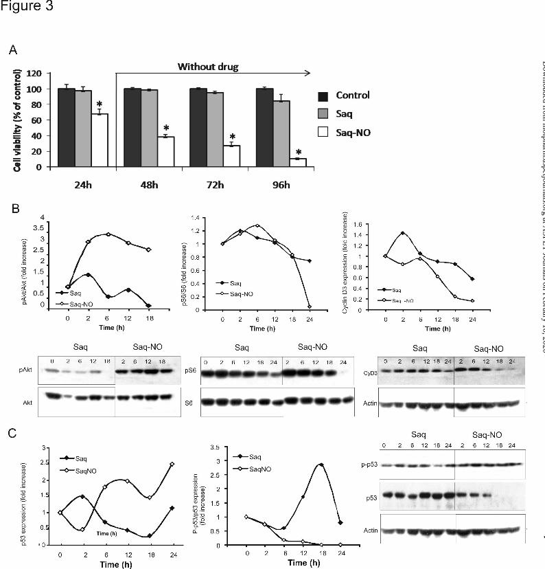
Saq-NO Suppressed Proliferation of Colon Cancer Cells. The lack of apoptosis and cell
cycle arrest observed in both cell lines upon the treatment with Saq-NO indicated that inhibition of
proliferation could result in decreased viability. To prove this, the cells were stained with CFSE and
then treated with an IC50 dose of Saq-NO or the same dose of original drug and flow cytometric
analysis was performed after 72 h. While treatment of CT26CL25 cells with Saq exerted no effect
on cellular proliferation, Saq-NO almost completely blocked the division of cells (11.9% of divided
cells vs 86.6% and 90.1% in control and Saq treated cells, respectively). Inhibition of HCT116 cell
proliferation was less pronounced (50% of divided cells vs 90% and 70% in control and Saq treated
cells, respectively. We next examined if this effect of Saq-NO required the continuous presence of
the drug. For that purpose, CT26CL25 cells were pretreated with Saq-NO for 24 h and then the drug
was removed and cell viability was measured after an additional 24, 48 or 72h of incubation (Fig.
3A). Exposure of CT26CL25 cells to Saq-NO irreversibly blocked the proliferative ability of the
cells. To clarify molecular background of this effect, PI-3K/Akt signaling pathway as a pivotal
regulator of cell proliferation was examined. While expression of p-Akt oppositely to original drug
was transiently up-regulated upon the treatment with NO modified Saq, downstream target of S6
kinase, S6 protein was continuously inactivated through decrease rate of phosphorylation on
Ser240/244 (Fig 3B). This phenomenon showed similar pattern in both cell lines tested and is
compatible with previously observed decrease of cell dividing potential. Decreased expression of
cyclin D3 revealed by Western blot in CT25CL26 cells indirectly confirmed abolished activity of
MOL #77842This article has not been copyedited and formatted. The final version may differ from this version.
Molecular Pharmacology Fast Forward. Published on July 13, 2012 as DOI: 10.1124/mol.112.077842 at A
SPET
Journals on February 10, 2020m
olpharm.aspetjournals.org
Dow
nloaded from
16
downstream part of PI-3K /Akt pathway (Fig 3C). In addition, Saq-NO remarkable elevated p53
protein expression without preventing its degradation as judged by the level of p53 phosphorylated
on Ser20 (Fig 3C). Oppositely to modified drug, Saq strongly induced phosphorylation of p53 which
protected this molecule from MDM-mediated degradation while total p-53 expression was not
significantly affected (Fig 3C) (Mijatovic et al. 2010).
Saq-NO Significantly Reduced Metastatic Properties of Colon Cancer Cells In Vitro and
In Vivo. To evaluate the anti-metastatic potential of original and NO-modified Saq, we examined the
growth of cells on reconstituted basement membrane (Matrigel) during 3 days in the presence of
subtoxic doses of the drugs. CT26CL25 and HCT116 cells elicited different patterns of growth on
Matrigel. While HCT116 cells showed rounded morphology and formed small clusters (Fig. 4A left
panel), CT26CL25 cells made net-like structures and displayed elongated morphology (Fig. 4A right
panel). Independently of the morphologies of the colonies, Saq-NO treatment during 24 h strongly
inhibited Matrigel growth in both types of colon cancer cells while the inhibitory potential of novel
modified drug was greater than the unmodified Saq (Fig. 4A). In a similar manner Saq-NO was
found to be superior in decreasing migration, invasion and adhesive properties of cells than its
parental compound (Fig. 4B). To confirm the permanent reduction of metastatic properties, the cells
were pretreated with drugs and formation of lung metastases was examined. At day 18, one animal in
control group died while the 2 animals displayed massive lung metastases (>120 nodules per mouse,
diameter~3mm each). Pretreatment of the cells with Saq resulted in lower presence of metastases
(n1=120 nodules, diameter~3mm; n2=90; n3=30, diameter~2mm), but its efficacy was significantly
less potent than Saq-NO. In the group of animals receiving Saq-NO- pretreated cells only one animal
exhibited mild metastases (n=59, diameter~1mm). Taken together, it is evident that Saq-NO exerted
its tumoricidal activity against colon cancer cells through inhibition of proliferation and a decrease of
metastatic potential.
MOL #77842This article has not been copyedited and formatted. The final version may differ from this version.
Molecular Pharmacology Fast Forward. Published on July 13, 2012 as DOI: 10.1124/mol.112.077842 at A
SPET
Journals on February 10, 2020m
olpharm.aspetjournals.org
Dow
nloaded from

17
Role of NO in the Anti-Tumor Effects of NO-Modified Compounds. We next compared
the release of NO into the culture medium of CT26CL25 cells in the presence of the NO-modified
drugs or NO donors such as SNAP, SIN-1, SNP and DetaNONOate. Whilst both GIT-27NO and
Saq-NO released lower amounts of NO than the NO donors they exhibited more powerful reduction
of cell viability than the latter (Fig. 5A, B). Even if GIT-27NO formed higher amount of nitrites in
comparison to Saq-NO, the diminished number of viable cells in cultures did not correlate with
accumulated nitrites. Only GIT-27NO efficiently induced NO release inside the cells (Fig. 5C). On
the other hand, both NO-modified compounds induced production of ROS (Fig. 5D). More sensitive
measurement of NO release carried out by flow cytometric analysis confirmed the previous results
(Fig. 5E, F). Concomitant treatment with hemoglobin, an extracellular scavenger of NO, resulted in
restored viability of cells exposed to GIT-27NO confirming the contribution of released NO to the
tumoricidal action of the drug (Fig. 5G). NO rapidly reacts with ROS forming peroxynitrites,
therefore we next scavenged them by adding MEG sulphate. MEG sulphate restored cell viability in
GIT-27NO but not Saq-NO (Fig. 5H) treated cultures confirming the crucial role of NO-induced
peroxynitrites in GIT-27NO mediated toxicity.
MOL #77842This article has not been copyedited and formatted. The final version may differ from this version.
Molecular Pharmacology Fast Forward. Published on July 13, 2012 as DOI: 10.1124/mol.112.077842 at A
SPET
Journals on February 10, 2020m
olpharm.aspetjournals.org
Dow
nloaded from

18
Discussion
In our study we demonstrated in two different colon cancer cell lines strong sensitivity to
treatment with two compounds designed by covalent attachment of NO moiety to the original anti-
inflammatory or anti-viral core drug. The mode of action of NO-modified drugs was profoundly
altered in comparison to original drugs. In agreement with previous data (Maksimovic-Ivanic et al.,
2008; Mijatovic et al., 2008; Mijatovic et al., 2010; Donia et al., 2009), the anti-inflammatory agent
VGX-1027 did not affect colon cancer cell viability. Analyses of cell cycle distribution as well as
AnnV/PI double staining revealed that GIT-27NO triggered apoptosis in both tested colon cell lines.
However, in contrast to its action in Ta3H mammary cells, this process involves caspase activation as
well as enhanced expression of caspases 8 and 3 at gene level accompanied with mitochondrial
membrane depolarization in mouse but not in Bax deficient human colon cell line (Mijatovic et al.,
2010). Obviously, different intracellular profile of apoptotic process pulsed by the treatment as
reflection of intrinsic specificity of tested lines did not disturb prevalence of the pro-apoptotic signal
and completing of dying process (Mijatovic et al., 2010). This cytotoxic mode of action coincided
with a slightly affected rate of cell proliferation that failed to influence the metastatic properties of
both colon cell lines when it was administered at sub-toxic doses (not shown). As previously
demonstrated, the cytocidal effects of drug GIT-27NO are related to the intracellular introduction of
high levels of NO (Maksimovic-Ivanic et al., 2008; Mijatovic et al., 2010). Even if GIT-27 NO
released lower amounts of NO in comparison to corresponding doses of exogenous NO donors, the
uptake of NO in colon cancer cells exposed to GIT-27NO, was impressively high. Subsequently, cell
damage promoted by the toxic amount of NO released from the GIT-27NO and other free radicals
generated in this cascade were reflected on cell viability. Furthermore, the cytocidal effects of GIT-
27NO on colon cancer cells were successfully blocked by the peroxynitrite scavenger MEG sulphate
confirming the crucial role of released NO and OONO- in drug mediated toxicity. Interestingly, the
MOL #77842This article has not been copyedited and formatted. The final version may differ from this version.
Molecular Pharmacology Fast Forward. Published on July 13, 2012 as DOI: 10.1124/mol.112.077842 at A
SPET
Journals on February 10, 2020m
olpharm.aspetjournals.org
Dow
nloaded from
19
intrinsic properties of HCT116 cells in redox status make them more tolerant to the oxidative stress
generated by applied doses of GIT-27NO (Dang et al., 2005). This resulted in a requirement of a
prolonged period of exposure to the drug for induction of massive apoptotic cell death.
On the other hand, Saq-NO decreased the viability of colon cancer cells primarily through
inhibition of cell proliferation but not through induction of cell death (Maksimovic-Ivanic et al.,
2009). Moreover, a short pulse exposure of the colon cancer cells to Saq-NO was sufficient to induce
irreversible loss of their dividing potential indicating permanent changes in cell physiology. A long-
lasting blockade in cell division was observed upon prolonged Saq-NO treatment which affected
significantly the metastatic potential of both cell lines independently of their previously documented
differences in metastasis (Sahai and Marshall, 2003). We previously showed that permanent
inhibition of cellular proliferation correlated with induction of differentiation and upregulated p53
expression in cancer cells of neuroectodermal origin (Maksimovic-Ivanic et al. 2009). Described
phenomenon was connected to p53 involvement in process of differentiation of transformed cells.
Strong upregulation of p53 through de novo synthesis rather than disturbed proteolitical degradation
can be involved in decreased cellular proliferation and also lost of malignant properties. In
concordance with our previous data, transient up-regulation of Akt activity was detected
(Maksimovic-Ivanic et al., 2009; Mijatovic et al., 2011). However, inhibition of downstream part of
PI3K/Akt pathway manifested through down-regulation of S6 protein observed in both lines upon the
treatment with Saq-NO could be essential for both- lost of proliferative potential and decreased
metastatic properties. The same effect was described in prostate cancer LNCap cells upon the
treatment with this drug (Mojic et al., 2012). Upstream regulator of S6 protein, S6K is multiple
incharged for regulation of these processes (Fenton and Gout, 2011) offering the explanation for
decreased capacity of tested cell lines to adhere, migrate and invade. In addition, it was recently
reported that Saq was able to inhibit metastatic properties through inhibition of angiogenesis and
MOL #77842This article has not been copyedited and formatted. The final version may differ from this version.
Molecular Pharmacology Fast Forward. Published on July 13, 2012 as DOI: 10.1124/mol.112.077842 at A
SPET
Journals on February 10, 2020m
olpharm.aspetjournals.org
Dow
nloaded from

20
MMP-2 and MMP-9 expression (Toschi et al., 2011; Sgadari et al., 2002). Moreover, the results
obtained in vitro were confirmed in a syngeneic metastatic model of colon cancer where pretreatment
of CT26CL26 cells with Saq-NO for 24 h was sufficient to permanently affect their metastatic
potential.
Absence of highly destructive RNS concurs with the cytostatic mode of action of the
compound (Calcerrada et al., 2011). It is evident that oxidative stress triggered by Saq-NO may not
be triggered by the discrete quantity of NO released that probably falls within concentration ranges
required by this gas to regulate cell physiology (Sing and Gupta, 2011; Hickok and Thomas, 2010).
The efficacy of certain newly synthesized drugs against colon cancer cells in tumor transplant studies
in animal cells is improving (Maksimovic-Ivanic et al., 2008; Mijatovic et al., 2008; Mijatovic et al.,
2010; Donia et al., 2009; Maksimovic-Ivanic et al., 2009; Canducci et al., 2011; Mijatovic et al.,
2011; Donia et al., 2011; Rotweiler et al., 2010). Since NO is a short lived and highly reactive
molecule, the advantage of GIT-27NO in comparison to other NO donors is related to its ability to
directly deliver NO into the cell and therefore prevent its loss before reaching a target. Moreover,
rapid amplification of the toxic signal through formation of peroxynitrite makes it highly effective in
inducing cell death.
In contrast to GIT-27NO, Saq-NO was created from a drug which already possessed
anticancer properties. Addition of a NO moiety to Saq markedly enhanced the anticancer action of
Saq in a manner apparently independent on NO release. We demonstrate for the first time in these
colon cancer cell lines that their high sensitivity to Saq-NO was associated with suppression by the
drug of migratory and invasive capacities. These findings reveals an additional and important anti-
neoplastic property of Saq-NO, that along with its direct cytostatic effects, as well as, chemo- and
immuno-sensitizing properties qualify it as an important chemotherapeutic agent worthy of further
research and potentially clinical evaluations.
MOL #77842This article has not been copyedited and formatted. The final version may differ from this version.
Molecular Pharmacology Fast Forward. Published on July 13, 2012 as DOI: 10.1124/mol.112.077842 at A
SPET
Journals on February 10, 2020m
olpharm.aspetjournals.org
Dow
nloaded from

21
Acknowledgements
We thank Dr. Natasa Nestorovic for help in evaluation of migration and invasion assays.
Authorship Contributions
Participated in research design: Mijatovic, Maksimovic-Ivanic, McCubrey, Mai-Britt Zocca and Nicoletti
Conducted experiments: Mijatovic, Maksimovic-Ivanic, Mojic, Miljkovic, Stankovic, Donia, Fagone Mangano and Travali
Contributed new reagents and analytic tools: Al-Abed
Performed data analysis: Mijatovic, Maksimovic-Ivanic, and Mojic
Wrote or contributed to the writing of the manuscript: Mijatovic, Maksimovic-Ivanic, Mojic, Stosic-Grujicic, Mai-Britt Zocca and Nicoletti
MOL #77842This article has not been copyedited and formatted. The final version may differ from this version.
Molecular Pharmacology Fast Forward. Published on July 13, 2012 as DOI: 10.1124/mol.112.077842 at A
SPET
Journals on February 10, 2020m
olpharm.aspetjournals.org
Dow
nloaded from

22
References
Bernstein WB, Dennis PA (2008) Repositioning HIV protease inhibitors as cancer therapeutics.
Curr Opin HIV AIDS 3:666-675.
Bower M, Fox P, Fife K, Gill J, Nelson M, and Gazzard B (1999) Highly active anti-retroviral
therapy (HAART) prolongs time to treatment failure in Kaposi's sarcoma. AIDS 13:2105-2111.
Calcerrada P, Peluffo G, and Radi R (2011) Nitric Oxide-derived Oxidants with a Focus on
Peroxynitrite: Molecular Targets, Cellular Responses and Therapeutic Implications. Curr Pharm
Des [Epub ahead of print]
Canducci F, Ceresola ER, Saita D, Al-Abed Y, Garotta G, Clementi M, and Nicoletti F (2011) The
new and less toxic protease inhibitor saquinavir-NO maintains anti-HIV-1 properties in vitro
indistinguishable from those of the parental compound saquinavir. Antiviral Res 91:292-295.
Chow WA, Jiang C, and Guan M (2009) Anti-HIV drugs for cancer therapeutics: back to the
future? Lancet Oncol 10:61-71.
Coimbra M, Kuijpers SA, van Seters SP, Storm G, and Schiffelers RM (2009) Targeted delivery of
anti-inflammatory agents to tumors. Curr Pharm Des 15:1825-1843.
Dang DT, Chen F, Kohli M, Rago C, Cummins JM, and Dang LH (2005) Glutathione S-transferase
pi1 promotes tumorigenicity in HCT116 human colon cancer cells. Cancer Res 15;65(20):9485-
94.
Donia M, Maksimovic-Ivanic D, Mijatovic S, Mojic M, Miljkovic D, Timotijevic G, Fagone P,
Caponnetto S, Al-Abed Y, McCubrey J, Stosic-Grujicic S, and Nicoletti F (2011) In vitro and in
MOL #77842This article has not been copyedited and formatted. The final version may differ from this version.
Molecular Pharmacology Fast Forward. Published on July 13, 2012 as DOI: 10.1124/mol.112.077842 at A
SPET
Journals on February 10, 2020m
olpharm.aspetjournals.org
Dow
nloaded from
23
vivo anticancer action of Saquinavir-NO, a novel nitric oxide-derivative of the protease inhibitor
saquinavir, on hormone resistant prostate cancer cells. Cell Cycle 10:492-499.
Donia M, Mijatovic S, Maksimovic-Ivanic D, Miljkovic D, Mangano K, Tumino S, Biondi A,
Basile F, Al-Abed Y, Stosic-Grujicic S, and Nicoletti F (2009) The novel NO-donating
compound GIT-27NO inhibits in vivo growth of human prostate cancer cells and prevents murine
immunoinflammatory hepatitis. Eur J Pharmacol 615:228-233.
Fenton TR and Gout IT (2011) Functions and regulation of the 70kDa ribosomal S6 kinases. Int J
Biochem Cell Biol 43, 47-59
Flick DA and Gifford GE (1984) Comparison of in vitro cell cytotoxic assays for tumor necrosis
factor. J Immunol Methods 68:167-175.
Gallagher DJ and Kemeny N (2010) Metastatic colorectal cancer: from improved survival to
potential cure. Oncology 78: 237-248. Gao J, Liu X, and Rigas B (2005) Nitric oxide-donating
aspirin induces apoptosis in human colon cancer cells through induction of oxidative stress. Proc
Natl Acad Sci U S A. 102:17207-17212.
Giuliani F, De Vita F, Colucci G, and Pisconti S (2010) Maintenance therapy in colon
cancer.Cancer Treat Rev 36:S42-S45.
Gupta AK, Cerniglia GJ, Mick R, McKenna WG, and Muschel RJ (2005) HIV protease inhibitors
block Akt signaling and radiosensitize tumor cells both in vitro and in vivo. Cancer Res 65:8256-
8265.
Hickok JR and Thomas DD (2010) Nitric oxide and cancer therapy: the emperor has NO clothes.
Curr Pharm Des 16:381-391.
MOL #77842This article has not been copyedited and formatted. The final version may differ from this version.
Molecular Pharmacology Fast Forward. Published on July 13, 2012 as DOI: 10.1124/mol.112.077842 at A
SPET
Journals on February 10, 2020m
olpharm.aspetjournals.org
Dow
nloaded from

24
Kashfi K and Rigas B (2007) The mechanism of action of nitric oxide-donating aspirin. Biochem
Biophys Res Commun 358:1096-1101.
Kleinman HK, McGarvey ML, Liotta LA, Robey PG, Tryggvason K, and Martin GR (1982)
Isolation and characterization of type IV procollagen, laminin, and heparan sulfate proteoglycan
from the EHS sarcoma. Biochemistry 21:6188-6193.
Labianca R and Merelli B (2010) Screening and diagnosis for colorectal cancer: present and future.
Tumori 96:889-901.
Lebbé C, Blum L, Pellet C, Blanchard G, Vérola O, Morel P, Danne O, and Calvo F (1998)
Clinical and biological impact of antiretroviral therapy with protease inhibitors on HIV-related
Kaposi's sarcoma. AIDS 12:F45-49.
Maksimovic-Ivanic D, Mijatovic S, Harhaji L, Miljkovic D, Dabideen D, Fan Cheng K, Mangano
K, Malaponte G, Al-Abed Y, Libra M, Garotta G, Nicoletti F, and Stosic-Grujicic S (2008)
Anticancer properties of the novel nitric oxide-donating compound (S,R)-3-phenyl-4,5-dihydro-
5-isoxazole acetic acid-nitric oxide in vitro and in vivo. Mol Cancer Ther 7:510-520.
Maksimovic-Ivanic D, Mijatovic S, Miljkovic D, Harhaji-Trajkovic L, Timotijevic G, Mojic M,
Dabideen D, Cheng KF, McCubrey JA, Mangano K, Al-Abed Y, Libra M, Garotta G, Stosic-
Grujicic S, and Nicoletti F (2009) The antitumor properties of a nontoxic, nitric oxide-modified
version of saquinavir are independent of Akt. Mol Cancer Ther 8:1169-1178.
Mijatovic S, Maksimovic-Ivanic D, Mojic M, Malaponte G, Libra M, Cardile V, Miljkovic D,
Harhaji L, Dabideen D, Cheng KF, Bevelacqua Y, Donia M, Garotta G, Al-Abed Y, Stosic-
Grujicic S, and Nicoletti F (2008) Novel nitric oxide-donating compound (S,R)-3-phenyl-4,5-
MOL #77842This article has not been copyedited and formatted. The final version may differ from this version.
Molecular Pharmacology Fast Forward. Published on July 13, 2012 as DOI: 10.1124/mol.112.077842 at A
SPET
Journals on February 10, 2020m
olpharm.aspetjournals.org
Dow
nloaded from

25
dihydro-5-isoxazole acetic acid-nitric oxide (GIT-27NO) induces p53 mediated apoptosis in
human A375 melanoma cells. Nitric Oxide 19:177-183.
Mijatovic S, Maksimovic-Ivanic D, Mojic M, Timotijevic G, Miljkovic D, Mangano K, Donia M,
Di Cataldo A, Al-Abed Y, Cheng KF, Stosic-Grujicic S, and Nicoletti F (2011) Cytotoxic and
immune-sensitizing properties of nitric oxide-modified Saquinavir in iNOS-positive human
melanoma cells. J Cell Physiol 226:1803-1812.
Mijatovic S, Maksimovic-Ivanic D, Timotijevic G, Miljkovic D, Donia M, Libra M, Coco M,
McCubrey J, Al-Abed Y, Korac A, Stosic-Grujicic S, and Nicoletti F (2010) Induction of
caspase-independent apoptotic-like cell death of mouse mammary tumor TA3Ha cells in vitro
and reduction of their lethality in vivo by the novel chemotherapeutic agent GIT-27NO. Free
Radic Biol Med 48:1090-1099.
Mojic M, Mijatovic S, Maksimovic-Ivanic D, Dinic S, Grdovic N, Miljkovic D, Stosic-Grujicic S,
Tumino S, Fagone P, Mangano K, Zocca MB, Al-Abed Y, McCubrey JA, Nicoletti F (2012)
Saquinavir-NO-targeted S6 protein mediates sensitivity of androgen-dependent prostate cancer
cells to TRAIL. Cell Cycler 11:1174-1182.
NicOx, S (2007) NicOx provides an update on NCX 4016 (Press release).
http://www.nicox.com/files/pdf/PR2007061800EN.pdf
Niehues T, Horneff G, Megahed M, Schroten H, and Wahn V (1999) Complete regression of
AIDS-related Kaposi's sarcoma in a child treated with highly active antiretroviral therapy. AIDS
13:1148-1149.
Rigas B and Williams JL ( 2008) NO-donating NSAIDs and cancer: an overview with a note on
whether NO is required for their action. Nitric Oxide 19:199-204.
MOL #77842This article has not been copyedited and formatted. The final version may differ from this version.
Molecular Pharmacology Fast Forward. Published on July 13, 2012 as DOI: 10.1124/mol.112.077842 at A
SPET
Journals on February 10, 2020m
olpharm.aspetjournals.org
Dow
nloaded from
26
Rothweiler F, Michaelis M, Brauer P, Otte J, Weber K, Fehse B, Doerr HW, Wiese M, Kreuter J,
Al-Abed Y, Nicoletti F, and Cinatl J Jr (2010) Anticancer effects of the nitric oxide-modified
saquinavir derivative saquinavir-NO against multidrug-resistant cancer cells. Neoplasia 12:1023-
1030.
Rothwell PM, Fowkes FG, Belch JF, Ogawa H, Warlow CP and Meade TW (2011) Effect of daily
aspirin on long-term risk of death due to cancer: analysis of individual patient data from
randomised trials. Lancet 377:31-41.
Rothwell PM, Wilson M, Elwin CE, Norrving B, Algra A, Warlow CP, and Meade TW (2010)
Long-term effect of aspirin on colorectal cancer incidence and mortality: 20-year follow-up of
five randomised trials. Lancet 376:1741-1750.
Sahai E and Marshall CJ (2003) Differing modes of tumour cell invasion have distinct
requirements for Rho/ROCK signalling and extracellular proteolysis. Nat Cell Biol 5:711-719.
Schütt M, Zhou J, Meier M, and Klein HH (2004) Long-term effects of HIV-1 protease inhibitors
on insulin secretion and insulin signaling in INS-1 beta cells. J Endocrinol 183:445-454.
Sgadari C, Barillari G, Toschi E, Carlei D, Bacigalupo I, Baccarini S, Palladino C, Leone P,
Bugarini R, Malavasi L, Cafaro A, Falchi M, Valdembri D, Rezza G, Bussolino F, Monini P, and
Ensoli B (2002) HIV protease inhibitors are potent anti-angiogenic molecules and promote
regression of Kaposi sarcoma. Nat Med 8:225-232.
Sgadari C, Monini P, Barillari G, and Ensoli B (2003) Use of HIV protease inhibitors to block
Kaposi's sarcoma and tumour growth. Lancet Oncol 4:537-547.
MOL #77842This article has not been copyedited and formatted. The final version may differ from this version.
Molecular Pharmacology Fast Forward. Published on July 13, 2012 as DOI: 10.1124/mol.112.077842 at A
SPET
Journals on February 10, 2020m
olpharm.aspetjournals.org
Dow
nloaded from

27
Singh S and Gupta AK (2011) Nitric oxide: role in tumour biology and iNOS/NO-based anticancer
therapies. Cancer Chemother Pharmacol 67:1211-1224.
Stojanovic I, Cuzzocrea S, Mangano K, Mazzon E, Miljkovic D, Wang M, Donia M, Al Abed Y,
Kim J, Nicoletti F, Stosic-Grujicic S, and Claesson M (2007) In vitro, ex vivo and in vivo
immunopharmacological activities of the isoxazoline compound VGX-1027: modulation of
cytokine synthesis and prevention of both organ-specific and systemic autoimmune diseases in
murine models. Clin Immunol 123:311-323.
Stosic-Grujicic S, Cvetkovic I, Mangano K, Fresta M, Maksimovic-Ivanic D, Harhaji L, Popadic
D, Momcilovic M, Miljkovic D, Kim J, Al-Abed Y, and Nicoletti F (2007) A potent
immunomodulatory compound, (S,R)-3-Phenyl-4,5-dihydro-5-isoxazole acetic acid, prevents
spontaneous and accelerated forms of autoimmune diabetes in NOD mice and inhibits the
immunoinflammatory diabetes induced by multiple low doses of streptozotocin in CBA/H mice.
J Pharmacol Exp Ther 320:1038-1049.
Toschi E, Sgadari C, Malavasi L, Bacigalupo I, Chiozzini C, Carlei D, Compagnoni D, Bellino S,
Bugarini R, Falchi M, Palladino C, Leone P, Barillari G, Monini P, and Ensoli B (2011) Human
immunodeficiency virus protease inhibitors reduce the growth of human tumors via a
proteasome-independent block of angiogenesis and matrix metalloproteinases. Int J Cancer
128:82-93.
Van Geelen CM, de Vries EG, de Jong S (2004) Lessons from TRAIL-resistance mechanisms in
colorectal cancer cells: paving the road to patient-tailored therapy. Drug Resist Updat 7:345-358.
Wolpin BM and Mayer RJ (2008) Systemic treatment of colorectal cancer. Gastroenterology
134:1296-1310.
MOL #77842This article has not been copyedited and formatted. The final version may differ from this version.
Molecular Pharmacology Fast Forward. Published on July 13, 2012 as DOI: 10.1124/mol.112.077842 at A
SPET
Journals on February 10, 2020m
olpharm.aspetjournals.org
Dow
nloaded from

28
Yeh RK, Chen J, Williams JL, Baluch M, Hundley TR, Rosenbaum RE, Kalala S, Traganos F,
Benardini F, del Soldato P, Kashfi K, and Rigas B (2004) NO-donating nonsteroidal
antiinflammatory drugs (NSAIDs) inhibit colon cancer cell growth more potently than traditional
NSAIDs: a general pharmacological property? Biochem Pharmacol 67:2197-2205.
Zhang Y, Yuan J, Zhang HY, Simayi D, Li PD, Wang YH, Li F, Zhang WJ (2011) Natural
resistance to apoptosis correlates with resistance to chemotherapy in colorectal cancer cells. Clin
Exp Med doi:10.1007/s10238-011-0146-5.
MOL #77842This article has not been copyedited and formatted. The final version may differ from this version.
Molecular Pharmacology Fast Forward. Published on July 13, 2012 as DOI: 10.1124/mol.112.077842 at A
SPET
Journals on February 10, 2020m
olpharm.aspetjournals.org
Dow
nloaded from
29
This work was supported in part by the Serbian Ministry of Education and Science [Grant 173013].
Disclosure of interest: MD, MBZ, YAA and FN are shareholders of Onconox
MOL #77842This article has not been copyedited and formatted. The final version may differ from this version.
Molecular Pharmacology Fast Forward. Published on July 13, 2012 as DOI: 10.1124/mol.112.077842 at A
SPET
Journals on February 10, 2020m
olpharm.aspetjournals.org
Dow
nloaded from

30
Figure Legends:
Figure 1. NO modified drugs decreased the tumor cells growth. (A) HCT116 and CT26CL25
cells (1 x 10⁴ cells/well) were treated with a range of concentrations of the indicated drugs for 24 h,
and then cell viability was determined by CV assay. The data are presented as mean ± SD from
representative of three independent experiments. *p < 0.05, refers to untreated cultures. (B) Tumors
were induced by subcutaneous implantation of 5 x 10⁶ CT26CL25 cells. Then after 12 days to allow
for the tumors to take in the mice, the mice were injected intraperitoneally daily with the drug for 14
days. Tumor volumes were measured two times a week until the end of experiment.
Figure 2. GIT-27NO induced caspase-dependent apoptosis. Cells were treated with an IC50 dose
of the drugs and cell cycle analysis (A), staining by AnnV/PI (B) and apostat (C) were performed by
flow cytometry after 24 h or 48 h for CT26CL25 and HCT116 cells, respectivly. RT-PCR analyses
for caspase-3, caspase-8 were performed after 6 h of incubation in the presence of the drug and data
are presented as relative expression of mRNA (D). Mitochondrial membrane depolarization of JC-1
stained CT26CL25 cells was determined by flow cytometry after 24 h (E). RT-PCR analyses for
Bax, Bcl-2 and Bcl-XL were done as mentioned above (F). Presence of p-53in CT26CL25 cells after
incubation for 24 h without (control) or with GIT-27NO detected by immunocytochemistry and
analyzed by light microscopy.
Figure 3. Saq-NO suppressed cell proliferation. (A) CT26CL25 cells were treated for 24 h with
Saq or Saq-NO, or left untreated (Control) and cell viabilities in the absence of the drugs were
measured by CV after additional 24, 48 and 72 h. The data are presented as mean ± SD from
representative of three independent experiments. *p < 0.05 refers to untreated cultures. Cells were
treated with IC50 dose of Saq-NO and equal dose of Saq for indicated time and Akt, S6 protein,
cyclinD3 (B), p-53 and p-p53 Ser20 (C) expression in indicated time points was analyzed by western
MOL #77842This article has not been copyedited and formatted. The final version may differ from this version.
Molecular Pharmacology Fast Forward. Published on July 13, 2012 as DOI: 10.1124/mol.112.077842 at A
SPET
Journals on February 10, 2020m
olpharm.aspetjournals.org
Dow
nloaded from

31
blot. Densitometric analysis of data from representative of three experiments was presented as fold
increase relative to control.
Figure 4. Saq-NO abbrogates metastatic potential of the cells. (A) Light microscopy and Matrigel
growth of cells after 72 h of treatment with Saq or Saq-NO for HCT116 (right panel) and CT26CL25
(left panel). (B) Cells were pretreated for 24 h with Saq or Saq-NO, or left untreated (0) and adhesion
was measured after 12 or 18 h, and migration and invasion of CT26CL25 and HCT116 was
examined after 24 or 36 h, respectivly. The data are presented as mean ± SD from representative of
two independent experiments. *p < 0.05, refers to untreated cultures. Representative photographs of
lungs from animals which received controls, Saq or Saq-NO pretreated tumor cells are presented.
Figure 5. The contribution of NO, ROS and RNS to the tumoricidal effects of the drugs. Cells
(1 x 10⁴ cells/well) were treated with a range of doses of indicated NO donors, Saq-NO or GIT-
27NO for 24 h, after which accumulation of nitrites was determined by Griess reaction (A), and cell
viability was examined by CV test (B). The data are presented as mean values while SD were less
than 10 %. Intracellular NO production was determined by DAF-FM indicator and ROS/RNI
production by DHR staining. Intensity of fluorencence was analized by Chameleon multiplate reader
(C,D respectively) or FACS Calibur flow cytometer (E, F). Cells were cultivated with IC50
concentrations of the drugs in the presence of Hb (25 μM) (G) or MEG (100 μM) (H) for 24 h and
viability was measured by CV. The data are presented as mean ± SD from representative of three
independent experiments *p < 0.05, refers to untreated cultures.
MOL #77842This article has not been copyedited and formatted. The final version may differ from this version.
Molecular Pharmacology Fast Forward. Published on July 13, 2012 as DOI: 10.1124/mol.112.077842 at A
SPET
Journals on February 10, 2020m
olpharm.aspetjournals.org
Dow
nloaded from

This article has not been copyedited and formatted. The final version may differ from this version.Molecular Pharmacology Fast Forward. Published on July 13, 2012 as DOI: 10.1124/mol.112.077842
at ASPE
T Journals on February 10, 2020
molpharm
.aspetjournals.orgD
ownloaded from

This article has not been copyedited and formatted. The final version may differ from this version.Molecular Pharmacology Fast Forward. Published on July 13, 2012 as DOI: 10.1124/mol.112.077842
at ASPE
T Journals on February 10, 2020
molpharm
.aspetjournals.orgD
ownloaded from
This article has not been copyedited and formatted. The final version may differ from this version.Molecular Pharmacology Fast Forward. Published on July 13, 2012 as DOI: 10.1124/mol.112.077842
at ASPE
T Journals on February 10, 2020
molpharm
.aspetjournals.orgD
ownloaded from

This article has not been copyedited and formatted. The final version may differ from this version.Molecular Pharmacology Fast Forward. Published on July 13, 2012 as DOI: 10.1124/mol.112.077842
at ASPE
T Journals on February 10, 2020
molpharm
.aspetjournals.orgD
ownloaded from

This article has not been copyedited and formatted. The final version may differ from this version.Molecular Pharmacology Fast Forward. Published on July 13, 2012 as DOI: 10.1124/mol.112.077842
at ASPE
T Journals on February 10, 2020
molpharm
.aspetjournals.orgD
ownloaded from