
Mariana de Oliveira Mendesrepositorio.ul.pt/bitstream/10451/34258/1/TM_Mariana_Mendes.pdf · A...
95
Universidade de Lisboa Faculdade de Farmácia Neuroprotective effects of TUDCA in Parkinson's disease: Dissecting the anti-oxidant and anti-inflammatory effects of this bile acid in the mouse cerebral cortex Mariana de Oliveira Mendes Orientador: Margarida Castro-Caldas, PhD, Faculdade de Ciências e Tecnologia, Universidade Nova de Lisboa Co-orientador: Maria João Gama, PhD Faculdade de Farmácia, Universidade de Lisboa Dissertação elaborada para obtenção do Grau de Mestre em Ciências Biofarmacêuticas Outubro, 2017
Transcript of Mariana de Oliveira Mendesrepositorio.ul.pt/bitstream/10451/34258/1/TM_Mariana_Mendes.pdf · A...
Universidade de Lisboa
Faculdade de Farmácia
Neuroprotective effects of TUDCA in Parkinson's disease: Dissecting
the anti-oxidant and anti-inflammatory effects of this bile acid in the
mouse cerebral cortex
Mariana de Oliveira Mendes
Orientador: Margarida Castro-Caldas, PhD, Faculdade de Ciências e Tecnologia,
Universidade Nova de Lisboa
Co-orientador: Maria João Gama, PhD Faculdade de Farmácia, Universidade de Lisboa
Dissertação elaborada para obtenção do Grau de Mestre em Ciências Biofarmacêuticas
Outubro, 2017


i | P a g e
Part of the results discussed in this thesis were presented in the following meetings:
M.O. Mendes, A.I. Rosa, M. J. Nunes, A. Carvalho, S. Duarte-Silva, A. Silva-Fernandes, E.
Rodrigues, P. Maciel, C.M.P. Rodrigues, M.J. Gama, M. Castro-Caldas. Neuroprotective
effects of Tauroursodeoxycholic acid in MPTP model of Parkinson's disease: A study in the
mouse cortex. MitoPorto - Advances in mitochondrial research, 26th May, 2017,
Universidade do Porto. [Poster communication];
M.O. Mendes, A.I. Rosa, M. J. Nunes, A. Carvalho, S. Duarte-Silva, A. Silva-Fernandes, E.
Rodrigues, P. Maciel, C.M.P. Rodrigues, M.J. Gama, M. Castro-Caldas. Extended
neuroprotective effect of Tauroursodeoxycholic acid to the mouse cerebral cortex in a MPTP
model of Parkinson's disease. 9th iMed.UL post-graduate students meeting, 13th-14th July,
2017, Faculty of Pharmacy, Universidade de Lisboa. [Poster communication];
This work was supported by National funds (Fundação para a Ciência e Tecnologia – FCT,
Portugal) with the project PTDC/NEUNMC/0248/2012 and with the project PEst-
OE/SAU/UI4013/2011.

ii | P a g e
Abstract
Parkinson’s disease (PD) is a progressive neurological disorder, mainly characterized by
the progressive loss of dopaminergic neurons in the substantia nigra pars compacta (SNpc)
and by the presence of intracellular inclusions, known as Lewy bodies. Although the main
cause of this disorder remains elusive, neurodegeneration has been associated with different
mechanisms of cell damage including oxidative stress, mitochondrial dysfunction and
neuroinflammation. Despite SNpc being considered the primary affected region in PD, the
neuropathological features may not solely be confined to the nigro-striatal axis. It is
conceivable that other brain regions are also affected, namely the cerebral cortex, being the
neurologic insult to this brain region still not completely unraveled.
Tauroursodeoxycholic acid (TUDCA) is an endogenous bile acid that has been shown to
have antioxidant properties and to exhibit a neuroprotective effect in 1-methyl-4-phenyl-
1,2,3,6-tetrahydropyridine (MPTP) mice model of PD. Moreover, TUDCA anti-inflammatory
properties have been reported in glial cells, making it a prominent therapeutic agent for PD
treatment and relevant the pursuit of its mechanism of action in this disease.
In this thesis we used C57BL/6 mice injected with MPTP in a sub-acute paradigm aiming
to investigate if the neurotoxic effects of MPTP could be extended to the cerebral cortex.
Moreover, we intended to dissect the anti-oxidant and neuroprotective effects of TUDCA in
this brain region. In addition, we proposed to investigate the anti-inflammatory mechanisms
triggered by this bile acid, in the cerebral cortex and in microglia cells, namely its ability to
modulate Annexin-1 (ANXA1) expression and secretion.
Our results revealed that in mice cortex TUDCA: i) increases the expression of nuclear
factor erythroid 2 related factor 2 antioxidant downstream targets, namely Glutathione
peroxidase 1 and Heme oxygenase 1 enzymes, upon MPTP exposure; ii) up-regulates parkin
expression and AMP-activated protein kinase activation, and prevents MPTP-induced ATP
depletion; and iii) decreases inflammatory makers of astrogliosis and microgliosis, and up-
regulates the expression of the anti-inflammatory protein ANXA1. Additionally, results from
cellular models corroborate TUDCA modulation of ANXA1 synthesis and secretion and point
to the possibility of this protein being a direct target of this bile acid.
Together, these results reinforce the potential benefits of using TUDCA as a therapeutic
strategy in the treatment of PD.
Keywords: ANXA1, Mitochondrial Dysfunction, MPTP, Neuroinflammation, TUDCA;

iii | P a g e
Resumo
A doença de Parkinson (DP) é uma doença neurodegenerativa progressiva, caracterizada
principalmente pela perda gradual de neurónios dopaminérgicos na substantia nigra pars
compacta (SNpc) e pela presença de inclusões intracelulares denominados corpos de Lewy.
Apesar da principal causa desta doença permanecer desconhecida, o processo de
neurodegenerescência tem sido associado a diferentes mecanismos de dano celular tais como
stress oxidativo, disfunção mitocondrial e neuroinflamação. Apesar da SNpc ser considerada
a principal região afectada na DP, as características neuropatológicas podem não estar
exclusivamente confinadas ao eixo nigro-estriado. É concebível que outras regiões cerebrais
possam estar também afectadas, nomeadamente o córtex cerebral, sendo que o insulto
neurológico nesta região cerebral ainda não está completamente desvendado.
O ácido tauroursodesoxicólico (TUDCA) é um ácido biliar endógeno que tem mostrado
ter propriedades antioxidantes e exibir um efeito neuroprotector num modelo da DP em
murganhos tratados com 1-metil-4-fenil-1,2,3,6-tetrahidropiridina (MPTP). Para além disso,
as propriedades anti-inflamatórias do TUDCA também foram descritas em células da glia
tornando-o num agente terapêutico importante para o tratamento da DP, e tornando relevante
a investigação sobre o seu mecanismo de acção nesta doença.
Nesta tese usámos murganhos C57BL/6 injectados com MPTP, num regime
experimental sub-agudo, com o objectivo de investigar se os efeitos neurotóxicos do MPTP se
estendem ao córtex cerebral. Para além disso, dissecámos os efeitos antioxidantes e
neuropotectores do TUDCA nesta região cerebral. Procurámos ainda investigar o mecanismo
anti-inflamatório despoletado por este ácido biliar, no córtex cerebral e em células da
micróglia, nomeadamente a capacidade do TUDCA modular a expressão e secreção da
Anexina-1 (ANXA1).
Os nossos resultados, no córtex cerebral de murganhos, revelaram que o TUDCA: i)
aumenta a expressão dos alvos antioxidantes do nuclear factor erythroid 2 related factor 2,
nomeadamente as enzimas Glutationa Peroxidase 1 e Heme-oxigenase 1, após exposição ao
MPTP; ii) regula a expressão proteica da parkin, induz a activação da AMP-activated protein
kinase, e previne a depleção de ATP induzida pelo MPTP; e iii) diminui os marcadores
inflamatórios característicos de astrogliose e microgliose, e regula a expressão da proteína
anti-inflamatória ANXA1. Além disso, os resultados dos modelos celulares corroboraram a
regulação da expressão e secreção da ANXA1 pelo TUDCA, sugerindo a possibilidade desta
proteína ser um alvo directo deste ácido biliar.

iv | P a g e
Em conjunto, os resultados apresentados neste trabalho contribuem para reforçar os
potenciais benefícios do uso do TUDCA como estratégia terapêutica no tratamento da DP.
Palavras-chave: ANXA1, Disfunção mitocondrial, MPTP, Neuroinflamação, TUDCA;
v | P a g e
Agradecimentos
As minhas primeiras palavras de agradecimento são dirigidas à Professora Doutora Cecília
Rodrigues pela disponibilidade e orientação ao longo do Mestrado em Ciências Biofarmacêuticas,
em especial pela ajuda que me deu na procura do tema desta tese e por me ter recebido no seu
grupo “Cellular Function and Therapeutic Targeting”.
Um enorme agradecimento à Professora Doutora Margarida Castro-Caldas por me ter
proporcionado esta oportunidade e pela ajuda e orientação constantes. Obrigada por todo o rigor,
exigência, sugestões e conhecimento transmitidos. Agradeço ainda a disponibilidade constante
para me esclarecer dúvidas e aconselhar (mesmo quando não podia estar presencialmente), toda a
paciência que teve comigo e todas as palavras de encorajamento e motivação (até quando os
resultados não eram assim tão promissores). Aprendi e cresci bastante neste ano e grande parte
deve-o a si, espero não a ter desapontado.
À Professora Doutora Maria João Gama agradeço a disponibilidade em co-orientar esta tese,
as valiosas sugestões que me deu e todas as dúvidas que se propôs a responder. Agradeço ainda a
preocupação e palavras de incentivo transmitidas ao longo deste ano, principalmente na recta
final.
À Professora Doutora Elsa Rodrigues agradeço o rigor e boas práticas que exigiu desde o
primeiro dia no laboratório, essenciais para o meu crescimento e sem as quais já não consigo
trabalhar. Agradeço ainda a boa disposição constante, a prontidão para esclarecer as minhas
dúvidas e todas as vezes que no final de um longo dia validava o meu trabalho dizendo que “ todo
o meu esforço não era em vão e iria certamente dar frutos”.
À Alexandra deixo o meu obrigada, apesar de não teres estado comigo “no meio” da tese,
estiveste presente no início e no fim, e a tua orientação e ajuda foi fundamental para que este
trabalho começasse com o pé direito.
À Andreia, a “Doutorada de Serviço”, quero agradecer a simpatia, a ajuda e o encorajamento
que me deste. As tuas histórias de experiências trágicas e finais felizes serviram para me dar um
pouco de alento quando as coisas não corriam tão bem e a tua boa disposição e relativização dos
problemas ajudou-me a descontrair um pouco mais. Obrigada.
A todos os elementos do CellFun quero expressar a minha gratidão, não só pela maneira
como me integraram mas também por toda a ajuda que me deram e dúvidas que esclareceram ao
longo deste ano. Não é fácil ser a pessoa nova no grupo, muito menos uma aluna de mestrado no
vi | P a g e
meio de alunos de doutoramento e professores. Dito isto, acredito que também seja complicado
lidar com este vai-e-vem de alunos verdinhos e propensos a fazer asneiras. Contudo o
companheirismo, a preocupação e a disponibilidade em ajudar são evidentes neste grupo de
pessoas fantásticas que nunca me fizeram sentir excluída nem sentir que as minhas questões não
eram válidas, por isso muito obrigada Maria, Pedro, André, Simão, Diane, Sofia, Tânia, Marta,
Hugo, Sara, Vanda, Susana, Rui, Joana e Dionísio (especialmente por todas as células que me
passaste e o tempo que perdeste comigo a desvendar o feitio temperamental das mesmas).
À Carolina, a minha companheira de cave e portuense favorita (sinceramente não conheço
muitas pessoas do norte) queria agradecer a companhia, os risos, os desabafos e o desespero
conjunto, aquela cave seria muito mais depressiva se não a partilhasse contigo. Orgulho-me em
dizer que no final deste ano já estávamos uma “máquina bem oleada” a trabalhar em conjunto, e a
única coisa que me arrependo foi não começarmos a ouvir rádio no laboratório mais cedo.
À Marta tenho a agradecer a minha sanidade mental! Obrigada pelos cafés, almoços e
jantares, por me aturares 8 a 12 horas por dia, por me fazeres companhia até às 10h da noite no
CPM só para eu não ficar sozinha, pelas conversas, gargalhadas e muito mais. Este ano teria sido
muito diferente se não o tivesse partilhado contigo, e ainda bem que assim foi.
Queria agradecer a minha família, sem a qual todo o meu percurso académico não seria
possível. Um obrigada especial à minha Mãe por tudo o que atura e por ser o meu porto de abrigo,
não há palavras para descrever o quão grata eu estou de me teres “calhado na rifa”. Ao meu Pai e
ao meu Irmão, que também estão neste barco e tem que levar com as minhas diversas
tempestades, mas no final estão sempre lá para me apoiar e encorajar. À minha querida Avó, que
apesar de não perceber porque é que eu passo tanto tempo ao computador, está sempre aqui para
me mimar, como só as avós sabem. Não podia deixar de agradecer à minha Tia Lurdes por todo o
apoio e concelhos que me dá, e ao resto da minha família que estando longe ou perto sei que com
eles posso sempre contar.
Por fim, e como não podia deixar de ser, quero agradecer aos meus amigos que me perdoam
as longas ausências e estão sempre dispostos a receber-me de volta de braços abertos. Posso não
mostrar e ser uma amiga desnaturada, mas sem vocês este meu ser “anti-social” seria muito
infeliz, por isso OBRIGADA do fundo do coração pela vossa amizade, paciência e compreensão!
P.S: Já estou de volta ao mundo real!

vii | P a g e
Table of Contents
Abbreviations ............................................................................................................................ xi
I. Introduction ............................................................................................................................. 1
1. Parkinson’s disease ............................................................................................................. 1
2. Experimental Models of PD ............................................................................................... 4
2.1 Neurotoxin based models of PD ................................................................................... 5
2.1.1 MPTP mechanism of action ................................................................................... 5
2.1.2 Animal models of PD ............................................................................................. 7
3. Mitochondrial dysfunction and oxidative stress ................................................................. 8
3.1 Mitochondrial dysfunction in PD ................................................................................ 10
3.1.1 Oxidative stress in PD .......................................................................................... 11
3.1.2 Mitophagy in PD .................................................................................................. 13
3.1.2.1 PINK1/parkin pathway in mitophagy ............................................................ 13
3.1.2.2 AMPK pathway in PD ................................................................................... 14
4. Glia Activation and Neuroinflammation in PD ................................................................ 16
4.1 Microglia activation in PD .......................................................................................... 16
4.2 Reactive Astrocytes in PD .......................................................................................... 18
4.3 Anti-Inflammatory mechanisms ................................................................................. 19
4.3.1 Annexin-1: an important anti-inflammatory protein ............................................ 20
5. Tauroursodeoxycholic acid............................................................................................... 22
5.1 TUDCA in PD ............................................................................................................. 23
6. Aims.................................................................................................................................. 24
II. Materials and Methods ........................................................................................................ 25
1. Materials ........................................................................................................................... 25
1.1 Supplements and chemicals ........................................................................................ 25
1.2 Antibodies ................................................................................................................... 26
2. Methods ............................................................................................................................ 27
2.1 Animal treatments ....................................................................................................... 27
2.2 Culture conditions and cell treatment ......................................................................... 30
2.3 Western Blot Analysis ................................................................................................ 32
2.4 Measurement of intracellular ROS production ........................................................... 33
2.5 Measurement of ATP levels ........................................................................................ 34
2.6 Immunohistochemistry ............................................................................................... 34
viii | P a g e
2.7 Statistical analysis ....................................................................................................... 35
III. Results ................................................................................................................................ 36
1. Evaluation of the antioxidant role of TUDCA in the MPTP mouse model of PD ........... 36
1.1 TUDCA up-regulates the expression levels of Nrf2 downstream targets in the cortex
of C57BL/6 mice upon MPTP administration .................................................................. 36
1.2 Role of TUDCA on ROS generation in C57BL/6 mice cortex upon MPTP
administration ................................................................................................................... 38
2. Evaluation of TUDCA effect on autophagy and mitophagy associated proteins in
C57BL/6 mice cortex upon MPTP administration ............................................................... 39
2.1 TUDCA up-regulates the expression of PINK1 and parkin proteins in C57BL/6 mice
cortex ................................................................................................................................. 39
2.2 Neuroprotective role of TUDCA against MPTP-induced ATP depletion and AMPK
phosphorylation in mice cortex ......................................................................................... 42
3. Evaluation of the anti-inflammatory role of TUDCA in the MPTP mouse model of PD 43
3.1 TUDCA decreases reactive astrogliosis marker in C57BL/6 mice cortex in the
presence of MPTP ............................................................................................................. 43
4. Evaluation of TUDCA neuroprotective role in the MPTP mouse model of PD .............. 47
5. Evaluation of the anti-inflammatory role of TUDCA in in vitro models ......................... 49
5.1 TUDCA modulation of ANXA1 expression in mice primary neurons treated with
MPP+ ................................................................................................................................. 49
5.2 TUDCA modulation of ANXA1 expression in BV2 microglia cell line .................... 50
5.3 Unraveling the mechanism underlying the modulation of ANXA1 by TUDCA ....... 51
5.4 TUDCA modulation of DJ-1 expression in BV2 microglia cell line .......................... 52
IV. Discussion & Conclusion ................................................................................................... 54
V. Future Perspectives .............................................................................................................. 63
VI. Bibliography ....................................................................................................................... 64
ix | P a g e
Index of figures
I. Introduction
Figure I.1 – Clinical Parkinson symptoms associated with over time disease progression. ...... 2
Figure I.2 – Molecular mechanisms involved in Parkinson’s disease. ...................................... 4
Figure I.3– Schematic representation of MPTP metabolism and MPP+ intracellular pathways.
.................................................................................................................................................... 7
Figure I.4– Schematic illustration of mitochondrial electron transport chain. ........................... 9
Figure I.5 – Mitochondrial respiration dysfunction in Parkinson’s disease. ............................ 11
Figure I.6 – Alterations in mitochondrial DNA and mitochondrial dynamics, and Parkinson’s
disease ...................................................................................................................................... 15
Figure I.7 – Beneficial and detrimental functions of microglia in the brain ............................ 18
Figure I.8 – Schematic anti-inflammatory and proresolving effects of ANXA1 ..................... 21
II. Materials and Methods
Figure II.1 – Schematic representation of mice treatments, short and term evaluation ........... 29
Figure II.2 – Schematic representation of three independent experiences in BV2 cells .......... 30
III. Results
Figure III.1 – Effect of TUDCA on the expression of Nrf2 downstream target enzymes in the
cortex of mice subjected to MPTP administration. .................................................................. 38
Figure III.2 – Role of TUDCA on ROS generation in the cortex of mice subjected to MPTP
administration. .......................................................................................................................... 39
Figure III.3 – TUDCA induces expression of mitophagy related preoteins parkin and PINK1
in mice administered with MPTP. ............................................................................................ 41
Figure III.4 – TUDCA protects against MPTP-induced ATP depletion and promotes AMPK
activation in mice cortex. ......................................................................................................... 43
Figure III.5 – TUDCA prevents MPTP-induced astrocyte activation in mice cortex .............. 45
Figure III.6 – TUDCA prevents MPTP-induced glial activation in mice cortex ..................... 46
Figure III.7 – TUDCA induces the expression of ANXA1 in cortex from mice injected with
MPTP ....................................................................................................................................... 47
Figure III.8 – Evaluation of TUDCA effect in neuronal death, in mice cortex, after MPTP
administration.. ......................................................................................................................... 48
Figure III.9 – TUDCA effect in ANXA1 expression in mice primary neurons treated with
MPP+. ....................................................................................................................................... 50
Figure III.10 – TUDCA increases ANXA1 expression and secretion in a mouse microglia cell
line. ........................................................................................................................................... 51
Figure III.11 - TUDCA effect in ANXA1expression in a mouse microglia cell line treated
with selective inhibitors. .......................................................................................................... 52
Figure III.12 - TUDCA effect in DJ-1 expression in a mouse microglia cell line. .................. 53

x | P a g e
Index of Tables
II. Materials and Methods
Table II.1 – Primary antibodies used for Western Blot analysis .............................................. 26
Table II.2 – Primary antibodies used in immunohistochemistry ............................................. 26
Table II.3 – Secondary antibodies used for Western Blot analysis and immunohistochemistry
assay ......................................................................................................................................... 26

xi | P a g e
Abbreviations
6-OHDA 6-hydroxydopamine
ADP Adenosine diphosphate
AKT Protein kinase B
AMP Adenosine monophosphate
AMPK AMP-activated protein kinase
ANXA1 Annexin-1
ARE Antioxidant response element
ARG1 Arginase 1 enzyme
ATP Adenosine triphosphate
BAX Bcl-2-associated X
BBB Brain blood barrier
BDNF Brain-derive neurotrophic factor
BSA Bovine serum albumin
cAMP Cyclic adenosine monophosphate
COX-2 Cyclooxygenase 2
CNS Central nervous system
CRE cAMP responsive element
CREB cAMP responsive element binding protein
DA Dopamine
DAMPs Danger associated molecular patterns
DAT Dopamine transporter
DCF 2’,7’-dichlorofluorescein
DCF-DA 2’,7’-dichloroflurescein diacetate
DNA Deoxyribonucleic acid
ESR Electron Spin Resonance

xii | P a g e
ETC Electron transport chain
FADH2 Flavin adenine dinucleotide
FBS Fetal bovine serum
FDA Food and Drug Administration
GFAP Glial fibrillary acidic protein
GPBAR1 G protein-coupled bile acid receptor 1
GPxs Glutathione peroxidases
HBSS Han’s balanced salt solution
HBSS-2 HBSS without Ca2+
and Mg2+
HOs Heme oxygenases
Iba-1 Ionized calcium binding adaptor molecule-1
IL-1β/4/6/10/12/13 Interleukins
iNOS Inducible nitic oxide synthase
i.p Intraperitoneally
Keap1 Kelch-like ECH-associated protein 1
LB Lewy bodies
LPS Lipopolysaccharide
LRRK2 Leucine-rich repeat kinase 2
MAO-A Monoamine oxidase A
MAO-B Monoamine oxidase B
MAF Musculoaponeurotic fibrosarcoma
MAP2 Microtubule-associated protein 2
MHC Major Histocompatibility Complex
MPP Mitochondrial processing peptidase
MPP+
1-methyl-4-phenylpyridinium
MPPP 1-methyl-4-phenyl-4-propionpiperidine
MPTP 1-methyl-4-phenyl-1,2,3,6-tetrahydropyridine

xiii | P a g e
mRNA Messenger RNA
mtDNA Mitochondrial DNA
NADH Nicotinamide adenine dinucleotide
NF-E2 Nuclear factor erythroid 2
NFkB Nuclear factor-kappa B
NO Nitric Oxide
NRF-1 Nuclear regulatory factors
Nrf2 Nuclear factor erythroid 2 (NF-E2) - related factor 2
OCT-3 Organic cation transporter 3
OMM Outer mitochondrial membrane
PAMPs Pathogen-associated molecular patterns
PARIS parkin-interacting substrate
PARL Presenilin-associated rhomboid-like protein
PD Parkinson’s disease
PGC-1α Peroxisome proliferator-activated receptor γ coactivator -
1α
PINK1 Phosphatase and tensin homolog (PTEN)-induced
putative kinase 1
PKA Protein kinase A
PRRs Pattern recognition receptors
PVDF Polyvinyl difluoride
ROS Reactive oxygen spices
SDS-PAGE Sodium dodecyl sulphate-polyacrilamide gel
electrophoresis
SNpc Substantia nigra pars compacta
TGF-β Transforming growth factor β
TGR5 Takeda G protein-coupled receptor 5
TIM Translocase of the inner membrane

xiv | P a g e
TLR Toll-like receptors
TNF-α Tumor necrosis factor-α
TOM Translocase of the outer membrane
TUDCA Tauroursodeoxycholic acid
TUNEL TdT-mediated dUTP nick end labeling
UDCA Ursodeoxycholic acid
ULK1 UNC-51-like kinase 1
VMAT2 Vesicular monoamine transporter-2
VTA Ventral tegmental area

1 | P a g e
I. Introduction
1. Parkinson’s disease
This year marks the 200th
anniversary of James Parkinson publication on “An Essay on
the Shaking Palsy” where he first described a condition that would one day bear is name
(Parkinson, 2002). Parkinson’s disease (PD) is the second most common neurodegenerative
disorder, following Alzheimer’s disease. This progressive motor disorder is pathologically
characterized not only by the loss of dopaminergic neurons in the Substantia nigra pars
compacta (SNpc) and the subsequent depletion of the neurotransmitter dopamine (DA) in
striatum, but also by the presence of intracellular inclusions composed by aggregates of α-
synuclein and other proteins, known as Lewy bodies (LB) (Dauer & Przedborski, 2003;
Goedert et al., 2013; Jellinger, 1999).
PD affects approximately 0,3% of the entire world population (Pringsheim et al., 2014).
It is believed that incidence of this disorder increases sharply with age, making the prevalence
of PD about 3%, in a population over 65 years of age (Poewe et al., 2017). Therefor an early-
onset, occurrence bellow 50 years of age, is rare and usually associated with genetic factors
(Klein & Westenberger, 2012). PD prevalence is also reported to vary with gender and
geographic distribution, being twice more common in men than in women, probably due to
the protective effect of female sex hormones, gender associated genetic mechanisms or
differences in exposure to environmental risk factors (Poewe et al., 2017).
Initial motor manifestations characteristics of PD are strongly linked to the main
pathological marker of the disorder, progressive loss of dopaminergic neurons in SNpc. The
nigrostriatal pathway is comprised of dopaminergic neurons rich in neuromelanin and their
projections to the putamen (Mhyre et al., 2012). Neurodegeneration in this area leads to a

2 | P a g e
Introduction
SNpc depigmentation, a reduction of the striatal DA and a consequent difficulty in regulating
movement activation and cognitive functions (Song & Kim, 2016). In early stages of the
disease, neuron loss is confined to the ventrolateral substantia nigra leaving other midbrain
areas exempts, though it becomes more widespread by the end-stage of the disease,
corroborating that neurodegeneration and LB formation are not only restricted to
dopaminergic neurons but also affect noradrenergic, serotonergic and cholinergic systems, as
well as the cerebral cortex, olfactory bulb, and autonomic nervous system in PD (Dauer &
Przedborski, 2003; Poewe et al., 2017).
Even though the initial diagnosis of PD rests upon clinical observation of prominent
motor features, symptoms can be divided in two categories: motor symptoms, including rest
tremor, rigidity, bradykinesia (slowness of movement) and postural instability; and non-motor
symptoms, including depression and cognitive decline (Rodriguez-Oroz et al., 2009).
Additionally, other important non-motor and motor symptoms, as well as their manifestation
over time are recount in Figure I.1. However, by the time clinical manifestations of PD are
noticeable 50% to 70% of dopaminergic neurons have already been lost, making it more
difficult to apply neuroprotective therapies in an early state of the disease (George et al.,
2009).
Figure I.1 – Clinical Parkinson symptoms associated with over time disease progression. Parkinson’s
disease diagnosis occurs with the onset of motor symptoms (early-stage Parkinson disease), but can remain
undiagnosed and with manifestation of specific non-motor symptoms for years (prodromal Parkinson disease).
Over disease progression, non-motor symptoms become more pronounced and widespread and motor symptoms
increase severity. REM – Rapid eye movement; PD – Parkinson’s Disease; Adapted from Poewe et al. (2017).

3 | P a g e
Introduction
So far, PD existing therapies such as replacement of DA by L-Dopa or deep brain
stimulation can only relive disease symptoms but cannot stop disease progression. Therefore,
furthering our understanding of PD pathogenesis it is crucial to the development of successful
therapeutic strategies (Orth & Tabrizi, 2003).
Despite the extensive research, mechanisms underlying neuropathology of PD are still
largely unknown. However it is believed that multiple genetic as well as environmental
factors play critical roles in the development of PD. Heritable forms of this disease (Familial
PD) only account for 5–10% of incidence, which means that most cases are a sporadic form
of the disorder, and likely represent the interplay between genetic and environmental factors
(Lill, 2016; Warner & Schapira, 2003). Mutations in genes indisputably related to heritable
monogenic PD are responsible for either autosomal dominant or recessive forms of the
disease. Loss of function mutations in α-synuclein and Leucine-rich repeat kinase 2 (LRRK2)
genes cause underlie dominant PD forms, whereas parkin, Phosphatase and tensin homolog
(PTEN)-induced putative kinase 1 (PINK1) and DJ-1 lead to recessive forms of the disease
(Gasser, 2015; Klein & Westenberger, 2012).
Familial PD although rare, has had an enormous contribute in unraveling molecular
pathways involved in dopaminergic cell loss, including α-synuclein proteostasis,
mitochondrial function, oxidative stress, calcium homeostasis, axonal transport and
neuroinflammation, expanding our understanding and making possible the discover of new
therapeutic targets, illustrated in Figure I.2 (Poewe et al., 2017).

4 | P a g e
Introduction
Figure I.2 – Molecular mechanisms involved in Parkinson’s disease. Schematic representation of the
molecular pathways implicated in PD pathogenesis and the interplay between them. Adapted from Poewe et al.
(2017).
2. Experimental Models of PD
Regardless of the progress that has been made to understand the underlying mechanisms
that leads to the development of PD and the molecular pathways involved in the progression
of this disease, there is still a lot to unravel. Nonetheless, the important advances made in 200
years of PD research are greatly due to experimental models of the disease. These models can
be divided in two categories, genetic and neurotoxic models used in both in vivo and in vitro
experiments (Blesa & Przedborski, 2014). Although experimental models are not perfect and
cannot precisely reproduce all features of PD, they are essential to broaden or knowledge
about the disease in hopes of discovering new therapeutic targets and strategies for combating
it.

5 | P a g e
Introduction
2.1 Neurotoxin based models of PD
Ideally, models of PD should portrait both pathological and clinical features of the
disease. This means that to be considered a fitted model of PD, non-dopaminergic and
dopaminergic systems should be involved, as well as non-motor and motor symptoms (Tieu,
2011). Even though none of the current models display all those attributes, the neurotoxin
based models remain preferable to genetic ones. Despite giving us great insight into the
molecular mechanisms of PD, present genetic models are incapable of reproducing a
considerable degeneration of dopaminergic neurons in contrast with some neurotoxic models
that generate selective neuronal death (Bove et al., 2005; Chesselet et al., 2008; Dawson et
al., 2010).
Within the group of different neurotoxic compounds responsible for inducing
degeneration of dopaminergic neurons such as rotenone, paraquat, methamphetamines, 6-
hydroxydopamine (6-OHDA), the 1-methyl-4-phenyl-1,2,3,6-tetrahydropyridine (MPTP)
serves as the most widely used and better characterized neurotoxic experimental model of PD
(Blesa & Przedborski, 2014; Duty & Jenner, 2011; Orth & Tabrizi, 2003).
MPTP was first discovered around 1982 in California, when a group of young drug
addicts started to develop severe symptoms similar to the ones in PD, after intravenously
administrating a street preparation of 1-methyl-4-phenyl-4-propionpiperidine (MPPP), a
meperidine analog. Thereafter, it was revealed that this new “synthetic heroin” was not only
comprised of MPPP but was in fact a mixture of this compound and a synthesis by-product,
MPTP. Further investigation in to patients’ condition etiology and progression, unveil MPTP
as the culprit behind their clinical state and responsible for nigral cell death, accessed after
patients’ postmortem evaluation (Langston et al., 1983; Langston et al., 1999) .
2.1.1 MPTP mechanism of action
After the discovery of MPTP parkinsonogenic effects it was necessary to understand its
underlying molecular mechanisms of action and particularly the high targeted dopaminergic
neurons. Although MPTP by itself is not toxic, as a highly lipophilic compound it can easily
cross the brain blood barrier (BBB) after being systemically administered (Langston, 2017).
Once in the brain, MPTP is oxidize into its toxic metabolite, 1-methyl-4-phenylpyridinium
(MPP+) by the monoamine oxidase B (MAO-B) enzyme, within astrocytes and serotonergic
neurons (Chiba et al., 1984; Langston et al., 1984; Ransom et al., 1987; Shen et al., 1985).

6 | P a g e
Introduction
Additional studies narrowed down further by proving that solely MAO-B, not even
monoamine oxidase A (MAO-A), was responsible for this biotransformation, but also that
MAO-B inhibition would prevent MPTP-induced loss of dopaminergic neurons in SNpc
(Heikkila et al., 1984).
MPP+ is then release by astrocytes and serotonergic neurons into the extracellular space
via the organic cation transporter 3 (OCT-3) (Cui et al., 2009). Since it is a polar molecule
and cannot easily pass through the plasmatic membrane, the questions were how and why this
toxic metabolite selectively targeted dopaminergic neurons. Both Shen and colleagues (1985)
and Javitch and collaborators (1985) drew attention to the fact that MPP+ has high affinity for
the membrane protein responsible for DA uptake, Dopamine Active Transporter (DAT) thus
explaining how MPP+ is taken up and concentrated into dopaminergic neurons (Javitch et al.,
1985; Shen et al., 1985). Considering that in the ventral tegmental area (VTA) dopaminergic
neurons membranes are enriched in DAT, MPP+ uptake by those membrane transporters does
not clarify the selectivity damage induced by MPTP in the nigrostriatal pathway. However,
differences in uptake affinity or the presence of calbindin in VTA neurons could be the reason
for a lower susceptibility to MPP+ when compared with SNpc neurons (Blanchard et al.,
1994; Lavoie & Parent, 1991).
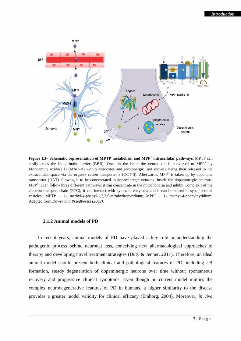
Once taken up by dopaminergic neurons, MPP+ can follow three different pathways; i) it
can bind to the vesicular monoamine transporter-2 (VMAT2) and be stored in synaptosomal
vesicles. Vesicular sequestration of MPP+ seems to represent an important protective
mechanism against neurodegeneration, being that a reduction in VMAT2 activity or a lower
DAT/VMAT2 ratio can enhance MPP+ toxicity (Miller et al., 1999; Speciale et al., 1998); ii)
it can remain in the cytosol and interact with cytosolic enzymes (Dauer & Przedborski, 2003;
Klaidman et al., 1993); and/or iii) it can be concentrated in the mitochondria against a
concentration gradient via an uptake mechanism energized by transmembrane potential
(Ramsay et al., 1986). Once in the mitochondria, MPP+ impairs oxidative phosphorylation by
blocking the Complex I of the mitochondrial electron transport chain (ETC) (Nicklas et al.,
1985). Consequently, this inhibition leads to a decrease in ATP synthesis and an increase in
reactive oxygen and nitrogen species production harmful to the cells (Chan et al., 1991).
MPTP mode of action is depicted in Figure I.3.
7 | P a g e
Introduction
Figure I.3– Schematic representation of MPTP metabolism and MPP+ intracellular pathways. MPTP can
easily cross the blood-brain barrier (BBB). Once in the brain the neurotoxic is converted to MPP+ by
Monoamine oxidase B (MAO-B) within astrocytes and serotonergic (not shown), being then released to the
extracellular space via the organix cation transporter 3 (OCT-3). Afterwards, MPP+ is taken up by dopamine
transporter (DAT) allowing it to be concentrated in dopaminergic neurons. Inside the dopaminergic neurons,
MPP+ it can follow three different pathways: it can concentrate in the mitochondria and inhibit Complex I of the
electron transport chain (ETC); it can interact with cytosolic enzymes; and it can be stored in synaptosomal
vesicles. MPTP – 1- methyl-4-phenyl-1,2,3,6-tetrahydropyridium; MPP+ – 1- methyl-4-phenylpyridium;
Adapted from Dawer and Przedborski (2003).
2.1.2 Animal models of PD
In recent years, animal models of PD have played a key role in understanding the
pathogenic process behind neuronal loss, conceiving new pharmacological approaches to
therapy and developing novel treatment strategies (Duty & Jenner, 2011). Therefore, an ideal
animal model should present both clinical and pathological features of PD, including LB
formation, steady degeneration of dopaminergic neurons over time without spontaneous
recovery and progressive clinical symptoms. Even though no current model mimics the
complex neurodegenerative features of PD in humans, a higher similarity to the disease
provides a greater model validity for clinical efficacy (Emborg, 2004). Moreover, in vivo

8 | P a g e
Introduction
models are essential for evaluating neuroprotective capacity of pharmacologic compounds
against insults, treatment related complications and side effects (Emborg, 2004).
Each animal model presents specific advantages and disadvantages, being the choice of
species, age and sex determined by different aspects including the neurotoxin administered
and the questions proposed to be answered. In comparison with non-human primates, cats and
dogs, rodent models present various advantages, they are widely available, have high
reproductive rates, require reduced cost of maintenance and living space, and bring up less
ethical concerns being extensively used as the preferential model of PD (Emborg, 2004;
Potashkin et al., 2010).
MPTP susceptibility and lesion not only varies between species, with rats presenting less
sensibility to this toxin than mice, but also is affected by gender, age and body weight making
male C57BL/6 mice, the optimal reproducible animal model of PD (Przedborski et al., 2001).
3. Mitochondrial dysfunction and oxidative stress
Mitochondria are unique double membrane organelles that carry their own DNA and
whose main function is the generation of energy in the form of ATP by oxidative
phosphorylation (Keane et al., 2011). Apart from being the powerhouses of eukaryotic cells,
mitochondria also play important roles in regulation of cell death via apoptosis, calcium
homeostasis and control of cell division and growth (Bose & Beal, 2016; Keane et al., 2011;
Szabadkai & Duchen, 2008; Tait & Green, 2013).
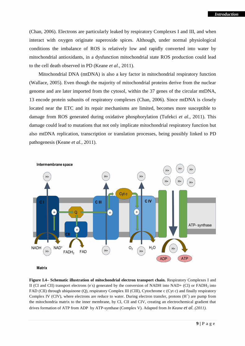
Mitochondrial energy production is accomplished by the transport of electrons through a
series of five complexes, located in the mitochondrial inner membrane, known as the ETC
(Keane et al., 2011). Electrons from oxidative substrates nicotinamide adenine dinucleotide
(NADH) or flavin adenine dinucleotide (FADH2) are conveyed along ETC complexes until
they are reduced to water, at the respiratory Complex IV (Winklhofer & Haass, 2010). In the
process, enough energy to create an electrochemical gradient is generated and protons are
transferred from the mitochondria matrix to the inner membrane by respiratory Complexes I,
III and IV. Lastly, as protons re-enter the mitochondrial matrix, ATP is synthesized by
respiratory Complex V, ATP synthase (Chan, 2006), depicted in Figure I.4. Even though the
electron transport process during oxidative phosphorylation is highly efficient it still can lead
to electron leakage and the consequent production of harmful reactive oxygen spices (ROS)
9 | P a g e
Introduction
(Chan, 2006). Electrons are particularly leaked by respiratory Complexes I and III, and when
interact with oxygen originate superoxide spices. Although, under normal physiological
conditions the imbalance of ROS is relatively low and rapidly converted into water by
mitochondrial antioxidants, in a dysfunction mitochondrial state ROS production could lead
to the cell death observed in PD (Keane et al., 2011).
Mitochondrial DNA (mtDNA) is also a key factor in mitochondrial respiratory function
(Wallace, 2005). Even though the majority of mitochondrial proteins derive from the nuclear
genome and are later imported from the cytosol, within the 37 genes of the circular mtDNA,
13 encode protein subunits of respiratory complexes (Chan, 2006). Since mtDNA is closely
located near the ETC and its repair mechanisms are limited, becomes more susceptible to
damage from ROS generated during oxidative phosphorylation (Tufekci et al., 2011). This
damage could lead to mutations that not only implicate mitochondrial respiratory function but
also mtDNA replication, transcription or translation processes, being possibly linked to PD
pathogenesis (Keane et al., 2011).
Figure I.4– Schematic illustration of mitochondrial electron transport chain. Respiratory Complexes I and
II (CI and CII) transport electrons (e-s) generated by the conversion of NADH into NAD+ (CI) or FADH2 into
FAD (CII) through ubiquinone (Q), respiratory Complex III (CIII), Cytochrome c (Cyt c) and finally respiratory
Complex IV (CIV), where electrons are reduce to water. During electron transfer, protons (H+) are pump from
the mitochondria matrix to the inner membrane, by CI, CII and CIV, creating an electrochemical gradient that
drives formation of ATP from ADP by ATP-synthase (Complex V). Adapted from In Keane et al. (2011).

10 | P a g e
Introduction
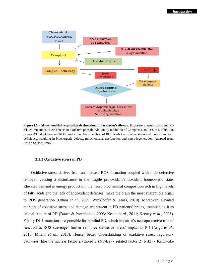
3.1 Mitochondrial dysfunction in PD
Mitochondria dysfunction was first implicated in PD upon the discovery of MPTP’s
ability to induce a parkinsonian syndrome in humans, by inhibiting mitochondrial respiratory
Complex I (Gautier et al., 2014). This hypothesis was further sustained by reports of Complex
I deficiency in SNpc, frontal cortex, peripheral tissues and skeletal muscle, in a postmortem
analysis of PD patients tissues (Bindoff et al., 1991; Haas et al., 1995; Parker et al., 2008;
Schapira et al., 1990).
Complex I impairment causes a decrease in mitochondrial ATP production and an
increase in ROS formation. In turn, severe ROS imbalance can lead to protein oxidation, lipid
peroxidation, and damage in DNA and mtDNA, thereby affecting components of the
respiratory chain and other mitochondrial factors, triggering a vicious cycle between
mitochondrial dysfunction and oxidative stress that increases neurodegeneration and PD
progression (Exner et al., 2012; Keane et al., 2011).
Even though the exact mechanisms that lead to neurodegeneration in PD remain elusive,
studies suggest that together with oxidative stress, defects in mitochondrial dynamics and
inflammation contribute to disease pathogenesis (Bose & Beal, 2016). Causes and
consequences of mitochondrial dysfunctions are represented in Figure I.5 and Figure I.6.
11 | P a g e
Introduction
Figure I.5 – Mitochondrial respiration dysfunction in Parkinson’s disease. Exposure to neurotoxins and PD
related mutations cause defects in oxidative phosphorylation by inhibition of Complex I. In turn, this Inhibition
causes ATP depletion and ROS production. Accumulation of ROS leads to oxidative stress and more Complex I
deficiency, resulting in bioenergetic defects, mitochondrial dysfunction and neurodegeneration. Adapted from
Bose and Beal, 2016.
3.1.1 Oxidative stress in PD
Oxidative stress derives from an increase ROS formation coupled with their defective
removal, causing a disturbance in the fragile pro-oxidant/antioxidant homeostatic state.
Elevated demand in energy production, the neuro biochemical composition rich in high levels
of fatty acids and the lack of antioxidant defenses, make the brain the most susceptible organ
to ROS generation (Uttara et al., 2009; Winklhofer & Haass, 2010). Moreover, elevated
markers of oxidative stress and damage are present in PD patients’ brains, establishing it as
crucial feature of PD (Dauer & Przedborski, 2003; Keane et al., 2011; Keeney et al., 2006).
Finally DJ-1 mutations, responsible for familial PD, which impair it’s neuroprotective role of
function as ROS scavenger further reinforce oxidative stress’ impact in PD (Ariga et al.,
2013; Milani et al., 2013). Hence, better understanding of oxidative stress regulatory
pathways, like the nuclear factor erythroid 2 (NF-E2) - related factor 2 (Nrf2) - Kelch-like

12 | P a g e
Introduction
ECH-associated protein 1 (Keap1) pathway and its downstream targets is integral to develop
novel therapeutic strategies or unveil new pathogenic avenues to PD.
A vital cellular defense mechanism against oxidative stress is the activation of Nrf2
pathway (Nguyen et al., 2009). Nrf2 is a basic leucine zipper transcription factor that
regulates gene expression by interacting with the antioxidant response element (ARE) present
in the promotor region of many cytoprotective genes (Yu & Kensler, 2005). Under
physiological conditions, Nrf2 can be found in the cytosol bound to its inhibitor, Keap1.
Keap1 is an oxidative stress sensor that regulates the rate of Nrf2 turnover through ubiquitin
signaling and proteosomal degradation, by facilitating the interaction of Cullin-based E3
ligase complex with Nrf2 (Tong et al., 2006). During oxidative stress Keap1 is oxidized and
the latch to Nrf2 is weaken, changing the conformation of Nrf2-Keap1 complex and stopping
ubiquitination and degradation of the nuclear factor (Stepkowski & Kruszewski, 2011). This
permits stabilization of Nrf2 and its consequent translocation to the nucleus. Once it reaches
the chromatin, Nrf2 can form a heterodimer with a basic leucine zipper transcription factor,
small musculoaponeurotic fibrosarcoma (MAF), bind to ARE sequence and activate the
transcription of Nrf2 dependent genes. Thereafter, when basal conditions are restored, Nrf2
returns to the cytoplasm were Keap1 continues to promote its sequestration and degradation
(de Vries et al., 2008).
The activation of the Nrf2 pathway induces the transcription of several endogenous
antioxidant enzymes including glutathione peroxidases (GPxs) and heme oxygenases (HOs).
GPxs are a group of eight enzymes (Gpx1-8) that play an important antioxidant role in
catalyzing the reduction of hydrogen peroxide to water. Amongst the four major selenium
dependent GPx isozymes in mammalian tissues, the classical GPx1 stands out. This enzyme is
present in both neurons and glia cells, where it can be found in the mitochondria, nucleus and
the cytosol (Smeyne & Smeyne, 2013). PD patients’ brain studies revealed that GPx1 was
present in highest levels in microglia, but was expressed in an overall low concentration in
brain (Power & Blumbergs, 2009). In addition, mice lacking GPx1 gene showed increased
vulnerability towards MPTP exposure than control mice (Klivenyi et al., 2000).
Interestingly, has been reported that overexpression of GPx enzymes decreases
accumulation of oxidizing agents and neuronal loss, highlighting its importance as antioxidant
defense mechanism (Wang et al., 2003).
HOs are a family of enzymes responsible for the degradation of intracellular heme into
free iron, carbon monoxide and biliverdin. Subsequently, free iron is sequestrated by ferritin
and biliverdin is converted into bilirubin, a powerful ROS scavenger (Hung et al., 2008).

13 | P a g e
Introduction
Besides bilirubin, carbon monoxide also has an important anti-apoptotic and antioxidant role,
highlighting HOs neuroprotective ability, not only by degrading proteins such as oxidases and
perioxidases, but also generating important antioxidants end products (Hung et al., 2008;
Zhang et al., 2013).
In mammals, two HOs isoforms where characterized, one whose expression is inducible
(HO-1), and another that is constitutively expressed, (HO-2). In contrast with HO-2, whose
expression does not vary in response to environmental stress, HO-1 mRNA and protein
expression are reported to be increased in stress conditions (Takeda et al., 1996). Postmortem
analysis of PD patients’ brains, shown a greater upregulation of HO-1 in SN in comparison to
other brain regions, emphasizing the importance of this antioxidant protein in the stress
context of this disease (Schipper et al., 1998; Schipper et al., 2009).
The evidence that Nrf2 expression is altered in neurodegenerative disorders combined
with reports that its downstream targets are increased in stress conditions as a tentative
endogenous defense mechanism, emphasize its antioxidant and neuroprotective role in PD as
well as its importance as a therapeutic strategy for regulating oxidative stress.
3.1.2 Mitophagy in PD
Mitochondrial dynamics and mtDNA maintenance play an integral part in preserving
mitochondria health (Bose & Beal, 2016). Consequently, removal of damage mitochondria
through autophagy (mitophagy) is crucial to protect cellular homeostasis and function (Ni et
al., 2015). Impairments in this process result in the persistence of dysfunctional mitochondria
and a subsequent increase in ROS production, which culminates in cellular stress and death,
as illustrated in Figure I.6.
3.1.2.1 PINK1/parkin pathway in mitophagy
PINK1 selective activation of parkin is an important mitophagic pathway, vital for
mitochondria quality control (Scarffe et al., 2014). The mitochondrial damage sensing
mechanism relies on PINK1 degradation rate. In healthy mitochondria PINK1 is transferred
form the translocase of the outer membrane (TOM) complex into the translocase of the inner
membrane (TIM) complex where it is first cleaved by the mitochondrial processing peptidase
(MPP) and then by presenilin-associated rhomboid-like protein (PARL). PARL proteolysis
14 | P a g e
Introduction
generates a 52 kDa N-terminal fragment of PINK1 that is later released into the cytosol.
There, cleaved PINK1 is recognized by an N-degron type 2 E3 ubiquitin ligase and marked
for proteosomal degradation. Conversely, in damage mitochondria PINK1’s import to TIM
complex is disrupted, preventing MPP and PARL proteolysis and forcing the accumulation of
full-length PINK1, bound to TOM complex, in the outer mitochondrial membrane (OMM).
Subsequently, PINK1 precedes with selective parkin recruitment to the mitochondria, by
phosphorylating ubiquitin chains linked to OMM. Once in the mitochondria, parkin can be
both activated by phospho-ubiquitin and PINK1 itself, promoting a maximal parkin activity.
parkin is an E3 ubiquitin ligase that once activated ubiquitynates a number of mitochondrial
proteins, triggering the recruitment of autophagy receptors to mitochondria and the general
autophagy process (Pickrell & Youle, 2015).
Mutations in certain genes such as PINK1 and parkin cause mitochondrial dysfunction
and are known to be associated with familial PD (Bose & Beal, 2016). Studies revealed that in
mice lacking parkin mitochondria integrity is reduced, Complexes I and IV have lower
activity, respiratory capacity decreases and mice become highly susceptible to neurotoxic
compounds like rotenone (Casarejos et al., 2006; Palacino et al., 2004). In turn, models of
PINK1 knockdown present reduced rates of mitochondria respiration and as a result, a
decrease in ATP synthesis. Moreover, mice lacking PINK1 appear to be more sensitive to the
oxidative stress toxic effect and to an increase in mitochondrial dysfunction (Gautier et al.,
2008; Liu et al., 2009). PINK1 deficiency also reported to affect brain regions in distinct
manners. For example, cortex tissues appear to compensate mitophagy failure with
stimulation of alternative catabolic processes, increasing its resistance against oxidative
damage (Diedrich et al., 2011). In addition, mutations found in PD patients in both PINK1
and parkin are also accountable for preventive recruitment of parkin to the mitochondria by
PINK1 thus disrupting this mitophagic pathway (Pickrell & Youle, 2015).
Together, these results reinforce the role of mitochondrial dysfunction in sporadic PD
and the importance of PINK1/parkin pathway as a possible therapeutic target (Pickrell &
Youle, 2015).
3.1.2.2 AMPK pathway in PD
AMP-activated protein kinase (AMPK) is a highly conserved energy and nutrient sensor
that its activated in cases of ATP depletion and/or glucose starvation (Mihaylova & Shaw,
2011). AMPK is essential in mitochondrial homeostasis regulation, and it is involved in both

15 | P a g e
Introduction
processes of mitochondrial biogenesis and clearance, sharing some similarities with parkin
(Canto et al., 2010; Jager et al., 2007; Kim et al., 2011; Zong et al., 2002). Upon energy
stress conditions, including mitochondria dysfunction, intracellular ATP/AMP ratio decreases
leading to AMPK phosphorylation (He & Klionsky, 2009). This allows the direct
phosphorylation of UNC-51-like kinase 1 (ULK1) and the consequent promotion of
autophagy processes, including mitophagy (Hang et al., 2015). Therefore, it is proposed that
AMPK-ULK1 pathway may play a neuroprotective role by rescuing mitophagy defects in
parkin deficient cases. Moreover, in a MPTP model of PD, AMPK activation is recognized as
a protective strategy against the neurotoxic stimuli (Choi et al., 2010). Collectively, this
secondary pathway to mitophay gains relevance as a compensatory neuroprotective strategy
in PD and once more exposes mitochondrial dysfunction role in this disease.
Figure I.6 – Alterations in mitochondrial DNA and mitochondrial dynamics, and Parkinson’s disease.
Alterations in mitochondrial DNA (mtDNA) can increase generation of reactive oxygen species (ROS) and lead
to mtDNA damage and ultimately neurodegeneration. Parkinson’s disease related mutations in parkin, PINK1,
DJ-1, α-synuclein and LRRK2 can also be the source of mitochondrial dysfunction. In addition, environmental
neurotoxins or mutations in parkin and PINK1 lead to alterations in mitochondrial dynamics that result in altered
mitochondrial clearence, turnover and transport, and eventually leading to neurodegeneration. Adapted from
Bose and Beal, 2016.
16 | P a g e
Introduction
4. Glia Activation and Neuroinflammation in PD
The initial neuroinflammation process is a protective mechanism that restores damaged
neuronal cells and glial cells in the central nervous system (CNS) (Kempuraj et al., 2016).
Nevertheless, a sustain neuroinflammatory response can become detrimental and contribute to
the cascade of events that lead to neuronal degeneration, thus playing and important role in
the onset and progression of neurodegenarive disorders like PD (Kempuraj et al., 2016).
Neuroinflammation is mostly mediated by neurons, infiltrating leukocytes and glial cells,
including microglia and astrocytes (Carson et al., 2006). These innate immune system cells
can express pattern recognition receptors (PRRs), sensible to pathogen-associated molecular
patterns (PAMPs) and host-derived PRR ligands, which can trigger inflammatory signaling
pathways. Host-derived PRR ligands, also known a danger associated molecular patterns
(DAMPs), can be found in brain diseases in the form of misfolded or aggregated proteins,
mislocalized nucleic acids and damaged cells, serving has direct triggers of inflammation. The
sustain exposure to pro-inflammatory mediators diverts immune-competent cells from their
beneficial “housekeeping” role and contributes to the development and progression of
neurodegenerative diseases (Heneka et al., 2014).
4.1 Microglia activation in PD
Microglia represents a key cellular component of the innate immune system in the CNS.
These cells are able to interact with neighboring neurons, blood vessels and astrocytes, and
have a number of transporters, channels and receptors in its cell surface that recognize
neurotransmitters, cytokines, chemokines and PRRs (Heneka et al., 2014).
Under physiological conditions, microglia actively maintain homeostasis,
neuroprotection and neuro repair by releasing growth factors such as brain-derive
neurotrophic factor (BDNF) and transforming growth factor β (TGF-β), while retaining a
ramified morphology. Under pathological conditions and in response to PAMPs and DAMPs,
microglia becomes activated, proliferates and changes its conformation to a macrophage-like
morphology (Wang, Tan, et al., 2015). Depending on the pathologic event and their activation
status, microglia can acquire one of two phenotypes with polar inflammatory responses, M1
microglia displaying a pro-inflammatory response whereas M2 microglia displays an anti-

17 | P a g e
Introduction
inflammatory reaction, making the balance between them essential to maintain homeostasis
(Tang & Le, 2016).
Classic microglia activation state, M1 phenotype, is associated with the production of
pro-inflammatory cytokines such as tumor necrosis factor-α (TNF-α), interleukin-1β (IL-1β)
and others (IL-6 and IL-12), superoxide, nitric oxide (NO), ROS and proteases (Wang, Liu, et
al., 2015). In turn, M2 phenotype comprises both states of alternative activation and acquired
deactivation. Alternative activation is dependent on IL-4 or IL-13 treatment, and associated to
genes that promote anti-inflammation, tissue repair and extracellular matrix reconstitution.
Acquired deactivation is induced by uptake of apoptotic cells and exposure to anti-
inflammatory cytokines like IL-10 and TGF-β counteracting the acute inflammation (Tang &
Le, 2016). Microglia interaction and active state are illustrated in Figure I.7.
In 1988, the first evidence of neuroinflammation involvement in PD pathogenesis was
proven by the presence of reactive microglia in the SNcp region of human brain (McGeer et
al., 1988). Further postmortem studies revealed the presence of microglia markers, cytokines
like TNF-α and IL-1β, NO and inducible NO synthase (iNOS) in SNcp as well as the presence
of TGF-β, TNF, IL-6 and IL-1β in the striatum of PD patients (Hirsch & Hunot, 2009).
Moreover, PD patients serum and cerebrospinal fluid analyzed before death, showed an
increased in cytokines (Hirsch & Hunot, 2009). MPTP models of PD were also investigated
and showed the presence of reactive microglia in both mice and monkey models (Hurley et
al., 2003; Liberatore et al., 1999).
In addition to cytokines and Major Histocompatibility Complex (MHC), the ionized
calcium binding adaptor molecule-1 (Iba-1) is also a widely used marker for active microglia
in tissues (Walker & Lue, 2015). Iba-1 is found to be expressed in microglia alone, both in
vivo and in vitro, making it a good marker of this monocyte cell type (Ito et al., 1998).
Moreover, Doorn and colleagues described an increase in Iba-1 in microglia found in the
substancia nigra of PD patients compared with the control (Doorn et al., 2014).

18 | P a g e
Introduction
Figure I.7 – Beneficial and detrimental functions of microglia in the brain. In physiological conditions,
microglia is responsible for maintaining tissue homeostasis, neuroprotection and neuro repair by secretion of
neurotrophic factors, and synaptic remodeling. Danger-associated molecular patterns (DAMPS) and pathogen-
associated molecular patterns (PAMPs) can bind to pattern recognition receptors (PRR) like Toll-like receptors
(TLR), expressed on the surface of microglia and promote their activation. Depending on the signal microglia
may adopt “M1-like” or “M2-like” phenotype, and can either respond by secreting inflammatory mediators or by
enhancing the removal of the stimulant. “M1-like” activation of microglia is associated with the expression of
inducible nitic oxide synthase (iNOS), production of reactive oxygen species (ROS) and pro-inflammatory
mediators by NF-kB. “M2-like” activation of microglia is associated with the increase secretion of neurotrophic
factors and the production pro-resolving cytokines. The difference responses determine whether microglial cell
activity leads to debris clearance and resolution of the inflammatory response or leads to chronic
neuroinflammation. MPTP - 1- methyl-4-phenyl-1,2,3,6-tetrahydropyridium; LPS – Lipopolysaccharide;
Adapted from Heneka et al., (2014) and Tang & Le (2016).
4.2 Reactive Astrocytes in PD
Astrocytes are greatly abundant in the CNS. These glial cells make numerous
contributions to maintain brain homeostasis, by providing metabolites and growth factors to
neurons, participating in synaptic formation and plasticity, regulating blood flow and
19 | P a g e
Introduction
maintaining the extracellular balance of neurotransmitters, fluid and ions (Sofroniew, 2009).
Astrocytes also play a vital role in the inflammatory process. Just like microglia, these
immune-competent cells are able to detect PAMPs and DAMPs and respond through the
secretion of cytokines and chemokines, thus activating an adaptive immune defense
(Colombo & Farina, 2016). Reactive astrogliosis is characterized by upregulation of glial
fibrillary acidic protein (GFAP), a main component of astrocyte intermediate filaments, and
morphological changes comprised of proliferation and glial scar formation in the most severe
cases (Pekny & Pekna, 2014).
The interplay between astrocytes and other immune-competent cells is also important for
the inflammatory process. Astrocytes are responsible for the production of chemokines
essential for the infiltration of macrophages and lymphocytes in the brain, thus promoting an
increased inflammatory response (Barcia, 2013). In turn, astrocytes response can also be
amplified by the conjugation of CNS immune stimulus with pro-inflammatory mediators
released by active microglia, leading to a potentially prejudicial sustained neuroinflammatory
response (Wang, Liu, et al., 2015).
Various studies in PD models as well as in affected brain regions of PD patients have
presented characteristics of reactive astrogliosis, namely increase expression of GFAP and
morphologic transformations in astrocytes, highlighting the contribution of
neuroinflammation in PD progression (Hirsch & Hunot, 2009; Wang, Liu, et al., 2015).
4.3 Anti-Inflammatory mechanisms
Even though the mechanism underlying the progressive neurodegeneration in PD is still
elusive, the hypothesis that links neuroinflammation with PD progression is more and more
plausible (Kempuraj et al., 2016).
An insult to the CNS triggers and immune response mainly carried out by microglia.
Microglia activation leads to production of inflammatory mediators that stimulate the
recruitment of others immune competent cells, and together promote clearance of cell debris
and secretion of neurotrophic factors. The initial protective inflammatory mechanism can
rapidly become detrimental. Inflammatory mediators not only target immune cells but also act
on neurons, promoting neurodegeneration. In turn, neuronal death acts as a stimulus for the
inflammatory response, creating a vicious cycle between inflammation and neuronal death
that contributes to neurodegeneration (Rocha et al., 2015). Therefore, anti-inflammatory
20 | P a g e
Introduction
mechanisms are essential for maintaining homeostasis and avoid the damaging effects of
chronic inflammation, making them an interesting therapeutic target to be explored in PD.
4.3.1 Annexin-1: an important anti-inflammatory protein
Inflammation process regulation is of the utmost importance. In physiological conditions,
inflammation does not progress to a chronic state due to a resolution response comprised of
downregulating the production of inflammatory mediators and clearance of leukocytes and
cellular debris from the inflammation site (Perretti et al., 2017). This crucial process is carried
out by anti-inflammatory and pro-resolving molecules like Annexin-1 (ANXA1), and is
essential for restoring tissue structure, function and homeostasis (Sugimoto et al., 2016).
ANXA1 is a phospholipid-calcium-binding protein that intracellularly plays a role in
differentiation, proliferation, plasma membrane repair and apoptosis. Additionally,
extracellular ANXA1 is also implicated in important anti-inflammatory processes, namely
regulation of neutrophil migration and apoptosis, glucocorticoids activation, and modulation
of macrophage phagocytosis and reprogramming (Solito et al., 2008; Sugimoto et al., 2016).
ANXA1 is a 37 kDa protein that in is full-length form remains inactive. However in
inflammatory conditions this protein can be cleaved by neutrophil elastase and proteinase-3,
generating a 33 kDa cleaved isoform and an active peptide derived from ANXA1 N-terminus.
Once the inflammation state is resolved ANXA1 regains its full-length form (Sugimoto et al.,
2016). Schematic representation of ANXA1 mediated inflammatory processes are illustrated
in Figure I.8.

21 | P a g e
Introduction
Figure I.8 – Schematic anti-inflammatory and proresolving effects of ANXA1. ANXA1 modulates a range
of cellular mechanisms that are activated to resolve the inflammatory response. This anti-inflammatory protein
decreases neutrophil (PMN) movement and adhesion to the endothelium, increases detachment of adherent cells
and inhibits neutrophil migration to the tissue. ANXA1 induces apoptosis of neutrophils while surmounting the
prosurvival signals to these cells. In addition, endogenous and exogenous ANXA1 also promote monocyte
recruitment and clearance mediated by macrophages, coupled with the release of anti-inflamatory signals and
decrease in proinflammatory cytokines levels. Besides, ANXA1is also involved in macrophages reprogramming,
from a proinflammatory to a proresolving phenotype. Adapted from Sugimoto et al., (2016).
In the brain, ANXA1 is mostly expressed in glial cells, especially microglia, but absent
in the majority of neurons (McArthur et al., 2010). This anti-inflammatory protein was
implicated in PD when Knott and colleagues reported an increased expression of ANXA1 in
activated microglia surrounding degenerating dopaminergic neurons, in SNpc of PD patients.
A raise in ANXA1 expression was not only detected in PD but also in other
neurodegenerative disorders such as Alzheimer’s disease and Multiple Sclerosis. This
upregulation could present a neuronal rescue mechanism, making it relevant the study of
ANXA1 anti-inflammatory mechanism as a therapeutic strategy in PD (McArthur et al., 2010;
Solito et al., 2008).

22 | P a g e
Introduction
5. Tauroursodeoxycholic acid
Tauroursodeoxycholic acid (TUDCA) is an endogenous hydrophilic bile acid formed in
liver by conjugation of ursodeoxycholic acid (UDCA) with taurine (Gronbeck et al., 2016).
Bile acids are chemical detergents produced in the liver crucial in the absorption and
transport of fats and lipid-soluble nutrients (Ackerman & Gerhard, 2016). Some of these
molecules are considered cytotoxic while others like UCDA appear to be cytoprotective.
Mostly, UDCA protective role derives from its ability to reduce the apoptotic threshold in
certain cell types (Amaral et al., 2009). Moreover, UDCA is approved by the U.S. Food and
Drug Administration (FDA) for human therapeutic use, being clinically adopted in primary
biliary cirrhosis treatment (Yanguas-Casas et al., 2017). Clinical results obtain with UDCA
led the way to TUDCA administration, which comparatively had a greater cytoprotective
effect (Rodrigues et al., 1995). Additionally, among its therapeutic advantages TUDCA is
quickly bioavailable via oral, subcutaneous, or intravenous administration, has no associated
toxicity and crosses the BBB (Ackerman & Gerhard, 2016; Rodrigues et al., 2002).
The neuroprotective role of TUDCA has been supported by various experimental studies,
being currently used in a promising clinical trial for the treatment of amyotrophic lateral
sclerosis (Elia et al., 2016). Rodrigues and colleagues demonstrated that TUDCA modulates
apoptosis by suppressing mitochondrial membrane perturbation and consequently inhibiting
Bcl-2-associated X (BAX) protein association to the mitochondria (Rodrigues et al., 2003).
Those results were proven to reduce neurodegeneration in Alzheimer's and Huntington's
disease models as well as to protect against locomotor and cognitive deficits in the 3-
nitropropionic rat model of Huntington's disease (Keene et al., 2001; Rodrigues et al., 2000;
Sola et al., 2006). In addition, Yanguas-Casás and co-workers recently revealed that TUDCA
displayed anti-inflammatory properties in microglia cells by decreasing nuclear factor-kappa
B (NF-κB) activation and/or by increasing cyclic adenosine monophosphate (cAMP) levels
that mediate an anti-inflammatory response (Yanguas-Casas et al., 2017). Together,
TUDCA’s neuroprotective characteristic along with its antioxidant and anti-inflammatory
capacity make this bile acid a promising therapeutic compound for neurodegenerative
disorders as well as other traumatic brain injuries (Ackerman & Gerhard, 2016; Gronbeck et
al., 2016).

23 | P a g e
Introduction
5.1 TUDCA in PD
Despite the intensive research in PD field, there are still no effective therapies to delay,
stop or revert neurodegeneration and disease progression. Current therapeutic strategies are
only capable of relieve PD symptoms and restore some quality of life to patients (Taylor et
al., 2013).
Giving the age relevance in PD and the increasing life expectancy of the world
population, incidence of this disease has been growing exponentially, making the discovery of
new approaches to treatment an urgent matter (Valadas et al., 2015). PD’s late diagnose,
mainly based on motor symptoms, together with the fact that this disease is believed to be
multifactorial and differs from case to case, hinders the strategy development process (Poewe
et al., 2017).
Although the cause of PD remains elusive, neurodegeneration has been associated with
different mechanisms of cell damage including inflammation, mitochondrial dysfunction,
oxidative stress, apoptosis and protein aggregation (Emborg, 2004). As a result, TUDCA’s
neuroprotective and antioxidant features appeared to represent a potentially successful
therapeutic agent in PD treatment.
Our group began to demonstrate that TUDCA prevented MPTP-induced dopaminergic
cell loss and ROS production, in a mice model of PD (Castro-Caldas, Carvalho, Rodrigues,
Henderson, Wolf, Rodrigues, et al., 2012). Furthermore, TUDCA’s antioxidant role was
determine by promoting Nrf2 activation in a MPP+ cell model as well as by increasing
expression of Nrf2 and its downstream antioxidant targets in striatum and midbrain regions of
an MPTP mice model (Moreira et al., 2017). Additionally, TUDCA was showed to display a
neuroprotective role by modulating PINK1/parkin mitophagic pathway and promoting
mitochondrial biogenesis in response to MPP+/MPTP models of PD (Rosa et al., 2017).
Together, these results validated the beneficial effects of TUDCA reported by other
studies in striatum and midbrain regions of an MPTP mice model of PD. However, little is
known about the TUDCA’s broad neuroprotective response in other brain regions such as
cortex, making it a relevant study to better understand the mechanisms involved in this
prominent therapeutic agent for PD treatment.

24 | P a g e
Introduction
6. Aims
The main goal of this thesis is to investigate if the neurotoxic effects of MPTP are
extended to the cerebral cortex in a mouse model of PD. This study also consists in dissecting
the anti-oxidant effects of TUDCA in this brain region, comparing the obtained results with
our previous data from MPTP mice models. In addition, we propose to investigate the anti-
inflammatory mechanism of this bile acid, in the cortex region and in glia cells, and explore
its capacity to modulate an important anti-inflammatory protein, ANXA1.
Our specific aims are:
Validate MPTP neurotoxic effects in the cerebral cortex region. We will start by
confirming MPTP toxicity in the cortex region of mice subjected to short and long
term evaluation. The expression level of proteins involved in mitochondrial
dysfunction as well as main dysfunctional markers such as ROS production and ATP
depletion will be assessed.
Determine TUDCA anti-oxidant effects in cerebral cortex region. The broad
neuroprotective effect of TUDCA in response to MPTP toxicity will be evaluated.
Expression of proteins involved in mitophagy and oxidative stress will be analyzed
and compared to the previous results obtained by our group in the striatum and
midbrain regions from mice models.
Explore TUDCA anti-inflammatory effect in an MPTP mice model. We will
explore TUDCA role in resolving/preventing neuroinflammation and glia activation.
Glia activation markers such as Iba-1 and GFAP and the expression of ANXA1 anti-
inflammatory protein will be analyzed in the brain cortex region.
Explore the direct effect of TUDCA in ANXA1. Lastly, we will examine TUDCA
direct effect on ANXA1 and microglia’s activity in vitro. We hope to establish a direct
connection, highlighting the promising therapeutic role of both TUDCA and ANXA1.
Finally, in this work we intent to provide extended knowledge into TUDCA broad
neuroprotective effect and draw attention to its promising therapeutic role in PD treatment.

25 | P a g e
II. Materials and Methods
1. Materials
1.1 Supplements and chemicals
Bio-Rad’s Protein Assay Reagent was acquired from Bio-Rad Laboratories (Hercules,
CA, USA); Polyvinyl difluoride (PVDF) membrane was obtained from Millipore (Bedford,
MA, USA). ATP-Glo Bioluminometric Cell Viability was purchased from Biotium
Inc. (Fremont, CA, USA); ECL Western blotting detection reagent and Western
BrightTM
ECL-Spray were bought from GE Healthcare (Buckinghamshire, UK)
and Advansta Inc. (CA, USA), respectively. SuperSignal® West Femto Maximum
Sensitivity Substrate was acquired from Thermo Scientific (Rockford, USA); RPMI Media
1640, fetal bovine serum (FBS) Penicilin/streptomycin and TriplE Express were
purchased from GIBCO ® (Life Technologies, Inc., Grand Islands, USA);
TUDCA, MPTP, 2’,7’-dichloroflurescein diacetate (DCF-DA) probe, Bovine
Serum Albumin (BSA) (fraction V), Hoechst 33258 dye, Triton X-100, and
Mowiol mounting media, were obtained from Sigma Aldrich (St Louis, MO,
USA); Other chemicals and analytical grade reagents were ordered from local commercial
sources.

26 | P a g e
Materials and Methods
1.2 Antibodies
Table II.1 – Primary antibodies used for Western Blot analysis
Primary Antibody (antigen) Host Brand Dilution
p-AMPK Rabbit Cell Signaling Technology (MA, USA) 1:1000
AMPK Rabbit Cell Signaling Technology (MA, USA) 1:1000
ANXA1 Rabbit Invitrogen (CA, USA) 1:1000
β-III-tubulin Mouse Covance (NJ, USA) 1:1000
β-Actin Mouse Sigma Aldrich (MO, USA) 1:80000
DJ-1 Rabbit Millipore (MA, USA) 1:1000
GFAP Mouse Millipore (MA, USA) 1:20000
Gpx1 Rabbit Abcam (Cambridge, UK) 1:1000
HO-1 Rabbit Enzo Life Sciences (NYC, USA) 1:1000
p-parkin Rabbit Abcam (Cambridge, UK) 1:300
Parkin Mouse Cell Signaling Technology (MA, USA) 1:1000
PINK-1 Rabbit Novus Biologicals (CO, USA) 1:1000
Table II.2 – Primary antibodies used in immunohistochemistry
Primary Antibody
Host
Brand
Dilution
GFAP Rabbit Sigma Aldrich (MO, USA) 1:200
Iba-1 Rabbit Wako Pure Chemicals (VA, USA) 1:100
Table II.3 – Secondary antibodies used for Western Blot analysis and immunohistochemistry assay
Secondary Antibody
Host
Brand
Dilution
Anti-rabbit Alexa Fluor® 488 Goat ThermoFisher Scientific (IL, USA) 1:200
Horseradish perioxidase conjugated anti-mouse Goat Bio-Rad Laboratories (CA, USA) 1:5000
Horseradish perioxidase conjugated anti-rabbit Goat Bio-Rad Laboratories (CA, USA) 1:5000

27 | P a g e
Materials and Methods
2. Methods
2.1 Animal treatments
All animal experiments were carried through in accordance with the institutional,
Portuguese and European guidelines (Diário da República, 2.ª série N.º121 of 27 June 2011;
and 2010/63/EU European Council Directive) and methods were approved by the Direcção
Geral de Alimentação e Veterinária, the Órgão Responsável pelo Bem-Estar Animal
(ORBEA) of the Faculty of Pharmacy, University of Lisbon and the Animal Ethics
Committee of the Life and Health Sciences Research Institute, University of Minho (SECVS
013/2017). Animal facilities and the people directly involved in animal experiments were
certified by the Portuguese regulatory entity - Direcção Geral de Alimentação e Veterinária.
Twelve-week-old male C57BL/6 mice were purchased from Harlan (Spain) and Charles
River Laboratories and subjected to short and long term MPTP treatments, respectively.
Animals were housed in groups of 4 or 5 animals in filter-topped polysulfone cages 267 × 207
× 140 mm (370 cm2 floor area) (Tecniplast, Buguggiate, Italy), with corncob bedding (Scobis
Due, Mucedola SRL, Settimo Milanese, Italy) in a conventional animal facility. All animals
were maintained under standard laboratory conditions: an artificial 12 h light/dark cycle
(lights on from 8:00 to 20:00 h), with an ambient temperature of 21±1°C and a relative
humidity of 50–60%; the mice were given a standard diet (4RF21, Mucedola SRL, Settimo
Milanese, Italy) and water ad libitum. Health monitoring was performed according to
FELASA guidelines (Guillen, 2012; Nicklas et al., 2002) confirming the Specified Pathogens
status of sentinel animals maintained in the same animal room. Humane endpoints for
experiments were defined (20% reduction of the body weight, inability to reach food and
water, presence of wounds in the body, dehydration), but not needed in practice, as animals
did not reach these endpoints in the study period.
TUDCA and MPTP were dissolved in saline and administered intraperitoneally (i.p.).
TUDCA was daily injected (1 injection per day), for three consecutive days, at a dose of 50
mg/Kg body weight whereas MPTP was administered at a single dose of 40 mg/Kg body
weight, as previously described (Castro-Caldas, Carvalho, Rodrigues, Henderson, Wolf,
Rodrigues, et al., 2012).
Mice evaluated for a short period of time were divided in 6 groups: i) mice that received
saline (Control group); ii) mice treated with TUDCA for 3 consecutive days, and sacrificed 6
h after the last TUDCA injection (TUDCA group); iii) mice injected with MPTP and
28 | P a g e
Materials and Methods
sacrificed 3 h after MPTP administration (MPTP, 3h group); iv) mice that received daily
injection of TUDCA during 3 consecutive days, followed by i.p. administration of MPTP on
day 3, 6 h after the last TUDCA injection, and sacrificed 3 h after MPTP administration
(T+M, 3h group); v) mice injected with MPTP followed by TUDCA injection 3 h after
neurotoxin administration, and sacrificed on day 3, 6 after the last TUDCA injection (M+T,
3h group); and vi) mice injected with MPTP and sacrificed at 3 days after MPTP
administration (MPTP, 3D group);
Mice evaluated during a long period of time were divided in 4 groups: 1) mice that
received saline (Control group); 2) mice injected with MPTP and sacrificed 52 days after
MPTP administration (MPTP, 52D group); 3) mice that received daily injection of TUDCA
during 3 consecutive days, followed by i.p. administration of MPTP on day 3, 6 h after the
last TUDCA injection, and sacrificed 52 days after the first MPTP administration (T+M, 52D
group); and 4) mice injected with MPTP followed by a daily injection of TUDCA during 3
consecutive days, starting 3 h after the neurotoxin administration, and sacrificed 52 days after
MPTP injection (M+T, 52D group).
Treatments were carried out in at least 3 independent experiments (n=3) for short and
long term evaluation experiences respectively. Since previous studies showed that evaluated
parameters in control animals did not change throughout the time course, control animals
were sacrificed together with TUDCA-treated animals in order to avoid increasing the number
of animals needed for the experiments. The schematic representation of mice treatments is
illustrated in Figure II.1.

29 | P a g e
Materials and Methods
Figure II.1 – Schematic representation of mice treatments, short and term evaluation. C57BL/6 male mice
were i.p injected with TUDCA (50mg/Kg body weight), for three consecutive days. MPTP was administered i.p
at a single dose of 40 mg/Kg body weight. Short term evaluation treatments are comprised of: 1.A – Control
mice received saline. 1.B – Mice were injected with TUDCA for three consecutive days, and were sacrificed 6h
after TUDCA last injection. 1.C – Mice were treated with a single dose of MPTP for 3h. 1.D – MPTP injection
was administered 6h after the last TUDCA injection, on day 3, and mice were sacrificed 3h after MPTP
administration. 1.E – TUDCA was administered for three consecutive days, 3h after MPTP injection on day 1.
Mice were sacrificed 6h after the last TUDCA injection. 1.F – Mice were injected on day 1 with MPTP and
sacrificed 3Days after neurotoxin administration. Long term evaluation treatments consisted of: 2.A – Control
mice received saline. 2.B – Mice were injected on day 1 with MPTP and sacrificed 52 Days after neurotoxin
administration. . 2.C – MPTP injection was administered 6h after the last TUDCA injection, on day 3, and mice
were sacrificed 52 days after MPTP administration. 2.D – TUDCA was administered for three consecutive days,
3h after MPTP injection on day 1. Mice were sacrificed 52 Days after MPTP injection. i.p – intra-peritoneally;
MPTP – 1-methyl-4-phenyl-1,2,3,6-tetrahydropyridine; TUDCA – Tauroursodeoxycholic acid.

30 | P a g e
Materials and Methods
2.2 Culture conditions and cell treatment
In vitro studies were carried out using an immortalized murine microgial cell line, BV2
(kindly provided by E. Rodrigues, Faculdade de Farmácia, Lisboa). This cell line expresses
properties of activated microglial cells and therefore, it can be used as alternative model
system for primary microglia cultures or for animal experiments examining brain
inflammation (Blasi et al., 1990; Bocchini et al., 1992; Henn et al., 2009).
Cells were maintained in T75 flasks in RPMI 1640 medium supplemented with 10% FBS
and 1% of Penicilin/streptomycin, at 37ºC in a humidified atmosphere of 5% CO2, in
HERAcell 150 incubators (Thermo Scientific, Waltham, MA, USA). When cells reached
about 80% confluence, they were detached from the T-flasks using 1.5 mL of TryplE express,
for 5 min at 37ºC and were then counted, with a hemocytometer, and diluted in RPMI 1640
medium, according to their initial concentration and specific assay.
Cells were plated in 60mm culture dishes, at a concentration of 1x10⁶ cells/mL and left to
stabilize for 24 h. BV2 cells were treated with 100 μM of TUDCA for different time points (3,
6 or 24 h). In another set of experiments BV2 cells were treated with 100 μM of TUDCA for
6 h after a pre-treatment for 1h with either 100ɳM of Wortmannin,100ɳM of K252A or 5μM
of H89. Controls were always included, and consisted of treating the cells with vehicle
(control). Treatments were carried out in at least two independent experiments. The schematic
representation of the experiences time course treatment is illustrated in Figure II.2.
Figure II.2 – Schematic representation of three independent experiences in BV2 cells. Experience 1, BV2
cells were treated with TUDCA for 3h, 6h or 24h. Experience 2, BV2 treatments included TUDCA incubation
for 6h or conjugated treatments of TUDCA for 6h, after 1h incubation with Wortmannin, K252A or H89
inhibitors. Controls were always included and treated with a vehicle. Incub. – Incubation; TUDCA -
Tauroursodeoxycholic acid;
31 | P a g e
Materials and Methods
After treatments, BV2 cells were washed with ice-cold PBS supplemented with 1mM of
EDTA in order to seize extracellular ANXA1 (Castro-Caldas et al., 2003). Subsequently, cells
were lysed in ice-cold lysis buffer (50mM Tris-HCl pH 7,4, 180 mM NaCl, 1mM EDTA and
1% Triton-X 100) plus Complete Mini protease inhibitor, 200mM Na3VO4, and incubated on
ice for 30 min. Cells were collected and sonicated, in the Ultrasonic Processor UP100H
(Hielscher-Ultrasound Technology, Teltow, Germany), three times for 5 sec each, on ice.
Intracellular extracts were centrifuged at 13,000xg for 10 min, at 4°C, whereas extracellular
extracts were centrifuged at 1,000xg for 5 min, at 4°C, the supernatants were collected and
stored at -80°C. Total Protein concentration was determined by the Bradford method
(Bradford, 1976), using Bio-Rad’s Protein Assay Reagent.
Proteins were also extracted from primary mouse neurons cultures previously subjected
to 0.5mM of MPP+ (Sigma Aldrich) and/or 100 μM of TUDCA. Neurons were treated with
either MPP+
for 3 h or the conjugated treatment of a TUDCA 6 h incubation followed by
MPP+
treatment for 3 h. Controls were included, and consisted of treating the cells with
vehicle (control).
Primary cultures of C57BL/6 mice cortical neurons were prepared from 17 to 18 day old
fetuses (Rosa et al., 2017). Succinctly, pregnant mice were sacrificed in a CO2 chamber and
the fetuses were collected in Han’s balanced salt solution (HBSS) (Sigma Aldrich, MO, USA)
and rapidly decapitated. After removal of meninges and white matter, the brain cortex was
collected in HBSS without Ca2+
and Mg2+
(HBSS-2). The cortex was then mechanically
fragmented, transferred into a 0.05% trypsin solution (Sigma Aldrich, MO, USA), and
incubated for 15 min at 37 °C. After trypsinization, cells were washed twice in HBSS-2
containing 10% FBS and resuspended in Neurobasal medium, supplemented with 0.5 mM L-
glutamine, 25 μM L-glutamine acid, B-27 supplement and 12 mg/ml gentamicin (GibcoTM
,
Thermo Scientific, MA, USA). Isolated neurons were plated in a density around 640
cells/mm2 on culture plates pre-coated with poly-D-lysine (Sigma Aldrich), and maintained at
37ºC in a humidified atmosphere of 5% CO2. Half of the neuronal primary cultures medium
was changed every 3-4 days. Glutamic acid was only added to the medium when plating the
cells. All the media changes and cell treatments afterwards were free from glutamic acid.
32 | P a g e
Materials and Methods
2.3 Western Blot Analysis
Mice were decapitated, after being anesthetized with sodium pentobarbital (50 mg/Kg,
i.p.), and the cerebral cortex was isolated at the level of the nigrostriatal axis (Bregma −3.20
to 1.00). Cortex region samples were then stored at -80°C until needed.
Mice cortex fragments were homogenized in cold PBS supplemented with Complete
Mini Protease Inhibitor Cocktail, using a tissue grinder and centrifuged at 3.000 r.p.m. for 10
min, at 4ºC. Pellets were then suspended in lysis buffer (20 mM Tris-HCl (pH 7.5), 150mM
NaCl, 1mM Na2EDTA, 1mM EGTA, 1% Triton, 2.5mM sodium pyrophosphate, 1mM β-
glycerophosphate, 1mM Na3VO4, 1μg/ml leupeptin) supplemented with Complete Mini
Protease Inhibitor Cocktail, 200mM Na3VO4 and 1M NaF, and incubated on ice for 30 min.
After sonication, in the Ultrasonic Processor UP100H (Hielscher-Ultrasound Technology,
Teltow, Germany), six times for 5 sec each, on ice, samples were centrifuged at 13000 rpm
for 15 min, at 4ºC. Supernatants were collected and frozen at -80ºC. Total Protein
concentration was determined by the Bradford method (Bradford, 1976), using Bio-Rad’s
Protein Assay Reagent.
Protein extracts from both animal and cell treatments were added (5:1) to loading buffer
5x SDS-PAGE (0.25 mM Tris-HCl, 4% SDS, 40% glycerol, 0.2% bromophenol blue, 1% β-
mercaptoethanol, pH 6.8) and boiled for 5 min. Samples were then resolved on a 12,5%
sodium dodecyl sulfate-polyacrylamide gel electrophoresis (SDS-PAGE), in running buffer
(25 mM Tris Base, 190 mM glycine, 0.1% SDS, pH 8.3) with a fixed amperage of 35 mA per
gel, for about 3 h. Subsequently, proteins were electrotransferred to an activated PVDF
membrane (1 min in ethanol, 2 min in H2O, 5 min in transfer buffer), in transfer buffer (25
mM Tris, 190 mM glycine, 20% methanol), during 2 h at a fixed amperage of 500 mA.
Membranes were then stained with Amido black for 4 min, washed with distaining solution
(25% isopropanol and 10% acetic acid) for 10 min, and dried at room temperature. After re-
hydration with ethanol for 1 min, wash with distillated water for 2 min and equilibrate for 5
min in TBS-T (25 mM Tris Base, 137 mM NaCl, 2.7 mM KCl, 0.05% Tween 20, pH 7.4),
membranes were blocked with 5% (w/v) non-fat dry milk in TBS-T (25 mM Tris Base, 137
mM NaCl, 2.7 mM KCl, 0.05% Tween 20, pH 7.4), for at least, 1 h at room temperature,
being then incubated with the specific primary antibodies: rabbit anti-Gpx1 (1:1000, 3%
BSA), rabbit anti-HO-1 (1:1000, 3% BSA), rabbit anti-p-parkin (1:300, 3% BSA), mouse
anti-parkin (1:1000, 5% non-fat dry Milk), rabbit anti-PINK1 (1:1000, 3% BSA), rabbit anti-
p-AMPK (1:1000, 3% BSA), rabbit anti-AMPK (1:1000, 3% BSA), mouse anti-GFAP

33 | P a g e
Materials and Methods
(1:20000, 3% BSA), rabbit anti-ANXA1 (1:1000, 3% BSA), mouse anti-β-III-Tubulin
(1:1000, 3% BSA), rabbit anti-DJ-1 (1:1000, 3% BSA), overnight, at 4ºC, with agitation.
Overnight incubation was followed by incubation with horseradish peroxidase-conjugated
anti-mouse (1:5000, 3% non-fat dry milk) or anti-rabbit (1:5000, 3% non-fat dry milk)
secondary antibodies, for at least, 1 h at room temperature. After washing the membranes
with TBS-T, the chemiluminescent immunocomplexes were detected using ECL or Femto
reagents. The relative intensities of protein bands were analyzed with Image Lab TM analysis
software, after being scanned with ChemiDoc™, both from Bio-Rad Laboratories (Hercules,
CA, USA). After the evaluation of the different proteins, membranes were stripped, with
stripping solution (1.5% glycine, 40% glacial acetic acid, 1% SDS, 10% Tween 20) for 10
min, and washed several times in TBS-T. Stripped membranes were then blocked as
previously described and incubated with mouse anti-β-actin (1:80000, 5% non-fat dry milk)
primary antibody, followed by incubation with horseradish peroxidase-conjugated anti-mouse
secondary antibody. β-actin expression analyzed in stripped membranes was used as a loading
control.
2.4 Measurement of intracellular ROS production
Intracellular ROS production was measured with the cell permeant and non-fluorescent
reagent 2’,7’-dichlorofluorescin diacetate (DCF-DA) that measures hydroxyl, peroxyl and
other ROS activity within the cell. After diffusion into the cell, the DCF-DA probe is
deacetylated by cellular esterases to a non-fluorescent compound, which is later oxidized by
ROS into 2’,7’-dichlorofluorescein (DCF), a highly fluorescent compound.
Measurement of ROS was performed in cortex tissue extracts from long term evaluation
experiments. Primarily, 50µg of protein from our samples were incubated with 10 μM of
DCF-DA probe, diluted in PBS, at 37ºC for 1 h, in the dark. After incubation, the florescence
intensity of the oxidized probe, DCF, was measured in the microplate reader GloMax® Multi
Detection System Promega (Sunnyvale, CA, USA), at an excitation an emission wavelength
of 485nm and 528nm, respectively. Blanks wavelengths, comprised of PBS incubated with
DCF-DA probe were subtracted to our results. Each assay from three different experiments,
was performed in duplicate and the results were normalized to the percentage of control.
34 | P a g e
Materials and Methods
2.5 Measurement of ATP levels
ATP levels were measured using with the ATP-Glo™ Bioluminometric Cell Viability
Assay (Biotium, Hayward, CA, USA). The premise of this assay is based on a Bioluminescent
reaction catalyzed by Firefly luciferase. This Protein uses the amount of ATP available in
each sample to oxidize D-Luciferin and as a byproduct of the reaction producing light.
Measurement of ATP levels was performed in cortex tissue extracts from long term
evaluation experiments, according to the manufacturer’s instructions. The Luciferase activity
in each sample was then measured in a luminometer (Berthold Systems). Negative controls
were always included, and results were normalized to the total protein concentration in each
well, as previously described (Rosa et al., 2017).
2.6 Immunohistochemistry
The detection of reactive astrocytes and microglia activation markers (GFAP and Iba-1,
respectively) was performed by immunohistochemistry, in cerebral tissue coronal sections
from long term evaluated mice that were treated with MPTP and TUDCA as previously
described.
Tissue sections stored at -80ºC were left to dry 1 h, at room temperature being later
fixated with 4% PFA in PBS for 10 min. Tissue sections were then washed three times and
permeated with PBS supplemented with 0.2% of Triton X-100 for 30 min, at room
temperature. After being washed three times with PBS for 10 min, sections were blocked with
blocking solution (2% BSA, 0.05% Tween-20 in PBS), for 1 h at room temperature. Sections
were then incubated overnight, at 4ºC, with the specific primary antibodies rabbit anti-GFAP
(1:200 in blocking solution) and rabbit anti-Iba-1 (1:100 in blocking solution), in a dark
humidified chamber. Overnight incubation was followed by incubation with the secondary
antibody goat anti-rabbit Alexa Fluor® 488 (1:200 in blocking solution) for 2 h, at room
temperature. After staining cell nuclei with Hoechst 33258 dye (5μg/mL), the sections were
mounted in glass slides with Mowiol anti-fading mounting medium.
Green (for both GFAP and Iba-1) and blue (for nuclei) fluorescence and UV images of,
at least, fifteen random microscopic fields belonging to mice cortex region, were acquired per
mice, under 400x magnification, using a fluorescence microscope (model AxioScope.A1)
with integrated camera AxioCam HR (Carl Zeiss, Inc. – North America). The results were

35 | P a g e
Materials and Methods
expressed as the percentage of area occupied by GFAP and Iba-1 positive cells, measured by
using the ImageJ software analysis (National Institutes of Health, USA).
2.7 Statistical analysis
All results are expressed as mean ± SEM values. Data were analyzed by T-student test or
one way ANOVA, where differences between groups were determined by post hoc
Bonferroni’s test and comparison of treatments to a single control by post hoc Dunnett’s test,
using GraphPad Prism 6.0 (San Diego, CA, USA). Means were considered statistically
significant at a p value bellow 0.05.

36 | P a g e
III. Results
1. Evaluation of the antioxidant role of TUDCA in the MPTP mouse model of PD
1.1 TUDCA up-regulates the expression levels of Nrf2 downstream targets in the cortex of
C57BL/6 mice upon MPTP administration
Nrf2 is a key regulator of cellular redox status (Sandberg et al., 2014). This transcription
factor is responsible for promoting the expression of cytoprotective genes, including
antioxidant enzymes, anti-inflammatory mediators and other transcription factors involved in
mitochondrial biogenesis (Tufekci et al., 2011). Therefore, Nrf2 system is widely expressed
in the CNS and integral for a neuroprotective response in acute cerebral insults and in
neurodegenerative diseases. Indeed it is reported that Nrf2 expression plays an important role
in protecting against neurotoxin-induced oxidative stress (Jakel et al., 2007; Sandberg et al.,
2014; Tufekci et al., 2011). Consequently, Nrf2 pathway dysregulation has been deeply
implicated in PD pathogenesis, making its activation and the up-regulation of antioxidant
downstream targets a promising strategy for regulating oxidative damage in PD. As
previously described, TUDCA antioxidant properties present a hopeful approach against
oxidative stress in PD. In accordance, our group disclosed that TUDCA treatment prior to or
after MPTP administration increased Nrf2 expression as well as an overall increase in GPx1
and HO-1 expression in mice striatum and midbrain (Moreira et al., 2017). Thus, to evaluate
the broad effect of TUDCA antioxidant role, we investigated its ability to modulate the
expression of Nrf2 downstream targets, GPx1 and HO-1 in the cortex of MPTP-injected mice.
The expression levels of these antioxidant enzymes were evaluated in the cortex of mice
either subjected to short or long term evaluation (Figure III.1) by Western blot. In short term
evaluation samples, both HO-1 and GPx1 expression were apparently increased in the cortex
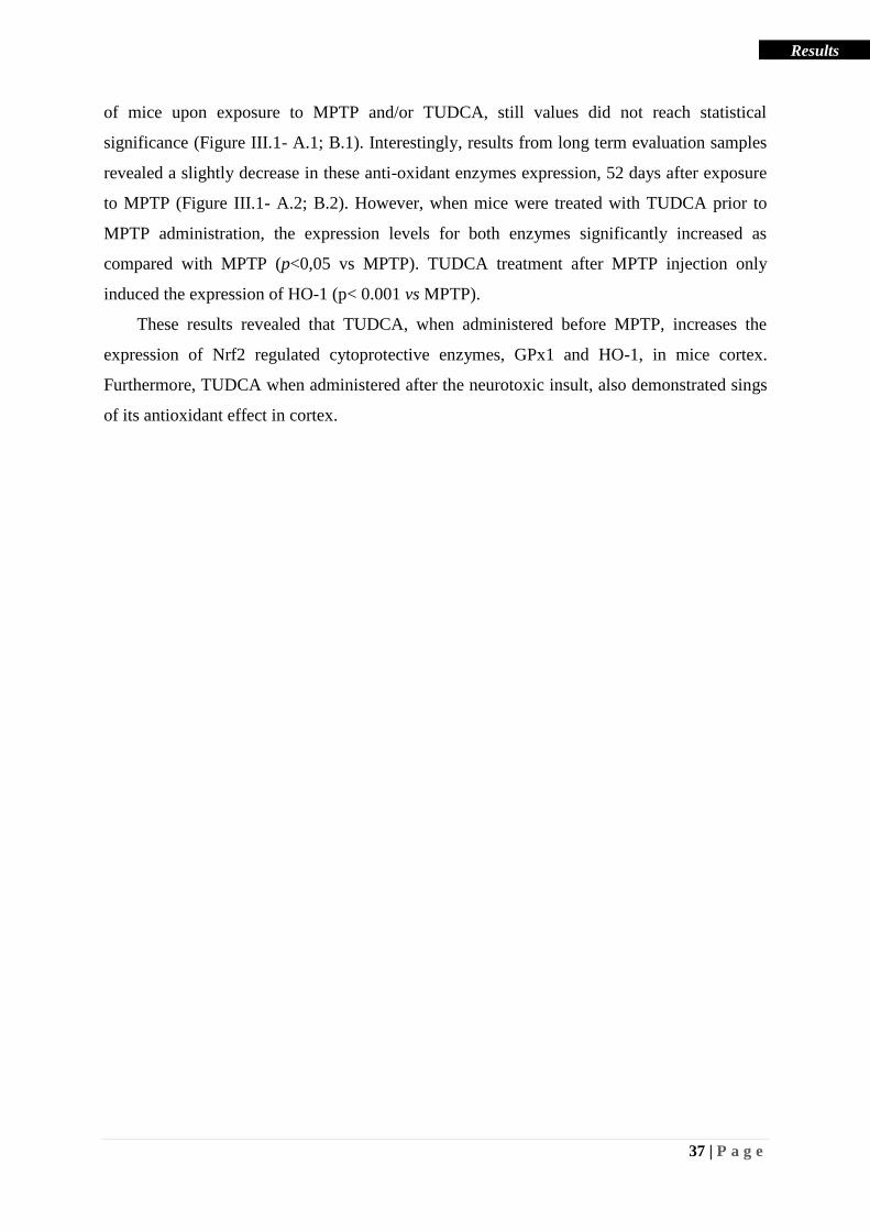
37 | P a g e
Results
of mice upon exposure to MPTP and/or TUDCA, still values did not reach statistical
significance (Figure III.1- A.1; B.1). Interestingly, results from long term evaluation samples
revealed a slightly decrease in these anti-oxidant enzymes expression, 52 days after exposure
to MPTP (Figure III.1- A.2; B.2). However, when mice were treated with TUDCA prior to
MPTP administration, the expression levels for both enzymes significantly increased as
compared with MPTP (p<0,05 vs MPTP). TUDCA treatment after MPTP injection only
induced the expression of HO-1 (p< 0.001 vs MPTP).
These results revealed that TUDCA, when administered before MPTP, increases the
expression of Nrf2 regulated cytoprotective enzymes, GPx1 and HO-1, in mice cortex.
Furthermore, TUDCA when administered after the neurotoxic insult, also demonstrated sings
of its antioxidant effect in cortex.

38 | P a g e
Results
Figure III.1 – Effect of TUDCA on the expression of Nrf2 downstream target enzymes in the cortex of
mice subjected to MPTP administration. C57BL/6 mice were treated with TUDCA and/or MPTP and were
sacrificed after a short time treatment with MPTP (A.1 and B.1) or 52 days after MPTP injection (A.2 and B.2),
as indicated in Methods. Tissue extracts from mice cortex were subjected to SDS-PAGE, and the blots were
probed with antibodies against GPx (A.1, A.2) or HO-1 (B.1, B.2). β-actin was used as a loading control. The
intensity of the bands was quantified using computerized image analysis (Image Lab). Representative
immunoblots for each protein are shown over the correspondent graphs. Data are expressed as the mean values ±
SEM from four independent experiments, indicated as percentage of control. *ρ<0.05, and *** ρ<0.001 vs
Control or MPTP using one-way ANOVA with Bonferroni’s post hoc test.
1.2 Role of TUDCA on ROS generation in C57BL/6 mice cortex upon MPTP
administration
As previously described above, oxidative stress derives from ROS generation coupled
with a defective free radical scavenging system, causing a disturbance in the pro-
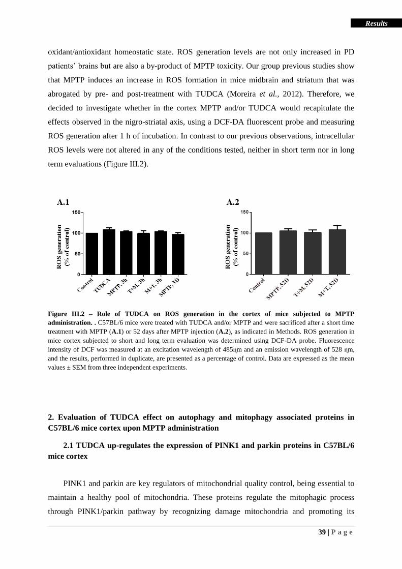
39 | P a g e
Results
oxidant/antioxidant homeostatic state. ROS generation levels are not only increased in PD
patients’ brains but are also a by-product of MPTP toxicity. Our group previous studies show
that MPTP induces an increase in ROS formation in mice midbrain and striatum that was
abrogated by pre- and post-treatment with TUDCA (Moreira et al., 2012). Therefore, we
decided to investigate whether in the cortex MPTP and/or TUDCA would recapitulate the
effects observed in the nigro-striatal axis, using a DCF-DA fluorescent probe and measuring
ROS generation after 1 h of incubation. In contrast to our previous observations, intracellular
ROS levels were not altered in any of the conditions tested, neither in short term nor in long
term evaluations (Figure III.2).
Figure III.2 – Role of TUDCA on ROS generation in the cortex of mice subjected to MPTP
administration. . C57BL/6 mice were treated with TUDCA and/or MPTP and were sacrificed after a short time
treatment with MPTP (A.1) or 52 days after MPTP injection (A.2), as indicated in Methods. ROS generation in
mice cortex subjected to short and long term evaluation was determined using DCF-DA probe. Fluorescence
intensity of DCF was measured at an excitation wavelength of 485ɳm and an emission wavelength of 528 ɳm,
and the results, performed in duplicate, are presented as a percentage of control. Data are expressed as the mean
values ± SEM from three independent experiments.
2. Evaluation of TUDCA effect on autophagy and mitophagy associated proteins in
C57BL/6 mice cortex upon MPTP administration
2.1 TUDCA up-regulates the expression of PINK1 and parkin proteins in C57BL/6
mice cortex
PINK1 and parkin are key regulators of mitochondrial quality control, being essential to
maintain a healthy pool of mitochondria. These proteins regulate the mitophagic process
through PINK1/parkin pathway by recognizing damage mitochondria and promoting its

40 | P a g e
Results
clearance (Scarffe et al., 2014). In turn, mutations or impairments in PINK1 and/or parkin
proteins can cause mitochondrial dysfunction and lead to PD pathogenesis and progression,
being implicated in both familial and sporadic forms of the disease (Bose & Beal, 2016).
Therefore, modulation of these proteins may represent a relevant therapeutic strategy for PD.
To understand TUDCA neuroprotective mechanism of action our group focused on this bile
acid ability to modulate mitophagy associated proteins. Previous studies showed that TUDCA
treatment prior to MPTP administration significantly increased the expression of PINK1 and
parkin, as well as parkin activation (phosphorylation) in mice striatum and midbrain (Rosa et
al., 2017). Thus to better understand TUDCA neuroprotective effect in the mice model of
PD, we investigated its ability to modulate PINK1 and parkin in the cortex of MPTP
administered animals.
The expression levels of p-parkin, parkin and PINK1 were evaluated by Western blot, in
cortex of mice either subjected to short or long term evaluation treatments (Figure III.3).
Regarding phosphorylated parkin expression in short term evaluation samples, (Figure III.3,
A.1), there is a tendency for increase in all time points evaluated, especially when animals
were pre-treated with TUDCA, although no significant results were obtained. Results from
panel B.1 show that the levels of parkin were significantly increased in all conditions
evaluated (p<0.001 vs control). Importantly, pre-treatment with TUDCA induced an increase
in parkin expression that was statistically different from MPTP-administered mice samples
(p<0.001 vs MPTP). In respect to full length PINK1 expression in samples from short term
evaluation treatment, no significant differences were found in any condition tested.
Unexpectedly, the expression levels of these proteins were not significantly affected in the
cortex of animals subjected to long term evaluation protocol.
These results indicate that TUDCA treatment modulates parkin levels in the cortex of
MPTP-intoxicated mice. Additionally, the regulation of parkin levels and activity by TUDCA
may be transient, as previously described for other brain regions (Rosa et al., 2017).

41 | P a g e
Results
Figure III.3 – TUDCA induces expression of mitophagy related preoteins parkin and PINK1 in mice
administered with MPTP. C57BL/6 mice were treated with TUDCA and/or MPTP and were sacrificed after a
short time treatment with MPTP (A.1, B.1 and C.1), or 52 days after MPTP injection (A.2, B.2 and C.2), as
indicated in Methods. Tissue extracts from mice cortex were subjected to SDS-PAGE, and the blots were probed

42 | P a g e
Results
with antibodies against p-parkin (A.1, A.2), parkin (B.1, B.2) or full length PINK1 (C.1, C.2). β-actin was used
as a loading control. The intensity of the bands was quantified using computerized image analysis (Image Lab).
Representative immunoblots for each protein are shown over the correspondent graphs. Data are expressed as the
mean values ± SEM from four independent experiments, indicated as percentage of control. *** ρ<0.001 vs
control or MPTP using one-way ANOVA with Bonferroni’s post hoc test.
2.2 Neuroprotective role of TUDCA against MPTP-induced ATP depletion and
AMPK phosphorylation in mice cortex
ATP depletion can be perceived as an evidence of mitochondrial dysfunction that
ultimately leads to cell death. As already mentioned, MPTP actively participates in decreasing
ATP, by directly inhibiting the Complex I of the ETC, contributing for a disruption in the
oxidative phosphorylation process. Complex I impairment increases ROS formation and the
subsequent damage to components of the respiratory chain and other mitochondrial factors,
triggering a vicious cycle between mitochondrial dysfunction and oxidative stress that affects
neighboring cells and increases neurodegeneration and PD progression.
The enzyme AMPK is essential for regulating mitochondrial homeostasis, being involved
biogenesis and autophagic processes. The autophagy pathway mediated by active
(phosphorylated) AMPK is triggered upon energy stress conditions, increases in ROS and
calcium concentration (Sundararaman et al., 2016). Since we observed the ability of TUDCA
to up-regulate the expression of antioxidant enzymes and the mitophagic associated protein
parkin, we decided to explore TUDCA role in ATP depletion and AMPK activation as
outcomes of its neuroprotective role in cortex. ATP levels were quantified using the ATP-
Glo™ Bioluminometric Cell Viability Assay and expression levels of p-AMPK and AMPK
were evaluated by Western blot, in the cortex of mice subjected to long term evaluation
(Figure III.4). Results from Figure III.4 - A show that ATP levels had a notable decrease in
the cortex of mice upon exclusive MPTP exposure, being significantly different from the
control (p<0.01 vs control). Interestingly, in mice pre-treated with TUDCA, ATP levels were
increased, being statistically different form MPTP samples (p<0.001 vs MPTP). In contrast,
TUDCA administration after MPTP could not restore ATP levels, showing ATP levels similar
to the ones found in MPTP-treated animals (p<0.01 vs control). Accordingly, AMPK
activation levels in the cortex from mice treated exclusively with MPTP, were found to be
significantly lower than the control (p<0.05 vs control), and pre-treatment with TUDCA
raised the expression levels of p-AMPK (p<0.01 vs MPTP). However, the levels of p-AMPK

43 | P a g e
Results
were not different between control and animals treated with TUDA after MPTP
administration.
In the cortex, at the time points evaluated, administration of this bile acid after MPTP
exposure does not appear to be effective in restoring ATP levels, nor inducing AMPK. But
together these results reveal that TUDCA, when administered before MPTP, prevents MPTP-
dependent ATP depletion in mice cortex probably through multiple mechanisms involving
antioxidant defense responses as well as activation of AMPK.
.
Figure III.4 – TUDCA protects against MPTP-induced ATP depletion and promotes AMPK activation in
mice cortex. C57BL/6 mice were treated with TUDCA and/or MPTP and were sacrificed 52 days after MPTP
injection as indicated in Methods for the long term evaluation group. A – ATP levels in cortex were quantified
using ATP-GloTM
Bioluminometric Cell Viability Assay. The Luciferase activity was measure in a luminometer
and the results are presented as a percentage of control. B – Tissue extracts from mice cortex were subjected to
SDS-PAGE , and the blots were probed with antibodies against p-AMPK, AMPK or β-actin as a loading control.
The intensity of the bands was quantified using computerized image analysis (Image Lab). Data are expressed as
the mean values ± SEM at least three independent experiments, indicated as percentage of control. * ρ<0.05, **
ρ<0.01 and *** p<0,001 vs control or MPTP using one-way ANOVA with Bonferroni’s post hoc test.
3. Evaluation of the anti-inflammatory role of TUDCA in the MPTP mouse model of PD
3.1 TUDCA decreases reactive astrogliosis marker in C57BL/6 mice cortex in the
presence of MPTP
Astrocytes are key players of the innate immune system in the CNS. These cells are
capable of detecting harmful stimulus and respond by activating an adaptive immune defense.
Reactive astrocytes are characterized by upregulation of GFAP protein and morphological
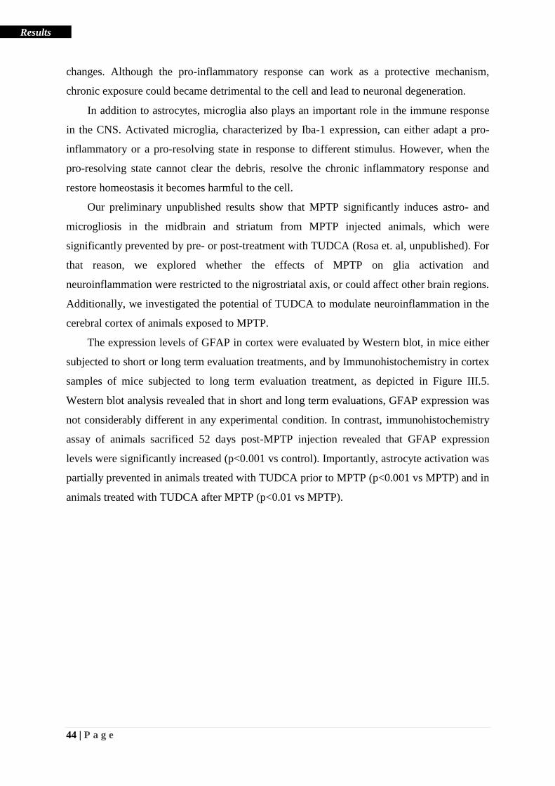
44 | P a g e
Results
changes. Although the pro-inflammatory response can work as a protective mechanism,
chronic exposure could became detrimental to the cell and lead to neuronal degeneration.
In addition to astrocytes, microglia also plays an important role in the immune response
in the CNS. Activated microglia, characterized by Iba-1 expression, can either adapt a pro-
inflammatory or a pro-resolving state in response to different stimulus. However, when the
pro-resolving state cannot clear the debris, resolve the chronic inflammatory response and
restore homeostasis it becomes harmful to the cell.
Our preliminary unpublished results show that MPTP significantly induces astro- and
microgliosis in the midbrain and striatum from MPTP injected animals, which were
significantly prevented by pre- or post-treatment with TUDCA (Rosa et. al, unpublished). For
that reason, we explored whether the effects of MPTP on glia activation and
neuroinflammation were restricted to the nigrostriatal axis, or could affect other brain regions.
Additionally, we investigated the potential of TUDCA to modulate neuroinflammation in the
cerebral cortex of animals exposed to MPTP.
The expression levels of GFAP in cortex were evaluated by Western blot, in mice either
subjected to short or long term evaluation treatments, and by Immunohistochemistry in cortex
samples of mice subjected to long term evaluation treatment, as depicted in Figure III.5.
Western blot analysis revealed that in short and long term evaluations, GFAP expression was
not considerably different in any experimental condition. In contrast, immunohistochemistry
assay of animals sacrificed 52 days post-MPTP injection revealed that GFAP expression
levels were significantly increased (p<0.001 vs control). Importantly, astrocyte activation was
partially prevented in animals treated with TUDCA prior to MPTP (p<0.001 vs MPTP) and in
animals treated with TUDCA after MPTP (p<0.01 vs MPTP).

45 | P a g e
Results
Figure III.5 – TUDCA prevents MPTP-induced astrocyte activation in mice cortex. . C57BL/6 mice were
treated with TUDCA and/or MPTP and were sacrificed after a short time treatment with MPTP (A.1), or 52 days
after MPTP injection (A.2), as indicated in Methods. Tissue extracts from mice cortex were subjected to SDS-
PAGE, and the blots were probed with antibodies against GFAP and β-actin, used as a loading control. The
intensity of the bands was quantified using computerized image analysis (Image Lab). Representative
immunoblots for each protein are shown over the correspondent graphs. Data are expressed as the mean values ±
SEM at least three independent experiments, indicated as percentage of control.
Fluorescence microscopy images show glial fibrillary acid protein (GFAP) staining of Control (B.1), MPTP
(B.2), TUDCA prior to MPTP (B.3) and MPTP before TUDCA (B.4) treated mice. GFAP was stained in green
and nuclei were counterstained with Hoechst dye 33258. Scale bar= 10μm. Graphic C represents the percentage
of area occupied by Iba-1 positive cells, measured using the ImageJ software. Data shown are mean values ±
SEM of at least three independent experiences. **p<0,01 and ***p<0,001 vs Control or MPTP using one way
ANOVA with Bonferroni’s post hoc test.
After evaluating TUDCA’s neuroprotective effect in astrogliosis we decided to further
characterize anti-inflammatory role of this bile acid in microglia activation. Iba-1 expression
46 | P a g e
Results
levels were evaluated by Immunohistochemistry in cortex samples of mice subjected to long
term evaluation treatments (Figure III.6). Treatment with MPTP in the presence or absence of
TUDCA significantly increased Iba-1 expression in the cortex (p<0.001). Importantly, pre-
treatment with TUDCA efficiently prevented MPTP-induced Iba-1 expression (p<0.001 vs
MPTP).
Taken together, these results reveal that, when administered before MPTP, TUDCA
exerts an efficient anti-inflammatory effect, by preventing the expression of these glia
activation markers.
Figure III.6 – TUDCA prevents MPTP-induced glial activation in mice cortex. C57BL/6 mice were treated
with TUDCA and/or MPTP and were sacrificed 52 days after MPTP injection as indicated in Methods for the
long term evaluation group. Fluorescence microscopy images show ionized calcium biding adaptor molecule-1
(Iba-1) staining of Control (A.1), MPTP (A.2), TUDCA prior to MPTP (A.3) and MPTP before TUDCA (A.4)
treated mice. Iba-1 was stained in green and nuclei were counterstained with Hoechst dye 33258. Scale bar=
10μm. Graphic B represents the percentage of area occupied by Iba-1 positive cells, measured using the ImageJ
software. Data shown are mean values ± SEM of at least three independent experiences. **p<0,01 and
***p<0,001 vs Control or MPTP using one way ANOVA with Bonferroni’s post hoc test.
After evaluating the effects of TUDCA in glial activation, we decided to explore the role
of this bile acid on the expression of the anti-inflammatory protein ANXA1. ANXA1 is an
anti-inflammatory protein that in inflammatory conditions is cleaved into an active state,
essential for resolving the pro-inflammatory response and for helping restore tissue structure,
function and homeostasis.
The expression levels of ANXA1 in cortex were evaluated by Western blot in cortex
samples from mice either subjected to short or long term evaluation protocols (Figure III.7).

47 | P a g e
Results
In the cortex of animals from the short term evaluation group MPTP and/or TUDCA did not
significantly differed ANXA1 expression levels in any experimental condition. Interestingly,
when animals were sacrificed 52 days post-MPTP TUDCA significantly increased ANXA1
expression when administered before (p<0.001 vs control, p<0.01 vs MPTP) or after (p<0.05
vs control) the neurotoxin
Altogether, our results show that MPTP induces a pro-inflammatory response in the
cortex that is efficiently ameliorated when animals are treated with TUDCA either before or
after administration of the neurotoxin. The modulation of ANXA1 expression by TUDCA
conveys a possible mechanism of action for this bile acid.
Figure III.7 – TUDCA induces the expression of ANXA1 in cortex from mice injected with MPTP.
C57BL/6 mice were treated with TUDCA and/or MPTP and were sacrificed after a short time treatment with
MPTP (A.1), or 52 days after MPTP injection (A.2), as indicated in Methods. Tissue extracts from mice cortex
were subjected to SDS-PAGE, and the blots were probed with antibodies against ANXA1 and β-actin, used as a
loading control. The intensity of the bands was quantified using computerized image analysis (Image Lab).
Representative immunoblots for each protein are shown over the correspondent graphs. Data are expressed as the
mean values ± SEM from four independent experiments, indicated as percentage of control. *ρ<0.05, **ρ<0.01
and *** ρ<0.001 vs control or MPTP using one-way ANOVA with Bonferroni’s post hoc test.
4. Evaluation of TUDCA neuroprotective role in the MPTP mouse model of PD
Finally, to complete our study regarding the effect of MPTP and TUDCA effect in the
mice cortex, we investigated the role of this compound in neuronal death. In order to examine
neuronal loss, we assessed the expression levels of a microtubule element found in neurons,

48 | P a g e
Results
β-III-tubulin in cortex by Western blot, in mice either subjected to short or long term
evaluation protocols. Results presented in Figure III.8 show that the expression of β-III-
tubulin was not significantly altered in any of the conditions tested.
These results suggest that although we have observed deleterious MPTP effects in mouse
cortex, these effects do not culminate with neuronal cell death at the time points evaluated.
However, cell death should be tested at longer time points and by other experimental
approaches in order to exclude neurodegeneration in the cortex triggered by MPTP.
Figure III.8 – Evaluation of TUDCA effect in neuronal death, in mice cortex, after MPTP administration.
C57BL/6 mice were treated with TUDCA and/or MPTP and were sacrificed after a short time treatment with
MPTP (A.1), or 52 days after MPTP injection (A.2), as indicated in Methods. Tissue extracts from mice cortex
were subjected to SDS-PAGE, and the blots were probed with antibodies against β-III-tubulin and β-actin, was
used as a loading control. The intensity of the bands was quantified using computerized image analysis (Image
Lab). Representative immunoblots for each protein are shown over the correspondent graphs. Data are expressed
as the mean values ± SEM from at least three independent experiments, indicated as percentage of control.
Collectively, the results obtained in this work, in mice cortex, emphasize the role of
TUDCA as an antioxidant, anti-inflammatory and cytoprotective compound in a mice PD
model and strengthened its promising therapeutic effects in PD.

49 | P a g e
Results
5. Evaluation of the anti-inflammatory role of TUDCA in in vitro models
5.1 TUDCA modulation of ANXA1 expression in mice primary neurons treated
with MPP+
Our group has previously explored the antioxidant role of TUDCA in experimental
models of PD (Moreira et al., 2017). Interestingly, in the present work we found that not only
most of the mechanisms triggered by MPTP and TUDCA are recapitulated in the cerebral
cortex, but we also demonstrate an important anti-inflammatory role of this bile acid. Here we
show that TUDCA abrogates glia activation, and most notably induces the expression of the
anti-inflammatory protein ANXA1 in the cortex, in this experimental model of PD. Therefore,
we decided to further explore the ability of TUDCA to induce ANXA1 expression and/or
secretion. For that, we used an in vitro approach starting by assessing ANXA1 expression in
mice primary neuron cultures treated with MPP+, in the presence or absence of TUDCA.
Results presented in Figure III.9 show that ANXA1 levels were increased about 3 times the
control value when cells were incubated with MPP+, in the presence or absence of TUDCA.
Still these values do not present statistical significance, due to a low number of experiments
(n=3) and/or to the fact that probably ANXA1 expression in response to MPP+ happens in a
wave-like response. The fact that ANXA1 is secreted upon its synthesis further increases the
difficulty of this analysis. Unfortunately, in these cells secreted membrane-bound ANXA1
could not be detected due to low concentration, since these assays were performed in a 1.9
cm2 plate that accommodates a low number of cells even in confluency.

50 | P a g e
Results
Figure III.9 – TUDCA effect in ANXA1 expression in mice primary neurons treated with MPP+. Primary
neurons were treated with TUDCA and/or MPP+ as described in Methods. Cell extracts were subjected to SDS-
PAGE, and the blots were probed with antibodies against ANXA1 and β-actin, was used as a loading control.
The intensity of the bands was quantified using computerized image analysis (Image Lab). Representative
immunoblots for each protein are shown over the correspondent graphs. Data are expressed as the mean values ±
SEM from three independent experiments, indicated as percentage of control.
5.2 TUDCA modulation of ANXA1 expression in BV2 microglia cell line
After evaluating the effect of TUDCA in ANXA1 expression levels, both in in vivo and
in vitro PD models we decided to explore the mechanism by which this bile acid could
modulate ANXA1 expression. Since this inflammatory protein, in the brain, is found to be
almost exclusively expressed in glial cells, we focused our study in the BV2 microglia cell
line (Solito et al., 2008). In addition to evaluate intracellular levels of ANXA1 we also
assessed its membrane-bound extracellular levels, considering the extracellular role of this
protein in several inflammatory processes. The intra- and extracellular expression levels of
ANXA1 in response to different time-points of treatment with TUDCA were evaluated by
Western blot, and are shown in Figure III.10. Intracellular protein levels of ANXA1 (Figure
III.10-A) do not seemed to change upon TUDCA administration. However, the extracellular
levels of ANXA1 were significantly increased after 6 h exposure to TUDCA (p<0.05). These
results indicate that TUDCA induces the secretion of ANXA1 and replenishes the
intracellular content of the protein, strongly suggesting de novo synthesis of ANXA1. Since
the peak of extracellular protein was found at 6 h post-TUDCA addition, this time-point was
used in the subsequent experiments aiming to determine the mechanisms underlying ANXA1
expression.

51 | P a g e
Results
Figure III.10 – TUDCA increases ANXA1 expression and secretion in a mouse microglia cell line. BV2
cells were treated with TUDCA and extracellular ANXA1 was obtained after washing the cells with 1mM
EDTA buffer, as described in Methods. Cell extracts (A) and products of EDTA buffer wash (membrane-bound
extracellular protein) (B) were subjected to SDS-PAGE, and the blots were probed with antibodies against
ANXA1 and β-actin (loading control). The intensity of the bands was quantified using computerized image
analysis (Image Lab). Representative immunoblots are shown over the correspondent graphs. Data are expressed
as the mean values ± SEM from three independent experiments, indicated as percentage of control. *p<0.05
using T-student test.
5.3 Unraveling the mechanism underlying the modulation of ANXA1 by TUDCA
The promotor region of ANXA1 gene contains a cAMP responsive element (CRE) that
requires an activated CRE binding protein (CREB) to induce ANXA1 mRNA synthesis
(Antonicelli et al., 2001). CREB activation occurs upon its phosphorylation, a process
regulated by several signaling pathways. Among those, cAMP is thought to play a critical
role, since production of this secondary messenger activates a protein kinase A (PKA)
responsible for CREB phosphorylation (Hagiwara et al., 1993) . Besides PKA, protein kinase
B (AKT) has also been implicated in CREB activation (Peltier et al., 2007). Taking these
reports in to account and together with the fact that TUDCA increases cAMP levels in
primary cultures of microglia cells (Yanguas-Casas et al., 2017), we evaluated whether PKA
or AKT mediate TUDCA-induced ANXA1 synthesis, using specific pharmacological
inhibitors of each kinase. Thus, BV2 cells were treated with K252A and H89, as PKA
inhibitors, and Wortmannin has an AKT inhibitor, for 1 h prior to TUDCA treatment. The
expression levels of intracellular ANXA1 were apparently decreased when cells were re-

52 | P a g e
Results
treated with each of the inhibitors prior to TUDCA (Figure III.11). As expected, the more
evident result was obtained when we used the PKA inhibitor H89. However, these results
only represent data from two different experiments, therefore we were not able to test
statistical significance in the experimental conditions tested. To confirm these results,
corroborative experiments are ongoing. Additionally, extracellular membrane bound protein
from these experimental conditions could not be detected due to technical problems, therefore
these assays have to be repeated with another batch of samples. Taken these preliminary
results in consideration, we decided to proceed our studies using H89.
Figure III.11 - TUDCA effect in ANXA1expression in a mouse microglia cell line treated with selective
inhibitors. BV2 microglia cell line, were treated with TUDCA and/or Inhibitors as described in Methods. Cell
extracts were subjected to SDS-PAGE, and the blots were probed with antibodies against ANXA1 and β-actin
(loading control). The intensity of the bands was quantified using computerized image analysis (Image Lab).
Representative immunoblots for each protein are shown over the correspondent graphs. Data are indicated as
percentage of control. H – H89; K – K252A; T – TUDCA; and W – Wortmannin;
Taken together, these results indicate that TUDCA up-regulates ANXA1 synthesis
through a PKA mediated mechanism. Although, this pathway has already been described for
other ANXA1 inducers, further assays are needed to confirm the obtained data with TUDCA.
5.4 TUDCA modulation of DJ-1 expression in BV2 microglia cell line
Evidences of the ability of TUDCA to modulate DJ-1 expression in midbrain and
striatum from a mice PD model were previously discussed by our group in Moreira et al.,
2017. Interestingly, DJ-1 is reported to modulate activation and cleavage of a member from

53 | P a g e
Results
the Annexin family, Annexin 2 (Yamane et al., 2015). Since neuroinflammation and oxidative
stress are intimately related, and both DJ-1 and ANXA1 proteins are noted to be modulated
by TUDCA in mice brain, we decided to verify DJ-1 expression in microglia BV2 cells upon
treatment with TUDCA. Results depicted in Figure III.12, show that and DJ-1 expression in
response to TUDCA increased over time, being significantly different from control at 24 h of
exposure to TUDCA (p<0.05).
Figure III.12 - TUDCA effect in DJ-1 expression in a mouse microglia cell line. BV2 microglia cell line,
were treated with TUDCA as described in Methods. Cell extracts were subjected to SDS-PAGE, and the blots
were probed with antibodies against DJ-1 and β-actin (loading control). The intensity of the bands was
quantified using computerized image analysis (Image Lab). Representative immunoblots for each protein are
shown over the correspondent graphs. Data are expressed as the mean values ± SEM from at least three
independent experiments, indicated as percentage of control. *p<0.05 using one-way ANOVA with Dunnett’s
post hoc test.
Together these results show that TUDCA modulates synthesis of ANXA1 de novo in
microglia cells, reinforcing the results obtained in mice cortex, suggesting that the anti-
inflammatory role of TUDCA is, at least partially, mediated by ANXA1. In accordance, in
microglia cells TUDCA also up-regulates the expression of DJ-1, a protein that is involved is
several cellular processes including anti-oxidant and anti-inflammatory mechanisms, that may
also serve as a mediator of the cytoprotective effects of TUDCA.
However, our preliminary results in BV2 cells were insufficient to provide a clear
explanation for TUDCA mechanism of action and effects in ANXA1, being necessary to
acquire supplementary data.

54 | P a g e
IV. Discussion & Conclusion
Over this last 200 years PD research and knowledge has greatly advanced. Nonetheless,
the main cause for neurodegeneration is still elusive. This may explain why there are still no
effective therapies to delay, stop or prevent neurodegeneration in PD. However, throughout
the years several mechanisms such as mitochondrial dysfunction, oxidative stress and
neuroinflammation have been deeply implicated in the pathogenesis of PD (Blesa et al., 2015;
Hirsch et al., 2012; Winklhofer & Haass, 2010). Therefore, the search for novel therapeutic
strategies that help maintain mitochondria homeostasis, counteract ROS mediated damage
and promote pro-resolving anti-inflammatory mechanisms is of utmost importance. In that
matter TUDCA, an endogenous bile acid that has been reported to have antioxidant, anti-
inflammatory and neuroprotective properties in several experimental models of
neurodegenerative diseases (Keene et al., 2001; Sola et al., 2006), including PD (Castro-
Caldas, Carvalho, Rodrigues, Henderson, Wolf, Rodrigues, et al., 2012; Moreira et al., 2017;
Rosa et al., 2017) is a promising therapeutic agent for PD. Additionally, although the most
vulnerable region in PD is considered to be the nigro-striatal axis (Fahn, 2003) the spread of
neurologic insult to other brain regions is still not completely unraveled. However, with PD
progression damage in regions such as cerebral cortex is unavoidable (Poewe et al., 2017).
Therefore, understanding the broader damage, as well as the effects of therapeutic strategies
in those areas is essential.
Thus, in this thesis we investigated whether the deleterious effects of MPTP in the mouse
brain were also evident in the cerebral cortex. In parallel, we investigated the effect of
TUDCA on key mediators implicated in the down-regulation of oxidative stress and
mitochondrial dysfunction. We have also discovered the anti-inflammatory potential of
TUDCA in the mouse cortex of mice exposed to MPTP. Finally, we have conducted in vitro

55 | P a g e
Discussion & Conclusion
experiments to further dissect a possible mechanism underlying the anti-inflammatory
potential of TUDCA in the brain.
A major endogenous cellular defense mechanism against oxidative stress involves the
activation of the Nrf2 pathway (Nguyen et al., 2009). This transcription factor is responsible
for inducing the expression of several cytoprotective enzymes including GPx1 and HO-1
(Brigelius-Flohe, 2006; Choi & Alam, 1996). Under pathological conditions, including
experimental models of PD (Cook et al., 2011; Jakel et al., 2007; Ramsey et al., 2007; Xiao et
al., 2011) the protective role of Nrf2 against oxidative stress highlights the importance of
activating this pathway and upregulating Nrf2-driven antioxidant enzymes (de Vries et al.,
2008). So we decided to evaluate the effect of TUDCA on Nrf2 downstream targets
expression in the cortex of C57BL/6 mice upon MPTP administration. Our results revealed
that in a short term evaluation protocol exposure to MPTP and/or TUDCA increased protein
levels of both HO-1 and GPx1 enzymes in cortex. The fact that MPTP by itself up-regulates
the expression of these enzymes may indicate a possible compensatory mechanism in
response to MPTP-induced oxidative stress. Indeed, in previous studies our group found that
Nrf2 was activated with concomitant increase in its downstream targets in striatum and
midbrain of mice exposed to MPTP (Carvalho et al., 2016; Moreira et al., 2017).
Accordingly, other groups also suggested that Nrf2 activation is part of an adaptive response
by the cells against oxidative stress, a reported repercussion of MPTP exposure (Chen et al.,
2009; Cleeter et al., 1992; Wang et al., 2014). Notably, results from long term evaluation
protocol showed that pre-treatment with TUDCA further enhanced the expression of GPx1
and HO-1. The differences observed in protein expression levels of both enzymes when
animals were treated with TUDCA after MPTP exposure could be due to different regulatory
mechanisms, and different kinetics of induction by Nrf2. In fact, GPx1 is a primary defense
network enzyme, whereas and HO-1 is an inducible detoxifying enzyme (Bajpai et al., 2017;
Loboda et al., 2016; Lubos et al., 2011). To have a better idea of the antioxidant role of
TUDCA we should not only have analyzed protein expression levels, but also we should have
determined the biological enzymatic activity of these proteins. Although we have these data
for midbrain and striatum (Carvalho et al., 2016; Moreira et al., 2017) it would have been
interesting to understand the ability of TUDCA and/or MPTP to modulate the activity of these
enzymes in the cortex. Nevertheless, the differences obtained in protein expression levels for
HO-1 and GPx1 upon TUDCA treatment do not undermine the antioxidant properties of
TUDCA in the cortex and its effectiveness before and after the toxicity onset.

56 | P a g e
Discussion & Conclusion
To further explore the antioxidant role of TUDCA we analyzed the ability of this bile
acid to modulate ROS generation in the cerebral cortex in the presence of MPTP. As
mentioned in the Chapter I of this thesis, oxidative stress derives from excessive ROS
production, which can result from the inhibition of ETC Complex I, PD associated mutations,
protein aggregation, among others (Gautier et al., 2008; Junn & Mouradian, 2002; Schapira et
al., 1990; Taira et al., 2004). Oxidative stress is, indeed, an important hallmark of sporadic
and familial PD, and is recapitulated in models of the disease in response to neurotoxins, such
as MPTP (Bose & Beal, 2016; Hwang, 2013; Mizuno et al., 1987; Schapira et al., 1990).
Interestingly, our results showed that intracellular ROS levels were invariable in all the
experimental conditions tested, for both short- and long-term treatments, contradicting our
initial predictions and previous studies (Castro-Caldas, Carvalho, Rodrigues, Henderson,
Wolf, Rodrigues, et al., 2012; Cleeter et al., 1992; Sriram et al., 1997). Probably this could be
due to lack of ROS induction by MPTP in the cerebral cortex, yet our results regarding ATP
determination are in contradiction with this hypothesis. Moreover, we have previously found
that in MPTP-exposed mice midbrain or striatum determination of ROS generation by this
experimental approach shows waves with transient peaks. For example, we detected
significant ROS generation at 1 h post-MPTP injection, but failed to determine any increase at
3 or 6 h post MPTP administration (Moreira et al., 2017, and unpublished data). Additionally,
evidences for ROS generation in waves were also reported in in vitro MPP+ models (Zawada
et al., 2011). Therefore, we probably failed to detect the peak of ROS production in cerebral
cortex. Different time-points of MPTP exposure together with experimental approaches, such
as Electron Spin Resonance (ESR) or the utilization of different fluorescent probes
(Dihydrorhodamine 123 or Dihydroethidium), should be used in order to clarify this result.
Mitochondrial dysfunction has been deeply associated with PD, therefore in parallel to
evaluation of the antioxidant effects of TUDCA we also analyzed its role in the expression of
proteins involved in mitochondrial homeostasis and mitophagy pathways. PINK1 and parkin
are key players in a major mitophagy pathway which is essential for ensuring mitochondrial
quality control (Truban et al., 2017). Besides parkin role in the elimination of damaged and
dysfunctional mitochondria, this protein is also implicated in other mitochondrial-turnover
mechanisms such as mitochondrial biogenesis (Shin et al., 2011). In fact, parkin mediates
parkin-interacting substrate (PARIS) ubiquitination, which prevents PARIS from biding to the
peroxisome proliferator-activated receptor γ coactivator - 1α (PGC-1α) and suppressing its
expression (Shin et al., 2011). In turn, PGC-1α regulates the expression of transcription
factors such as nuclear regulatory factors (NRF-1) and promotes mitochondrial biogenesis,

57 | P a g e
Discussion & Conclusion
serving as a key player in mitochondrial and energy homeostasis (Scarpulla, 2011; Wu et al.,
1999). Since PINK1 and parkin are involved in mitophagy, mitochondrial biogenesis and
energy metabolism, dysregulation or dysfunction of these proteins are deeply implicated in
PD, highlighting the importance of a therapeutic approach based on enhancing mitochondria
turnover in order to halt the progression of the disease (Nardin et al., 2016; Ryan et al., 2015).
Our results reveal that in both short and long term evaluation the expression levels of active
parkin (p-parkin) and full length PINK1 were not significantly altered in any condition tested.
Results show a tendency with lack of statistical significance, indicating that further studies
should be performed in different animal samples to increase data. In contrast, short term
evaluation results showed a significant increase in parkin expression levels in the presence of
MPTP, indicating a possible compensatory mechanism for MPTP-induced mitochondrial
dysfunction. Interestingly, pre-treatment with TUDCA increased significantly parkin protein
expression when compared to MPTP only, evidencing TUDCA neuroprotective effect by
mediating this protein expression in cortex, and corroborating the results obtained by our
group in the striatum and midbrain region (Rosa et al., 2017). These results also highlight the
potential of TUDCA as an inducer of parkin expression in different brain areas and cells.
The capacity of TUDCA to modulate important endogenous targets as well as activate
downstream pathways, as a neuroprotective strategy makes this bile acid a powerful
therapeutic molecule to be used in neurodegenerative conditions.
As an outcome of the neurotoxic/neuroprotective effects of MPTP/TUDCA in the
cerebral cortex we investigated ATP levels and AMPK activation. ATP levels are extremely
important for intracellular homeostasis and take part in the majority of the metabolic events.
Most intracellular ATP derives from glycolysis and oxidative phosphorylation processes. The
last process comprises the oxidation of reduced cofactors trough the ETC, in the
mitochondria, which culminates in ATP synthesis by Complex V of the ETC, the ATP
synthase (Brown, 1992). In neurons, MPTP inhibits the Complex I of the ETC, contributing to
mitochondrial dysfunction in neurons, and leading to a decrease in ATP production (Chan et
al., 1991; Nicklas et al., 1985). Consequently, severe depletion of ATP triggers a cascade of
events that culminates in cellular death (Moon & Paek, 2015). In turn, AMPK is a highly
conserved energy sensor that regulates energy metabolism, autophagic degradation and stress
resistance (Salminen & Kaarniranta, 2012). Deregulation of AMPK can be attributed to
protein aggregates, arisen from defective clearance mechanisms, excitotoxicity, metabolic
alterations and oxidative stress, present in neurodegenerative disorders like PD (Domise &
Vingtdeux, 2016). AMPK active form (p-AMPK) not only has been reported to be expressed
58 | P a g e
Discussion & Conclusion
in PD patients brains but also being induced by PD neurotoxins in in vivo and in vitro studies,
ultimately triggering the autophagic pathway (Choi et al., 2010; Jiang et al., 2013; T. W. Kim
et al., 2013; Xu et al., 2014). Even though, some studies view AMPK activation detrimental
in PD, by contributing to neurodegeneration, others highlight its neuroprotective role by
enhancing autophagy (Dulovic et al., 2014; Hou et al., 2015; Wu et al., 2011). Moreover,
studies also reveal the beneficial role of AMPK activation in the regulation of mitochondrial
function in PD, including the participation of this protein in mitochondrial biogenesis in
disease models (Ferretta et al., 2014; Ng et al., 2012). Although, the advantages of AMPK
activation are still controversial, this protein can be an interesting therapeutic target to
approach mitochondrial turnover (Hang et al., 2015).
Our results show that ATP levels, exhibit a significant decrease in MPTP treated mice
evidencing a neurotoxic detrimental effect in cortex, coincident with previous observations in
PD patients brains and MPTP models of the disease (Chan et al., 1991; Przedborski et al.,
2000). Notably, pre-treatment with TUDCA prevented the impairment of intracellular ATP
levels, proving the neuroprotective effects of this bile acid in vivo, confirming our previous
observation in an in vitro cellular model of PD (Rosa et al., 2017). In this experimental
condition, post-treatment with TUDCA had no effect on MPTP-triggered ATP depletion.
In respect to AMPK activation, our results revealed that in the presence of MPTP, the
expression of active kinase (p-AMPK) decreased. The same results were not confirmed in
similar studies using the MPTP model of PD (Choi et al., 2010). The reason for this
difference could be the distinct assays used to investigate AMPK activation, as well as the
brain region studied, the MPTP dosage used (twice the concentration used in this work),
and/or the time-point of evaluation. Since AMPK is involved in several regulatory processes
its active form might be tightly regulated and have transient levels of activation (Castro-
Caldas, Carvalho, Rodrigues, Henderson, Wolf, & Gama, 2012; Salminen & Kaarniranta,
2012) Interestingly, p-AMPK expression increased when animals were treated with TUDCA
prior to MPTP administration, highlighting the neuroprotective effects of this bile acid,
already described in other PD models since this protein is involved in important cellular
survival pathways (Hang et al., 2015). Moreover, TUDCA administration after toxic onset did
not altered p-AMPK expression, which is in accordance with the results regarding ATP
determination. Together these results indicate a concomitant activation of AMPK and
preservation of ATP levels when animals are pre-treated with TUDCA, but not when TUDCA
is administered after MPTP.
59 | P a g e
Discussion & Conclusion
To evaluate the susceptibility of cortical neurons to MPTP, we assessed
neurodegeneration by detection of β-III-tubulin expression. This protein is expressed
exclusively in neurons, and can be used as a neuronal marker, being helpful in recognizing
disease-related alterations in brain neuronal composition (Geisert & Frankfurter, 1989;
Sullivan, 1988). In contradiction to other studies and our initial expectations, our results
showed unaltered β-III-tubulin expression levels upon MPTP administration. Even though,
results regarding ATP pool indicate a cytotoxic effect of MPTP that may not be solely
directed to neurons. Moreover, the time-point of actual cell death can occur latter after MPTP
administration. In order to have a better understanding of neurodegeneration in cerebral cortex
in the conditions tested, different experimental approaches, should have been performed
including immunohistochemistry analysis of β-III-tubulin expression or the employment of
different neuronal markers such as microtubule-associated protein 2 (MAP2) (Izant &
McIntosh, 1980). Moreover, in conjugation with specific neurons and/or glia markers,
methods to evaluate general cell death should have been used, such as TdT-mediated dUTP
nick end labeling (TUNEL) assay or assessment of apoptotic markers (like cleaved caspase 3
and/or downstream targets), to evaluate whether other brain cells are being affected by MPTP
(Korzhevskiy & Kirik, 2015; Lyck et al., 2008; Mangili et al., 1999; Regan, 1988; Yang et
al., 2002).
To further explore the role of TUDCA in this PD model, we decided to investigate anti-
inflammatory effect of this bile acid in cerebral cortex. The neuroinflammation process,
although initially beneficial can rapidly became detrimental after chronic exposure, being
often linked to propagation of the initial trigger in neurodegenerative disorders (Kempuraj et
al., 2016). Chronic inflammation is characterized by lasting glia activation which results in a
sustained release of inflammatory mediators. This leads to an increase in ROS, perpetuating
the inflammatory cycle and further prolonging inflammation (Chen et al., 2016). Therefore, a
useful and promising therapeutic strategy relies on targeting mechanisms that drive this
process (Heneka et al., 2014; Tansey et al., 2007).
Collectively, our results showed an increased expression of astro- and microgliosis
markers, namely GFAP and Iba-1 proteins, respectively, in the presence of MPTP, consistent
with the toxic effects of this compound described in other studies (Khan et al., 2013;
Lofrumento et al., 2011). Notably, TUDCA treatment prevented or decreased the expression
levels of both these indicators of glia activation, having a more effective role when
administrated before MPTP. These results support the anti-inflammatory effect of TUDCA
described by Yanguas-Casás and colleagues (Yanguas-Casas et al., 2014, 2017), which states

60 | P a g e
Discussion & Conclusion
that TUDCA decreases pro-inflammatory cytokines attenuating the inflammatory response
and can activate G protein-coupled bile acid receptor 1/Takeda G protein-coupled receptor 5
(GPBAR1/TGR5), increasing cAMP and mediating anti-inflammatory effects on microglia.
To better understand the mechanism underlying the anti-inflammatory effects of TUDCA we
explored how this bile acid modulated ANXA1 expression. Our results revealed that in the
presence of MPTP, expression levels of ANXA1 were lower, possibly suggesting an
endogenous inflammatory state with depiction of microglia M1-like phenotype instead of a
pro-resolving/anti-inflammatory M2-like phenotype. Under inflammatory condition, in this
case triggered by MPTP, NF-κB dissociates from its inhibitor in the cytosol and translocates
to the nucleus in its free active form. There, NF-κB mediates the production of pro-
inflammatory cytokines such as IL-1β, IL-6, TNF-α, cyclooxygenase 2 (COX-2), iNOS and
ROS (Kiani-Esfahani et al., 2016; Tang & Le, 2016). These cytokines not only target the
source of inflammation but also nonspecific neurons leading to neurodegeneration, creating a
vicious cycle between M1-like phenotype and neuronal death, and preventing an effective
pro-inflammatory response (Heneka et al., 2014; Rocha et al., 2015). Notably, results showed
an increase of ANXA1 expression, when animals were treated with TUDCA either before or
after the neurotoxic insult. These results not only reinforce the anti-inflammatory effect of
TUDCA but also could describe a possible mechanism of action. The ability of TUDCA to
modulate ANXA1 expression has never been described, but is in accordance with the anti-
inflammatory potential of TUDCA and the mechanisms already described to modulate
ANXA1 expression in several cell types. Other studies showed that the promotor region of
ANXA1 gene contains a CRE that can be induced by increased levels of cAMP (Antonicelli
et al., 2001; Castro-Caldas et al., 2003; Lima et al., 2017), and that TUDCA was been shown
to upregulate cAMP levels through activation of TGR5 (Yanguas-Casas et al., 2017).
Therefore, we decided to further explore the ability of TUDCA to modulate ANXA1
synthesis and secretion using a microglia cell line.
ANXA1 is an anti-inflammatory protein, that in the brain is mostly expressed in
microglia cells, whose intracellular function range from playing a role in differentiation to
apoptosis (de Coupade et al., 2000; McArthur et al., 2010; Solito et al., 2001). Moreover,
extracellular ANXA1 has also been reported to play a role in anti-inflammatory processes,
making the secretion of this protein a possible indicator of a pro-resolving response mediated
by ANXA1 (Buckingham & Flower, 1997; Fan et al., 2004; Flower, 1988).
We began our in vitro studies by analyzing MPP+ and/or TUDCA modulation of ANXA1
expression levels, in mouse primary neuronal cultures. Our results revealed that in cells

61 | P a g e
Discussion & Conclusion
incubated with MPP+, in the presence or absence of TUDCA, expression levels of ANXA1
were not significantly altered, despite presenting an increase 3 times superior to the control.
Moreover, due to technical problems, which could not be overcome in the course of this
thesis, secreted membrane-bound ANXA1 was not detected. Thus, resulting in insufficient
data to draw conclusions regarding MPP+ and TUDCA modulation of ANXA1 protein, in this
experimental model of PD.
Nonetheless, in a microglia cell line, TUDCA significantly promoted ANXA1 secretion,
without changes in the intracellular pool of the protein. The up-regulation of ANXA1
secretion, together with de novo synthesis of this protein possibly reveals one mechanism by
which TUDCA exerts its anti-inflammatory effects. In dissecting the mechanisms underlying
TUDCA modulation of ANXA1 expression our results, even though preliminary (n=2),
showed a decrease in protein expression upon pre-incubation with the PKA inhibitor, H89.
Since PKA is a downstream target of cAMP, and also an upstream regulator of CREB
activation, our data could possibly unveil the pathway responsible for TUDCA regulation of
this anti-inflammatory protein expression.
Finding new factors for inflammation relief has become more and more significant,
especially in neurodegenerative disorders. Our work, despite being in the initial stages, shows
a potential role in ANXA1 upregulation as an anti-inflammatory strategy induced by
TUDCA. However, much more work needs to be done in order to understand the ability of
TUDCA to regulate this protein.
Neuroinflammation and mitochondrial dysfunction are common features of PD. Both can
increase oxidative stress by excessive release of ROS, further promoting neuronal damage
and subsequent inflammation, resulting in a vicious cycle of chronic neurodegeneration
(Fischer & Maier, 2015). DJ-1 is a stress sensor, whose mutations and deficiency have been
associated with PD pathogenesis (Ariga et al., 2013; Bonifati et al., 2003; Lev et al., 2006).
Besides DJ-1 antioxidant role, this protein as also been implicated in the inflammatory
process, being postulated that DJ-1 deficiency increases microglia neurotoxicity. Since
neuroinflammation and oxidative stress are intimately related, and DJ-1 is noted to be
modulated by TUDCA in mice brain (Moreira et al., 2017), we explored this protein
expression in microglia cells, upon treatment with TUDCA. Our results revealed a significant
up-regulation of DJ-1 expression over time, which suggests that together with ANXA1, DJ-1
may serve as a mediator of TUDCA cytoprotective effects. Moreover, further investigation of
the relation between ANXA1 and DJ-1 would be interesting to investigate, not only because
DJ-1 is reported to modulate activation and cleavage of a member from the Annexin family,

62 | P a g e
Discussion & Conclusion
Annexin 2 (Yamane et al., 2015), but also in hopes to obtain a better insight of a possible
TUDCA mechanism of action.
Shortly, results discussed here not only confirm the effects of MPTP in cerebral cortex,
but also validate TUDCA broad antioxidant, neuroprotective and anti-inflammatory effects in
this brain region. In addition, results from cellular models reinforce TUDCA anti-
inflammatory and antioxidant effects, presenting ANXA1 as a possible direct target of this
bile acid, paving the way for future work.
Altogether, our work in association with promising results from previous studies,
reinforce the potential benefits of using TUDCA as a therapeutic strategy in the treatment of
PD.

63 | P a g e
V. Future Perspectives
The main question left unanswered in this work was the discovery of the molecular
mechanisms involved on ANXA1 synthesis and secretion in response to TUDCA. Our
preliminary results helped us to establish a possible pathway dependent on cAMP/PKA but
there is still a lot to be unraveled.
Therefore, our future goals are:
Understand how TUDCA modulates ANXA1 expression and/or secretion in the
presence of an inflammatory stimulus. For that, we could analyze ANXA1
expression in response to lipopolysaccharide (LPS), in the presence or absence of
TUDCA. Pharmacologic inhibitors of PKA, such as H89, as well as
agonists/antagonists of cAMP and antagonists of TGR5 could be used in order to
study the involvement of this kinase on TUDCA effects in inflammatory conditions.
The expression levels of p-CREB, p-AKT and pro-inflammatory cytokines should also
be assessed, to further evaluate TUDCA mechanism of action.
Investigate if ANXA1 mediates the anti-inflammatory effects of TUDCA. For that
we should carry experiments using microglia cells transfected with ANXA1 small
interfering RNA to analyze the importance of ANXA1 in TUDCA anti-inflammatory
role.
Ultimately, the prosecution of this work would give us more insights into TUDCA
mechanisms of action and would help us to explore ANXA1 as a potential target of this bile
acid, with implications in several neurodegenerative diseases that have an inflammatory
component.

64 | P a g e
VI. Bibliography
Ackerman, H. D., & Gerhard, G. S. (2016). Bile Acids in Neurodegenerative Disorders. Front
Aging Neurosci, 8, 263.
Amaral, J. D., Viana, R. J., Ramalho, R. M., Steer, C. J., & Rodrigues, C. M. (2009). Bile
acids: regulation of apoptosis by ursodeoxycholic acid. J Lipid Res, 50(9), 1721-1734.
Antonicelli, F., De Coupade, C., Russo-Marie, F., & Le Garrec, Y. (2001). CREB is involved
in mouse annexin A1 regulation by cAMP and glucocorticoids. Eur J Biochem,
268(1), 62-69.
Ariga, H., Takahashi-Niki, K., Kato, I., Maita, H., Niki, T., & Iguchi-Ariga, S. M. (2013).
Neuroprotective function of DJ-1 in Parkinson's disease. Oxid Med Cell Longev, 2013,
683920.
Bajpai, V. K., Alam, M. B., Quan, K. T., Kwon, K. R., Ju, M. K., Choi, H. J., Lee, J. S.,
Yoon, J. I., Majumder, R., Rather, I. A., Kim, K., Lee, S. H., & Na, M. (2017).
Antioxidant efficacy and the upregulation of Nrf2-mediated HO-1 expression by (+)-
lariciresinol, a lignan isolated from Rubia philippinensis, through the activation of
p38. Sci Rep, 7, 46035.
Barcia, C. (2013). Glial-mediated inflammation underlying parkinsonism. Scientifica (Cairo),
2013, 357805.
Bindoff, L. A., Birch-Machin, M. A., Cartlidge, N. E., Parker, W. D., Jr., & Turnbull, D. M.
(1991). Respiratory chain abnormalities in skeletal muscle from patients with
Parkinson's disease. J Neurol Sci, 104(2), 203-208.
Blanchard, V., Raisman-Vozari, R., Vyas, S., Michel, P. P., Javoy-Agid, F., Uhl, G., & Agid,
Y. (1994). Differential expression of tyrosine hydroxylase and membrane dopamine
transporter genes in subpopulations of dopaminergic neurons of the rat
mesencephalon. Brain Res Mol Brain Res, 22(1-4), 29-38.
Blasi, E., Barluzzi, R., Bocchini, V., Mazzolla, R., & Bistoni, F. (1990). Immortalization of
murine microglial cells by a v-raf/v-myc carrying retrovirus. J Neuroimmunol, 27(2-
3), 229-237.
Blesa, J., & Przedborski, S. (2014). Parkinson's disease: animal models and dopaminergic cell
vulnerability. Front Neuroanat, 8, 155.
Blesa, J., Trigo-Damas, I., Quiroga-Varela, A., & Jackson-Lewis, V. R. (2015). Oxidative
stress and Parkinson's disease. Front Neuroanat, 9, 91.
Bocchini, V., Mazzolla, R., Barluzzi, R., Blasi, E., Sick, P., & Kettenmann, H. (1992). An
immortalized cell line expresses properties of activated microglial cells. J Neurosci
Res, 31(4), 616-621.
Bonifati, V., Rizzu, P., van Baren, M. J., Schaap, O., Breedveld, G. J., Krieger, E., Dekker,
M. C., Squitieri, F., Ibanez, P., Joosse, M., van Dongen, J. W., Vanacore, N., van
Swieten, J. C., Brice, A., Meco, G., van Duijn, C. M., Oostra, B. A., & Heutink, P.
(2003). Mutations in the DJ-1 gene associated with autosomal recessive early-onset
parkinsonism. Science, 299(5604), 256-259.
65 | P a g e
Bibliography
Bose, A., & Beal, M. F. (2016). Mitochondrial dysfunction in Parkinson's disease. J
Neurochem, 139 Suppl 1, 216-231.
Bove, J., Prou, D., Perier, C., & Przedborski, S. (2005). Toxin-induced models of Parkinson's
disease. NeuroRx, 2(3), 484-494.
Brigelius-Flohe, R. (2006). Glutathione peroxidases and redox-regulated transcription factors.
Biol Chem, 387(10-11), 1329-1335.
Brown, G. C. (1992). Control of respiration and ATP synthesis in mammalian mitochondria
and cells. Biochem J, 284 ( Pt 1), 1-13.
Buckingham, J. C., & Flower, R. J. (1997). Lipocortin 1: a second messenger of
glucocorticoid action in the hypothalamo-pituitary-adrenocortical axis. Mol Med
Today, 3(7), 296-302.
Canto, C., Jiang, L. Q., Deshmukh, A. S., Mataki, C., Coste, A., Lagouge, M., Zierath, J. R.,
& Auwerx, J. (2010). Interdependence of AMPK and SIRT1 for metabolic adaptation
to fasting and exercise in skeletal muscle. Cell Metab, 11(3), 213-219.
Carson, M. J., Thrash, J. C., & Walter, B. (2006). The cellular response in
neuroinflammation: The role of leukocytes, microglia and astrocytes in neuronal death
and survival. Clin Neurosci Res, 6(5), 237-245.
Carvalho, A. N., Marques, C., Guedes, R. C., Castro-Caldas, M., Rodrigues, E., van Horssen,
J., & Gama, M. J. (2016). S-Glutathionylation of Keap1: a new role for glutathione S-
transferase pi in neuronal protection. FEBS Lett, 590(10), 1455-1466.
Casarejos, M. J., Menendez, J., Solano, R. M., Rodriguez-Navarro, J. A., Garcia de Yebenes,
J., & Mena, M. A. (2006). Susceptibility to rotenone is increased in neurons from
parkin null mice and is reduced by minocycline. J Neurochem, 97(4), 934-946.
Castro-Caldas, M., Carvalho, A. N., Rodrigues, E., Henderson, C., Wolf, C. R., & Gama, M.
J. (2012). Glutathione S-transferase pi mediates MPTP-induced c-Jun N-terminal
kinase activation in the nigrostriatal pathway. Mol Neurobiol, 45(3), 466-477.
Castro-Caldas, M., Carvalho, A. N., Rodrigues, E., Henderson, C. J., Wolf, C. R., Rodrigues,
C. M., & Gama, M. J. (2012). Tauroursodeoxycholic acid prevents MPTP-induced
dopaminergic cell death in a mouse model of Parkinson's disease. Mol Neurobiol,
46(2), 475-486.
Castro-Caldas, M., Mendes, A. F., Duarte, C. B., & Lopes, M. C. (2003). Dexamethasone-
induced and estradiol-induced CREB activation and annexin 1 expression in CCRF-
CEM lymphoblastic cells: evidence for the involvement of cAMP and p38 MAPK.
Mediators Inflamm, 12(6), 329-337.
Chan, D. C. (2006). Mitochondria: dynamic organelles in disease, aging, and development.
Cell, 125(7), 1241-1252.
Chan, P., DeLanney, L. E., Irwin, I., Langston, J. W., & Di Monte, D. (1991). Rapid ATP loss
caused by 1-methyl-4-phenyl-1,2,3,6-tetrahydropyridine in mouse brain. J
Neurochem, 57(1), 348-351.
Chen, W.W., Vargas, M. R., Pani, A. K., Smeyne, R. J., Johnson, D. A., Kan, Y. W., &
Johnson, J. A. (2009). Nrf2-mediated neuroprotection in the MPTP mouse model of
Parkinson's disease: Critical role for the astrocyte. Proc Natl Acad Sci U S A, 106(8),
2933-2938.

66 | P a g e
Bibliography
Chen, P. C., Zhang, X., & Huang, W. J. (2016). Role of neuroinflammation in
neurodegenerative diseases (Review). Mol Med Rep, 13(4), 3391-3396.
Chesselet, M. F., Fleming, S., Mortazavi, F., & Meurers, B. (2008). Strengths and limitations
of genetic mouse models of Parkinson's disease. Parkinsonism Relat Disord, 14 Suppl
2, S84-87.
Chiba, K., Trevor, A., & Castagnoli, N., Jr. (1984). Metabolism of the neurotoxic tertiary
amine, MPTP, by brain monoamine oxidase. Biochem Biophys Res Commun, 120(2),
574-578.
Choi A. M., & Alam, J. (1996). Heme oxygenase-1: function, regulation, and implication of a
novel stress-inducible protein in oxidant-induced lung injury. Am J Respir Cell Mol
Biol, 15(1), 9-19.
Choi, J. S., Park, C., & Jeong, J. W. (2010). AMP-activated protein kinase is activated in
Parkinson's disease models mediated by 1-methyl-4-phenyl-1,2,3,6-
tetrahydropyridine. Biochem Biophys Res Commun, 391(1), 147-151.
Cleeter, M. W., Cooper, J. M., & Schapira, A. H. (1992). Irreversible inhibition of
mitochondrial complex I by 1-methyl-4-phenylpyridinium: evidence for free radical
involvement. J Neurochem, 58(2), 786-789.
Colombo, E., & Farina, C. (2016). Astrocytes: Key Regulators of Neuroinflammation. Trends
Immunol, 37(9), 608-620.
Cook, A. L., Vitale, A. M., Ravishankar, S., Matigian, N., Sutherland, G. T., Shan, J.,
Sutharsan, R., Perry, C., Silburn, P. A., Mellick, G. D., Whitelaw, M. L., Wells, C. A.,
Mackay-Sim, A., & Wood, S. A. (2011). NRF2 activation restores disease related
metabolic deficiencies in olfactory neurosphere-derived cells from patients with
sporadic Parkinson's disease. PLoS One, 6(7), e21907.
Cui, M., Aras, R., Christian, W. V., Rappold, P. M., Hatwar, M., Panza, J., Jackson-Lewis,
V., Javitch, J. A., Ballatori, N., Przedborski, S., & Tieu, K. (2009). The organic cation
transporter-3 is a pivotal modulator of neurodegeneration in the nigrostriatal
dopaminergic pathway. Proc Natl Acad Sci U S A, 106(19), 8043-8048.
Dauer, W., & Przedborski, S. (2003). Parkinson's disease: mechanisms and models. Neuron,
39(6), 889-909.
Dawson, T. M., Ko, H. S., & Dawson, V. L. (2010). Genetic animal models of Parkinson's
disease. Neuron, 66(5), 646-661.
de Coupade, C., Gillet, R., Bennoun, M., Briand, P., Russo-Marie, F., & Solito, E. (2000).
Annexin 1 expression and phosphorylation are upregulated during liver regeneration
and transformation in antithrombin III SV40 T large antigen transgenic mice.
Hepatology, 31(2), 371-380.
de Vries, H. E., Witte, M., Hondius, D., Rozemuller, A. J., Drukarch, B., Hoozemans, J., &
van Horssen, J. (2008). Nrf2-induced antioxidant protection: a promising target to
counteract ROS-mediated damage in neurodegenerative disease? Free Radic Biol
Med, 45(10), 1375-1383.
Diedrich, M., Kitada, T., Nebrich, G., Koppelstaetter, A., Shen, J., Zabel, C., Klose, J., &
Mao, L. (2011). Brain region specific mitophagy capacity could contribute to selective
neuronal vulnerability in Parkinson's disease. Proteome Sci, 9, 59.
67 | P a g e
Bibliography
Domise, M., & Vingtdeux, V. (2016). AMPK in Neurodegenerative Diseases. EXS, 107, 153-
177.
Doorn, K. J., Moors, T., Drukarch, B., van de Berg, W., Lucassen, P. J., & van Dam, A. M.
(2014). Microglial phenotypes and toll-like receptor 2 in the substantia nigra and
hippocampus of incidental Lewy body disease cases and Parkinson's disease patients.
Acta Neuropathol Commun, 2, 90.
Dulovic, M., Jovanovic, M., Xilouri, M., Stefanis, L., Harhaji-Trajkovic, L., Kravic-Stevovic,
T., Paunovic, V., Ardah, M. T., El-Agnaf, O. M., Kostic, V., Markovic, I., &
Trajkovic, V. (2014). The protective role of AMP-activated protein kinase in alpha-
synuclein neurotoxicity in vitro. Neurobiol Dis, 63, 1-11.
Duty, S., & Jenner, P. (2011). Animal models of Parkinson's disease: a source of novel
treatments and clues to the cause of the disease. Br J Pharmacol, 164(4), 1357-1391.
Elia, A. E., Lalli, S., Monsurro, M. R., Sagnelli, A., Taiello, A. C., Reggiori, B., La Bella, V.,
Tedeschi, G., & Albanese, A. (2016). Tauroursodeoxycholic acid in the treatment of
patients with amyotrophic lateral sclerosis. Eur J Neurol, 23(1), 45-52.
Emborg, M. E. (2004). Evaluation of animal models of Parkinson's disease for
neuroprotective strategies. J Neurosci Methods, 139(2), 121-143.
Exner, N., Lutz, A. K., Haass, C., & Winklhofer, K. F. (2012). Mitochondrial dysfunction in
Parkinson's disease: molecular mechanisms and pathophysiological consequences.
EMBO J, 31(14), 3038-3062.
Fahn, S. (2003). Description of Parkinson's disease as a clinical syndrome. Ann N Y Acad Sci,
991, 1-14.
Fan, X., Krahling, S., Smith, D., Williamson, P., & Schlegel, R. A. (2004). Macrophage
surface expression of annexins I and II in the phagocytosis of apoptotic lymphocytes.
Mol Biol Cell, 15(6), 2863-2872.
Ferretta, A., Gaballo, A., Tanzarella, P., Piccoli, C., Capitanio, N., Nico, B., Annese, T., Di
Paola, M., Dell'aquila, C., De Mari, M., Ferranini, E., Bonifati, V., Pacelli, C., &
Cocco, T. (2014). Effect of resveratrol on mitochondrial function: implications in
parkin-associated familiar Parkinson's disease. Biochim Biophys Acta, 1842(7), 902-
915.
Fischer, R., & Maier, O. (2015). Interrelation of oxidative stress and inflammation in
neurodegenerative disease: role of TNF. Oxid Med Cell Longev, 2015, 610813.
Flower, R. J. (1988). Eleventh Gaddum memorial lecture. Lipocortin and the mechanism of
action of the glucocorticoids. Br J Pharmacol, 94(4), 987-1015.
Gasser, T. (2015). Usefulness of Genetic Testing in PD and PD Trials: A Balanced Review. J
Parkinsons Dis, 5(2), 209-215.
Gautier, C. A., Corti, O., & Brice, A. (2014). Mitochondrial dysfunctions in Parkinson's
disease. Rev Neurol (Paris), 170(5), 339-343.
Gautier, C. A., Kitada, T., & Shen, J. (2008). Loss of PINK1 causes mitochondrial functional
defects and increased sensitivity to oxidative stress. Proc Natl Acad Sci U S A,
105(32), 11364-11369.
Geisert, E. E., Jr., & Frankfurter, A. (1989). The neuronal response to injury as visualized by
immunostaining of class III beta-tubulin in the rat. Neurosci Lett, 102(2-3), 137-141.

68 | P a g e
Bibliography
George, J. L., Mok, S., Moses, D., Wilkins, S., Bush, A. I., Cherny, R. A., & Finkelstein, D. I.
(2009). Targeting the progression of Parkinson's disease. Curr Neuropharmacol, 7(1),
9-36.
Goedert, M., Spillantini, M. G., Del Tredici, K., & Braak, H. (2013). 100 years of Lewy
pathology. Nat Rev Neurol, 9(1), 13-24.
Gronbeck, K. R., Rodrigues, C. M., Mahmoudi, J., Bershad, E. M., Ling, G., Bachour, S. P.,
& Divani, A. A. (2016). Application of Tauroursodeoxycholic Acid for Treatment of
Neurological and Non-neurological Diseases: Is There a Potential for Treating
Traumatic Brain Injury? Neurocrit Care, 25(1), 153-166.
Guillen, J. (2012). FELASA guidelines and recommendations. J Am Assoc Lab Anim Sci,
51(3), 311-321.
Haas, R. H., Nasirian, F., Nakano, K., Ward, D., Pay, M., Hill, R., & Shults, C. W. (1995).
Low platelet mitochondrial complex I and complex II/III activity in early untreated
Parkinson's disease. Ann Neurol, 37(6), 714-722.
Hagiwara, M., Brindle, P., Harootunian, A., Armstrong, R., Rivier, J., Vale, W., Tsien, R., &
Montminy, M. R. (1993). Coupling of hormonal stimulation and transcription via the
cyclic AMP-responsive factor CREB is rate limited by nuclear entry of protein kinase
A. Mol Cell Biol, 13(8), 4852-4859.
Hang, L., Thundyil, J., & Lim, K. L. (2015). Mitochondrial dysfunction and Parkinson
disease: a Parkin-AMPK alliance in neuroprotection. Ann N Y Acad Sci, 1350, 37-47.
He, C., & Klionsky, D. J. (2009). Regulation mechanisms and signaling pathways of
autophagy. Annu Rev Genet, 43, 67-93.
Heikkila, R. E., Manzino, L., Cabbat, F. S., & Duvoisin, R. C. (1984). Protection against the
dopaminergic neurotoxicity of 1-methyl-4-phenyl-1,2,5,6-tetrahydropyridine by
monoamine oxidase inhibitors. Nature, 311(5985), 467-469.
Heneka, M. T., Kummer, M. P., & Latz, E. (2014). Innate immune activation in
neurodegenerative disease. Nat Rev Immunol, 14(7), 463-477.
Henn, A., Lund, S., Hedtjarn, M., Schrattenholz, A., Porzgen, P., & Leist, M. (2009). The
suitability of BV2 cells as alternative model system for primary microglia cultures or
for animal experiments examining brain inflammation. ALTEX, 26(2), 83-94.
Hirsch, E. C., & Hunot, S. (2009). Neuroinflammation in Parkinson's disease: a target for
neuroprotection? Lancet Neurol, 8(4), 382-397.
Hirsch, E. C., Vyas, S., & Hunot, S. (2012). Neuroinflammation in Parkinson's disease.
Parkinsonism Relat Disord, 18 Suppl 1, S210-212.
Hou, Y. S., Guan, J. J., Xu, H. D., Wu, F., Sheng, R., & Qin, Z. H. (2015). Sestrin2 Protects
Dopaminergic Cells against Rotenone Toxicity through AMPK-Dependent Autophagy
Activation. Mol Cell Biol, 35(16), 2740-2751.
Hung, S. Y., Liou, H. C., Kang, K. H., Wu, R. M., Wen, C. C., & Fu, W. M. (2008).
Overexpression of heme oxygenase-1 protects dopaminergic neurons against 1-
methyl-4-phenylpyridinium-induced neurotoxicity. Mol Pharmacol, 74(6), 1564-1575.
Hurley, S. D., O'Banion, M. K., Song, D. D., Arana, F. S., Olschowka, J. A., & Haber, S. N.
(2003). Microglial response is poorly correlated with neurodegeneration following
chronic, low-dose MPTP administration in monkeys. Exp Neurol, 184(2), 659-668.
69 | P a g e
Bibliography
Hwang, O. (2013). Role of oxidative stress in Parkinson's disease. Exp Neurobiol, 22(1), 11-
17.
Ito, D., Imai, Y., Ohsawa, K., Nakajima, K., Fukuuchi, Y., & Kohsaka, S. (1998). Microglia-
specific localisation of a novel calcium binding protein, Iba1. Brain Res Mol Brain
Res, 57(1), 1-9.
Izant, J. G., & McIntosh, J. R. (1980). Microtubule-associated proteins: a monoclonal
antibody to MAP2 binds to differentiated neurons. Proc Natl Acad Sci U S A, 77(8),
4741-4745.
Jager, S., Handschin, C., St-Pierre, J., & Spiegelman, B. M. (2007). AMP-activated protein
kinase (AMPK) action in skeletal muscle via direct phosphorylation of PGC-1alpha.
Proc Natl Acad Sci U S A, 104(29), 12017-12022.
Jakel, R. J., Townsend, J. A., Kraft, A. D., & Johnson, J. A. (2007). Nrf2-mediated protection
against 6-hydroxydopamine. Brain Res, 1144, 192-201.
Javitch, J. A., D'Amato, R. J., Strittmatter, S. M., & Snyder, S. H. (1985). Parkinsonism-
inducing neurotoxin, N-methyl-4-phenyl-1,2,3,6 -tetrahydropyridine: uptake of the
metabolite N-methyl-4-phenylpyridine by dopamine neurons explains selective
toxicity. Proc Natl Acad Sci U S A, 82(7), 2173-2177.
Jellinger, K. A. (1999). Post mortem studies in Parkinson's disease--is it possible to detect
brain areas for specific symptoms? J Neural Transm Suppl, 56, 1-29.
Jiang, P., Gan, M., Ebrahim, A. S., Castanedes-Casey, M., Dickson, D. W., & Yen, S. H.
(2013). Adenosine monophosphate-activated protein kinase overactivation leads to
accumulation of alpha-synuclein oligomers and decrease of neurites. Neurobiol Aging,
34(5), 1504-1515.
Junn, E., & Mouradian, M. M. (2002). Human alpha-synuclein over-expression increases
intracellular reactive oxygen species levels and susceptibility to dopamine. Neurosci
Lett, 320(3), 146-150.
Keane, P. C., Kurzawa, M., Blain, P. G., & Morris, C. M. (2011). Mitochondrial dysfunction
in Parkinson's disease. Parkinsons Dis, 2011, 716871.
Keene, C. D., Rodrigues, C. M., Eich, T., Linehan-Stieers, C., Abt, A., Kren, B. T., Steer, C.
J., & Low, W. C. (2001). A bile acid protects against motor and cognitive deficits and
reduces striatal degeneration in the 3-nitropropionic acid model of Huntington's
disease. Exp Neurol, 171(2), 351-360.
Keeney, P. M., Xie, J., Capaldi, R. A., & Bennett, J. P., Jr. (2006). Parkinson's disease brain
mitochondrial complex I has oxidatively damaged subunits and is functionally
impaired and misassembled. J Neurosci, 26(19), 5256-5264.
Kempuraj, D., Thangavel, R., Natteru, P. A., Selvakumar, G. P., Saeed, D., Zahoor, H.,
Zaheer, S., Iyer, S. S., & Zaheer, A. (2016). Neuroinflammation Induces
Neurodegeneration. J Neurol Neurosurg Spine, 1(1).
Khan, M. M., Kempuraj, D., Thangavel, R., & Zaheer, A. (2013). Protection of MPTP-
induced neuroinflammation and neurodegeneration by Pycnogenol. Neurochem Int,
62(4), 379-388.
Kiani-Esfahani, A., Kazemi Sheykhshabani, S., Peymani, M., Hashemi, M. S., Ghaedi, K., &
Nasr-Esfahani, M. H. (2016). Overexpression of Annexin A1 Suppresses Pro-

70 | P a g e
Bibliography
Inflammatory Factors in PC12 Cells Induced by 1-Methyl-4-Phenylpyridinium. Cell J,
18(2), 197-204.
Kim, Kundu, M., Viollet, B., & Guan, K. L. (2011). AMPK and mTOR regulate autophagy
through direct phosphorylation of Ulk1. Nat Cell Biol, 13(2), 132-141.
Kim, T. W., Cho, H. M., Choi, S. Y., Suguira, Y., Hayasaka, T., Setou, M., Koh, H. C.,
Hwang, E. M., Park, J. Y., Kang, S. J., Kim, H. S., Kim, H., & Sun, W. (2013). (ADP-
ribose) polymerase 1 and AMP-activated protein kinase mediate progressive
dopaminergic neuronal degeneration in a mouse model of Parkinson's disease. Cell
Death Dis, 4, e919.
Klaidman, L. K., Adams, J. D., Jr., Leung, A. C., Kim, S. S., & Cadenas, E. (1993). Redox
cycling of MPP+: evidence for a new mechanism involving hydride transfer with
xanthine oxidase, aldehyde dehydrogenase, and lipoamide dehydrogenase. Free Radic
Biol Med, 15(2), 169-179.
Klein, C., & Westenberger, A. (2012). Genetics of Parkinson's disease. Cold Spring Harb
Perspect Med, 2(1), a008888.
Klivenyi, P., Andreassen, O. A., Ferrante, R. J., Dedeoglu, A., Mueller, G., Lancelot, E.,
Bogdanov, M., Andersen, J. K., Jiang, D., & Beal, M. F. (2000). Mice deficient in
cellular glutathione peroxidase show increased vulnerability to malonate, 3-
nitropropionic acid, and 1-methyl-4-phenyl-1,2,5,6-tetrahydropyridine. J Neurosci,
20(1), 1-7.
Korzhevskiy, D. E., & Kirik, O. V. (2015). [Cerebral Microglia and Microglial Markers].
Morfologiia, 147(3), 37-44.
Langston, J. W. (2017). The MPTP Story. J Parkinsons Dis, 7(s1), S11-S22.
Langston, J. W., Ballard, P., Tetrud, J. W., & Irwin, I. (1983). Chronic Parkinsonism in
humans due to a product of meperidine-analog synthesis. Science, 219(4587), 979-
980.
Langston, J. W., Forno, L. S., Tetrud, J., Reeves, A. G., Kaplan, J. A., & Karluk, D. (1999).
Evidence of active nerve cell degeneration in the substantia nigra of humans years
after 1-methyl-4-phenyl-1,2,3,6-tetrahydropyridine exposure. Ann Neurol, 46(4), 598-
605.
Langston, J. W., Irwin, I., Langston, E. B., & Forno, L. S. (1984). 1-Methyl-4-
phenylpyridinium ion (MPP+): identification of a metabolite of MPTP, a toxin
selective to the substantia nigra. Neurosci Lett, 48(1), 87-92.
Lavoie, B., & Parent, A. (1991). Dopaminergic neurons expressing calbindin in normal and
parkinsonian monkeys. Neuroreport, 2(10), 601-604.
Lev, N., Roncevic, D., Ickowicz, D., Melamed, E., & Offen, D. (2006). Role of DJ-1 in
Parkinson's disease. J Mol Neurosci, 29(3), 215-225.
Liberatore, G. T., Jackson-Lewis, V., Vukosavic, S., Mandir, A. S., Vila, M., McAuliffe, W.
G., Dawson, V. L., Dawson, T. M., & Przedborski, S. (1999). Inducible nitric oxide
synthase stimulates dopaminergic neurodegeneration in the MPTP model of Parkinson
disease. Nat Med, 5(12), 1403-1409.
Lill, C. M. (2016). Genetics of Parkinson's disease. Mol Cell Probes, 30(6), 386-396.
Lima, K. M., Vago, J. P., Caux, T. R., Negreiros-Lima, G. L., Sugimoto, M. A., Tavares, L.
P., Arribada, R. G., Carmo, A. A. F., Galvao, I., Costa, B. R. C., Soriani, F. M., Pinho,

71 | P a g e
Bibliography
V., Solito, E., Perretti, M., Teixeira, M. M., & Sousa, L. P. (2017). The resolution of
acute inflammation induced by cyclic AMP is dependent on annexin A1. J Biol Chem,
292(33), 13758-13773.
Liu, G., Zhang, C., Yin, J., Li, X., Cheng, F., Li, Y., Yang, H., Ueda, K., Chan, P., & Yu, S.
(2009). alpha-Synuclein is differentially expressed in mitochondria from different rat
brain regions and dose-dependently down-regulates complex I activity. Neurosci Lett,
454(3), 187-192.
Loboda, A., Damulewicz, M., Pyza, E., Jozkowicz, A., & Dulak, J. (2016). Role of Nrf2/HO-
1 system in development, oxidative stress response and diseases: an evolutionarily
conserved mechanism. Cell Mol Life Sci, 73(17), 3221-3247.
Lofrumento, D. D., Saponaro, C., Cianciulli, A., De Nuccio, F., Mitolo, V., Nicolardi, G., &
Panaro, M. A. (2011). MPTP-induced neuroinflammation increases the expression of
pro-inflammatory cytokines and their receptors in mouse brain.
Neuroimmunomodulation, 18(2), 79-88.
Lubos, E., Loscalzo, J., & Handy, D. E. (2011). Glutathione peroxidase-1 in health and
disease: from molecular mechanisms to therapeutic opportunities. Antioxid Redox
Signal, 15(7), 1957-1997.
Lyck, L., Dalmau, I., Chemnitz, J., Finsen, B., & Schroder, H. D. (2008).
Immunohistochemical markers for quantitative studies of neurons and glia in human
neocortex. J Histochem Cytochem, 56(3), 201-221.
Mangili, F., Cigala, C., & Santambrogio, G. (1999). Staining apoptosis in paraffin sections.
Advantages and limits. Anal Quant Cytol Histol, 21(3), 273-276.
McArthur, S., Cristante, E., Paterno, M., Christian, H., Roncaroli, F., Gillies, G. E., & Solito,
E. (2010). Annexin A1: a central player in the anti-inflammatory and neuroprotective
role of microglia. J Immunol, 185(10), 6317-6328.
McGeer, P. L., Itagaki, S., Boyes, B. E., & McGeer, E. G. (1988). Reactive microglia are
positive for HLA-DR in the substantia nigra of Parkinson's and Alzheimer's disease
brains. Neurology, 38(8), 1285-1291.
Mhyre, T. R., Boyd, J. T., Hamill, R. W., & Maguire-Zeiss, K. A. (2012). Parkinson's disease.
Subcell Biochem, 65, 389-455.
Mihaylova, M. M., & Shaw, R. J. (2011). The AMPK signalling pathway coordinates cell
growth, autophagy and metabolism. Nat Cell Biol, 13(9), 1016-1023.
Milani, P., Ambrosi, G., Gammoh, O., Blandini, F., & Cereda, C. (2013). SOD1 and DJ-1
converge at Nrf2 pathway: a clue for antioxidant therapeutic potential in
neurodegeneration. Oxid Med Cell Longev, 2013, 836760.
Miller, G. W., Gainetdinov, R. R., Levey, A. I., & Caron, M. G. (1999). Dopamine
transporters and neuronal injury. Trends Pharmacol Sci, 20(10), 424-429.
Mizuno, Y., Sone, N., & Saitoh, T. (1987). Effects of 1-methyl-4-phenyl-1,2,3,6-
tetrahydropyridine and 1-methyl-4-phenylpyridinium ion on activities of the enzymes
in the electron transport system in mouse brain. J Neurochem, 48(6), 1787-1793.
Moon, H. E., & Paek, S. H. (2015). Mitochondrial Dysfunction in Parkinson's Disease. Exp
Neurobiol, 24(2), 103-116.
Moreira, S., Fonseca, I., Nunes, M. J., Rosa, A., Lemos, L., Rodrigues, E., Carvalho, A. N.,
Outeiro, T. F., Rodrigues, C. M. P., Gama, M. J., & Castro-Caldas, M. (2017). Nrf2

72 | P a g e
Bibliography
activation by tauroursodeoxycholic acid in experimental models of Parkinson's
disease. Exp Neurol, 295, 77-87. doi: 10.1016/j.expneurol.2017.05.009
Nardin, A., Schrepfer, E., & Ziviani, E. (2016). Counteracting PINK/Parkin Deficiency in the
Activation of Mitophagy: A Potential Therapeutic Intervention for Parkinson's
Disease. Curr Neuropharmacol, 14(3), 250-259.
Ng, C. H., Guan, M. S., Koh, C., Ouyang, X., Yu, F., Tan, E. K., O'Neill, S. P., Zhang, X.,
Chung, J., & Lim, K. L. (2012). AMP kinase activation mitigates dopaminergic
dysfunction and mitochondrial abnormalities in Drosophila models of Parkinson's
disease. J Neurosci, 32(41), 14311-14317.
Nguyen, T., Nioi, P., & Pickett, C. B. (2009). The Nrf2-antioxidant response element
signaling pathway and its activation by oxidative stress. J Biol Chem, 284(20), 13291-
13295.
Ni, H. M., Williams, J. A., & Ding, W. X. (2015). Mitochondrial dynamics and mitochondrial
quality control. Redox Biol, 4, 6-13.
Nicklas, W., Baneux, P., Boot, R., Decelle, T., Deeny, A. A., Fumanelli, M., & Illgen-Wilcke,
B. (2002). Recommendations for the health monitoring of rodent and rabbit colonies
in breeding and experimental units. Lab Anim, 36(1), 20-42.
Nicklas, W.J., Vyas, I., & Heikkila, R. E. (1985). Inhibition of NADH-linked oxidation in
brain mitochondria by 1-methyl-4-phenyl-pyridine, a metabolite of the neurotoxin, 1-
methyl-4-phenyl-1,2,5,6-tetrahydropyridine. Life Sci, 36(26), 2503-2508.
Orth, M., & Tabrizi, S. J. (2003). Models of Parkinson's disease. Mov Disord, 18(7), 729-737.
Palacino, J. J., Sagi, D., Goldberg, M. S., Krauss, S., Motz, C., Wacker, M., Klose, J., &
Shen, J. (2004). Mitochondrial dysfunction and oxidative damage in parkin-deficient
mice. J Biol Chem, 279(18), 18614-18622.
Parker, W. D., Jr., Parks, J. K., & Swerdlow, R. H. (2008). Complex I deficiency in
Parkinson's disease frontal cortex. Brain Res, 1189, 215-218.
Parkinson, J. (2002). An essay on the shaking palsy. 1817. J Neuropsychiatry Clin Neurosci,
14(2), 223-236; discussion 222.
Pekny, M., & Pekna, M. (2014). Astrocyte reactivity and reactive astrogliosis: costs and
benefits. Physiol Rev, 94(4), 1077-1098.
Peltier, J., O'Neill, A., & Schaffer, D. V. (2007). PI3K/Akt and CREB regulate adult neural
hippocampal progenitor proliferation and differentiation. Dev Neurobiol, 67(10),
1348-1361.
Perretti, M., Di Filippo, C., D'Amico, M., & Dalli, J. (2017). Characterizing the anti-
inflammatory and tissue protective actions of a novel Annexin A1 peptide. PLoS One,
12(4), e0175786.
Pickrell, A. M., & Youle, R. J. (2015). The roles of PINK1, parkin, and mitochondrial fidelity
in Parkinson's disease. Neuron, 85(2), 257-273.
Poewe, W., Seppi, K., Tanner, C. M., Halliday, G. M., Brundin, P., Volkmann, J., Schrag, A.
E., & Lang, A. E. (2017). Parkinson disease. Nat Rev Dis Primers, 3, 17013.
Potashkin, J. A., Blume, S. R., & Runkle, N. K. (2010). Limitations of animal models of
Parkinson's disease. Parkinsons Dis, 2011, 658083.

73 | P a g e
Bibliography
Power, J. H., & Blumbergs, P. C. (2009). Cellular glutathione peroxidase in human brain:
cellular distribution, and its potential role in the degradation of Lewy bodies in
Parkinson's disease and dementia with Lewy bodies. Acta Neuropathol, 117(1), 63-73.
Pringsheim, T., Jette, N., Frolkis, A., & Steeves, T. D. (2014). The prevalence of Parkinson's
disease: a systematic review and meta-analysis. Mov Disord, 29(13), 1583-1590.
Przedborski, S., Jackson-Lewis, V., Djaldetti, R., Liberatore, G., Vila, M., Vukosavic, S., &
Almer, G. (2000). The parkinsonian toxin MPTP: action and mechanism. Restor
Neurol Neurosci, 16(2), 135-142.
Przedborski, S., Jackson-Lewis, V., Naini, A. B., Jakowec, M., Petzinger, G., Miller, R., &
Akram, M. (2001). The parkinsonian toxin 1-methyl-4-phenyl-1,2,3,6-
tetrahydropyridine (MPTP): a technical review of its utility and safety. J Neurochem,
76(5), 1265-1274.
Ramsay, R. R., Dadgar, J., Trevor, A., & Singer, T. P. (1986). Energy-driven uptake of N-
methyl-4-phenylpyridine by brain mitochondria mediates the neurotoxicity of MPTP.
Life Sci, 39(7), 581-588.
Ramsey, C. P., Glass, C. A., Montgomery, M. B., Lindl, K. A., Ritson, G. P., Chia, L. A.,
Hamilton, R. L., Chu, C. T., & Jordan-Sciutto, K. L. (2007). Expression of Nrf2 in
neurodegenerative diseases. J Neuropathol Exp Neurol, 66(1), 75-85.
Ransom, B. R., Kunis, D. M., Irwin, I., & Langston, J. W. (1987). Astrocytes convert the
parkinsonism inducing neurotoxin, MPTP, to its active metabolite, MPP+. Neurosci
Lett, 75(3), 323-328.
Regan, C. M. (1988). Neuronal and glial markers of the central nervous system. Experientia,
44(8), 695-697.
Rocha, N. P., de Miranda, A. S., & Teixeira, A. L. (2015). Insights into Neuroinflammation in
Parkinson's Disease: From Biomarkers to Anti-Inflammatory Based Therapies.
Biomed Res Int, 2015, 628192.
Rodrigues, C. M., Kren, B. T., Steer, C. J., & Setchell, K. D. (1995). Tauroursodeoxycholate
increases rat liver ursodeoxycholate levels and limits lithocholate formation better
than ursodeoxycholate. Gastroenterology, 109(2), 564-572.
Rodrigues, C. M., Sola, S., Sharpe, J. C., Moura, J. J., & Steer, C. J. (2003).
Tauroursodeoxycholic acid prevents Bax-induced membrane perturbation and
cytochrome C release in isolated mitochondria. Biochemistry, 42(10), 3070-3080.
Rodrigues, C. M., Spellman, S. R., Sola, S., Grande, A. W., Linehan-Stieers, C., Low, W. C.,
& Steer, C. J. (2002). Neuroprotection by a bile acid in an acute stroke model in the
rat. J Cereb Blood Flow Metab, 22(4), 463-471.
Rodrigues, C. M., Stieers, C. L., Keene, C. D., Ma, X., Kren, B. T., Low, W. C., & Steer, C. J.
(2000). Tauroursodeoxycholic acid partially prevents apoptosis induced by 3-
nitropropionic acid: evidence for a mitochondrial pathway independent of the
permeability transition. J Neurochem, 75(6), 2368-2379.
Rodriguez-Oroz, M. C., Jahanshahi, M., Krack, P., Litvan, I., Macias, R., Bezard, E., &
Obeso, J. A. (2009). Initial clinical manifestations of Parkinson's disease: features and
pathophysiological mechanisms. Lancet Neurol, 8(12), 1128-1139.
Rosa, A. I., Fonseca, I., Nunes, M. J., Moreira, S., Rodrigues, E., Carvalho, A. N., Rodrigues,
C. M. P., Gama, M. J., & Castro-Caldas, M. (2017). Novel insights into the

74 | P a g e
Bibliography
antioxidant role of tauroursodeoxycholic acid in experimental models of Parkinson's
disease. Biochim Biophys Acta, 1863(9), 2171-2181.
Ryan, B. J., Hoek, S., Fon, E. A., & Wade-Martins, R. (2015). Mitochondrial dysfunction and
mitophagy in Parkinson's: from familial to sporadic disease. Trends Biochem Sci,
40(4), 200-210.
Salminen, A., & Kaarniranta, K. (2012). AMP-activated protein kinase (AMPK) controls the
aging process via an integrated signaling network. Ageing Res Rev, 11(2), 230-241.
Sandberg, M., Patil, J., D'Angelo, B., Weber, S. G., & Mallard, C. (2014). NRF2-regulation in
brain health and disease: implication of cerebral inflammation. Neuropharmacology,
79, 298-306.
Scarffe, L. A., Stevens, D. A., Dawson, V. L., & Dawson, T. M. (2014). Parkin and PINK1:
much more than mitophagy. Trends Neurosci, 37(6), 315-324.
Scarpulla, R. C. (2011). Metabolic control of mitochondrial biogenesis through the PGC-1
family regulatory network. Biochim Biophys Acta, 1813(7), 1269-1278.
Schapira, A. H., Cooper, J. M., Dexter, D., Clark, J. B., Jenner, P., & Marsden, C. D. (1990).
Mitochondrial complex I deficiency in Parkinson's disease. J Neurochem, 54(3), 823-
827.
Schipper, H. M., Liberman, A., & Stopa, E. G. (1998). Neural heme oxygenase-1 expression
in idiopathic Parkinson's disease. Exp Neurol, 150(1), 60-68.
Schipper, H. M., Song, W., Zukor, H., Hascalovici, J. R., & Zeligman, D. (2009). Heme
oxygenase-1 and neurodegeneration: expanding frontiers of engagement. J
Neurochem, 110(2), 469-485.
Shen, R. S., Abell, C. W., Gessner, W., & Brossi, A. (1985). Serotonergic conversion of
MPTP and dopaminergic accumulation of MPP+. FEBS Lett, 189(2), 225-230.
Shin, J. H., Ko, H. S., Kang, H., Lee, Y., Lee, Y. I., Pletinkova, O., Troconso, J. C., Dawson,
V. L., & Dawson, T. M. (2011). PARIS (ZNF746) repression of PGC-1alpha
contributes to neurodegeneration in Parkinson's disease. Cell, 144(5), 689-702.
Smeyne, M., & Smeyne, R. J. (2013). Glutathione metabolism and Parkinson's disease. Free
Radic Biol Med, 62, 13-25.
Sofroniew, M. V. (2009). Molecular dissection of reactive astrogliosis and glial scar
formation. Trends Neurosci, 32(12), 638-647.
Sola, S., Amaral, J. D., Borralho, P. M., Ramalho, R. M., Castro, R. E., Aranha, M. M., Steer,
C. J., & Rodrigues, C. M. (2006). Functional modulation of nuclear steroid receptors
by tauroursodeoxycholic acid reduces amyloid beta-peptide-induced apoptosis. Mol
Endocrinol, 20(10), 2292-2303.
Solito, E., de Coupade, C., Canaider, S., Goulding, N. J., & Perretti, M. (2001). Transfection
of annexin 1 in monocytic cells produces a high degree of spontaneous and stimulated
apoptosis associated with caspase-3 activation. Br J Pharmacol, 133(2), 217-228.
Solito, E., McArthur, S., Christian, H., Gavins, F., Buckingham, J. C., & Gillies, G. E. (2008).
Annexin A1 in the brain--undiscovered roles? Trends Pharmacol Sci, 29(3), 135-142.
Song, J., & Kim, J. (2016). Degeneration of Dopaminergic Neurons Due to Metabolic
Alterations and Parkinson's Disease. Front Aging Neurosci, 8, 65.

75 | P a g e
Bibliography
Speciale, S. G., Liang, C. L., Sonsalla, P. K., Edwards, R. H., & German, D. C. (1998). The
neurotoxin 1-methyl-4-phenylpyridinium is sequestered within neurons that contain
the vesicular monoamine transporter. Neuroscience, 84(4), 1177-1185.
Sriram, K., Pai, K. S., Boyd, M. R., & Ravindranath, V. (1997). Evidence for generation of
oxidative stress in brain by MPTP: in vitro and in vivo studies in mice. Brain Res,
749(1), 44-52.
Stepkowski, T. M., & Kruszewski, M. K. (2011). Molecular cross-talk between the
NRF2/KEAP1 signaling pathway, autophagy, and apoptosis. Free Radic Biol Med,
50(9), 1186-1195.
Sugimoto, M. A., Vago, J. P., Teixeira, M. M., & Sousa, L. P. (2016). Annexin A1 and the
Resolution of Inflammation: Modulation of Neutrophil Recruitment, Apoptosis, and
Clearance. J Immunol Res, 2016, 8239258.
Sullivan, K. F. (1988). Structure and utilization of tubulin isotypes. Annu Rev Cell Biol, 4,
687-716.
Sundararaman, A., Amirtham, U., & Rangarajan, A. (2016). Calcium-Oxidant Signaling
Network Regulates AMP-activated Protein Kinase (AMPK) Activation upon Matrix
Deprivation. J Biol Chem, 291(28), 14410-14429.
Szabadkai, G., & Duchen, M. R. (2008). Mitochondria: the hub of cellular Ca2+ signaling.
Physiology (Bethesda), 23, 84-94.
Taira, T., Saito, Y., Niki, T., Iguchi-Ariga, S. M., Takahashi, K., & Ariga, H. (2004). DJ-1
has a role in antioxidative stress to prevent cell death. EMBO Rep, 5(2), 213-218.
Tait, S. W., & Green, D. R. (2013). Mitochondrial regulation of cell death. Cold Spring Harb
Perspect Biol, 5(9).
Takeda, A., Kimpara, T., Onodera, H., Itoyama, Y., Shibahara, S., & Kogure, K. (1996).
Regional difference in induction of heme oxygenase-1 protein following rat transient
forebrain ischemia. Neurosci Lett, 205(3), 169-172.
Tang, Y., & Le, W. (2016). Differential Roles of M1 and M2 Microglia in Neurodegenerative
Diseases. Mol Neurobiol, 53(2), 1181-1194.
Tansey, M. G., McCoy, M. K., & Frank-Cannon, T. C. (2007). Neuroinflammatory
mechanisms in Parkinson's disease: potential environmental triggers, pathways, and
targets for early therapeutic intervention. Exp Neurol, 208(1), 1-25.
Taylor, J. M., Main, B. S., & Crack, P. J. (2013). Neuroinflammation and oxidative stress: co-
conspirators in the pathology of Parkinson's disease. Neurochem Int, 62(5), 803-819.
Tieu, K. (2011). A guide to neurotoxic animal models of Parkinson's disease. Cold Spring
Harb Perspect Med, 1(1), a009316.
Tong, K. I., Katoh, Y., Kusunoki, H., Itoh, K., Tanaka, T., & Yamamoto, M. (2006). Keap1
recruits Neh2 through binding to ETGE and DLG motifs: characterization of the two-
site molecular recognition model. Mol Cell Biol, 26(8), 2887-2900.
Truban, D., Hou, X., Caulfield, T. R., Fiesel, F. C., & Springer, W. (2017). PINK1, Parkin,
and Mitochondrial Quality Control: What can we Learn about Parkinson's Disease
Pathobiology? J Parkinsons Dis, 7(1), 13-29.
Tufekci, K. U., Civi Bayin, E., Genc, S., & Genc, K. (2011). The Nrf2/ARE Pathway: A
Promising Target to Counteract Mitochondrial Dysfunction in Parkinson's Disease.
Parkinsons Dis, 2011, 314082.
76 | P a g e
Bibliography
Uttara, B., Singh, A. V., Zamboni, P., & Mahajan, R. T. (2009). Oxidative stress and
neurodegenerative diseases: a review of upstream and downstream antioxidant
therapeutic options. Curr Neuropharmacol, 7(1), 65-74.
Valadas, J. S., Vos, M., & Verstreken, P. (2015). Therapeutic strategies in Parkinson's
disease: what we have learned from animal models. Ann N Y Acad Sci, 1338, 16-37.
Walker, D. G., & Lue, L. F. (2015). Immune phenotypes of microglia in human
neurodegenerative disease: challenges to detecting microglial polarization in human
brains. Alzheimers Res Ther, 7(1), 56.
Wallace, D. C. (2005). A mitochondrial paradigm of metabolic and degenerative diseases,
aging, and cancer: a dawn for evolutionary medicine. Annu Rev Genet, 39, 359-407.
Wang, H., Cheng, E., Brooke, S., Chang, P., & Sapolsky, R. (2003). Over-expression of
antioxidant enzymes protects cultured hippocampal and cortical neurons from necrotic
insults. J Neurochem, 87(6), 1527-1534.
Wang, Q., Liu, Y., & Zhou, J. (2015). Neuroinflammation in Parkinson's disease and its
potential as therapeutic target. Transl Neurodegener, 4, 19.
Wang, X. Y., Tan, M. S., Yu, J. T., & Tan, L. (2015). Role of pro-inflammatory cytokines
released from microglia in Alzheimer's disease. Ann Transl Med, 3(10), 136.
Wang, X. L., Xing, G. H., Hong, B., Li, X. M., Zou, Y., Zhang, X. J., & Dong, M. X. (2014).
Gastrodin prevents motor deficits and oxidative stress in the MPTP mouse model of
Parkinson's disease: involvement of ERK1/2-Nrf2 signaling pathway. Life Sci, 114(2),
77-85.
Warner, T. T., & Schapira, A. H. (2003). Genetic and environmental factors in the cause of
Parkinson's disease. Ann Neurol, 53 Suppl 3, S16-23; discussion S23-15.
Winklhofer, K. F., & Haass, C. (2010). Mitochondrial dysfunction in Parkinson's disease.
Biochim Biophys Acta, 1802(1), 29-44.
Wu, Y., Li, X., Zhu, J. X., Xie, W., Le, W., Fan, Z., Jankovic, J., & Pan, T. (2011).
Resveratrol-activated AMPK/SIRT1/autophagy in cellular models of Parkinson's
disease. Neurosignals, 19(3), 163-174.
Wu, Z., Puigserver, P., Andersson, U., Zhang, C., Adelmant, G., Mootha, V., Troy, A., Cinti,
S., Lowell, B., Scarpulla, R. C., & Spiegelman, B. M. (1999). Mechanisms controlling
mitochondrial biogenesis and respiration through the thermogenic coactivator PGC-1.
Cell, 98(1), 115-124.
Xiao, H., Lv, F., Xu, W., Zhang, L., Jing, P., & Cao, X. (2011). Deprenyl prevents MPP(+)-
induced oxidative damage in PC12 cells by the upregulation of Nrf2-mediated NQO1
expression through the activation of PI3K/Akt and Erk. Toxicology, 290(2-3), 286-
294.
Xu, Y., Liu, C., Chen, S., Ye, Y., Guo, M., Ren, Q., Liu, L., Zhang, H., Xu, C., Zhou, Q.,
Huang, S., & Chen, L. (2014). Activation of AMPK and inactivation of Akt result in
suppression of mTOR-mediated S6K1 and 4E-BP1 pathways leading to neuronal cell
death in in vitro models of Parkinson's disease. Cell Signal, 26(8), 1680-1689.
Yamane, T., Yamamoto, Y., Nakano, Y., Nakagaki, T., Ohkubo, I., & Ariga, H. (2015).
Expression and protease activity of mouse legumain are regulated by the
oncogene/transcription co-activator, DJ-1 through p53 and cleavage of annexin A2 is
increased in DJ-1-knockout cells. Biochem Biophys Res Commun, 467(3), 472-477.

77 | P a g e
Bibliography
Yang, X., Yang, S., Zhang, J., Xue, L., & Hu, Z. (2002). Role of Caspase 3 in neuronal
apoptosis after acute brain injury. Chin J Traumatol, 5(4), 250-253.
Yanguas-Casas, N., Barreda-Manso, M. A., Nieto-Sampedro, M., & Romero-Ramirez, L.
(2014). Tauroursodeoxycholic acid reduces glial cell activation in an animal model of
acute neuroinflammation. J Neuroinflammation, 11, 50.
Yanguas-Casas, N., Barreda-Manso, M. A., Nieto-Sampedro, M., & Romero-Ramirez, L.
(2017). TUDCA: An Agonist of the Bile Acid Receptor GPBAR1/TGR5 With Anti-
Inflammatory Effects in Microglial Cells. J Cell Physiol, 232(8), 2231-2245.
Yu, X., & Kensler, T. (2005). Nrf2 as a target for cancer chemoprevention. Mutat Res, 591(1-
2), 93-102.
Zawada, W. M., Banninger, G. P., Thornton, J., Marriott, B., Cantu, D., Rachubinski, A. L.,
Das, M., Griffin, W. S., & Jones, S. M. (2011). Generation of reactive oxygen species
in 1-methyl-4-phenylpyridinium (MPP+) treated dopaminergic neurons occurs as an
NADPH oxidase-dependent two-wave cascade. J Neuroinflammation, 8, 129.
Zhang, M., An, C., Gao, Y., Leak, R. K., Chen, J., & Zhang, F. (2013). Emerging roles of
Nrf2 and phase II antioxidant enzymes in neuroprotection. Prog Neurobiol, 100, 30-
47.
Zong, H., Ren, J. M., Young, L. H., Pypaert, M., Mu, J., Birnbaum, M. J., & Shulman, G. I.
(2002). AMP kinase is required for mitochondrial biogenesis in skeletal muscle in
response to chronic energy deprivation. Proc Natl Acad Sci U S A, 99(25), 15983-
15987.


![expo tabaco.ppt [Modo de Compatibilidade] - Início · •Doença de Crohn •Lúpus •Cataratas •Osteoporose Outros MALEFÍCIOS DE FUMAR ... 1 a 9 meses • O risco de doença](https://static.fdocuments.us/doc/165x107/5be425a109d3f219598c6126/expo-modo-de-compatibilidade-inicio-doenca-de-crohn-lupus-cataratas.jpg)

