Management of suprastomal and tracheal granulomas: An Update
Upload
truongphucCategory
view
221download
1
Management of
suprastomal and
tracheal granulomas:
An Update
Shraddha Mukerji, MD
Didactic Day - November 22, 2010
The University of Texas Medical Branch
Department of Otolaryngology

Overview
• Etiology
• Incidence
• Indications for treatment
• Techniques for treatment
• Algorithm
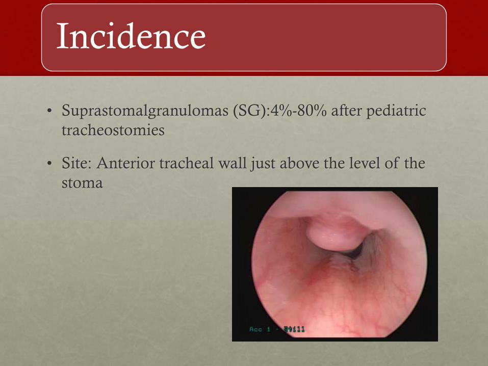
Incidence
• Suprastomalgranulomas (SG):4%-80% after pediatric
tracheostomies
• Site: Anterior tracheal wall just above the level of the
stoma

Incidence contd
• <3 months: soft and friable, >6 months firm and fibrous
• Increased incidence with use of endoscopy to evaluate the
trachea
• Incidence increased with use of modern circular curve
shaped tracheostomy tubes
• Increased incidence with inappropriate sized tubes, cuffed
tubes
Shires et al. Management of suprastomal tracheal fibroma: Introduction of a new
technique and comparison with other techniques. Int J PedOtorhinolary 2009.

Etiology of SG
• Frictional trauma of the tube
• Exposure of the stoma to the environment
• Secondary infection
• Stasis of secretions at the entry site of the tracheotomy tube

Etiology of tracheal granulomas
• Mucosal injury and necrosis from
suction tips
• Frictional trauma from the tip of the tube

When to treat?
• Majority of SG are asymptomatic and do not require
treatment
• Treatment is indicated if SG and tracheal granulomas
are associated with
• Bleeding
• Airway Obstruction
• Dysphonia, Aphonia
• Prior to decannulation
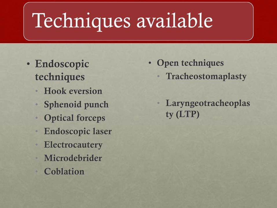
Techniques available
• Endoscopic
techniques
• Hook eversion
• Sphenoid punch
• Optical forceps
• Endoscopic laser
• Electrocautery
• Microdebrider
• Coblation
• Open techniques
• Tracheostomaplasty
• Laryngeotracheoplas
ty (LTP)

Endoscopic techniques
• Laryngeal suspension
• Ventilating
bronchoscope or Rigid
0 degree Hopkins
telescope
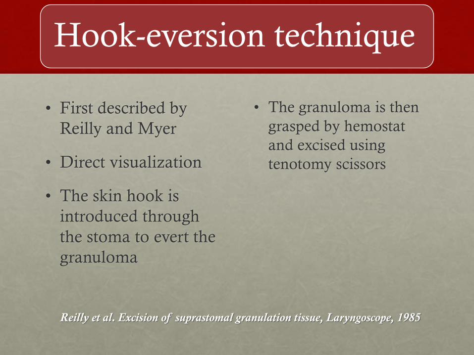
Hook-eversion technique
• First described by
Reilly and Myer
• Direct visualization
• The skin hook is
introduced through
the stoma to evert the
granuloma
• The granuloma is then
grasped by hemostat
and excised using
tenotomy scissors
Reilly et al. Excision of suprastomal granulation tissue, Laryngoscope, 1985
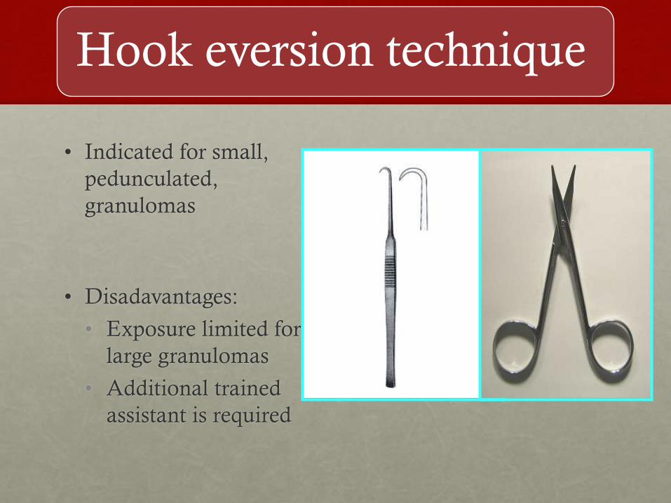
Hook eversion technique
• Indicated for small,
pedunculated,
granulomas
• Disadavantages:
• Exposure limited for
large granulomas
• Additional trained
assistant is required
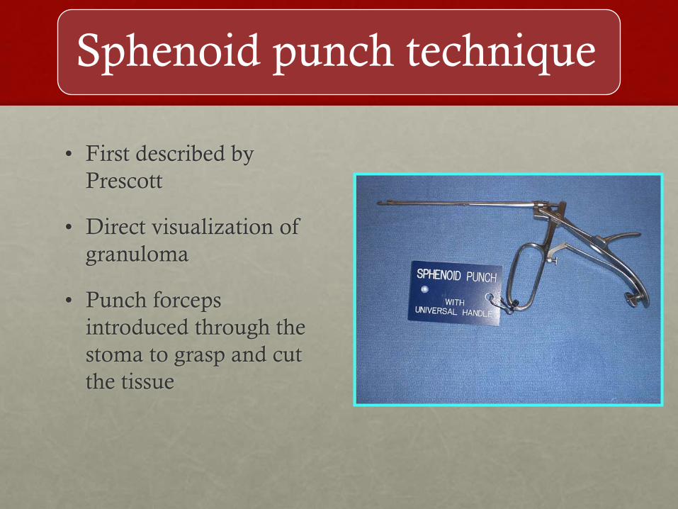
Sphenoid punch technique
• First described by
Prescott
• Direct visualization of
granuloma
• Punch forceps
introduced through the
stoma to grasp and cut
the tissue

Sphenoid punch
• Advantages: Curve allows easy introduction, easy
removal, minimal bleeding
• Used primarily for fibrous granulomas
• Disdadvantage: Cannot be used for large, obstructing
granulomas: difficult to bypass the mass
Prescott CAJ. Persistent complications of pediatric tracheotomy. Int
J PediatrOtorhinolaryngol, 1992

Optical Forceps
• Cupped optical forceps
• Used with a rigid
Hopkins system
• Indicated for small
friable granulomas
• Can cause bleeding due
to piecemeal
granuloma removal
Electrocautery
• It consists of a long skinny wire passed through the
endoscopic bronchoscope
• The tip of the wire cauterizes the tissue with minimal
bleeding
• Advantages: Direct delivery of energy, minimal
bleeding
• Disdavantages: Learning curve, scarring

Granulomas amenable to cold techniques

Endoscopic laser
• CO2 laser is considered to be the work horse of
pediatric airway surgery
• The pediatric airway is smaller and has less tissue as
compared to an adult larynx.
• This precludes widespread use of KTP laser as it has
deeper penetrating properties
• CO2 laser: Shallow depth of penetration and minimal
non-specific thermal affect

CO2 laser fiber
• This device delivers CO2 laser energy to target tissue
through a hollow, flexible wire
• The flexible wire can be introduced through the
ventilating bronchoscope or through custom designed
hand pieces
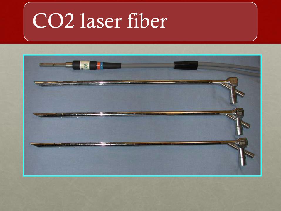
CO2 laser fiber

CO2 laser Fiber

CO2 laser Fiber
• Advantages:
• CO2 laser properties are maintained
• Ease of use
• Direct delivery of energy to difficult to reach anatomical
areas such as distal trachea
• The tip of the carrier can be used for dissection
• Cumbersome, articulated line of sight CO2 delivery
systems are avoided

Tip of CO2 fiber
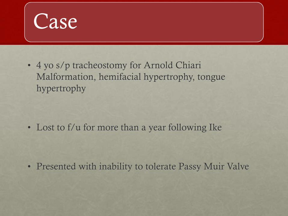
Case
• 4 yo s/p tracheostomy for Arnold Chiari
Malformation, hemifacial hypertrophy, tongue
hypertrophy
• Lost to f/u for more than a year following Ike
• Presented with inability to tolerate Passy Muir Valve

Large SG/Tracheal
Granuloma
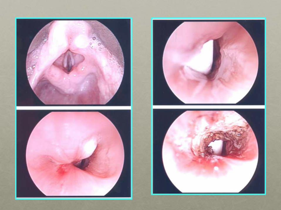
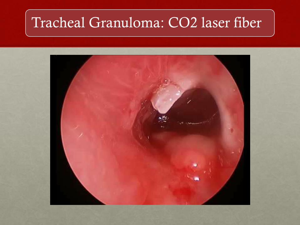
Tracheal Granuloma: CO2 laser fiber

Microdebrider
• Can use a tricut or a
skimmer blade
• Usually indicated for small
fibrous tracheal granulomas
• Can be introduced through
the stoma to reach distal
granulomas
• Disadvantage: Bleeding

Laryngeal coblation
• This consists of using a laryngeal coblation wand for
suprastomal and tracheal granuloma removal
• Only a few case reports have been published in the
literature showing good results
Kitsko et al. Coblation removal of large suprastomal tracheal granulomas
Laryngoscope 2009

Advantages of laryngeal coblation
• Less bleeding as compared to hook-eversion and
optical forceps technique
• Has a suction port, so less chances of loss of
granuloma into the distal airway
• Direct visualization and ease of use
• Laser precautions are avoided, external scars for open
procedures are avoided

Laryngeal coblation technique
• Suspension of the larynx
• Introduction of an appropriate sized bronchoscope
into the larynx just above the stoma
• The coblation wand is slightly bent and introduced
through the tracheostoma
• Coblation is carried out at a setting of 7
• The shape of the wand, electrodes and suction prevent
injury to posterior and lateral tracheal walls.

Laryngeal coblation wand

Coblation
PRE
POST
Indications for Open Procedures
• Large, broad-based obstructing granulomas especially
if planning for decannulation
• Associated anterior tracheal wall collapse
• Failure of Endoscopic Management
Gupta et al. Pediatric suprastomal granulomas: Management and
Treatment. Otolaryngol Head and Neck Surg, 2004
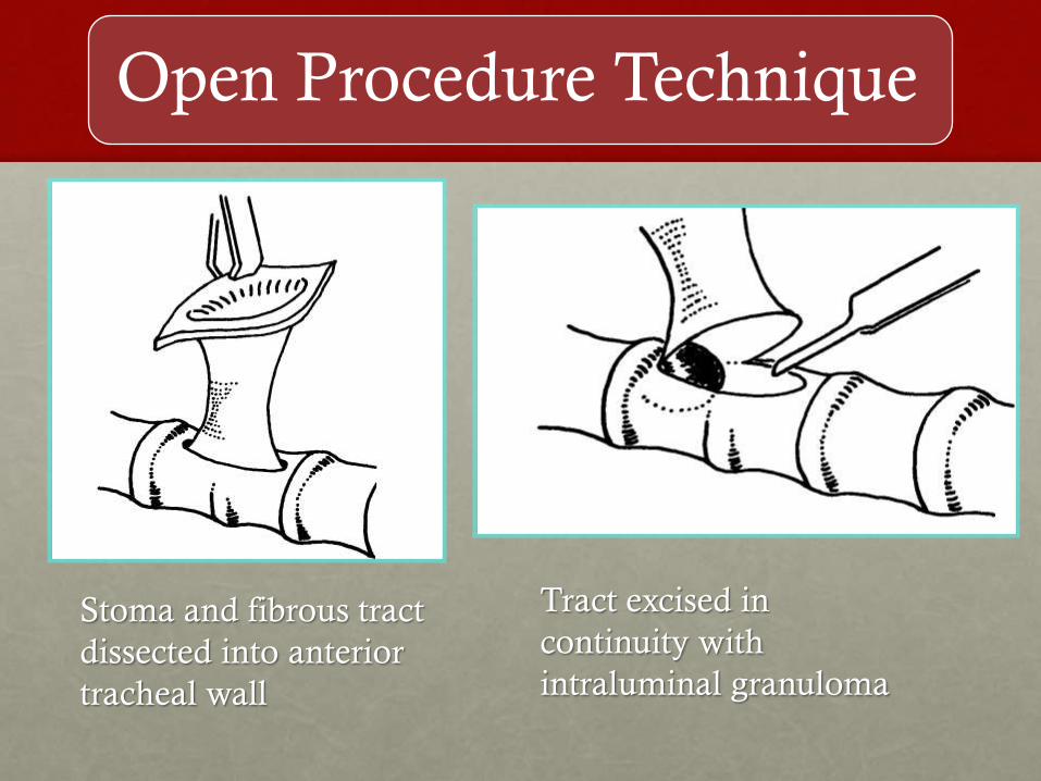
Open Procedure Technique
Stoma and fibrous tract
dissected into anterior
tracheal wall
Tract excised in
continuity with
intraluminal granuloma

Open procedure technique
• If there is associated anterior tracheal wall collapse, the
trachea may be hitched forward and sutured to the
strap muscles on either side.
• Closure of the tracheal opening with PDS suture
• Post-operative ICU monitoring for 48 hours (patient
remains intubated
• Steroids and antibiotics
Al-Saati et al. Surgical decannulation of children with tracheostomy,
Journal of Laryngology and Otology, 1993
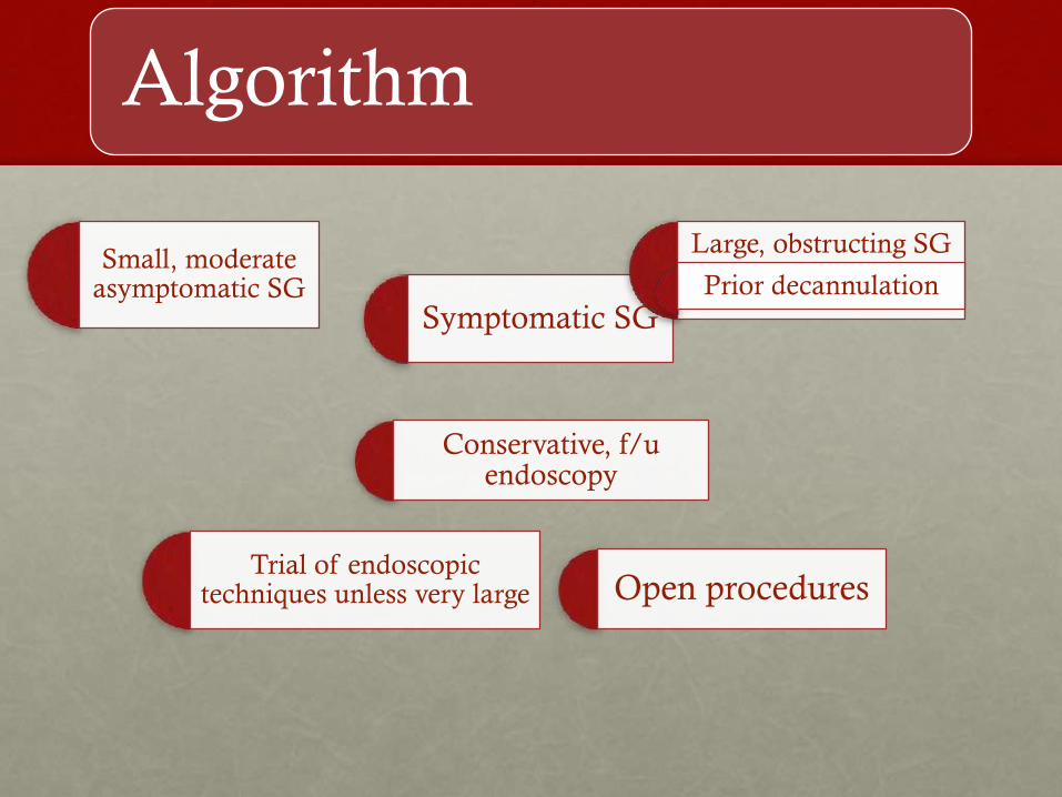
Algorithm
Small, moderate asymptomatic SG
Conservative, f/u endoscopy
Trial of endoscopic techniques unless very large Open procedures
Symptomatic SG
Large, obstructing SG
Prior decannulation
Summary
• Suprastomal granulomas occur very commonly after
pediatric tracheotomies
• Majority are asymptomatic and do not require
treatment
• Endoscopic excision should be tried first for small or
moderate sized granulomas
• Open procedures should be carried out as a last resort
for specific indications