Management of delayed cerebral ischemia after subarachnoid ... · and/or cerebral infarction,...
12
REVIEW Open Access Management of delayed cerebral ischemia after subarachnoid hemorrhage Charles L. Francoeur 1 and Stephan A. Mayer 2,3* Abstract For patients who survive the initial bleeding event of a ruptured brain aneurysm, delayed cerebral ischemia (DCI) is one of the most important causes of mortality and poor neurological outcome. New insights in the last decade have led to an important paradigm shift in the understanding of DCI pathogenesis. Large-vessel cerebral vasospasm has been challenged as the sole causal mechanism; new hypotheses now focus on the early brain injury, microcirculatory dysfunction, impaired autoregulation, and spreading depolarization. Prevention of DCI primarily relies on nimodipine administration and optimization of blood volume and cardiac performance. Neurological monitoring is essential for early DCI detection and intervention. Serial clinical examination combined with intermittent transcranial Doppler ultrasonography and CT angiography (with or without perfusion) is the most commonly used monitoring paradigm, and usually suffices in good grade patients. By contrast, poor grade patients (WFNS grades 4 and 5) require more advanced monitoring because stupor and coma reduce sensitivity to the effects of ischemia. Greater reliance on CT perfusion imaging, continuous electroencephalography, and invasive brain multimodality monitoring are potential strategies to improve situational awareness as it relates to detecting DCI. Pharmacologically-induced hypertension combined with volume is the established first-line therapy for DCI; a good clinical response with reversal of the presenting deficit occurs in 70 % of patients. Medically refractory DCI, defined as failure to respond adequately to these measures, should trigger step-wise escalation of rescue therapy. Level 1 rescue therapy consists of cardiac output optimization, hemoglobin optimization, and endovascular intervention, including angioplasty and intra-arterial vasodilator infusion. In highly refractory cases, level 2 rescue therapies are also considered, none of which have been validated. This review provides an overview of current state-of-the-art care for DCI management. Keywords: Delayed cerebral ischemia, Subarachnoid hemorrhage, Vasospasm, Multimodality monitoring Abbreviations: ADR, Alpha/delta ratio; CBF, Cerebral blood flow; CO, Cardiac output; CPP, Cerebral perfusion pressure; CSD, Cortical spreading depolarization; CTA, Computed tomographic angiography; CTP, Computed tomographic perfusion; DCI, Delayed cerebral ischemia; DSA, Digital subtraction angiography; EBI, Early brain injury; ECoG, Electrocorticography; EEG, Electroencephalogram; GCS, Glasgow Coma Scale; ICE, Intracortical electroencephalography; ICP, Intracerebral pressure; MAP, Mean arterial blood pressure; MCA, Middle cerebral artery; MMM, Multimodality monitoring; PTCA, Percutaneous transluminal balloon angioplasty; RCT, Randomized controlled trial; SAH, Subarachnoid hemorrhage; SBP, Systolic blood pressure; TCD, Transcranial Doppler; WFNSS, World Federation of Neurological Surgeons Scale * Correspondence: [email protected] 2 Department of Neurology (Neurocritical Care), Mount Sinai, New York, NY, USA 3 Institute for Critical Care Medicine, Icahn School of Medicine at Mount Sinai, One Gustave L. Levy Place, Box 1522, New York, NY 10029-6574, USA Full list of author information is available at the end of the article © 2016 The Author(s). Open Access This article is distributed under the terms of the Creative Commons Attribution 4.0 International License (http://creativecommons.org/licenses/by/4.0/), which permits unrestricted use, distribution, and reproduction in any medium, provided you give appropriate credit to the original author(s) and the source, provide a link to the Creative Commons license, and indicate if changes were made. The Creative Commons Public Domain Dedication waiver (http://creativecommons.org/publicdomain/zero/1.0/) applies to the data made available in this article, unless otherwise stated. Francoeur and Mayer Critical Care (2016) 20:277 DOI 10.1186/s13054-016-1447-6
Transcript of Management of delayed cerebral ischemia after subarachnoid ... · and/or cerebral infarction,...
REVIEW Open Access
Management of delayed cerebral ischemiaafter subarachnoid hemorrhageCharles L. Francoeur1 and Stephan A. Mayer2,3*
Abstract
For patients who survive the initial bleeding event of a ruptured brain aneurysm, delayed cerebral ischemia (DCI) isone of the most important causes of mortality and poor neurological outcome. New insights in the last decadehave led to an important paradigm shift in the understanding of DCI pathogenesis. Large-vessel cerebral vasospasmhas been challenged as the sole causal mechanism; new hypotheses now focus on the early brain injury,microcirculatory dysfunction, impaired autoregulation, and spreading depolarization. Prevention of DCI primarilyrelies on nimodipine administration and optimization of blood volume and cardiac performance. Neurologicalmonitoring is essential for early DCI detection and intervention. Serial clinical examination combined withintermittent transcranial Doppler ultrasonography and CT angiography (with or without perfusion) is the mostcommonly used monitoring paradigm, and usually suffices in good grade patients. By contrast, poor grade patients(WFNS grades 4 and 5) require more advanced monitoring because stupor and coma reduce sensitivity to theeffects of ischemia. Greater reliance on CT perfusion imaging, continuous electroencephalography, and invasivebrain multimodality monitoring are potential strategies to improve situational awareness as it relates to detectingDCI. Pharmacologically-induced hypertension combined with volume is the established first-line therapy for DCI; agood clinical response with reversal of the presenting deficit occurs in 70 % of patients. Medically refractory DCI,defined as failure to respond adequately to these measures, should trigger step-wise escalation of rescue therapy.Level 1 rescue therapy consists of cardiac output optimization, hemoglobin optimization, and endovascularintervention, including angioplasty and intra-arterial vasodilator infusion. In highly refractory cases, level 2 rescuetherapies are also considered, none of which have been validated. This review provides an overview of currentstate-of-the-art care for DCI management.
Keywords: Delayed cerebral ischemia, Subarachnoid hemorrhage, Vasospasm, Multimodality monitoring
Abbreviations: ADR, Alpha/delta ratio; CBF, Cerebral blood flow; CO, Cardiac output; CPP, Cerebral perfusionpressure; CSD, Cortical spreading depolarization; CTA, Computed tomographic angiography; CTP, Computedtomographic perfusion; DCI, Delayed cerebral ischemia; DSA, Digital subtraction angiography; EBI, Early brain injury;ECoG, Electrocorticography; EEG, Electroencephalogram; GCS, Glasgow Coma Scale; ICE, Intracorticalelectroencephalography; ICP, Intracerebral pressure; MAP, Mean arterial blood pressure; MCA, Middle cerebral artery;MMM, Multimodality monitoring; PTCA, Percutaneous transluminal balloon angioplasty; RCT, Randomized controlledtrial; SAH, Subarachnoid hemorrhage; SBP, Systolic blood pressure; TCD, Transcranial Doppler; WFNSS, WorldFederation of Neurological Surgeons Scale
* Correspondence: [email protected] of Neurology (Neurocritical Care), Mount Sinai, New York, NY,USA3Institute for Critical Care Medicine, Icahn School of Medicine at Mount Sinai,One Gustave L. Levy Place, Box 1522, New York, NY 10029-6574, USAFull list of author information is available at the end of the article
© 2016 The Author(s). Open Access This article is distributed under the terms of the Creative Commons Attribution 4.0International License (http://creativecommons.org/licenses/by/4.0/), which permits unrestricted use, distribution, andreproduction in any medium, provided you give appropriate credit to the original author(s) and the source, provide a link tothe Creative Commons license, and indicate if changes were made. The Creative Commons Public Domain Dedication waiver(http://creativecommons.org/publicdomain/zero/1.0/) applies to the data made available in this article, unless otherwise stated.
Francoeur and Mayer Critical Care (2016) 20:277 DOI 10.1186/s13054-016-1447-6
BackgroundAmong subarachnoid hemorrhage (SAH) patients whosurvive the initial bleed of a ruptured aneurysm, delayedcerebral ischemia (DCI) is the most important prevent-able cause of mortality and poor neurological outcome.DCI affects up to 30 % of patients, and leaves the majorityof survivors with motor deficits, cognitive dysfunction,and reduced quality of life [1]. The risk of DCI is primarilyrelated to the severity of the initial hemorrhage, with agreater amount of cisternal and intraventricular blood oninitial imaging and a poor post-resuscitation neurologicalexamination being the strongest predictors of an unfavor-able evolution [2].State-of-the art management in the ICU does influence
the outcome of DCI. In order to provide optimal care, cli-nicians must grasp the underlying concepts behind DCIand must all use the same terminology. Evidence-basedinterventions can be implemented to reduce the risk ofdeveloping DCI, adequate monitoring must be offered toallow early detection, and timely intervention should beoffered to reverse DCI as rapidly as possible before the is-chemic process progresses to infarction. We offer here apractical algorithm for managing DCI in the ICU basedon the best available evidence, and on our expertise andexperience in situations where firm data are lacking. Theaim is to provide bedside clinicians with a structured andcoherent approach in order to provide optimal care totheir patients.
Concepts and definitionsHistorically, arterial narrowing with subsequent down-stream low flow and ischemia was considered the solecause of delayed neurological deterioration in SAH pa-tients with vasospasm. This tenet of the SAH literature,however, has been challenged recently. Although themajority of SAH patients develop angiographic vasocon-striction (up to 70 %), only around 20–30 % developDCI [2]. Cerebral infarction sometimes develops in theabsence of demonstrable vasoconstriction, or in a vascularterritory unaffected by vasospasm. Successful treatmentof angiographic vasoconstriction does not necessarilylead to better functional outcome [3]. Clazosentan, anendothelin receptor antagonist, is successful in redu-cing angiographic vasospasm but has no significant ef-fect on mortality, functional outcome, or the frequencyof cerebral infarction [4]. Finally, nimodipine is the onlypharmacological intervention shown to improve out-come in SAH patients, although it has no impact onlarge-vessel vasospasm [5].Large artery vasospasm still doubtlessly plays an im-
portant role in the pathogenesis of DCI, but the scien-tific community has now turned its interest towardalternative explanations for a process that may be muchmore complex than was previously thought. The main
thrust of this paradigm shift is general agreement thatdemonstration of large-vessel narrowing is no longer re-quired to make the diagnosis of DCI. In line with recentpublications and guidelines [6–8], we reserve the termsvasospasm for narrowing of large cerebral arteries as evi-denced by imaging, DCI for cerebral infarction or neuro-logical deterioration, or both, when the cause is thoughtto be vasospasm, and cerebral infarction as an infarct fromany cause demonstrated on CT or MRI within 6 weeks ofaneurysm rupture (see Table 1). The latter is now recog-nized as the primary determinant of long-term cognitiveor motor deficits after SAH [9].
PathogenesisAlthough in-depth exploration of the pathophysiology ofDCI is beyond the scope of this review, a basic under-standing of the prevailing hypotheses is useful to theclinician. As mentioned earlier, large-vessel narrowingwith subsequent low flow might be one of multiplemechanisms of DCI, but the causal framework now alsoincludes early brain injury (EBI), microcirculatory dys-function with loss of autoregulation, cortical spreadingdepolarization (CSD), and microthrombosis [10]. EBI en-compasses the multiple physiological derangements thatare thought to occur in the first 72 hours after the ictus.The initial ICP crisis and global hypoperfusion triggerglial activation, endothelial dysfunction, and inflammatorypathways. Animal and human data suggest an ultra-earlydiffuse neuroinflammatory process that predicts later is-chemic damage [11]. Associated necrosis and apoptosis,
Table 1 Harmonized definition of delayed cerebral ischemiaand cerebral infarction
Delayed cerebral ischemia
Focal (hemiparesis, aphasia, hemianopia, or neglect) or global (2 pointsdecrease on GCS) neurological impairment lasting for at least 1 hourand/or cerebral infarction, which:
▪ Is not apparent immediately after aneurysm occlusion
▪ Is attributable to ischemia
▪ Is not attributed to other causes (i.e. surgical complication, metabolicderangements) after appropriate clinical, imaging, and laboratoryevaluation
Cerebral infarction
Presence of cerebral infarction on CT or MR scan of the brain within6 weeks after SAH, or on the latest CT or MR scan made before deathwithin 6 weeks, or proven at autopsy; that is:
▪ Not present on the CT or MR scan between 24 and 48 hours afterearly aneurysm occlusion
▪ Not attributable to other causes such as surgical clipping orendovascular treatment
▪ Not due to a nonischemic lucency related to a ventricular catheter,intraparenchymal hematoma, or brain retraction injury
Based on references [101, 102]GCS Glasgow Coma Scale, CT computed tomography, MR magnetic resonance,SAH, subarachnoid hemorrhage
Francoeur and Mayer Critical Care (2016) 20:277 Page 2 of 12
as well as endothelial dysfunction, lead to neuronal lossand cerebral edema, respectively. CSD represents a waveof electrical depolarization that propagates across thecerebral gray matter at a speed of 2–5 mm/min, with en-suing depression of ECoG activity for 5–15 min [12]. Thisprocess is accompanied by neurovascular uncoupling: asthe energy expenditure of neurons is reaching its peak,paradoxical vasoconstriction occurs, resulting in corticalhypoperfusion and energy failure. CSD is present in 80 % ofpoor grade SAH patients, has a biphasic distribution withpeak frequency on SAH days 0 and 7, and has an uncertainrelationship to large-vessel vasospasm and concurrent seiz-ure activity [13]. Endothelial and platelet dysfunction, co-agulation cascade activation, and impaired fibrinolysis alloccur after SAH. Numerous biological markers of theseevents have been associated with DCI and poor outcome.Postmortem studies have found evidence of microthrombi,particularly in areas of cerebral infarction, after SAH. Infact, this correlates better with cerebral infarction lesionsthan vasospasm or aneurysm location [14].
PreventionNimodipineDCI prevention has been the Holy Grail of SAH re-search for decades, but few options are available and un-fortunately most attempts have yielded disappointingresults (see Table 2). Nimodipine, a dihydropyridine
calcium channel antagonist, is the only pharmacologicintervention so far associated with better outcome inSAH patients. Multiple trials have demonstrated abenefit [15], with the seminal trial showing an impres-sive reduction in cerebral infarction, poor neurologicaloutcome, and death with oral nimodipine 60 mg givenevery 4 hours for 21 days [16]. This is now the recom-mended regimen, although intravenous nimodipine isapproved as an alternative in Europe. Since nimodipinecan cause hypotension, the dose can be divided into30 mg every 2 hours or reduced to 30 mg every 4 hours.An ongoing phase 3 trial evaluating a single administra-tion of intraventricular nimodipine (600 mg) micropar-ticles to optimize its efficacy and reduce its side effectsis in progress [17].
Enhanced blood clearanceThe presence of blood and its breakdown products isstrongly associated with vasospasm. Numerous attemptshave been made to accelerate clearance of subarachnoidblood, with the hope that this might result in the pre-vention of delayed arterial spasm. The only randomizedcontrolled trial (RCT) investigating the use of intraop-erative administration of rt-PA failed to show any ef-fect on outcome [18]. Lumbar drainage of CSF wasalso unsuccessful at improving mRS [19] or GOS [20]scores at 6 months in two RCTs. Different other in-terventions, including cisternal irrigation or use ofurokinase, have been evaluated for feasibility and re-ported mixed results. Use of such techniques cannotbe advocated at present.
Avoidance of hypovolemia and hyponatremiaHyponatremia and hypovolemia occur frequently afterSAH due to physiological changes favoring excessivenatriuresis and inappropriate antidiuretic hormone ele-vation, and have been associated with impending DCI[21]. Retrospective data indicate that fluid restriction,the typical treatment for syndrome of inappropriateantidiuretic hormone (SIADH), can be deleterious andincreases the risk of DCI due to aggravation of hypo-volemia [22]. Isotonic crystalloid fluid resuscitation tar-geting normal serum sodium values and euvolemia ispresently the favored fluid management strategy forpreventing DCI. The latter is notoriously difficult to as-sess in critically ill patients, and the readers are referredto papers dedicated to this specific subject for a morein-depth approach to the matter [23–26]. Administra-tion of fludrocortisone (between 0.2 and 0.4 mg/day)has been shown to reduce the occurrence of hyponatre-mia [27], with some indication towards DCI reduction.Anecdotal evidence indicates that correction of acutesymptomatic hyponatremia with hypertonic saline (3 %)infusion is usually effective.
Table 2 Selected pharmacologic interventions that have beenevaluated for DCI preventiona
Intervention Effect
Aspirin No effect on new lesion associated withneurological worsening [103]
Clazosentan No effect on mortality or vasospasm-relatedmorbidity [5]
Enoxaparin No effect on DCI or GOS at 3 months [104]
Erythropoietin Less neurological deficit with cerebral infarct;no difference in mRS or GOS at 6 months [105]
Fludrocortisone No effect on incidence of cerebral ischemia orindependent living [27]
Magnesium No difference in mRS at 3 months [106]
Methylprednisolone No effect on neurologic worsening; trendtowards better GOS at 6 months [107]
Nicardipine No effect on neurological worsening or GOSat 3 months [102]
Prophylacticangioplasty
No effect on new neurologic deficits or GOSat 3 months [86]
Prophylactichypervolemia
No effect on neurologic worsening or GOSat 3 months [69]
Statins No effect on DCI, death or mRS at6 months [108]
aExcluding nimodipine. Only randomized controlled trials are considered.References are either the most recent, most definitive, or most robust trialaccording to the authors’ opinionDCI delayed cerebral ischemia, GOS, Glasgow Outcome Scale, mRS modifiedRankin Scale
Francoeur and Mayer Critical Care (2016) 20:277 Page 3 of 12

Detection and diagnosisEarly detection of DCI is critical to allow for timely inter-vention. Although straightforward in relatively intact pa-tients, early detection is notoriously difficult in poor gradeSAH patients (Table 3). Depending on the context, thetechnique can vary from simple serial clinical examinationsto multiple advanced monitoring strategies, as described inthe following section.
Clinical examinationClinical examination in awake patients who can followcommands is the most reliable way to detect and diagnoseDCI. Neurological impairment can be focal or global. TheGlasgow Coma Scale (GCS) is the most commonly usedtool for measuring and documenting the level of con-sciousness in the ICU setting. Serial testing of attentionand concentration by reciting from 20 to 1 and fromDecember to January in good grade patents has been usedsuccessfully to quantify subtle changes in mental statusthat are not detected by the GCS [28]. However, poorgrade SAH patients, defined here as WFNS grades 4 and5, do not consistently manifest symptoms when DCI oc-curs, although they constitute the most at-risk group.More than 20 % will present DCI as asymptomatic cere-bral infarction, and these patients are less likely to receiveacute hypertensive therapy [29]. This is the primary ra-tionale for using other modalities, including invasive brainmultimodality monitoring (MMM) [30], in this specificsubgroup.
Transcranial Doppler ultrasonographyTranscranial Doppler (TCD) ultrasonography is a nonin-vasive test that allows indirect detection of large-vessel
narrowing based on quantification of acceleration offlow. Velocities lower than 120 cm/s in the middle cere-bral artery (MCA) show high negative predictive valuefor angiographic vasospasm, whereas velocities exceeding180 cm/s have high positive predictive value [31]. TheLindegaard ratio, defined as MCA mean cerebral bloodflow (CBF) velocity divided by extracranial internal carotidartery mean cerebral flow velocity, is an index thought tobe less affected by systemic hemodynamic variations. Usedas a screening tool in many tertiary centers, TCD ultra-sonography suffers from both technical and anatomicallimitations [32]. TCD ultrasonography provides no infor-mation about the distal vasculature and can be affected byhydrocephalus or elevated intracranial pressure. Propervessel insonation is highly operator dependent and at least10 % of patients do not have adequate bone windows. Fi-nally, just as with vascular imaging, TCD ultrasonographydetects vasospasm, but this does not directly translate intoa high risk of DCI. In one study, 40 % of SAH patientswho experienced DCI never had a MCA flow velocity thatexceeded 120 cm/s during the entire period of monitoring[33]. It is the authors’ opinion that the aforementionedcutoff values are specific enough to mandate additionalinvestigations if the clinical picture is compatible withimpending or ongoing DCI. However, due to its lowsensitivity, TCD ultrasonography should not be the solescreening examination in a patient with a poor clinicalexamination.
Vascular imagingImaging of the cerebral vasculature allows recognition ofarterial narrowing. A decrease in luminal diameter ofmore than 50 % is usually considered severe vasospasm
Table 3 Components of brain multimodality monitoring for poor grade SAH
Device Physiological parametermeasured
Normal range Pathological condition
Continuouselectroencephalography
Brain activityEpileptiform discharges
• Alpha/delta ratio > 50 %• No epileptiform discharges• Reactivity to stimuli
• Alpha/delta ratio < 50 %• Epileptiform discharges• No reactivity
Transcranial Dopplerultrasound
Mean blood flow velocity (FVm) • FVm MCA: 30–75 cm/s • MCA FVm 120–180 cm/s: intermediate probabilityof vasospasm
• MCA FVm >180 cm/s: high probability of vasospasm
Cerebral blood flowmonitor (Hemedex)
Cerebral blood flow (CBF) • >40 ml/100 g/min • <20 ml/100 g/min: indicative of ischemia assumingpreserved metabolic demand
Jugular venous oximetry Balance between oxygendelivery and consumption (SjO2)
• 50–75 % • <50 %; increased oxygen extraction fraction, indicativeof ischemia
Brain tissue oxygentension (Licox)
Regional parenchymal braintissue oxygen tension (PbtO2)
• 25–35 mmHg in whitesubcortical matter
• <20 mmHg: indicative of cerebral hypoxia
Cerebral microdialysis • Glucose• Lactate• Pyruvate• Lactate/pyruvate ratio• Glutamate• Glycerol
• 0.8–4.0 μmol/L• 0.7–3.0 μmol/L• Unknown• < 25• 2–10 μmol/L• 10–90 μmol/L
• <0.2 μmol/L• ≥4.0 μmol/L• Unknown• >40 indicative of anaerobic metabolism• >10 μmol/L• >90 μmol/L
SAH subarachnoid hemorrhage
Francoeur and Mayer Critical Care (2016) 20:277 Page 4 of 12

and is associated with lower CBF. Conventional angiog-raphy (digital subtraction angiography (DSA)) is the goldstandard and offers the possibility of endovascular treat-ment. Complication rates for diagnostic angiography arein the range of 1 %. Computed tomographic angiography(CTA) is a less invasive and a more readily available op-tion. Studies comparing CTA with DSA have found goodagreement, suggesting high sensitivity and specificity invasospasm diagnosis [34]. The authors use CTA as afirst-line screening tool for detecting large-vessel vaso-spasm, with the initial study timed to occur betweenSAH day 4 (for patients felt to be at greater risk) andday 8 (for lower risk patients). Lack of appreciable large-vessel spasm on SAH day 8 or later implies a very lowrisk of subsequent DCI, enabling fast-tracking out of theICU into a lower intensity, step-down setting.
Brain perfusion imagingDirectly assessing cerebral perfusion is appealing becauseit allows for evaluation of the functional consequences ofboth large-vessel and small-vessel vasospasm. Xenon CT,single photon emission computed tomography, positronemission tomography, MR perfusion, and computedtomographic perfusion (CTP) all allow tomographic CBFassessment. CTP is currently the most widely used andstudied modality [35]. Various cutoff values that correlatewith DCI have been reported, including a mean transittime (MTT) exceeding 5.0–6.4 s, or regional CBF below25–40 ml/100 g/min [36]. One detriment to this type ofanalysis is the high degree of variability due to differencesin equipment and postprocessing methods [37]. CTPseems to correlate fairly well with DCI, but focal flowreductions can also occur as a consequence of brain
retraction injury or perihematomal brain dysfunction.Many centers perform CTA and CTP together, as acomplement to serial TCD monitoring, in the critical timewindow for DCI onset (SAH days 4–8, see Fig. 1).
Continuous electroencephalographyContinuous electroencephalography provides noninva-sive, real-time continuous information about cortical ac-tivity, and quantitative electroencephalography allowsdecomposition of the data contained in the raw EEG. Inthe presence of cortical hypoperfusion leading to neur-onal dysfunction, EEG changes are detectable and mayprecede the onset of symptoms [38]. Recent data suggestthat reductions in the alpha/delta ratio (ADR) or inalpha variability are most sensitive and specific for pre-dicting DCI at a point where it is potentially reversible[39]. Even more interesting, reversal of those changescould serve as a surrogate target to titrate therapy. Forexample, as explained later, induced hypertension couldbe titrated to ADR normalization. Despite its theoreticalattractiveness, the intense manpower commitment re-quired to provide around-the-clock real-time neurotele-metry has hampered widespread adoption of continuouselectroencephalography for neuromonitoring after SAH.
Multimodality monitoringAdvanced neuromonitoring using MMM provides con-tinuous, real-time information allowing early detectionof physiological derangements, providing both a triggerand a target for intervention. In addition to acting as anearly warning system for improving situational aware-ness, MMM can be proactively used to create an opti-mized physiological environment for the injured brain,
Fig. 1 Mean maximal TCD values during SAH days 3–14 in patients who did or did not develop DCI. TCD examinations after the diagnosis of DCIwere censored. Histogram shows the number of patients with new onset DCI between SAH days 3 and 14. Nine patients had DCI between days15 and 29. Number (in parentheses) represents the number of TCD examinations performed for each corresponding SAH day. From reference [33],with permission. DCI delayed cerebral ischemia, mBFV mean blood flow velocity, SAH subarachnoid hemorrhage
Francoeur and Mayer Critical Care (2016) 20:277 Page 5 of 12
with the goal of secondary injury prevention. Many high-volume centers equipped with invasive MMM now rou-tinely use it in poor grade SAH patients, with various com-binations of ICP, brain tissue oxygen, CBF, and metabolicmonitoring, as well as intracranial electroencephalography.ICP monitoring is essential to any MMM bundle.
Intracranial hypertension is common in SAH, especiallyin poor grade patients where occurrence in up to 80 %of patients has been described [40]. It is associated withseverely deranged cerebral metabolism [41] and consist-ently leads to poor outcome [42, 43], warranting aggres-sive management. ICP monitoring also permits cerebralperfusion pressure (CPP) measurement. We have re-ported in poor grade patients that simply maintainingCPP >70 mmHg is associated with a lower risk of brainmetabolic crisis and tissue hypoxia [44], which may be auseful clinical guideline for minimizing the risk of sec-ondary brain injury in unmonitored patients.Parenchymal brain tissue oxygenation (PbtO2) monitor-
ing allows quantification of oxygen tension in the braininterstitial space and will detect episodes of cerebral com-promise even in the absence of elevated ICP or low CPP[30], underlying its role as a complement to conventionalneuromonitoring in SAH patients. This is probably helpfulin early detection of silent infarcts [29], and higher meanPbtO2 is associated with improved survival [30].Microdialysis allows determination of interstitial fluid
composition and cellular metabolism. The most com-mon targets of clinical microdialysis analysis are extra-cellular lactate levels and the lactate/pyruvate ratio(LPR) [45]. These metabolic derangements precede si-lent infarction by a few hours [29], are often detected inthe setting of normal ICP and even normal PbtO2 [30],and are fairly specific for DCI (0.89 for lactate levels >4 mmol) [46]. Microdialysis is actually superior to TCDultrasonography and DSA in predicting clinical deterior-ation secondary to DCI [47]. Some experienced centersalso use the biochemical profile to differentiate ischemiafrom mitochondrial dysfunction [48] or to monitor brainglucose metabolism [49], but these applications needfurther evaluation before being widely adopted.Intracranial electroencephalography includes subcortical
electrocorticography (ECoG) and intracortical electroen-cephalography (ICE). ECoG allows detection of CSD is-chemia, a potent mechanism of DCI [13] that decreasesbrain O2 supply and increases brain O2 consumption inSAH patients [50], providing a potential therapeutic target[51]. ICE, on the other hand, can detect ictal dischargesnot apparent on scalp EEG [52]; ICE ADR reduction mayoutperform scalp quantitative electroencephalography inearly DCI detection [53].Finally, ICP or PbtO2 monitoring also permits dynamic
evaluation of autoregulation through moving linear cor-relation coefficients such as the pressure reactivity index
(PRx, which correlates MAP with ICP) or the PtiO2 pres-sure reactivity index (ORx, which correlates PbtO2 withCPP) [54]. Early autoregulatory failure is predictive of DCI[55] and is associated with poor outcome in SAH patients[56]. Theoretically, these indices might also be used to de-fine the optimal CPP for a given patient [57].Proper positioning in at risk cerebral region is essential,
but offers no guarantee that other brain regions are not is-chemic [58]. We prefer to place the MMM bolt in thefrontal anterior and middle cerebral territory watershedregion ipsilateral to the ruptured aneurysm, or in the non-dominant hemisphere in the case of a midline aneurysm.The invasive and regional nature of MMM, its associatedcost, and the required expertise are the main obstacles toits implementation.
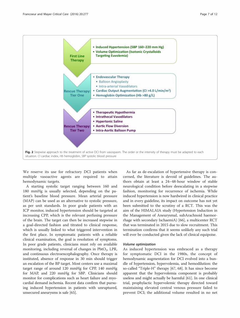
TreatmentSAH patients are complex and should be cared for inspecialized, high-volume centers to maximize good out-come [59]. The suggested approach below assumes thatthe standards of care in all other aspects of treatmentare followed. An organized approach that has beenagreed upon in advance by all stakeholders minimizesconflicts and streamlines the process of care. Althoughpresented as a three-stage algorithm (Fig. 2), manage-ment should always be tailored to the individual patient,to the available resources, and in a contextualized fashion.Our approach to treatment divides interventions into: first-line therapy for new-onset DCI, which can manifest asneurological deterioration, characteristic imaging findings,or MMM abnormalities indicative of ischemia; and second-line “rescue therapy” for refractory DCI, indicating inad-equate reversal of ischemia in response to first-line therapy.
First-line therapy for new-onset DCIInduced hypertensionSuccessful reversal of neurological symptoms followinginduced hypertension has been described in case seriessince the late 1970s, and most clinicians caring for SAHpatients can testify to its benefit. Use of vasopressors toaugment blood pressure is still the cornerstone of first-line therapy for DCI. A normal saline bolus (15 ml/kg over1 hour) at the institution of therapy increases CBF [60].Norepinephrine [61], dopamine [62], and phenylephrine-induced [63] hypertension all have been demonstrated tosignificantly improve CBF and/or cerebral oxygenation,resulting in clinical improvement of the neurologicaldeficit in approximately 70 % of patients. The authorsuse norepinephrine as the first-line treatment of choicedue to its combination of alpha and beta receptorstimulation, the low frequency of tachycardia, and thereliable hemodynamic response that results. Argininevasopressin has also been reported as a safe supplemen-tary vasopressor in a small group of SAH patients [64].
Francoeur and Mayer Critical Care (2016) 20:277 Page 6 of 12
We reserve its use for refractory DCI patients whenmultiple vasoactive agents are required to attainhemodynamic targets.A starting systolic target ranging between 160 and
180 mmHg is usually selected, depending on the pa-tient’s baseline blood pressure. Mean arterial pressure(MAP) can be used as an alternative to systolic pressure,as per unit standards. In poor grade patients with anICP monitor, induced hypertension should be targeted atincreasing CPP, which is the relevant perfusing pressureof the brain. The target can then be increased stepwise ina goal-directed fashion and titrated to clinical response,which is usually linked to what triggered intervention inthe first place. In symptomatic patients with a reliableclinical examination, the goal is resolution of symptoms.In poor grade patients, clinicians must rely on availablemonitoring, including reversal of changes in PbtO2, LPR,and continuous electroencephalography. Once therapy isinstituted, absence of response in 30 min should triggeran escalation of the BP target. Most centers use a maximaltarget range of around 120 mmHg for CPP, 140 mmHgfor MAP, and 220 mmHg for SBP. Clinicians shouldmonitor for complications such as heart failure and myo-cardial demand ischemia. Recent data confirm that pursu-ing induced hypertension in patients with unruptured,unsecured aneurysms is safe [65].
As far as de-escalation of hypertensive therapy is con-cerned, the literature is devoid of guidelines. The au-thors obtain at least a 24–48-hour window of stableneurological condition before deescalating in a stepwisefashion, monitoring for recurrence of ischemia. Whileinduced hypertension is now hardwired in clinical practiceand in every guideline, its impact on outcome has not yetbeen submitted to the scrutiny of a RCT. This was theaim of the HIMALAIA study (Hypertension Induction inthe Management of AneurysmaL subArachnoid haemor-rhage with secondary IschaemiA) [66], a multicenter RCTthat was terminated in 2015 due to slow recruitment. Thistermination confirms that it seems unlikely any such trialwill ever be conducted given the lack of clinical equipoise.
Volume optimizationAs induced hypertension was embraced as a therapyfor symptomatic DCI in the 1980s, the concept ofhemodynamic augmentation for DCI evolved into a bun-dle of hypertension, hypervolemia, and hemodilution: theso-called “Triple-H” therapy [67, 68]. It has since becomeapparent that the hypervolemia component is probablyuseless and might actually be harmful [61]. In one clinicaltrial, prophylactic hypervolemic therapy directed towardmaintaining elevated central venous pressure failed toprevent DCI; the additional volume resulted in no net
Fig. 2 Stepwise approach to the treatment of active DCI from vasospasm. The order or the intensity of therapy must be adapted to eachsituation. CI cardiac index, Hb hemoglobin, SBP systolic blood pressure
Francoeur and Mayer Critical Care (2016) 20:277 Page 7 of 12
increase in cumulative fluid balance, blood volume, orCBF [69]. Other studies have shown that hypervolemictherapy increases the risk of pulmonary edema, especiallyin the setting of cardiac dysfunction [70], and that positivefluid balance in SAH is associated with poor outcome[71]. Current guidelines suggest that isotonic fluids beused judiciously to correct hypovolemia, with the ultimategoal of maintaining a euvolemic state while avoiding fluidoverload [7].
Rescue therapy for medically-refractory DCITier One interventions
Hemoglobin optimization Based on current evidencefrom randomized clinical trials in the general ICU popu-lation [72], a restrictive strategy aiming for a hemoglobinlevel above 70 g/L is the favored approach for SAH pa-tients prior to the onset of DCI. It is questionable, how-ever, whether this is the appropriate threshold forpatients with active and ongoing brain ischemia. Anemiais seen in more than 50 % of SAH patients [73] and isconsistently associated with poor outcome [74, 75].Moreover, hemoglobin levels of less than 90 g/L, andeven less than 100 g/L, are associated with brain tissuehypoxia and metabolic distress in poor grade patients[76]. Packed red blood cell transfusion successfully in-creases brain tissue oxygen tension in poor grade SAHpatients with a baseline hemoglobin level of 80 g/L [77].This makes the use of red blood cell transfusion tooptimize cerebral oxygen delivery appealing when facingactive brain ischemia refractory to first-line therapies.However, blood transfusions are also associated withmedical complications [78], poor outcome [79], and evenhigher mortality in the [80] SAH population. The on-going RCT Aneurysmal Subarachnoid Hemorrhage: RedBlood Cell Transfusion and Outcome (SAHaRA Pilot)comparing RBC transfusion triggers from 100 g/L downto 80 g/L will hopefully shed light on this debate. In themeantime, the Neurocritical Care Society guidelines [7]suggest a transfusion threshold of 80 g/L in SAH pa-tients without DCI, with a more aggressive transfusiontrigger of 90–100 g/L as a Tier One rescue therapy incases of DCI unresponsive to first-line therapy.
Endovascular therapy When confronted with medicallyrefractory DCI—cases in which significant neurologicdeficits exist despite hemodynamic optimization—endo-vascular treatment should be the next step [81]. In recentyears, indications for deploying intra-arterial therapy haveevolved and this treatment is introduced much earlier, es-pecially if there are reasons to believe that medical therapyis at high risk of failure or in the face of complicationsresulting from heart failure, fluid overload, or myocardialischemia [82].
Endovascular therapy can be subdivided into mechan-ical dilation and intra-arterial infusion of vasodilators.Percutaneous transluminal balloon angioplasty (PTCA)is based on mechanical stretching and dilation of vaso-spastic arteries. The technique is limited to proximalvessels, mainly the internal carotid artery and vertebralor basilar artery, M1 and sometimes M2 segments of theMCA, and A1 and P1 segments of the anterior and pos-terior cerebral artery respectively. The success rate inmost case series is over 90 % and long-lasting [83], withoccasional cases of recurrence that require repeatedprocedures. Improvement in CBF post PTCA has alsobeen clearly demonstrated [84]. Observational studiessuggest that early intervention (less than 2 hours afterneurological decline) results in a better clinical re-sponse [85]. The drawback of PTCA is that seriouscomplications can occur in up to 5 % of patients, in-cluding embolism, thrombosis, dissection, and vesselrupture. The only published RCT to date evaluatedPTCA as a prophylactic measure in good grade patientswith large amounts of cisternal clot [86]. Three patientsdied of vessel perforation and there was no differencein frequency of DCI, condemning this indication. If theclinician is convinced that ongoing ischemia is ex-plained by the visualized local vasospasm, PTCA is apotent therapy.Numerous cases series have shown various degrees of
reversal of vasospasm with intra-arterial vasodilators,evaluated by angiography, TCD ultrasonography, XenonCBF, cerebral oxygenation, or angiographic cerebral cir-culation time. Over the years, numerous agents havebeen evaluated, including papaverine, nicardipine, ver-apamil, nimodipine, milrinone, amrinone, and fasudil.None of these have ever been tested objectively in aclinical trial against a control group. Intra-arterial vaso-dilators have several advantages over PTCA: better dis-tal penetration, a more diffuse effect, and a better safetyprofile. It is most often used with balloon angioplasty,for more distal or diffuse vasospasm. Disadvantagesinclude recurrent vasospasm due to the short-lastingeffect of these agents, increased ICP secondary to vaso-dilation [87], and potential hypotension due to systemiceffects. Today the most commonly used agents areintra-arterial nicardipine 10–20 mg or verapamil 20–40 mg, infused over about 1 hour. Doses of up to720 mg per treatment have been described in refractorysevere vasospasm [88].
Cardiac output augmentation Several authors havedemonstrated that increasing cardiac output (CO) withfluids and inotropes is feasible and can improve brainperfusion after SAH [89]. CO augmentation with dobuta-mine has been shown to increase CBF by almost 50 % inSAH patients with severe vasospasm, which is comparable
Francoeur and Mayer Critical Care (2016) 20:277 Page 8 of 12
with the effect of phenylephrine [63]. Milrinone, a select-ive inhibitor of the phosphodiesterase III isoenzyme, pro-vides more effective inotropy than dobutamine in thesetting of neurogenic stunned myocardium, which isassociated with beta-receptor desensitization [90]. TheMontreal Neurological Institute published an uncon-trolled case series in which high-dose milrinone(0.75–1.25 μg/kg/min) was used as first-line therapy withgood results, without CO monitoring, to improve microcir-culatory flow [91]. By contrast, the authors and most cen-ters use CO augmentation as a second-line hemodynamicintervention once arterial BP has been optimized. The au-thors recommend the use of a validated CO monitoringdevice, such as a transpulmonary thermodilution (PICCO;Maquet Medical) or a pulmonary artery catheter, to titratefluids, pressors, and inotropes [92], targeting a cardiacindex of >4.0 L/min/m2.
Tier Two interventionsWhen facing evidence of ongoing neurological injurydespite the aforementioned measures, the clinician is leftwith the option of pursuing nonevidence-based therap-ies. These interventions should only be instituted in cen-ters with the appropriate expertise and monitoring, andshould be proportionate to the global goals of care. In-fusion of hypertonic saline (2 ml/kg of HTS 23.5 % over20 min) has been shown to improve CBF [93] in poorgrade patients and can be considered if facing elevatedICP concomitantly to DCI. Most clinicians will favoradvanced fever control, even if it requires heavier sed-ation or paralysis [94]. The next step involves targetedtemperature management to attain hypothermia to levelsbetween 33 and 36 °C [95], with or without use of barbitu-rates [96]. Although this has been described, no objectivesubstantiation of success or safety is available.Experimental interventions include aortic flow diver-
sion, intrathecal nicardipine, and intra-aortic balloonpump (IABP) counter-pulsation. The aortic flow diver-sion NeuroFlo System™ (Zoll Medical) partially occludesthe descending aorta in order to divert a greater propor-tion of the CO towards the brain, resulting in higherperfusion pressure and microcirculatory flow [97]. Itsuse remains investigational and in the USA is limited toa Food and Drug Administration Humanitarian DeviceExemption. Intrathecal nicardipine, given via a ventricu-lar catheter, has been reported to reduce TCD velocitieswithin 8 hours of administration and has been used off-label as rescue therapy for patients with refractory DCI[98]. Intrathecal nitroprusside has also been evaluated asa potential therapy for refractory vasospasm [99]. Finally,anecdotal reports mention successful use of an IABP incases of refractory DCI associated with severe cardiacdysfunction, making it another option to consider in ex-treme cases [100].
ConclusionDCI prevention, detection, and reversal are among thetop priorities of clinicians caring for SAH patients. Basedon the best available evidence, nimodipine administra-tion and maintenance of euvolemia are the surest way toprevent DCI. Detection of delayed ischemia can rely onsimple clinical examination in intact patients, but re-quires advanced MMM in poor grade patients. Earlydiagnosis and treatment is the key to treating active,symptomatic DCI. Induced hypertension and volumeoptimization are the cornerstone of first-line therapy.Rescue therapy for medically refractory vasospasm reliesprimarily on endovascular intervention and circulatoryoptimization. A shift from the paradigm emphasizinglarge-vessel narrowing to recognition that vasospasmrepresents a complex, multifaceted pathophysiologicalprocess involving the microcirculation, disturbed auto-regulation, and spreading depolarization should allowfor new insights and novel therapeutic targets in the fu-ture. Fast-paced developments in imaging and advancedneuromonitoring also promise better understanding andearlier detection of DCI. Although fraught with manydifficulties, dogma not being the least of them, new in-terventions will have to face rigorous trials in order tomove towards a better outcome for our patients.
Authors’ contributionsCLF contributed by reviewing the current literature and writing themanuscript. SAM contributed by reviewing the manuscript and makingsubstantive intellectual contributions. Both authors read and approved themanuscript.
Competing interestsSAM has received consulting fees from Edge Therapeutics and Actelion.There are no other competing interests. CLF has no competing interests.
Author details1Critical Care Division, Department of Anesthesiology and Critical Care, CHUde Québec-Université Laval, Québec, Canada. 2Department of Neurology(Neurocritical Care), Mount Sinai, New York, NY, USA. 3Institute for CriticalCare Medicine, Icahn School of Medicine at Mount Sinai, One Gustave L.Levy Place, Box 1522, New York, NY 10029-6574, USA.
References1. Schweizer TA, Al-Khindi T, MacDonald RL. Mini-Mental State Examination
versus Montreal Cognitive Assessment: rapid assessment tools for cognitiveand functional outcome after aneurysmal subarachnoid hemorrhage. JNeurol Sci. 2012;316(1–2):137–40.
2. Millikan CH. Cerebral vasospasm and ruptured intracranial aneurysm. ArchNeurol. 1975;32:433–49.
3. Etminan N, DI Vergouwen M, Ilodigwe D, Macdonald RL. Effect ofpharmaceutical treatment on vasospasm, delayed cerebral ischemia, andclinical outcome in patients with aneurysmal subarachnoid hemorrhage:a systematic review and meta-analysis. J Cereb Blood Flow Metab.2011;31(6):1443–51.
4. Macdonald RL, Higashida RT, Keller E, Mayer SA, Molyneux A, Raabe A,Vajkoczy P, Wanke I, Bach D, Frey A, Marr A, Roux S, Kassell N. Clazosentan,an endothelin receptor antagonist, in patients with aneurysmalsubarachnoid haemorrhage undergoing surgical clipping: a randomised,double-blind, placebo-controlled phase 3 trial (CONSCIOUS-2). LancetNeurol. 2011;10(7):618–25.
Francoeur and Mayer Critical Care (2016) 20:277 Page 9 of 12
5. Petruck KC, West M, Mohr G, Weir BK, Benoit BG, Gentilli F, Disney LB, KhanMI, Grace M, Holness RO, Karwon MS, Ford RM, Cameron S, Tucker WS,Purves GB, Miller JDR, Hunter KM, Richard MT, Durity FA, Chan R, Cleain LJ,Maroun FB, Godon A. Nimodipine treatment in poor-grade aneurysmpatients. J Neurosurg. 1988;68:505–17.
6. Connolly ES, Rabinstein AA, Carhuapoma JR, Derdeyn CP, Dion J, HigashidaRT, Hoh BL, Kirkness CJ, Naidech AM, Ogilvy CS, Patel AB, Thompson BG,Vespa P, Council on Cardiovascular Radiology and Intervention, on behalf ofthe American Heart Association Stroke Council Council on CardiovascularNursing, Council on Cardiovascular Surgery and Anesthesia, and Council onClinical Cardiology. Guidelines for the Management of Aneurysmal SubarachnoidHemorrhage: A Guideline for Healthcare Professionals From the American HeartAssociation/American Stroke Association. Stroke. 2012;43(6):1711–37.
7. Diringer MN, Bleck TP, Claude Hemphill J, Menon D, Shutter L, Vespa P,Bruder N, Connolly ES, Citerio G, Gress D, Hänggi D, Hoh BL, Lanzino G, LeRoux P, Rabinstein A, Schmutzhard E, Stocchetti N, Suarez JI, Treggiari M,Tseng M-Y, Vergouwen MDI, Wolf S, Zipfel G, Roux P, Rabinstein A,Schmutzhard E, Stocchetti N, Suarez JI, Treggiari M, Tseng M-Y, VergouwenMDI, Wolf S, Zipfel G. Critical care management of patients followinganeurysmal subarachnoid hemorrhage: recommendations from theNeurocritical Care Society’s Multidisciplinary Consensus Conference.Neurocrit Care. 2011;15(2):211–40.
8. Steiner T, Al-Shahi Salman R, Beer R, Christensen H, Cordonnier C, Csiba L,Forsting M, Harnof S, Klijn CJM, Krieger D, Mendelow AD, Molina C,Montaner J, Overgaard K, Petersson J, Roine RO, Schmutzhard E,Schwerdtfeger K, Stapf C, Tatlisumak T, Thomas BM, Toni D, Unterberg A,Wagner M. European Stroke Organisation (ESO) guidelines for the managementof spontaneous intracerebral hemorrhage. Int J Stroke. 2014;9:840–55.
9. Kreiter KT, Copeland D, Bernardini GL, Bates JE, Peery S, Claassen J, Du E,Stern Y, Connolly ES, Mayer SA. Predictors of cognitive dysfunction aftersubarachnoid hemorrhage. Stroke. 2002;33(1):200–9.
10. Budohoski KP, Guilfoyle M, Helmy A, Huuskonen T, Czosnyka M, Kirollos R,Menon DK, Pickard JD, Kirkpatrick PJ. The pathophysiology and treatment ofdelayed cerebral ischaemia following subarachnoid haemorrhage. J NeurolNeurosurg Psychiatry. 2014;85(12):1343–53.
11. Sehba FA, Hou J, Pluta RM, Zhang JH. The importance of early brain injuryafter subarachnoid hemorrhage. Prog Neurobiol. 2012;97(1):14–37.
12. Dreier JP, Woitzik J, Fabricius M, Bhatia R, Major S, Drenckhahn C, LehmannT-N, Sarrafzadeh A, Willumsen L, Hartings JA, Sakowitz OW, Seemann JH,Thieme A, Lauritzen M, Strong AJ. Delayed ischaemic neurological deficitsafter subarachnoid haemorrhage are associated with clusters of spreadingdepolarizations. Brain. 2006;129(12):3224–37.
13. Dreier JP, Major S, Manning A, Woitzik J, Drenckhahn C, Steinbrink J, ToliasC, Oliveira-Ferreira AI, Fabricius M, Hartings JA, Vajkoczy P, Lauritzen M,Dirnagl U, Bohner G, Strong AJ. Cortical spreading ischaemia is a novelprocess involved in ischaemic damage in patients with aneurysmalsubarachnoid haemorrhage. Brain. 2009;132(7):1866–81.
14. Vergouwen MDI, Vermeulen M, Coert BA, Stroes ESG, Roos YBWEM.Microthrombosis after aneurysmal subarachnoid hemorrhage: an additionalexplanation for delayed cerebral ischemia. J Cereb Blood Flow Metab.2008;28(11):1761–70.
15. Allen GS, Ahn HS, Preziosi TJ, Battye R, Boone SC, Chou SN, Kelly DL, WeirBK, Crabbe RA, Lavik PJ, Rosenbloom SB, Dorsey FC, Ingram CR, Mellits DE,Bertsch LA, Boisvert DPJ, Hundley MB, Johnson RK, Strom JA, Transou CR.Cerebral arterial spasm—a controlled trial of nimodipine in patients withsubarachnoid hemorrhage. N Engl J Med. 1983;308(11):619–24.
16. Pickard JD, Murray GD, Illingworth R, Shaw MDM, Teasdale GM, Foy PM,Humphrey PRD, Lang DA, Nelson R, Richards P, Sinar J, Nailey S, Skene A.Effect of oral nimodipine on cerebral infarction and outcome aftersubarachnoid haemorrhage: British aneurysm nimodipine trial. Br Med J.1989;298:636–42.
17. Hänggi D, Etminan N, Macdonald RL, Steiger HJ, Mayer S a, Aldrich F,Diringer MN, Hoh BL, Mocco J, Strange P, Faleck HJ, Miller M. NEWTON:Nimodipine Microparticles to Enhance Recovery While Reducing Toxicityafter Subarachnoid Hemorrhage. Neurocrit Care. 2015;23:274–84.
18. Amin-Hanjani S, Ogilvy CS, Barker II FG, Dumont AS, Kassell NF, Dempsey RJ,Lawton MT, Haines SJ. Does intracisternal thrombolysis prevent vasospasmafter aneurysmal subarachnoid hemorrhage? A meta-analysis. Neurosurgery.2004;54(2):326–35.
19. Al-Tamimi YZ, Bhargava D, Feltbower RG, Hall G, Goddard AJ, Quinn AC,Ross SA. Lumbar drainage of cerebrospinal fluid after aneurysmal
subarachnoid hemorrhage: a prospective, randomized, controlled trial(LUMAS). Stroke. 2012;43(3):677–82.
20. Park S, Yang N, Seo E. The effectiveness of lumbar cerebrospinal fluid drainageto reduce the cerebral vasospasm after surgical clipping for aneurysmalsubarachnoid hemorrhage. J Korean Neurosurg Soc. 2015;57(3):167–73.
21. Hasan D, Wijdicks EF, Vermeulen M. Hyponatremia is associated withcerebral ischemia in patients with aneurysmal subarachnoid hemorrhage.Ann Neurol. 1990;27(1):106–8.
22. Wijdicks EFM, Vermeulen M, Hijdra A, van Gijn J. Hyponatremia and cerebralinfarction in patients with ruptured intracranial aneurysms: Is fluid restrictionharmful? Ann Neurol. 1985;17(2):137–40.
23. Participants in the International Multi-Disciplinary Consensus Conference onthe Critical Care Management of Subarachnoid Hemorrhage, Gress DR.Monitoring of volume status after subarachnoid hemorrhage. NeurocritCare. 2011;15(2):270–4.
24. Wolf S. Routine management of volume status after aneurysmalsubarachnoid hemorrhage. Neurocrit Care. 2011;15(2):275–80.
25. Obata Y, Takeda J, Sato Y, Ishikura H, Matsui T, Isotani E. A multicenterprospective cohort study of volume management after subarachnoidhemorrhage: circulatory characteristics of pulmonary edema aftersubarachnoid hemorrhage. J Neurosurg. 2015;125:1–10.
26. Tagami T, Kuwamoto K, Watanabe A, Unemoto K, Yokobori S, Matsumoto G,Yokota H. Optimal range of global end-diastolic volume for fluidmanagement after aneurysmal subarachnoid hemorrhage: a multicenterprospective cohort study. Crit Care Med. 2014;42(6):1348–56.
27. Hasan D, Lindsay KW, Wijdicks EF, Murray GD, Brouwers PJ, Bakker WH, vanGijn J, Vermeulen M. Effect of fludrocortisone acetate in patients withsubarachnoid hemorrhage. Stroke. 1989;20(9):1156–61.
28. Mayer SA, Dennis LJ, Peery S, Fitsimmons B-F, Du YE, Bernardini GL,Commichau C, Eldaief M. Quantification of lethargy in the neuro-ICU: the60-second test. Neurology. 2003;61(4):543–5.
29. Helbok R, Madineni RC, Schmidt MJ, Kurtz P, Fernandez L, Ko SB, Choi A,Stuart MR, Connolly ES, Lee K, Badjatia N, Mayer SA, Khandji AG, Claassen J.Intracerebral monitoring of silent infarcts after subarachnoid hemorrhage.Neurocrit Care. 2011;14(2):162–7.
30. Chen HI, Stiefel MF, Oddo M, Milby AH, Maloney-Wilensky E, Frangos S,Levine JM, Kofke WA, LeRoux PD. Detection of cerebral compromise withmultimodality monitoring in patients with subarachnoid hemorrhage.Neurosurgery. 2011;69(1):53–63.
31. Miller C, Armonda R, Participants in the International Multi-disciplinaryConsensus Conference on Multimodality Monitoring, Miller C, Armonda R.Monitoring of cerebral blood flow and ischemia in the critically ill. NeurocritCare. 2014;21(S2):121–8.
32. Washington CW, Zipfel GJ. Detection and monitoring of vasospasm anddelayed cerebral ischemia: a review and assessment of the literature.Neurocrit Care. 2011;15(2):312–7.
33. Carrera E, Schmidt JM, Oddo M, Fernandez L, Claassen J, Seder D, Lee K,Badjatia N, Connolly ES, Mayer SA. Transcranial Doppler for predictingdelayed cerebral ischemia after subarachnoid hemorrhage. Neurosurgery.2009;65(2):316–24.
34. Wilson CD, Jai J, Shankar S. Diagnosing vasospasm after subarachnoidhemorrhage: CTA and CTP. Can J Neurol Sci. 2014;41:314–9.
35. Cremers CHP, van der Schaaf IC, Wensink E, Greving JP, Rinkel GJE, VelthuisBK, Vergouwen MDI. CT perfusion and delayed cerebral ischemia inaneurysmal subarachnoid hemorrhage: a systematic review and meta-analysis. J Cereb Blood Flow Metab. 2014;34(2):200–7.
36. Mir DIA, Gupta A, Dunning A, Puchi L, Robinson CL, Epstein HBAB, SanelliPC. CT perfusion for detection of delayed cerebral ischemia in aneurysmalsubarachnoid hemorrhage: a systematic review and meta-analysis. AJNR AmJ Neuroradiol. 2014;35(5):866–71.
37. Sanelli PC, Ugorec I, Johnson CE, Tan J, Segal AZ, Fink M, Heier LA, Tsiouris AJ,Comunale JP, John M, Stieg PE, Zimmerman RD, Mushlin AI. Using quantitativeCT perfusion for evaluation of delayed cerebral ischemia following aneurysmalsubarachnoid hemorrhage. AJNR Am J Neuroradiol. 2011;32(11):2047–53.
38. Claassen J, Hirsch LJ, Kreiter KT, Du EY, Sander Connolly E, Emerson RG,Mayer SA. Quantitative continuous EEG for detecting delayed cerebralischemia in patients with poor-grade subarachnoid hemorrhage. ClinNeurophysiol. 2004;115(12):2699–710.
39. Rots ML, van Putten MJAM, Hoedemaekers CWE, Horn J. Continuous EEGmonitoring for early detection of delayed cerebral ischemia in subarachnoidhemorrhage: a pilot study. Neurocrit Care. 2016;24:207–216.
Francoeur and Mayer Critical Care (2016) 20:277 Page 10 of 12
40. Zoerle T, Lombardo A, Colombo A, Longhi L, Zanier ER, Rampini P,Stocchetti N. Intracranial pressure after subarachnoid hemorrhage. Crit CareMed. 2014;43(1):168–76.
41. Nagel A, Graetz D, Schink T, Frieler K, Sakowitz O, Vajkoczy P,Sarrafzadeh A. Relevance of intracranial hypertension for cerebralmetabolism in aneurysmal subarachnoid hemorrhage. Clinical article JNeurosurg. 2009;111(1):94–101.
42. Magni F, Pozzi M, Rota M, Vargiolu A, Citerio G. High-resolution intracranialpressure burden and outcome in subarachnoid hemorrhage. Stroke. 2015;46(9):2464–9.
43. Heuer GG, Smith MJ, Elliott JP, Winn HR, Leroux PD. Relationship betweenintracranial pressure and other clinical variables in patients with aneurysmalsubarachnoid hemorrhage. J Neurosurg. 2004;101(3):408–16.
44. Schmidt JM, Ko S-B, Helbok R, Kurtz P, Stuart RM, Presciutti M, Fernandez L,Lee K, Badjatia N, Connolly ES, Claassen J, Mayer SA. Cerebral perfusionpressure thresholds for brain tissue hypoxia and metabolic crisis after poor-grade subarachnoid hemorrhage. Stroke. 2011;42(5):1351–6.
45. Hutchinson PJ, Jalloh I, Helmy A, Carpenter KLH, Rostami E, Bellander BM,Boutelle MG, Chen JW, Claassen J, Dahyot-Fizelier C, Enblad P, Gallagher CN,Helbok R, Hillered L, Le Roux PD, Magnoni S, Mangat HS, Menon DK,Nordström CH, O’Phelan KH, Oddo M, Perez Barcena J, Robertson C, Ronne-Engström E, Sahuquillo J, Smith M, Stocchetti N, Belli A, Carpenter TA, ColesJP, Czosnyka M, Dizdar N, Goodman JC, Gupta AK, Nielsen TH, Marklund N,Montcriol A, O’Connell MT, Poca MA, Sarrafzadeh A, Shannon RJ, Skjøth-Rasmussen J, Smielewski P, Stover JF, Timofeev I, Vespa P, Zavala E,Ungerstedt U. Consensus statement from the 2014 InternationalMicrodialysis Forum. Intensive Care Med. 2015;41(9):1517–28.
46. Sarrafzadeh AS, Sakowitz OW, Kiening KL, Benndorf G, Lanksch WR,Unterberg AW. Bedside microdialysis: a tool to monitor cerebral metabolismin subarachnoid hemorrhage patients? Crit Care Med. 2002;30(5):1062–70.
47. Unterberg AW, Sakowitz OW, Sarrafzadeh AS, Benndorf G, Lanksch WR. Roleof bedside microdialysis in the diagnosis of cerebral vasospasm followinganeurysmal subarachnoid hemorrhage. J Neurosurg. 2001;94(5):740–9.
48. Jacobsen A, Nielsen TH, Nilsson O, Schalén W, Nordström CH. Bedsidediagnosis of mitochondrial dysfunction in aneurysmal subarachnoidhemorrhage. Acta Neurol Scand. 2014;130(3):156–63.
49. Helbok R, Schmidt JM, Kurtz P, Hanafy KA, Fernandez L, Stuart RM, PresciuttiM, Ostapkovich ND, Connolly ES, Lee K, Badjatia N, Mayer SA, Claassen J.Systemic glucose and brain energy metabolism after subarachnoidhemorrhage. Neurocrit Care. 2010;12(3):317–23.
50. Bosche B, Graf R, Ernestus RI, Dohmen C, Reithmeier T, Brinker G, Strong AJ,Dreier JP, Woitzik J. Recurrent spreading depolarizations after subarachnoidhemorrhage decreases oxygen availability in human cerebral cortex. AnnNeurol. 2010;67(5):607–17.
51. Chung DY, Oka F, Ayata C. Spreading depolarizations: A therapeutic targetagainst delayed cerebral ischemia after subarachnoid hemorrhage?. J ClinNeurophysiol. 2016;33:196–202.
52. Waziri A, Claassen J, Morgan Stuart R, Arif H, Michael Schmidt J, MayerSA, Badjatia N, Kull LL, Sander Connolly E, Emerson RG, Hirsch LJ.Intracortical electroencephalography in acute brain injury. Ann Neurol.2009;66(3):366–77.
53. Stuart RM, Waziri A, Weintraub D, Schmidt MJ, Fernandez L, Helbok R, KurtzP, Lee K, Badjatia N, Emerson R, Mayer SA, Connolly ES, Hirsch LJ, Claassen J.Intracortical eeg for the detection of vasospasm in patients with poor-gradesubarachnoid hemorrhage. Neurocrit Care. 2010;13(3):355–8.
54. Budohoski KP, Czosnyka M, Kirkpatrick PJ, Smielewski P, Steiner LA, PickardJD. Clinical relevance of cerebral autoregulation following subarachnoidhaemorrhage. Nat Rev Neurol. 2013;9(3):152–63.
55. Jaeger M, Schuhmann MU, Soehle M, Nagel C, Meixensberger J. Continuousmonitoring of cerebrovascular autoregulation after subarachnoidhemorrhage by brain tissue oxygen pressure reactivity and its relation todelayed cerebral infarction. Stroke. 2007;38(3):981–6.
56. Jaeger M, Soehle M, Schuhmann MU, Meixensberger J. Clinical significanceof impaired cerebrovascular autoregulation after severe aneurysmalsubarachnoid hemorrhage. Stroke. 2012;43(8):2097–101.
57. Rasulo FA, Girardini A, Lavinio A, De Peri E, Stefini R, Cenzato M, Nodari I,Latronico N. Are optimal cerebral perfusion pressure and cerebrovascularautoregulation related to long-term outcome in patients with aneurysmalsubarachnoid hemorrhage? J Neurosurg Anesthesiol. 2012;24(00):3–8.
58. Le Roux P, Menon DK, Citerio G, Vespa P, Bader MK, Brophy GM, DiringerMN, Stocchetti N, Videtta W, Armonda R, Badjatia N, Böesel J, Chesnut R,
Chou S, Claassen J, Czosnyka M, De Georgia M, Figaji A, Fugate J, Helbok R,Horowitz D, Hutchinson P, Kumar M, McNett M, Miller C, Naidech A, OddoM, Olson D, O’Phelan K, Provencio JJ, Puppo C, Riker R, Robertson C,Schmidt M, Taccone F. Consensus summary statement of the InternationalMultidisciplinary Consensus Conference on Multimodality Monitoring inNeurocritical Care: a statement for healthcare professionals from theNeurocritical Care Society and the European Society of Intensive. NeurocritCare. 2014;21:S1–26.
59. Berman MF, Solomon RA, Mayer SA, Johnston SC, Yung PP. Impact ofhospital-related factors on outcome after treatment of cerebral aneurysms.Stroke. 2003;34(9):2200–7.
60. Jost SC, Diringer MN, Zazulia AR, Videen TO, Aiyagari V, Grubb RL, PowersWJ. Effect of normal saline bolus on cerebral blood flow in regions with lowbaseline flow in patients with vasospasm following subarachnoidhemorrhage. J Neurosurg. 2005;103(1):25–30.
61. Muench E, Horn P, Bauhuf C, Roth H, Philipps M, Hermann P, Quintel M,Schmiedek P, Vajkoczy P. Effects of hypervolemia and hypertension on regionalcerebral blood flow, intracranial pressure, and brain tissue oxygenation aftersubarachnoid hemorrhage. Crit Care Med. 2007;35(8):1844–51. quiz 1852.
62. Darby JM, Yonas H, Marks EC, Durham S, Snyder RW, Nemoto EM. Acutecerebral blood flow response to dopamine-induced hypertension aftersubarachnoid hemorrhage. J Neurosurg. 1994;80:857–64.
63. Kim DH, Joseph M, Ziadi S, Nates J, Dannenbaum M, Malkoff M. Increases incardiac output can reverse flow deficits from vasospasm independent ofblood pressure: a study using xenon computed tomographic measurementof cerebral blood flow. Neurosurgery. 2003;53(5):1044–52.
64. Muehlschlegel S, Dunser MW, Gabrielli A, Wenzel V, Layon JA. Argininevasopressin as a supplementary vasopressor in refractory hypertensive,hypervolemic, hemodilutional therapy in subarachnoid hemorrrhage.Neurocrit Care. 2007;6:3–10.
65. Reynolds MR, Buckley RT, Indrakanti SS, Turkmani AH, Oh G, Crobeddu E,Fargen KM, El Ahmadieh TY, Naidech AM, Amin-hanjani S, Lanzino G, HohBL, Bendok BR, Zipfel GJ. The safety of vasopressor-induced hypertension insubarachnoid hemorrhage patients with coexisting unruptured,unprotected intracranial aneurysms. J Neurosurg. 2015;123:862–71.
66. Gathier CS, van den Bergh WM, Slooter AJC, Group H-S. HIMALAIA(Hypertension Induction in the Management of AneurysmaL subArachnoidhaemorrhage with secondary IschaemiA): a randomized single-blindcontrolled trial of induced hypertension vs. no induced hypertension in thetreatment of delayed cerebral ischem. Int J Stroke. 2014;9(3):375–80.
67. Originato TC, Wacher TM, Reichman HO, Anderson DE. Sustained increasedcerebral blood flow with prophylactic hypertensive hypervolemichemodilution (“Triple-H” therapy) after subarachnoid hemorrhage.Neurosurery. 1990;27(5):729–40.
68. Naidech AM, Shaibani A, Garg RK, Duran IM, Liebling SM, Bassin SL, BendokBR, Bernstein RA, Batjer HH, Alberts MJ. Prospective, randomized trial ofhigher goal hemoglobin after subarachnoid hemorrhage. Neurocrit Care.2010;13(3):313–20.
69. Lennihan L, Mayer SA, Fink ME, Beckford A, Paik MC, Zhang H, Wu YC,Klebanoff LM, Raps EC, Solomon RA. Effect of hypervolemic therapy oncerebral blood flow after subarachnoid hemorrhage: a randomizedcontrolled trial. Stroke. 2000;31:383–91.
70. Raabe A, Beck J, Keller M, Vatter H, Zimmermann M, Seifert V. Relativeimportance of hypertension compared with hypervolemia for increasingcerebral oxygenation in patients with cerebral vasospasm aftersubarachnoid hemorrhage. J Neurosurg. 2005;103(6):974–81.
71. Kissoon NR, Mandrekar JN, Fugate JE, Lanzino G, Wijdicks EFM, RabinsteinAA. Positive fluid balance is associated with poor outcomes in subarachnoidhemorrhage. J Stroke Cerebrovasc Dis. 2015;24(10):2245–51.
72. Hébert PC, Wells G, Blajchman MA, Marshall J, Martin C, Pagliarello G,Tweeddale M, Schweitzer I, Yetisir E. A multicenter, randomized,controlled clinical trial of transfusion requirements in critical care. NEngl J Med. 1999;340(6):409–17.
73. Le Roux PD. Anemia and transfusion after subarachnoid hemorrhage.Neurocrit Care. 2011;15(2):342–53.
74. Naidech AM, Jovanovic B, Wartenberg KE, Parra A, Ostapkovich N, ConnollyES, Mayer SA, Commichau C. Higher hemoglobin is associated withimproved outcome after subarachnoid hemorrhage. Crit Care Med. 2007;35(10):2383–9.
75. Kramer AH, Zygun DA, Bleck TP, Dumont AS, Kassell NF, Nathan B.Relationship between hemoglobin concentrations and outcomes across
Francoeur and Mayer Critical Care (2016) 20:277 Page 11 of 12
subgroups of patients with aneurysmal subarachnoid hemorrhage.Neurocrit Care. 2009;10(2):157–65.
76. Kurtz P, Schmidt JM, Claassen J, Carrera E, Fernandez L, Helbok R, PresciuttiM, Stuart RM, Connolly ES, Badjatia N, Mayer SA, Lee K. Anemia is associatedwith metabolic distress and brain tissue hypoxia after subarachnoidhemorrhage. Neurocrit Care. 2010;13(1):10–6.
77. Kurtz P, Helbok R, Claassen J, Schmidt JM, Fernandez L, Stuart RM, ConnollyES, Lee K, Mayer S a, Badjatia N. The effect of packed red blood celltransfusion on cerebral oxygenation and metabolism after subarachnoidhemorrhage. Neurocrit Care. 2015;24:10–13
78. Levine J, Kofke A, Cen L, Chen Z, Faerber J, Elliott JP, Winn HR, Le Roux P.Red blood cell transfusion is associated with infection and extracerebralcomplications after subarachnoid hemorrhage. Neurosurgery. 2010;66(2):312–8.
79. Kramer AH, Gurka MJ, Nathan B, Dumont AS, Kassell NF, Bleck TP.Complications associated with anemia and blood transfusion in patientswith aneurysmal subarachnoid hemorrhage. Crit Care Med. 2008;36(7):2070–5.
80. Festic E, Rabinstein AA, Freeman WD, Mauricio EA, Robinson MT, MandrekarJ, Zubair AC, Lee AS, Gajic O. Blood transfusion is an important predictor ofhospital mortality among patients with aneurysmal subarachnoidhemorrhage. Neurocrit Care. 2013;18(2):209–15.
81. Kimball MM, Velat GJ, Hoh BL. Critical care guidelines on the endovascularmanagement of cerebral vasospasm. Neurocrit Care. 2011;15:336–41.
82. Hollingworth M, Chen PR, Goddard AJP, Coulthard A, Söderman M, BulsaraKR. Results of an international survey on the investigation and endovascularmanagement of cerebral vasospasm and delayed cerebral ischemia. WorldNeurosurg. 2015;83(6):1120–6. e1.
83. Chalouhi N, Tjoumakaris S, Thakkar V, Theofanis T, Hammer C, Hasan D,Starke RM, Wu C, Gonzalez LF, Rosenwasser R, Jabbour P. Endovascularmanagement of cerebral vasospasm following aneurysm rupture: outcomesand predictors in 116 patients. Clin Neurol Neurosurg. 2014;118:26–31.
84. Firlik AD, Kaufmann AM, Jungreis CA, Yonas H. Effect of transluminalangioplasty on cerebral blood flow in the management of symptomaticvasospasm following aneurysmal subarachnoid hemorrhage. J Neurosurg.1997;86(5):830–9.
85. Rosenwasser RH, Armonda RA, Thomas JE, Benitez RP, Gannon PM,Harrop J. Therapeutic modalities for the management of cerebralvasospasm: timing of endovascular options. Neurosurgery.1999;44(5):975–9. discussion 979–80.
86. Zwienenberg-Lee M, Hartman J, Rudisill N, Madden LK, Smith K, Eskridge J,Newell D, Verweij B, Bullock MR, Baker A, Coplin W, Mericle R, Dai J, RockeD, Muizelaar JP. Effect of prophylactic transluminal balloon angioplasty oncerebral vasospasm and outcome in patients with fisher grade IIisubarachnoid hemorrhage: Results of a phase II multicenter, randomized,clinical trial. Stroke. 2008;39:1759–65.
87. McAuliffe W, Townsend M, Eskridge JM, Newell DW, Grady MS, Winn HR.Intracranial pressure changes induced during papaverine infusion fortreatment of vasospasm. J Neurosurg. 1995;83(3):430–4.
88. Albanese E, Russo A, Quiroga M, Willis RN, Mericle RA, Ulm AJ. Ultrahigh-dose intraarterial infusion of verapamil through an indwelling microcatheterfor medically refractory severe vasospasm: initial experience. Clinical article.J Neurosurg. 2010;113(4):913–22.
89. Kurtz P, Helbok R, Ko S, Claassen J, Schmidt JM, Fernandez L, Stuart RM,Connolly ES, Badjatia N, Mayer SA, Lee K. Fluid responsiveness and braintissue oxygen augmentation after subarachnoid hemorrhage. NeurocritCare. 2013;20(2):247–54.
90. Naidech A, Du Y, Kreiter KT, Parra A, Fitzsimmons BF, Lavine SD, Connolly ES,Mayer SA, Commichau C. Dobutamine versus milrinone after subarachnoidhemorrhage. Neurosurgery. 2005;56(1):21–6.
91. Lannes M, Teitelbaum J, Pilar Cortés M, Cardoso M, Angle M, del Pilar CM,Cardoso M, Angle M. Milrinone and homeostasis to treat cerebralvasospasm associated with subarachnoid hemorrhage: the MontrealNeurological Hospital Protocol. Neurocrit Care. 2012;16(3):1–9.
92. Taccone FS, Citerio G. Participants in the International Multi-disciplinaryConsensus Conference on Multimodality Monitoring. Advanced monitoringof systemic hemodynamics in critically ill patients with acute brain injury.Neurocrit Care. 2014;21(S2):38–63.
93. Tseng MY, Al-Rawi PG, Pickard JD, Rasulo FA, Kirkpatrick PJ. Effect ofhypertonic saline on cerebral blood flow in poor-grade patients withsubarachnoid hemorrhage. Stroke. 2003;34(6):1389–96.
94. Oddo M, Frangos S, Milby A, Chen I, Maloney-Wilensky E, Mac Murtrie E,Stiefel M, Kofke WA, Le Roux PD, Levine JM. Induced normothermia
attenuates cerebral metabolic distress in patients with aneurysmalsubarachnoid hemorrhage and refractory fever. Stroke. 2009;40:1913–6.
95. Nagao S, Irie K, Kawai N, Nakamura T, Kunishio K, Matsumoto Y. The use ofmild hypothermia for patients with severe vasospasm: a preliminary report.J Clin Neurosci. 2003;10(2):208–12.
96. Seule MA, Muroi C, Mink S, Yonekawa Y, Keller E. Therapeutic Hypothermiain patients with aneurysmal subarachnoid hemorrhage, refractory intracranialhypertension, or cerebral vasospasm. Neurosurgery. 2009;64(1):86–92.
97. Appelboom G, Strozyk D, Hwang BY, Prowda J, Badjatia N, Helbok R, MeyersPM. Bedside use of a dual aortic balloon occlusion for the treatment ofcerebral vasospasm. Neurocrit Care. 2010;13(3):385–8.
98. Ehtisham A, Taylor S, Bayless L, Samuels OB, Klein MW, Janzen JM. Use ofintrathecal nicardipine for aneurysmal subarachnoid hemorrhage-inducedcerebral vasospasm. South Med J. 2009;102(2):150–3.
99. Thomas JE, Rosenwasser RH, Armonda RA, Harrop J, Mitchell W, Galaria I.Safety of intrathecal sodium nitroprusside for the treatment andprevention of refractory cerebral vasospasm and ischemia in humans.Stroke. 1999;30(7):1409–16.
100. Lazaridis C, Pradilla G, Nyquist PA, Tamargo RJ. Intra-aortic balloon pumpcounterpulsation in the setting of subarachnoid hemorrhage, cerebralvasospasm, and neurogenic stress cardiomyopathy. Case report and reviewof the literature. Neurocrit Care. 2010;13(1):101–8.
101. Vergouwen MDI, Vermeulen M, van Gijn J, Rinkel GJE, Wijdicks EF, MuizelaarJP, Mendelow AD, Juvela S, Yonas H, Terbrugge KG, Macdonald RL, DiringerMN, Broderick JP, Dreier JP, Roos YBWEM. Definition of delayed cerebralischemia after aneurysmal subarachnoid hemorrhage as an outcome eventin clinical trials and observational studies: proposal of a multidisciplinaryresearch group. Stroke. 2010;41(10):2391–5.
102. Haley EC, Kassell NF, Torner JC. A randomized controlled trial of high-doseintravenous nicardipine in aneurysmal subarachnoid hemorrhage. A reportof the Cooperative Aneurysm Study. J Neurosurg. 1993;78(4):537–47.
103. van den Bergh WM, Algra A, Dorhout Mees SM, van Kooten F, Dirven CMF,van Gijn J, Vermeulen M, Rinkel GJE. Randomized controlled trial ofacetylsalicylic acid in aneurysmal subarachnoid hemorrhage: the MASHStudy. Stroke. 2006;37:2326–30.
104. Siironen J, Juvela S, Varis J, Porras M, Poussa K, Ilveskero S, Hernesniemi J,Lassila R. No effect of enoxaparin on outcome of aneurysmal subarachnoidhemorrhage: a randomized, double-blind, placebo-controlled clinical trial.J Neurosurg. 2003;99:953–9.
105. Tseng M-Y, Hutchinson PJ, Richards HK, Czosnyka M, Pickard JD, Erber WN,Brown S, Kirkpatrick PJ. Acute systemic erythropoietin therapy to reducedelayed ischemic deficits following aneurysmal subarachnoid hemorrhage:a Phase II randomized, double-blind, placebo-controlled trial. Clinical article.J Neurosurg. 2009;111(1):171–80.
106. Mees SMD, Rinkel GJE, Vandertop WP, Pablo AA, Lavados M, van Kooten F,Kuijsten HAJM, Boiten J, van Oostenbrugge RJ, Salman RA-S, van den BerghWM. Magnesium for aneurysmal subarachnoid haemorrhage (MASH-2): arandomised placebo-controlled trial. Lancet. 2012;380(9836):44–9.
107. Gomis P, Graftieaux JP, Sercombe R, Hettler D, Scherpereel B, Rousseaux P.Randomized, double-blind, placebo-controlled, pilot trial of high-dosemethylprednisolone in aneurysmal subarachnoid hemorrhage. J Neurosurg.2010;112(3):681–8.
108. Kirkpatrick PJ, Turner CL, Smith C, Hutchinson PJ, Murray GD. Simvastatin inaneurysmal subarachnoid haemorrhage (STASH): a multicentre randomisedphase 3 trial. Lancet Neurol. 2014;13(7):666–75.
Francoeur and Mayer Critical Care (2016) 20:277 Page 12 of 12