Management of Brain Arteriovenous Malformationswcm/@sop/...Slides Prepared by Members of the ......
49
AHA/ASA Scientific Statement Management of Brain Arteriovenous Malformations A Scientific Statement for Healthcare Professionals from the American Heart Association/American Stroke Association The American Academy of Neurology affirms the value of this statement as an educational tool for neurologists. (PENDING) Endorsed by the American Association of Neurological Surgeons and Congress of Neurological Surgeons (PENDING) Endorsed by the Society of NeuroInterventional Surgery (PENDING) science is why
Transcript of Management of Brain Arteriovenous Malformationswcm/@sop/...Slides Prepared by Members of the ......

science is whyscience is why
AHA/ASA Scientific Statement
Management of Brain
Arteriovenous Malformations
A Scientific Statement for Healthcare Professionals
from the American Heart Association/American Stroke
Association
The American Academy of Neurology affirms the value of this statement as an
educational tool for neurologists. (PENDING)
Endorsed by the American Association of Neurological Surgeons and Congress of
Neurological Surgeons (PENDING)
Endorsed by the Society of NeuroInterventional Surgery (PENDING)
science is why

science is whyscience is why
Colin P. Derdeyn, MD, FAHA, Chair; Gregory J. Zipfel, MD, FAHA, Vice-Chair; Felipe C. Albuquerque, MD;
Daniel L. Cooke, MD; Edward Feldmann, MD, FAHA; Jason P. Sheehan, MD, PhD; James C. Torner, PhD, MS,
FAHA;
On behalf of the American Heart Association Stroke Council
Authors
©2017 American Heart Association, Inc. All rights reserved. Unauthorized use prohibited.
science is whyscience is why
• Daniel L. Cooke MD
• Majaz Moonis MD, MRCPI,FRCPE,FAAN, FAHA,
FAASM
Slides Prepared by Members of the Stroke Professional Committee
©2017 American Heart Association, Inc. All rights reserved. Unauthorized use prohibited.

science is whyscience is why
I. Introduction
II. Methods
III. Epidemiology and Biology
IV. Natural History
V. Imaging Diagnosis and Evaluation
VI. Treatment: Microsurgery
VII. Treatment: Stereotactic Radiosurgery
VIII.Treatment: Endovascular
IX. Management: Unruptured bAVMs
X. Management: Ruptured bAVMs
XI. Research
XII. Summary
Outline
©2017 American Heart Association, Inc. All rights reserved. Unauthorized use prohibited.
science is whyscience is why
• Brain Arteriovenous Malformations (bAVMs) are
uncommon and may present with intracranial
hemorrhage (ICH).
• Treatment options include conservative management,
surgical resection, stereotactic radiosurgery,
endovascular embolization, or combinations of these
treatments.
• The primary goal of these interventions is to prevent
hemorrhagic stroke.
• The purpose of this statement is to review the current
data, make recommendations for management of
patients with bAVMs, and to provide an update to the
2001 AHA statement.
Introduction
©2017 American Heart Association, Inc. All rights reserved. Unauthorized use prohibited.

science is whyscience is why
• Online searches were conducted independently by each author of
all English-language papers on bAVMs in humans, following the
AHA Task Force on Practice Guidelines for literature searches
(https://professional.heart.org/professional/GuidelinesStatements/P
ublicationDevelopment/UCM_320470_Methodologies-and-
Policies-from-the-ACCAHA-Task-Force-on-Practice-
Guidelines.jsp).
• The chair and vice chair revised the document in response to peer
review, and the document was again sent to the entire writing
group for additional suggestions and approval
• Formal recommendations with grades and levels of evidence
include the Primary and Secondary Prevention Guidelines and the
Intracerebral Hemorrhage Guideline. This Scientific statements is
more narrowly focused serves to increase the knowledge and
awareness of healthcare professionals.
Methods
©2017 American Heart Association, Inc. All rights reserved. Unauthorized use prohibited.

science is whyscience is why
• Brain AVMs have an asymptomatic prevalence on brain
Magnetic Resonance (MR) studies of 0.05% (95% CI
0.01-0.10), and asymptomatic or symptomatic detection
of bAVM in the population of 10–18 per 100,000 adults
(0.010-0.018%).
• New detection rate (incidence) is approximately 1.3 per
100,000 person-years.
• Symptomatic bAVMs manifest with hemorrhagic stroke
(58%), epileptic seizure(s) (34%), or other symptoms
such as progressive neurological deficit (8%).
Epidemiology and Biology
©2017 American Heart Association, Inc. All rights reserved. Unauthorized use prohibited.

science is whyscience is why
Epidemiology and Biology
• Brain AVMs are characterized by
direct connections from artery to vein
with an intervening tangle of
abnormal dilated channels, neither
arterial nor venous, termed the nidus.
• Blood is shunted from artery to vein
through the nidus, resulting in higher
than normal flow in both feeding
arteries and draining veins, and
higher than normal pressure on the
venous side.
©2017 American Heart Association, Inc. All rights reserved. Unauthorized use prohibited.

science is whyscience is why
Epidemiology and Biology
• Factors that may contribute to
complex vascular physiology
include high flow rates and shear
stress, nidal flow impedance,
and/or venous outflow obstruction.
• Anatomic features associated with
hemorrhagic presentation include
the presence of intranidal
aneurysms (large arrow) or deep
venous drainage (drainage into the
galenic system), and deep or
infratentorial location.
©2017 American Heart Association, Inc. All rights reserved. Unauthorized use prohibited.

science is whyscience is why
• While new molecular genetic information has no immediate impact
on current recommendations for management bAVM, it has great
potential for defining future therapeutic options or rupture risk.
• Hereditary hemorrhagic telangiectasia (HHT) is an autosomal
dominant vascular disease and the most common genetic cause of
bAVMs with the causative mutations for HHT involve
haploinsufficiency of signaling genes for transforming growth factor
(TGF)-β.
• Mutations in RASA1 are associated with the capillary
malformation-AVM (CM-AVM) syndrome.
• Evidence supporting multi-hit somatic mutation in AVM
pathogenesis along with de novo bAVM observation support the
idea that many of these lesions are acquired and not congenital.
Epidemiology and Biology
©2017 American Heart Association, Inc. All rights reserved. Unauthorized use prohibited.

science is whyscience is why
• The natural history of bAVMs often centers on stroke with many
studies focusing on ICH event rate, as it represents the most
common and morbid clinical manifestation of the disease.
• The untreated clinical course of bAVMs is based on observational
research studies of everyday clinical practice and the conservative
management group in the ARUBA trial.
• ICH is not the only long-term consequence of bAVMs and many
patients may develop seizure disorders.
Natural History
©2017 American Heart Association, Inc. All rights reserved. Unauthorized use prohibited.
science is whyscience is why
• An individual patient data meta-analysis of 2,525 patients with
141 ICHs during 6,074 person-years of follow-up in a variety of
population- and hospital-based studies provides the most
reliable data on bAVM untreated clinical course.
• The annual risk of ICH is 2.3% (95% CI 2.0 to 2.7) per year over
10 years, however the annual risk differs according to whether a
bAVM was unruptured (1.3% [95% CI 1.0 to 1.7]) or ruptured
(4.8% [95% CI 3.9 to 5.9]) when first diagnosed.
Natural History
©2017 American Heart Association, Inc. All rights reserved. Unauthorized use prohibited.
science is whyscience is why
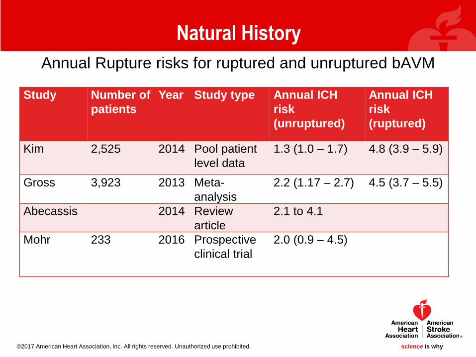
Annual Rupture risks for ruptured and unruptured bAVM
Natural History
Study Number of
patients
Year Study type Annual ICH
risk
(unruptured)
Annual ICH
risk
(ruptured)
Kim 2,525 2014 Pool patient
level data
1.3 (1.0 – 1.7) 4.8 (3.9 – 5.9)
Gross 3,923 2013 Meta-
analysis
2.2 (1.17 – 2.7) 4.5 (3.7 – 5.5)
Abecassis 2014 Review
article
2.1 to 4.1
Mohr 233 2016 Prospective
clinical trial
2.0 (0.9 – 4.5)
©2017 American Heart Association, Inc. All rights reserved. Unauthorized use prohibited.

science is whyscience is why
• The most consistently reported prognostic factor for ICH after
diagnosis is initial presentation with ICH.
• Increasing age is another prognostic factor significantly associated
with future ICH with a 1.34-fold increase per decade.
• Exclusively deep venous drainage may be another prognostic
factor for ICH, conferring a 1.6-2.4-fold increase in annual risk.
• Deep AVM nidus location and associated arterial aneurysms may
also be prognostic factors, though better-powered studies are
needed.
Natural History
©2017 American Heart Association, Inc. All rights reserved. Unauthorized use prohibited.

science is whyscience is why
Natural History
Hazard Ratios for rupture risk for clinical and anatomic features from longitudinal studies of unruptured bAVMs
Study Number Year Study
Type
Exclusively
Deep
Venous
Drainage
Any Deep
Venous
Drainage
Increasing
Age at
Diagnosis
Deep
Nidus
Location
Associated
Aneurysms
Female
Gender
Size < 3
cm
Kim 2,525 2014 Pooled
patient
level data
1.60 (0.95 –
2.68)
1.34 (1.17
– 1.53)
1.49 (0.96-
2.30)
1.02 (0.90-
1.16)
Gross 3,923 2013 Meta-
analysis
2.4 (1.1 –
3.8)
1.3 (0.9 –
1.75)
1.0 (0.4 -
1.6)
2.4 (1.4-
3.4)
1.8 (1.6-2.0) 1.4 (0.6-
2.1)
1.0 (0.8 –
1.2)
©2017 American Heart Association, Inc. All rights reserved. Unauthorized use prohibited.

science is whyscience is why
• Angioarchitectural risk factors for ICH from smaller series include
deep and infratentorial location (OR 2.718, p = 0.007), single
draining vein (OR 0.404, p = 0.008), venous varices (OR 0.488, p =
0.018), and aneurysm all type (OR 8.541, p = 0.002) or flow-related
(OR 2.923, p = 0.002).
• bAVM size has been implicated as a risk factor, though in the
larger cohorts this association has not been replicated.
• There is no evidence that any of HHT or RASA1 genotypes confer
higher ICH risk or a particular bAVM appearance..
Natural History
©2017 American Heart Association, Inc. All rights reserved. Unauthorized use prohibited.

science is whyscience is why
Natural History
Angioarchitectural features associated with ruptured bAVMs (retrospective studies comparing ruptured to unruptured bAVMs
(potential prognostic significance)
Study number Year Larger Size Aneurysm Venous
stenosis
Venous
ectasia
Exclusively
deep draining
Single
draining vein
Stapf 464 2006 Yes Yes Yes Yes
Sahlein 122 2014 Yes Yes Yes
Alexander 519 2015 Yes Yes Yes
Lv 302 2011 Yes Yes Yes Yes
©2017 American Heart Association, Inc. All rights reserved. Unauthorized use prohibited.

science is whyscience is why
• Clinical outcomes from bAVM ICH is less well-defined in part due
to entanglement with treatment effects.
• Admission GCS, age, and ICH volume are similarly predict bAVM-
related ICH clinical outcomes as in the setting of primary ICH.
• There is limited data describing angioarchitectural details and ICH-
related clinical outcomes, though AVM volume does not appear
associated ICH volume.
• There is evidence that posterior fossa bAVMs may present more
often with smaller ICH volumes and flow-related aneurysms as well
as poorer clinical outcomes relative supratentorial bAVMs.
Natural History
©2017 American Heart Association, Inc. All rights reserved. Unauthorized use prohibited.

science is whyscience is why
• ICH presentation is more common in children (56% vs.
43%) , though similar annual ICH rates compared to
adults (2.0% vs. 2.2%).
• There is no evidence in support of an increased rate of
ICH during pregnancy or puerperium.
Natural History
©2017 American Heart Association, Inc. All rights reserved. Unauthorized use prohibited.
science is whyscience is why
• Brain AVMs may cause focal or secondary generalized seizures, or
both
• The 5-year risk of first seizure is 8% (95% CI 0-20%) for patients
with bAVM and in the setting of ICH or focal neurologic deficit
increased the risk of seizure to 23% for patients with bAVM.
• The 5-year risk of developing epilepsy following a first seizure was
58%.
• Angioarchitectural features associated with seizures include a
cortical location, superficial venous drainage, varices, and
positioning within the frontal and particularly the temporal lobe.
Natural History
©2017 American Heart Association, Inc. All rights reserved. Unauthorized use prohibited.

science is whyscience is why
• The imaging evaluation of bAVMs may be separated into three
clinical settings: diagnosis, treatment planning, and follow-up.
• The definitive diagnosis of a bAVM is currently by digital
subtraction angiography (DSA), although many bAVMs can be
reliably identified by CT (computed tomography) and MR (magnetic
resonance) imaging, including angiographic imaging (CTA and
MRA).
• Non-contrast CT has > 90% sensitivity for acute subarachnoid
hemorrhage and hemorrhagic stroke and though limited in
detecting bAVMs, it can demonstrate features, including enlarged
or calcified vessels along the margin of the hemorrhage or regions
of increased density corresponding to the vascular nidus,
suggestive of an underlying vascular anomaly.
Imaging Diagnosis and Evaluation
©2017 American Heart Association, Inc. All rights reserved. Unauthorized use prohibited.
science is whyscience is why
• For any new ICH with non-contrast CT findings
and/or epidemiological and/or clinical variables
suspicious for a secondary vascular etiology and
cross-sectional angiogram should be performed.
• CTA and MRA have excellent accuracy for the
detection of secondary vascular anomalies in the
setting of ICH.
• Both CT and MR provide information about the
bAVM and the adjacent brain, this latter information
essential as it relates to assessing treatment
planning.
Imaging Diagnosis and Evaluation
©2017 American Heart Association, Inc. All rights reserved. Unauthorized use prohibited.

science is whyscience is why
Imaging Diagnosis and Evaluation
• For a subset of acute hemorrhagic
bAVM patients as well as those
without ICH presentation, MR may
identify prior subclinical
microhemorrhage (white arrow) using
susceptibility-weighted imaging.
• MR offers the ability to perform
advanced methods including temporal
encoding, functional imaging, and
flow-related parameters all of which
may help in treatment planning.
©2017 American Heart Association, Inc. All rights reserved. Unauthorized use prohibited.

science is whyscience is why
• Digital subtraction angiography (DSA) is the reference standard for
the diagnosis providing the most detailed and accurate information
on bAVM angio-architecture and hemodynamics.
• In addition to bAVM size, location, and venous drainage pattern
other angioarchtetural details that should be noted include: number
of veins, presence of subependymal venous involvement, number
of veins reaching a sinus, venous ectasia, venous reflux or
occlusion, flow-related or nidal arterial aneurysms, angiopathy,
angiogenesis, nidal border, and/or pial-pial collaterals.
• Associated aneurysms occur 15-30% of patients, and may be
remote, arising from arterial afferents, or intra-nidal in location, with
the latter being associated with a higher annual rate of ICH.
Imaging Diagnosis and Evaluation
©2017 American Heart Association, Inc. All rights reserved. Unauthorized use prohibited.

science is whyscience is why
• There is limited data on the utility of imaging surveillance for
untreated bAVMs, though long term monitoring is important after
treatment, particularly following stereotactic radiosurgery and in the
pediatric setting.
• DSA remains the reference standard, though MRA has improved in
its ability to assess for residual bAVM, particularly in the post
radiosurgical treatment setting.
Imaging Diagnosis and Evaluation
©2017 American Heart Association, Inc. All rights reserved. Unauthorized use prohibited.
science is whyscience is why
I. The definitive treatment of bAVMs should be complete elimination of
the nidus and the arteriovenous shunt. Partial nidal obliteration does
not appear to reduce hemorrhage risk
II. Additional interventions to reduce hemorrhagic risks with partial bAVM
obliteration
I. Microsurgical resection. This may be performed primarily or after
endovascular embolization to reduce bleeding risks during surgery
and facilitate complete and uncomplicated removal.
II. Stereotactic radiosurgery. Which may be done primarily or after
embolization to reduce nidal volumes and potentially improve nidal
obliteration rates.
III. Endovascular embolization .
Treatment: Overview
©2017 American Heart Association, Inc. All rights reserved. Unauthorized use prohibited.
science is whyscience is why
I. Microsurgical resection via craniotomy is a common approach for
treating patients with bAVMs with a goal to achieve definitive cure.
II. The sequential steps in this approach include:
a. Craniotomy to obtain adequate exposure to the bAVM including its
arterial feeders and venous outflow
b. Isolate and divide its arterial feeders;
c. Circumferentially dissect the nidus from the adjacent brain
parenchyma and surrounding neurovascular structures
d. Disconnect the venous outflow
e. Wound closure.
Treatment: Microsurgical Resection
©2017 American Heart Association, Inc. All rights reserved. Unauthorized use prohibited.

science is why
Microsurgical Resection: Advantages and Disadvantages
Advantages
• High rate of complete nidus obliteration
• Ability to immediately eliminate hemorrhage risk
• Long-term durability.
Disadvantages
• Invasiveness,
• Prolonged length of recovery
• Procedural risks.
©2017 American Heart Association, Inc. All rights reserved. Unauthorized use prohibited.

science is whyscience is why
• Functional magnetic resonance imaging (fMRI) and diffusion tensor imaging-based tractography(dTI) have been applied to more accurately determine the proximity of bAVMs to eloquent cortex and critical white matter tracts
• Stereotactic neuronavigation has been used to permit smaller, more accurate, and more effective approaches to bAVM surgery. This technique involves quantitative spatial fusion of the patient’s pre-operatively obtained CT and/or MR images with a fiducial coordinate system that permits guidance of surgical exposure and real-time localization of the bAVM and surrounding neurovascular structures
• Intraoperative vascular Imaging using DSA, Fluorescein videoangiography to map angioarchitecture
• Adjunct endovascular embolization aims to:
– Reduce in flow and/or nidus volume that would permit safer surgical removal
– Treatment of feeding artery and intra-nidal aneurysms.
Adjuncts to Improve Outcomes with Microsurgery
©2017 American Heart Association, Inc. All rights reserved. Unauthorized use prohibited.

science is whyscience is why
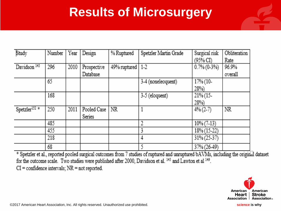
Spetzler-Martin (SM) Grading Scale for AVMs
SM grade I = 4% (CI: 2-7%); SM grade II = 10% (CI: 7-13%); SM grade III = 18% (CI: 15-
22%); SM grade IV = 31% (CI: 25-37%); and SM grade V = 37% (CI: 26-49%) 152. These
results demonstrate microsurgical removal is best suited for low-grade bAVMs (SM I and II),
while surgical removal of high-grade bAVMs (SM IV and V) carries high risk of poor patient
outcome.
©2017 American Heart Association, Inc. All rights reserved. Unauthorized use prohibited.

science is whyscience is why
SM grade III bAVMs are subcategorized based on specific combinations of
size, location, and venous drainage.
1. SM grade III- bAVMs (combination of small size, eloquent location, and
deep venous drainage) have good surgical outcomes
2. SM grade III bAVMs (combination of medium size, non-eloquent
location, and deep venous drainage) and SM grade III+ bAVMs
(combination of medium size, eloquent location, and superficial venous
drainage) have worse surgical outcomes similar to that reported for
high-grade bAVMs
Management: Unruptured bAVMs: Further Categorization
©2017 American Heart Association, Inc. All rights reserved. Unauthorized use prohibited.

science is whyscience is why
• Improving the predictive value of bAVM outcomes
Supplementing the traditional SM grading scheme:
1) patient age (<20 y = 1 point; 20-40 y = 2 points; >40 y = 3 points)
2) bleeding or hemorrhagic presentation (yes = 0 points; no = 1 point)
3) Nidus configuration (compact = 0 points; diffuse = 1 point)
– More accurate at predicting patient outcome vs. the SM system
alone.
– This scale has been referred to as the Lawton-Young supplementary
grading scale and has been validated in a separate cohort of 1009
patients
Management of Unruptured AneurysmsLawton-Young Supplementary Grading Scale
©2017 American Heart Association, Inc. All rights reserved. Unauthorized use prohibited.
science is whyscience is why
Results of Microsurgery
©2017 American Heart Association, Inc. All rights reserved. Unauthorized use prohibited.

science is whyscience is why
• SRS is used in patients where it is very risky to perform micro resection either due to the AVM itself or the patients medical condition.
• Leads to endothelial cell proliferation, necrosis, concentric vessel wall thickening and eventual closure.
• Time to obliteration is prolonged and complications of radiation injury, edema and cyst formation as well as early hemorrhage are some of the disadvantages of the procedure.
• Long term follow-up shows an obliteration rate of 70-80%
• In situations where SRS is the best option and resulted in incomplete obliteration, repeat SRS can be considered
Stereotactic Radiosurgery (SRS)
©2017 American Heart Association, Inc. All rights reserved. Unauthorized use prohibited.

science is whyscience is why
Unruptured bAVM: Radiosurgery
Radiosurgery may be the best option
Series Patients Follow-
up
(months)
Radiation
Induced
changes
Annual
hemorrhage
rate
Clinical
deteriora
tion
Cause of
deterioration
Obliteration
Rates
Ding 444 86 13.7%
(temp)
2%
(permanent)
1.6%/yr 12.3% Early
hemorrhage62%
Starke 2236 60.3%
Pollock 174 64 4% 11.5%at
10years
4% Radiation
necrosis78.9%
©2017 American Heart Association, Inc. All rights reserved. Unauthorized use prohibited. science is why

science is whyscience is why
• Age, lesion size, location relative to eloquent cortex, number feeding vessels and draining veins
• In a study of obliteration outcomes in 139 patients undergoing radiosurgery, Taeshineetanakul et al. reported factors predictive of bAVM obliteration. These included the following
– bAVM size (OR 0.88, CI 0.81 – 0.96),
– non-eloquent location (OR 3.2, CI 1.29 – 7.93)
– low flow pattern (OR 3.47, CI 1.6 – 7.53)
– Absence of perinidal angiogenesis (OR 2.61, CI 1.21 – 5.64)
.
Pre-treatment Radiation ScalesFactors and Validation
©2017 American Heart Association, Inc. All rights reserved. Unauthorized use prohibited.
science is whyscience is why
Endovascular Treatment
• Commonly used as an pre-procedural adjunct microsurgery or SRS with the aim of reducing vascularity and thus reducing the chances of other subsequent interventional complications
• Sometimes used as a stand alone procedure to obliterate AVMs. Here ethyl vinyl alcohol copolymer (EVOH) or NBCA are used. Detachable micro -catheter tips are helpful in reducing complications
• As a palliative treatment to improve symptoms caused by vascular steal secondary to the bAVM
©2017 American Heart Association, Inc. All rights reserved. Unauthorized use prohibited.

science is whyscience is why
• A five point scale that takes into account the following
• Arterial Feeders (<3, 3-6. >6)
• Location of the bAVM
• Size >3 cm
• Diameter depth >6 cm
• Deep venous drainage
• Proximity to eloquent cortex
Embolization Scales
©2017 American Heart Association, Inc. All rights reserved. Unauthorized use prohibited.

science is whyscience is why
• The two most common complications of embolization are intracerebral
hemorrhage and ischemic stroke
• The causes of ischemic stroke include thromboembolic complications of
catheterization as well as non-target embolization
• Brain hemorrhage may occur from vessel wall injury or AVM rupture.
– Microcatheter or wire perforation of arterial feeders may occur owing
to access through small tortuous pial arteries.
– The AVM nidus may rupture during embolization or in the hours to
days following the procedure. This may be due to inadvertent closure
of the draining vein prior complete nidal elimination or to other in the
nidal pressure flow dynamic changes
Complications of Embolization
©2017 American Heart Association, Inc. All rights reserved. Unauthorized use prohibited.

science is whyscience is why
• Initial single or staged embolization with the aim of reducing vascularity and bAVM volume
• Subsequent microsurgery or SRS
• Follow-up radiological and angiographic studies to determine the outcomes of the proceedure
Multimodal Treatment
©2017 American Heart Association, Inc. All rights reserved. Unauthorized use prohibited.

science is whyscience is why
Studies of Multimodal Approach and outcomes: embolization with subsequent SRS
©2017 American Heart Association, Inc. All rights reserved. Unauthorized use prohibited.

science is whyscience is why
• Result of the single randomized trial ARUBA did not support interventional treatment as opposed to conservative management.
– Primary Outcome: Stroke or Death
Interventional group 30.7%
Medical group 10.1%
– Enrollment has been stopped and a longer follow-up of 5-years is ongoing.
• These results supported by a more recent prospective non-randomized observational study of 204 patients where medical management was superior to interventional treatment in reducing the likelihood of death, sustained disability or non-fatal symptomatic stroke
• Critique: bAVM time to rupture and bleed is long compared to the shorter follow-up in these studies which may confound results and exclusion of patients under 16 years of age.
– ARUBA most interventional patients were treated with embolization alone or as an adjunct to surgery or SRS alone.
– Results of surgery or SRS as primary treatments with low
grade SM aneurysms may have yielded better outcomes.
ARUBA Trial and Unruptured bAVMs
©2017 American Heart Association, Inc. All rights reserved. Unauthorized use prohibited.

science is whyscience is why
• After microsurgical resection of bAVMs, there is excellent seizure control with many series suggesting up to 90% (80% seizure free and 16% with medically controlled seizures in patients with preoperative seizures
• Deep arterial perforator supply to the bAVM was associated with continued seizures
• With SRS 44-88% achieve complete or partial freedom from seizures. Complete obliteration of the bAVM was associated with the greater (88%) cure.
• Microsurgery is superior to radiosurgery and embolization in achieving seizure control (78%, 66% and 50%) respectively
• Results were replicated in a very large meta analysis
Seizure control after various interventions for bAVMs
©2017 American Heart Association, Inc. All rights reserved. Unauthorized use prohibited.

science is whyscience is why
• No clear incidence or prevalence reported
• This is an area that needs more attention
• It is empirically suggested to avoid vasoconstrictors that might increase the risk of rupture
Headaches after bAVM treatment
©2017 American Heart Association, Inc. All rights reserved. Unauthorized use prohibited.

science is whyscience is why
Management of Ruptured bAVMs
• Management of ruptured bAVMs is similar to the management of unruptured bAVMs
• Use of surgery, embolization, SRS or a combination of these modalities
• Embolization may be used alone in very high risk cases or as a means of reducing the nidus facilitating subsequent surgery
• NBCA (N0butyl cyanoacrylate) and EVOH (embolization materials) are commonly used for embolization and detachable micro catheter tips improve the chances of complete obliteration through a catheter based intervention
• A continuum of care starting from prehospital notification to the emergency department with ready access to neurology, neuroradiology and neurosurgery are essential in management
©2017 American Heart Association, Inc. All rights reserved. Unauthorized use prohibited.

science is whyscience is why
• In non-emergent cases transfer to NICU
• Anticonvulsants when patients have seizures
• Control of blood pressure, hyperthermia and cerebral edema as needed
• Refer to the ICH and AVM guidelines 2011 and 2015 from the AHA and ASA for complete management details.
Supportive Care: Ruptured bAVMs
©2017 American Heart Association, Inc. All rights reserved. Unauthorized use prohibited.

science is whyscience is why
• Presently the research base knowledge needs to be expanded to attempt to answer the following questions– Can case and bAVM selection determine whether conservative
treatment or interventional treatment should be selected
– Sub-selection in a new trial similar to ARUBA to see if low risk aneurysms are better treated conservatively or with intervention
– Greater understanding of the molecular genetics for patient intervention
An ongoing registry capturing these various treatment options and devising a new trial such a ARUBA to specifically address management based on patient and bAVM selection is required.
Future options in the pipeline
Ongoing Research
©2017 American Heart Association, Inc. All rights reserved. Unauthorized use prohibited.

science is whyscience is why
• Uncertainties still exist regarding the best approach to management of bAVMs.
• While medical management of unruptured bAVMs seems to be superior to intervention, subsets of patients may do better in terms of reduced risk of rupture and seizures with intervention and complete resection.
• Microsurgery offers the best option in selected cases for prevention of cerebral hemorrhage and seizure control.
• Where microsurgery is not feasible, SRS and Embolization may be options.
• Long term follow-up of patients in the ARUBA trial and other prospectively collected data may provide more definite pathways to the best management of bAVMs.
Summary
©2017 American Heart Association, Inc. All rights reserved. Unauthorized use prohibited.

science is whyscience is why