Management of Acute Exacerbation of Asthma and …€¦ · Management of Acute Exacerbation of...
23
Management of Acute Exacerbation of Asthma and Chronic Obstructive Pulmonary Disease in the Emergency Department Salvador J. Suau, MD*, Peter M.C. DeBlieux, MD INTRODUCTION Acute asthma and chronic obstructive pulmonary disease (COPD) exacerbations are the most common respiratory diseases requiring emergent medical evaluation and treatment. Asthma accounts for more than 2 million visits to emergency departments (EDs), and approximately 4000 annual deaths in the United States. 1 In a similar fashion, COPD is a major cause of morbidity and mortality. It affects more than 14.2 million Americans (9.8 million who may be undiagnosed). 2 COPD accounts for more than 1.5 million yearly ED visits and is the fourth leading cause of death Disclosures: None. Louisiana State University, University Medical Center of New Orleans, 2000 Canal Street, D&T 2nd Floor - Suite 2720, New Orleans, LA 70112, USA * Corresponding author. E-mail address: [email protected] KEYWORDS Asthma Asthmatic crisis COPD AECOPD KEY POINTS Management of severe asthma and chronic obstructive pulmonary disease (COPD) exac- erbations require similar medical interventions in the acute care setting. Capnography, electrocardiography, chest x-ray, and ultrasonography are important diag- nostic tools in patients with undifferentiated shortness of breath. Bronchodilators and corticosteroids are first-line therapies for both asthma and COPD exacerbations. Noninvasive ventilation, magnesium, and ketamine should be considered in patients with severe symptoms and in those not responding to first-line therapy. A detailed plan reviewed with the patient before discharge can decrease the number of future exacerbations. Emerg Med Clin N Am 34 (2016) 15–37 http://dx.doi.org/10.1016/j.emc.2015.08.002 emed.theclinics.com 0733-8627/16/$ – see front matter Ó 2016 Elsevier Inc. All rights reserved.
Transcript of Management of Acute Exacerbation of Asthma and …€¦ · Management of Acute Exacerbation of...
Management of AcuteExacerbation of Asthma
and Chronic ObstructivePulmonary Disease in theEmergency DepartmentSalvador J. Suau, MD*, Peter M.C. DeBlieux, MD
KEYWORDS
� Asthma � Asthmatic crisis � COPD � AECOPD
KEY POINTS
� Management of severe asthma and chronic obstructive pulmonary disease (COPD) exac-erbations require similar medical interventions in the acute care setting.
� Capnography, electrocardiography, chest x-ray, and ultrasonography are important diag-nostic tools in patients with undifferentiated shortness of breath.
� Bronchodilators and corticosteroids are first-line therapies for both asthma and COPDexacerbations.
� Noninvasive ventilation, magnesium, and ketamine should be considered in patients withsevere symptoms and in those not responding to first-line therapy.
� A detailed plan reviewed with the patient before discharge can decrease the number offuture exacerbations.
INTRODUCTION
Acute asthma and chronic obstructive pulmonary disease (COPD) exacerbations arethe most common respiratory diseases requiring emergent medical evaluation andtreatment. Asthma accounts for more than 2 million visits to emergency departments(EDs), and approximately 4000 annual deaths in the United States.1 In a similarfashion, COPD is a major cause of morbidity and mortality. It affects more than 14.2million Americans (�9.8 million who may be undiagnosed).2 COPD accounts formore than 1.5 million yearly ED visits and is the fourth leading cause of death
Disclosures: None.Louisiana State University, University Medical Center of New Orleans, 2000 Canal Street, D&T2nd Floor - Suite 2720, New Orleans, LA 70112, USA* Corresponding author.E-mail address: [email protected]
Emerg Med Clin N Am 34 (2016) 15–37http://dx.doi.org/10.1016/j.emc.2015.08.002 emed.theclinics.com0733-8627/16/$ – see front matter � 2016 Elsevier Inc. All rights reserved.
Suau & DeBlieux16
worldwide.3,4 Both asthma and COPD exacerbations impose an enormous economicburden on the US health care budget with estimates of more than $56 billion annuallyfor asthma,5 and $49.9 billion annually for COPD.4 A recent study found that, despitesignificant efforts to educate the public and increase disease awareness, the rates ofCOPD hospitalizations have increased by 20% to 30% between 2002 and 2012. Theinpatient monetary charges for these hospitalizations have increased by an alarming125%, and the rate of hospital readmissions for patients with poorly controlledCOPD remains at 21%.6
Asthma and COPD are chronic, debilitating disease processes that have beendifferentiated traditionally by the presence or absence of reversible airflow obstruc-tion. In daily clinical practice, it is difficult to differentiate these 2 obstructive processesbased on their symptoms, and on their nearly identical acute treatment strategies.Their major differences are important only when discussing anatomic sites involved,long-term prognosis, and the nature of inflammatory markers. These aspects affectdisease response to certain pharmacologic treatment options.7
DEFINITIONS
The Global Initiative for Asthma (GINA) described asthma as an allergic disease, typi-cally commencing in childhood,2,8 and characterized by increased bronchial hyperres-ponsiveness, increased vascular permeability, bronchial smooth muscle spasm, andthe release of inflammatory mediators. This pathophysiology translates into recurrentepisodes of wheezing, difficulty breathing, chest tightness, and coughing.9
Asthma exacerbations are variable and episodic. Asthma can be triggered by aplethora of environmental agents, infectious precipitants, emotional or exercisestates, and diverse exposure to ingested or inhaled agents, typically resolvingcompletely either spontaneously or with treatment.8,10
The Global Initiative for Chronic Obstructive Lung Disease (GOLD) guidelines defineCOPD as an acquired and preventable disease resulting primarily from tobacco smok-ing, and characterized by persistent airflow obstruction, and decline in progressivelung function.2,4 It usually develops after the fourth decade of life, and it is character-ized by shortness of breath, cough, and sputum production.4 The airflow limitationsare classically progressive and associated with an abnormal inflammatory responseto diverse inhaled agents, gases, and particles.7
PATHOPHYSIOLOGY
There is not strong evidence suggesting histopathologic overlap between these 2obstructive entities, known as the asthma–COPD overlap syndrome.2 Themost impor-tant pathologic difference between asthma and COPD is the inflammatory cells thatmediate each respective disease process. Eosinophils and CD4 cells mainly mediateasthma, whereas neutrophils and CD8 cells mediate COPD.2 This basic difference al-lows inhaled corticosteroids (CS) to be more efficacious against eosinophilic-mediated asthma, and largely ineffective against theprimarily neutrophilic inflammationseen in COPD.2,7 Regardless of their pathologic differences or their similar incitingagents, it is paramount that emergent risk stratification and treatment modalities beinitiated expeditiously to decrease clinical deterioration, morbidity, and mortality.
RISK STRATIFICATION
Risk stratification of the severely short of breath (SOB) patient requires several stepsand can be a difficult feat when an undifferentiated patient with SOB presents to the
ED Management of Asthma and COPD 17
ED. The practitioner should undertake a methodologic approach to optimize theacquisition of a pertinent history and quickly determine the best managementpathway. Box 1 provides some high-yield questions that will aid in the initial assess-ment of the dyspneic patient.4,11
After these initial questions, the severity of the exacerbation can be assessed withobjective physical findings such as vital signs, including oxygen saturation, heart rate(HR), and respiratory rate; degree of wheezing and air movement; use of accessorymuscles; degree of difficulty with speech; peak expiratory flow; and end-tidal carbondioxide (ETCO2) monitoring.4,11 It is imperative to understand that the absence ofseverity markers does not exclude the presence of a life-threatening disease process.A helpful algorithm to aid in differentiating between mild, moderate, and severe exac-erbation is presented in Fig. 1.The final step during the primary assessment of the patient with SOB is the essential
consideration that wheezing and respiratory distress can also be found in multipleother disease states. An adequate differential diagnosis must be formulated to preventthe creation of an anchoring bias, which would prevent a clinician from maintaining abroad differential diagnosis. Box 2 illustrates a differential diagnosis of wheezing inadults and children.
ACUTE DECOMPENSATED HEART FAILURE
The acutely undifferentiated patient with SOB may have multiple comorbidities thatmight contribute or disguise the exact inciting disease process. Two commonlyencountered examples are heart failure (HF) and COPD. These 2 entities are frequentlyencountered in the elderly and tobacco smoker. Several studies estimate the preva-lence of HF in COPD patients to be somewhere between 20% and 30%.12 Similarstudies have also reported that the presence of HF in COPD is associated with worseclinical outcomes.13,14
DIAGNOSISSpirometry
GOLD, GINA, and other evidenced-based guidelines have been developed as blue-prints for the identification, prevention, and treatment of both these obstructive
Box 1
Important risk factors in the asthmatic/COPD patient
� Previous endotracheal intubations
� Previous intensive care unit admissions
� �2 non-ICU hospitalizations in the past 1 year
� �3 ED visits in the past month
� Chronic use of oral corticosteroids
� Medication noncompliance
� Living in poverty with no access to health care
� Using �2 SABA pressurized metered dose inhalers monthly
Abbreviations: COPD, chronic obstructive pulmonary disease; ED, emergency department; ICU,intensive care unit; SABA, short-acting b-agonist.

Fig. 1. Dyspneic exacerbation severity algorithm. BGM, blood glucose monitor; BP, bloodpressure; ETCO2, end-tidal carbon dioxide; FEV1, forced expiratory volume in 1 second;HR, heart rate; PEF, peak expiratory flow; RR, respiratory rate; WNL, within normal limits.
Suau & DeBlieux18
entities. Both GOLD and GINA recommend baseline spirometry to diagnose and clas-sify these diseases.4,8 Despite this standard recommendation, there is no clinicalbenefit to performing spirometry in the acute care setting. Spirometry is not a suitabletool for the emergent management of the undifferentiated dyspneic patient.
Box 2
Differential diagnosis of wheezing
Adults Children
� Upper respiratory tract infection
� Pneumonia
� Asthma
� Chronic obstructive pulmonary disease
� Congestive heart failure
� Chronic bronchitis
� Gastroesophageal reflux disease
� Acute coronary syndrome
� Pulmonary embolism
� Foreign body
� Pneumothorax
� Cystic fibrosis
� Vocal cord dysfunction
� Upper respiratory tract infection
� Croup
� Tracheomalasia
� Bronchiolitis
� Asthma
� Pneumonia
� Foreign body

ED Management of Asthma and COPD 19
Laboratory Tests
There is currently no laboratory test that will specifically identify asthma or acute ex-acerbations of COPD (AECOPD) as the etiology of the acutely patient with SOB. Anystandard serum laboratory studies should only be drawn to assist in deciphering theetiology of the acute decompensation. Sputum testing is unreliable and should notbe gathered, unless tuberculosis is suspected as the underlying etiology of the exac-erbation. GOLD only recommends sputum testing in the AECOPD patient who hasfailed initial antibiotic therapy.4 Viruses are strongly associated with AECOPD; there-fore, testing for influenza may provide important implications in management of thesepatients.15
Blood Gas Analysis
Arterial blood gas analysis is a routine test performed in the severe asthmatic andAECOPD patient. Several guidelines recommend its use in moderate and severe res-piratory exacerbations: when the pulse oxygen saturation (SaO2) is less than 92% onroom air; and to follow pH, partial pressure of carbon dioxide (PCO2), and partial pres-sure of oxygen. One must question the benefit of an arterial over a venous blood gasgiven the pain severity, the possibility of aneurysmal formation, arterial laceration,infection, and infrequently, the loss of limb.16–26 These possible risks of the proceduremust be coupled with the understanding that a normal PCO2 in a venous blood gasanalysis excludes arterial hypercarbia, making this painful and possibly complicatedprocedure unnecessary.16–26
Capnography
ETCO2 during an AECOPD may be useful in the risk stratification of these patients.Do�gan and colleagues27 found that, when measuring with mainstream capnographydevices, ETCO2 levels were higher in admitted patients than those who were dis-charged from the ED. These levels must be obtained before any bronchodilator treat-ment. After the first bronchodilator treatment was completed, the ETCO2 between the2 groups showed no difference. This study also showed a strong correlation betweenETCO2 and arterial PCO2, previously demonstrated by Cinar and colleagues.28
Electrocardiogram
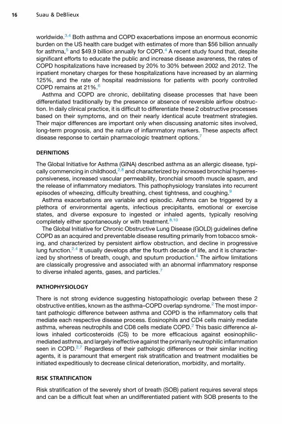
Electrocardiography is an essential component in the acute evaluation of the patientwith SOB. Part of the reported 58% increased mortality of patients with COPD be-tween 1990 and 2010 has been linked to adverse cardiovascular events.29 Althoughthe exact pathophysiologic link remains unclear, data suggest that this could becaused partly by cardiac dysrhythmias.30,31 Fig. 2 demonstrates commonly encoun-tered ECG changes that may be found in the AECOPD. These changes can be attrib-uted to the clockwise rotation of the heart and the right atrial and ventricularhypertrophy that is seen in the COPD patient. Furthermore, P-wave verticalization islikely caused by the downward displacement of the heart owing to the progressiveflattening of the diaphragms. This pathology is owing to the right atrium being physi-cally anchored to the diaphragm by a strong aponeurosis.32
Other ECG findings in COPD include:
� S waves in leads I, II, and III;� R/S ratio less than 1 in leads V5 or V6; and� The lead I sign—isoelectric P wave, QRS amplitude less than 1.5 mm, and Twave amplitude less than 0.5 mm in lead I.
Fig. 2. (1) Tachycardia. Multifocal atrial tachycardia is rare, but specific to chronic obstruc-tive pulmonary disease (COPD).33 (2) Right axis deviation. (3) P wave axis of greater than60� (considered to be 96% sensitive for COPD.34) (4) Low-voltage QRS amplitude in I, aVL,V5-V6. (May be found in leads II, III, or aVF [<5 mm]). (5) P pulmonale (peaked P waves inleads II, III, or aVF [>2.5 mm]). (From Burns E. The ECG in chronic obstructive pulmonary dis-ease. Life in the Fast Lane. 2012. Available at: http://lifeinthefastlane.com/ecg-library/copd/.Accessed May 29, 2015. Life in the Fast Lane is licensed under a Creative Commons ShareA-like 4.0 International User’s License http://creativecommons.org/licenses/by-sa/4.0/.)
Suau & DeBlieux20
If these criteria seem overly complex, a more simplified diagnostic marker is findinga P wave in lead aVL, or the P wave amplitude in lead III greater than in lead I.35
Radiography
The posteroanterior and lateral chest radiograph is the most widely used imaging mo-dality in the evaluation of the acutely dyspneic patient. Typical findings include a flat-tened diaphragm, an increased anteroposterior diameter, an enlarged retrosternalairspace, a narrow vertical cardiac silhouette, and bullae.36 Although none of thesefindings is diagnostic, a chest x-ray is more importantly obtained to rule out othercauses of shortness of breath, such as pneumothorax, pulmonary infiltrates, or pulmo-nary edema. Tsai and colleagues37 found that 21% of patients had their managementaltered by an initial chest x-ray. Pulmonary embolism has been found in 3% of COPDpatients presenting to the ED.38
Chest radiography should be considered routine in the patient with an AECOPD. Inpatients with established asthma, there is more room for clinical judgment, and prac-titioners should consider a chest x-ray in patients who (1) are in extremis, (2) have clin-ical markers of pneumonia or pneumothorax, (3) are not responding to conventionaltherapy, (4) are presenting with new onset wheezing, and presumed de novo asthma,and (5) are at risk for an alternative diagnosis, for example, HF in the older adult andforeign body aspiration in the young child with wheezing.
Ultrasonography
Cardiopulmonary ultrasonography has become an important diagnostic tool in the EDsetting because it decreases exposure to radiation. Three main protocols have comeinto favor. These include Lung Ultrasound in the Critically Ill (LUCI), Bedside Lung Ul-trasonography in Emergency (BLUE), and Fluid Administration Limited by Lung So-nography (FULL).39 Gallard and colleagues40 found that ultrasonography has anaccuracy of 95% in diagnosing COPD or asthma exacerbations. This reinforced thefindings of Silva and colleagues,41 who found a 92% accuracy of ultrasonography indiagnosing these conditions.
ED Management of Asthma and COPD 21
TREATMENTOxygen
Oxygen therapy is a key feature in the management of an undifferentiated patient withSOB. In an acute asthma exacerbation, GINA and the British Thoracic Society recom-mend that oxygen be the first-line treatment. They strongly emphasize this recommen-dation with the understanding that hypoxia must be addressed expeditiously andoxygen administration shouldbemonitored closely for efficacy. This differs significantlyfrom their guidelines for the AECOPD patient. The FiO2 provided to this patient popula-tion should be no greater than 28%. Bronchodilators are to be given with compressedair rather than oxygen. These recommendations stem from the knowledge that hyper-oxia leads to decreased minute ventilation and hypercapnia.42 Such increases in car-bon dioxide are more likely to be seen in older patients and those with a homeoxygen dependence, and can cause neurologic and cardiac depression.43
Austin and colleagues44 showed a reduced mortality in COPD patients with titratedoxygen therapy. Oxygen administration guidelines should therefore be in place in boththe prehospital setting as well as in the ED. Oxygen can be titrated according to a satu-ration of peripheral oxygen (SpO2), with no oxygen given at an SpO2 of greater than92%, 2 to 3 L via nasal cannula at an SpO2 of 85% to 92%, and a face mask withhigher flows used for an SpO2 of less than 85%.45 An arterial blood gas can then beobtained to further guide oxygen requirements.
Bronchodilators
The first-line pharmacotherapy in the emergent management of the asthmatic crisisand AECOPD is the administration of bronchodilators.46 These agents target the bron-chial hyperactivity and attempt to reverse, or ameliorate airflow obstruction. AlthoughCOPD is usually considered an irreversible process, most acute COPD exacerbationshave a reversible component that must be targeted. The primary pharmacotherapyagents used are short-acting b2-agonists (SABA) and ipratropium bromide.
Short-acting b2-receptor agonistsSABA relax pulmonary smooth muscle by stimulating airway b2-adrenergic receptors,increasing intracellular cyclic adenosine monophosphate. This increase in cyclicadenosine monophosphate inhibits smooth muscle bronchoconstriction. SABA’stypical time of onset is seconds to minutes, with peak effect at 30 minutes and ahalf-life of 4 to 6 hours.4,8 The most widely used SABA is albuterol, a racemic mixtureof 2 enantiomers, namely (R)-albuterol and (S)-albuterol. (R)-albuterol is the activeform, binding to b2-receptors and provides the desired bronchodilation. This alsocauses the more undesired, tachycardia, tremors, and anxiety/restlessness. (S)-albu-terol, the inert form, is hypothesized to possibly have detrimental effects on airwayfunction.46 This was the premise of the development of levalbuterol, a purified versionof the (R)-albuterol enantiomer that was marketed as having fewer of the unwantedcardiac adverse effects than racemic albuterol. Multiple studies have shown thatcontinuous nebulized levalbuterol is not superior to continuous nebulized albuteroland that levalbuterol had no beneficial effects on HR.47,48 In ametaanalysis of 7 clinicaltrials conducted by Jat and Khairwa,49 there was no evidence supporting the theorythat levalbuterol is superior to albuterol regarding efficacy and patient safety.
Long-acting b2-receptor agonistsLong-acting b2-receptor agonists (LABAs) such as salmeterol and formoterol werewidely used in the early 1990s because they provided approximately 12 hours of bron-chodilation. In 1993, Castle and colleagues50 showed significant evidence that
Suau & DeBlieux22
salmeterol had a 3-fold mortality increase in asthmatic patients. This finding wasquickly confirmed in 1996 by the US Food and Dug Administration’s Salmeterol Multi-centre Asthma Research Trial (SMART). The study had to be prematurely stoppedowing to increased exacerbations and mortality.51 An additional study performed byMann and colleagues52 also demonstrated increased asthma exacerbations.This is in contrast with current recommendations provided by the American College
of Chest Physicians and the Canadian Thoracic Society to prevent AECOPD. LABAshave been shown to improve quality of life and lung function while decreasing moder-ate and severe exacerbations in COPD patients. Rate of adverse events and mortalitywere not increased compared with placebo in this patient population.53 A LABA com-bined with an inhaled CS is preferable to monotherapy with either agent.
AnticholinergicsInhaled ipratropium bromide (Atrovent) elicits its bronchodilatory effect by competi-tively inhibiting the muscarinic acetylcholine receptors of the pulmonary smooth mus-cle. Its time of onset is approximately 15 minutes, with a peak effect at 60 to90 minutes and half-life of 6 to 8 hours, making it slower in onset and longer in durationthan SABA.54 This explains the common practice of using these inhaled agents incombination. The GOLD guidelines recommend a SABA as a first-line agent owingto its faster onset of action, followed by anticholinergics if a prompt response is notattained clinically.4 The authors of this article agree with the findings of Vezina and col-leagues,55 who found that combined pharmacotherapy is more effective in decreasingED admissions with no evidence of adverse effects. Ipratropium bromide can also beconsidered as a good alternative in patients who are intolerant of SABA side effects.The agent has been linked to lower ED admission rates in acutely severe exacerba-tions and may decrease the overall ED duration of stay.56–58 A similar, but longer-acting antimuscarinic, tiotropium, has been shown to be an effective maintenancebronchodilator in both COPD and asthma patients. Kerstjens and associates59
demonstrated that tiotropium improved symptomatic control in patients with poorlycontrolled symptoms who were on inhaled CS and LABAs and reduced severe exac-erbations by 21%. In the first 24 hours of the respiratory obstructive crisis, somebelieve that the adrenergic receptors, which constitute the majority of pulmonaryairway receptors, are downregulated and perhaps temporarily unresponsive to b2-re-ceptor agonists. During this time, pulmonary muscarinic acetylcholine receptorsremain functional leading to their contribution in bronchodilation.60–62
Delivery modeMethod of pharmacotherapy delivery is via a pressurized metered dose inhaler with aholding chamber or an oxygen-driven nebulizer. The current literature does not showany difference in outcomes based on route of administration, except for slightlyshorter ED duration of stay in those treated with gas-driven nebulizers.63,64
Magnesium sulfateIntravenous (IV) magnesium sulfate (MgSO4) is suggested to produce pulmonarysmooth muscle relaxation via calcium receptor blockade or by activation of adenylcyclase at the smooth muscle cellular level.65 Regardless of its mechanism of action,its efficacy on the acute asthmatic crisis or the AECOPD remains uncertain, despiteguidelines like GINA and GOLD advocating its use.4,8 Two studies were recently un-dertaken to ascertain this agent’s efficacy. The first, conducted by Goodacre and col-leagues,66 failed to show that either IV or nebulized MgSO4 provided any clinicallyrelevant benefit in adults with severe acute asthma. On the contrary, a second studyperformed by Kew and colleagues67 found that IV MgSO4 reduced hospital
ED Management of Asthma and COPD 23
admissions and improved lung function when other pharmacotherapy had failed toameliorate the acute exacerbation.
Corticosteroids
CS treatment is also considered first-line in the emergent management of the asth-matic crisis and AECOPD. CS have a complex mechanism of action that ultimatelyleads to the inhibition of potent inflammatory mediators and the reduction of airwayinflammation. A recent Cochrane review conducted by Walters and colleagues68
demonstrated that the use of CS was associated with a greater than 50% reductionin treatment failure. The number of patients needed to treat with CS to prevent 1 treat-ment failure was 9. This same study also showed that CS provided a 30-day relapserate reduction, and a shorter hospital duration of stay, despite no association withdecreased mortality.68 The choice of which systemic CS (SCS) to use has beendebated, and common practice dictates the use of glucocorticoids (prednisone, pred-nisolone, or methylprednisolone), because they are the most widely studied makingthem the preferred choice over SCS with mineralocorticoid effects like hydrocorti-sone.69 There is still ongoing research regarding the most appropriate dose, routeof administration, and duration of therapy. Currently, there is good consensus thatthere is no inferiority between oral and parenteral treatment with regards to rates oftreatment failure, relapse rate, and mortality.68,70 Therefore, if the patient can toleratean oral agent, provide therapy orally and reserve parenteral treatment for those pa-tients who cannot tolerate oral treatment.Of note, a new pilot study conducted by Mendes and colleagues71 regarding the
emerging use of inhaled CS showed that, in adults with airflow obstruction, a singlestandard dose of an inhaled CS provided simultaneously with inhaled albuterol acutelypotentiated the effects of the albuterol-induced pulmonary smooth muscle relaxationand increased the forced expiratory volume in 1 second (FEV1) response versus thestandard method.Initial CS dosages have also been a topic of great debate secondary to the miscon-
ception that severity of disease warrants higher dosages of treatment. In 2013, Chengand colleagues72 performed a metaanalysis of 12 studies totaling 1172 patients; theywere not able to demonstrate any benefit to CS dosages of greater than 80 mg/d.These findings were consistent with Alia and colleagues’s73 study from 2011, whichdemonstrated that SCS dosages of 0.5 mg/kg every 6 hours were sufficient, andhigher dosages were unwarranted for achieving clinical outcomes. For example,higher dosages did not have decreased duration of stay, decreased length of ventila-tion, or decreased treatment failure with noninvasive ventilation (NIV). Despite nobenefit found in larger dosages of SCS therapy, Schacke and colleagues74,75 haveshown that the risk of adverse effects increases with increased doses of CS. Themain adverse reactions with larger doses of CS were hyperglycemia, myopathies,neurologic effects like anxiety and delirium, increased rate of infection, hypertension,and gastrointestinal bleeding.74,75 These adverse effects were also documented byKiser and colleagues,76 who found the association of increased rates of hyperglyce-mia, need for insulin therapy, and increased rates of invasive fungal infections in pa-tients that were given CS doses of greater than 240 mg/d. Dosages of greater than2 mg/kg per day do not provide any clinical benefit, and will likely provide greaterside effects in the management of the critically ill asthmatics or AECOPD.The last vastly debated concept in CS treatment is the duration of treatment ther-
apy. The literature has described a wide range of therapy from 5 days to 8 weeks.The Reduction in the Use of Corticosteroids in Exacerbated COPD (REDUCE) trialconducted by Leuppi and colleagues77 demonstrated that short-term therapy was

Suau & DeBlieux24
noninferior to a longer duration. It showed no difference in mortality, rate of relapse, orchange in recovery of lung function based on treatment duration.77
Upon completion of these initial interventions, any additional treatment is based onthe patient’s clinical status. Fig. 3 provides a suggested treatment algorithm for pa-tients with severe asthma.78 It can also serve as a helpful algorithm in the treatmentof AECOPD.
Antibiotics
Antimicrobial pharmacotherapy is perhaps the only emergent treatment recommen-dation that will differ between the asthmatic and the AECOPD. As opposed to an asth-matic event, it is estimated that approximately 8 of every 10 AECOPD episodesprecipitated by a pulmonary infection, with either a bacterial, viral, or fungal path-ogen.79–81 Furthermore, these authors estimate that 50% of the microbial-induced ex-acerbations are attributed to a bacterial pathogen, warranting antibiotic therapy. Whiteand colleagues82 have also suggested that antibiotic therapy in an AECOPD willdecrease the risk of progression to pneumonia and will catalyze bacterial eradicationthat in turn will improve airway inflammation in the acute exacerbation. Macrolides arethe best class of antibiotics class for the treatment of AECOPD. Martinez and col-leagues83 determined that macrolides have added antiinflammatory and immunomod-ulatory effects, in addition to their antibacterial efficacy. Barnes84 has postulated thatmacrolide therapy may increase the pulmonary smooth muscle’s response to CS viathe increased recruitment of the enzyme histone deacetylase 2 that is integral in theinflammatory response seen in COPD patients. Finally, although macrolides mayhave additional benefits over other classes of antibiotics, if an allergic reaction de-velops, other commonly used agents such as b-lactams and fluoroquinolones aredeemed appropriate alternatives.84
Noninvasive Ventilation
The airway management of a decompensating asthmatic and COPD patient must bemonitored meticulously and prompt changes must be made if first-line therapy is notameliorating the respiratory crisis. The early recognition and appropriate modificationin management may prevent further decompensation and increased mortality. Carsonand colleagues85 report that patients who are decompensating warrant NIV. NIV is themainstay of therapy in the acute management of most reversible respiratory emergen-cies. This treatment modality is best provided via full facial mask, although other deliv-erymethods are available. It is postulated that NIV is a direct bronchodilator.86 NIV alsorecruits alveoli secondary toexternal positive endexpiratory pressure (PEEP), offsettingintrinsic PEEP.87 Alveolar recruitment improves ventilation–perfusionmismatch by pre-venting airway closure and reducing the work of breathing.88 NIV can be used for shortperiods of time as deemed clinically necessary and carries a lower risk of nosocomialpneumonia than endotracheal intubation (ETI).89 Fig. 3 suggests starting inspiratorypositive airway pressure and expiratory positive airway pressure at 8 cm H2O and3 cm H2O, respectively. The authors of this article strongly encourage that the practi-tioner remain at the patient’s bedside directly monitoring work of breathing and seriallyincreasing both inspiratory positive airway pressure and expiratory positive airwaypressure to higher pressures within a 30 minute trial to optimize nebulizer and overalltreatment time. A recent Cochrane review indicated that patients with acute asthmaexacerbation who were treated with NIV had decreased admission rates, decreasedduration of ICU stay, and an overall shorter hospital stay, although there was no clearbenefit for reduced ETI or mortality.90 Last, NIV has been shown to be safe in pregnantpatients and in pediatric populations.85 Relative contraindications include facial or
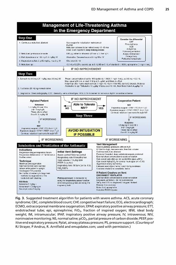
Fig. 3. Suggested treatment algorithm for patients with severe asthma. ACS, acute coronarysyndrome;CBC, completebloodcount; CHF, congestiveheart failure; ECG,electrocardiograph;ECMO,extracorporealmembraneoxygenation; EPAP, expiratorypositive airwaypressure; ETT,endotracheal tube; epi, epinephrine; FiO2, fraction of inspired oxygen; IBW, ideal bodyweight; IM, intramuscular; IPAP, inspiratory positive airway pressure; IV, intravenous; NIV,noninvasivemonitoring;NS,normal saline;pCO2,partialpressureof carbondioxide; PEEP,pos-itive endexpiratorypressure; Pplat, airwayplateaupressure; PS, pressure support. (CourtesyofRJ Strayer, P Andrus, R. Arntfield and emupdates.com; used with permission.)
ED Management of Asthma and COPD 25
Suau & DeBlieux26
esophageal trauma or surgery, deformities of the upper airway, copious secretions, oran uncooperative patient. Absolute contraindications are cardiac or respiratoryarrest.91
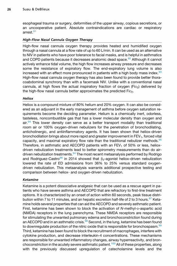
High-Flow Nasal Cannula Oxygen Therapy
High-flow nasal cannula oxygen therapy provides heated and humidified oxygenthrough a nasal cannula at a flow rate of up to 60 L/min. It can be used as an alternativeto NIV in patients who have poor tolerance to facial masks, and is helpful in asthmaticsand COPD patients because it decreases anatomic dead space.92 Although it cannotactively enhance tidal volume, the high flow increases airway pressure and decreasessome the resistance of expiratory flow. The end-expiratory lung volume is alsoincreased with an effect more pronounced in patients with a high body mass index.93
High-flow nasal cannula oxygen therapy has also been found to provide better thora-coabdominal synchrony than with a facemask NIV. Unlike with a conventional nasalcannula, at high flows the actual inspiratory fraction of oxygen (FiO2) delivered bythe high-flow nasal cannula better approximates the predicted FiO2.
Heliox
Heliox is a compound mixture of 80% helium and 20% oxygen. It can also be consid-ered as an adjuvant in the early management of asthma before oxygen saturation re-quirements become the deciding parameter. Helium is a chemically inert, odorless,tasteless, noncombustible gas that has a lower molecular density than oxygen andair.94 This lower density can serve as a better transport modality than traditionalroom air or 100% oxygen-driven nebulizers for the penetration of bronchodilating,anticholinergic, and antiinflammatory agents. It has been shown that heliox-drivenbronchodilation brings about more rapid and greater improvement in FEV1, forced vitalcapacity, and maximal expiratory flow rate than the traditional nebulizer methods.95
Therefore, in asthmatic and AECOPD patients with an FEV1 of 50% or less, heliox-driven nebulization treatments lead to better spirometry measurements than do air-driven nebulization treatments.94 The most recent metaanalysis performed by Rodrigoand Rodriguez-Castro96 in 2014 showed that b2-agonist heliox-driven nebulizationlowered the rate of ED admissions from 36% to 25% versus standard oxygen-driven nebulization. This metaanalysis warrants additional prospective testing andcomparison between heliox- and oxygen-driven nebulization.
Ketamine
Ketamine is a potent dissociative analgesic that can be used as a rescue agent in pa-tients who have severe asthma and AECOPD that are refractory to first-line treatmentoptions. It is characterized by an onset of action within 60 seconds, peak tissue distri-bution within 7 to 11 minutes, and an hepatic excretion half-life of 2 to 3 hours.97 Keta-mine holds several properties that can aid the AECOPDand severely asthmatic patient.First, ketamine has been shown to block the activation of N-methyl-D-aspartic acid(NMDA) receptors in the lung parenchyma. These NMDA receptors are responsiblefor stimulating the unwanted pulmonary edema and bronchoconstriction found duringan AECOPD and in an asthmatic crisis.98 Second, in the lung, ketamine has been foundto downregulate production of the nitric oxide that is responsible for bronchospasm.99
Third, ketamine has been found to block the recruitment ofmacrophages, interferewithcytokine production, and decrease interleukin-4 concentrations. These mechanismsare responsible for unwanted inflammatory changes, airway hyperreactivity, and bron-choconstriction in the acutely severe asthmatic patient.100 All of these properties, alongwith the previously discussed upregulation of catecholamine levels and the

ED Management of Asthma and COPD 27
anticholinergic effects on bronchial smooth muscle, argue strongly for ketamine as anadvantageousadjuvant agent in themanagementof theAECOPDanddecompensatingasthmatic. Last, it is important to emphasize that ketamine, like all analgesic, amnestic,anesthetic, and muscle relaxants, be administered in a monitored environment whereSaO2, ETCO2, blood pressure (BP), HR, and appropriate nursing presence is availablecontinuously.
Epinephrine
Perhaps the most underappreciated and underused pharmacotherapy in our arma-mentarium for the treatment of an asthmatic crisis and the AECOPD is epinephrine.Some iconic practitioners would strongly argue that this agent should be placed onthe top of every first-line treatment algorithm published by medical societies, a senti-ment that both authors of this article share. The concern for a possible adverse cardio-vascular event, increased risk of hypertension in an already uncontrolled hypertensivepatient and the possible risk of aggravating the tachycardic state in an already tachy-cardic patient has placed this previously first-line agent at the bottom, if notcompletely off treatment algorithms. In 1988, Cydulka and colleagues101 statedthat, despite concerns of old age, concerns of potential adverse cardiac events, orconcerns of increased risk of exacerbating BP and HR, epinephrine, dosed at startingdoses of 0.3 mg of 1:1000 solution, did not cause any of these adverse reactions.Epinephrine, like many of the first-line agents today, may cause tremors, anxiety,and nausea, and most patients will tolerate these doses with no contraindications.101
Even increased BP and tachycardia, which can also be attributed to severe anxiety,hypoxia, hypercapnia, and increased work of breathing, actually declined in the oldercohort of patients, and only minimally increased in the younger group. In the youngergroup, BP and HR normalized when patients attained relief from bronchospasm.101
A second study published that same year was a study by Spiteri and colleagues102
that compared terbutaline with epinephrine for the treatment of acute asthma, and itdemonstrated that neither agent produced any significant increases in BP or HR, notreatment-related ECG abnormalities, and no observed adverse cardiovascular ef-fects. Both of these studies showed benefit and no significant harm in the use ofepinephrine for the treatment an asthmatic crisis. It is time to revisit this topic in a pro-spective and randomized manner.
Endotracheal Intubation
The decision to intubate should not be taken lightly as manipulation of the airway in apatient with AECOPD or an asthmatic crisis can cause laryngospasm, worsen bron-chospasm, and may even increase morbidity. It is estimated that the mortality rateof ICU patients who are intubated for severe asthma is 10% to 20%.103 Some studieshave advocated that the severe asthmatic and COPD patient can be adequatelymanaged without resorting to ETI.104,105 Despite attempting to refrain from intubatinga decompensating asthmatic, once first- and second-line therapies have failed, thepractitioner must seriously consider ETI. The clinical decision regarding when to intu-bate a decompensating asthmatic patient can be aided by clinical signs such as anSaO2 of less than 90% with maximal supplemental oxygen, bradypnea leading to hy-percapnia and respiratory acidosis, altered level of consciousness, and physicalexhaustion.106 The only absolute indications for intubation are respiratory or cardiacarrest.107
The usual method for intubating a patient in AECOPD and asthmatic crisis ismaximal preoxygenation followed by rapid sequence induction. Ketamine and propo-fol are both valid options for induction agents. Ketamine creates a catecholamine
Suau & DeBlieux28
release that causes bronchodilation by relaxing bronchial smooth muscles.108
Because this release of catecholamines can cause hypertension and arrhythmias, ke-tamine should be avoided in patients with active dysrhythmias. Propofol also hassome bronchodilating effects, and it can cause hypotension. Patient selection forthis drug also should be considered carefully.Succinylcholine and rocuronium bromide are the 2 main choices of muscle relaxant
for rapid sequence intubation in the ED. It is essential that their respective benefits andpossible side effects be understood before selecting an agent. Traditionally, rocuro-nium has been considered to have a slower time of onset than succinylcholine. Impor-tantly, onset is slower only if rocuronium is used at lower doses of 0.6 to 0.9 mg/kg IV.If a dose of 1.2 mg/kg IV is used, no difference exists in the time of onset of ideal intu-bating conditions, although the higher dose will lengthen the duration of paralysis.109
The duration of paralysis with rocuronium is also dose dependent; time to paralysisrecovery is reported to occur as early as 30 minutes with a dose of 0.6 mg/kg IV,and it will be double or even triple after a 1.2 mg/kg dose.109 Table 1 provides thecommon side effects encountered with succinylcholine. The most troubling andrare, side effect of rocuronium is anaphylaxis. Regardless of the agent used for paral-ysis, understanding respective mechanisms of action and side effects is essential.In the patient with severe asthma and COPD, ETI and positive-pressure ventilation
may cause abrupt increases in intrathoracic pressure that in turn decrease venous re-turn and therefore cardiac output. For this reason, time permitting, it is important tooptimize the patient’s preload before ETI is attempted. A preintubation fluid bolus(as well as use of the ventilator strategies described elsewhere in this article) mayhelp to prevent or attenuate abrupt decreases in BP immediately after ETI.
Post Endotracheal Intubation Documentation
ED and critical care specialists should be diligent in documenting the intubation pro-cedure. A detailed medical record will greatly aid the clinician who attempts extuba-tion when the pathophysiologic state has been reversed. The physician shouldalways document the Cormack–Lehane grading scale (Fig. 4), the laryngoscope bladeused, any airway adjuvants used, the number of intubation attempts made, a descrip-tion of any complications, and any confirmation modalities used in the airwaymanagement.
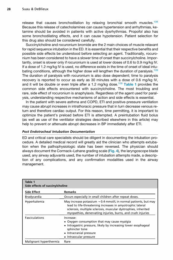
Table 1Side effects of succinylcholine
Side Effect Remarks
Bradycardia Occurs especially in small children after repeat doses.
Hyperkalemia May increase potassium w0.4 mmol/L in normal patients, but maylead to life-threatening increases in amyotrophic lateralsclerosis, multiple sclerosis, muscular dystrophies, inheritedmyopathies, denervating injuries, burns, and crush injuries
Fasciculations Increase:� Oxygen consumption that may cause myalgia� Intragastric pressure, likely by increasing lower esophageal
sphincter tone� Intracranial pressure� Intraocular pressure
Malignant hyperthermia Rare
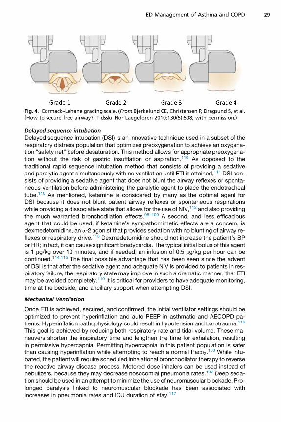
Fig. 4. Cormack–Lehane grading scale. (From Bjerkelund CE, Christensen P, Dragsund S, et al.[How to secure free airway?] Tidsskr Nor Laegeforen 2010;130(5):508; with permission.)
ED Management of Asthma and COPD 29
Delayed sequence intubationDelayed sequence intubation (DSI) is an innovative technique used in a subset of therespiratory distress population that optimizes preoxygenation to achieve an oxygena-tion “safety net” before desaturation. This method allows for appropriate preoxygena-tion without the risk of gastric insufflation or aspiration.110 As opposed to thetraditional rapid sequence intubation method that consists of providing a sedativeand paralytic agent simultaneously with no ventilation until ETI is attained,111 DSI con-sists of providing a sedative agent that does not blunt the airway reflexes or sponta-neous ventilation before administering the paralytic agent to place the endotrachealtube.110 As mentioned, ketamine is considered by many as the optimal agent forDSI because it does not blunt patient airway reflexes or spontaneous respirationswhile providing a dissociative state that allows for the use of NIV,112 and also providingthe much warranted bronchodilation effects.98–100 A second, and less efficaciousagent that could be used, if ketamine’s sympathomimetic effects are a concern, isdexmedetomidine, an a-2 agonist that provides sedation with no blunting of airway re-flexes or respiratory drive.113 Dexmedetomidine should not increase the patient’s BPor HR; in fact, it can cause significant bradycardia. The typical initial bolus of this agentis 1 mg/kg over 10 minutes, and if needed, an infusion of 0.5 mg/kg per hour can becontinued.114,115 The final possible advantage that has been seen since the adventof DSI is that after the sedative agent and adequate NIV is provided to patients in res-piratory failure, the respiratory state may improve in such a dramatic manner, that ETImay be avoided completely.110 It is critical for providers to have adequate monitoring,time at the bedside, and ancillary support when attempting DSI.
Mechanical Ventilation
Once ETI is achieved, secured, and confirmed, the initial ventilator settings should beoptimized to prevent hyperinflation and auto-PEEP in asthmatic and AECOPD pa-tients. Hyperinflation pathophysiology could result in hypotension and barotrauma.116
This goal is achieved by reducing both respiratory rate and tidal volume. These ma-neuvers shorten the inspiratory time and lengthen the time for exhalation, resultingin permissive hypercapnia. Permitting hypercapnia in this patient population is saferthan causing hyperinflation while attempting to reach a normal PaCO2.
103 While intu-bated, the patient will require scheduled inhalational bronchodilator therapy to reversethe reactive airway disease process. Metered dose inhalers can be used instead ofnebulizers, because they may decrease nosocomial pneumonia rates.107 Deep seda-tion should be used in an attempt to minimize the use of neuromuscular blockade. Pro-longed paralysis linked to neuromuscular blockade has been associated withincreases in pneumonia rates and ICU duration of stay.117
Suau & DeBlieux30
Extracorporeal Membrane Oxygenation
The last resort in a clinically decompensating asthmatic patient would be the initiationof extracorporeal membrane oxygenation. This technology is considered in those pa-tients who cannot be maintained on mechanical ventilation with adequate oxygena-tion. Extracorporeal membrane oxygenation requires a dedicated support staff andequipment and is beyond the capabilities of the typical ED.
Treatment Options Beyond the Emergency Department
The following therapies may also be used in the treatment of both severe asthmaticand COPD patients, but there is no evidence to support their use in the acute settingof the ED.
i. Leukotriene modifiersii. Mast cell stabilizersiii. Anti-immunoglobulin therapyiv. Specific immunotherapy
MANAGEMENT BEYOND THE EMERGENCY DEPARTMENT
Weaning and extubation criteria have not been adequately studied in the acutely asth-matic or AECOPD patient.103 AECOPD and asthma exacerbations that require ETItypically are slow to resolve and require aggressive therapy for more than 24 hoursbefore weaning and extubation can be considered. Before assessing the patient forpossible extubation, the practitioner must confirm that the asthmatic pathophysiologicstate that warranted intubation has resolved. First, all sedation and muscle relaxantsmust be discontinued and prophylactic antiemetic treatment provided. The head ofthe bed should be raised to greater than 45�. Adequate time must be allowed forthe patient to be able to follow simple commands, such as opening his eyes, trackingwith his eyes, grasping with both hands, and protruding the tongue on command withno evidence of bronchospasm or hemodynamic compromise. Once appropriate timehas elapsed, the cuff leak test should be performed. This test is used to evaluate forany laryngeal edema that might have occurred during the ETI and throughout the treat-ment. In the absence of laryngeal edema a noticeable airleak should be audible at thepatient’s bedside when the endotracheal tube cuff is deflated. Zhou and colleagues118
state that the cuff test is accurate in finding significant differences in laryngeal edema,but that it does not accurately predict the need for reintubation. When no mucosalswelling is evident, the third step is the assessment of oxygenation and ventilation.Adequate oxygenation and ventilation can be assessed with a spontaneous breathingtrial on reduced pressure support of 5 cm H2O. If the patient is able to maintain thefollowing parameters with no bronchospasm, an attempt at extubation can beconsidered119:
� SaO2 of greater than 92% (PaO2 >70) on FiO2 less than 40% and PEEP is less than5 cm H2O;
� Tidal volume of greater than 5 mL/kg;� Mean arterial pressure of greater than 60 with no aid of vasopressor agents;� Respiratory rate of less than 30 and greater than 6 breaths/min; and� HR of less than 100 and greater than 60 beats/min.
If the patient remains stable with no evidence of bronchospasm for approximately30 minutes, one can move forward with the negative inspiratory force test. A valuegreater than –30 cm H2O (normal, –90 to –120) indicates that the strength of the
ED Management of Asthma and COPD 31
diaphragm and other inspiratory muscles is adequate to attempt extubation. A finalassessment modality to predict a successful extubation is the rapid shallow breathingindex. This index relies on the idea that patients on a ventilator who cannot tolerateindependent breathing tend to breathe with high frequency and shallow tidal volume.Therefore, a score of less than approximately 100 is considered by most an adequateindication of weaning readiness.120 Upon successful completion of all of these steps,extubation may be undertaken. Safe extubation of a patient requires equipment suchas suction, oral airway, supplemental oxygen, and equipment that may be needed ifreintubation is required. A nonrebreathing mask and NIV should be at the bedsidebecause extubation may elicit laryngeal edema, bronchospasm, and postextubationstridor that require nebulized epinephrine and further treatment. In short, extubationshould always be approached in a logical and cautious manner. Every step shouldbe anticipated meticulously and executed cautiously to prevent reexacerbation orother complications.
ACUTE EXACERBATIONS OF CHRONIC OBSTRUCTIVE PULMONARY DISEASE ANDASTHMA CARE PLANS
The final component of post-AECOPD and asthmatic crisis care is a detailed COPDand asthma care plan that includes explicit discharge instructions, necessary medica-tions and education in how to use them, education in self-assessment, a future actionplan for managing recurrence of airflow obstruction, and an explicit follow-up appoint-ment. COPD and asthma care plans, including education and casemanagement, havebeen associated with improved outcomes and medication compliance. For the patienthospitalized with severe asthma or COPD, it is recommended that follow-up with anasthma/COPD-specialized clinician occur within 1 week of discharge. Pulmonaryrehabilitation within the first 4 weeks after AECOPD is recommended to prevent acuteexacerbations.53 The final moments before the patient returns home after an asth-matic or AECOPD crisis are the ideal opportunity for clinicians to provide appropriatecare plans that will assist patients with future exacerbations, encourage partnershipwith primary care physicians, and promote ongoing discussions of home asthma/COPD care.
REFERENCES
1. Moorman JE, Akinbami LJ, Bailey CM, et al. National surveillance of asthma:United States, 2001–2010. National Center for Health Statistics. Vital HealthStat 3 2012;(35):1–58. Available at: http://www.cdc.gov/nchs/data/series/sr_03/sr03_035.pdf.
2. Papaiwannou A, Zarogoulidis P, Porpodis K, et al. Asthma-chronic obstructivepulmonary disease overlap syndrome (ACOS): current literature review.J Thorac Dis 2014;6(Suppl 1):S146–51.
3. American Lung Association. Chronic obstructive pulmonary disease (COPD) factsheet. Available at: http://www.lung.org/lung-disease/copd/resources/facts-figures/COPD-Fact-Sheet.html. Accessed November 24, 2014.
4. Global Initiative for Chronic Obstructive Lung Disease (GOLD). Global strategyfor the diagnosis, management, and prevention of chronic obstructive pulmo-nary disease. 2013. Available at: http://www.goldcopd.org. Accessed January,2015.
5. CDC’s Asthma’s Impact on the Nation. Available at: http://www.cdc.gov/asthma/most_recent_data.htm. Accessed January, 2015.
Suau & DeBlieux32
6. Ford ES. Hospital discharges, readmissions, and ED visits for COPD or bronchi-ectasis among US adults: findings from the Nationwide Inpatient Sample2001-2012 and Nationwide Emergency Department Sample 2006-2011. Chest2015;147(4):989–98.
7. PostmaDS,ReddelHK, tenHackenNH, et al. Asthmaandchronic obstructivepul-monary disease: similarities and differences. Clin Chest Med 2014;35(1):143–56.
8. Global Initiative for Asthma (GINA). Global strategies for asthma managementand prevention. 2015. Available at: www.ginasthma.org. Accessed January,2015.
9. Sellers WFS. Inhaled and intravenous treatment in acute severe and life-threatening asthma. Br J Anaesth 2013;110(2):183–90.
10. Murata A, Ling PM. Asthma diagnosis and management. Emerg Med Clin NorthAm 2012;30:203–22.
11. National Asthma Education and Prevention Program. Expert panel report III:guidelines for the diagnosis and management of asthma. Bethesda (MD): Na-tional Heart, Lung, and Blood Institute; 2007. Available at: http://www.columbia.edu/cgi-bin/cul/resolve?clio7740011.
12. Fisher KA, Stefan MS, Darling C, et al. Impact of COPD on the mortality andtreatment of patients hospitalized with acute decompensated heart failure: theWorcester Heart Failure Study. Chest 2015;147(3):637–45.
13. De Blois J, Simard S, Atar D, et al. COPD predicts mortality in HF: the NorwegianHeart Failure Registry. J Card Fail 2010;16(3):225–9.
14. Macchia A, Monte S, Romero M, et al. The prognostic influence of chronicobstructive pulmonary disease in patients hospitalised for chronic heart failure.Eur J Heart Fail 2007;9(9):942–8.
15. Mohan A, Chandra S, Agarwal D, et al. Prevalence of viral infection detected byPCR and RT-PCR in patients with acute exacerbation of COPD: a systematic re-view. Respirology 2010;15(3):536–42.
16. Kelly AM. Agreement between arterial and venous blood gases in emergencymedical care: a systematic review Hong Kong. J Emerg Med 2013;20:166–71.
17. Kelly AM, Kerr D, Middleton P. Validation of venous pCO2 to screen for arterialhypercarbia in patients with chronic obstructive airways disease. J Emerg Med2005;28:377–9.
18. Kelly AM, Kyle E, McAlpine R. Venous pCO2 and pH can be used to screen forsignificant hypercarbia in emergency patients with acute respiratory disease.J Emerg Med 2002;22:15–9.
19. Sur E. COPD: is it all in the vein? Thorax 2013;68(Suppl 3):P182.20. McCanny P, Bennett K, Staunton P, et al. Venous vs. arterial blood gases in the
assessment of patients presenting with an exacerbation of chronic obstructivepulmonary disease. Am J Emerg Med 2012;30:896–900.
21. Ibrahim I, Ooi SBS, Huak CY, et al. Point-of-care bedside gas analyzer: limiteduse of venous pCO2 in emergency patients. J Emerg Med 2011;41:117–23.
22. Lim BL, Kelly AM. A meta-analysis on the utility of peripheral venous blood gasanalysis in exacerbations of chronic obstructive pulmonary disease in the emer-gency department. Eur J Emerg Med 2010;17:246–8.
23. Razi E, Moosavi GA. Comparison of arterial and venous blood gases analysis inpatients with exacerbation of chronic obstructive pulmonary disease. SaudiMed J 2007;28:862–5.
24. Ak A, Ogun CO, Bayor A, et al. Prediction of arterial blood gas values fromvenous blood gas values in patients with acute exacerbation of chronic obstruc-tive pulmonary disease. Tohoku J Exp Med 2006;210:285–90.

ED Management of Asthma and COPD 33
25. RangLCF,MurrayHE,WellsGA, et al. Canperipheral venousbloodgases replacearterial blood gases in emergency department patients? CJEM 2002;4:7–15.
26. Elborn JS, Finch MB, Stanford CF. Non-arterial assessment of blood gas statusin patients with chronic pulmonary disease. Ulster Med J 1991;60:164–7.
27. Do�gan NO, S‚ ener A, Gunaydın GP, et al. The accuracy of mainstream end-tidalcarbon dioxide levels to predict the severity of chronic obstructive pulmonarydisease exacerbations presented to the ED. Am J Emerg Med 2014;32(5):408–11.
28. Cinar O, Acar YA, Arziman I, et al. Can mainstream end-tidal carbon dioxidemeasurement accurately predict the arterial carbon dioxide level of patientswith acute dyspnea in ED. Am J Emerg Med 2012;30:358–61.
29. Murray CJ, Atkinson C, Bhalla K, et al. The State of US health, 1990-2010:burden of diseases, injuries, and risk factors. JAMA 2013;310:591–608.
30. Curkendall SM, DeLuise C, Jones JK, et al. Cardiovascular disease in patientswith chronic obstructive pulmonary disease, Saskatchewan Canada cardiovas-cular disease in COPD patients. Ann Epidemiol 2006;16:63–70.
31. Schneider C, Bothner U, Jick SS, et al. Chronic obstructive pulmonary diseaseand the risk of cardiovascular diseases. Eur J Epidemiol 2010;25:253–60.
32. Shah NS, Koller SM, Janower ML, et al. Diaphragm levels as determinants of Paxis in restrictive vs obstructive pulmonary disease. Chest 1995;107(3):697–700.
33. Rodman DM, Lowenstein SR, Rodman T. The electrocardiogram in chronicobstructive pulmonary disease. J Emerg Med 1990;8:607–15.
34. Thomas AJ, Apiyasawat S, Spodick DH. Electrocardiographic detection ofemphysema. Am J Cardiol 2011;107:1090–2.
35. Khalid N, Chhabra L, Spodick DH. Electrocardiographic screening of emphy-sema: lead aVL or Leads III and I? Acta Inform Med 2013;21(3):223.
36. Friedman PJ. Imaging studies in emphysema. Proc Am Thorac Soc 2008;5:494–500.
37. Tsai T, Gallagher E, Lombardi G, et al. Guidelines for the selective ordering ofadmission chest radiography in adult obstructive airway disease. Ann EmergMed 1993;22:1854–8.
38. Rizkallah J, Man SF, Sin DD. Prevalence of pulmonary embolism in acute exac-erbations of COPD: a systematic review and metaanalysis. Chest 2009;135:786–93.
39. Lichtenstein D, van Hooland S, Elbers P, et al. Ten good reasons to practice ul-trasound in critical care. Anaesthesiol Intensive Ther 2014;46(5):323–35.
40. Gallard E, Redonnet JP, Bourcier JE, et al. Diagnostic performance of cardiopul-monary ultrasound performed by the emergency physician in the managementof acute dyspnea. Am J Emerg Med 2015;33(3):352–8.
41. Silva S, Biendel C, Ruiz J, et al. Usefulness of cardiothoracic chest ultrasound inthe management of acute respiratory failure in critical care practice. Chest 2013;144(3):859–65.
42. Sassoon CSH, Hassell KT, Mahutte CK. Hypoxic induced hypercapnia in stablechronic obstructive pulmonary disease. Am Rev Respir Dis 1987;135:907–11.
43. Susanto C, Thomas PS. Assessing the use of initial oxygen therapy in chronicobstructive pulmonary disease patients: a retrospective audit of pre-hospitaland hospital emergency management. Intern Med J 2015;45(5):510–6.
44. Austin M, Wills E, Blizzard L, et al. Effect of high flow oxygen on mortality onchronic obstructive pulmonary disease patients in prehospital setting: rando-mised controlled trial. BMJ 2010;341:c546.
Suau & DeBlieux34
45. Beasley R, Aldington S, Robinson G. Is it time to change the approach to oxy-gen therapy in the breathless patient? Thorax 2007;62:840–1.
46. Ameredes BT, Calhoun WJ. Albuterol enantiomers: pre-clinical and clinicalvalue? Front Biosci (Elite Ed) 2010;2:1081–92.
47. Andrews T, McGintee E, Mittal MK, et al. High-dose continuous nebulized leval-buterol for pediatric status asthmaticus: a randomized trial. J Pediatr 2009;155:205–10.
48. Wilkinson M, Bulloch B, Garcia-Filion P, et al. Efficacy of racemic albuterolversus levalbuterol used as a continuous nebulization for the treatment of acuteasthma exacerbations: a randomized, double-blind, clinical trial. J Asthma2011;48(2):188–93.
49. Jat KR, Khairwa A. Levalbuterol versus albuterol for acute asthma: A systematicreview and meta-analysis. Pulm Pharmacol Ther 2013;26(2):239–48.
50. Castle W, Fuller R, Hall J, et al. Serevent nationwide surveillance study: compar-ison of salmeterol with salbutamol in asthmatic patients who require regularbronchodilator treatment. BMJ 1993;306:1034–7.
51. Nelson HS, Weiss ST, Bleecker ER, et al. The salmeterol multicenter asthmaresearch trial: a comparison of usual pharmacotherapy for asthma or usualpharmacotherapy plus salmeterol. Chest 2006;129:15–26.
52. Mann M, Chowdhury B, Sullivan E, et al. Serious asthma exacerbations in asth-matics treated with high-dose formoterol. Chest 2003;124:70–4.
53. Criner GL, Bourbeau J, Diekemper RL, et al. Prevention of Acute Exacerbationsof COPD: American College of Chest Physicians and Canadian Thoracic SocietyGuideline. Chest 2015;147(4):894–942.
54. Rennard SI. Treatment of stable chronic obstructive pulmonary disease. Lancet2004;364(9436):791–802.
55. Vezina K, Chauhan BF, Ducharme FM. Inhaled anticholinergics and short-actingbeta(2)-agonists versus short-acting beta2-agonists alone for children withacute asthma in hospital. Cochrane Database Syst Rev 2014;(7):CD010283.
56. Rodrigo GJ, Castro-Rodriguez JA. Anticholinergics in the treatment of childrenand adults with acute asthma: a systematic review with meta-analysis. Thorax2005;60:740–6.
57. Plotnick LH, Ducharme FM. Acute asthma in children and adolescents: shouldinhaled anticholinergics be added to beta(2)-agonists? Am J Respir Med2003;2:109–15.
58. Zorc JJ, Pusic MV, Ogborn CJ, et al. Ipratropium bromide added to asthma treat-ment in thepediatric emergencydepartment. Pediatrics 1999;103(4Pt 1):748–52.
59. Kerstjens HA, Engel M, Dahl R, et al. Tiotropium in asthma poorly controlled withstandard combination therapy. N Engl J Med 2012;367:1198–207.
60. Teoh L, Cates CJ, Hurwitz M, et al. Anticholinergic therapy for acute asthma inchildren. Cochrane Database Syst Rev 2012;(4):CD003797.
61. Craven D, Kercsmar CM, Myers TR, et al. Ipratropium bromide plus nebulizedalbuterol for the treatment of hospitalized children with acute asthma.J Pediatr 2001;138(1):51–8.
62. Goggin N, Macarthur C, Parkin PC. Randomized trial of the addition of ipra-tropium bromide to albuterol and corticosteroid therapy in children hospitalizedbecause of an acute asthma exacerbation. Arch Pediatr Adolesc Med 2001;155(12):1329–34.
63. Cates CJ, Crilly JA, Rowe BH. Holding chambers (spacers) versus nebulisersfor beta-agonist treatment of acute asthma. Cochrane Database Syst Rev2006;(2):CD000052.
ED Management of Asthma and COPD 35
64. Turner MO, Gafni A, Swan D, et al. A review and economic evaluation of bron-chodilator delivery methods in hospitalized patients. Arch Intern Med 1996;156:2113–8.
65. Rodrigo GL. Advances in acute asthma. Curr Opin Pulm Med 2015;21(1):22–6.66. Goodacre S, Cohen J, Bradburn M, et al. Intravenous or nebulised magnesium
sulphate versus standard therapy for severe acute asthma (3Mg trial): a double-blind, randomised controlled trial. Lancet Respir Med 2013;1:293–300.
67. Kew KM, Kirtchuk L, Michell CI. Intravenous magnesium sulfate for treatingadults with acute asthma in the emergency department. Cochrane DatabaseSyst Rev 2014;(5):CD010909.
68. Walters JA, Tan DJ, White CJ, et al. Systemic corticosteroids for acute exacer-bations of chronic obstructive pulmonary disease. Cochrane Database SystRev 2014;(9):CD001288.
69. Kiser TH, Vandivier RW. Severe acute exacerbations of chronic obstructive pul-monary disease: does the dosage of corticosteroids and type of antibiotic mat-ter? Curr Opin Pulm Med 2015;21:142–8.
70. Lindenauer PK, Pekow PS, Lahti MC, et al. Association of corticosteroid doseand route of administration with risk of treatment failure in acute exacerbationof chronic obstructive pulmonary disease. JAMA 2010;303:2359–67.
71. Mendes ES, Cadet L, Arana J, et al. Acute effect of an inhaled glucocorticoste-roid on albuterol-induced bronchodilation in patients with moderately severeasthma. Chest 2015;147(4):1037–42.
72. Cheng T, Gong Y, Guo Y, et al. Systemic corticosteroid for COPD exacerbations,whether the higher dose is better? A meta-analysis of randomized controlled tri-als. Clin Respir J 2013;7:305–18.
73. Alia I, de la Cal MA, Esteban A, et al. Efficacy of corticosteroid therapy in pa-tients with an acute exacerbation of chronic obstructive pulmonary diseasereceiving ventilatory support. Arch Intern Med 2011;171:1939–46.
74. Schacke H, Schottelius A, Docke WD, et al. Dissociation of transactivation fromtransrepression by a selective glucocorticoid receptor agonist leads to separationof therapeutic effects fromsideeffects.ProcNatlAcadSciUSA2004;101:227–32.
75. Schacke H, Docke WD, Asadullah K. Mechanisms involved in the side effects ofglucocorticoids. Pharmacol Ther 2002;96:23–43.
76. Kiser TH, Allen RR, Valuck RJ, et al. Outcomes associated with corticosteroiddosage in critically ill patients with acute exacerbations of chronic obstructivepulmonary disease. Am J Respir Crit Care Med 2014;189:1052–64.
77. Leuppi JD, Schuetz P, Bingisser R, et al. Short-term vs conventional glucocorti-coid therapy in acute exacerbations of chronic obstructive pulmonary disease:the REDUCE randomized clinical trial. JAMA 2013;309:2223–31.
78. Available at: http://emupdates.com/2011/12/14/when-the-patient-cant-breathe-and-you-cant-think-the-emergency-departement-life-threatening-asthma-flowsheet/.Accessed January, 2015.
79. Domenech A, Puig C, Marti S, et al. Infectious etiology of acute exacerbations insevere COPD patients. J Infect 2013;67:516–23.
80. Decramer M, Janssens W, Miravitlles M. Chronic obstructive pulmonary dis-ease. Lancet 2012;379:1341–51.
81. Sethi S, Murphy TF. Infection in the pathogenesis and course of chronic obstruc-tive pulmonary disease. N Engl J Med 2008;359:2355–65.
82. White AJ, Gompertz S, Bayley DL, et al. Resolution of bronchial inflammation isrelated to bacterial eradication following treatment of exacerbations of chronicbronchitis. Thorax 2003;58:680–5.
Suau & DeBlieux36
83. Martinez FJ, Curtis JL, Albert R. Role of macrolide therapy in chronic obstructivepulmonary disease. Int J Chron Obstruct Pulmon Dis 2008;3:331–50.
84. Barnes PJ. Corticosteroid resistance in patients with asthma and chronicobstructive pulmonary disease. J Allergy Clin Immunol 2013;131:636–45.
85. Carson KV, Usmani ZA, Smith BJ. Noninvasive ventilation in acute severeasthma: current evidence and future perspectives. Curr Opin Pulm Med 2014;20:118–23.
86. Buda AJ, Pinsky MR, Ingels NB Jr, et al. Effect of intrathoracic pressure on leftventricular performance. N Engl J Med 1979;301:453–9.
87. Broux R, Foidart G, Mendes P, et al. Use of PEEP in management of life- threat-ening status asthmaticus: a method for the recovery of appropriate ventilation-perfusion ratio. Appl Cardiopulm Pathophysiol 1991;4:79–83.
88. Soroksky S, Stav D, Shpirer I. A pilot prospective, randomized, placebo-controlled trial of bilevel positive airway pressure in acute asthmatic attack.Chest 2003;123:1018–25.
89. Nourdine K, Combes P, Carton MJ, et al. Does noninvasive ventilation reducethe ICU nosocomial infection risk? A prospective clinical survey. IntensiveCare Med 1999;25:567–73.
90. Lim WJ, Mohammed Akram R, Carson KV, et al. Noninvasive positive & pressureventilation for treatment of respiratory failure due to severe acute exacerbationsof asthma. Cochrane Database Syst Rev 2012;(12):CD004360.
91. Yeow ME, Santanilla JI. Noninvasive positive pressure ventilation in the emer-gency department. Emerg Med Clin North Am 2008;26:835–47.
92. Nishimura M. High-flow nasal cannula oxygen therapy in adults. J Intensive Care2015;3:15.
93. Corley A, Caruana LR, Barnett AG, et al. Oxygen delivery through high-flownasal cannulae increase end-expiratory lung volume and reduce respiratoryrate in post-cardiac surgical patients. Br J Anaesth 2011;107(6):998–1004.
94. El-Khatib MF, Jamaleddine G. Effect of heliox- and air-driven nebulized broncho-dilator therapy on lung function in patients with asthma. Lung 2014;192:377–83.
95. Bag R, Bandi V, Fromm RE Jr, et al. The effect of heliox-driven bronchodilatoraerosol therapy on pulmonary function tests in patients with asthma. J Asthma2002;39(7):659–65.
96. Rodrigo GJ, Rodriguez-Castro JA. Heliox-driven b2-agonists nebulization forchildren and adults with acute asthma: a systematic review with meta-analysis.Ann Allergy Asthma Immunol 2014;112:29–34.
97. Stevenson C. Ketamine: a review. Update Anaesth 2005;20:25–9.98. Sato T, Hirota K, Matsuki A, et al. The role of the N-Methyl-D-Aspartic acid re-
ceptor in the relaxant effect of ketamine on tracheal smooth muscle. Anesth An-alg 1998;87:1383–8.
99. Zhu MM, Qian YN, Zhu W, et al. Protective effects of ketamine on allergen-induced airway inflammatory injure and high airway reactivity in asthma: exper-iment with rats. Zhonghua Yi Xue Za Zhi 2007;87:1308–13.
100. Goyal S, Agrawal A. Ketamine in status asthmaticus: a review. Indian J Crit CareMed 2013;17(3):154–61.
101. Cydulka R, Davison R, Grammer L, et al. The use of epinephrine in the treatmentof older adult asthmatics. Ann Emerg Med 1988;17(4):322–6.
102. Spiteri MA, Millar AB, Pavia D, et al. Subcutaneous adrenaline versus terbutalinefor the treatment of acute severe asthma. Thorax 1988;43(1):19–23.
103. Brenner B, Corbridge T, Kazzi A. Intubation and mechanical ventilation of thepatient in respiratory failure. J Emerg Med 2009;37(2S):S23–34.
ED Management of Asthma and COPD 37
104. Braman SS, Kaemmerlen JT. Intensive care of status asthmaticus. A 10-yearexperience. JAMA 1990;264:366–8.
105. Mountain RD, Sahn SA. Acid-base disturbances in acute asthma. Chest 1990;98:651–5.
106. Murase K, Tomii K, Chin K, et al. The use of non-invasive ventilation for life-threatening asthma attacks: changes in the need for intubation. Respirology2010;15:714–20.
107. Schauer SG, Cuenca PJ, Johnson JJ, et al. Management of acute asthma in theemergency department. Emerg Med Pract 2013;15(6):1–28.
108. Brown RH, Wagner EM. Mechanisms of bronchoprotection by anesthetic induc-tion agents: propofol versus ketamine. Anesthesiology 1999;90:822–8.
109. Perry JJ, Lee JS, Sillberg VA, et al. Rocuronium versus succinylcholine for rapidsequence induction intubation. Cochrane Database Syst Rev2008;(2):CD002788.
110. Weingart SD. Preoxygenation, reoxygenation, and delayed sequence intubationin the emergency department. J Emerg Med 2011;40(6):661–7.
111. Walls RM, Murphy MF. Manual of emergency airway management. 3rd edition.Philadelphia: Lippincott Williams & Wilkins; 2008.
112. Aroni F, Iacovidou N, Dontas I, et al. Pharmacological aspects and potential newclinical applications of ketamine: reevaluation of an old drug. J Clin Pharmacol2009;49:957–64.
113. Carollo DS, Nossaman BD, Ramadhyani U. Dexmedetomidine: a review of clin-ical applications. Curr Opin Anaesthesiol 2008;21:457–61.
114. Abdelmalak B, Makary L, Hoban J, et al. Dexmedetomidine as sole sedative forawake intubation in management of the critical airway. J Clin Anesth 2007;19:370–3.
115. Bergese SD, Khabiri B, Roberts WD, et al. Dexmedetomidine for conscioussedation in difficult awake fiberoptic intubation cases. J Clin Anesth 2007;19:141–4.
116. Lougheed MD, Fisher T, O’Donnell DE. Dynamic hyperinflation during broncho-constriction in asthma: implications for symptom perception. Chest 2006;130:1072–81.
117. Adnet F, Racine SX, Lapostolle F, et al. Full reversal of hypercapnic coma bynoninvasive positive pressure ventilation. Am J Emerg Med 2001;19:244–6.
118. Zhou T, Zhang HP, Chen WW, et al. Cuff-leak test for predicting postextubationairway complications: a systematic review. J Evid Based Med 2011;4(4):242–54.
119. Salam A, Tilluckdharry L, Amoateng-Adjepong Y, et al. Neurologic status,cough, secretions and extubation outcomes. Intensive Care Med 2004;30:1334–9.
120. Meade M, Guyatt G, Cook D, et al. Predicting success in weaning from mechan-ical ventilation. Chest 2001;120(6 Suppl):400S–24S.