
Mammalian Heme Peroxidases and Mycobacterium...
18
15 Mammalian Heme Peroxidases and Mycobacterium tuberculosis Amit K. Singh, Nisha Pandey, Mau Sinha, Sujata Sharma and Tej P. Singh Department of Biophysics, All India Institute of Medical Sciences, New Delhi, India 1. Introduction Tuberculosis (TB) is a lethal infectious disease which is caused by Mycobacterium tuberculosis. The alarming rate at which the incidence of bacterial resistance to known antibiotics has been rising is a serious cause of concern. At present, the two well known anti-tuberculosis drugs, isonicotinic acid hydrazide (INH, isoniazid) and pyrazinamide (PZA, pyrazin-2- carboxamide) which are important components of the current course of the first-line TB chemotherapy suffer from increasing bacterial resistance. The other drugs of the combination therapy include rifampicin and ethambutol. It may be noted that both INH and PZA are prodrugs and require specific enzymes to convert them into drugs. INH is activated by a bacterial heme enzyme catalase peroxidase (MtCP) into a free radical form (Scheme I) (Zhang et al., 1992). The structure of unliganded MtCP is known (Bertrand et al., 2004) and detailed information is available about the substrate-binding site and the residues that might be involved in the binding and conversion of INH into a beneficial product. However, a precise mode of binding and the mechanism of action are not yet clearly understood because the structure of INH bound MtCP is not yet determined. On the other hand, PZA is metabolized into its active form pyrazinoic acid (POA) by amidase activity of the Mycobacterium tuberculosis nicotinamidase/pyrazinamidase (PncA) (Scheme II) (Konno et al., 1967). Although the crystal structure of pyrazinamidase in complex with POA is known but the structure of the complex with the original compound PZA is not yet determined. Therefore, the mode of binding of PZA with PncA has not so far been revealed. As shown by the crystal structure of the complex of LPO with INH, the binding of INH to lactoperoxidase (LPO) occurs through the distal heme cavity where INH interacts with a conserved water molecule W1 which is hydrogen bonded to ferric iron (Singh et al., 2010). Similarly, as revealed by the structure determination of the complex formed between LPO and PZA, PZA has been located in the substrate-binding site and interacts with substrate recognition residues of LPO (PDB ID: 3R4X) indicating a possible role of LPO in the conversion of PZA into an active form. Although the crystal structure of the PZA bound PncA is not known but a piece of information is available on the possible mode of ligand binding based on the molecular modeling data (Petrella et al., 2011). Therefore, it is of great interest that both prodrugs, INH and PZA bind to LPO specifically at the substrate-binding site on the distal heme side as the substrates bind to LPO (Singh et al., 2009) so that these www.intechopen.com
Transcript of Mammalian Heme Peroxidases and Mycobacterium...

15
Mammalian Heme Peroxidases and Mycobacterium tuberculosis
Amit K. Singh, Nisha Pandey, Mau Sinha, Sujata Sharma and Tej P. Singh Department of Biophysics, All India Institute of Medical Sciences,
New Delhi, India
1. Introduction
Tuberculosis (TB) is a lethal infectious disease which is caused by Mycobacterium tuberculosis. The alarming rate at which the incidence of bacterial resistance to known antibiotics has been rising is a serious cause of concern. At present, the two well known anti-tuberculosis drugs, isonicotinic acid hydrazide (INH, isoniazid) and pyrazinamide (PZA, pyrazin-2-carboxamide) which are important components of the current course of the first-line TB chemotherapy suffer from increasing bacterial resistance. The other drugs of the combination therapy include rifampicin and ethambutol. It may be noted that both INH and PZA are prodrugs and require specific enzymes to convert them into drugs. INH is activated by a bacterial heme enzyme catalase peroxidase (MtCP) into a free radical form (Scheme I) (Zhang et al., 1992). The structure of unliganded MtCP is known (Bertrand et al., 2004) and detailed information is available about the substrate-binding site and the residues that might be involved in the binding and conversion of INH into a beneficial product. However, a precise mode of binding and the mechanism of action are not yet clearly understood because the structure of INH bound MtCP is not yet determined. On the other hand, PZA is metabolized into its active form pyrazinoic acid (POA) by amidase activity of the Mycobacterium tuberculosis nicotinamidase/pyrazinamidase (PncA) (Scheme II) (Konno et al., 1967). Although the crystal structure of pyrazinamidase in complex with POA is known but the structure of the complex with the original compound PZA is not yet determined. Therefore, the mode of binding of PZA with PncA has not so far been revealed. As shown by the crystal structure of the complex of LPO with INH, the binding of INH to lactoperoxidase (LPO) occurs through the distal heme cavity where INH interacts with a conserved water molecule W1 which is hydrogen bonded to ferric iron (Singh et al., 2010). Similarly, as revealed by the structure determination of the complex formed between LPO and PZA, PZA has been located in the substrate-binding site and interacts with substrate recognition residues of LPO (PDB ID: 3R4X) indicating a possible role of LPO in the conversion of PZA into an active form. Although the crystal structure of the PZA bound PncA is not known but a piece of information is available on the possible mode of ligand binding based on the molecular modeling data (Petrella et al., 2011). Therefore, it is of great interest that both prodrugs, INH and PZA bind to LPO specifically at the substrate-binding site on the distal heme side as the substrates bind to LPO (Singh et al., 2009) so that these
www.intechopen.com

Understanding Tuberculosis – Deciphering the Secret Life of the Bacilli
278
compounds are converted into useful antimicrobial products. Since LPO is able to bind and oxidize both of these compounds, the role of LPO in the treatment of TB appears to be quite plausible. It may be mentioned here that the peroxidase activity of MtCP was shown to be associated with the activation of isoniazid (Zhang et al., 1992). It may also be mentioned here that the role of LPO has already been demonstrated in the bacterial clearance of airways by inhaling INH because LPO and H2O2 are present in the mucus of airways (Sawatdee et al., 2006). Thus, understanding the mode of binding of INH and PZA to LPO as well as the mechanisms of action of LPO with respect to these compounds will provide important insights on the possible mode of bindings of INH and PZA to bacterial enzymes MtCP and PncA respectively.
O NH
N
NH2
MtCP / LPO
N
CO
Isoniazid Isonicotinoyl radical Scheme 1.
Pyrazinamide Pyrazinoic acid
N
N C
O
NH2
PncAN
N C
O
OH
Scheme 2.
2. Lactoperoxidase
Lactoperoxidase (EC.1.11. 1.7) (LPO) belongs to the family of mammalian heme peroxidases which also includes myeloperoxidase (MPO), eosinophil peroxidase (EPO) and thyroid peroxidase (TPO). LPO is present in exocrine secretions such as milk, saliva and tears. Although it is produced at different sites in human body by various glands such as mammary, salivary and lachrymal with varying amino acid sequences but these were found to be chemically and immunologically similar (Kussendrager & van Hooijdonk, 2000). The
www.intechopen.com

Mammalian Heme Peroxidases and Mycobacterium tuberculosis
279
primary function of LPO is to catalyze the bielectronic oxidation of pseudohalide (SCN- ion) or halides (I-, Br- and Cl-) to pseudohypohalide (OSCN- ion) or hypohalides (OI-, OBr- and OCl- ions) (Oram & Reiter, 1996; Hoogendoorn et al., 1977) at the expense of hydrogen peroxide (H2O2) in order to generate reactive products with a wide range of antimicrobial activities (Reiter & Harnulv, 1984; Reiter & Perraudin, 1991; Wolfson & Sumner, 1993). LPO also catalyzes the bielectronic oxidation (by two 1-electron steps) of a number of physiologically relevant aromatic organic compounds (Ciaccio et al., 2004; Zhang & Dunford 1993; Monzani et al., 1997; Metodiewa et al., 1989; Metodiewa et al., 1989; Ferrari et al., 1993; Doerge & Decker, 1994; Sipe, 1994; Cavalieri et al., 1997; Ghibaudi et al., 2000; Ramakrishna et al., 1993).
The biological significance of lactoperoxidase is related to its involvement in the natural host defense system against invading micro-organisms (Reiter & Harnulv, 1984; Reiter & Perraudin, 1991; Wolfson & Sumner, 1993). Apart from that, it was also reported to be involved in the antiviral activity (Mikola et al., 1995; Pourtois et al., 1990; Shin et al., 2005), degradation of various carcinogens and protection of animal cells against peroxidative effects (Tenovuo et al, 1985). It may be noted that the reaction products generated by the catalytic action of lactoperoxidase are harmless to mammalian cells (Reiter & Harnulv, 1984).
Lactoperoxidase is a heme-containing single chain protein with 595 amino acid residues. Its molecular mass is approximately 68 kDa. LPO is a basic protein with an isoelectric point of 8.2. The carbohydrate content of this protein molecule is about 10% for the four glycosylation sites (Carlstrom, 1969). LPO contains a covalently linked prosthetic group in the catalytic centre which is a derivative of protoporphyrin IX (Thanabal & La Mar, 1989). The iron content of LPO is 0.07% (Paul & Ohlsson, 1985) corresponding to one iron atom per LPO molecule which is a part of the heme prosthetic group. The overall molecular structure of LPO is stabilized by a calcium ion which is strongly bound to LPO molecule through seven-fold coordination.
2.1 Mechanism of action
LPO catalyzes a set of reactions where the resting ferric enzyme (Fe3+) is oxidized rapidly by hydrogen peroxide to form compound I (Kussendrager & van Hooijdonk, 2000), an oxyferryl porphyryl radical species where an oxygen is coupled by a double bond to the iron (Dolphin et al., 1973) which subsequently oxidizes two aromatic substrate molecules as follows:
LPO + H2O2 Compound I + H2O (1)
Compound I + S Compound II + S* (2)
Compound II + S LPO + S* + H2O (3)
Where S is an aromatic substrate and S* is an 1-electron oxidized form of substrate.
3. Structure of lactoperoxidase
Lactoperoxidase folds into an oval-shaped structure which is largely -helical with 20 -helices and two small anti-parallel β-strands (Figure 1) (Singh et al., 2008). The central core of the protein consists of five long -helices, H2, H5, H6, H8 and H12. The N-terminal
www.intechopen.com
Understanding Tuberculosis – Deciphering the Secret Life of the Bacilli
280
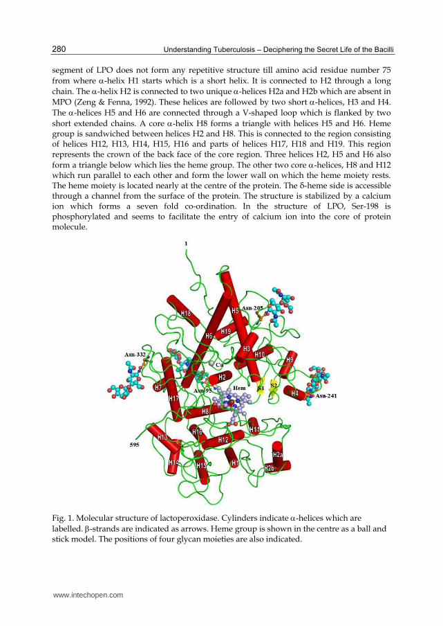
segment of LPO does not form any repetitive structure till amino acid residue number 75 from where -helix H1 starts which is a short helix. It is connected to H2 through a long chain. The -helix H2 is connected to two unique -helices H2a and H2b which are absent in MPO (Zeng & Fenna, 1992). These helices are followed by two short -helices, H3 and H4. The -helices H5 and H6 are connected through a V-shaped loop which is flanked by two short extended chains. A core -helix H8 forms a triangle with helices H5 and H6. Heme group is sandwiched between helices H2 and H8. This is connected to the region consisting of helices H12, H13, H14, H15, H16 and parts of helices H17, H18 and H19. This region represents the crown of the back face of the core region. Three helices H2, H5 and H6 also form a triangle below which lies the heme group. The other two core -helices, H8 and H12 which run parallel to each other and form the lower wall on which the heme moiety rests. The heme moiety is located nearly at the centre of the protein. The δ-heme side is accessible through a channel from the surface of the protein. The structure is stabilized by a calcium ion which forms a seven fold co-ordination. In the structure of LPO, Ser-198 is phosphorylated and seems to facilitate the entry of calcium ion into the core of protein molecule.
Fig. 1. Molecular structure of lactoperoxidase. Cylinders indicate -helices which are labelled. -strands are indicated as arrows. Heme group is shown in the centre as a ball and stick model. The positions of four glycan moieties are also indicated.
www.intechopen.com

Mammalian Heme Peroxidases and Mycobacterium tuberculosis
281
3.1 Heme moiety
The heme moiety in LPO is a derivative of protoporphyrin IX (Thanabal & La Mar, 1989) in which the methyl groups on pyrrole rings A and C are modified to allow formation of ester linkages with carboxylic groups of Glu258 and Asp108 respectively (Figure 2).
Fig. 2. The structure of the heme moiety showing a standard nomenclature. The two covalent linkages to protein are indicated.
The ferric iron atom is coordinated to four heme nitrogen atoms in a slightly distorted planar arrangement. The fifth coordination is provided by proximal His351 while on the sixth side a conserved water molecule W1 is located at a hydrogen bonded distance from the heme iron. The heme moiety is deeply buried inside the protein molecule while the heme cavity is surrounded by a number of -helices from three sides. The two β-strands, S1 and S2 are situated on the upper side of the opening to the heme cavity (Figure 1). Overall, the plane of heme protoporphyrin IX moiety is slightly distorted from planarity. The pyrrole rings A, C and D are essentially planar while pyrrole ring B is slightly distorted from planarity. The iron position is shifted slightly towards the proximal side. The carboxyl group of the pyrrole ring D propionate interacts with the guanidinium groups of Arg348 and Arg440. In contrast, the ring C propionate interacts with Asp112 Oδ2, Ala112 N and a water molecule.
3.2 Substrate specificity
The substrate binding site is formed on the distal heme side. In the native structure of LPO, the substrate-binding space is occupied by six water molecules W1, W2, W3, W4¸ W5 and W6 (Figure 3). In the resting state, W1 is linked to ferric iron at a hydrogen bonded distance.
www.intechopen.com

Understanding Tuberculosis – Deciphering the Secret Life of the Bacilli
282
When H2O2 is supplied, it expels W1 and forms the sixth coordination. On this side, His109 works as proton donor-acceptor residue as it is linked to a chain of water molecules that facilitate proton relay (Figure 4). When ligands bind to LPO in the substrate binding site on the distal heme side, it plays an essential role in the enzymatic action. The substrate-binding site is surrounded by heme moiety on one side while residues, His109, Phe113, Phe254 and Arg255 occupy the opposite side. The front end of the site consists of Gln105 while Glu258 supports it from below. The other wall is made up of residues, Phe381, Phe422, Gln423 and Pro424. The substrate-binding site is connected by a long channel formed by hydrophobic aromatic residues including Pro234, Pro236, Phe380, Phe381 and Phe254 on one side while Leu421, Phe422, Gln423 and Pro424 on the opposite (Figure 5). The length of substrate diffusion channel in LPO is approximately 22Å while its diameter is about 10Å. The substrate-binding site is connected to the surface of the protein through this diffusion channel.
Fe3+
Phe-254
W 5
W 1
Arg 255
Heme
W 6
W 4
W 3W 2
Gln 423
Pro 424
Phe 381
Phe 422
His 351
His 109
Glu 258
Gln 105
Phe 113
Proximal side
Distal side
Fig. 3. The structure of the substrate-binding site on the distal heme side in LPO and the positions of six conserved water molecules, W1, W2, W3, W4¸ W5 and W6 as observed in the unliganded structure. The proximal and distal sites have been indicated.
www.intechopen.com

Mammalian Heme Peroxidases and Mycobacterium tuberculosis
283
Fig. 4. Hydrogen bonded chain involving His351, Fe3+, W1, His109, W2, His266, W3, W4, W5 and W6 where W1 is hydrogen bonded to Fe3+ and W6 is located near the protein surface.
Fig. 5. A schematic representation of the diffusion channel and substrate-binding site in LPO drawn using PDB-ID: 3GC1.
3.3 Specificity for aromatic compounds
The substrate diffusion channel in LPO is long and surrounded by aromatic residues from all sides. As a result it allows the diffusion of small aromatic compounds from surface to the substrate binding site on the distal heme side. The aromatic compounds such as acetyl
www.intechopen.com

Understanding Tuberculosis – Deciphering the Secret Life of the Bacilli
284
salicylic acid (ASA) and salicylhydroxamic acid (SHA) have been shown to bind to LPO at the substrate-binding site (Figure 6) (Singh et al., 2009). It has been observed that the aromatic compounds which bind to LPO but do not expel the conserved water molecule, W1 from its original position as has been observed in the case of acetyl salicylic acid act as substrates (Figure 6A) while those that expel the conserved water molecule, W1 and coordinate directly with the heme iron act as inhibitors as found in the case of salicylhydroxamic acid (Figure 6B). Therefore, the mode of binding of substrates and inhibitors differ in terms of their positioning in the substrate-binding site.
(A) (B) Fig. 6. (A) Acetyl salicylic acid (ASA) bound to LPO as a substrate at the substrate-binding site (PDB-ID: 3GCL). The hydrogen bonded interactions are indicated by dotted lines. (B) Salicylhydroxamic acid (SHA) bound to LPO as an inhibitor in the substrate-binding site (PDB-ID: 3GCJ). The hydrogen bonded interactions are indicated by dotted lines.
3.4 Structure of the LPO complex with Isoniazid
Since LPO has been shown to bind to small aromatic compounds, the binding properties of LPO with INH were examined using surface plasmon resonance technique. The protein molecule was immobilized on the chip while INH was used as a solution in a mobile phase. These measurements gave a value of 1.1 10-6 M for the dissociation constant (Kd) for the binding of LPO with INH. In order to determine the interactions between LPO and INH, the crystal structure of the INH-bound LPO was determined (Singh et al., 2010). The structure of the LPO-INH complex showed that INH upon binding to LPO in the substrate-binding site on the distal heme side expelled four water molecules W2, W4, W5 and W6 from the substrate-binding site on the distal heme side. Two out of six conserved water molecules, W1 and W3 remained unperturbed. INH binds to LPO with an appropriate orientation as its pyridine ring nitrogen atom is at a hydrogen bonded distance from the conserved water molecule W1 (Figure 7). Therefore, when H2O2 is introduced into the solution containing LPO and INH, it expels the water molecule W1 and the reaction gets initiated. The product isonicotinyl radical is formed as described by
www.intechopen.com

Mammalian Heme Peroxidases and Mycobacterium tuberculosis
285
equations 1 to 3. This product is similar to that produced by Mycobacterium tuberculosis catalase peroxidase (MtCP) (Bertrand et al., 2004).
Fe3+
Phe 254
His 109
W 1
Arg 255
Heme
W 3′
Gln 423
Pro 424
Phe 381
Phe 422
His 351
Isoniazid
Fig. 7. Showing binding of INH to LPO as a substrate. The dotted lines indicate hydrogen bonds.
3.5 Complex of LPO with Pyrazinamide
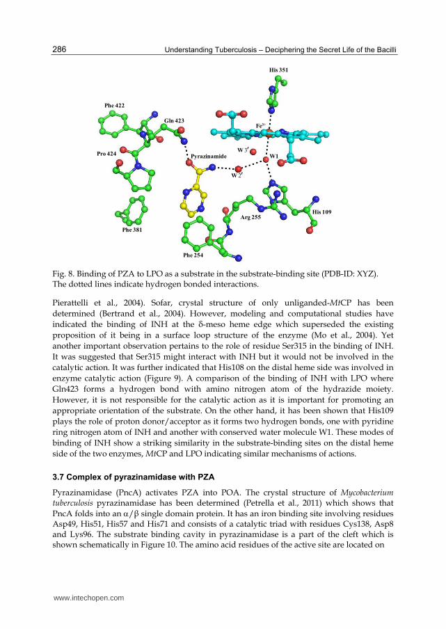
As revealed by binding studies using surface Plasmon resonance technique, pyrazinamide (PZA) has also been found to bind to LPO with a slightly lower affinity than that of INH (dissociation constant, Kd = 1.2 10-5 M). The structure determination of PZA-bound LPO revealed that it occupies a position in the centre of the substrate-binding site on the distal heme side. Upon binding to pyrazinamide, three water molecules, W4, W5 and W6 were expelled from the substrate-binding site. It retained three water molecules, W1, W2 and W3. The nearest nitrogen atom of PZA is about 5.5Å away from the oxygen atom of conserved water molecule W1. PZA and conserved water molecule W1 are separated from each other by another water molecule W2. The carboxamide nitrogen atom of PZA forms a hydrogen bond with W2 which in turn is hydrogen bonded at W1 (Figure 8). It reflects a slightly weaker affinity of PZA towards the position of W1. However, when H2O2 is introduced, it is expected to move closer to H2O2 and the product may be formed. The position occupied by PZA in the substrate-binding site appears to be suitable for the catalytic action by LPO. However, the nature of product is not characterized clearly.
3.6 Structure of catalase peroxidase
Mycobacterium tuberculosis catalase peroxidase (MtCP) is a dimeric bi-functional heme-dependent enzyme of molecular mass of 160 kDa. Its primary function is of catalase activity. However, its role as a peroxidase is well established and its peroxidative activity is comparable with those of other mono-functional heme peroxidases (Metcalfe et al., 2008;
www.intechopen.com
Understanding Tuberculosis – Deciphering the Secret Life of the Bacilli
286
Pro 424
Gln 423Fe3+
Phe 254
Phe 381
Arg 255
W1
Phe 422
His 351
His 109
Pyrazinamide
W 2
W 3
Fig. 8. Binding of PZA to LPO as a substrate in the substrate-binding site (PDB-ID: XYZ). The dotted lines indicate hydrogen bonded interactions.
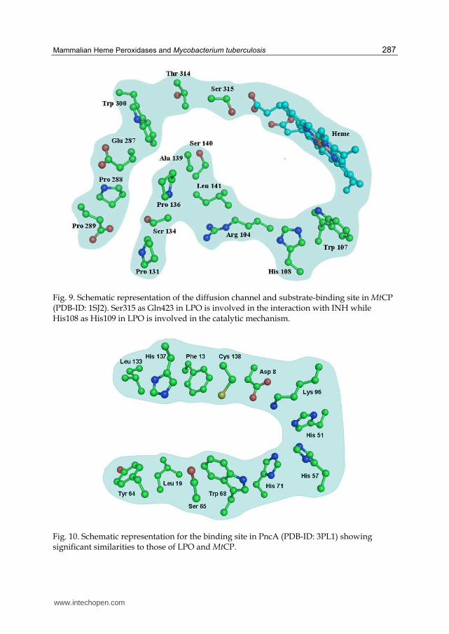
Pierattelli et al., 2004). Sofar, crystal structure of only unliganded-MtCP has been determined (Bertrand et al., 2004). However, modeling and computational studies have indicated the binding of INH at the δ-meso heme edge which superseded the existing proposition of it being in a surface loop structure of the enzyme (Mo et al., 2004). Yet another important observation pertains to the role of residue Ser315 in the binding of INH. It was suggested that Ser315 might interact with INH but it would not be involved in the catalytic action. It was further indicated that His108 on the distal heme side was involved in enzyme catalytic action (Figure 9). A comparison of the binding of INH with LPO where Gln423 forms a hydrogen bond with amino nitrogen atom of the hydrazide moiety. However, it is not responsible for the catalytic action as it is important for promoting an appropriate orientation of the substrate. On the other hand, it has been shown that His109 plays the role of proton donor/acceptor as it forms two hydrogen bonds, one with pyridine ring nitrogen atom of INH and another with conserved water molecule W1. These modes of binding of INH show a striking similarity in the substrate-binding sites on the distal heme side of the two enzymes, MtCP and LPO indicating similar mechanisms of actions.
3.7 Complex of pyrazinamidase with PZA
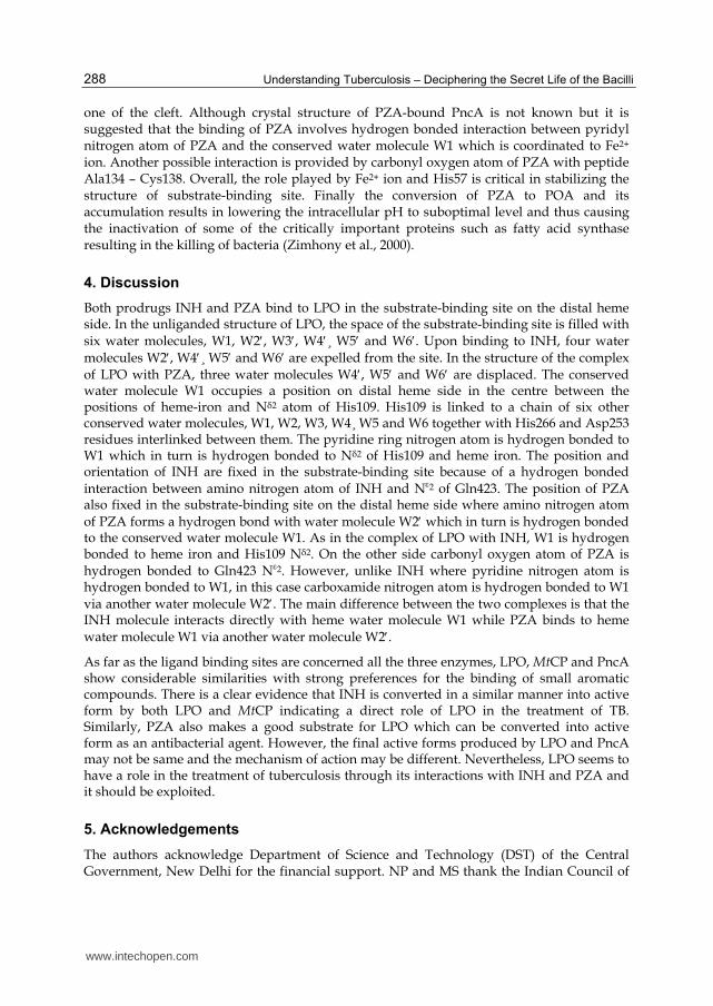
Pyrazinamidase (PncA) activates PZA into POA. The crystal structure of Mycobacterium tuberculosis pyrazinamidase has been determined (Petrella et al., 2011) which shows that PncA folds into an /β single domain protein. It has an iron binding site involving residues Asp49, His51, His57 and His71 and consists of a catalytic triad with residues Cys138, Asp8 and Lys96. The substrate binding cavity in pyrazinamidase is a part of the cleft which is shown schematically in Figure 10. The amino acid residues of the active site are located on
www.intechopen.com
Mammalian Heme Peroxidases and Mycobacterium tuberculosis
287
Fig. 9. Schematic representation of the diffusion channel and substrate-binding site in MtCP (PDB-ID: 1SJ2). Ser315 as Gln423 in LPO is involved in the interaction with INH while His108 as His109 in LPO is involved in the catalytic mechanism.
Fig. 10. Schematic representation for the binding site in PncA (PDB-ID: 3PL1) showing significant similarities to those of LPO and MtCP.
www.intechopen.com
Understanding Tuberculosis – Deciphering the Secret Life of the Bacilli
288
one of the cleft. Although crystal structure of PZA-bound PncA is not known but it is suggested that the binding of PZA involves hydrogen bonded interaction between pyridyl nitrogen atom of PZA and the conserved water molecule W1 which is coordinated to Fe2+ ion. Another possible interaction is provided by carbonyl oxygen atom of PZA with peptide Ala134 – Cys138. Overall, the role played by Fe2+ ion and His57 is critical in stabilizing the structure of substrate-binding site. Finally the conversion of PZA to POA and its accumulation results in lowering the intracellular pH to suboptimal level and thus causing the inactivation of some of the critically important proteins such as fatty acid synthase resulting in the killing of bacteria (Zimhony et al., 2000).
4. Discussion
Both prodrugs INH and PZA bind to LPO in the substrate-binding site on the distal heme side. In the unliganded structure of LPO, the space of the substrate-binding site is filled with six water molecules, W1, W2, W3, W4¸ W5 and W6. Upon binding to INH, four water molecules W2, W4¸ W5 and W6 are expelled from the site. In the structure of the complex of LPO with PZA, three water molecules W4, W5 and W6 are displaced. The conserved water molecule W1 occupies a position on distal heme side in the centre between the positions of heme-iron and Nδ2 atom of His109. His109 is linked to a chain of six other conserved water molecules, W1, W2, W3, W4¸ W5 and W6 together with His266 and Asp253 residues interlinked between them. The pyridine ring nitrogen atom is hydrogen bonded to W1 which in turn is hydrogen bonded to Nδ2 of His109 and heme iron. The position and orientation of INH are fixed in the substrate-binding site because of a hydrogen bonded interaction between amino nitrogen atom of INH and N2 of Gln423. The position of PZA also fixed in the substrate-binding site on the distal heme side where amino nitrogen atom of PZA forms a hydrogen bond with water molecule W2 which in turn is hydrogen bonded to the conserved water molecule W1. As in the complex of LPO with INH, W1 is hydrogen bonded to heme iron and His109 Nδ2. On the other side carbonyl oxygen atom of PZA is hydrogen bonded to Gln423 N2. However, unlike INH where pyridine nitrogen atom is hydrogen bonded to W1, in this case carboxamide nitrogen atom is hydrogen bonded to W1 via another water molecule W2. The main difference between the two complexes is that the INH molecule interacts directly with heme water molecule W1 while PZA binds to heme water molecule W1 via another water molecule W2. As far as the ligand binding sites are concerned all the three enzymes, LPO, MtCP and PncA show considerable similarities with strong preferences for the binding of small aromatic compounds. There is a clear evidence that INH is converted in a similar manner into active form by both LPO and MtCP indicating a direct role of LPO in the treatment of TB. Similarly, PZA also makes a good substrate for LPO which can be converted into active form as an antibacterial agent. However, the final active forms produced by LPO and PncA may not be same and the mechanism of action may be different. Nevertheless, LPO seems to have a role in the treatment of tuberculosis through its interactions with INH and PZA and it should be exploited.
5. Acknowledgements
The authors acknowledge Department of Science and Technology (DST) of the Central Government, New Delhi for the financial support. NP and MS thank the Indian Council of
www.intechopen.com
Mammalian Heme Peroxidases and Mycobacterium tuberculosis
289
Medical Research, New Delhi and Council of Scientific and Industrial Research, New Delhi for the award of fellowships. TPS thanks Department of Biotechnology (DBT) for the award of Distinguished Biotechnology Research Professorship.
6. References
Bertrand, T., Eady, N. A., Jones, J. N., Jesmin, Nagy, J. M., Jamart-Gregoire, B., Raven, E. L., & Brown, K. A. (2004). Crystal structure of Mycobacterium tuberculosis catalase-peroxidase. Journal of Biological Chemistry, Vol.279, No.37, (September 2004), pp. 38991-38999, ISSN 0021-9258
Carlstrom, A. (1969). Lactoperoxidase: Identification of multiple molecular forms and their interrelationships. Acta Chemica Scandinavica, Vol.23, No.1, pp. 171-184, ISSN 0001-5393
Cavalieri, E. L., Stack, D. E., Devanesan, P. D., Todorovic, R., Dwivedy, I., Higginbotham, S., Johansson, S. L., Patil, K. D., Gross, M. L., Gooden, J. K., Ramanathan, R., Cerny, R. L. & Rogan, E. G. (1997). Molecular origin of cancer: catechol estrogen-3,4-quinones as endogenous tumor initiators. Proceedings of the National Academy of Sciences, USA, Vol.94, No.20, (September 1997), pp. 10937-10942, ISSN 0027-8424
Ciaccio, C., De Sanctis, G., Marini, S., Sinibaldi, F., Santucci, R., Arcovito, A., Bellelli, A., Ghibaudi, E., Ferrari, R. P., & Coletta, M. (2004). Proton linkage for CO binding and redox properties of bovine lactoperoxidase. Biophysical Journal, Vol.86, No.1 Pt 1, (January 2004), pp. 448-454, ISSN 0006-3495
Doerge, D. R. & Decker, C. J. (1994). Inhibition of peroxidase-catalyzed reactions by arylamines: mechanism for the anti-thyroid action of sulfamethazine. Chemical
Research in Toxicology, Vol.7, No.2, (March-April 1994), pp. 164-169, ISSN 0893-228X Dolphin, D., Muljiani, Z., Rousseau, K., Borg, D. C., Fajer, J., & Felton, R. H. (1973). The
chemistry of porphyrin pi-cations. Annals of the New York Academy of Sciences, Vol.206, (October 1973), pp. 177-200, ISSN 0077-8923
Ferrari, R. P., Laurenti, E., Casella, L., & Poli, S. (1993). Oxidation of catechols and catecholamines by HRP and LPO: ESR spin stabilization approach combined with optical methods. Spectrochimica Acta, Vol.49A, pp. 1261–1267, ISSN 1386-1425
Ghibaudi, E. M., Laurenti, E., Beltramo, P. & Ferrari, R. P. (2000). Can estrogenic radicals, generated by lactoperoxidase, be involved in the molecular mechanism of breast carcinogenesis? Redox Report, Vol.5, No.4, pp. 229-235, ISSN 1351-0002
Hoogendoorn, H., Piessens, J. P., Scholtes, W., & Stoddard, L. A. (1977) Hypothiocyanite ion; the inhibitor formed by the system lactoperoxidase-thiocyanate-hydrogen peroxide. I. Identification of the inhibiting compound. Caries Research, Vol.11, No.2, pp. 77-84, ISSN 0008-6568
Konno, K., Feldmann, F. M., & McDermott, W. (1967). Pyrazinamide susceptibility and amidase activity of tubercle bacilli. American Review of Respiratory Diseases, Vol.95, (March 1967), pp 461-469, ISSN 0003-0805
Kussendrager, K. D, & van Hooijdonk, A. C. (2000). Lactoperoxidase: physico-chemical properties, occurrence, mechanism of action and applications. British Journal of
Nutrition, Vol.84, Suppl 1, (November 2000), pp. S19-25, ISSN 0007-1145
www.intechopen.com

Understanding Tuberculosis – Deciphering the Secret Life of the Bacilli
290
Metcalfe, C., Macdonald, I. K., Murphy, E. J., Brown, K. A., Raven, E. L., & Moody, P. C. (2008).The tuberculosis prodrug isoniazid bound to activating peroxidases. Journal
of Biological Chemistry, Vol.283, No.10 (March 2008), pp. 6193-6200, ISSN 0021-9258 Metodiewa, D., Reszka, K. & Dunford, H. B. (1989). Evidence for a peroxidatic oxidation of
norepinephrine, a catecholamine, by lactoperoxidase. Biochemical and Biophysical
Research Communications, Vol.160, No.3, (May 1989), pp.1183-1188, ISSN 0006-291X Metodiewa, D., Reszka, K. & Dunford, H. B. (1989). Oxidation of the substituted catechols
dihydroxyphenylalanine methyl ester and trihydroxyphenylalanine by lactoperoxidase and its compounds. Archives of Biochemistry and Biophysics, Vol.274, No.2, (November 1989), pp. 601-608, ISSN 0003-9861
Mikola, H., Waris, M., & Tenovuo, J. (1995). Inhibition of herpes simplex virus type 1, respiratory syncytial virus and echovirus type 11 by peroxidase-generated hypothiocyanite. Antiviral Research, Vol.26, No.2, (March 1995), pp. 161-171, ISSN 0166-3542
Mo, L., Zhang, W., Wang, J., Weng, X. H., Chen, S., Shao, L. Y., Pang, M. Y. & Chen, Z. W. (2004). Three-dimensional model and molecular mechanism of Mycobacterium tuberculosis catalase-peroxidase (KatG) and isoniazid-resistant KatG mutants. Microbial Drug Resistance, Vol.10, No.4, 269-279, ISSN 1076-6294
Monzani, E., Gatti, A. L., Profumo, A., Casella, L. & Gullotti, M. (1997). Oxidation of phenolic compounds by lactoperoxidase. Evidence for the presence of a low-potential compound II during catalytic turnover. Biochemistry, (February 1997), Vol.36, No.7, pp. 1918-1926, ISSN 0006-2960
Oram, J. D., & Reiter, B. (1966). The inhibition of streptococci by lactoperoxidase, thiocyanate and hydrogen peroxide. The oxidation of thiocyanate and the nature of the inhibitory compound. Biochemical Journal, (August 1966), Vol.100, No.2, pp. 382-388, ISSN 0264-6021
Paul, K. G., & Ohlsson, P. I. (1985). The chemical structure of lactoperoxidase, In: The
Lactoperoxidase System, Chemistry and Biological Significance, Pruitt, K. M., Tenovuo, J. O. (ed.), pp. 15-29, Marcel Dekker, New York, ISBN 0824772989
Petrella, S., Gelus-Ziental, N., Maudry, A., Laurans, C., Boudjelloul, R., & Sougakoff, W. (2011). Crystal structure of the pyrazinamidase of Mycobacterium tuberculosis: insights into natural and acquired resistance to pyrazinamide. PLoS One, Vol.6, No.1, (January 2011), pp. e15785, ISSN 1932-6203
Pierattelli, R., Banci, L., Eady, N. A., Bodiguel, J., Jones, J. N., Moody, P. C., Raven, E. L., Jamart-Gregoire, B., & Brown, K. A. (2004). Enzyme-catalyzed mechanism of isoniazid activation in class I and class III peroxidases. Journal of Biological
Chemistry, Vol.279, No.37, (September 2004), pp. 39000-39009. ISSN 0021-9258 Pourtois, M., Binet, C., Van Tieghem, N., Courtois, P., Vandenabbeele, A., Thiry, L. (1990).
Inhibition of HIV infectivity by lactoperoxidase-produced hypothiocyanite. Journal
de biologie buccale, Vol.18, No.4, (December 1990), pp. 251-253. ISSN 0301-3952 RamaKrishna, N. V., Li, K. M., Rogan, E. G., Cavalieri, E. L., George, M., Cerny, R. L. &
Gross, M. L. (1993). Adducts of 6-methylbenzo[a]pyrene and 6-fluorobenzo[a]pyrene formed by electrochemical oxidation in the presence of
www.intechopen.com

Mammalian Heme Peroxidases and Mycobacterium tuberculosis
291
deoxyribonucleosides. Chemical Research in Toxicology, Vol.6, No.6, (November-December 1993), pp. 837-845, ISSN 0893-228X
Reiter, B., & Harrnulv, G. (1984). Lactoperoxidase antibacterial system: natural occurrence, biological functions and practical applications. Journal of Food Protection, Vol.47, (Month 1984), pp. 724-732, ISSN 0362-028X
Reiter, B., & Perraudin, J. P. (1991). Lactoperoxidase: biological functions. In: Peroxidases in
Chemistry and Biology. pp. 143-180, Boca Raton: CRC Press, ISBN 0849369630 Sawatdee, S., Worakul, N., & Srichana, T. (2006) Preparation of isoniazid as dry powder
formulations for inhalation by physical mixing and spray drying. Malaysian Journal
of Pharmaceutical Sciences, Vol.4, No.1, pp. 43-63, ISSN 2180-429X Shin, K., Wakabayashi, H., Yamauchi, K., Teraguchi, S., Tamura, Y., Kurokawa, M. &
Shiraki, K. (2005). Effects of orally administered bovine lactoferrin and lactoperoxidase on influenza virus infection in mice. Journal of Medical Microbiology, Vol.54, No.8, (August 2005), pp. 717-723, ISSN 0022-2615
Singh, A. K., Kumar, R. P., Pandey, N., Singh, N., Sinha, M., Bhushan, A., Kaur, P., Sharma, S., & Singh, T. P. (2010) Mode of binding of the tuberculosis prodrug isoniazid to heme peroxidases: binding studies and crystal structure of bovine lactoperoxidase with isoniazid at 2.7 Å resolution. Journal of Biological Chemistry, Vol.285, No.2, (January 2010), pp. 1569-1576, ISSN 0021-9258
Singh, A. K., Singh, N., Sharma, S., Singh, S. B., Kaur, P., Bhushan, A., Srinivasan, A. & Singh, T. P. (2008). Crystal structure of lactoperoxidase at 2.4 Å resolution. Journal of
Molecular Biology, Vol.376, No.4, (February 2008), pp. 1060-1075, ISSN 0022-2836 Singh, A. K., Singh, N., Sinha, M., Bhushan, A., Kaur, P., Srinivasan, A., Sharma, S. & Singh,
T. P. (2009). Binding modes of aromatic ligands to mammalian heme peroxidases with associated functional implications: crystal structures of lactoperoxidase complexes with acetylsalicylic acid, salicylhydroxamic acid, and benzylhydroxamic acid. Journal of Biological Chemistry, Vol.284, No.30, (July 2009), pp. 20311-20318, ISSN 0021-9258
Sipe, H. J., Jr., Jordan, S. J., Hanna, P. M. & Mason, R. P. (1994). The metabolism of 17 beta-estradiol by lactoperoxidase: a possible source of oxidative stress in breast cancer. Carcinogenesis, (November 1994), Vol.15, No.11, pp. 2637-2643, ISSN 0143-3334
Tenovuo, J., Makinen, K. K. & Sievers, G. (1985). Antibacterial effect of lactoperoxidase and myeloperoxidase against Bacillus cereus. Antimicrobial Agents and Chemotherapy, (January 1985) Vol.27, No.1, pp. 96-101, ISSN 0066-4804
Thanabal, V., & La Mar, G. N. (1989). A nuclear Overhauser effect investigation of the molecular and electronic structure of the heme crevice in lactoperoxidase. Biochemistry. Vol.28, No.17, (August 1989), pp. 7038-7044, ISSN 0006-2960
Wolfson, L. M., & Sumner, S. S. (1993). Antibacterial activity of the lactoperoxidase system: a review. Journal of Food Protection, Vol.56, pp. 887-892, ISSN 0362-028X
Zeng, J., & Fenna, R. E. (1992). X-ray crystal structure of canine myeloperoxidase at 3 Å resolution. Journal of Molecular Biology, Vol.226, No.1, (July 1992), pp. 185-207, ISSN 0022-2836
www.intechopen.com

Understanding Tuberculosis – Deciphering the Secret Life of the Bacilli
292
Zhang, H., and Dunford, H. B. (1993). Hammett pσ correlation for reactions of lactoperoxidase compound II with phenols. Canadian Journal of Chemistry, Vol.71, No.12, pp. 1990-1994, ISSN 0008-4042
Zhang, Y., Heym, B., Allen, B., Young, D., & Cole, S. (1992). The catalase-peroxidase gene and isoniazid resistance of Mycobacterium tuberculosis. Nature, Vol.358, No.6387, (August 1992), pp. 591-593, ISSN 0028-0836
Zimhony, O., Cox, J. S., Welch, J. T., Vilcheze, C., & Jacobs, W. R. Jr. (2000). Pyrazinamide inhibits the eukaryotic-like fatty acid synthetase I (FASI) of Mycobacterium
tuberculosis. Nature Medicine, Vol.6, No.9, (September 2000), pp. 1043-1047, ISSN 1061-4036
www.intechopen.com

Understanding Tuberculosis - Deciphering the Secret Life of theBacilliEdited by Dr. Pere-Joan Cardona
ISBN 978-953-307-946-2Hard cover, 334 pagesPublisher InTechPublished online 17, February, 2012Published in print edition February, 2012
InTech EuropeUniversity Campus STeP Ri Slavka Krautzeka 83/A 51000 Rijeka, Croatia Phone: +385 (51) 770 447 Fax: +385 (51) 686 166www.intechopen.com
InTech ChinaUnit 405, Office Block, Hotel Equatorial Shanghai No.65, Yan An Road (West), Shanghai, 200040, China
Phone: +86-21-62489820 Fax: +86-21-62489821
Mycobacterium tuberculosis, as recent investigations demonstrate, has a complex signaling expression, whichallows its close interaction with the environment and one of its most renowned properties: the ability to persistfor long periods of time under a non-replicative status. Although this skill is well characterized in other bacteria,the intrinsically very slow growth rate of Mycobium tuberculosis, together with a very thick and complex cellwall, makes this pathogen specially adapted to the stress that could be generated by the host against them. Inthis book, different aspects of these properties are displayed by specialists in the field.
How to referenceIn order to correctly reference this scholarly work, feel free to copy and paste the following:
Amit K. Singh, Nisha Pandey, Mau Sinha, Sujata Sharma and Tej P. Singh (2012). Mammalian HemePeroxidases and Mycobacterium tuberculosis, Understanding Tuberculosis - Deciphering the Secret Life of theBacilli, Dr. Pere-Joan Cardona (Ed.), ISBN: 978-953-307-946-2, InTech, Available from:http://www.intechopen.com/books/understanding-tuberculosis-deciphering-the-secret-life-of-the-bacilli/mammalian-hemeperoxidases-and-mycobacterium-tuberculosis

© 2012 The Author(s). Licensee IntechOpen. This is an open access articledistributed under the terms of the Creative Commons Attribution 3.0License, which permits unrestricted use, distribution, and reproduction inany medium, provided the original work is properly cited.


![Burst ORAM: Minimizing ORAM Response Times for Bursty ...elaine/docs/burstoram.pdf · due to physical tampering [6, 15, 16, 20]. Since on-chip trusted cache is expensive, such ORAM](https://static.fdocuments.us/doc/165x107/5fc7308991682453423feb36/burst-oram-minimizing-oram-response-times-for-bursty-elainedocs-due-to.jpg)


![Three-Party ORAM for Secure Computation · ORAM for Secure Computation (SC-ORAM) [17], which is a protocol that securely implements a RAM functionality, i.e. given a secret-sharing](https://static.fdocuments.us/doc/165x107/5f0ac6817e708231d42d4814/three-party-oram-for-secure-computation-oram-for-secure-computation-sc-oram-17.jpg)


![SCORAM: Oblivious RAM for Secure Computationhubert/ccs14a.pdf · ORAM by Shi et al. [30] performs the same as a naively implemented Path ORAM [34], although Path ORAM is asymptotically](https://static.fdocuments.us/doc/165x107/5f0ac9767e708231d42d56e3/scoram-oblivious-ram-for-secure-computation-hubert-oram-by-shi-et-al-30.jpg)
