Malignant lymphoma associated with internal fixation of a fractured tibia
3
Malignant Lymphoma Associated with Internal Fixation of a Fractured Tibia IAN McDONALD MB, CHB, FRCS((;LAS) A case of malignant lymphoma in association with the platimg of a fractured tibia 17 years previously is presented. The literature is briefly reviewed, and attention is drawn to the reported association between tumor and internal fixation in animals. Cancer 48:1009-1011, 1981. N ASSOCIATION between internal fixation of frac- A tures and the subsequent development of neo- plasia has only occasionally been described. A case is reported here. The Vitallium plate and screws were not analyzed and the degree of corrosion was not recorded. Case Report A 48-year-old farmer was seen in July 1978. Seventeen years before, his left tibia had been plated following a frac- ture. The bone had healed uneventfully, and the patient had had no symptoms that could be traced to the leg until two years prior to presentation. At that time, slight aching had been experienced in the area of the plate after strenuous work or exercise. The discomfort had gradually increased, and night sweats and general malaise appeared. An x-ray film (Fig. 1) showed a well-healed fracture of the tibia, with slight osteoporosis and cortical irregularity around the plate. A diagnosis of low-grade infection was made and removal of the plate was recommended. At operation, the surrounding soft tissues appeared friable and necrotic. The plate and screws remained firmly fixed to the bone, and several screw heads had to be sheared off to allow removal of the plate. Biopsy was done of bone and adjacent soft tissue and initial histology showed a malignant lymphoma, with the tumor actively infiltrating bone (Fig. 2). A subsequent bone marrow biopsy from the iliac crest showed diffuse involvement of the marrow with malignant lymphoma cells of the histiocytic type (Fig. 3). This repre- sented a “worst type of” tumor histology; in view of this, the patient received local radiotherapy to the leg, CHOPP (cyclophosphamide, Adriamycin, vincristine, and pred- nisolone) plus prophylactic intrathecal rnethotrexate and cranial irradiation. He is at present asymptomatic. From the Departmen1 of Orthopaedic Surgery. Christchurch Hos- The author thanks A. B. Mackenzie for permission to report his Accepted for publication July 17. 1980. pital. Christchurch, New Zealand. case. FI~,. I. X-ray film showing cortical irregularity and periosteal reaction overlying the plate at the site of tumor involvement. 000~-543>(/8I/0X51/1009 $0.65 0 .4merican Cancer Society 1009
-
Upload
ian-mcdonald -
Category
Documents
-
view
213 -
download
0
Transcript of Malignant lymphoma associated with internal fixation of a fractured tibia

Malignant Lymphoma Associated with Internal Fixation of a Fractured Tibia
IAN McDONALD MB, CHB, FRCS((;LAS)
A case of mal ignant lymphoma in association wi th t h e platimg of a fractured t ibia 17 years previously is presented. T h e l i terature is briefly reviewed, a n d a t ten t ion is d r a w n to t h e reported association between t u m o r and internal fixation i n animals.
Cancer 48:1009-1011, 1981.
N ASSOCIATION between internal fixation of frac- A tures and the subsequent development of neo- plasia has only occasionally been described. A case is reported here.
The Vitallium plate and screws were not analyzed and the degree of corrosion was not recorded.
Case Report
A 48-year-old farmer was seen in July 1978. Seventeen years before, his left tibia had been plated following a frac- ture. The bone had healed uneventfully, and the patient had had no symptoms that could be traced to the leg until two years prior to presentation. At that time, slight aching had been experienced in the area of the plate after strenuous work or exercise. The discomfort had gradually increased, and night sweats and general malaise appeared.
An x-ray film (Fig. 1) showed a well-healed fracture of the tibia, with slight osteoporosis and cortical irregularity around the plate. A diagnosis of low-grade infection was made and removal of the plate was recommended.
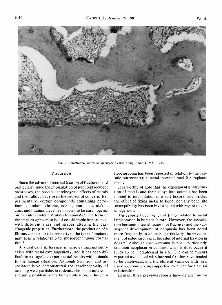
At operation, the surrounding soft tissues appeared friable and necrotic. The plate and screws remained firmly fixed to the bone, and several screw heads had to be sheared off to allow removal of the plate. Biopsy was done of bone and adjacent soft tissue and initial histology showed a malignant lymphoma, with the tumor actively infiltrating bone (Fig. 2 ) .
A subsequent bone marrow biopsy from the iliac crest showed diffuse involvement of the marrow with malignant lymphoma cells of the histiocytic type (Fig. 3 ) . This repre- sented a “worst type of” tumor histology; in view of this, the patient received local radiotherapy to the leg, C H O P P (cyclophosphamide, Adriamycin, vincristine, and pred- nisolone) plus prophylactic intrathecal rnethotrexate and cranial irradiation. H e is at present asymptomatic.
From the Departmen1 of Orthopaedic Surgery. Christchurch Hos-
The author thanks A . B. Mackenzie for permission to report his
Accepted for publication July 17. 1980.
pital. Christchurch, New Zealand.
case. F I ~ , . I . X-ray film showing cortical irregularity and periosteal reaction overlying the plate at the site of tumor involvement.
000~-543>(/8I/0X51/1009 $0.65 0 .4merican Cancer Society
1009
1010 CANCER September 15 1981 Vol. 48
FIG. 2. Intertrabecular spaces occupied by infiltrating tumor (H & E, ~ 2 5 ) .
Discussion
Since the advent of internal fixation of fractures, and particularly since the implantation ofjoint replacement prostheses, the possible carcinogenic effects of metals and their alloys have been the subject of concern. Ex- perimentally, certain compounds containing beryl- lium, cadmium, chrome, cobalt, iron, lead, nickel, zinc, and titanium have been shown to be carcinogenic on parenteral administration to animals.8 The form of the implant appears to be of considerable importance, with different sizes and shapes altering the car- cinogenic properties. Furthermore, the production of a fibrous capsule, itself a property of the type of implant, may bear a relationship to subsequent tumor forma- tion.2
A significant difference in species susceptibility exists with metal carcinogenicity, and it has been dif- ficult to extrapolate experimental results with animals to the human situation. Although Swanson and as- sociates!' have demonstrated the carcinogenicity of total hip wear particles in rodents, this is not now con- sidered a problem in the human situation, although a
fibrosarcoma has been reported in relation to the cap- sule surrounding a metal-to-metal total hip replace- ment.'
It is worthy of note that the experimental introduc- tion of metals and their alloys into animals has been limited to implantation into soft tissues, and neither the effect of fixing metal to bone, nor any bone site susceptibility has been investigated with regard to car- cinogenesis.
The reported occurrence of tumor related to metal implantation in humans is rare. However, the associa- tion between internal fixation of fractures and the sub- sequent development of neoplasia has been noted more frequently in animals, particularly the develop- ment of osteosarcoma at the sites of internal fixation in dog^.^*^ Although osteosarcoma is not a particularly common neoplasm in canines, when it does occur it tends to be metaphyseal in site. The canine tumors reported associated with internal fixation have tended to be diaphyseal, and therefore at variance with their usual location, giving supportive evidence for a causal relationship.
In man, three previous reports have. detailed an as-

No. 6 MALIGNANT LYMPHOMA A N D INTERNAL FIXATION . McDonald 1011
FIG. 3. Anaplastic large-celled tumor with ovoid nuclei showing prominent nucleoli (H & E, x 100).
sociation between internal fixation of fractures and subsequent tumor formation. McDougal16 described a Ewing-like sarcoma that developed at the site of plat- ing of a fractured humerus after a 30-year interval. Dube and Fisher4 described a hemangioendothelioma that occurred after internal fixation of a fractured tibia at the end of a 30-year period, and Delgado3 reported an osteosarcoma that developed three years after internal fixation of a fractured tibia.
In this case, it is impossible to say whether the lymphoma arose in the tibia and subsequently became generalized, or whether an initially diffuse lymphoma presented first in the tibia at the site of bone plating. However, it is very unlikely that a generalized histio- cytic lymphoma would, untreated, have been as- sociated with a survival of over two years from onset of symptoms until diagnosis, a feature that indicates an initially localized tumor. However, with regard to the number of metallic implants that have been placed in humans and left in situ over considerable periods of
time, the association with tumor formation continues to be rare.
REFERENCES
I . Arden GI], Ansell BM. The Surgical Management of Juvenile Chronic Polyai-thritis. London: Academic Press, 1978; 270.
2. Bates R, Klein M. Importance of a smooth surface in carcino- genesis by plastic film. J N N / / Crrwcer Ins/ 1966; 37:145-150.
3 . Delgado ER. Sarcoma following a surgically treated fractured tibia. Clin Ortliop 1958; 12:315-318.
4. Dube VE, Fisher DE. Haemangioendothelioma of the leg fol- lowing metallic fixation of the tibia. Comer 1972; 30:1260- 1266.
5 . Harrison JW, McLain DL, Hohn RB, Wilson GP, Chalman JA, MacGowan KN. Osteosarcoma associated with metallic implants. Report of two cases in dogs. Clin Orthop 1976; 116:253-257.
6 . McDougall A. Malignant tumour at the site of bone plating. J Bone Join1 S u q (BY) 1956; 38:709-713.
7. Sinibaldi K, Rosen H, Liu SK, deAngelis M. Tumours as- sociated with metallic implants in animals. Clin Orthop 1976;
8 . Sunderman FW Jr. Metal carcinogenesis in experimental animals. Food Cosmet Toxicol 1971; 9:105- 120.
9. Swanson SAV, Freeman MAR, Heath JC. Laboratory tests on total joint replacement prostheses. J Bone Joinr Surg (Br) 1973; 55:759- 773.
118~257-266.