
MALDI-TOF: A Rapid Identification of Dairy Pathogens
89
St. Cloud State University theRepository at St. Cloud State Culminating Projects in Biology Department of Biology 7-2017 MALDI-TOF: A Rapid Identification of Dairy Pathogens Nusrat Jahan St. Cloud State University, [email protected] Follow this and additional works at: hps://repository.stcloudstate.edu/biol_etds is esis is brought to you for free and open access by the Department of Biology at theRepository at St. Cloud State. It has been accepted for inclusion in Culminating Projects in Biology by an authorized administrator of theRepository at St. Cloud State. For more information, please contact [email protected]. Recommended Citation Jahan, Nusrat, "MALDI-TOF: A Rapid Identification of Dairy Pathogens" (2017). Culminating Projects in Biology. 26. hps://repository.stcloudstate.edu/biol_etds/26
Transcript of MALDI-TOF: A Rapid Identification of Dairy Pathogens

St. Cloud State UniversitytheRepository at St. Cloud State
Culminating Projects in Biology Department of Biology
7-2017
MALDI-TOF: A Rapid Identification of DairyPathogensNusrat JahanSt. Cloud State University, [email protected]
Follow this and additional works at: https://repository.stcloudstate.edu/biol_etds
This Thesis is brought to you for free and open access by the Department of Biology at theRepository at St. Cloud State. It has been accepted forinclusion in Culminating Projects in Biology by an authorized administrator of theRepository at St. Cloud State. For more information, please [email protected].
Recommended CitationJahan, Nusrat, "MALDI-TOF: A Rapid Identification of Dairy Pathogens" (2017). Culminating Projects in Biology. 26.https://repository.stcloudstate.edu/biol_etds/26

MALDI-TOF: A Rapid Identification of Dairy Pathogens
by
Nusrat Annie Jahan
A Thesis
Submitted to the Graduate Faculty of
St. Cloud State University
In Partial Fulfillment of the Requirements
for the Degree of
Master of Science
in Cellular and Molecular Biology
July, 2017
Thesis Committee:
Ryan C. Fink, Chairperson
Matthew P. Davis
Omar Al-Azzam

2
Abstract
The proposed research study is a field validation study to benchmark against proven
methods, a new methodology for the detection of microorganisms (Matrix-Assisted Laser
Desorption Ionization Time of Flight Mass Spectrometry or MALDI-ToF) isolated from dairy
farm and critical for safety and quality. The MALDI-TOF is a relatively new molecular
technique extremely advantageous in terms of cost effectiveness, sample preparation easiness,
turn-around time and result analysis accessibility. Although already successfully deployed in
clinical diagnostic, it has not been evaluated for agricultural applications yet. In the dairy
industry, Mastitis causes the most financial loss and a rapid diagnostic method as MALDI-TOF,
will assist in the control and prevention program of mastitis, in addition to the sanitation and
safety level of the dairy farms and processing facility. In the present study, we prospectively
compared MALDI-TOF MS to the conventional 16S rRNA sequencing method for the
identification of environmental mastitis isolates (481) and thermoduric isolates of pasteurized
milk (248). Among the 481 environmental isolates, 454 (94.4%) were putatively identified to the
genus level by MALDI-TOF MS and 426 (88.6%) were identified to the species level, but no
reliable identification was obtained for 17 (3.5%), and 27 (5.6%) discordant results were
identified. Future studies can help to overcome the limitation of MALDI database and additional
sample preparation steps might help to reduce the number of discordance in identification. In
conclusion, our results show that MALDI-TOF MS is a fast and reliable technique which has the
potential to replace conventional identification methods for most dairy pathogens, routinely
isolated from the milk and dairy products. Thus it’s adoption will strengthen the capacity,
quality, and possibly the scope of diagnostic services to support the dairy industry.
3
Acknowledgement
This work is supported by the grant form dairy industries in Minnesota and Veterinary
Diagnostic Lab (VDL). We are grateful to VDL (College of veterinary medicine), Udder Health
Lab (UHL) and Department of Food Science and Nutrition, University of Minnesota for their
technical assistance. Special thanks to Dr. Sandra Godden and Jennifer Timmerman for
conducting a major part of the study and providing samples and results. I appreciate my fellow
graduate students; Billie Johnson, Cassey Kipping, Rene Martin, Wesley Davis, Rebecca Jensch,
Sandra hinz and Miezan Echimane, who guided me and assisted in the lab and related
coursework. I would further like to thank Mary Norbeck, Sadhana Bom, Mathew Yang, Ryan
Wolfe, Megan Stein, Megan Jones, Brittany Campion, and Kavitha Gobalan, for their assistance
in the sample preparation and conducting related experiments of the study. I also thank Professor
Louise Millis for her constant guidance and inspiration. A special thanks to my graduate
committee members: Dr. Matthew P. Davis and Dr. Omar Al-Azzam for all of their knowledge
and assistance. Finally I would like to extend a heartfelt thanks to my mentor Dr. Ryan C. Fink
for all his guidance and moral support during the study. I further thank him for his confidence in
me that I will be able to finish this extensive research work. Last but not the least, I would like to
thank my husband and my parents for believing in me and extending their constant support
towards my study and career goals.

4
Table of Contents
Chapter Page
I: Literature Review ............................................................................................................9
Mastitis ....................................................................................................................9
Mastitis Physiology ...............................................................................................10
Pathogens ..............................................................................................................11
Threat to Animal and Human health .....................................................................12
Effect on Milk Composition .................................................................................14
Effect on Dairy Products .......................................................................................15
Impact on Economy of Dairy Industry .................................................................16
Mastitis Impact across the World .........................................................................17
Mastitis Impact in the U.S ....................................................................................19
Importance of Rapid Diagnosis ............................................................................21
Biochemical Testing .............................................................................................22
16S rRNA Sequencing ..........................................................................................23
MALDI-TOF MS System .....................................................................................25
Objective and Hypothesis .....................................................................................29
II: Materials and Methods .................................................................................................30
Sample Collection .................................................................................................30
Environmental Isolates ..........................................................................................30
Thermoduric Isolates ............................................................................................31
Stocking and Storage of Isolates ...........................................................................34
Preparation of BHI Broth ......................................................................................34

5
Preparation of 20% Glycerol Solution ..................................................................35
Preparation of 20% Glycerol Stock ......................................................................35
16S rRNA Sequencing ..........................................................................................36
PCR Amplification ................................................................................................37
Primer Working Stock Preparation .......................................................................38
PCR Reaction and Sample Preparation .................................................................39
Gel Electrophoresis ...............................................................................................41
Running Buffer Preparation ..................................................................................42
1% Agarose Gel Preparation .................................................................................43
Sample Preparation and Loading Gel ...................................................................44
PCR Product Purification ......................................................................................45
Data Analysis of 16S rRNA Sequencing ..............................................................45
Data Analysis of MALDI-TOF .............................................................................46
III: Results .........................................................................................................................47
Thermoduric Isolate Identification .......................................................................47
Prevalent Genera in Milk Samples .......................................................................50
Environmental Isolates ..........................................................................................53
Prevalent Genera in Environmental Samples .......................................................56
Comparison of 16S rRNA Sequencing and MALDI-TOF Identification .............60
Complete and Partial Misidentification by MALDI-TOF ....................................62
Unreliable Identification by MALDI-TOF ...........................................................63
IV: Discussion ...................................................................................................................65
References .........................................................................................................................76

6
List of Tables
Table Page
1. Different Category showing Economic loss due to
Mastitis per cow on average ................................................................................21
2. Milk samples received from DQCI ......................................................................32
3. The components and amount for BHI broth preparation .....................................34
4. The components and amount for making 20% Glycerol solution .......................35
5. The components and amount for making 20% Glycerol Stock ...........................36
6. The list of primers for the amplification of the template .....................................37
7. Preparation of 100 µM primer stock ....................................................................39
8. The list of reagents and amount used for a single PCR reaction .........................39
9. Thermal cycler program for PCR reaction ...........................................................41
10. The list of reagents used to make 5X TBE ..........................................................43
11. The list of reagents used to make 1% Agarose Gel .............................................43
12. The list of reagents used to prepare sample and loading the Gel .........................44
13. List of Genus and Species of isolates collected from heat-treated
milk samples ........................................................................................................47
14. List of Prevalent Genera from the heat-treated milk samples ..............................51
15. List of Genus and Species of isolates collected from
environmental samples .........................................................................................53
16. List of Prevalent Genera from the Environmental samples .................................57
17. Comparison of 16S rRNA Sequencing and MALDI-TOF
identification of the Environmental isolates .......................................................60

7
Table Page
18. Complete mismatch of genus and species of
isolates by MALDI-TOF identification ...............................................................62
19. Species mismatch of Environmental isolates by
MALDI-TOF identification .................................................................................63
8
List of Figures
Figure Page
1. Prevalence of Mastitis in different countries of Asia ...........................................18
2. The 16S rRNA gene sequencing method showing gradual steps ........................24
3. MALDI-TOF MS spectrometry machine at VDL ...............................................28
4. Isolation of Mastitis pathogens (Bacteria) on 5% sheep blood agar ....................31
5. Isolation of Thermoduric organisms from heat-treated milk samples .................33
6. PCR reaction preparation and Thermal Cycler ....................................................38
7. Gel Electrophoresis Chamber ..............................................................................42
8. Pie Chart of different prevalent Genera isolated from the Milk samples ............52
9. Pie Chart of different prevalent Genera isolated from the
Environmental samples ........................................................................................59
10. The comparison of MALDI-TOF and 16S sequencing
identification of Bacterial isolates .......................................................................61

9
Chapter I: Literature Review
Mastitis
Mastitis, a complex and multi-etiological infectious disease, is widespread in dairy cattle.
It is defined by the inflammation of the mammary gland usually in response to injury by different
agents. The inflammatory response is triggered to destroy or neutralize the source of the
infection and to start the healing process of the udder (Harmon, 1994). The injury triggering the
inflammation can have different sources such as physical trauma, chemical irritants, or microbes
and their toxins. In dairy cattle, microorganisms, particularly bacteria, are the main cause of
mastitis (Jones & Bailey, 2009). Pathogenic bacteria invade the udder, multiply in the milk-
producing tissues, and produce toxins that are the immediate cause of tissue damage (Harmon,
1994).
Mastitis can be classified as clinical or subclinical. Clinical Mastitis is the presence of
disease with visible signs that can be categorized as mild (e.g., flakes or clots in the milk, slight
swelling of infected quarter) and severe (e.g., abnormal secretions, hot and swollen quarter or
udder, fever, rapid pulse, loss of appetite, dehydration and depression). The most severe cases
can be fatal (Erskine, Eberhart, Hutchinson, Spencer, & Campbell, 1988; Bradley, 2002). The
clinical form of mastitis is the main cause of financial loss to dairy farmers through lowered milk
production (Halasa, Huijps, Østerås, & Hogeveen, 2007). For every clinical case of mastitis, 15
to 40 subclinical cases will occur (Cremonesi, et al., 2009).
In the subclinical mastitis, there are no visible signs of the disease. However, the somatic
cell count (SCC) of the milk will be above normal levels (above 300,000) indicating
inflammation of the udder. If infectious, bacteriological culturing of milk will be generally
positive for the presence of bacteria (Erskine, Eberhart, Hutchinson, Spencer, & Campbell,
10
1988). Concerning husbandry practices, the animals affected by sub-clinical mastitis can be
source of infection for herd mates.
Mastitis Pathophysiology
Bovine mastitis, characterized as inflammation of the mammary gland, can have an
infectious or non-infectious etiology (Bradley, 2002). The bovine mammary gland is composed
of glandular tissue, gland cistern and branching network of ducts formed of epithelial cells
ending in alveolar clusters that are the sites of milk secretion (McManaman & Neville, 2003)
There is only one type of secretory epithelial cell that surround each alveolus within these
clusters, forming a single layer over the cells (Linzell & Peaker, 1971). The apical junction
complex that is composed of adherens- and tight-junctional elements connects all the secretory
cells to each other (Linzell & Peaker, 1971; McManaman & Nevile, 2003). The function of the
tight-junction is to inhibit any direct exchange of substances between vascular and milk
compartments during lactation (Linzell & Peaker, 1971; McManaman & Nevile, 2003).
Mastitis occurs when potentially pathogenic microorganisms present in the environment
enter the udder through the teat cistern colonizing it. The invading organism multiplies in the teat
and mammary cisterns (Auldist and Hubble, 1998). As part of the host immune response, the
intramammary infection is quickly followed by an influx of leucocytes, predominantly
polymorphonuclear neutrophils (PMN) into the milk and elevated somatic cell counts of the milk
(Auldist and Hubble, 1998; Bruckmaier & Blum, 2004). The tight junction permeability
(Holdaway, 1990) across endothelial and epithelial layers increases due to the inflammatory
reaction products including histamine, TNF, IFN-g and acute phase proteins (Nguyen, Beeman,
& Neville, 1998; Pyorala, 2003). The increase in permeability of the tight junction allows
immune components to reach the infection site (Nguyen, Beeman, & Neville, 1998).

11
Polymorphonuclear neutrophils (PMN) are the predominant leucocytes present in milk during
the infection that are consequently responsible for the high somatic cell counts (SCC) (Auldist &
Hubble, 1998; Pyorala, 2003; Bruckmaier & Blum, 2004). Considerable tissue damage of
secretory cells is observed once the immune effector cells begin to combat the invading
pathogens and their toxins. Furthermore, subsequent releases of enzymes like N-acetyl-b -D-
glucosaminidase (NAG-ase) and Lactate dehydrogenase (LDH) are increased in the milk with
the onset of mastitis (Burvenich, et al., 1994). Also necrotic mammary epithelial cells can be
found with histological examination of mastitic glands (Nguyen, Beeman, & Neville, 1998).
Pathogens
There are several different bacteria that can be responsible for mastitis. These are generally
present in the environment and categorized as infectious pathogens. The ability of these bacteria
to colonize the outside surface and the internal locales of the mammary gland leads to spreading
of the infection within a dairy cattle herd during milking. A series of recent surveys found that
the most common contagious pathogens are Staphylococcus aureus, Streptococcus agalactiae,
Mycoplasma spp. and Corynebacterium bovis (Carrillo-Casas & Miranda-Morales, 2012).
The environmental pathogens are those that are present in the environment of the animals
such as moisture, mud and manure. They are the primary sources of exposure for environmental
mastitis pathogens. The most frequently isolated environmental pathogens are environmental
streptococci (usually S. uberis and S. disgalactiae) and gram-negative bacteria such as
Escherichia coli, Klebsiella spp. and Enterobacter spp, coliforms etc (Harmon, 1994).

12
Threat to Animal and Human Health
Although mastitis is highly morbid and can cause much pain in the affected animals, it is
rarely lethal. Two studies from France and Ireland, concerning the lethality of mastitis in dairy
cattles, reported that mastitis had annual mortality rate of 0.22% (Faye & Perochon, 1995) and
0.19% (Menzies, Bryson, Mccallion, & Matthews, 1995). The cows infected with mastitis are at
higher risk of being culled as the cost of treatment might be a burden for the dairy farmer and
replacement of the sick cow would save the unwanted cost.
Because of the importance of milk and dairy products consumption in human diets,
mastitis can be a health concern for human as well. In fact, diseases as tuberculosis, sore-throat,
Q-fever, brucellosis, and leptospirosis are all caused by pathogens responsible for udder
infections that can contaminate the milk rendering it unfit for human consumption (Sharif et al.,
2009). As a result, mastitis is listed as a significant zoonosis in the World Organization for
Animal Health Terrestrial Animal Health Code (World Organization for Animal Health, 2014).
Some of the mastitis-causing bacteria are also responsible for human infection instances such as
Brucella, Campylobacter, Listeria, E.coli etc. A lot of them cause intoxication of foods resulting
in food poisoning such as the toxins produced by S. aureus. Although, pasteurization reduces the
number of viable microorganisms but often does not destroy toxins produced by bacterial
pathogens, hence it is very likely to get infected with bacterial toxins when raw milk is
consumed or when pasteurization is faulty.
Moreover, some bacteria produce different heat stable toxins that can endure the boiling
temperature, hence withstanding the pasteurization and sterilization processes. The transfer of
heat-stable toxins produced by mastitis-causing pathogens in milk is a serious potential concern.
Enterotoxins produced by enterotoxigenic strains of S. aureus have frequently been implicated in
13
cases of food poisoning. Campylobacter, Salmonella also found in the environment of the herd
and also in bulk tank milk. Some strains of E. coli produces shiga toxin that can lead to severe
conditions as bloody diarrhea and hemolytic uremic syndrome (Centers for Disease Control and
Prevention, 2012). Despite the fact that it is brought about frequently by the consumption of
ground beef, cases of contamination through raw milk consumption have been reported as well
(Iowa Department of Public Health, 2014). The E. coli O157:H7 is the most studied enteric
pathogen among the Enterohemorrhagic Escherichia coli (EHEC). There are many outbreaks In
the US caused by the Shiga toxin-producing E. coli, including the occurrence in 2012 that caused
29 outbreaks and 500 illnesses with 98 hospitalizations (CDC, 2012). Moreover in a Brazilian
study, 5.8% of mastitic milk samples were contaminated with E. coli strains, and among them
64.5% belonged to the STEC group (Kobori, Rigobelo, Macedo, Marin, & Avila, 2004). The E.
coli O157 and non-O157 STEC that causes human illness are considered highly infectious such
that the encounter of these organisms are recommended to report to the Nationally Notifiable
Diseases Surveillance System in the US (CDC, 2012).
In fact, milk from affected animals can be a threat to human health, especially if consumed
by vulnerable people (children, pregnant, old people, people living with HIV-AIDS), and if it is
consumed raw or not properly pasteurized. Antibiotic residue is a major public health concern, as
people allergic to antibiotics, and development of antibiotic-resistant strains of bacteria (Pol &
Ruegg, 2007). There is also concern for the safety of dried milk products used for infant formula,
due to possible contamination with Cronobacter that can survive for longer period in such low
water activity foods (Farakos & Frank, 2014). Bacillus cereus, a human pathogen found in both
pasteurized and dried milk is a psychrotroph that can survive milk pasteurization (Notermans, et
al., 1997; Lin, 1998). B. cereus has been frequently found in dried milk powders, however it

14
requires large number of growth (>10 5CFU/g) for causing an outbreak (Farakos & Frank, 2014).
Again spores of B. cereus can survive for long periods, germinate and grow in foods that are not
properly processed or under poor storage condition (Beuchat, et al., 2013).
Effect on Milk Composition
Mastitis changes the chemical and physical properties of the milk. The major milk protein
casein, of high nutritional quality, declines in mastitic milk due to the invading organism. Such
as, E. coli has a direct or indirect role in casein proteolysis that still needs to be determined
(Moussaoui, et al., 2004). Also S. aureus produces proteases including serine protease, cysteine
protease and metalloprotease that are involved in the casein proteolysis (Karlsson & Arvidson,
2002). Moreover, similar studies reported that E. coli and S. aureus are associated with increased
levels of lactoferrin, protein content, proteose peptone, plasmin and lower levels of the
casein/protein ratio, calcium, and phosphorus (Kawai, Hagiwara, Anri, & Nagahata, 1999;
Coulona, et al., 2002; Hagiwara, Kawai, Anri, & Nagahata, 2003; Leitner, Krifucks, Merin, Lavi,
& Silanikove, 2006). The whey proteins that derive from the blood mammary barrier disruption,
has important implications for the manufacturing potential of the milk, particularly, but not
exclusively, for cheese manufacture (Auldist & Hubble, 1998).
Also, Na+ and Cl
- increase in mastitic milk, while K+
, normally the predominant mineral
in milk, declines (Auldist, Coats, Rogers, & Mcdowell, 1995). Because most calcium in milk is
associated with casein, the disruption of casein synthesis contributes to lowered calcium in milk
(Maréchal, Thiéry, Vautor, & Loir, 2011). Again serum proteins are found in the milk such as
the immunoglobulins and serum albumin. The presence of immunoglobulins in milk induces the
formation of agglutins, which can inhibit acid production in raw and pasteurized whole or skim
milk, causing problems in the manufacture of cottage cheese (Salih & Sandine, 1984)

15
Effect on Dairy Products
To ensure the safety and quality of dairy products for human consumption, it is necessary
for the industry to rapidly and accurately detect and identify milk-borne bacterial contaminants.
In particular, the presence of bacterial contaminants (e.g. Bacillus spp., Paenibacillus spp.,
Listeria spp., etc.) in dairy foods is of significant concern to milk processors for several reasons
including food quality (e.g. reduced shelf life, reduced cheese quality) as well as food safety. For
example, S. dysgalactiae has such a huge impact on milk composition that no curd has been
produced from infected milk in experimental cheese making. S. dysgalactiae infection results in
reduced yields in both cheese and yogurt production. S. dysgalactiae directly generates (through
its enzymatic activities) or activates the formation of short-chain peptides, which interfere with
the coagulation process. Clotting time has also been shown to be significantly higher in S. aureus
mastitic milk than in normal milk and curd firmness slightly decreased. Altogether these data
show that most mastitis pathogens directly or indirectly affect milk coagulation by impacting
either rennet or starter activity (Maréchal, Thiéry, Vautor, & Loir, 2011). The ability to rapidly
screen milk, dry dairy powders and cheese for contamination without the use of culturing
strategies would be highly beneficial to the dairy products industry.
Organisms that affect quality and safety can come with the raw milk, or gain entry to
pasteurized product in plant equipment during processing and packaging. Thermoduric
organisms including mesophiles, psychrophiles, and especially gram-positive spore forming
bacteria can survive milk pasteurization, causing early spoilage or lowered shelf life of the dairy
products. Losses of fluid milk due to spoilage at the consumer level were estimated to be 18% in
the US in 2008, which equates to approximately $4.2 billion worth of product (Buzby & Hyman,
2012). This loss can be due to the product reaching the shelf-life expiration date before being

16
consumed and then discarded (even if still consumable), or due to actual microbial spoilage
because of the activity of psychrotrophic microorganisms or temperature abuse, resulting in
spoilage. To further lengthen the shelf life of conventionally processed milk, reductions in spore
forming organisms that survive pasteurization must be addressed.
Impact on Economy of Dairy Industry
Dairy is a vital part of the global food system and a universal agricultural production. The
worlds total milk production was estimated at 748.7 million tonnes in the year of 2011, of which
620.7 million tonnes was cow’s milk, produced by 260 million cows (IDF World Dairy Situation
report, 2012). The data from the FAO showed that the gross production value of agriculture
equals 3282 billion USD, where raw milk produced across the world equals 292 billion USD.
The value of milk represented 8.9% of the value of all agricultural products in 2010, on a global
scale (FAO, IFCN, 2010). The Dairy business plays a significant role to the agricultural
economies of some particular countries where the milk production value accounts for more than
20% of the total agricultural value, whereas the average value represents between 8.5% and
10.5% depending on the year. Such countries include New Zealand (35%), Finland (26%), India
(24%), Luxembourg (23%), Estonia (23%), Switzerland (21%) and Latvia (20%) (FAO, IFCN,
2010).
In the year of 2011, excluding trade within the European Union; world trade of dairy
products (e.g. butter, skim milk powder, whole milk powder, condensed milk, yoghurt and
cheese) summed up to 58.2 million tonnes in milk equivalents which represents 7.8% of world
milk production (IDF World Dairy Situation report, 2012). FAO estimates, the trade of dairy
products across the world to be at 64 billion USD, which is 5.9% of all of the agricultural
products trade. Therefore, dairy industry plays a key role in the sustainability of the economy

17
across the world and financial loss in this sector reflects the forfeit in the entire economical
system.
Mastitis Impact across the World
Domestic animal husbandry is a growing economic sector in most developing or
underdeveloped countries. It is a noteworthy income source of the poor and particularly of
women in developing countries. Bovine mastitis is one of the most significant production
diseases of dairy animals, which directly or indirectly affect the economy of the dairy farmers
and consequently affect the economy of the country. The major groups of mastitis causing
organisms in Asia are Staphylococcus aureus, Streptococci, E. coli, Corynebacterium spp. and
Klebsiella spp (Sharma, Pandey, & Sudhan, 2010).
In the year 2008 buffalo milk production in Asia represented 96.78% of the total volumes
of world's buffalo milk production of 89.2 Million tons, where India and Pakistan from South
and Southwest region principally contributed 93.17% (FAO, 2010). Therefore, buffaloes can be
considered as the major sources of milk in this sub-region contributing as high as 68.35% of the
total milk yield in Pakistan and 56.85% in total milk production in India (FAO, 2010). The
prevalence of mastitis in Asia are shown below (Figure 1).
18
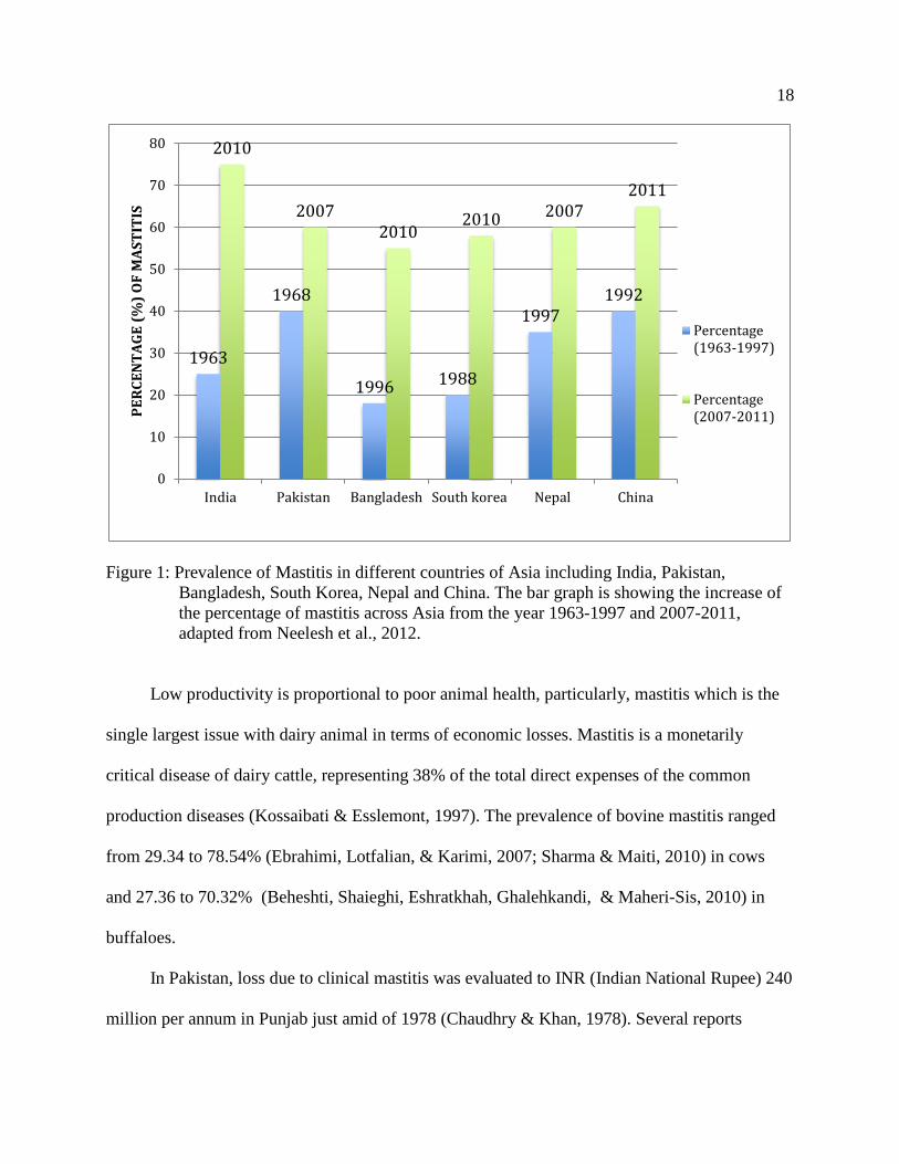
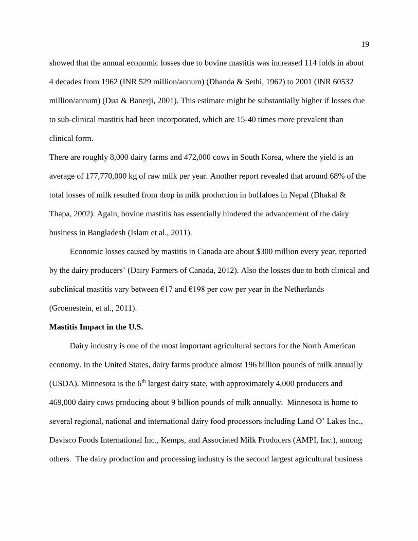
Figure 1: Prevalence of Mastitis in different countries of Asia including India, Pakistan,
Bangladesh, South Korea, Nepal and China. The bar graph is showing the increase of
the percentage of mastitis across Asia from the year 1963-1997 and 2007-2011,
adapted from Neelesh et al., 2012.
Low productivity is proportional to poor animal health, particularly, mastitis which is the
single largest issue with dairy animal in terms of economic losses. Mastitis is a monetarily
critical disease of dairy cattle, representing 38% of the total direct expenses of the common
production diseases (Kossaibati & Esslemont, 1997). The prevalence of bovine mastitis ranged
from 29.34 to 78.54% (Ebrahimi, Lotfalian, & Karimi, 2007; Sharma & Maiti, 2010) in cows
and 27.36 to 70.32% (Beheshti, Shaieghi, Eshratkhah, Ghalehkandi, & Maheri-Sis, 2010) in
buffaloes.
In Pakistan, loss due to clinical mastitis was evaluated to INR (Indian National Rupee) 240
million per annum in Punjab just amid of 1978 (Chaudhry & Khan, 1978). Several reports
1963
1968
1996 1988
19971992
2010
20072010
2010 20072011
0
10
20
30
40
50
60
70
80
India Pakistan Bangladesh South korea Nepal China
PE
RC
EN
TA
GE
(%
) O
F M
AS
TIT
IS
Percentage(1963-1997)
Percentage(2007-2011)
19
showed that the annual economic losses due to bovine mastitis was increased 114 folds in about
4 decades from 1962 (INR 529 million/annum) (Dhanda & Sethi, 1962) to 2001 (INR 60532
million/annum) (Dua & Banerji, 2001). This estimate might be substantially higher if losses due
to sub-clinical mastitis had been incorporated, which are 15-40 times more prevalent than
clinical form.
There are roughly 8,000 dairy farms and 472,000 cows in South Korea, where the yield is an
average of 177,770,000 kg of raw milk per year. Another report revealed that around 68% of the
total losses of milk resulted from drop in milk production in buffaloes in Nepal (Dhakal &
Thapa, 2002). Again, bovine mastitis has essentially hindered the advancement of the dairy
business in Bangladesh (Islam et al., 2011).
Economic losses caused by mastitis in Canada are about $300 million every year, reported
by the dairy producers’ (Dairy Farmers of Canada, 2012). Also the losses due to both clinical and
subclinical mastitis vary between €17 and €198 per cow per year in the Netherlands
(Groenestein, et al., 2011).
Mastitis Impact in the U.S.
Dairy industry is one of the most important agricultural sectors for the North American
economy. In the United States, dairy farms produce almost 196 billion pounds of milk annually
(USDA). Minnesota is the 6th largest dairy state, with approximately 4,000 producers and
469,000 dairy cows producing about 9 billion pounds of milk annually. Minnesota is home to
several regional, national and international dairy food processors including Land O’ Lakes Inc.,
Davisco Foods International Inc., Kemps, and Associated Milk Producers (AMPI, Inc.), among
others. The dairy production and processing industry is the second largest agricultural business
20
in Minnesota, generating approximately $5.6 billion per year, with a total economic impact of
about $11.5 billion, and supporting over 38,000 jobs in the state (Thiesse, 2012).
Mastitis costs the U.S. dairy industry about $1.7-2 billion annually or 11% of total U.S.
milk production. The annual losses per cow from mastitis in the United States of America in
1976 were estimated to be US$ 117.35 per cow per year and a total loss of US$ 1.294 billion
(Blosser, 1979); two decades later these losses had increased to US$ 185 to $ 200 per cow per
year and the total loss increased to US$ 2 billion (Costello, 2004; Viguier, Arora, Gilmartin,
Welbeck, & O’Kennedy, 2009). Despite decades of ongoing advancements, mastitis continues to
be the most costly infectious disease on dairy farms (Erskine, Eberhart, Hutchinson, Spencer, &
Campbell, 1988; Sargeant, Scott, Leslie, Ireland, & Bashiri, 1998; Erskine, Wagner, & Degraves,
2003; Riekerink, Barkema, Kelton, & Scholl, 2007; and Fetrow, 2000).
Much of the cost due to mastitis, is attributed to reduced milk production, discarded milk,
and replacements, which are estimated at $102, $24, and $33 per cow per year as shown below
(Table 1). The obvious costs for treatment medication, labor, and veterinary services are low,
estimated to total $13 per cow (Costello, 2004).

21
Table 1: Different Category of Economic loss due to Mastitis, includes reduced milk production,
discarded milk, replacements and treatment of the animal (Costello, 2004).
It must be recognized that mastitis cannot be completely eliminated from a herd, as most
of the pathogens involved in causing mastitis are the natural inhabitants of the environmental
flora of the cow barns. However, the total cost of mastitis in the average herd enrolled in DHI
(Dairy Herd Improvement) is approximately $171 per cow. In general, it is assumed that milk
had to be discarded for 6 days including 3 days treatment and 3 days withholding period due to
the possible secretion of antibiotic residues in the milk. The treatment cost includes the
veterinarian fees and the cost of drugs.
Importance of Rapid Diagnosis
Early diagnosis is of the utmost importance due to the high costs of mastitis to the dairy
industry. Mastitis has several causative agents and the proper rapid detection of the pathogen is
very important to address the control and prevention measures of Mastitis. A reliable rapid
diagnostic test is a dire need for the wellbeing of the dairy industry, rapid turnaround time being
the key factor because the storage of perishable foods for longer period is cost-effective and a big
problem in such instances. Though the traditional milk culture techniques are useful for the
Category of loss due to Mastitis Cost US$ Per Cow
Reduced Milk production 102
Discarded Milk 24
Replacement 33
Treatment 13
Total Cost 171

22
primary identification of important bacterial pathogens in mastitic milk samples, but most of
them require longer time of incubation and adequate training to perform the tests. Also
Biochemical tests can be nonspecific, slow, costly, and more importantly result interpretation can
be critical in relation to the diagnosis of mastitis pathogens.
Biochemical Testing
Most of the biochemical tests are conducted on site to identify the infection of the cow
through testing the milk and usually not helpful in detecting the presence of any pathogenic
organisms. Some of the commonly used diagnostic techniques are California Mastitis Test
(CMT), Culture test, Enzyme test, pH test, Portacheck, Fossomatic SCC, Electrical Conductivity
test etc. (Viguier, Arora, Gilmartin, Welbeck, & O’Kennedy, 2009). Biochemical tests include
catalase or coagulase tests where the enzyme catalase or coagulase is detected by adding specific
reagent and API system (Analytical Profile Index), which is a biochemical panel containing
chemically-defined dehydrated media for the manual identification and differentiation of bacteria
to the species level.
California mastitis test (CMT) assay indirectly measures the somatic cell count (SCC) in
milk samples. The CMT test applies a detergent that contains bromocresol-purple, which is used
to break down the cell membrane of somatic cells, and the subsequent release and aggregation of
nucleic acid forms a gel-like matrix with a viscosity that is proportional to the leukocyte number.
The CMT assay is very cost effective where 350 tests costs about US$ 12 (Dingwell, Kelton, &
Leslie, 2003). Also, it can be used ‘on-site’ or in the laboratory. It is a rapid and user-friendly
assay, but it can be difficult to interpret and has low sensitivity and no information on the
possible pathogens can be obtained through this rapid test.

23
Mastitis causes the increase of ionic particles in the milk and the Electrical conductivity
(EC) test measures the increase in conductance in milk caused by the increase in levels of ions
such as sodium, potassium, calcium, magnesium and chloride during inflammation (Norberg, et
al., 2004). It can be used ‘on-site’ but non-mastitis-related variations in EC can present problems
in diagnosis, as the test does not refer to any possible presence of pathogens or mastitis infection.
Different selective cultures are used to identify different microorganisms involved in
causing mastitis. Some selective culture medium (e.g. Gram-negative or Gram-positive,
Coliform specific) can identify specific pathogens causing mastitis. Although it can be used only
in laboratory and the waiting time for results can be 24 to 48 hours. (Viguer, Arora, Gilmartin,
Welbeck, & O’Kennedy, 2009)
The pH is a good indicator as well and can be used in mastitis detection. Milk pH rises due
to mastitis and can be detected using bromothymol blue (Kitchen, 1981). It is easy to use, cost
effective and rapid. On the other hand, it is not as sensitive as other tests. It is only an indicator
of the inflammation and no information can be obtained about the possible pathogens.
Also some Enzyme assays are used as well to detect enzymes involved in mastitis immune
response, such as NAGase and LDH (Kitchen, 1976). However, such assays are rapid but might
be laboratory-based only.
16S rRNA Sequencing
There are also a number of molecular techniques that are used for the identification of
pathogens such as PCR, DNA sequencing or other DNA-based methods that are very sensitive
and rapid. One such technique is the 16S rRNA sequencing (Figure 2) which is considered as the
gold standard for bacterial identification purpose, as it provides very specific characterization in
species level as well (Edwards, Rogall, Blöcker, Emde, & Böttger, 1989).

24
Figure 2. The 16S rRNA gene sequencing method showing gradual steps starting from the DNA
extraction of pure bacterial colony, Amplification, Agarose gel electrophoresis,
Sequencing and characterization through data analysis using 16S rRNA library.

25
It is important to have a rapid technique to evaluate products and sources of organisms
along the product chain in an industrial setup (from farm through the processing plant), so that
mitigation strategies can be developed. However, these techniques are relatively very time
consuming and so are used almost exclusively for research but not for service samples (Zadoks,
Middleton, McDougall, Katholm, & Schukken, 2014). Similarly, many of the systems employed
for the isolation and identification of bacterial contaminants in dairy foods require expensive
biochemical tests such as pathogen specific (e.g. Salmonella spp., E. coli 0157, Listeria
monocytogenes, Campylobacter spp.) media or Petrifilm and/or DNA and RNA-based molecular
techniques. These analyses are also time-consuming; increasing the holding time of the food
products and thus impacting the production costs.
MALDI-TOF MS System
A new method of identification called MALDI-ToF, may overcome many of the
aforementioned diagnostic limitations. MALDI-ToF, or matrix-assisted laser desorption
ionization time-of-flight analysis, is a mass spectrometry-based method of identification.
Simply, bacteria are shattered to protein fragments (peptides) with a laser beam and those
peptides rise up an evacuated detection tube. The time taken by the peptides to reach the
detection tube is called the “time of flight” or “ToF” and it is specific for the mass to charge ratio
of the peptide. From this information, the peptide and protein composition can be presented as a
mass spectrometry profile.
The comparison of the mass spectrometry profile with the MALDI-TOF database then
allows for identification of bacterial genus and species based on the protein composition. The
MALDI-TOF database is commercially available and can be updated by individual laboratory
personnel or through purchasing updated version of the library. However, it is faster, less
26
expensive and potentially more universal (i.e. can identify more organisms) than biochemical or
sequence-based identification of cultured isolates (Zadoks, Middleton, McDougall, Katholm, &
Schukken, 2014). The great advantages of the technique includes rapid turnaround time,
MALDI-TOF MS has been shown to identify organisms in <15 minutes whereas traditional
methods take 5 to 48 hours depending on the method used (Cobo, 2013); a single MALDI-TOF
MS system can be used for gram-positive bacteria, gram- negative bacteria, and yeast as well;
the MALDI-TOF MS reference spectra database can be increased by editing commercially
available software updates or by internal laboratory personnel; using MALDI-TOF MS in the
clinical laboratory has been shown to lower costs for hospitals upwards of 53% annually (Seng,
et al., 2009).
Also, while the current proposal focuses on detection of dairy microorganisms, because
MALDI-ToF equipment can be used for detection of many kinds of pathogens. MALDI-TOF
technology has already been successfully used in the field of virology where studies showed the
comparison between the MALDI-TOF results and the other conventional methods (e.g. viral
culture, PCR, nucleic acid based techniques). The concordance rate between MALDI-TOF and
these established conventional techniques were high. Several approaches have been developed to
detect different viruses in clinical specimen such as Human Herpes Viruses (HHVs), Poliovirus,
Coxsackie virus A and B and Echo virus (Sjöholm, Dillner, & Carlson, 2007). Furthermore,
MALDI-TOF could be useful for detecting drug resistance against some antivirals and antibiotic
susceptibility testing (Zürcher, et al., 2012). Despite the fact that MALDI-ToF has already been
validated and adopted for use by many human diagnostic laboratories (e.g. Rochester’s Mayo
Clinic), the library used by this method includes many human-derived pathogens that differ from

27
some animal-derived pathogens. As such, the method must be validated separately for use in
veterinary diagnostic laboratories.
In the last few years, several European groups have investigated the use of MALDI-ToF
for identification of a limited number of mastitis pathogens including several Streptococcus,
Enterococcus and Staphylococcus species (Moser, Stephan, Ziegler, & Johler, 2013; Raemy, et
al., 2013; and Werner, et al., 2012). However, before this instrument can be wholly accepted by
the U.S. veterinary diagnostic laboratories, field validation of the MALDI-ToF must be
completed using a broad range of North American-derived bacterial isolates of importance to
animal health (e.g. bovine mastitis), food quality or food safety. During 2013 and early 2014,
several veterinary diagnostic laboratories across western Europe, Canada and the U.S. have
begun the process of evaluating MALDI-ToF for detection of mastitis, and other animal
pathogens. The VDL is currently at the forefront of this process. The University of Minnesota
VDL has acquired a MALDI-ToF instrument (Figure 3) and has been involved since 2013 in
completing some preliminary internal validation studies evaluating the method’s ability to detect
common animal pathogens, including a large number of mastitis pathogens.

28
Figure 3. MALDI-TOF Mass Spectrometry machine at VDL.
With many advantages of using MALDI-TOF MS in the clinical microbiology
laboratory, there are a few limitations that are noteworthy. A pure culture of the microorganism
is currently still required and MALDI-TOF MS cannot be used with mixed cultures. In addition,
traditional antimicrobial susceptibility testing is still required when a pathogenic organism is
isolated and identified from culture. In that case, pure culture isolation is required which might
be a problem in some instances. MALDI-TOF will fail to identify any new organism that is
absent in the database and will show as unreliable identity. Moreover, there is also complexity in
distinguishing some organisms that are closely related strains, due to the great sequence
similarity of the microorganisms. These include Streptococcus pneumoniae and some of the
viridans Streptococci, or Shigella species and Escherichia coli (Wieser, Schneider, Jung, &
29
Schubert, 2012). In such instances additional test needs to be conducted including, traditional
biochemical tests, antigen detection, or molecular methods.
Objectives & Hypotheses
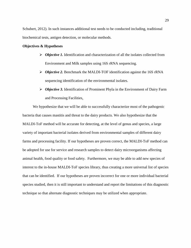
➢ Objective 1. Identification and characterization of all the isolates collected from
Environment and Milk samples using 16S rRNA sequencing.
➢ Objective 2. Benchmark the MALDI-TOF identification against the 16S rRNA
sequencing identification of the environmental isolates.
➢ Objective 3. Identification of Prominent Phyla in the Environment of Dairy Farm
and Processing Facilities.
We hypothesize that we will be able to successfully characterize most of the pathogenic
bacteria that causes mastitis and threat to the dairy products. We also hypothesize that the
MALDI-ToF method will be accurate for detecting, at the level of genus and species, a large
variety of important bacterial isolates derived from environmental samples of different dairy
farms and processing facility. If our hypotheses are proven correct, the MALDI-ToF method can
be adopted for use for service and research samples to detect dairy microorganisms affecting
animal health, food quality or food safety. Furthermore, we may be able to add new species of
interest to the in-house MALDI-ToF species library, thus creating a more universal list of species
that can be identified. If our hypotheses are proven incorrect for one or more individual bacterial
species studied, then it is still important to understand and report the limitations of this diagnostic
technique so that alternate diagnostic techniques may be utilized when appropriate.

30
Chapter II: Materials and Methods
Sample Collection
The technicians in the UHL (Udder Health Laboratory) collected approximately 810
individual isolates that included Gram-positive and Gram-negative organisms. These were
recovered by routine culture methods from milk samples from individual cows, already
submitted by dairy producers to the UHL. No more than 10 bacterial isolates were derived from
any one farm of origin. Selected isolates were frozen in a blood and/or glycerin suspension and
retained at -80°C for later use in the project. Also participating food scientists collected samples
of post-processed fluid milk or other dairy products. Samples were de-identified prior to
culturing to maintain processor confidentiality. Processor milk samples or other dairy products
were from unique lots. Samples were submitted, on ice, to the food science microbiology
laboratory where they sampled, diluted and homogenized, using standard microbial laboratory
practices (Wehr, 2004). 100 processor-sourced milk samples were collected for mesophilic and
psychrophilic bacterial spore formers present that have survived pasteurization. Once recovered,
isolates were grown on a non-selective agar medium and stored in glycerol at – 80 °C for later
use. Between producer and processor sourced isolates, approximately 1,247 individual bacterial
isolates were collected for use in the study.
Environmental Isolates
A total of 810 environmental isolates were collected form VDL (Veterinary Diagnostic
Lab). The bacterial isolates will be grown in the appropriate culture medium for further
identification using the 16S rRNA sequencing. Blood Agar (with 5% sheep blood and Tryptic
Soy Agar) was used for the isolation and cultivation (Figure 4), as it is an appropriate medium
for the growth of a wide variety of fastidious microorganisms. Moreover, the morphology can be

31
observed clearly and hemolysis activity of the bacteria can be seen on blood agar, which serves
as a way of partial identification and categorization of the pathogen (Faddin,1985).
Figure 4. Isolation of Mastitis pathogens (Bacteria) on 5% sheep blood agar.
Thermoduric Isolates
Milk samples were obtained from the DQCI (Dairy Quality Control Institute) for the
isolation of mesophilic and psychrophilic bacterial cultures. The samples were prepared in three
different ways as reported below (Table 2):

32
Table 2. The list of Milk samples received from DQCI.
No. Sample Received Treatment
1 Raw Milk 87 Frozen raw milk
2 Heat Treated 87 Heated to 80°C for 12 minutes then frozen
3 Heat Treated 87 Heated to 80°C for 12 minutes and held at refrigeration
temperature for 5 to 7 days before freezing.
For the isolation of thermoduric and spore forming organisms, only heat-treated milk
samples were used. A total of 87 milk samples were collected from different farms by DQCI.
Three replicates from each of the 87 milk samples were made, where the first set was kept as raw
without any treatment. The other two sample sets were heated to 80°C for 12 minutes, and
storage was done in two different ways for experimental purposes, where one of them were
frozen right after heating and the other set was kept in the refrigerator for 5 to 7 days before
freezing. The two different storage conditions were intended to examine the growth of mesopilic
and psychrophilic spore forming organisms, respectively.
For the primary isolation of bacteria from the milk samples, three different agar media
were used including NA (Nutrient agar), LB agar (Luria broth) and BHI agar (Brain heart
infusion). Among them the BHI agar performed best with more bacterial growth comparatively,
as the purpose was to grow most bacteria including fastidious ones (Figure 5). At first, 500 μl of
each milk sample from both replicate sets were spread on the agar medium using sterile
spreading rods. After plating, the plates were placed at 37°C for the growth of mesophilic spores
and at 7°C for 10 days to grow psychrophilic spores. Depending on the amount of growth, the
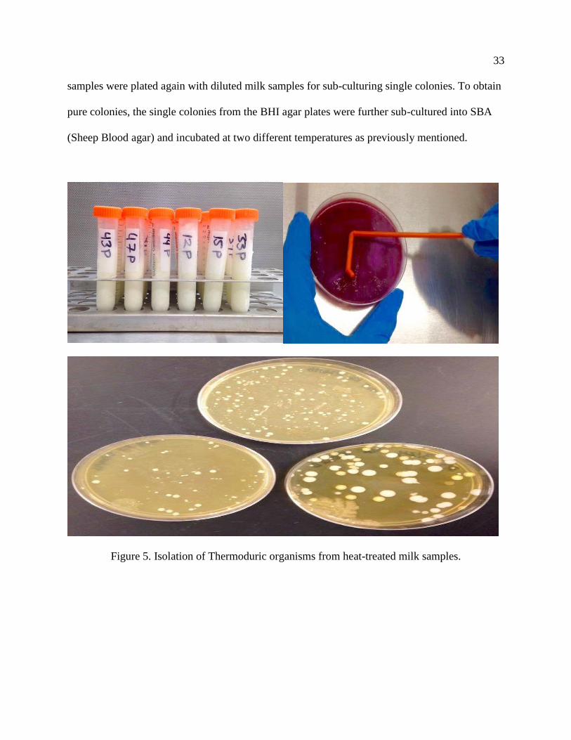
33
samples were plated again with diluted milk samples for sub-culturing single colonies. To obtain
pure colonies, the single colonies from the BHI agar plates were further sub-cultured into SBA
(Sheep Blood agar) and incubated at two different temperatures as previously mentioned.
Figure 5. Isolation of Thermoduric organisms from heat-treated milk samples.

34
Stocking and Storage of Isolates
Glycerol stock of bacteria can serve the purpose for the long-term storage and future use
of the bacterial isolates (Swift, 1920). After the isolation of bacteria on blood agar, they were
further processed for making glycerol stocks. For each of the 1,247 isolates, two glycerol stocks
were made. 20% glycerol was used for the stocking, where overnight grown bacterial culture in
BHI broth were pipetted in 20% glycerol solution and vigorously vortex to generate uniform
mixture. The glycerol stock vials were kept in -80° C for preservation and future use.
Preparation of BHI broth
Brain-heart infusion broth (BHI) is a general-purpose highly nutritious growth medium
made with cow or porcine heart and brain (Table 3). It is used for culturing both fastidious and
non-fastidious microorganisms (Faddin, 1985).
Table 3. The components and amount for BHI broth preparation.
At first the BHI powder was weighed to 37g and put in an Erlenmeyer flask, following
Instructions from the manufacturer (Hardy Diagnostics, California, U.S.). Using a graduated
cylinder, 1000 ml DI water was added slowly to the flask. Magnetic stir bar was inserted and the
top of the flask was wrapped with aluminum foil. Then the flask was placed on a magnetic stirrer
with hot plate. The heating plate was adjusted to boiling temperature and speed was set on
medium for the stirring. The heat and stirrer were turned off when the broth started boiling. Then
Component Amount
BHI powder 37 g
DI H2O 1000 ml

35
the magnetic stir bar was removed cautiously and the broth was autoclaved at load cycle 3
(Liquid medium < 6L). After cooling down to room temperature, the broth was used to make
bacterial culture and stock.
Preparation of 20% Glycerol solution
The addition of glycerol to the culture stabilizes the bacterial plasmid, protects from
damage to the cell membranes and keeps the cells alive in freezing temperature (Gibson &
Khoury, 1986). The glycerol stock of bacteria can be stored stably at -80°C for many years.
In a glass beaker 20 ml of Glycerol was added using a pipetter and 80 ml of DI water was
added using graduated cylinder (Table 4). The solution was mixed thoroughly using the pipetter
and autoclaved at Load Cycle 3 (Liquid medium < 6L) in the autoclave. After cooling down to
room temperature the 20% Glycerol solution was used to prepare the bacterial stock.
Table 4. The components and amount for making 20% Glycerol solution.
Preparation of 20% Glycerol stock
After both the BHI broth and 20% glycerol was made and cooled down to room
temperature, 5 ml of BHI broth were transferred to falcon tubes. Pure cultures of bacterial isolates
were introduced into the broth using sterile loop and incubated overnight at 37°C. At first, 800 μl
of 20% Glycerol solution was added to 1000 μl cryovials and then 200 μl of overnight pure
Component Amount
Glycerol 20 ml
DI H2O 80 ml
36
bacterial culture was added (Table 5). The turbidity must be observed for bacterial culture in the
broth to ensure growth. Also the cryovials were labeled carefully according to the sample ID.
Finally the mixture was vortexed vigorously and kept in the -80°C freezer in separate labeled
boxes for later use.
Table 5. The components and amount for making 20% Glycerol Stock.
16S rRNA Sequencing
Isolates obtained from blood plates were initially classified by 16S rRNA sequencing, as
described by Lane (1991). Each of the isolate has a unique number and other related information,
that were recorded carefully. Four different universal primers were tested to select the best
working primer set (Forward and Reverse primer) that works for most isolates (Greisen,
Loeffelholz, Purohit, & Leong, 1994; Hou, Fink, Radtke, Sadowsky, & Diez-Gonzalez, 2013).
The universal primers that were used in this study are as followed in the table below (Table 6):
Component Amount
20% Glycerol solution 800 µl
Overnight bacterial culture in BHI broth 200 µl

37
Table 6. The list of primers for the amplification of the template.
Primer Sequence (5’--------3’)
1492R TACGGYTACCTTGTTACGACTT
27F AGAGTTTGATCMTGGCTCAG
8F AGA GTT TGA TCC TGG CTC AG
63F-1389R 5’- CAGGCCTAACACATGCAAGTT-3’
BACT2F 5’-CAAACAGGATTAGATACCCTG-3’
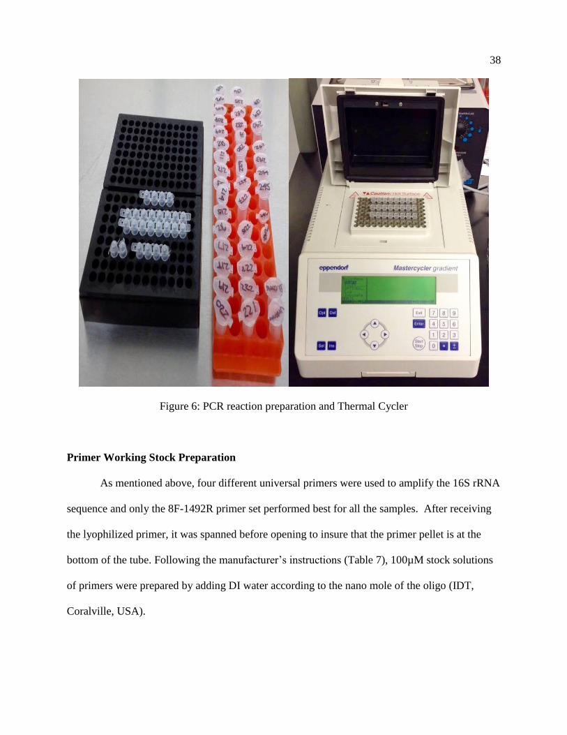
PCR Amplification
Polymerase chain reaction, better known as PCR, is a technique used extensively in
molecular biology. The technique uses a single piece of DNA and amplifies it to thousand to
millions of copies. It requires two primers for forward and reverse reaction, DNA template to be
amplified, DNA polymerase to elongate the strand, Deoxynucleoside triphosphates (dNTPs) to
provide nucleotide to the growing strand and buffer solution providing suitable environment for
the reaction (Lane, 1991). In this project, PCR amplification of the 16S rRNA sequence was
done using 4 different forward primers and reverse primer for all samples (Figure 6). The
forward primers are usually named according to the restriction site and the target site of the
promoter where they will bind (Dieffenbach, Lowe, & Dveksler, 1993).
38
Figure 6: PCR reaction preparation and Thermal Cycler
Primer Working Stock Preparation
As mentioned above, four different universal primers were used to amplify the 16S rRNA
sequence and only the 8F-1492R primer set performed best for all the samples. After receiving
the lyophilized primer, it was spanned before opening to insure that the primer pellet is at the
bottom of the tube. Following the manufacturer’s instructions (Table 7), 100µM stock solutions
of primers were prepared by adding DI water according to the nano mole of the oligo (IDT,
Coralville, USA).

39
Table 7. Preparation of 100 µM primer stock.
Oligo Amount (nmoles) Amount of DI H2O
8F- 20.8 nmole 208 µl
1492R- 25.4 nmole 254 µl
From the 100µM primer stock, 10 µM working stock was prepared by 1:10 dilution. In
two separate sterile 1 ml tubes, 100 µl of Forward and Reverse primer were added following the
addition of 900 µl of RNAase free water. The 100µM primer stocks were stored at -20°C for
later use.
PCR Reaction and Sample Preparation
Fast Cycling PCR Kit from QIAGEN was used to amplify the templates. A 20µl reaction
mixture was prepared following the manufacturer’s instructions (Table 8). Amount of all the
reagents used for PCR is given in table below.
Table 8. The list of reagents and amount used for a single PCR reaction.
Components Amount for 1 reaction For *110 reaction
Master Mix 10 µl 1100 µl
Q Solution 4 µl 440 µl
Forward & Reverse Primer 1 µl (0.5 each) 110 µl
RNAase free Water 3 µl 330 µl
Template DNA 2 µl ---
Total = 20 µl Total= 1,980 µl

40
A 96 well PCR plate was used for mixing the PCR reaction and amplification in the
thermal cycler. In the 96 well plate, 95 samples and one control were run for amplification. The
PCR reaction mixture was made for a total of 110 reactions instead of 96, to avoid the shortage
due to pipetting loss. In a sterile plastic vial 1100 µl of Fast cycling PCR Master Mix was added.
Then 440 µl Q solution, 330 µl of RNAase free H2O and 110 µl of Forward & Reverse primer
solution were added to the vial. The mixture was vortexed briefly to evenly mix all the
components.
For a single PCR run, 95 samples were prepared at a time. In a sterile 1.5ml plastic tube,
200 µl of DI water was added. Using the tip of the pipette tips, very little amount of culture was
taken form the center of the pure single colony, to avoid contamination. The culture was mixed
thoroughly using vortex. All the tubes were labeled carefully according to the sample ID.
At first, 18 µl of PCR reaction mixture was added to the entire well in the 96 plate and
then using a multichannel pipetter 2 µl of sample was added to each well with vigorous mixing.
The DNA templates (Sample) were added at last to reduce contamination. The PCR plate was
covered tightly with domed shape strip caps and placed inside the thermal cycler for
amplification run.
The thermal cycler program was set for 40 cycles with the denaturation temperature at
95° C, annealing temperature at 55°C and elongation temperature at 72°C (Table 9). Annealing
temperature varied for different samples so adjustments were made, ranging from 50°C, 55°C,
65°C and 68°C (QIAGEN Fast Cycling Handbook, 2012).

41
Table 9. Thermal cycler program for PCR reaction.
Stage Cycle Step Temperature (°C) Time
Hold Initial denaturation 95 10 min
Cycle (25 to 40
cycles)
Denature 95 30 sec
Anneal 55 30 sec
Extend 72 60 sec
Hold Final Extension 72 7 min
Hold Final Hold 4 ∞
Gel Electrophoresis
Gel electrophoresis is a standard tool in biological laboratories in purpose of separating
DNA and RNA fragments according to their size. It uses a gel as medium containing the sample,
which runs through an electrical field (Johansson, 1972). There are types of gel used for
electrophoresis based on the size of sample. For this project Agarose gel was used for which is
easy to cast and samples can easily be recovered from the gel (Figure 7).

42
Figure 7: Gel Electrophoresis Chamber.
Running Buffer Preparation
Tris-borate-EDTA (TBE) buffer was used for making Agarose gel and as running buffer.
A beaker was filled with 800 ml of filtered DI (Deionized) water and put in a stir bar. All the
components were weighed according to the amount shown in the chart below (Table 10). At first
54g of Tris base was added, then 2.9g of EDTA acid. Boric acid (27.5g) was added after the
EDTA acid was fully dissolved. Another 200 ml of filtered DI water was added using graduated
cylinder to reach the final concentration of 1000 ml or 1 Liter. After mixing for a couple of
minutes, the mixture was poured into a sterilized 1 Liter glass bottle. Finally the glass bottle was
autoclaved on cycle 3. To reach the working solution of 0.5X buffer, the 1 Liter buffer was
diluted with 9 Liter DI water.

43
Table 10. The list of reagents used to make 5X TBE.
Components Amount
Tris Base 54g
EDTA Acid 2.9 g
Boric Acid 27.5g
DI Water 1 Liter
1% Agarose Gel Preparation
In this project, after amplifying the 16S rRNA sequence of the templates, gel
electrophoresis was used to detect the amplified desired product size of 1400 to 1500 bp (Base
pair). For the gel electrophoresis 1% Agarose gel was used. Amount of each reagent in
preparation of gel and sample loading is given in the table (Table 11).
Table 11. The list of reagents used to make 1% Agarose Gel.
Components Amount
Agarose Powder 1 g
0.5X TBE Buffer 100 ml
1g Agarose gel powder was measured and taken in a sterile bottle and mixed with 0.5X
TBE buffer. The top of the bottle was covered with plastic wrap and placed into a microwave for
3 minutes. The heating was observed carefully in case of overflow of liquid due to boiling. The
mixture was placed into a water bath at 50°C and was not poured until it reached down to the
temperature from boiling point. For gel with smaller pore size, 2% gel can be prepared when

44
needed. Amount of all the reagents are same as 1% Agarose, only instead of using 1g of gel
powder, 2 g can be used.
Sample Preparation and Loading on Gel
After cooling down to 50°C, the liquid gel was poured on top of the Gel electrophoresis
chamber and left for 30 minutes to set and solidify. After the gel was solidified, it was put on the
electrophoresis tank. The wells in the gel were loaded with the amplified template and DNA
ladder (Table 12).
Table 12. The list of reagents used to prepare sample and loading the Gel.
Components Amount
DNA Ladder 6 µl
Loading Dye 2 µl
Template 10 µl
One well was loaded with 6 µl DNA ladder (GelPilot 100 bp plus-QIAGEN) mixed with 2
µl loading dye (EZ-VISION DNA dye), for measuring the size of the amplified sequences.
Amplified samples of 10 µl were mixed with 2 µl of loading dye. All the wells were loaded with
12 µl sample. After loading the sample, gel was run at 70V (voltage) for 45 minutes. Images of
the bands forming were visualized under UV light and picture was taken for recording the
presence of desired bands.

45
PCR Product Purification (ExoSAP-IT)
When PCR amplification is complete, any unconsumed dNTPs and primers remaining in
the PCR product mixture will interfere with these methods (Bell, 2008). ExoSAP-IT is a reagent
that removes these contaminants. After the amplification of the PCR product and Gel
Electrophoresis run, templates with desired product size or DNA band (1400-1500 bp) were
identified under UV light observation and selected to send out for sequencing. PCR products
were purified using ExoSAP-IT reagent by Affymetrix. The procedure was followed as
manufacturer’s instructions with adding 2 µl of the ExoSAP-IT clean up reagent to 5 µl PCR
product. After mixing the mentioned amount, the mixture was incubated at 37°C for 15 minutes
and heated at 80°C for another 15 minutes to inactivate ExoSAP-IT. The entire program
including the incubation and heating temperature and time were set in the Thermal cycler
machine for repeated use.
Data Analysis of 16S rDNA Sequencing
The amplified samples were sent for sequencing to GENEWIZ, which is a reknowned
research organization providing different adavanced research services. Using the sanger
sequencing method, forward and reverse primer sequences were obtained for the amplified
samples. The forward and reverese primer sequences were assembeled together using the
software Geneious version R10 (Kearse, et al., 2012). After retrieving the whole sequences for
each sample, the taxonomic classification of 16S rRNA PCR products were assigned using NCBI
Targeted Loci BLAST, comparing with the 16S rRNA library and further identify the unknown
isolates. In the NCBI Targeted Loci BLAST 16S rRNA database, the result shows a combination
of the highest alignment score (Max score), the total alignment scores (Total score), the
percentage of query covered by alignment to the database sequence, the best (lowest) Expect

46
value (E-value) of all alignments, and the highest percent identity (Max identity) of all query-
subject alignments from the sequence database (NCBI Resource Coordinators, 2016). Generally,
the first hit shows the highest Max and total score, lowest E value, and highest Identity
percentage. However, the percentage of query cover does not reflect the highest number all the
time, it might be same or slightly lower than the highest score.
Data Analysis of MALDI-TOF
The result data from MALDI-TOF is analyzed by the commercial software and referrence
library in the laboratory. It generates result very fast as a single run takes around 20 mins and it
gives the specific name of the unknown bacteria by matching up with library. Also it provides a
second prediction as well and no reading if there is not sufficient amount of cell debris of the
bacteria or if there is no record in the referrence library. It is very easy to analyse the result of the
MALDI-TOF as it provides the species name of the bacteria tested.
The MALDI-ToF method is run in duplicate for each isolate tested, with the 2 closest
matches from the isolate library reported for each test, and a ‘diagnostic certainty score’ reported
for each. The manufacturer (Bruker Corp., Germany) recommends a diagnostic certainty score of
2.0 or greater be attained to be confident in the accuracy of a diagnosis at the species level and a
score between 1.8 and 2.0 is accurate to the genus level.
Therefore, the 16S rRNA sequencing was used to validate the MALDI-ToF performed by
the Diagnostic lab at the University of Minnesota. All the isolates were analyzed and the
comparison confirmed true genus and species identification for each organism tested. The results
of the 16S rRNA sequence analysis were compared to the MALDI-ToF identification and the
discrepancies were further analyzed in the result section.

47
Chapter III: Results
Thermoduric Isolate Identification
From the heat-treated milk samples, mesophilic and psychrophilic bacteria were isolated.
Culturing the milk samples in the appropriate agar media collected a total of 447 isolates, where
the numbers of psychrophilic isolates were 226 and mesophilic isolates were 221. For the
identification purpose, unknown amplified samples were sent out for obtaining the complete 16S
rRNA gene sequences. Due to reaction failure during the Sanger sequencing, only 248 sequences
were retrieved combining 118 psychrophilic isolates and 130 mesophilic isolates. The table
below (Table 13) shows all the Genus and Species obtained from the milk samples using the 16S
sequencing identification method.
Table 13: List of Genus and Species of isolates collected from heat-treated milk samples.
NO. Genus Species
1 Acinetobacter radioresistens
2 Arthrobacter agilis
3 Aureimonas phyllosphaerae
4 Bacillus paralicheniformis
5 Bacillus aryabhattai
6 Bacillus safensis
7 Bacillus kochii
8 Bacillus siralis
9 Bacillus pumilus
10 Bacillus oleronius

48
NO. Genus Species
11 Bacillus axarquiensis
12 Bacillus circulans
13 Bacillus aerius
14 Bacillus ginsengi
15 Bacillus subtilis
16 Bacillus galliciensis
17 Bacillus pseudomycoides
18 Bacillus hisashii
19 Bacillus amyloliquefaciens
20 Bacillus licheniformis
21 Bacillus clausii
22 Bacillus sonorensis
23 Bacillus galactosidilyticus
24 Bacillus flexus
25 Bacillus lonarensis
26 Bacillus thermoamylovorans
27 Brachybacterium muris
28 Brachybacterium nesterenkovii
29 Brevibacterium frigoritolerans
30 Brevundimonas bacteroides
31 Clavibacter michiganensis
32 Curtobacterium oceanosedimentum
33 Enterococcus faecalis

49
NO. Genus Species
34 Frigoribacterium faeni
35 Hydrogenophaga caeni
36 Janibacter hoylei
37 Kocuria varians
38 Lysinibacillus halotolerans
39 Microbacterium lacticum
40 Microbacterium aurum
41 Microbacterium lacus
42 Micrococcus flavus
43 Micrococcus yunnanensis
44 Micromonospora aurantiaca
45 Oceanobacillus sojae
46 Oceanobacillus caeni
47 Paenibacillus rhizosphaerae
48 Paenibacillus xylanexedens
49 Paenibacillus tundrae
50 Paenibacillus borealis
51 Paenibacillus barengoltzii
52 Paenibacillus amylolyticus
53 Paenibacillus lactis
54 Rothia dentocariosa
55 Salmonella enterica
56 Sphingomonas trueperi

50
NO. Genus Species
57 Sporosarcina psychrophila
58 Sporosarcina siberiensis
59 Sporosarcina contaminans
60 Staphylococcus epidermidis
61 Staphylococcus cohnii
62 Staphylococcus chromogenes
63 Staphylococcus hominis
64 Staphylococcus wameri
65 Streptococcus oralis
66 Streptococcus mitis
67 Streptococcus salivarius
68 Streptococcus rubneri
69 Virgibacillus halotolerans
70 Virgibacillus proomii
Prevalent Genera in Milk Samples
The characterization of the isolates showed frequent occurrence of some predominant
genera. Many different species were found from the same genus with repeated occurrence. The
table below (Table 14) shows the list of genera with the number of occurrences.

51
Table 14: List of Prevalent Genera from the heat-treated milk samples.
Genus Species Occurrence Total Species Occurrence
Bacillus 23 135
Paenibacillus 7 13
Staphylococcus 5 12
Streptococcus 4 7
Microbacterium 3 37
Sporosarcina 3 6
Micrococcus 2 5
Oceanobacillus 2 4
Other 21 29
A total of 71 different species were identified from the milk samples where the different
prevalent genera included Bacillus, Paenibacillus, Staphylococcus, Streptococcus,
Microbacterium, Micrococcus and Oceanobacillus (Figure 8). The most frequent genus was
Bacillus with a total of 135 repeated occurrences and 23 different species. Some of the most
frequent species of the genus Bacillus were Bacillus licheniformis with 38 occurrences, Bacillus
safensis with 18 occurrences, Bacillus paralicheniformis with 16 occurrences, Bacillus aerius
with 13 occurrences, Bacillus circulans with 9 occurrences and Bacillus pumilus with 8
occurrences. There were 7 different species of Paenibacillus including Paenibacillus
amylolyticus with 4 occurrences, Paenibacillus tundra with 3 occurrences and Paenibacillus
borealis with 2 occurrences. Then Microbacterium with 3 different species and a total 37
occurrences included the species Microbacterium lacticum with 33 occurrences, Microbacterium

52
aurum with 2 occurrences and Microbacterium lacus with 2 occurrences. Again the 5 different
species of Staphylococcus included Staphylococcus chromogenes, Staphylococcus epidermidis,
Staphylococcus cohnii, Staphylococcus hominis and Staphylococcus wameri. Also 4 different
Streptococcus species were found such as Streptococcus mitis, Streptococcus oralis,
Streptococcus salivaris and Streptococcus rubneri. Some other comparatively less frequent
genus were Sporosarcina, Micrococcus, Oceanobacillus, Virgibacillus, lysinibacillus and
Brachybacterium.
Figure 8: The pie chart in above figure is showing the occurrence rate (in percentage %) of the
different prevalent Genera isolated from the milk samples. The Bacillus with 54.4%,
Microbacterium 15%, Paenibacillus 5.2%, Staphylococcus 5%, Streptococcus 3% and
other genera 14.1%.
Occurrence level %
Bacillus 54.4%
Microbacterium 15%
Paenibacillus 5.2%
Staphylococcus 5%
Streptococcus 3%
Other 14.1%

53
The Bacillus genus represented 54.4% of the total bacterial isolates collected from the
pasteurized milk samples. The occurrence level of Microbacterium was 15%, Paenibacillus
5.2%, Staphylococcus 5%, Streptococcus 3% and other genera constituted about 14.1% of the
total bacterial population collected from the heat-treated milk samples. Some very less frequent
genera that represented the other category included Acinetobacter, Arthrobacter, Aureimonas,
Clavibacter, Brevundimonas, Curtobacterium, Frigoribacterium, Hydrogenophaga, Janibacter,
Kocuria, Micromonospora, Rothia, Sphingomonas, Enterococcus and Salmonella.
Environmental Isolates
The Environmental samples were collected from different dairy farms across Minnesota.
The total number of isolates received from the Veterinary Diagnostic Lab (VDL) were 810 and
664 isolates were amplified. After the Sanger sequencing 481 sequences were retrieved due to
reaction failures. The table below (Table 15) shows all the Genus and Species identified from the
environmental samples using 16S sequencing technique.
Table 15: List of Genus and Species of isolates collected from environmental samples.
NO. Genus Species
1 Acinetobacter gandensis
2 Aerococcus viridans
3 Aerococcus urinaeequi
4 Citrobacter freundii
5 Corynebacterium argentoratense
6 Enterobacter cloacae
7 Enterobacter xiangfangensis

54
NO. Genus Species
8 Enterobacter ludwigii
9 Enterobacter kobei
10 Enterobacter asburiae
11 Enterococcus faecium
12 Enterococcus faecalis
13 Enterococcus thailandicus
14 Enterococcus hirae
15 Enterococcus saccharolyticus
16 Enterococcus casseliflavus
17 Escherichia fergusonii
18 Escherichia hermanni
19 Klebsiella pneumoniae`
20 Klebsiella oxytoca
21 Lactococcus lactis
22 Lactococcus formosensis
23 Lactococcus garvieae
24 Listeria innocua
25 Lysinibacillus fusiformis
26 Macrococcus caseolyticus
27 obesumbacterium proteus
28 Ochrobactrum tritici
29 Pantoea agglomerans
30 Pantoea brenneri

55
NO. Genus Species
31 Paracoccus denitrificans
32 Psedomonas guariconesis
33 Pseudomonas putida
34 Pseudomonas punonensis
35 Pseudomonas knackmussi
36 Pseudomonas indoloxydans
37 Pseudomonas protegens
38 Pseudomonas lundensis
39 Pseudomonas guariconensis
40 Pseudomonas pictorum
41 Pseudoxanthomonas suwonensis
42 Psychrobacter maritimus
43 Raoultella terrigena
44 Riemerella anatipestifer
45 Shigella flexneri
46 Shigella dysenteriae
47 Staphylococcus aureus
48 Staphylococcus chromogenes
49 Staphylococcus sciuri
50 Staphylococcus simulans
51 Staphylococcus haemolyticus
52 Staphylococcus saprophyticus
53 Staphylococcus agnetis

56
NO. Genus Species
54 Staphylococcus cohnii
55 Staphylococcus devriesei
56 Staphylococcus epidermidis
57 Staphylococcus xylosus
58 Staphylococcus hominis
59 Streptococcus pyogenes
60 Streptococcus agalactiae
61 Streptococcus uberis
62 Streptococcus dysgalactiae
63 Streptococcus porcorum
64 Trueperella abortisuis
65 Weissella parameseneroides
Prevalent Genera in Environmental Samples
After the identification of the isolates, it was seen that some genera were more frequent
than the others. Again many different species were found to be from the same genus with
repeated species occurrence. The table below (Table 16) shows the list of genera with the
number of individual species occurrences.

57
Table 16: List of Prevalent Genera from the Environmental samples.
Bacterial Genera Species occurrence Total Species occurrence
Staphylococcus 11 157
Streptococcus 4 86
Escherichia 3 55
Klebsiella 2 53
Lactococcus 3 31
Enterococcus 6 29
Pseudomonas 9 11
Citrobacter 1 7
Aerococcus 2 6
Shigella 2 4
Other 22 42
Total 65 481
From the environmental samples, 64 different species were identified. The frequency of
occurrence for some of the genus was more than the others (Figure 9), such as Staphylococcus
was found to be the most prevalent genus for having 11 different species and a total occurrence
number of 157 isolates. The different species of Staphylococcus were Staphylococcus aureus,
Staphylococcus chromogenes, Staphylococcus cohnii, Staphylococcus simulans, Staphylococcus
epidermidis, Staphylococcus haemolyticus, Staphylococcus saprophyticus, Staphylococcus

58
agnetis, Staphylococcus devriesei, Staphylococcus xylosus and Staphylococcus hominis etc. The
second most prevalent genus was Streptococcus with a total occurrence of 86 isolates of 4
different species including Streptococcus pyogenes, Streptococcus agalactiae, Streptococcus
uberis and Streptococcus dysgalactiae. Then Escherichia with 3 different species and 55 isolates
included Escherichia fergusonii, Escherichia coli and Escherichia hermanni. The genus
Klebsiella with 53 isolates and 2 different species included Klebsiella oxytoca and Klebsiella
pneumonia. The Lactococcus genus had 31 isolates with 3 different species including
Lactococcus lactis, Lactococcus formensis and Lactococcus garviae. The other prevalent genus
Enterococcus with 6 different species included Enterococcus faecalis, Enterococcus hirae,
Enterococcus thailandicus, Enterococcus saccharolyticus, Enterococcus casseliflavus and
Enterococcus faecium. Again Pseudomonas had 9 different species, such as Pseudomonas
putida, Pseudomonas lundensis, Pseudomonas guariconesis, Pseudomonas punonensis,
Pseudomonas knackmussi, Pseudomonas indoloxydans, Pseudomonas protegens, Pseudomonas
parafulva and Pseudomonas pictorum. Some other prevalent species were Citrobacter freundii,
Aerococcus viridans, Aerococcus urinaeequi, Shigella flexneri and Shigella dysenteriae.

59
Figure 9: The pie chart in above figure is showing the occurrence rate (in percentage %) of the
different prevalent Genera isolated from the Environmental samples. The Staphylococcus
occupied 35%, Streptococcus occupied 19%, Escherichia occupied 12.2%, Klebsiella
occupied 12%, Lactococcus occupied 7%, Enterococcus occupied 6%, Pseudomonas 2%
and other genera occupied 6.8%.
The most prevalent genus Staphylococcus represented 35% of the total bacterial
population of the environmental isolates. The other frequent genera Streptococcus occupied
19%, Escherichia occupied 12.2%, Klebsiella occupied 12%, Lactococcus occupied 7%,
Enterococcus occupied 6%, Pseudomonas 2% and other genera occupied 6.8%. Some less
frequent genera that occurred only once or twice over the entire bacterial population in the
environmental samples, were categorized as the other genera and included Acinetobacter,
Enterobacter, Corynebacterium, Enterobacter, Listeria, Lysinibacillus, Macrococcus,
Obesumbacterium, Ochrobactrum, Pantoea, Paracoccus, Pseudoxanthomonas, Psychrobacter,
Raoultella, Riemerella, Trueperella and Weissella.
Occurrence Level %
Staphylococcus 35%
Streptococcus 19%
Escherichia 12.2%
Klebsiella 12%
Lactococcus 7%
Enterococcus 6%
Pseudomonas 2%
Other 6.8%

60
Comparison of 16S rRNA Sequencing and MALDI-TOF Identification
The MALDI-TOF identification was compared with the 16S sequencing results to verify
the accuracy of the test method. The MALDI-TOF identification result was obtained only for the
environmental isolates. Hence, only environmental (481) isolates 16S identification was
compared against the MALDI-TOF identification at both genus and species level. The table
below (Table 17) is showing the outcome of the comparison.
Table 17: Comparison of 16S rRNA Sequencing and MALDI-TOF identification of the
Environmental isolates.
16S sequencing and MALDI-TOF
Number of isolates
Total number of Isolates retrieved 481
Number of Genus level match 454
Number of Species level match 426
Number of complete mismatch 27
Number of mismatch at only Species level 26
Number of unreliable identification 17
The comparison of the identification of the environmental isolates included both genus
and species level match, where all the 481 isolates 16S identification was compared against the
MALDI-TOF identification (Figure 10). At the genus level, 454 isolates matched out of the total
481 isolates. The number of species level match was 426 out of 481 isolates, which makes the
species level mismatch for 26 isolates out of the total. The number of complete mismatch where
both genus and species did not match for 16S sequencing and MALDI-TOF identification was
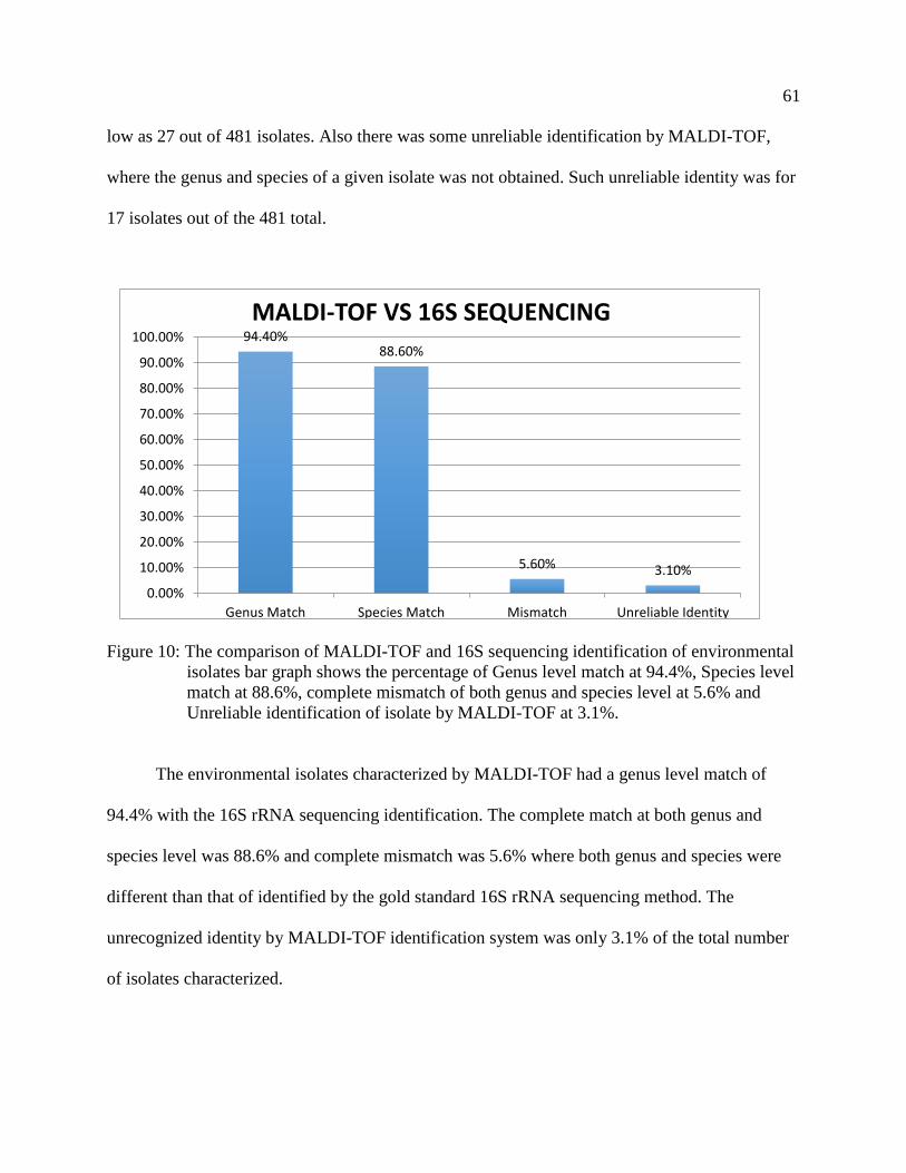
61
low as 27 out of 481 isolates. Also there was some unreliable identification by MALDI-TOF,
where the genus and species of a given isolate was not obtained. Such unreliable identity was for
17 isolates out of the 481 total.
Figure 10: The comparison of MALDI-TOF and 16S sequencing identification of environmental
isolates bar graph shows the percentage of Genus level match at 94.4%, Species level
match at 88.6%, complete mismatch of both genus and species level at 5.6% and
Unreliable identification of isolate by MALDI-TOF at 3.1%.
The environmental isolates characterized by MALDI-TOF had a genus level match of
94.4% with the 16S rRNA sequencing identification. The complete match at both genus and
species level was 88.6% and complete mismatch was 5.6% where both genus and species were
different than that of identified by the gold standard 16S rRNA sequencing method. The
unrecognized identity by MALDI-TOF identification system was only 3.1% of the total number
of isolates characterized.
94.40%88.60%
5.60% 3.10%
0.00%
10.00%
20.00%
30.00%
40.00%
50.00%
60.00%
70.00%
80.00%
90.00%
100.00%
Genus Match Species Match Mismatch Unreliable Identity
MALDI-TOF VS 16S SEQUENCING

62
Complete and Partial Misidentification by MALDI-TOF
The misidentification of the environmental bacterial isolates by MALDI-TOF was either
complete mismatch or partial mismatch. The complete mismatch were those, where both genus
and species were not the same as the 16S identification. Whereas the partial mismatch had only
the genus level match with the 16S characterization but the species were different. The table
below (Table 18) shows the complete misidentification by the MALDI-TOF mass spectrometry
system.
Table 18: Complete mismatch of genus and species of isolates by MALDI-TOF identification.
16S rRNA identification MALDI-ToF identification
Shigella flexneri Escherichia coli
Pantoea agglomerans Enterobacter cloacae
Citrobacter gillenii Klebsiella oxytoca
The complete mismatch shows that all the Shigella flexneri that were characterized by 16S
rRNA sequencing were identified as Escherichia coli by the MALDI-TOF. Also Pantoea
agglomerans was identified as Enterobacter cloacae by the newer technique MALDI-TOF. All
the Citrobacter gillenii were characterized as Klebsiella oxytoca by MALDI-TOF identification
system.
The table below (Table 19) shows the misidentification of species from the same genus by
the MALDI-TOF characterization method.
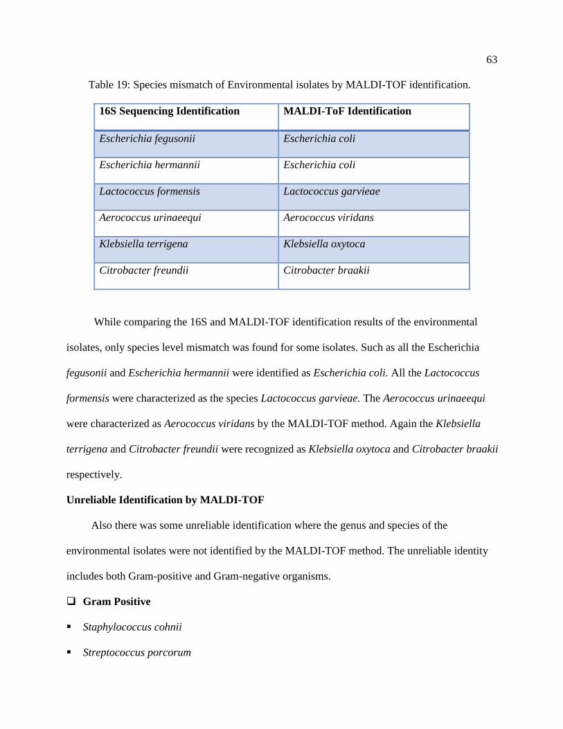
63
Table 19: Species mismatch of Environmental isolates by MALDI-TOF identification.
16S Sequencing Identification MALDI-ToF Identification
Escherichia fegusonii Escherichia coli
Escherichia hermannii Escherichia coli
Lactococcus formensis Lactococcus garvieae
Aerococcus urinaeequi Aerococcus viridans
Klebsiella terrigena Klebsiella oxytoca
Citrobacter freundii Citrobacter braakii
While comparing the 16S and MALDI-TOF identification results of the environmental
isolates, only species level mismatch was found for some isolates. Such as all the Escherichia
fegusonii and Escherichia hermannii were identified as Escherichia coli. All the Lactococcus
formensis were characterized as the species Lactococcus garvieae. The Aerococcus urinaeequi
were characterized as Aerococcus viridans by the MALDI-TOF method. Again the Klebsiella
terrigena and Citrobacter freundii were recognized as Klebsiella oxytoca and Citrobacter braakii
respectively.
Unreliable Identification by MALDI-TOF
Also there was some unreliable identification where the genus and species of the
environmental isolates were not identified by the MALDI-TOF method. The unreliable identity
includes both Gram-positive and Gram-negative organisms.
Gram Positive
▪ Staphylococcus cohnii
▪ Streptococcus porcorum

64
▪ Macrococcus caseolyticus
▪ Acinetobacter gandensi
▪ Weissella parameseneroides
▪ Enterococcus hirae
Gram Negative
▪ Pseudomonas knackmussi
▪ Pseudomonas guariconesis
▪ Riemerella anatipestifer
▪ Psychrobacter maritimus

65
Chapter IV: Discussion
Most of the isolates we identified from environmental samples belonged to the genera
Staphylococcus, Streptococcus, Escherichia, Klebsiella, and Pseudomonas; all reported as
common environmental mastitis pathogens in many studies (Harmon, 1994). We also identified
many Lactococcus and Enterococcus species (31 and 29 respectively). According to Plumed-
Ferrer, et.al, Lactococcus spp. are being identified from clinical mastitis more often recently than
in the past, possibly because this genus was misidentified either as Streptococcus or
Enterococcus by traditional culture techniques. Also further genotyping of the Lactococcus
lactis, and Lactococcus garvieae in the particular study showed that the strains were different
which implies the environmental transmission rather than contagious transmission of mastitis
(Plumed-Ferrer, Uusikylä, Korhonen, & Wright, 2013). A study was conducted at the
Wisconsin Veterinary Diagnostic Lab for the identification of mastitis pathogens from the
environmental samples that were taken from dirt, manure, bedding, and milking machines. The
different species that were found to be common with our findings included Citrobacter sp.,
Aerococcus sp., Shigella sp., Corynebacterium sp., Acinetobacter sp., Enterobacter sp., Listeria
sp., Pantoea sp., and Trueperella sp. According to the study, all these pathogens are reported as
common Environmental mastitis pathogens (Wisconsin VDL, 2016).
In the pasteurized milk samples, we identified mainly Bacillus, Paenibacillus,
Microbacterium, Streptococcus, Staphylococcus, Micrococcus, Sporosarcina, and
Oceanobacillus. The Bacillus spp. constituted more than half of the total bacterial species
identified from the heat-treated frozen milk samples. Bacillus spp. are Gram-positive spore-
forming bacteria that can survive the usual pasteurization temperature and have been very
commonly associated with milk and dairy product spoilage (Caceres, Castillo, & Pizarro, 1997;

66
Johnson, 2000; Rukure, & Bester, 2001). In agreement with other similar studies, the most
frequent species of the genus Bacillus were Bacillus licheniformis, Bacillus safensis Bacillus
paralicheniformis, Bacillus aerius, Bacillus circulans, and Bacillus pumilus, (SAMARŽIJA,
Zamberlin, & Pogačić ,2012). Some species of Bacillus such as B. cereus, licheniformis,, and
subtilis isolated from the milk samples, can produce different types of toxins implicated in food
borne diseases (Griffiths, 1990; Svensson, et al., 2006). Also, many aerobic spore-formers were
identified including Paenibacillus, Sporosarcina, and Oceanobacillus, which can be associated
with environmental, bedding, and feeding factors within dairy farms (De Jonghe, Shaheen,
Andersson, Salkinoja-Salonen, & Christiansson, 2006). Different extracellular enzymes
including proteases and lipases, plays an important role in the spoilage of dairy products. Being
resistant to heat, the extracellular enzymes can help the organisms to survive pasteurization
(72°C for 15 s) and even ultrahigh temperature processing (UHT; 138°C for 2 s or 149°C for 10
s) (Cousin, 1982; Koka & Weimer, 2001). Microbacterium spp., and Streptococcus spp. that
were frequently isolated from the milk are very common dairy spoilage organisms, secreting
such extracellular enzymes causing the spoilage of milk, and dairy products (Adams, Barach, &
Speck, 1975).
The environmental samples and pasteurized milk samples had different predominant
genera, which can be justified by the different sources of the organisms. In fact, the
environmental isolates were collected from skin swabs of the cow where Staphylococcus and
Streptococcus species can be found predominantly. Also, some samples included swabs from the
cow bedding and feeding area where Escherichia, Pseudomonas, Klebsiella, Enterobacter, and
other coliform bacteria can be commonly isolated. On the other hand, in the milk samples,
bacteria can be introduced in the milk while in the processing facility after collection. Spore

67
forming bacteria can survive pasteurization and the heat shock of the high temperature activates
surviving spores so that they are primed to germinate at a favorable growth temperature (Cromie,
Schmidt, & Dommett, 1989). Also, Staphylococcus spp. and Streptococcus spp. were seen as
predominant genera in both milk and environmental samples. However, there was a significant
difference in the species level, such as Streptococcus mitis, Streptococcus oralis, Streptococcus
salivaris, and Streptococcus rubneri were found in the pasteurized milk samples, which are
common flora of human oral cavity, and upper respiratory tract (Davis, 1996), thus confirming
the contamination in the milk through the processing facility. The environmental samples had
more Streptococcus pyogenes, Streptococcus agalactiae, Streptococcus dysgalactiae, and
Streptococcus uberis, which are commonly reported environmental Streptococcus spp. that
causes mastitis (Petersson-Wolfe & Currin, 2012). Likewise, Staphylococcus epidermidis,
Staphylococcus cohnii, Staphylococcus chromogenes and Staphylococcus hominis were found in
the milk samples but all the other species of Staphylococcus including Staphylococcus aureus,
Staphylococcus sciuri, Staphylococcus hominis, Staphylococcus haemolyticus, Staphylococcus
agnetis, and Staphylococcus saprophyticus were found in the environmental samples.
As the 16S sequencing identification is generally considered more reliable, and the
database more updated than the MALDI-TOF diagnostic methodology, MALDI-TOF diagnoses
were benchmarked against the 16S sequencing. The comparison of the two molecular
identification methods result shows overall higher percentage of similarity in case of
identification of the dairy pathogens collected form environmental samples. The genus level
match was shown to be pretty high as 94.4% and species level match was moderately high as
86.6%. The species level match bears more importance as it shows both genus, and species
identification of the organisms. However, only genus identification might help in determining the

68
level of sanitation, and hygiene of a dairy farm, as well as processing facility. Again, MALDI-
TOF’s complete mismatch, and unreliable identity was very low as 5.6%, and 3.1% respectively.
This can be attributed to the database limitation of MALDI-TOF library, and fortunately this
limitation can be overcome with more similar studies, and expansion of the library of MALDI-
TOF.
Misidentification can occur due to an inaccurate taxonomic assignment of a given
spectra in the MALDI-TOF MS database or a recent change in the taxonomy of a given species.
Also, an error in the initial conventional identification system can be a possibility as well. In a
study by Bizzini and Durussel, the repetition of conventional method of misidentified isolates
showed that the initial identity obtained by the conventional method was incorrect and that the
identity obtained by MALDI-TOF MS was correct. Some MALDI-TOF misidentifications were
consistent by both genus and species level. For example, Shigella flexneri, Pantoea
agglomerans, and Citrobacter gillenii were erroneously identified as Escherichia coli,
Enterobacter cloacae, and Klebsiella oxytoca respectively. This misdiagnosis indicates that the
MALDI-TOF identification of closely related Gram-negative taxa might present some challenge.
Furthermore, in a related study on the evaluation of MALDI-TOF performance, Shigella isolates
were misidentified as E. coli and the discordances were associated with the limit of resolution of
the MALDI-TOF MS method (Bizzini, Durussel, Bille, Greub, & Prodhom, 2010). Again, being
closely related species with great genomic level similarity, Shigella spp. and E. coli are expected
to have similar proteomes that might prevent their differentiation by MALDI-TOF MS (Johnson
et al., 2000).
Again, MALDI-TOF could partially identify some organisms, where only the genus was
identified correctly not species. For example, MALDI-TOF could not distinguish among closely

69
related strains of the genus Escherichia, where Escherichia fegusonii, and Escherichia hermannii
were identified as Escherichia coli. Similarly, Lactococcus formensis was identified as
Lactococcus garvieae, Aerococcus urinaeequi was identified as Aerococcus viridans, Klebsiella
terrigena was identified as Klebsiella oxytoca, Citrobacter freundii was identified as Citrobacter
braakii. For these partially identified strains, where misidentifications were consistent by only
species level, the16S rRNA identification showed same query cover percentage (%) but slightly
different Max score, where mainly the highest max score, total score, Max identity percentage,
and lowest E-value were taken under consideration for species identification.
Among these partially identified organisms, some are Gram-positive cocci and some are
Gram-negative rods. This misdiagnosis indicates the MALDI-TOF MS identification systems
limitation in identifying closely related species of both Gram-positive cocci and Gram-negative
rods. The MALDI-TOF MS successfully distinguished between Gram-positive and Gram-
negative organisms. Otherwise, it would be very problematic and questionable for the
fundamentals of the detection system, as the cell wall composition of Gram-positive and Gram-
negative bacteria are significantly different from each other, where Gram-positive cell wall is
high in peptidoglycan and Gram-negative cell wall has more lipid content. So it can be
hypothesized that Gram-positive bacteria, might require an additional extraction step more
frequently for their cell wall structure to yield a valid MALDI score (Bizzini, Durussel, Bille,
Greub, & Prodhom, 2010).
MALDI-TOF technique was unable to identify some of the species where it showed the
result as unreliable identity. Gram-positive organisms that were unidentified by MALDI-TOF
included Staphylococcus cohnii, Streptococcus porcorum, Macrococcus caseolyticus,
Acinetobacter gandensi, Weissella parameseneroides, and Enterococcus hirae. Unidentified

70
Gram-negative organisms were Pseudomonas knackmussi, Pseudomonas guariconesis,
Riemerella anatipestifer, and Psychrobacter maritimus. The species that were unrecognized are
very less frequent, mostly unrepeated, and occurred only once or twice over the entire bacterial
isolates that were collected from the milk samples. So, it can be implied that these less frequent
species were not updated in the MALDI-TOF library, so that they can be identified. As
previously mentioned, this problem can be solved, as the database of MALDI-TOF can be
updated easily by an internal lab personnel or by simply purchasing the most updated database
software (Seng, et al., 2010).
Additionally, MALDI-TOF sample preparation is simple yet crucial, and requires to be
performed carefully, as it was observed that too much bacterial load could reduce the quality of
the results. Also, due to the small inter-spot distance on the MALDI plate, liquid smear between
spots may result in cross contamination, inducing the unreliable identification. The extraction
technique yields better result than the direct deposition technique. In addition, the quality of the
spectra obtained by this technique is superior to the quality of the spectra obtained by the direct
deposition technique. Therefore, better MALDI scores can be obtained. In a similar study, it was
shown that protein extraction increased the overall yield of valid results by 25% (Bizzini,
Durussel, Bille, Greub, & Prodhom, 2010). The rapid turnaround time is the biggest advantage of
MALDI-TOF MS method, starting from the sample preparation to result analysis, it can be done
by individuals with minimal training that can save the labor cost of the dairy industry greatly.
Also, food samples need to be stored the entire time of the testing, until the results, and data
analysis is done, and ensured that no repeat test is required. In this case, the rapidity of the test
method will save the dairy industry big time by reducing the cost of preservation or storage
process.

71
Both MALDI-TOF, and PCR requires pure bacterial culture, from there the steps of
sample preparation for MALDI-TOF is only adding formic acid on the culture to kill the
bacteria, and obtain cell debris; whereas PCR requires multiple steps of sample preparation
including DNA extraction (if needed), PCR reaction preparation, amplification, gel run
preparation, and detection of amplicons etc.
The result reading of MALDI-TOF is very easy, as it directly comes up with suspect of
genus, and species. On the other hand, most of the times amplified PCR reaction needs to be sent
out for sequencing to commercial gene sequencing labs, as most labs or institutions would not
have the expensive equipment or technical support for this purpose. Again, after the sequences
are obtained, bioinformatics tools need to be applied for retrieving the species identification,
which requires advanced level of trainings on the respective area.
Some limitations, and drawbacks will always be present in a research study. Such as the
sample size can be considered one of the study limitations, as bigger sample size always gives a
better, and statistically significant result. Initially the sample size was 1,247 (Environmental
isolate 800 & Milk isolate 447), but due to reaction failures in both PCR, and sequencing events,
the final number got down to 729 (Environmental isolate 481 & Milk isolate 248). So, if the
results from all the samples could be retrieved, it would surely contribute to more accurate
findings.
All the environmental samples were collected from different dairy farms of Minnesota,
including samples from skin and/or udder swab of Mastitis infected or normal looking cows,
surroundings of the animal e.g. mud, manure, moisture, and bedding etc., and the heat-treated
milk samples were collected from different farms, and dairy processing facility across Minnesota
by the DQCI (Dairy Quality Control Institution). So, the sample collection site can be a limiting
72
factor as well, because all the samples of this study are collected from similar ecological
environment, and climate, which resulted in less variation of bacterial population obtained.
Consequently, it is adding to the limitation of database of MALDI-TOF, as newer species
addition to the library can be possible only by conducting more studies with bigger sample size,
and different geographical sites with varying environmental conditions.
There were some obvious limitations of the test method itself. MALDI-TOF result
analysis shows some unreliable identities where no peaks were found for those unrecognized
species. So it might require additional sample preparation other than just adding formic acid. For
example, some organisms such as Mycoplasma spp. requires DNA extraction step prior to the
exposure to laser beam (Pereyre, et al., 2013). Also, further test of catalase, coagulase or
antibiotic susceptibility testing helps to identify some unrecognized identity. Therefore, the test
method still needs some improvements in sample preparation or some alternate test method to
avoid such kind of occurrences of unreliability.
Another big limitation of the study was part of the data collection, and result analysis.
MALDI-TOF MS system is an expensive one-time setup, and was available only at the VDL
(Veterinary Diagnostic Lab) of the University of Minnesota, thus all the MALDI-TOF testing
was conducted there. However, MALDI-TOF result was obtained only for the environmental
isolates, the pasteurized milk sample isolate test is still under progress, and the rest of the results
will be provided once they are tested.
Some other potential study can be done including bigger sample size with geographical
variety, as studies showed that the different season and weather condition had great impact on
the predominance of different species of bacteria in the environment (Hantsis-Zacharov &
Halpern, 2007). A comparison of predominant environmental flora of mastitis among different

73
farms, and across different states of the U.S. can be done. Also, even larger studies can include
different regions or countries of the world that has bigger dairy industry. It would be very
interesting to find the endemic pathogens of mastitis that are specific to ecological or
environmental conditions. Moreover, MALDI-TOF MS system has the potential to change the
way of functioning of microbiology laboratories. Hence, further prospective studies can be
conducted to assess its cost-effectiveness and time to results in comparison to those for
conventional techniques.
In the food industry, it is of high importance to detect foodborne pathogens as early as
possible in order to prevent outbreaks of foodborne diseases, and the spread of foodborne
pathogens. The conventional culture based methods can be very specific, and selective for
microorganisms, but they are mostly time consuming, and laborious as well. Again, rapid
detection methods are also more sensitive, specific, time-efficient, laborsaving, and reliable
compared to conventional methods. Hence various rapid methods have been developed to
overcome the limitations of conventional detection techniques.
One such method is the MALDI-TOF MS system, and it is full of possibility for the rapid
detection of microorganisms based on their mass spectrometry profile. From the results of the
current study, it can be concluded that MALDI-TOF MS has the potential to replace
conventional identification techniques for the majority of routine isolates in the milk and dairy
products. MALDI-TOF can detect the genus of the bacteria with high efficiency, and species
level detection is very possible with the same efficiency by expanding the library, and added
sample preparation steps. More similar studies can contribute to the availability of databases
specifically designed for the identification of clinically significant strains of Mastitis, which will
greatly serve the dairy industry. Moreover, disease or food category specific studies might help

74
to develop alternate techniques, and added sample preparation steps. Furthermore, laboratories
with limited microbiological expertise will benefit by using this newer, and faster detection
method.
The outcome of the current study includes successful characterization of bacterial isolates
derived from environmental samples and pasteurized milk samples, using 16S sequencing
method. The MALDI-TOF identification was benchmarked against conventional 16S sequencing
identification for a set of environmental isolates derived from dairy farms and processing facility,
where the comparison showed MALDI-TOF’s high accuracy of genus level identification and a
slightly lower species level identification. The study results were similar to the other relevant
studies (Bizzini, Durussel, Bille, Greub, & Prodhom, 2010), confirming certain limitation of the
MALDI-TOF MS system including database limitation and inadequate sample preparation. A
few misidentifications of the bacterial species were observed in our study for some particular
closely related strains of bacteria that were reported in other similar studies, thus confirming the
relevance of data across the board for environmental dairy pathogens (Bizzini, Durussel, Bille,
Greub, & Prodhom, 2010; Pereyre, et al., 2013). Also, we found many environmental bacterial
isolates that were characterized by 16S sequencing but remained unrecognized by the MALDI-
TOF system, those bacterial isolates can be added as new species of interest to the MALDI-ToF
species library. Finally, the proposed study will surely contribute to better understand the
advantages and limitations of the MALDI-TOF as a primary rapid identification system, let alone
providing potential species addition to the dairy pathogens list of MALDI-TOF database.
In conclusion, it bears great significance to develop a rapid, and sensitive method for
detection, and identification of dairy pathogens, particularly for commercial dairy industries all
over the world. The MALDI-TOF MS technique itself is simple to use, fast, and reliable. The

75
rapid detection of pathogen in the milk, and in the dairy farm processing facility will help to
address the sanitation, and hygiene practices. Also, the early detection of mastitis pathogens from
subclinically infected animals, will help to control the mastitis situation in the farm, and assist in
adopting prevention measures as well.
76
References
Adams, D., Barach, J., & Speck, M. (1975). Heat Resistant Proteases Produced in Milk by
Psychrotorophic Bacteria of Dairy Origin. Journal of Dairy Science,58(6), 828-834.
doi:10.3168/jds.s0022-0302(75)84645-5
Auldist, M., Coats, S., Rogers, G., & Mcdowell, G. (1995). Changes in the composition of milk
from healthy and mastitic dairy cows during the lactation cycle. Australian Journal of
Experimental Agriculture,35(4), 427. doi:10.1071/ea9950427
Auldist, M. J., & Hubble, I. B. (1998). Effects of mastitis on raw milk and dairy products.
Australian Journal of Dairy Technology, 53(1), 28.
Beheshti, R., Shaieghi, J., Eshratkhah, B., Ghalehkandi, J. G., & Maheri-Sis, N. (2010).
Prevalence and etiology of subclinical mastitis in ewes of the Tabriz region, Iran. Global
veterinaria, 4(3), 299-302.
Bell, J. (2008). A Simple Way to Treat PCR Products Prior to Sequencing Using ExoSAP-IT®.
BioTechniques,44(6), 834-834. doi:10.2144/000112890
Beuchat, L. R., Komitopoulou, E., Beckers, H., Betts, R. P., Bourdichon, F., Fanning, S., . . .
Kuile, B. H. (2013). Low–Water Activity Foods: Increased Concern as Vehicles of
Foodborne Pathogens. Journal of Food Protection,76(1), 150-172. doi:10.4315/0362-
028x.jfp-12-211
Bizzini, A., Durussel, C., Bille, J., Greub, G., & Prodhom, G. (2010). Performance of Matrix-
Assisted Laser Desorption Ionization-Time of Flight Mass Spectrometry for
Identification of Bacterial Strains Routinely Isolated in a Clinical Microbiology
Laboratory. Journal of Clinical Microbiology,48(5), 1549-1554. doi:10.1128/jcm.01794-
09
77
Blosser, T. (1979). Economic Losses from and the National Research Program on Mastitis in the
United States. Journal of Dairy Science,62(1), 119-127. doi:10.3168/jds.s0022-
0302(79)83213-0
Bradley, A. (2002). Bovine Mastitis: An Evolving Disease. The Veterinary Journal,164(2), 116-
128. doi:10.1053/tvjl.2002.0724
Bruckmaier R.M., O., C.E., & Blum, J.W. (2004), Fractionized milk composition in dairy cows
with subclinical mastitis. Veterinary medicine-Czech, 8(283-290).
Burvenich, C., Paape, M., Hill, A., Guidry, A., Miller, R., Heyneman, R., . . . Brand, A. (1994).
Role of the neutrophil leucocyte in the local and systemic reactions during experimentally
induced e.coli mastitis in cows immediately after calving. Veterinary Quarterly,16(1), 45-
50. doi:10.1080/01652176.1994.9694416
Buzby, J. C., & Hyman, J. (2012). Total and per capita value of food loss in the United States.
Food Policy, 37(5), 561-570.
Cáceres, P., Castillo, D., & Pizarro, M. (1997). Secondary flora of Casar de Cáceres cheese:
Characterization of Micrococcaceae. International Dairy Journal,7(8-9), 531-536.
doi:10.1016/s0958-6946(97)00045-9
Carrillo-Casas, E. M., & Miranda-Morales, R. E. (2012). Bovine mastitis pathogens: prevalence
and effects on somatic cell count. In Milk Production-An Up-to-Date Overview of
Animal Nutrition, Management and Health. InTech.
Chaudhry, N. A., & Khan, B. B. (1978). Estimation of economic losses due to animal diseases in
Pakistan. Final Technical Report, Univ. Agri., Faisalabad, Pakistan.
Cobo, F. (2013). Application of MALDI-TOF Mass Spectrometry in Clinical Virology: A
Review. The Open Virology Journal,7(1), 84-90. doi:10.2174/1874357920130927003

78
Costello, S. (2004). Consultant guide to economics of mastitis. In Penn State Nutrition
Conference.
Coulona, J., Gasquib, P., Barnouin, J., Ollier, A., Pradel, P., & Pomiès, D. (2002). Effect of
mastitis and related-germ on milk yield and composition during naturally-occurring udder
infections in dairy cows. Animal Research,51(05), 383-393.
doi:10.1051/animres:2002031
Cousin, M. A. (1982). Presence and Activity of Psychrotrophic Microorganisms in Milk and
Dairy Products: A Review. Journal of Food Protection,45(2), 172-207. doi:10.4315/0362-
028x-45.2.172
Cremonesi, P., Pisoni, G., Severgnini, M., Consolandi, C., Moroni, P., Raschetti, M., &
Castiglioni, B. (2009). Pathogen detection in milk samples by ligation detection reaction-
mediated universal array method. Journal of Dairy Science,92(7), 3027-3039.
doi:10.3168/jds.2008-1773
Cromie, S. J., Dommett, T. W., & Schmidt, D. (1989). Changes in the microflora of milk with
different pasteurization and storage conditions and aseptic packaging. Australian journal
of dairy technology, 44(2), 74-77.
Current concepts of bovine mastitis(4th ed.). (1996). Arlington, VA: National mastitis council.
Davis C. P. (1996). Normal Flora. In: Baron S, editor. Medical Microbiology. 4th edition.
Galveston (TX): University of Texas Medical Branch at Galveston; Chapter 6.
Database Resources of the National Center for Biotechnology Information. (2016). Nucleic
Acids Research,45(D1). doi:10.1093/nar/gkw1071

79
De Jonghe, A., Shaheen, R., Andersson, M. A., Salkinoja-Salonen, M., & Christiansson, A.
(2006). Occurrence of emetic toxin producing Bacillus cereus in the dairy production
chain. International Dairy Journal,16(7), 740-749. doi:10.1016/j.idairyj.2005.07.002
Dhakal, I. P., & Thapa, B. B. (2002). Economic impact of clinical mastitis in the buffaloes in
Nepal. Buffalo Journal, 18(2), 225-234.
Dhanda, M. R., & Sethi, M. S. (1962). Investigations of mastitis in India (Vol. 35). Indian
Council of Agricultural Research.
Dieffenbach, C. W., Lowe, T. M., & Dveksler, G. S. (1993). General concepts for PCR primer
design. Genome Research,3(3). doi:10.1101/gr.3.3.s30
Dingwell, R. T., Kelton, D. F., & Leslie, K. E. (2003). Management of the dry cow in control of
peripartum disease and mastitis. Veterinary Clinics of North America: Food Animal
Practice,19(1), 235-265. doi:10.1016/s0749-0720(02)00072-5
Dua, P., & Banerji, A. (2001). An indicator approach to business and growth rate cycles: The
case of India. Indian Economic Review, 55-78.
Ebrahimi, A., Lotfalian, S., & Karimi, S. (2007). Drug resistance in isolated bacteria from milk
of sheep and goats with subclinical mastitis in Shahrekord district. Iranian Journal of
Veterinary Research, 8(1), 76-79.
Edwards, U., Rogall, T., Blöcker, H., Emde, M., & Böttger, E. C. (1989). Isolation and direct
complete nucleotide determination of entire genes. Characterization of a gene coding for
16S ribosomal RNA. Nucleic Acids Research,17(19), 7843-7853.
doi:10.1093/nar/17.19.7843

80
Erskine, R. J., Eberhart, R. J., Hutchinson, L. J., Spencer, S. B., & Campbell, M. A. (1988).
Incidence and types of clinical mastitis in dairy herds with high and low somatic cell
counts. Journal of the American Veterinary Medical Association, 192(6), 761-765.
Erskine, R. J., Wagner, S., & Degraves, F. J. (2003). Mastitis therapy and pharmacology.
Veterinary Clinics of North America: Food Animal Practice,19(1), 109-138.
doi:10.1016/s0749-0720(02)00067-1
Farakos, S. M., & Frank, J. F. (2014). Challenges in the Control of Foodborne Pathogens in
Low-Water Activity Foods and Spices. The Microbiological Safety of Low Water
Activity Foods and Spices,15-34. doi:10.1007/978-1-4939-2062-4_2
Faye, B., & Pérochon, L. (1995). La mortalité des vaches laitières dans l'enquête
écopathologique Bretagne. Veterinary Research, 26(2), 124-131.
Fetrow, J. (2000). Mastitis: an economic consideration. In ANNUAL MEETING-NATIONAL
MASTITIS COUNCIL INCORPORATED (Vol. 39, pp. 3-47). National Mastitis
Council; 1999.
Gibson, L. F., & Khoury, J. T. (1986). Storage and survival of bacteria by ultra-freeze. Letters in
Applied Microbiology,3(6), 127-129. doi:10.1111/j.1472-765x.1986.tb01565.
Greisen, K., Loeffelholz, M., Purohit, A., & Leong, D. (1994). PCR primers and probes for the
16S rRNA gene of most species of pathogenic bacteria, including bacteria found in
cerebrospinal fluid. Journal of clinical microbiology, 32(2), 335-351.
Griffiths, M. W. (1990). Toxin Production by Psychrotrophic Bacillus spp. Present in Milk.
Journal of Food Protection,53(9), 790-792. doi:10.4315/0362-028x-53.9.790

81
Groenestein, C. M., Van Bruggen, C., De Haan, B. J., Hoogeveen, M. W., Huijsmans, J. F. M.,
Van De Sluis, S. M., & Velthof, G. L. (2011). NEMA: Dutch inventory of ammonia
emission from livestock production and focus on housing and storage. Emission of gas
and dust from livestock.
Hagiwara, S., Kawai, K., Anri, A., & Nagahata, H. (2003). Lactoferrin Concentrations in Milk
from Normal and Subclinical Mastitic Cows. Journal of Veterinary Medical
Science,65(3), 319-323. doi:10.1292/jvms.65.319.
Halasa, T., Huijps, K., Østerås, O., & Hogeveen, H. (2007). Economic effects of bovine mastitis
and mastitis management: A review. Veterinary Quarterly,29(1), 18-31.
doi:10.1080/01652176.2007.9695224.
Hantsis-Zacharov, E., & Halpern, M. (2007). Culturable Psychrotrophic Bacterial Communities
in Raw Milk and Their Proteolytic and Lipolytic Traits. Applied and Environmental
Microbiology,73(22), 7162-7168. doi:10.1128/aem.00866-07
Harmon, R. (1994). Physiology of Mastitis and Factors Affecting Somatic Cell Counts. Journal
of Dairy Science,77(7), 2103-2112. doi:10.3168/jds.s0022-0302(94)77153-8
Holdaway, R. J. (1990). A comparison of methods for the diagnosis of bovine subclinical
mastitis within New Zealand dairy herds: a thesis presented in partial fulfilment of the
requirements for the degree of Doctor of Philosophy in veterinary clinical science at
Massey University(Unpublished master's thesis).
Hou, Z., Fink, R. C., Radtke, C., Sadowsky, M. J., & Diez-Gonzalez, F. (2013). Incidence of
naturally internalized bacteria in lettuce leaves. International Journal of Food
Microbiology,162(3), 260-265. doi:10.1016/j.ijfoodmicro.2013.01.027

82
Islam, M. (2011). Rural Women’s Empowerment through Self-income Generating Activities: A
Study on NGO Development Programs in Bangladesh. Journal of Global Citizenship &
Equity Education, 1(1), pp-96.
Johansson, B. G. (1972). Agarose Gel Electrophoresis. Scandinavian Journal of Clinical and
Laboratory Investigation,29(Sup124), 7-19. doi:10.3109/00365517209102747
Johnson, J. R. (2000). Shigella and Escherichia coli at the crossroads: machiavellian
masqueraders or taxonomic treachery? Journal of Medical Microbiology,49(7), 583-585.
doi:10.1099/0022-1317-49-7-583
Jones, G. M., & Bailey, T. L. (2009). Understanding the basics of mastitis. Blacksburg, VA:
Virginia Cooperative Extension.
Karlsson, A., & Arvidson, S. (2002). Variation in Extracellular Protease Production among
Clinical Isolates of Staphylococcus aureus Due to Different Levels of Expression of the
Protease Repressor sarA. Infection and Immunity,70(8), 4239-4246.
doi:10.1128/iai.70.8.4239-4246.2002
Kawai, K., Hagiwara, S., Anri, A., & Nagahata, H. (1999). Lactoferrin concentration in milk of
bovine clinical mastitis. Veterinary Research Communications, 23(7), 391-398.
Kearse, M., Moir, R., Wilson, A., Stones-Havas, S., Cheung, M., Sturrock, S., . . . Drummond,
A. (2012). Geneious Basic: An integrated and extendable desktop software platform for
the organization and analysis of sequence data. Bioinformatics,28(12), 1647-1649.
doi:10.1093/bioinformatics/bts199

83
Kitchen, B. J. (1976). Enzymic methods for estimation of the somatic cell count in bovine milk:
1. Development of assay techniques and a study of their usefulness in evaluating the
somatic cell content of milk. Journal of Dairy Research,43(02), 251.
doi:10.1017/s0022029900015806
Kitchen, B. J. (1981). Bovine mastitis: milk compositional changes and related diagnostic tests.
Journal of Dairy Research,48(01), 167. doi:10.1017/s0022029900021580
Kobori, D., Rigobelo, E., Macedo, C., Marin, J. M., & Avila, F. A. (2004). Caractéristiques de la
virulence d’Escherichia coli, productrice de la toxine type Shiga, isolée à partir de cas de
mammite bovine au Brésil. Revue d’élevage et de médecine vétérinaire des pays
tropicaux,57(1-2), 15. doi:10.19182/remvt.9899
Koka, R., & Weimer, B. C. (2001). Influence of growth conditions on heat-stable phospholipase
activity in Pseudomonas. Journal of Dairy Research,68(1), 109-116.
doi:10.1017/s0022029900004647
Kossaibati, M., & Esslemont, R. (1997). The costs of production diseases in dairy herds in
England. The Veterinary Journal,154(1), 41-51. doi:10.1016/s1090-0233(05)80007-3
Lane, D. J. (1991). 16S/23S rRNA sequencing. Nucleic acid techniques in bacterial systematics.
Leitner, G., Krifucks, O., Merin, U., Lavi, Y., & Silanikove, N. (2006). Interactions between
bacteria type, proteolysis of casein and physico-chemical properties of bovine milk.
International Dairy Journal,16(6), 648-654. doi:10.1016/j.idairyj.2005.10.020
Lin, S. (1998). Identification of contamination sources of Bacillus cereus in pasteurized milk.
International Journal of Food Microbiology,43(3), 159-171. doi:10.1016/s0168-
1605(98)00105-6

84
Linzell, J. L., & Peaker, M. (1971). Mechanism of milk secretion. Physiological reviews, 51(3),
564-597.
Mac Faddin, J. F. (1985). Media for isolation-cultivation-identification-maintenance of medical
bacteria. Williams & Wilkins.
Maréchal, C. L., Thiéry, R., Vautor, E., & Loir, Y. L. (2011). Mastitis impact on technological
properties of milk and quality of milk products—a review. Dairy Science &
Technology,91(3), 247-282. doi:10.1007/s13594-011-0009-6
Mcmanaman, J. L., & Neville, M. C. (2003). Mammary physiology and milk secretion.
Advanced Drug Delivery Reviews,55(5), 629-641. doi:10.1016/s0169-409x(03)00033-4
Menzies, F., Bryson, D., Mccallion, T., & Matthews, D. (1995). A study of mortality among
suckler and dairy cows in Northern Ireland in 1992. Veterinary Record,137(21), 531-536.
doi:10.1136/vr.137.21.531
Moser, A., Stephan, R., Ziegler, D., & Johler, S. (2013). Species distribution and resistance
profiles of coagulase-negative staphylococci isolated from bovine mastitis in Switzerland.
Schweizer Archiv für Tierheilkunde,155(6), 333-338. doi:10.1024/0036-7281/a000468
Moussaoui, F., Vangroenweghe, F., Haddadi, K., Roux, Y. L., Laurent, F., Duchateau, L., &
Burvenich, C. (2004). Proteolysis in Milk During Experimental Escherichia coli Mastitis.
Journal of Dairy Science,87(9), 2923-2931. doi:10.3168/jds.s0022-0302(04)73423-2
Nguyen, D., Beeman, N., & Neville, M. (1998). Regulation of Tight Junction Permeability in the
Mammary Gland. Tight Junctions. doi:10.1201/9781420038538.ch18
Norberg, E., Hogeveen, H., Korsgaard, I., Friggens, N., Sloth, K., & Løvendahl, P. (2004).
Electrical Conductivity of Milk: Ability to Predict Mastitis Status. Journal of Dairy
Science,87(4), 1099-1107. doi:10.3168/jds.s0022-0302(04)73256-7

85
Notermans, S., Dufrenne, J., Teunis, P., Beumer, R., Giffel, M. T., & Weem, P. P. (1997). A risk
assessment study ofBacillus cereuspresent in pasteurized milk. Food Microbiology,14(2),
143-151. doi:10.1006/fmic.1996.0076
Petersson-Wolfe, C. S., & Currin, J. F. (2012). Environmental Streptococci and Enterococcus
spp.: A Practical Summary for Controlling Mastitis.
Pereyre, S., Tardy, F., Renaudin, H., Cauvin, E., Machado, L. D., Tricot, A., . . . Bebear, C.
(2013). Identification and Subtyping of Clinically Relevant Human and Ruminant
Mycoplasmas by Use of Matrix-Assisted Laser Desorption Ionization-Time of Flight
Mass Spectrometry. Journal of Clinical Microbiology,51(10), 3314-3323.
doi:10.1128/jcm.01573-13
Plumed-Ferrer, C., Uusikylä, K., Korhonen, J., & Wright, A. V. (2013). Characterization of
Lactococcus lactis isolates from bovine mastitis. Veterinary Microbiology,167(3-4), 592-
599. doi:10.1016/j.vetmic.2013.09.011
Pol, M., & Ruegg, P. (2007). Relationship Between Antimicrobial Drug Usage and
Antimicrobial Susceptibility of Gram-Positive Mastitis Pathogens. Journal of Dairy
Science,90(1), 262-273. doi:10.3168/jds.s0022-0302(07)72627-9
Presented at the Eleventh Annual Meeting of the British Association of Clinical Anatomists,
June 15-18, 1994, University of Texas Medical Branch, Galveston, Texas. (1995).
Clinical Anatomy,8(2), 154-161. doi:10.1002/ca.980080213
PrimerQuest® program, IDT, Coralville, USA. Retrieved 12 December, 2012.
Pyorala, S. (2003). Indicators of inflammation in the diagnosis of mastitis. Veterinary
Research,34(5), 565-578. doi:10.1051/vetres:2003026

86
Raemy, A., Meylan, M., Casati, S., Gaia, V., Berchtold, B., Boss, R., . . . Graber, H. U. (2013).
Phenotypic and genotypic identification of streptococci and related bacteria isolated from
bovine intramammary infections. Acta Veterinaria Scandinavica,55(1), 53.
doi:10.1186/1751-0147-55-53
Riekerink, R. O., Barkema, H., Kelton, D., & Scholl, D. (2007). Incidence Rate of Clinical
Mastitis on Canadian Dairy Farms. Journal of Dairy Science,91(4), 1366-1377.
doi:10.3168/jds.2007-0757
Rukure, G., & Bester, B. H. (2001). Survival and growth of Bacillus cereus during Gouda cheese
manufacturing. Food Control,12(1), 31-36. doi:10.1016/s0956-7135(00)00016-5
Salih, M. A., & Sandine, W. E. (1984). Rapid Test for Detecting Lactic Streptococcal
Agglutinins in Cheese Milk. Journal of Dairy Science,67(1), 7-23. doi:10.3168/jds.s0022-
0302(84)81261-8
Samaržija, D., Zamberlin, Š., & Pogačić, T. (2012). Psychrotrophic bacteria and milk and dairy
products quality. Mljekarstvo, 62(2), 77-95.
Sargeant, J. M., Scott, H. M., Leslie, K. E., Ireland, M. J., & Bashiri, A. (1998). Clinical mastitis
in dairy cattle in Ontario: frequency of occurrence and bacteriological isolates. The
Canadian Veterinary Journal, 39(1), 33.
Seng, P., Drancourt, M., Gouriet, F., Scola, B. L., Fournier, P., Rolain, J., & Raoult, D. (2009).
Ongoing Revolution in Bacteriology: Routine Identification of Bacteria by Matrix‐
Assisted Laser Desorption Ionization Time‐ of‐ Flight Mass Spectrometry. Clinical
Infectious Diseases,49(4), 543-551. doi:10.1086/600885
87
Seng, P., Rolain, J., Fournier, P. E., Scola, B. L., Drancourt, M., & Raoult, D. (2010). MALDI-
TOF-mass spectrometry applications in clinical microbiology. Future
Microbiology,5(11), 1733-1754. doi:10.2217/fmb.10.127
Sharif, A. A. M. I. R., Umer, M. U. H. A. M. M. A. D., & Muhammad, G. H. U. L. A. M. (2009).
Mastitis control in dairy production. Journal of Agriculture and Social Science, 5, 102-
105.
Sharma, A., Chhabra, R., & Sindhu, N. (2012). Prevalence of sub clinical mastitis in cows: its
etiology and antibiogram. Indian Journal of Animal Research, 46(4).
Sharma, N., & Maiti, S. K. (2010). Prevalence and etiology of sub-clinical mastitis in cows.
Indian Journal of Veterinary Research, 19(2), 45-54.
Sharma, N., Pandey, V., & Sudhan, N. A. (2010). Comparison of some indirect screening tests
for detection of subclinical mastitis in dairy cows. Bulgarian Journal of Veterinary
Medicine, 13(2), 98-103.
Sjoholm, M. I., Dillner, J., & Carlson, J. (2007). Multiplex Detection of Human Herpesviruses
from Archival Specimens by Using Matrix-Assisted Laser Desorption Ionization-Time of
Flight Mass Spectrometry. Journal of Clinical Microbiology,46(2), 540-545.
doi:10.1128/jcm.01565-07
Swift, H. F. (1920). Preservation Of Stock Cultures Of Bacteria By Freezing And Drying.
Journal of Experimental Medicine,33(1), 69-75. doi:10.1084/jem.33.1.69
Thiesse, K. 2012. Dairy Industry Facts. In C& S Digest Focus on Ag. June, 2012. Accessed
02/07/2014 at http://cornandsoybeandigest.com/blog/dairy-industry-facts.
88
Viguier, C., Arora, S., Gilmartin, N., Welbeck, K., & O’Kennedy, R. (2009). Mastitis detection:
current trends and future perspectives. Trends in Biotechnology,27(8), 486-493.
doi:10.1016/j.tibtech.2009.05.004
Wehr, H. M. (2004). Chapter 1Standard Methods. Standard Methods for the Examination of
Dairy Products. doi:10.2105/9780875530024ch01
Werner, G., Fleige, C., Feßler, A. T., Timke, M., Kostrzewa, M., Zischka, M., . . . Schwarz, S.
(2012). Improved identification including MALDI-TOF mass spectrometry analysis of
group D streptococci from bovine mastitis and subsequent molecular characterization of
corresponding Enterococcus faecalis and Enterococcus faecium isolates. Veterinary
Microbiology,160(1-2), 162-169. doi:10.1016/j.vetmic.2012.05.019
Wieser, A., Schneider, L., Jung, J., & Schubert, S. (2011). MALDI-TOF MS in microbiological
diagnostics—identification of microorganisms and beyond (mini review). Applied
Microbiology and Biotechnology,93(3), 965-974. doi:10.1007/s00253-011-3783-4
Zadoks, R. N., Middleton, J. R., McDougall, S., Katholm, J., & Schukken, Y. H. (2011).
Molecular epidemiology of mastitis pathogens of dairy cattle and comparative relevance
to humans. Journal of mammary gland biology and neoplasia, 16(4), 357-372.
Zürcher, S., Mooser, C., Lüthi, A. U., Mühlemann, K., Barbani, M. T., Mohacsi, P., . . . Flatz, L.
(2012). Sensitive and rapid detection of ganciclovir resistance by PCR based MALDI-
TOF analysis. Journal of Clinical Virology,54(4), 359-363. doi:10.1016/j.jcv.2012.04.019


