Malaria Detection by Third-Harmonic Generation Image ... · thick malaria blood films (MBFs) were...
8
Malaria Detection by Third-Harmonic Generation Image Scanning Cytometry Alexei Kazarine, † Fadi Baakdah, ‡ Angelica A. Gopal, † Wellington Oyibo, § Elias Georges, ‡ and Paul W. Wiseman* ,†,∥ † Department of Chemistry, McGill University, 801 Sherbrooke Street West, Montreal, Quebec H3A 0B8, Canada ‡ Institute of Parasitology, McGill University, Sainte Anne de Bellevue, Quebec H9X 3 V9, Canada § ANDI Centre of Excellence for Malaria Diagnosis, College of Medicine, University of Lagos, Idi-Araba, Lagos 100254, Nigeria ∥ Department of Physics, McGill University, 3600 University Street, Montreal, Quebec H3A 2T8, Canada * S Supporting Information ABSTRACT: Despite global efforts aimed at its elimination, malaria is still a significant health concern in many countries across the world. The disease is caused by blood-borne parasites, Plasmodium species, and is transmitted by female Anopheles mosquitoes and presents with generic febrile symptoms that are challenging to diagnose clinically. To adequately tackle this issue, an effective detection method is required for screening potential malaria patients for infection. To this day, the gold standard for malaria detection remains basic light microscopy of Giemsa- stained patient blood smears to first enable detection and manual counting to determine the parasite density by a microscopist. While effective at detecting parasites, this method requires both significant time and skilled personnel. As an alternate approach, we propose a new malaria detection method that we call third-harmonic generation image scanning cytometry (THGISC) based on the combination of third-harmonic generation imaging, high-speed motorized scanning, and automated software processing. Third-harmonic generation (THG) is a nonlinear optical process in which the frequency of incident photons is tripled within the sample material. We have previously demonstrated that hemozoin, a metabolic byproduct of the malaria parasite, presents a significant THG signal. We now present a practical approach that uses the selectivity of this contrast mechanism to perform label-free image scanning cytometry of patient blood smears for automated malaria detection. In this work, we applied this technique to lab-cultured parasites and parasites in whole blood obtained from malaria patients. We also compared its effectiveness to parasite counts obtained by classical methods. The ability to easily and rapidly determine parasitemia by THG offers potential not only for the easy confirmation of malaria diagnoses following symptoms, but also the tracking of treatment progress in existing patients, potentially allowing physicians to adjust medication and dosage for each individual. T o this day, malaria remains a significant global public health issue. This infectious disease is caused by Plasmodium species, blood-borne apicomplexan parasites transmitted by mosquitoes, with most cases and deaths caused by Plasmodium falciparum. 1 In 2017, the World Health Organization (WHO) reported over 216 million cases of infection, with over 90% occurring in Africa. 2 It is estimated that malaria causes over a hundred thousand deaths each year. The infection begins from sporozoites injected into the bloodstream during the mosquito’s blood meal. 3 This infective motile stage of the parasite travels to the liver where it begins reproducing asexually, releasing merozoites into the blood- stream. These merozoites proceed to infect erythrocytes to replicate, consuming the hemoglobin contained within, and enter the ring stage. The rings then mature to become trophozoites. After rapid growth within the red blood cell (RBC), they undergo multiple asexual fission (schizogony) developing into a schizont that eventually results in hemolysis and release of merozoites to continue the parasitic lifecycle. During hemolysis, the patient presents with general febrile symptoms, making clinical diagnosis difficult. 4,5 While the disease is curable, early and accurate confirmation of malaria parasites in suspected patients is critical for effective case management of malaria to prevent the onset of severe malaria that may be fatal. The current gold standard for malaria detection is light microscopy 6 that requires the preparation of Giemsa-stained blood smears that are then examined by a trained technician who would have to skillfully view the fields of the smear to detect or negate the presence of the parasite. Thick blood smears allow for more sensitive parasite detection, while thin blood smears permit more precise determination of the Received: October 18, 2018 Accepted: January 2, 2019 Published: January 2, 2019 Article pubs.acs.org/ac Cite This: Anal. Chem. 2019, 91, 2216-2223 © 2019 American Chemical Society 2216 DOI: 10.1021/acs.analchem.8b04791 Anal. Chem. 2019, 91, 2216−2223 Downloaded via MCGILL UNIV on February 20, 2019 at 19:44:14 (UTC). See https://pubs.acs.org/sharingguidelines for options on how to legitimately share published articles.
Transcript of Malaria Detection by Third-Harmonic Generation Image ... · thick malaria blood films (MBFs) were...

Malaria Detection by Third-Harmonic Generation Image ScanningCytometryAlexei Kazarine,† Fadi Baakdah,‡ Angelica A. Gopal,† Wellington Oyibo,§ Elias Georges,‡
and Paul W. Wiseman*,†,∥
†Department of Chemistry, McGill University, 801 Sherbrooke Street West, Montreal, Quebec H3A 0B8, Canada‡Institute of Parasitology, McGill University, Sainte Anne de Bellevue, Quebec H9X 3 V9, Canada§ANDI Centre of Excellence for Malaria Diagnosis, College of Medicine, University of Lagos, Idi-Araba, Lagos 100254, Nigeria∥Department of Physics, McGill University, 3600 University Street, Montreal, Quebec H3A 2T8, Canada
*S Supporting Information
ABSTRACT: Despite global efforts aimed at its elimination, malaria isstill a significant health concern in many countries across the world. Thedisease is caused by blood-borne parasites, Plasmodium species, and istransmitted by female Anopheles mosquitoes and presents with genericfebrile symptoms that are challenging to diagnose clinically. Toadequately tackle this issue, an effective detection method is requiredfor screening potential malaria patients for infection. To this day, the goldstandard for malaria detection remains basic light microscopy of Giemsa-stained patient blood smears to first enable detection and manualcounting to determine the parasite density by a microscopist. Whileeffective at detecting parasites, this method requires both significant timeand skilled personnel. As an alternate approach, we propose a new malaria detection method that we call third-harmonicgeneration image scanning cytometry (THGISC) based on the combination of third-harmonic generation imaging, high-speedmotorized scanning, and automated software processing. Third-harmonic generation (THG) is a nonlinear optical process inwhich the frequency of incident photons is tripled within the sample material. We have previously demonstrated that hemozoin,a metabolic byproduct of the malaria parasite, presents a significant THG signal. We now present a practical approach that usesthe selectivity of this contrast mechanism to perform label-free image scanning cytometry of patient blood smears for automatedmalaria detection. In this work, we applied this technique to lab-cultured parasites and parasites in whole blood obtained frommalaria patients. We also compared its effectiveness to parasite counts obtained by classical methods. The ability to easily andrapidly determine parasitemia by THG offers potential not only for the easy confirmation of malaria diagnoses followingsymptoms, but also the tracking of treatment progress in existing patients, potentially allowing physicians to adjust medicationand dosage for each individual.
To this day, malaria remains a significant global publichealth issue. This infectious disease is caused by
Plasmodium species, blood-borne apicomplexan parasitestransmitted by mosquitoes, with most cases and deaths causedby Plasmodium falciparum.1 In 2017, the World HealthOrganization (WHO) reported over 216 million cases ofinfection, with over 90% occurring in Africa.2 It is estimatedthat malaria causes over a hundred thousand deaths each year.The infection begins from sporozoites injected into thebloodstream during the mosquito’s blood meal.3 This infectivemotile stage of the parasite travels to the liver where it beginsreproducing asexually, releasing merozoites into the blood-stream. These merozoites proceed to infect erythrocytes toreplicate, consuming the hemoglobin contained within, andenter the ring stage. The rings then mature to becometrophozoites. After rapid growth within the red blood cell(RBC), they undergo multiple asexual fission (schizogony)developing into a schizont that eventually results in hemolysis
and release of merozoites to continue the parasitic lifecycle.During hemolysis, the patient presents with general febrilesymptoms, making clinical diagnosis difficult.4,5 While thedisease is curable, early and accurate confirmation of malariaparasites in suspected patients is critical for effective casemanagement of malaria to prevent the onset of severe malariathat may be fatal.The current gold standard for malaria detection is light
microscopy6 that requires the preparation of Giemsa-stainedblood smears that are then examined by a trained technicianwho would have to skillfully view the fields of the smear todetect or negate the presence of the parasite. Thick bloodsmears allow for more sensitive parasite detection, while thinblood smears permit more precise determination of the
Received: October 18, 2018Accepted: January 2, 2019Published: January 2, 2019
Article
pubs.acs.org/acCite This: Anal. Chem. 2019, 91, 2216−2223
© 2019 American Chemical Society 2216 DOI: 10.1021/acs.analchem.8b04791Anal. Chem. 2019, 91, 2216−2223
Dow
nloa
ded
via
MC
GIL
L U
NIV
on
Febr
uary
20,
201
9 at
19:
44:1
4 (U
TC
).
See
http
s://p
ubs.
acs.
org/
shar
ingg
uide
lines
for
opt
ions
on
how
to le
gitim
atel
y sh
are
publ
ishe
d ar
ticle
s.

Plasmodium species. However, this method presents severaldrawbacks, including the need for trained and experiencedpersonnel, low detection sensitivity for cases of low para-sitemia, and the subjective nature of manual observation. Avariety of alternative malaria diagnostic techniques have nowbeen developed including fluorescence microscopy,7 point-of-care rapid diagnostic tests8 (RDT) based on antigen detection,nucleic acid based analyses,9 microfluidic devices,10 and othersensors.11 While these methods show significant promise, theWHO still recommends the use of light microscopy as thediagnostic tool for potential malaria cases2 in addition tomalaria RDTs.Other detection methods have been elaborated based on the
detection of hemozoin,12 a biocrystalline byproduct of thePlasmodium parasite’s digestion of hemoglobin, instead ofdirect parasite detection. This metabolic byproduct ischaracteristic of malaria infection and is presently beingresearched as a biomarker for malaria detection13 as well as apotential target for antimalarial drugs. Hemozoin’s nature as abiological crystal endows it interesting optical properties thatcan be exploited for its detection. Several potential malariadiagnostic methods have now been demonstrated, includingthe use of cell-phone-based polarized light microscopy toexploit its birefringence14 and an acoustic method based on theproduction of nanobubbles generated by its interaction withultrafast laser pulses.15,16 Another technique based on simple invivo UV−vis absorption spectroscopy of hemozoin has alsobeen reported.17
Another remarkable optical property of hemozoin is itssignificant third-order nonlinear optical susceptibility resultingin efficient third-harmonic generation (THG). THG is anonlinear coherent optical process in which three photons ofincident light combine to form one photon at one-third of theexcitation wavelength. This process mainly occurs at interfacesbetween materials presenting different refractive indexes orthird-order nonlinear susceptibilities.18 THG imaging providesa simple yet powerful contrast method that does not requireany form of external labeling. We have previously demon-strated that hemozoin presents a highly specific THG signalwhen illuminated with near-infrared ultrafast pulses.19 Themethod was demonstrated on both cultured Plasmodiumparasites and synthetic hematin anhydride crystals, but itspractical effectiveness has not yet been established.20
In the development of an effective and practicalimplementation of THG-based malaria detection, a high-throughput approach is desired to maximize detection chancesand minimize time-to-answer. One such approach could be itsimplementation into flow cytometry. However, as THG is aninterface effect that requires scanning with a diffraction-limitedlaser beam, it is not directly compatible with flow cytometryinstrumentation which typically utilizes a widefield config-uration. A more practical approach to THG-based malariadetection is its implementation in the form of image scanningcytometry. Unlike flow cytometry, image scanning cytometryrelies on automated rapid scanning of microscopy slide areas toperform single-cell optical measurements on a high-throughputlevel.21,22 One additional advantage of imaged-based cytometryover standard flow cytometry is the ability to obtain detaileddata for each imaged cell, including morphological data such ascell size.23 Overall, while image scanning cytometry doespresent a lower throughput than flow cytometry, it is presentlythe favored option when it comes to THG cytometry due tothe unique interface-based nature of this contrast mechanism.
In this work, we combined THG imaging with motorizedscanning and easy-to-use processing software to establish anew method for malaria detection that we call third-harmonicgeneration image scanning cytometry (THGISC) (Figure 1).
Using a single detection channel, we can locate both red bloodcells and Plasmodium parasites via their hemoglobin andhemozoin crystals, respectively. This method enables screeningat the single red blood cell level to rapidly and effectivelydetermine parasitemia in whole patient blood, label-free.Thanks to the specificity of the THG signal and the use ofautomated image analysis, malaria detection using THGISCdoes not require prior user experience or skill. We have testedthe method on both cultured P. falciparum parasites at severalerythrocytic lifecycle stages and whole-blood samples obtainedfrom confirmed malaria patients. Method performance wascompared using two-photon fluorescence (TPF) microscopyof fluorescent nuclear stains using the same imaging system forthe cultured parasites. Classical light microscopy was used toindependently verify parasitemia for the patient study.
■ METHODSP. falciparum Asexual Blood Stages Cultivation. P.
falciparum 3D7-H strain was maintained in a continuoussynchronous culture as described previously by Trager andJensen24 with a few modifications. Culture reagents wereobtained from ThermoFisher Scientific, Massachusetts, unlessstated otherwise. Briefly, parasites were cultivated in RPMI1640 media supplemented with L-glutamine and HEPES in10% human plasma and A+ human erythrocytes (TheInterstate Blood Bank Inc., Tennessee) in a maintained packedcell volume of 2%. They were synchronized with 5% sorbitol(MilliporeSigma, Ontario, Canada), and each stage wascollected at 8% from rings, trophozoites, and schizonts. Oncethe parasites reached 8% they were washed with PBS and fixedin 4% paraformaldehyde (electron microscopy grade, ElectronMicroscopy Sciences, Pennsylvania) in PBS.
Patient Blood Collection from Clinical Cases andParasitemia Determination. Blood samples were collectedfrom patients that presented with malaria-related symptomswho consented to participating in an ongoing malaria researchin Ikorodu, Lagos, Nigeria. The study protocol was approvedby the Ethics Committee of the College of Medicine,University of Lagos, Lagos, Nigeria. In summary, 3 mL ofvenous blood was collected from patients and stored in a 5 mLEDTA blood bottle that was gently rotated to ensure thecollected blood mixed with the anticoagulant. Two thin andthick malaria blood films (MBFs) were prepared on the sameslide and stained with Giemsa using standard protocol.The stained MBFs were read independently by two WHO
grade I certified microscopists who confirmed the presence orabsence of Plasmodium species (detection), speciated into
Figure 1.Methodology schematic of malaria detection by THG imagescanning cytometry.
Analytical Chemistry Article
DOI: 10.1021/acs.analchem.8b04791Anal. Chem. 2019, 91, 2216−2223
2217
different species of Plasmodium species, reported the stage ofthe parasites, and enumerated the parasites in thick film usingstandard procedures. Where there was discordance in parasitedetection and counts, a third microscopist that served as anarbiter reread the MBF to resolve the discordant result. ThePlasmodium parasites seen were counted against a minimum of500 leucocytes, while 200 oil immersion fields were scanned todeclare an MBF negative. Parasite density was computed usingan estimated 8000 white blood cells and reported as parasitesper microliter of blood from parasites in thick film preparation.Sample Preparation from Cultured Plasmodium
Parasites. Thin blood smears were prepared from parasitescultured and synchronized at three different lifecycle stages:ring, trophozoite, and schizont. Samples were separatelystained with two fluorescent nuclear stains: acridine orange(MilliporeSigma, Ontario, Canada) or DAPI (4′,6-diamidino-2-phenylindole, ThermoFisher Scientific, Massachusetts,U.S.A.) for verification. Blood smears were also preparedfrom whole-blood samples obtained from malaria patients afterdilution in phosphate-buffered saline (DPBS, ThermoFisherScientific, Massachusetts, U.S.A.) by 50% to ensure thin films.Samples were rehydrated and mounted on coverslipsimmediately prior to imaging.Experimental Setup. THGISC was conducted using a
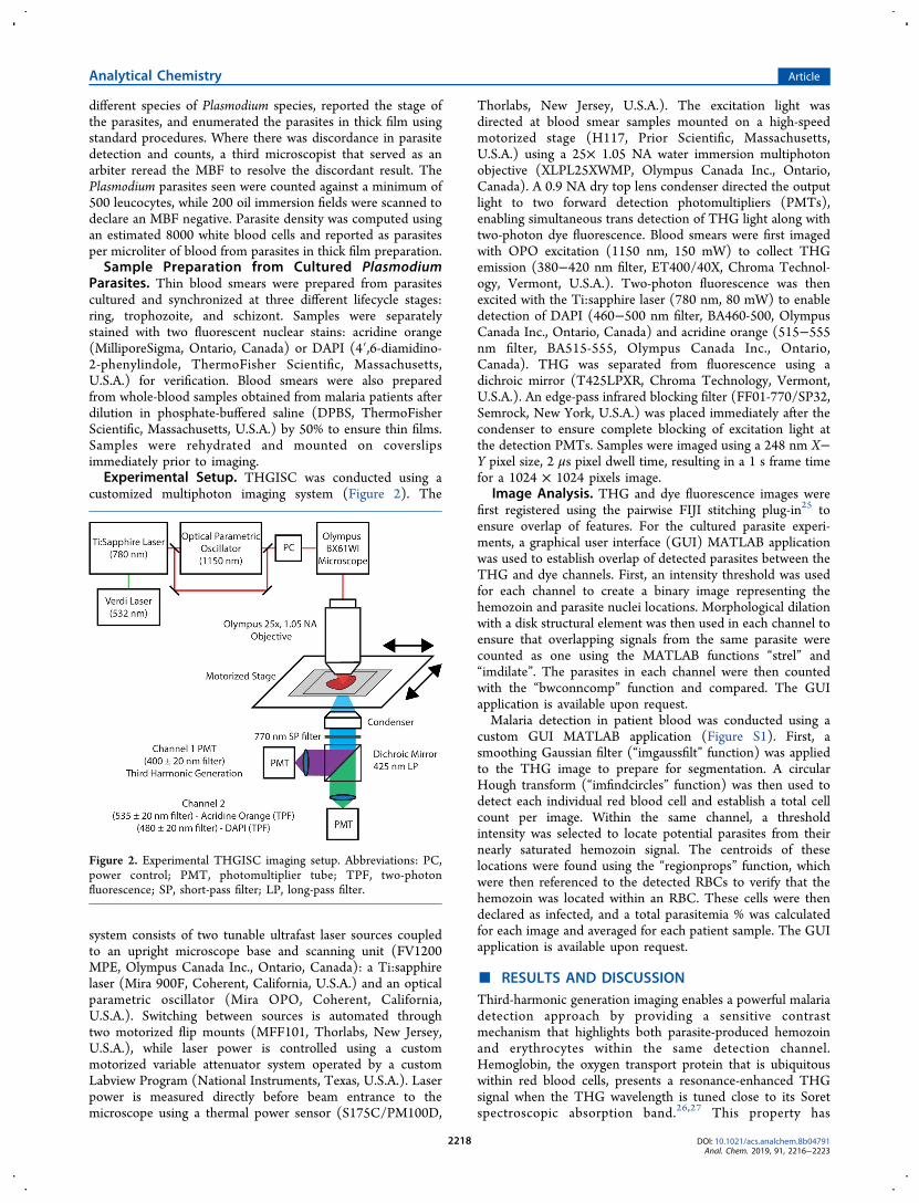
customized multiphoton imaging system (Figure 2). The
system consists of two tunable ultrafast laser sources coupledto an upright microscope base and scanning unit (FV1200MPE, Olympus Canada Inc., Ontario, Canada): a Ti:sapphirelaser (Mira 900F, Coherent, California, U.S.A.) and an opticalparametric oscillator (Mira OPO, Coherent, California,U.S.A.). Switching between sources is automated throughtwo motorized flip mounts (MFF101, Thorlabs, New Jersey,U.S.A.), while laser power is controlled using a custommotorized variable attenuator system operated by a customLabview Program (National Instruments, Texas, U.S.A.). Laserpower is measured directly before beam entrance to themicroscope using a thermal power sensor (S175C/PM100D,
Thorlabs, New Jersey, U.S.A.). The excitation light wasdirected at blood smear samples mounted on a high-speedmotorized stage (H117, Prior Scientific, Massachusetts,U.S.A.) using a 25× 1.05 NA water immersion multiphotonobjective (XLPL25XWMP, Olympus Canada Inc., Ontario,Canada). A 0.9 NA dry top lens condenser directed the outputlight to two forward detection photomultipliers (PMTs),enabling simultaneous trans detection of THG light along withtwo-photon dye fluorescence. Blood smears were first imagedwith OPO excitation (1150 nm, 150 mW) to collect THGemission (380−420 nm filter, ET400/40X, Chroma Technol-ogy, Vermont, U.S.A.). Two-photon fluorescence was thenexcited with the Ti:sapphire laser (780 nm, 80 mW) to enabledetection of DAPI (460−500 nm filter, BA460-500, OlympusCanada Inc., Ontario, Canada) and acridine orange (515−555nm filter, BA515-555, Olympus Canada Inc., Ontario,Canada). THG was separated from fluorescence using adichroic mirror (T425LPXR, Chroma Technology, Vermont,U.S.A.). An edge-pass infrared blocking filter (FF01-770/SP32,Semrock, New York, U.S.A.) was placed immediately after thecondenser to ensure complete blocking of excitation light atthe detection PMTs. Samples were imaged using a 248 nm X−Y pixel size, 2 μs pixel dwell time, resulting in a 1 s frame timefor a 1024 × 1024 pixels image.
Image Analysis. THG and dye fluorescence images werefirst registered using the pairwise FIJI stitching plug-in25 toensure overlap of features. For the cultured parasite experi-ments, a graphical user interface (GUI) MATLAB applicationwas used to establish overlap of detected parasites between theTHG and dye channels. First, an intensity threshold was usedfor each channel to create a binary image representing thehemozoin and parasite nuclei locations. Morphological dilationwith a disk structural element was then used in each channel toensure that overlapping signals from the same parasite werecounted as one using the MATLAB functions “strel” and“imdilate”. The parasites in each channel were then countedwith the “bwconncomp” function and compared. The GUIapplication is available upon request.Malaria detection in patient blood was conducted using a
custom GUI MATLAB application (Figure S1). First, asmoothing Gaussian filter (“imgaussfilt” function) was appliedto the THG image to prepare for segmentation. A circularHough transform (“imfindcircles” function) was then used todetect each individual red blood cell and establish a total cellcount per image. Within the same channel, a thresholdintensity was selected to locate potential parasites from theirnearly saturated hemozoin signal. The centroids of theselocations were found using the “regionprops” function, whichwere then referenced to the detected RBCs to verify that thehemozoin was located within an RBC. These cells were thendeclared as infected, and a total parasitemia % was calculatedfor each image and averaged for each patient sample. The GUIapplication is available upon request.
■ RESULTS AND DISCUSSIONThird-harmonic generation imaging enables a powerful malariadetection approach by providing a sensitive contrastmechanism that highlights both parasite-produced hemozoinand erythrocytes within the same detection channel.Hemoglobin, the oxygen transport protein that is ubiquitouswithin red blood cells, presents a resonance-enhanced THGsignal when the THG wavelength is tuned close to its Soretspectroscopic absorption band.26,27 This property has
Figure 2. Experimental THGISC imaging setup. Abbreviations: PC,power control; PMT, photomultiplier tube; TPF, two-photonfluorescence; SP, short-pass filter; LP, long-pass filter.
Analytical Chemistry Article
DOI: 10.1021/acs.analchem.8b04791Anal. Chem. 2019, 91, 2216−2223
2218

previously been exploited to examine and evaluate morphologyof stored RBCs within blood bags.28 Hemozoin, also known asβ-hematin, is a biocrystal formed during the Plasmodiumparasite’s digestion of hemoglobin.29 The digestion processreleases free hematin, which is toxic to the parasite, resulting inthe production of hemozoin as a detoxification mechanism.Owing to a similar resonance mechanism as hemoglobin,hemozoin has been found to be a very strong source of THG,generating signals over an order of magnitude higher thanthose of hemoglobin. By using THG exclusively as a contrastmechanism, it is possible to simultaneously detect RBCs viahemoglobin and malaria parasites via hemozoin with thesegmentation between the two easily implemented due to thelarge signal difference.As THG is a nonlinear optical effect that requires a tight
diffraction-limited beam focus to achieve the necessaryexcitation power, its usage is incompatible with standard flowcytometry instrumentation which uses a wide focus toilluminate whole cells at lower power. However, by combiningTHG laser scanning microscopy with rapid motorizedscanning, a high-throughput measurement can still be achievedwithout relying on flow processes. This approach brings thebenefit of image-based cytometry which provides single-cellcharacterization, enabling us to monitor hemozoin on asubcellular level in addition to visualizing morphologicalcellular features. Once images of the sampled erythrocytesare obtained, simple image processing can be used to identifyand count red blood cells and highlight potential infected onesvia the significant hemozoin signal.Validation Using Cultured Parasites. To establish our
method as a practical means for malaria detection, it wasimperative to characterize its performance using knowncontrols. We first applied our technique to P. falciparumparasites cultured in packed red blood cells at three differentlifecycle stages: ring (early trophozoite), trophozoite, andschizont stages. To validate our measurements, two-photonfluorescence (TPF) imaging was performed alongside THGimaging using two nuclear staining fluorescent dyes commonlyused for malaria parasite detection, acridine orange (AO)30
and DAPI (4′,6-diamidino-2-phenylindole).31 By performingtwo-photon fluorescence microscopy with the same imagingsystem as for THG, it was possible to obtain a directcomparison for parasite numbers obtained for the same fieldsof view with both methods. Since healthy RBCs do not possessDNA, any fluorescence signal present within an RBC afterstaining could be attributed to the presence of parasite DNA.Both AO and DAPI are intercalating DNA dyes and yieldincreased fluorescence upon binding to DNA. To obtainefficient two-photon excitation of the fluorescent dyes, aTi:sapphire laser source was used instead of the OPO, owingto its ability to generate excitation light at a lower wavelength(780 nm), closer to the peak of their two-photon absorbance.Example images obtained from this study can be found in
Figure 3 for the three different parasite stages examined,showing nearly exact overlap (white) between the parasitenuclei (green for AO and cyan for DAPI) and hemozoin(magenta) locations. It is important to note that the hemozoinis located within the malaria parasite’s digestive vacuole,whereas the AO/DAPI signals originate from the parasite’snucleus. As such, these two signals are not completelycolocalized depending on the parasites’ spatial orientation.The PMT sensitivity for the THG channel was adjusted toclearly visualize the RBCs in addition to the hemozoin,
resulting in a saturated signal associated with the presence ofhemozoin due to its high intrinsic third-order nonlinearsusceptibility. Using simple image processing, we determinedthe number of parasites per image obtained by THG and TPFfor parasites stained with AO (Figure 4A) and DAPI (Figure4B).Using the slopes of the THG versus AO graphs, it could be
seen that THGISC slightly underestimated ring (0.8186) andtrophozoite (0.8170) numbers while matching the numbersobtained for schizonts (1.0085). The numbers obtained forDAPI-stained parasites showed an improvement across allstages, with the trophozoite and schizont slopes beingsignificantly higher than for rings. The differences in parasitecounts obtained by THGISC in comparison to those obtainedfrom the fluorescent DNA probes could potentially beexplained by a difference in focal planes between the twodifferent laser sources used for excitation (780 nm Ti:sapphirevs 1150 nm OPO), which were manually switched for everyexperiment. As THG is an interface effect, it is significantlymore sensitive to small changes in focal plane compared tobulk fluorescence. For earlier parasite stages (ring andtrophozoite), it is possible that the THG excitation beamfailed to encompass their smaller hemozoin crystals at the samefocal plane in which fluorescence could still be seen from theirnuclei.To verify this hypothesis, an additional experiment (Figure
4C) was conducted using DAPI-strained trophozoites wherethe dye was excited using the same Ti:sapphire excitationsource as before along with the OPO that is normally used forTHG, this time manually switched to 1000 nm for each field,which was the shortest wavelength possible for this source.From Figure 4C, the precision of THGISC significantlyimproves (R2 of 0.96 vs 0.88) when the same laser source isused for the comparison, demonstrating the importance ofproper focal plane selection. This issue could potentially beresolved by automated autofocusing or the incorporation ofthree-dimensional image stacks for validation from which amaximum projection could be extracted instead of singleimages. However, in practical applications of our method,
Figure 3. Validation of method performance using nuclear stainingdyes on cultured P. falciparum parasites at the (A and D) ring, (B andE) trophozoite, and (C and F) schizont stages. Panels A−C werestained using acridine orange; panels D−F were stained using DAPI.Magenta, THG signal; green, TPF acridine orange; cyan, TPF DAPI.Scale bar: 10 μm. Separate channel images for panels A−C can befound in Figure S2, while separate channel images for panels D−F canbe found in Figure S3.
Analytical Chemistry Article
DOI: 10.1021/acs.analchem.8b04791Anal. Chem. 2019, 91, 2216−2223
2219

proper focusing using the THG image channel directly shouldminimize false negatives due to this effect. We implementedthis approach for our patient study.Proof of Concept Using Whole-Blood Samples from
Malaria Patients. To characterize our method for potentialclinical applications, we conducted THGISC screening ofwhole blood obtained from malaria patients. For each patient,parasite count was first established at the point of origin by
microscopy of thick blood films. Parasite count was conductedin thick MBF, and the estimated white blood cell count of thepatient (8000) was used in determining the parasitemia inparasite per microliter count32 (eq 1):
μ μ= ×
n n
nL8000 white blood cells
Lparasites parasites
white blood cells (1)
Using THGISC, parasite counts were established usingunlabeled methanol-fixed thin blood smears to determine apercentage of parasitemia for each patient. Parasitemia wasdetermined using eq 2:
= ×n
n% parasitemia 100 infected RBCs
total RBCs (2)
An example of images obtained from two different patients canbe found on Figure 5. We established a direct comparison for
the % parasitemia obtained from our technique against theparasites per microliter that were previously clinicallydetermined, which can be found on Figure 6. The relationshipbetween the two methods can be seen to be very linear (R2 of0.96), demonstrating that malaria detection by THGISCprovides comparable performance to the classical method ofmalaria detection.The larger variation between figures obtained for patients
having smaller parasite counts could potentially be explainedby parasite reproduction during transit from Nigeria to Canadaalong with variation in sampling during preparation of theblood films. To obtain accurate results for smaller parasitecounts, it is critical for the erythrocytes to be set in a propersingle layer with the blood homogeneously mixed to increasethe chances of finding single isolated parasites.
Figure 4. Results of method validation using fluorescent nuclearstaining dyes on three different parasite lifecycle stages: ring,trophozoite, and schizont. Number of parasites obtained by ourTHG method vs no. of parasites detected by TPF using (A) acridineorange, (B) DAPI, and (C) DAPI with same/different excitationsources for comparison for trophozoites only. A linear regression bestfit line was calculated for each data set (solid lines). N = 20−25 fieldsfor each parasite stage.
Figure 5. Raw and processed THGISC images for malaria detectionin patient blood. (A and B) Malaria parasites (arrows) detected inwhole-blood smears of two malaria patients. Inset: intensity crosssection through red blood cell and hemozoin THG signals. (C and D)Red blood cell segmentation and hemozoin detection for images inpanels A and B. Scale bar: 10 μm.
Analytical Chemistry Article
DOI: 10.1021/acs.analchem.8b04791Anal. Chem. 2019, 91, 2216−2223
2220

Thanks to the use of automated scanning, the number offields examined by THGISC could also be increased to covermost of the blood smear to increase detection probability inpatients with low parasitemia. Assuming that the parasitemiarate is constant throughout a blood sample, the probability offinding a small parasite count within the millions of uninfectedblood cells should follow a Poisson distribution. The higherthroughput enabled by our method could allow examiners tosample significantly more fields than is normally possible dueto labor constraints, which could increase the overallprobability of finding at least one parasite in an entire bloodsmear. Such an approach could also be beneficial for parasitescreening in populations to find potential asymptomaticcarriers, an important checkpoint on the road to completemalaria eradication.33 In addition, automated scanningremoves the subjectivity of manual malaria blood smearreading by microscopists, while increasing the screeningthroughput.The main drawback of the technique is the requirement for
an ultrafast mode-locked excitation laser source for THGimaging which carries a significant initial expense. However,while a research grade tunable OPO was used in ourdemonstration, a much less expensive setup using a single-wavelength fiber laser based setup could provide a moreeconomical option.34 With the rapid progress in laser oscillatordevelopment and optimization, it is conceivable to expect suchlaser sources to become more widely available and accessible.Other than cost, the power requirement of the excitation lasermust also be considered, rendering this method more viable foruse in larger hospitals and clinics where such power can bereliably present. In such locations, a quantitative and easy-to-use higher throughput method would be highly advantageousfor routinely tracking patient parasitemia throughout treat-ment, allowing earlier health care interventions.Another limitation of the technique is the throughput
obtained via laser scanning cytometry in comparison to flowcytometry. While much faster than manual counting, a higherthroughput would be desirable. Due to the nature of THG asan interface effect arising from the Gouy phase shift, theemitted signal is maximized when the sample being imaged ison the same physical scale as the laser beam.35 For hemozoincrystals, this requires diffraction-limited beam sizes to max-imize sensitivity.20 To incorporate this approach into a flowcytometry instrument, a high-speed line scanner could
potentially be adapted to continuously scan incoming RBCs.However, this would require a very tight focus of the cell flowin the axial dimension to avoid focal plane issues. Anotherpotential solution would be the utilization of scanned Besselbeams to increase the available depth of field to ensure thatparasites are detected along all axial positions in each RBC.However, a theoretical evaluation of the application of Besselbeams to THG imaging showed that it would potentiallyrequire significantly more excitation power than the standardGaussian beams typically used in THG microscopy, whichwould bring additional challenges to the practical implementa-tion.36 Biologically, the absence of hemozoin in the very earlyring stages of Plasmodium parasites (less than 6 h)37 may bemissed by THG and could explain the slightly reduceddetection compared with TPF.Malaria detection by THGISC shows promising results as
compared to the current gold standard, light microscopy;however, several improvements could be made to enhance themeasurements. While blood smears were prepared in methanolfor each patient sample to ensure sample preservation for theentire batch after receiving of the shipment, direct measure-ments without fixation could provide a benefit in sensitivity. Inlive samples, red blood cells present a more standardizedshape, resulting in better image segmentation. In addition, livesamples present a lower background signal due to a reducednumber of THG-emitting particles that can often beintroduced during sample processing.Treatment options for malaria patients typically consist of
artemisinin-based combination therapies (ACT) where anartemisinin derivative is combined with a quinine derivative.The specific combinations, treatment duration, and dosagedepend on patient factors such as endemicity, age, weight,pregnancy, and immunocompromised status, in addition toclinical diagnosis. According to the WHO,38 symptomaticmalaria patients with no signs of severity or organ dysfunctionare categorized as “uncomplicated malaria”, whereas “severemalaria” is defined with signs of severity and/or signs of vitalorgan dysfunction. It has been reported that some patients maybe symptomatic at parasitemia levels as low as 0.002%, whereaspatients that have developed a partial protective immunityowing to repeated exposure only show symptoms aboveparasitemia of 0.2%.39 The WHO defines hyperparasitemia as>4%, a level which increases the risk of deterioration fromuncomplicated to severe malaria. At a parasitemia >10%, theWHO considers the case to be severe malaria even if there isno evidence of vital organ dysfunction. We have demonstratedmalaria detection at physiological levels ranging from 0.2% to1.2%, consistent with detection using Giemsa-stained bloodsmear imaging. However, our method demonstrates anautomated higher throughput approach in comparison tomanual counting by microscopists.Current developments in adaptive optics and three-photon
fluorescence instrumentation have resulted in significantimprovements in deep tissue imaging, with some commercialobjectives allowing imaging at depths of up to 8 mm. Givensuch progress, it could be possible to apply THG-based malariadetection in vivo with imaging conducted directly in bloodvessels through patient skin with epi-based detection. Such anapproach was previously used for conducting human leukocytecytometry by using THG transdermally.40
Figure 6. Results of method characterization using patient samples. N= 11 patients. The percentage of infected RBCs is comparable toparasitemia obtained via imaging of thick films. A linear regressionbest fit line was calculated (solid line).
Analytical Chemistry Article
DOI: 10.1021/acs.analchem.8b04791Anal. Chem. 2019, 91, 2216−2223
2221

■ CONCLUSIONWe developed a new optical detection method anddemonstrated that it is a viable tool for malaria detection. Bycombining the sensitivity of THG imaging with high-speedmotorized scanning and image processing software, malariadetection at the single red blood cell level can be achieved. Thetechnique relies on intrinsic contrast obtained from the third-order susceptibilities of hemoglobin and hemozoin to obtainboth cytometry of red blood cells and detection of malariaparasites in a single channel. Unlike traditional methods, ourapproach does not require any labels or any preparationbeyond a simple blood smear. In addition, thanks to theautomated image processing, parasitemia counts can be rapidlydetermined in an objective manner without requiring skilledlabor. While the method was shown to be sensitive to properfocusing, this could be remedied with the use of an autofocusmodule.In control measurements, THGISC showed good linearity
and comparable results to parasite counts obtained by two-photon fluorescence of parasite DNA stains, with a slightunderestimation of smaller parasites from earlier stages (ringand trophozoite). In direct application to blood smearsobtained from confirmed malaria patients, the techniqueshowed great linearity and comparable performance tostandard thick blood smear measurements. The ability toeasily and rapidly determine parasitemia offers potential notonly for the easy confirmation of malaria diagnoses followingsymptoms, but also the tracking of treatment progress inexisting patients, potentially allowing physicians to adjustmedication and dosage for each individual. The techniquecould also be used to assist in the development of newantimalarial drugs, some of which have been theorized to beactively blocking hemozoin formation, resulting in a fataloutcome for the parasite.41
■ ASSOCIATED CONTENT*S Supporting InformationThe Supporting Information is available free of charge on theACS Publications website at DOI: 10.1021/acs.anal-chem.8b04791.
MATLAB application for red blood cell counting andhemozoin detection by THGISC image analysis andvalidation of method performance using acridine orangeand DAPI nuclear staining (PDF)
■ AUTHOR INFORMATIONCorresponding Author*E-mail: [email protected] W. Wiseman: 0000-0002-5732-2858NotesThe authors declare no competing financial interest.
■ ACKNOWLEDGMENTSThe authors gratefully acknowledge Discovery Grant supportfrom the Natural Sciences and Engineering Research Councilof Canada for P.W.W. (RGPIN-2018-05005) and E.G.(RGPIN-2017-05009). A.K. would like to acknowledge theFond de Recherche du QuebecNature et Technologies forscholarship support. The authors thank the Bio Ventures forGlobal Health Organization for helping establish this
collaboration. We thank the Lagos State Ministry of Healthfor approving the use of Health Facilities in Ikorodu, LagosState, Nigeria where samples for this study were obtained aswell as the management and staff of Ijede General Hospital andImota Primary Health Centre, Ikorodu, Lagos, Nigeria.
■ REFERENCES(1) Phillips, M. A.; Burrows, J. N.; Manyando, C.; van Huijsduijnen,R. H.; Van Voorhis, W. C.; Wells, T. N. C. Nat. Rev. Dis Primers 2017,3, 17050.(2) World Malaria Report 2017; World Health Organization:Geneva, Switzerland, 2017.(3) Miller, L. H.; Ackerman, H. C.; Su, X. Z.; Wellems, T. E. Nat.Med. 2013, 19 (2), 156−67.(4) White, N. J.; Pukrittayakamee, S.; Hien, T. T.; Faiz, M. A.;Mokuolu, O. A.; Dondorp, A. M. Lancet 2014, 383 (9918), 723−735.(5) Cowman, A. F.; Healer, J.; Marapana, D.; Marsh, K. Cell 2016,167 (3), 610−624.(6) Mathison, B. A.; Pritt, B. S. J. Clin. Microbiol. 2017, 55 (7),2009−2017.(7) Hathiwala, R.; Mehta, P. R.; Nataraj, G.; Hathiwala, S. J. InfectPublic Health 2017, 10 (6), 824−828.(8) Wilson, M. L. Arch. Pathol. Lab. Med. 2013, 137 (6), 805−11.(9) Britton, S.; Cheng, Q.; McCarthy, J. S. Malar. J. 2016, 15, 88.(10) Kolluri, N.; Klapperich, C. M.; Cabodi, M. Lab Chip 2018, 18(1), 75−94.(11) Ragavan, K. V.; Kumar, S.; Swaraj, S.; Neethirajan, S. Biosens.Bioelectron. 2018, 105, 188−210.(12) Coronado, L. M.; Nadovich, C. T.; Spadafora, C. Biochim.Biophys. Acta, Gen. Subj. 2014, 1840 (6), 2032−41.(13) Delahunt, C.; Horning, M. P.; Wilson, B. K.; Proctor, J. L.;Hegg, M. C. Malar. J. 2014, 13, 147.(14) Pirnstill, C. W.; Cote, G. L. Sci. Rep. 2015, 5, 13368.(15) Lukianova-Hleb, E. Y.; Campbell, K. M.; Constantinou, P. E.;Braam, J.; Olson, J. S.; Ware, R. E.; Sullivan, D. J., Jr.; Lapotko, D. O.Proc. Natl. Acad. Sci. U. S. A. 2014, 111 (3), 900−5.(16) Lukianova-Hleb, E.; Bezek, S.; Szigeti, R.; Khodarev, A.; Kelley,T.; Hurrell, A.; Berba, M.; Kumar, N.; D’Alessandro, U.; Lapotko, D.Emerging Infect. Dis. 2016, 22 (2), 344−344.(17) Burnett, J. L.; Carns, J. L.; Richards-Kortum, R. Biomed. Opt.Express 2015, 6 (9), 3462−74.(18) Weigelin, B.; Bakker, G. J.; Friedl, P. J. Cell Sci. 2016, 129 (2),245−55.(19) Belisle, J. M.; Costantino, S.; Leimanis, M. L.; Bellemare, M. J.;Bohle, D. S.; Georges, E.; Wiseman, P. W. Biophys. J. 2008, 94 (4),L26−8.(20) Tripathy, U.; Giguere-Bisson, M.; Sangji, M. H.; Bellemare, M.J.; Bohle, D. S.; Georges, E.; Wiseman, P. W. Anal. Bioanal. Chem.2013, 405 (16), 5431−40.(21) Pozarowski, P.; Holden, E.; Darzynkiewicz, Z. Methods Mol.Biol. 2006, 319, 165−92.(22) Pozarowski, P.; Holden, E.; Darzynkiewicz, Z. Methods Mol.Biol. 2012, 931, 187−212.(23) Han, Y.; Gu, Y.; Zhang, A. C.; Lo, Y. H. Lab Chip 2016, 16(24), 4639−4647.(24) Trager, W.; Jensen, J. Science 1976, 193 (4254), 673−675.(25) Preibisch, S.; Saalfeld, S.; Tomancak, P. Bioinformatics 2009, 25(11), 1463−5.(26) Chang, C. F.; Yu, C. H.; Sun, C. K. Journal of biophotonics 2010,3 (10−11), 678−85.(27) Clay, G. O.; Millard, A. C.; Schaffer, C. B.; Aus-Der-Au, J.; Tsai,P. S.; Squier, J. A.; Kleinfeld, D. J. Opt. Soc. Am. B 2006, 23 (5), 932−950.(28) Saytashev, I.; Glenn, R.; Murashova, G. A.; Osseiran, S.;Spence, D.; Evans, C. L.; Dantus, M. Biomed. Opt. Express 2016, 7 (9),3449−3460.(29) Egan, T. J. J. Inorg. Biochem. 2002, 91 (1), 19−26.
Analytical Chemistry Article
DOI: 10.1021/acs.analchem.8b04791Anal. Chem. 2019, 91, 2216−2223
2222

(30) Shute, G. T.; Sodeman, T. M. Bull. World Health Organ. 1973,48 (5), 591−596.(31) Hyman, B. C.; Macinnis, A. J. J. Parasitol. 1979, 65 (3), 421−5.(32) DPDx - Laboratory Identification of Parasites of Public HealthConcern. Determination of Parasitemia; Centers for Disease Controland Prevention: Atlanta, GA, 2014.(33) Krampa, F. D.; Aniweh, Y.; Awandare, G. A.; Kanyong, P.Diagnostics 2017, 7 (3), 54.(34) Millard, A. C.; Wiseman, P. W.; Fittinghoff, D. N.; Wilson, K.R.; Squier, J. A.; Muller, M. Appl. Opt. 1999, 38 (36), 7393−7397.(35) Beaurepaire, E.; Deb́arre, D.; Olivier, N. THG Microscopy ofCells and Tissues: Contrast Mechanisms and Applications. In SecondHarmonic Generation Imaging; CRC Press: Boca Raton, FL, 2016; pp70−99.(36) Olivier, N.; DeBarre, D.; Mahou, P.; Beaurepaire, E. Opt.Express 2012, 20 (22), 24886−902.(37) Delahunt, C.; Horning, M. P.; Wilson, B. K.; Proctor, J. L.;Hegg, M. C. Malar. J. 2014, 13, 147.(38) Guidelines for the Treatment of Malaria; World HealthOrganization: Geneva, Switzerland, 2015.(39) Hanscheid, T. Clin. Lab. Haematol. 1999, 21 (4), 235−45.(40) Wu, C. H.; Wang, T. D.; Hsieh, C. H.; Huang, S. H.; Lin, J. W.;Hsu, S. C.; Wu, H. T.; Wu, Y. M.; Liu, T. M. Sci. Rep. 2016, 6, 37210.(41) Kumar, S.; Guha, M.; Choubey, V.; Maity, P.; Bandyopadhyay,U. Life Sci. 2007, 80 (9), 813−28.
Analytical Chemistry Article
DOI: 10.1021/acs.analchem.8b04791Anal. Chem. 2019, 91, 2216−2223
2223