Magnetic techniques for the detection and … Magnetic techniques for the detection and...
17
REVIEW Magnetic techniques for the detection and determination of xenobiotics and cells in water Ivo Safarik & Katerina Horska & Kristyna Pospiskova & Mirka Safarikova Received: 15 February 2012 / Revised: 15 April 2012 / Accepted: 16 April 2012 / Published online: 12 May 2012 # Springer-Verlag 2012 Abstract Magnetic techniques based on the application of magnetic nanoparticles and microparticles and films have been successfully used for the determination and detection of different types of xenobiotics (e.g. herbicides, insecti- cides, fungicides, aromatic and polyaromatic hydrocarbons, pentachlorophenol and heavy metal ions) as well as viruses, microbial pathogens and protozoan parasites in water sam- ples. Preconcentration of xenobiotics from large volumes of samples can be performed using magnetic solid-phase extraction, stir-bar sorptive extraction and related proce- dures. This review provides basic information about these techniques. Published examples of successful applications document the importance of these simple and efficient pro- cedures employing magnetic materials. Keywords Bioanalytical methods . Enzymes . Immunoassays/ELISA . Organic compounds . Water . Preconcentration Introduction Human activity is accompanied by the production of both organic and inorganic xenobiotics (contaminants). These contaminants are usually analysed using highly sophisticat- ed analytical techniques. High-performance liquid chro- matography and gas chromatography systems equipped with different types of detectors are routinely used for assays of organic xenobiotics, whereas atomic absorption spectro- photometry and inductively coupled plasma mass spectrosco- py are used for assays of inorganic xenobiotics. These methods usually require extensive analyte preconcentration, experienced technical staff and expensive equipment and reagents. As a consequence, attention has also been focused on the development of new methods enabling relatively simple, rapid and sensitive assays. Immunoassays seem to be methods of choice, both for screening and for analytical purposes; they have been used for many years in clinical chemistry as reli- able, sensitive and selective methods to determine low con- centrations of organic compounds in, for example, blood, urine and tissue extracts [1]. The possibility to use immunoassays for environmental studies was recognized in the early 1970s. Immunoassays have been developed for a broad range of pesticides and contaminants of industrial origin. This interesting topic has been reviewed in several articles and book chapters [1–5]. Immunoassays can benefit from the application of mag- netic nanoparticles or microparticles. In general, magneti- cally responsive particles can be used in an enormous number of applications, ranging from molecular biology to wastewater treatment [6–8]. Magnetic particles exhibit the following important properties [8, 9]: 1. Selective separation and removal of magnetically respon- sive nanoparticles and microparticles and other relevant materials from complex samples using an external mag- netic field (e.g. an appropriate magnetic separator, perma- nent magnet, or electromagnet). 2. Targeting and localization of magnetic particles to the desired place using an external magnetic field (e.g. I. Safarik (*) : K. Horska : M. Safarikova Department of Nanobiotechnology, Institute of Nanobiology and Structural Biology of GCRC, Na Sadkach 7, 370 05 Ceske Budejovice, Czech Republic e-mail: [email protected] I. Safarik Regional Centre of Advanced Technologies and Materials, Palacky University, Slechtitelu 11, 783 71 Olomouc, Czech Republic K. Pospiskova Department of Biochemistry, Faculty of Science, Palacky University, Slechtitelu 11, 783 71 Olomouc, Czech Republic Anal Bioanal Chem (2012) 404:1257–1273 DOI 10.1007/s00216-012-6056-x
Transcript of Magnetic techniques for the detection and … Magnetic techniques for the detection and...
REVIEW
Magnetic techniques for the detection and determinationof xenobiotics and cells in water
Ivo Safarik & Katerina Horska & Kristyna Pospiskova &
Mirka Safarikova
Received: 15 February 2012 /Revised: 15 April 2012 /Accepted: 16 April 2012 /Published online: 12 May 2012# Springer-Verlag 2012
Abstract Magnetic techniques based on the application ofmagnetic nanoparticles and microparticles and films havebeen successfully used for the determination and detectionof different types of xenobiotics (e.g. herbicides, insecti-cides, fungicides, aromatic and polyaromatic hydrocarbons,pentachlorophenol and heavy metal ions) as well as viruses,microbial pathogens and protozoan parasites in water sam-ples. Preconcentration of xenobiotics from large volumes ofsamples can be performed using magnetic solid-phaseextraction, stir-bar sorptive extraction and related proce-dures. This review provides basic information about thesetechniques. Published examples of successful applicationsdocument the importance of these simple and efficient pro-cedures employing magnetic materials.
Keywords Bioanalytical methods . Enzymes .
Immunoassays/ELISA . Organic compounds . Water .
Preconcentration
Introduction
Human activity is accompanied by the production of bothorganic and inorganic xenobiotics (contaminants). These
contaminants are usually analysed using highly sophisticat-ed analytical techniques. High-performance liquid chro-matography and gas chromatography systems equippedwith different types of detectors are routinely used for assaysof organic xenobiotics, whereas atomic absorption spectro-photometry and inductively coupled plasma mass spectrosco-py are used for assays of inorganic xenobiotics. Thesemethods usually require extensive analyte preconcentration,experienced technical staff and expensive equipment andreagents.
As a consequence, attention has also been focused on thedevelopment of new methods enabling relatively simple,rapid and sensitive assays. Immunoassays seem to be methodsof choice, both for screening and for analytical purposes; theyhave been used for many years in clinical chemistry as reli-able, sensitive and selective methods to determine low con-centrations of organic compounds in, for example, blood,urine and tissue extracts [1].
The possibility to use immunoassays for environmentalstudies was recognized in the early 1970s. Immunoassayshave been developed for a broad range of pesticides andcontaminants of industrial origin. This interesting topichas been reviewed in several articles and book chapters[1–5].
Immunoassays can benefit from the application of mag-netic nanoparticles or microparticles. In general, magneti-cally responsive particles can be used in an enormousnumber of applications, ranging from molecular biology towastewater treatment [6–8]. Magnetic particles exhibit thefollowing important properties [8, 9]:
1. Selective separation and removal of magnetically respon-sive nanoparticles and microparticles and other relevantmaterials from complex samples using an external mag-netic field (e.g. an appropriate magnetic separator, perma-nent magnet, or electromagnet).
2. Targeting and localization of magnetic particles to thedesired place using an external magnetic field (e.g.
I. Safarik (*) :K. Horska :M. SafarikovaDepartment of Nanobiotechnology,Institute of Nanobiology and Structural Biology of GCRC,Na Sadkach 7, 370 05 Ceske Budejovice, Czech Republice-mail: [email protected]
I. SafarikRegional Centre of Advanced Technologies and Materials,Palacky University, Slechtitelu 11, 783 71 Olomouc,Czech Republic
K. PospiskovaDepartment of Biochemistry, Faculty of Science,Palacky University, Slechtitelu 11, 783 71 Olomouc,Czech Republic
Anal Bioanal Chem (2012) 404:1257–1273DOI 10.1007/s00216-012-6056-x

sealing the rotating objects using magnetic fluids ormagnetic drug targeting).
3. Heat production caused by magnetic particles subjectedto a high-frequency alternating magnetic field. Thisphenomenon is employed especially during magneticfluid hyperthermia, e.g., for cancer treatment.
4. Increase of a negative T2 contrast by magnetic iron oxidenanoparticles during magnetic resonance imaging.
5. Great increase of apparent viscosity of magnetorheolog-ical fluids when they are subjected to a magnetic field.
6. Magnetic nanoparticles and microparticles can be usedfor magnetic modification of diamagnetic biologicalmaterials (e.g. cells) and magnetic labelling of biologi-cally active compounds (e.g. antibodies and enzymes).This property can be efficiently employed in the devel-opment of magnetic assays for determination and detec-tion of xenobiotics and cells.
The conversion of classic immunoassays (performed, e.g.,in microtitration plates) to the magnetic version (performedmainly in test tubes with the help of magnetic separators)opened new possibilities for their applications, such as theiruse during field surveys. Two types of magnetic-particle-based immunoassays can be distinguished:
1. Immunomagnetic assays, where an appropriate antibody(or antibodies) is immobilized on a magnetic carrier(instead of in the wells of the microtitration plates)
2. Magnetoimmunoassays, where magnetic particles serveas a detectable label (instead of the enzymes, radio-nuclides or luminescent molecules used in standardimmunoassays)
Immunomagnetic assays are very similar to standardmicrotitration plate assays; the main difference is that spe-cific antibodies are bound to the magnetically responsiveparticles, which can be separated from the suspension usingappropriate magnetic separators based on strong rare-earthpermanent magnets.
Immunomagnetic techniques have also been successfullyused for the detection of pathogenic bacteria and viruses infood and clinical microbiology [10, 11]. This technique hasalso been employed in environmental microbiology for thedetection of target viruses, bacteria and protozoan parasitesin water samples of different origin.
Also enzymes (e.g. acetylcholinesterase) immobilized onmagnetic carriers can be used for the detection of xeno-biotics acting as enzyme inhibitors, such as carbofuran,paraoxon, malaoxon, and paraoxon-methyl [12].
In many cases the target analytes present in water sam-ples have to be preconcentrated before appropriate analyti-cal procedures are performed. Currently, preconcentration isusually performed using liquid–liquid extraction or solid-phase extraction (SPE) processes. Recently, a new
extraction technique based on the application of magnetical-ly responsive adsorbents has been developed; this techniqueis known as magnetic SPE (MSPE) [13]. Another technique,called stir-bar sorptive extraction (SBSE), is based on theuse of a magnetic stir bar covered with a layer of an appro-priate adsorbent [14].
The synthesis of magnetic ferrites has been used to removemetal pollutants from wastewater. The method involves theincorporation of metal ions in the ferrite structure during itssynthesis by a coprecipitation method [15–17].
It can be clearly seen that magnetically responsive mate-rials can be widely used in detection and analytical systemsbecause of the performance advantages they offer comparedwith similar solids that lack magnetic properties [15]. All theabove-mentioned procedures will be briefly summarized inthe following sections.
Immunomagnetic techniques for detectionand determination of xenobiotics, viruses and cells
Different types of magnetic nanoparticles and microparticlesare available commercially. The magnetic particles for theassays have to enable both efficient immobilization of anti-bodies and other biomolecules (e.g. enzymes) and theirrapid separation using a simple permanent magnet or mag-netic separator. For commercially available RaPID Assaykits, intended for determination of xenobiotics, silanizedmagnetic iron oxide microparticles are used as a carrier.Immunomagnetic separations of cells are usually based onthe use of Dynabeads.
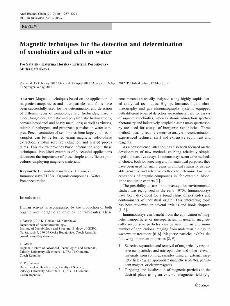
Currently, two commercially available assay systems fordetermination of xenobiotics based on the application ofmagnetic particles for antibody immobilization are used,namely, RaPID Assay (SDIX, formerly Strategic Diagnostics,USA) and Abraxis (Abraxis, USA) kits (see Fig. 1).
A typical assay can be performed as follows. In thisexample, the volumes and times for both SDIX and Abraxisatrazine assays will be reviewed. The protocols for otheranalytes are similar. First, 200 μL of water sample is directlycombined with 250 μL of the atrazine enzyme conjugateand 500 μL of antibody-coupled magnetic particle suspen-sion in a test tube. After vortexing, this mixture is incubatedfor 15 min, and then the magnetic particles are pulled to theside of the test tube by placing it in a special magnetic rackfor 2 min. The particles are rinsed twice with washingsolution and then 500 μL of colour reagent (containinghydrogen peroxide and 3,3′,5,5′-tetramethylbenzidine in anorganic base) is added. After 20 min, this reaction is stoppedwith 500 μL of sulfuric acid solution and the absorbance ofthe tube contents is measured (after magnetic separation) ina spectrophotometer at 450 nm. Because a competitiveimmunoassay is used, where the antigen (analyte) in the
1258 I. Safarik et al.
analysed sample competes with enzyme (horseradish perox-idase) labelled antigen to bind with immobilized antibodies,colour development is inversely proportional to the analyteconcentration; a darker colour corresponds to lower analyteconcentration and vice versa. The analyte concentration inan unknown sample is calculated using the standard calibra-tion curve generated from kit standards after performing thereaction and reading with a spectrophotometer.
The kits currently available commercially, together withthe applications described in the area of water contaminantstudies and the sensitivities of the kits, are presented inTable 1.
The spectrophotometric detection of the reaction productduring the immunoassays is very simple and is the basis ofportable systems enabling field analyses. However, manystudies have been performed recently where immunomag-netic particles were coupled with electrochemical sensors[18, 19]. The main advantage of such a combination is thesubstantial decrease of the detection limit (e.g. 50 timeslower for PCBs) in comparison with standard magneticimmunoassays with spectrophotometric detection [20, 21].
Immunomagnetic assays for determination of pesticideswere compared with “standard” analytical procedures, suchas GC-MS. In the case of atrazine determination, the RaPIDAssay kit was internally consistent with a variation of lessthan 3 % between 156 duplicate standards that were run toproduce standard curves. For the 217 samples examined,atrazine concentrations measured by the two analytical tech-niques were highly correlated (r00.96). Compared with theconcentrations measured by GC-MS, the magnetic assay pro-duced no false negatives and few false positives (5.53 %). Themagnetic assay tended to overestimate atrazine concentrationsslightly, probably the result of cross-reaction in the assay byatrazine metabolites and structurally related triazine herbi-cides. Even so, the immunomagnetic assays proved to be areliable, relatively simple and cost-effective screening tech-nique for atrazine contamination [22].
Not only the individual chemical compounds, but alsoviruses and whole cells can be successfully separated using
magnetic particles. Magnetic separation of viruses and cellshas several advantages in comparison with other techniquesused for the same purpose. It permits the target cells to beisolated directly from crude materials such as environmentalwater samples, sediments, soil, blood, bone marrow, tissuehomogenates, stool, fermentation media and food. Com-pared with other conventional methods of cell separation,magnetic separation is relatively simple and fast. The largedifferences between the magnetic permeabilities of the mag-netic and non-magnetic cells enable highly selective sepa-rations. Cells isolated by magnetic separation processes areusually pure, viable and unaltered. The whole separationprocess can be performed in the same tube running multiplesamples simultaneously in a fraction of the usual time.Separated microbial cells are usually grown on selectiveagars [10].
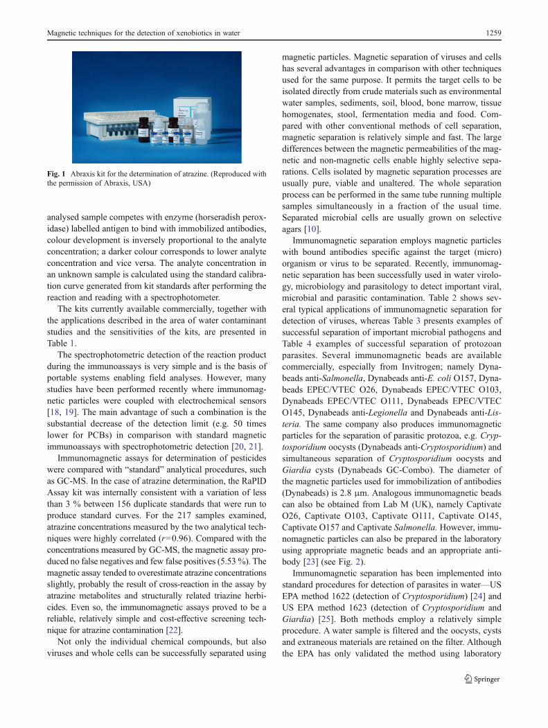
Immunomagnetic separation employs magnetic particleswith bound antibodies specific against the target (micro)organism or virus to be separated. Recently, immunomag-netic separation has been successfully used in water virolo-gy, microbiology and parasitology to detect important viral,microbial and parasitic contamination. Table 2 shows sev-eral typical applications of immunomagnetic separation fordetection of viruses, whereas Table 3 presents examples ofsuccessful separation of important microbial pathogens andTable 4 examples of successful separation of protozoanparasites. Several immunomagnetic beads are availablecommercially, especially from Invitrogen; namely Dyna-beads anti-Salmonella, Dynabeads anti-E. coli O157, Dyna-beads EPEC/VTEC O26, Dynabeads EPEC/VTEC O103,Dynabeads EPEC/VTEC O111, Dynabeads EPEC/VTECO145, Dynabeads anti-Legionella and Dynabeads anti-Lis-teria. The same company also produces immunomagneticparticles for the separation of parasitic protozoa, e.g. Cryp-tosporidium oocysts (Dynabeads anti-Cryptosporidium) andsimultaneous separation of Cryptosporidium oocysts andGiardia cysts (Dynabeads GC-Combo). The diameter ofthe magnetic particles used for immobilization of antibodies(Dynabeads) is 2.8 μm. Analogous immunomagnetic beadscan also be obtained from Lab M (UK), namely CaptivateO26, Captivate O103, Captivate O111, Captivate O145,Captivate O157 and Captivate Salmonella. However, immu-nomagnetic particles can also be prepared in the laboratoryusing appropriate magnetic beads and an appropriate anti-body [23] (see Fig. 2).
Immunomagnetic separation has been implemented intostandard procedures for detection of parasites in water—USEPA method 1622 (detection of Cryptosporidium) [24] andUS EPA method 1623 (detection of Cryptosporidium andGiardia) [25]. Both methods employ a relatively simpleprocedure. A water sample is filtered and the oocysts, cystsand extraneous materials are retained on the filter. Althoughthe EPA has only validated the method using laboratory
Fig. 1 Abraxis kit for the determination of atrazine. (Reproduced withthe permission of Abraxis, USA)
Magnetic techniques for the detection of xenobiotics in water 1259

Tab
le1
Examples
ofxeno
biotic
assays
inwater
samples
usingcommercially
availableim
mun
omagnetic
kitsandph
otom
eter
measurement
Xenobiotics
Characteristics
Kitused
Assay
range(ppb)
Water
system
Com
ments
Reference
Acetochlor
Herbicide,pesticide
Abraxis
0.10–2.5
Wastewater
treatm
entplanteffluents
Acetochloras
themajor
toxicant
intank
truck
cleaning
wastewater
effluent
[55]
Atrazine
Herbicide,pesticide
RaPID
Assay
0.4-5.0
Estuarine
waters
Com
parisonwith
GC
[56]
Ebroriverwater
Annualmonito
ring
(from
March
1995
toJune
1996)
[57]
Realestuarine,
coastalandspiked
water
samples
Evaluationof
2differentim
munoassays;solid
-phase
extractio
nfollo
wed
byLC-D
AD
[58]
Surface
water
inOntario,Canada
Spatialandseasonal
variations
measurement
[59]
Abraxis
0.1–5.0
Water
from
drinking
water
treatm
entplants
Study
ofatrazine
occurrence
intheUSA
[60]
Water
from
atrazine-spikedmicrocosm
sStudy
ofatrazine
asachem
ical
stressor
[61]
Water
samples
from
industrial
effluents
Com
parisonwith
SPEfollo
wed
byLC-A
PCI-MS
[62]
Carbofuran
Insecticide
RaPID
Assay
0.06-5.0
Water
samples
from
Daphnia
magna
experiments
Study
ofsimultaneousexposure
ofDaphnia
magna
tosuspendedsolid
sandcarbofuran
[63,
64]
Fish,
bluegills
(Lepom
ismacrochirus)
Com
parisonof
2commercial
immunoassays
[65]
2,4-Dichloro-
phenoxyacetic
acid
Herbicide
RaPID
Assay
0.7-50.0
12surfacewatersin
thePiedm
ont,USA
12%
ofsamples
tested
positiv
e[66]
17β-Estradiol
Estrogen
Abraxis
1.5-25.0
Realandspiked
water
samples
Com
parisonwith
chromatographytechniques
[67]
Glyphosate
Herbicide
Abraxis
0.075-4.0
Surface
water
Evaluationof
theanalytical
procedure
[68]
Metolachlor
Herbicide
RaPID
Assay
0.05-5.0
Surface
runoff,tiledrainage
Com
parisonwith
SPE-G
CandSPME-G
C[69]
12surfacewatersin
thePiedm
ont,USA
66%
ofsamples
tested
positiv
e[66]
Surface
water
samples
Com
parisonof
ELISA
andGC/M
S[70]
Surface
water
inOntario,Canada
Spatialandseasonal
variations
measurement
[59]
PAHs
PAHs
RaPID
Assay
0.7-50.0
River
water
samples
Com
parisonwith
GC-M
Stechniques
[71]
CarcinogenicPA
Hs
PAHswith
4or
morerings
RaPID
Assay
0.2-10.0
Environmentalsedimentsamples
from
thePatos
Lagoonestuary,
southern
Brazil
Com
parisonwith
GC-FID
[72]
Water
samples
from
industrial
effluents
Com
parisonwith
SPEfollo
wed
byLC-A
PCI-MS
[62]
PBDEs
PDBEs
Abraxis
0.025-1.0
Water
from
amunicipal
water
source,areservoir,alake
andapond
BDE-47andBDE-99congenersmostreadily
recognized
[73]
Brackishwatersin
theHaw
aiianIslands
Analysisof
PBDEsin
Haw
aiianeuryhalin
efish
andcrabs
[74]
PCP
Pesticide,woodpreservativ
eRaPID
Assay
0.06-10.0
Water
samples
from
industrial
effluents
Com
parisonwith
SPEfollo
wed
byLC-APCI-MS
[62]
Pyrethroids
Insecticide
Abraxis
1.0-15.0
Surface
waters
Monito
ring
inSan
Juan
County,WA,USA
[75]
Spinosad
Insecticide
RaPID
Assay
0.02-1.0
Spikedwater
samples
Methodology
developm
ent
[76]
Sulfamethazine
Antibacterial
agent
Abraxis
0.05-5.0
Wastewater
from
variousstages
inwastewater
treatm
entplants
SPEandLC-M
S/M
Sused
forcomparison
[77]
Triclopyr
Herbicide
RaPID
Assay
0.03-3.0
Leachatefrom
soilcolumns
Study
ofpesticidetransport
[78]
Triclosan
Antibacterial
agent
Abraxis
0.02-1.0
Wastewater
treatm
entplantinfluentsandeffluents
Allwastewater
samples
tested
show
edthepresence
oftriclosan
[79]
Wastewater
treatm
entplantinfluentsandeffluents
Triclosan
toxicity
onbiofilm
algaeandbacteria
exam
ined
[80]
The
kits
mentio
nedwereob
tained
from
SDIX
(formerly
Strategic
Diagn
ostics;
RaPID
Assay)andAbraxis
during
2011.Alsokits
fortheassays
ofalachlor,metolachlor
(Abraxis),beno
myl/
carbendazim
(RaPID
Assay)andPCBs(bothAbraxisandRaPID
Assay)wereavailable.
PAHpo
lycyclicarom
atichy
drocarbo
n,PDBEpo
lybrom
inated
diph
enyl
ether,PCPpentachlorop
heno
l,GCgaschromatog
raph
y,LCliq
uidchromatog
raph
y,DADdiod
e-arraydetection,
SPEsolid
-ph
aseextractio
n,ACPIatmosph
eric
pressure
chem
ical
ionizatio
n,MSmassspectrom
etry,SP
MEsolid
-phase
microextractio
n,FID
flam
eionizatio
ndetection
1260 I. Safarik et al.

filtration of bulk water samples shipped from the field, fieldfiltration may also be used. Then, materials on the filter areeluted and the eluate is centrifuged to pelletize the oocystsand cysts, and the supernatant fluid is aspirated. The oocystsand cysts are magnetized by attachment of magnetic beadsconjugated to anti-Cryptosporidium antibodies (method1622) or magnetic beads conjugated to anti-Cryptosporidiumand anti-Giardia antibodies (method 1623). The magnetizedoocysts and cysts are separated from the extraneous materialsusing a magnet, and the extraneous materials are discarded.The magnetic bead complex is then detached from the oocystsand cysts. The oocysts and cysts are stained on well slideswith fluorescently labelled monoclonal antibodies and 4′,6-diamidino-2-phenylindole. The stained sample is examinedusing fluorescence and differential interference contrast mi-croscopy. Qualitative analysis is performed by scanning eachslide well for objects that meet the size, shape and fluores-cence characteristics of Cryptosporidium oocysts or Giardiacysts. Quantitative analysis is performed by counting the totalnumber of objects on the slide confirmed as oocysts or cysts.
Pathogenic microorganisms obtained using immunomag-netic separation can also be detected using molecular biol-ogy methods, especially polymerase chain reaction (PCR).Detection limits using molecular methods such as PCR maybe lower when compared with conventional growth-basedassays, and also have the advantage of increased specificity.Achieving low detection limits in any environmental path-ogen assay is of paramount importance, especially in water
samples, where the presence of a single organism may resultin human illness. However, successful PCR requires nucleicacid that is free from inhibitors and interfering compounds.Nucleic acids have to be released from the concentrated micro-organisms after chemical or mechanical disintegration. Mag-netic particles coated with streptavidin can be used to bindbiotin-labelled oligonucleotide capture probes. The strong af-finity between biotin and streptavidin (KD010
-15 M) per-mits the separation of hybrid from non-target nucleic acid,interfering compounds and chemical species. This techniqueof combining magnetic capture hybridization with PCR hasbeen applied to pathogen detection in a wide variety of samplematrices, including water samples containing Salmonella[26].
Magnetic techniques with immobilized enzymes
The mechanism of action of many pesticides and otherxenobiotics is designed to impact on a specific enzymefound within the target organism or even an unintendedenzyme. Organophosphate and carbamate pesticides aredesigned to inhibit acetylcholinesterase, and this enzymehas been used most often in enzymatic detection of thesepesticides. Also, other enzymes can be inhibited by thepesticides and other xenobiotics and the extent of inhi-bition is correlated to the concentration of the analyte[27].
Table 2 Examples of (immuno)magnetic separation of viruses from water samples
Virus Water sample Magnetic system Comments Reference
Adenoviruses Water from theTamagawa river, Japan
Dynabeads M-280 sheep anti-mouseIgG with bound mouse antiadenovirusmonoclonal antibodies
Combination of IMS with directquantitative real-time PCR
[81]
Enteroviruses Seeded environmentalwater samples
Dynabeads M-280 sheep anti-mouseIgG coated with mouse antienterovirusmonoclonal antibody
Comparison with reportercell system responding to viralreplication based on fluorescentresonance energy transfer
[82]
Hepatitis A virus Spiked surface river waterand seawater samples
Dynabeads M-280 with bound murinemonoclonal antibodies
Combination of IMS with reversetranscription/nested PCR
[83]
Spiked environmentalwater samples
Streptavidin MagneSphereparamagnetic particles (Promega)with bound biotinylatedrabbit anti-HAV IgG
Combination of IMS with reversetranscription PCR
[84]
Noroviruses Seeded environmentalwater samples
MyOne streptavidin-coated magneticbeads with bound synthetic biotinylatedhisto-blood group antigens
Histo-blood group antigensused as binding moleculesinstead of antibodies
[85]
Norwalk-like virus Environmentalwater samples
Rabbit polyclonal antibodies bound toDynabeads M-280 with sheepanti-rabbit IgG
Virus capture was not influencedby the content of humic acidsin the samples
[86]
Rotavirus Seeded environmentalwater samples
Dynabeads protein A with boundantibodies against group A rotavirus
IMS combined with quantitativereverse transcription-PCR
[87]
Spiked water samples Dynabeads M-280, sheep anti-mouseIgG with bound murinemonoclonal antibodies
IMS combined with reversetranscription PCR
[88]
IMS immunomagnetic separation, PCR polymerase chain reaction
Magnetic techniques for the detection of xenobiotics in water 1261

Not only free enzymes, but also enzymes immobilized onmagnetic carriers can be used for the detection of xenobioticsacting as their inhibitors. Acetylcholinesterase immobilizedon magnetic particles and integrated in a flow-injection
system via a magnetic reactor was used for amperometricdetermination of carbofuran, paraoxon, malaoxon andparaoxon-methyl [12]. Later, an ultrasensitive method fordetermination of pesticides in a glass lab-on-a-chip by means
Table 3 Examples of immunomagnetic separation of pathogenic microorganisms from water samples
Microorganism Water sample Magnetic system Comments Reference
Desulfovibriovulgaris
Subsurface water samples Streptavidin-coupled Dynabeadswith bound biotin-labelled antibodies
Samples taken frombioremediation areas
[89]
Escherichia coli Beach waters Magnetic particles (Bangs) withbound anti-E. coli antibodies
IMS and ATP bioluminescenceused for selective captureand quantification
[90]
Real and spikedwater samples
Dynabeads M-280 streptavidinwith bound biotin-labelled polyclonalrabbit antibody to E. coli
IMS coupled with quantumdot labelling
[91]
Untreated wastewatersamples from California,North Carolina, and Ohio
Magnetic particles with boundrabbit polyclonal anti-E. coli antibody
IMS/ATP method used [92]
Escherichia coliO157:H7
Wastewater from municipalsewage treatment plantand slaughterhouses
Dynabeads anti-E. coli O157 E. coli O157 is commonlypresent in animal andhuman wastewaters
[93]
Spiked sewage andriver waters
Dynabeads anti-E. coli O157 IMS coupled with selectivemedia, immunoassaysand biochemical tests
[94]
Spiked water samples Dynabeads anti-E. coli O157and Dynabeads streptavidinwith bound biotinylatedmonoclonal antibodyto O157 LPS
Immunomagneticelectrochemiluminescence
[95]
Drinking water fromprivate water suppliesin the Netherlands
Dynabeads anti-E. coli O157 E. coli O157:H7 was isolatedfrom 2.7 % of the samplesthat otherwise met thedrinking water standards
[96]
Helicobacterpylori
Spiked water fromthe Fyris river
Dynabeads M-280 withbound polyclonal rabbitantibody to H. pylori
Combination ofIMS and PCR
[97]
Legionellapneumophila
Spiked water samples andwater from potablehot water systems
Dynabeads M-280 tosylactivatedwith bound polyclonalantibody to L. pneumophila
Combination of IMSand real-time PCR
[98]
Spiked water samplesand water fromenvironmental sources
Dynabeads M-280 tosylactivatedwith bound monoclonalantibody to L. pneumophila
IMS–culture assay inenvironmental sampleswith high levels ofinterfering microflora
[99]
Spiked water samples Dynabeads M-280 precoatedwith sheep anti-mouse IgGand coated with mouseanti-L. pneumophila IgG3k
Combination of IMS and PCR [100]
Dynabeads MyOne streptavidinwith bound biotinylatedpolyclonal anti-Legionella antibody
IMS and detection by sandwichELISA and PCR amplificationof the ompS gene
[23]
Mycobacteriumavium subsp.paratuberculosis
Water from watertreatment works.Spiked water samples
Dynabeads M-280 withbound polyclonal rabbitantibody to M. aviumsubsp. paratuberculosis
4.7 % of samples testedpositive. Combination ofIMS with PCR studied
[101] [102]
Mycobacteriumulcerans
Spiked water samples Dynabeads coated withsheep anti-mouse IgG
Magnetic bead sequencecapture PCR
[103]
Dynabeads precoated withsheep anti-mouse IgG andcoated with mouse polyclonal IgG
Combination of IMS and PCR [104]
ATP adenosine triphosphate
1262 I. Safarik et al.
of enzymatic inhibition of acetylcholinesterase immobi-lized on magnetic beads was developed. The reproduc-ible insertion of a controlled amount of enzyme-coupledmagnetic beads inside the chip channel and their immo-bilization in a capture region with the aid of a magnetic
field enables the easy renewal of the biosensing materialafter each determination in a highly reproducible man-ner. The detection of carbofuran (one of the most toxiccarbamate pesticides) has been achieved down to thenanomolar level [28].
Table 4 Examples of immunomagnetic separation of protozoan parasites from water samples
Parasite Water sample Magnetic system Comments Reference
Cryptosporidiumparvum
Spiked turbid water Dynabeads GC-Combo Nested PCR detection [105]
Karst water samples Dynabeads M-280streptavidin with boundbiotin-labelled antibodyto C. parvum
Comparison withimmunofluorescence assay
[106]
Spiked water samples Dynabeadanti-Cryptosporidium
Effects of pH and magneticmaterial on IMS
[107]
Spiked water samples Dynabeadanti-Cryptosporidium
High-quality DNA preparation [108]
Potomac river watershed Dybaneadsanti- Cryptosporidium
Separation for DNA extraction [109]
Wastewater concentrates Dynabeadanti-Cryptosporidium
Modified US EPA method 1622 [110]
Cryptosporidiumparvum andGiardia lamblia
Spiked water samples Dynabeads GC-Combo Fresh, aged, viable andheat inactivated oocystsdo not influence IMS
[111]
Evaluation of cyst lossin standard procedure
[112]
Cryptosporidiumparvum and Giardiaintestinalis
Spiked water samples Dynabeads GC-Combo Portable continuous flowcentrifugation as an alternativeconcentration method
[113]
Cryptosporidiumand Giardia
Filtered concentratedtap water, secondaryeffluent waterand purified water
Dynabeads GC-Combo Flow-through IMS system [114]
Wastewater samples fromthe effluents of secondarysedimentation and tertiarytreatment
Dynabeads GC-Combo Improvements of membranefiltration/elution and IMSsteps of modified USEPA method 1623
[115]
Raw and treated wastewaterand sludge samples
Dynabeads GC-Combo IMS followed byimmunofluorescentassay microscopy and PCR
[116]
Raw and treated wastewater,water from the Seineriver in Paris
Dynabeads GC-Combo Faecal bacterial indicators,enteroviruses and oocysts ofToxoplasma gondii also assessed
[117]
Water samples from 12wastewater treatment plants
Dynabeads GC-Combo PCR-RFLP analysis [118]
Enterocytozoonbieneusi
Spiked water samples Immunotech beads orDynabeads with boundspecific antibodies
PCR used for detection [119]
Giardia lamblia Spiked water samples Dynabeads GC-Combo Optimization of separationparameters
[120]
Dynabeads M-280streptavidin
Indirect IMS followedby PCR detection
[121]
Primary- and tertiary-treatedwastewater effluents
Anti-Giardia Dynabeads Infectivity of G. lamblia cystsassessed in Meriones unguiculatus
[122]
Real and spikedwater samples
Sheep anti-mouse IgGantibody-coatedmagnetite particles
Labelling of cysts with mouseIgG anti-Giardia antibody;82 % of seeded cysts recovered
[123]
Toxoplasmagondii
Unspiked and spikeddrinking and surface water
Goat anti-mouse IgM-coatedmagnetic beads withbound specific antibody
No positive environmentalsamples found
[124]
RFLP restriction fragment length polymorphism
Magnetic techniques for the detection of xenobiotics in water 1263

A highly specific and sensitive novel electrochemicalimmunosensing platform for screening exposure to organo-phosphorus agents consists of magnetic beads with immo-bilized butyrylcholinesterase (BChE) and beads with anti-BChE antibody. The system is based on simultaneousimmunodetection of enzyme activity and immunoassay ofthe total amount of enzyme in the samples. The differencebetween the total amount of enzyme and amount of activeenzyme is determined as a phosphorylated enzyme adduct.This method can detect 2 % BChE inhibition. Human plas-ma was used as a matrix; however, this procedure could alsohave interesting applications in water analyses [29].
A phenol biosensor based on the immobilization oftyrosinase on the surface of modified magnetic MgFe2O4
nanoparticles was developed. The tyrosinase was first cova-lently immobilized on core–shell (MgFe2O4–SiO2) magneticnanoparticles, which were modified with an amino group onthe surface. The resulting magnetic bionanoparticles wereattached to the surface of a carbon paste electrode with thehelp of a permanent magnet. The immobilization matrix pro-vided a goodmicroenvironment for retention of the bioactivityof tyrosinase. Phenol was determined by the direct reductionof biocatalytically generated quinone species at −150 mVversus the saturated calomel electrode. The linear range fordetermination of phenol concentration was from 1×10⁻⁶ to2.5×10-4 M, with a detection limit of 6.0×10⁻⁷ M [30].Alternatively, a Fe3O4 nanoparticle–chitosan nanocompositewas used for entrapment of tyrosinase for the detection ofphenolic compounds such as catechol. This biosensor was arapid, simple and cost-effective way of analysis with a linearrange of 8.3×10⁻⁸–7.0×10⁻⁵ M [31].
Different magnetic particles can be used for immobiliza-tion enzymes, such as in the case of an amperometricscreen-printing biosensor for the detection of bisphenol Awhere tyrosinase immobilized on amino-functionalizednickel nanoparticles were compared with nickel
nanoparticles immobilized on iron oxide and gold nano-particles. The biosensor based on magnetic nickel nanopar-ticles had a response time of less than 30 s, a linear rangefrom 9.1×10⁻⁷ to 4.8×10⁻⁵ M and a detection limit of 7.1×10⁻⁹ M, was stable for more than 100 assays and hadcharacteristics comparable to or better than those of theother two types of biosensors [32]. In another example,tyrosinase was immobilized on streptavidin magnetic par-ticles via glutaraldehyde activation and transferred to thesurface of a carbon paste electrode; this biosensor wasdesigned for the screening of the inhibitory potency ofdifferent skin-whitening agents [33].
Enzymes inhibited by the presence of heavy metal ionshave also been tested after their immobilization on magneticparticles; the procedure is useful especially when colouredsamples or samples containing suspended solid impuritiesare to be assayed. Selected heavy metal ions such as Ag+
and Pb2+ inhibited the activity of trypsin immobilized onmagnetic nanoparticles in the form of ferrofluid. The per-centage of inhibition was simply evaluated from the differ-ence in the activities of the non-inhibited and inhibitedtrypsin samples using spectrophotometric measurementwith an artificial substrate [34].
In some cases the enzyme can be substituted by alterna-tive materials. Magnetic magnetite nanoparticles can mimicperoxidase activity and thus they have been used for thedetermination of hydrogen peroxide in rainwater, based ontheir catalytic effect on the oxidation of N,N-diethyl-p-phe-nylenediamine sulfate to a coloured product with a strongabsorption maximum at 550 nm [35]. Alternatively, haemo-globin can be used for the same purpose [36].
Examples of enzymes and relevant proteins immobilizedon magnetic carriers and used for determination of xeno-biotics in water are given in Table 5.
Magnetic techniques for preconcentration of xenobiotics
Analysis of both organic and inorganic xenobiotics (as wellas of biologically active compounds) in water systems oftenrequires preconcentration of the target analyte(s) from largevolumes of solutions and/or suspensions. This process isoften accompanied by partial purification of the analyte(s).The sample preparation is often the most time-consumingstep in chemical analysis, accounting on average for 61 % ofthe time typically required to perform the analytical tasks[37]. The sample preparation is also the source of much ofthe imprecision and inaccuracy of the overall analysis [38].
Presently, considerable attention is being paid to SPE as away to isolate and preconcentrate desired components froma sample matrix. SPE offers an excellent alternative to theconventional sample preparation methods, such as liquid–liquid extraction. The separation and preconcentration of an
Fig. 2 Electron microscope image of Legionella pneumophila boundto immunomagnetic beads—Dynabeads MyOne streptavidin (Invitro-gen) with bound biotinylated polyclonal anti-Legionella antibody.(Reproduced with permission from [23])
1264 I. Safarik et al.

Tab
le5
Examples
ofenzymes
andrelatedproteins
immob
ilizedon
magnetic
carriers
andtheirapplicationin
water
analysis
Enzym
e/ECnu
mber
Xenob
iotics
Water
system
Magnetic
carrier/activ
ation(immob
ilizatio
n)Com
ments
References
Acetylcho
linesterase
/3.1.1.7
Carbo
furanand
malaoxo
n(pesticides)
Spikedwater
samples
Amino-term
inated
magnetic
particles
(BioMag
4100
)/glutaraldehy
deFlow
injectionanalysisof
pesticides
indrinking
water
[12,
125]
Dichlorvo
s(pesticide)
Water
samples
Au-do
pedmagnetic
Fe 3O4nano
particles
Electrochem
ical
sensor
forthe
detectionof
released
thiocholine
[126]
Carbo
furan(pesticide)
Spikedwater
samples
Dyn
abeads
M-270
epox
yLab-on-a-chip
assaywith
ultrasensitiv
ecarbofuran
detection
[28]
Acetylcho
linesterase
(genetically
engineered)
Chlorpy
riph
os-oxo
nand
chlorfenvinp
hos(pesticides)
Spikedwater
samples
Metal-chelate-fun
ctionalized
magnetic
microbeads/affinity
interaction
with
6-Histail
Amperometricmeasurements
ofreleased
thiocholine
[127]
Perox
idase/1.11.1.7
Hyd
rogenperoxide
Spikedsamples,
disinfectants
Magnetite–chito
sanmicrospheres
Amperometricbiosensorbasedon
amod
ifiedglassy
carbon
electrod
e[128]
Trypsin/3.4.21.4
Ag+,Pb2
+,4-am
inob
enzamidine,
bacitracin,safranin,thionin
Water
solutio
nsMagnetiteparticles(ferrofluid)/
water
solublecarbod
iimide
Spectroph
otom
etricdeterm
ination
ofdecrease
ofim
mob
ilized
tryp
sinactiv
ity
[34]
Tyrosinase/1.14
.18.1
Skin-whitening
agents
(tyrosinaseinhibitors—
kojic,
benzoicandazelaicacids)
Mod
elsolutio
nsStreptavidinMasterbeads
(magnetic
particles50
0nm
),Ademtech,
France/glutaraldehy
de
Carbo
npasteelectrod
e,am
perometricassay
[33]
Pheno
lIndu
strial
wastewater
Core–shell(M
gFe 2O4–SiO
2)magnetic
nano
particlessurfacemod
ifiedwith
aminegrou
ps/glutaraldehyd
e
Carbo
npasteelectrod
e,cyclic
voltammetricand
amperometricmetho
d
[30]
Pheno
liccompo
unds
Mod
elsolutio
nsFe 3O4nano
particle–chito
san
nano
compo
site/entrapm
ent
Glassycarbon
electrod
e,am
perometricdetection
[31]
Bisph
enol
AAqu
eous
solutio
nsAmino-functio
nalized
magnetic
Nior
Fe 3O4nano
particles,Aunano
particles/
glutaraldehy
de
Amperometricbiosensor,
screen-printed
graphite
electrod
e[32]
Haemog
lobin
Hyd
rogenperoxide
Spikedsamples,
disinfectants
Magnetite–chito
sanmicrospheres
Amperometricbiosensor,
glassy
carbon
electrod
e[36]
Magnetic techniques for the detection of xenobiotics in water 1265
analyte from large volumes of solution can take a lot of timeusing a standard column SPE. That is why a new procedurefor SPE based on the use of magnetic or magnetizableadsorbents called magnetic solid-phase extraction (MSPE)has been developed recently [13]. Magnetic particles havealso been implemented in other extraction and preconcen-tration procedures [39]. Other procedures, such as SBSE[14], rotating-disk sorptive extraction [40] and stir-rodsorptive extraction [151], employ a magnetic stir bar orother mixing element covered with an adsorbent layer.Stir membrane extraction uses an iron wire to enablemagnetic stirring of the membrane during the extractionprocess [152].
Magnetic solid-phase extraction
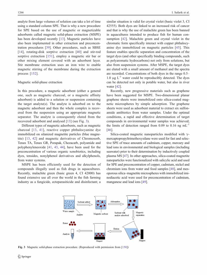
In this procedure, a magnetic adsorbent (either a generalone, such as magnetic charcoal, or a magnetic affinityadsorbent) is added to a solution or suspension containingthe target analyte(s). The analyte is adsorbed on to themagnetic adsorbent and then the whole complex is recov-ered from the suspension using an appropriate magneticseparator. The analyte is consequently eluted from therecovered adsorbent and analysed [13] (see Fig. 3).
Different types of magnetic adsorbents, such as magneticcharcoal [13, 41], reactive copper phthalocyanine dyeimmobilized on silanized magnetite particles (blue magne-tite) [13, 42] and magnetic derivatives of Chromosorb,Tenax TA, Tenax GR, Porapak, Chezacarb, polyamide andpolyphenyleneoxide [41, 43, 44], have been used for thepreconcentration of various organic xenobiotics, includingdyes, tensides, nonylphenol derivatives and alkylphenols,from water systems.
MSPE has been efficiently used for the detection ofcompounds illegally used as fish drugs in aquacultures.Recently, malachite green (basic green 4, CI 42000) hasfound extensive use all over the world in the fish farmingindustry as a fungicide, ectoparasiticide and disinfectant; a
similar situation is valid for crystal violet (basic violet 3, CI42555). Both dyes are linked to an increased risk of cancerand that is why the use of malachite green has been bannedin aquacultures intended to produce fish for human con-sumption [42]. Malachite green and crystal violet in thechromatic form specifically interact with copper phthalocy-anine dye immobilized on magnetic particles [45]. Thisfeature enables specific separation and concentration of thetarget dyes (and other specifically binding compounds, suchas polyaromatic hydrocarbons) not only from solutions, butalso from suspension systems. After MSPE, the target dyesare eluted with a small amount of methanol and the spectraare recorded. Concentrations of both dyes in the range 0.5–1.0 μg L-1 water could be reproducibly detected. The dyescan be detected not only in potable water, but also in riverwater [42].
Recently, new progressive materials such as graphenehave been suggested for MSPE. Two-dimensional planargraphene sheets were immobilized onto silica-coated mag-netic microspheres by simple adsorption. The graphenesheets were used as adsorbent material to extract six sulfon-amide antibiotics from water samples. Under the optimalconditions, a rapid and effective determination of targetcompounds in environmental water samples was achieved;the limits of detection ranged from 0.09 to 0.16 ng mL-1
[46].Silica-coated magnetic nanoparticles modified with γ-
mercaptopropyltrimethoxysilane were used for fast and selec-tive SPE of trace amounts of cadmium, copper, mercury andlead ions in environmental and biological samples (includingseawater) prior to their determination by inductively coupledplasma MS [47]. In other approaches, silica-coated magnetitenanoparticles were functionalized with salicylic acid and usedfor SPE and preconcentration of copper, cadmium, nickel andchromium ions from water and food samples [48], and mes-oporous silica–magnetite microspheres with immobilized imi-nodiacetic acid were used for preconcentration of cadmium,manganese and lead ions [49].
Fig. 3 Magnetic solid-phase extraction procedure. (Reproduced with permission from [150])
1266 I. Safarik et al.

Tab
le6
Examples
ofxeno
bioticsprecon
centratedby
magnetic
solid
-phase
extractio
n
Xenob
iotics
Characteristics
Water
system
Magnetic
system
Com
ments
Reference
Crystal
violet,safranin
ODyes
Spikedwater
samples
Reactivecopp
erph
thalocyanine
dye
attached
tosilanizedmagnetite
240-fold
enrichmentof
crystalviolet
from
800mLof
sample
[13]
Crystal
violet,safranin
ODyes
Spikedwater
samples
Magnetic
charcoal
460-fold
enrichmentof
crystalviolet
from
800mLof
sample
[13]
Malachite
green,
crystalviolet
Dyes
Spikedwater
samples
Magnetitewith
immob
ilized
copp
erph
thalocyanine
dye
0.5-1.0μg
ofdy
ein
1Lof
water
detected
[42]
Non
ionicsurfactants
Surfactants
Spikedwater
samples
Magnetic
derivativ
esof
charcoal,
poly(oxy
-2,6-dim
ethy
l-1,4-ph
enylene)
andpo
lyam
ide
Hyd
roph
obic
adsorbentsexhibitedthe
bestextractio
ncharacteristics
[41]
Alkylph
enolsand
oxyethylated
alky
lpheno
lsSurfactantsprecursors,
plasticizers
Spikedwater
samples,
riverandlake
samples
Magnetic
derivativ
esof
Chrom
osorb,
Tenax,Porapak,Chezacarb
and
polyph
enyleneoxide
Detectio
nlim
itswerebetween
0.7and1μg
L-1.
[43,
44,12
9]
Polyaromatic
hydrocarbo
nsHyd
rocarbon
sSpikedwater
samples
n-Octadecylph
osph
onic
acid
mod
ified
mesop
orou
smagnetic
nano
particles
Adsorbent
with
extrem
ely
hydrop
hobicprop
erties
[130]
Sulfonamides
Antim
icrobial
drug
Spikedandenvironm
ental
water
samples
Octadecyltrim
ethy
lammon
ium
brom
ide
adsorbed
onto
magnetitenano
particles
Aconcentrationfactor
of1,00
0was
achieved
(extractionof
500mLof
water
samples)
[131]
UV
filters
from
cosm
etic
prod
ucts
Organic
compo
unds
containing
benzenering
Water
samples
ofdifferent
origin
(tap,riverandsea).
Oleic
acid
coated
magnetic
nano
particles
GC-M
Sdeterm
ination
[132]
Polyaromatic
hydrocarbo
nsHyd
rocarbon
sLakewater
Carbo
n–ferrom
agnetic
nano
compo
site
GC-M
Sdeterm
ination
[133]
PAHs,ph
thalateesters
Organic
compo
unds
Sno
w,tapandriverwater
Barium
alginate
cagedFe 3O4–C-18
magnetic
nano
particles
HPLCassaywith
fluo
rescence
detection
[134]
Diethylstilb
estrol,
oestrone,oestriol
Estrogens
Tap,mineral
and
Pearlriverwater
Magnetic
silicaparticlescoated
with
hydrox
y-term
inated
multiw
alled
carbon
nano
tubes
The
extractio
nefficiencies
were
95.9,93
.9and52
.4%,respectiv
ely
[135]
Sarin,soman,tabu
n,cyclosarin
Nerve
agents
Spikedwater
samples
Magnetic
multiw
alledcarbon
nano
tubes
Elutio
nwith
chloroform
,recoveries
60–96
%[136]
Methy
lmercury
Organom
etallic
derivativ
eSeawater
Fe 3O4/polyanilin
enano
particles
GC-M
Sdeterm
ination
[137]
Cd,
Cu,
Hgand
Pbions
Heavy
metal
ions
Spikedandreal
water
samples
Silica-coatedmagnetic
nano
particlesmod
ified
with
γ-mercaptop
ropy
ltrim
etho
xysilane
Indu
ctivelycoup
ledplasmaMS
used
forthestud
y[47]
Ag,
Cd,
Cuand
Znions
Heavy
metal
ions
Tap
andmineral
water
samples
Magnetic
nano
particlescoated
with
3-(trimetho
xysilyl)-1-propanetio
land
mod
ifiedwith
2-am
ino-5-mercapto-
1,3,4-thiadiazole
Indu
ctivelycoup
ledplasmaop
tical
emission
spectroscopy
determ
ination
[138]
Fluoride
Anion
Spikedwater
samples
Magnetic
iron
oxides
nano
particles
Spectroph
otom
etricdeterm
ination
[139]
HPLChigh
-perform
ance
liquidchromatog
raph
y
Magnetic techniques for the detection of xenobiotics in water 1267

Inspiration can also be taken from food analysis, whereMSPE using a magnetic, phenyl-functionalized silica adsor-bent has been used for the determination of tetracyclines inmilk samples [50]; other magnetic adsorbents—e.g. magne-tite/silica/poly(methacrylic acid-co-ethylene glycol dimetha-crylate) composite microspheres and magnetic carbonnanotubes—have been used for sulfonamide preconcentra-tion [51] and oestrogen determination [52] from the samematrix.
Typical examples of MSPE for water analysis are shownin Table 6.
Incorporation of magnetic particles in other extractionprocedures
A new two-step microextraction technique, combining dis-persive liquid–liquid microextraction (DLLME) and disper-sive solid-phase microextraction (SPME), was developedfor the fast GC-MS determination of polycyclic aromatichydrocarbons in environmental samples. Any organic sol-vent immiscible with water can be used as an extractant inDLLME. In the approach presented, hydrophobic magneticnanoparticles were used to retrieve the extractant (1-octanol)together with the analyte. Because of the rapid mass transferassociated with the DLLME and the dispersive SPME steps,fast extraction could be achieved. The desorption of analyteswas performed by sonication with acetonitrile as the solvent.
Enrichment factors ranging from 110- to 186-fold wereobtained for the analytes. This two-step extraction methodwas successfully used for the fast determination of polycy-clic aromatic hydrocarbons in river water samples [39]. Thescheme of the procedure is shown in Fig. 4.
Stir-bar sorptive extraction and related procedures
SBSE is a new solventless sample preparation method forthe extraction and enrichment of organic compounds fromaqueous matrices. The method is based on the same princi-ples as SPME; a magnetic stir bar covered with an appro-priate adsorbent is used as the adsorbing element. Comparedwith SPME, a relatively large amount of extracting phase iscoated on a magnetic stir bar. Solutes are extracted into thecoating, based upon their octanol–water partitioning coeffi-cient and upon the sample–extraction medium phase ratio.The technique has been applied successfully to trace analy-sis in environmental, biomedical and food applications.Extremely low detection limits can be obtained [14].
Polydimethylsiloxane-coated stir bars are currently avail-able commercially (Gerstel, Müllheim an der Ruhr, Ger-many). These stir bars have three essential parts (Fig. 5).The first one is a magnetic stirring rod, which is necessaryfor transferring the rotating movement of a stirring plate tothe sample liquid. The second part of the stir bar is a thinglass jacket that covers the magnetic stirring rod. The third
Fig. 4 Dispersive liquid–liquidmicroextraction (DLLME)coupled with dispersivesolid-phase microextraction(D-μ-SPE). (Reproducedwith permission from [39])
Fig. 5 Magnetic stir bar covered with polydimethylsiloxane (PDMS)
1268 I. Safarik et al.
and outermost part is the layer of polydimethylsiloxanesorbent into which the analytes are extracted. The extractionprocedure is quite simple. SBSE of a liquid sample isperformed by placing a suitable amount of sample in aheadspace vial or other container. A polydimethylsiloxane-coated stir bar is added and the sample is stirred for 30–240 min. The extraction time is controlled kinetically and isdetermined by the sample volume, stirring speed and stir bardimensions; it has to be optimized for a given application.Optimization is normally accomplished by measuring theanalyte recovery as a function of the extraction time [14].
After extraction, the stir bar is removed, dipped on aclean paper tissue to remove water droplets and introducedto the thermal desorption unit, connected to a gas chromato-graph. In some cases, rinsing the stir bar slightly withdistilled water to remove adsorbed sugars, proteins or othersample components is recommended; this step will preventthe formation of non-volatile material during the thermal-desorption step. Rinsing does not cause solute loss, becausethe sorbed solutes are present in the polydimethylsiloxane
phase. Finally, the solutes are thermally desorbed. The de-sorption temperatures are application-dependent, primarilydetermined by the volatility of the solutes, and are typicallybetween 150 and 300 °C. Desorption can be accomplishedin 5–15 min under a 10–50 mL min-1 helium flow. As analternative to thermal desorption, organic solvents can beused for desorption [14].
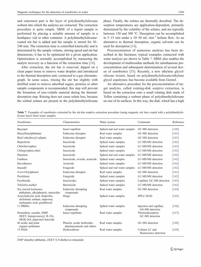
Preconcentration of numerous analytes has been de-scribed in the literature; typical examples connected withwater analysis are shown in Table 7. SBSE also enables thedevelopment of multiresidue methods for simultaneous pre-concentration and subsequent determination of several doz-en of xenobiotics [53]. Recently, a new ethylene glycol/silicone twister, based on polydimethylsiloxane/ethyleneglycol copolymer, has become available from Gerstel.
An alternative procedure for the preconcentration of tar-get analytes, called rotating-disk sorptive extraction, isbased on the extraction onto a small rotating disk made ofTeflon containing a sorbent phase of polydimethylsiloxaneon one of its surfaces. In this way, the disk, which has a high
Table 7 Examples of xenobiotics extracted by the stir-bar sorptive extraction procedure (using magnetic stir bars coated with a polydimethylsi-loxane layer) from water samples
Xenobiotics Characteristics Water system Comments Reference
Bayrepel Insect repellent Spiked and real water samples GC-MS detection [140]
Benzylbutylphthalate Endocrine disruptor Real water samples GC-MS detection [141]
Bis(2-ethylhexyl) adipate Endocrine disruptor Real water samples GC-MS detection [141]
Buprofezin Insecticide Spiked water samples LC-MS/MS detection [142]
Chlorfenvinphos Insecticide Spiked water samples LC-MS/MS detection [142]
Chlorpyriphos-ethyl Insecticide Spiked water samples LC-MS/MS detection [142]
Diazinon Insecticide Spiked and real water samples LC-MS/MS detection [142]
Fenthion Insecticide, avicide, acaricide Spiked water samples LC-MS/MS detection [142]
Hexythiazox Acaricide Spiked water samples LC-MS/MS detection [142]
Imazalil Fungicide Spiked and real water samples LC-MS/MS detection [142]
4-tert-Octylphenol Endocrine disruptor Real water samples GC-MS detection [141]
Prochloraz Fungicide Spiked water samples LC-MS/MS detection [142]
Pyrethroids Insecticides Spiked water samples Capillary GC-MS detection [143]
Tolclofos-methyl Bactericide Spiked water samples LC-MS/MS detection [142]
Sex steroid hormones,phthalates, alkylphenols, tamoxifen
Endocrine disruptingcompounds
Real water samples GC-MS detection [144]
Acetylsalicylic acid, ibuprofen,diclofenac sodium, naproxen,mefenamic acid, gemfibrozil
Drugs Spiked water samples HPLC-DAD [145]
11 PBDEs Endocrine disruptingcompounds
Spiked water samples Injection and capillaryGG-MS detection
[146]
Permethrin, icaridin, DMP,DEET, butopyronoxyl, R-326,MGK-264, piperonyl butoxide
Insect repellents Real water samples ThermodesorptionGC-MS detection
[147]
46 acidic and polarorganic pollutants
Phenols, acidic herbicides,pharmaceuticals and others
Real water samples GC-MS detection [148]
15 PAHs Hydrocarbons Real water samples Column LC andfluorescence detection
[149]
DMP dimethyl phthalate, DEET N,N-diethyl-m-toluamide
Magnetic techniques for the detection of xenobiotics in water 1269

surface area, contacts only the liquid sample, which can bestirred at higher velocity than with the stir bar used in SBSE,without damaging the phase while at the same time facili-tating analyte mass transfer to the polydimethylsiloxanesurface. With increasing rotational velocity, the amount ofextracted analyte significantly increases because the stag-nant layer concomitantly decreases. On the other hand, theextracted amount concomitantly increases with extractiontime, reaching equilibrium at approximately 20 min. Theprocedure was used for the determination of nonylphenol inreal water samples after elution of the adsorbed analyte withmethanol [40]. In the case of determination of colouredcompounds (e.g. malachite green), another approach hasbeen used. After extraction, the sorbent phase with theconcentrated analyte was separated from the Teflon diskand used directly for malachite green determination bysolid-phase spectrophotometry at 624 nm, without thenecessity of a desorption step [54]. The scheme of the diskand the separation process is shown in Fig. 6.
Stir-rod sorptive extraction was proposed as a new pro-cedure to avoid the friction loss of adsorbent coatings duringthe stirring processes. As shown in Fig. 7, a monolithic-polymer-coated stir rod is used. The polymer has octadecylgroups (hydrophobic) and sulfonic acid groups (ionexchange). A small magnet enables efficient movement ofthe stir rod in an analysed solution using a magnetic stirrer.This system was efficiently used for the separation of fluo-roquinolones [151]. The preconcentration of polycyclicaromatic hydrocarbons from water samples was performed
using poly(ethylene glycol dimethacrylate)/graphene com-posite as a stir bar coating [153].
Because extraction capabilities depend on the extractantgeometry, polymeric membranes are promising materialsthanks to their high sample/adsorbent contact surface. Anovel approach which combines the extraction capabilitiesof polymeric membranes with the advantages of their mag-netic stirring in the analysed solution has been developed;this system was used for monitoring of polycyclic aromatichydrocarbons in water samples [152].
Conclusions
The need for rapid, cost-effective and high-throughput ana-lytical methods for environmental monitoring is increasing.Magnetic nanoparticles and microparticles are very versa-tile, and the examples presented clearly document that mag-netically responsive materials can significantly simplify andimprove determination and detection of both organic andinorganic xenobiotics, nucleic acids and cellular pathogens.The sensitivity and selectivity of these methods depend onthe appropriate functionalization of magnetic materials andmolecules immobilized on their surfaces. In many cases,magnetic techniques can be performed more rapidly andwith lower consumption of chemicals than standard analyt-ical procedures; in addition, magnetic techniques are usuallymore environmentally friendly. Methods using magneticmaterials can be easily integrated into existing analyticaland microbiology laboratory protocols because only a fewadditional items, such as a magnetic separation rack andcommercially available kits, are needed. Water researchand technology can benefit greatly from the application ofsmart magnetic materials and the corresponding analyticalprocedures in laboratory practice.
Fig. 7 Stir-rod sorptive extraction. (Reproduced with permission,from[151])
Fig. 6 The rotating disk and the rotating-disk sorptive extractionprocedure. (Reproduced with permission from [40])
1270 I. Safarik et al.
Acknowledgment This research was supported by the Grant Agencyof the Czech Republic (project no. P503/11/2263).
References
1. Plaza G, Ulfig K, Tien AJ (2000) Pol J Environ Stud 9(4):231–236
2. Herzog DP (1997) Technical bulletin. Strategic Diagnostics,Newark
3. Hennion M-C (1998) Analusis Mag 26(6):M149–M1554. Marquette CA, Blum LJ (2006) Biosens Bioelectron 21:1424–
14335. Dankwardt A (2000) In: Meyers RA (ed) Encyclopedia of ana-
lytical chemistry. Chichester, Wiley6. Safarik I, Safarikova M (2002) Monatsh Chem 133:737–7597. Safarikova M, Safarik I (2001) Magn Electr Sep 10:223–2528. Safarik I, Safarikova M (2009) Chem Pap 63:497–5059. Safarik I, Safarikova M (2012) In: Thanh NTK (ed) Magnetic
nanoparticles: from fabrication to biomedical and clinical appli-cations. CRC, Boca Raton, pp 215–242
10. Safarik I, Safarikova M (1999) J Chromatogr B 722:33–5311. Safarik I, Safarikova M (2011) Veterinarstvi 61(4):199–20212. Gunther A, Bilitewski U (1995) Anal Chim Acta 300:117–12513. Safarikova M, Safarik I (1999) J Magn Magn Mater 194:108–11214. David F, Tienpont B, Sandra P (2003) LC GC Eur (July):2–715. Aguilar-Arteaga K, Rodriguez JA, Barrado E (2010) Anal Chim
Acta 674(2):157–16516. Barrado E, Vega M, Pardo R, Grande P, Del Valle JL (1996)
Water Res 30:2309–231417. Barrado E, Prieto F, Ribas J, Lopez FA (1999) Water Air Soil
Pollut 115:385–39418. Díaz-González M, González-García MB, Costa-García A (2005)
Electroanalysis 17:1901–191819. Centi S, Rozum B, Laschi S, Palchetti I, Mascini M (2006) Chem
Anal 51:963–97520. Lin Y-Y, Liu G, Wai CM, Lin Y (2008) Anal Chim Acta 612
(1):23–2821. Liu GD, Riechers SL, Timchalk C, Lin YH (2005) Electrochem
Commun 7:1463–147022. Gruessner B, Shambaugh NC, Watzin MC (1995) Environ Sci
Technol 29:251–25423. Reidt U, Geisberger B, Heller C, Friedberger A (2011) J Labor
Automat 16(2):157–16424. Environmental Protection Agency (2005) US EPA method 1622.
Cryptosporidium in water by filtration/IMS/FA. http://www.epa.gov/microbes/1622de05pdf
25. Environmental Protection Agency (2005) US EPA method 1623.Cryptosporidium and Giardia in water by filtration/IMS/FA.http://www.epa.gov/microbes/1623de05pdf
26. Thompson DE, Rajal VB, De Batz S, Wuertz S (2006) J WaterHealth 4:67–75
27. Van Dyk JS, Pletschke B (2011) Chemosphere 82(3):291–30728. Llopis X, Pumera M, Alegret S, Merkoci A (2009) Lab Chip 9
(2):213–21829. Du D, Wang J, Wang L, Lu D, Smith JN, Timchalk C, Lin Y
(2011) Anal Chem 83:3770–377730. Liu ZM, Liu YL, Yang HF, Yang Y, Shen GL, Yu RQ (2005)
Anal Chim Acta 533(1):3–931. Wang SF, Tan YM, Zhao DM, Liu GD (2008) Biosens Bioelec-
tron 23(12):1781–178732. Alkasir RSJ, Ganesana M, Won YH, Stanciu L, Andreescu S
(2010) Biosens Bioelectron 26(1):43–4933. Sima VH, Patris S, Aydogmus Z, Sarakbi A, Sandulescu R,
Kauffmann J-M (2011) Talanta 83(3):980–987
34. Safarik I, Ptackova L, Koneracka M, Safarikova M, Timko M,Kopcansky P (2002) Biotechnol Lett 24(5):355–358
35. Chang Q, Deng KJ, Zhu LH, Jiang GD, Yu C, Tang HQ (2009)Microchimica Acta 165(3–4):299–305
36. Tan XC, Zhang JL, Tan SW, Zhao DD, Huang ZW, Mi Y, HuangZY (2009) Sensors 9(8):6185–6199
37. Fritz JS, Dumont PJ, Schmidt LW (1995) J Chromatogr A 691(1–2):133–140
38. Berrueta LA, Gallo B, Vicente F (1995) Chromatographia 40(7–8):474–483
39. Shi Z-G, Lee HK (2010) Anal Chem 82(4):1540–154540. Richter P, Leiva C, Choque C, Giordano A, Sepulveda B (2009) J
Chromatogr A 1216(49):8598–860241. Safarikova M, Kibrikova I, Ptackova L, Hubka T, Komarek K,
Safarik I (2005) J Magn Magn Mater 293(1):377–38142. Safarik I, Safarikova M (2002) Water Res 36(1):196–20043. Komarek K, Safarikova M, Hubka T, Safarik I, Kandelova M,
Kujalova H (2009) Chromatographia 69(1):133–13744. Safarikova M, Lunackova P, Komarek K, Hubka T, Safarik I
(2007) J Magn Magn Mater 311(1):405–40845. Safarik I, Safarikova M, Vrchotova N (1995) Coll Czech Chem
Commun 60(1):34–4246. Luo Y-B, Shi Z-G, Gao Q, Feng Y-Q (2011) J Chromatogr A
1218(10):1353–135847. Huang C, Hu B (2008) Spectrochim Acta B 63(3):437–
44448. Shishehbore MR, Afkhami A, Bagheri H (2011) Chem Central J
5:4149. Zhang N, Peng HY, Wang S, Hu B (2011) Microchim Acta 175
(1–2):121–12850. Ibarra IS, Rodriguez JA, Miranda JM, Vega M, Barrado E (2011)
J Chromatogr A 1218(16):2196–220251. Gao QA, Luo D, Ding J, Feng YQ (2010) J Chromatogr A 1217
(35):5602–560952. Ding J, Gao Q, Li X-S, Huang W, Shi Z-G, Feng Y-Q (2011) J
Sep Sci 34(18):2498–250453. Bonet-Domingo E, Grau-Gonzalez S, Martin-Biosca Y, Medina-
Hernandez MJ, Sagrado S (2007) Anal Bioanal Chem 387(7):2537–2545
54. Richter P, Cańas A, Muńoz C, Leiva C, Ahumada I (2011) AnalChim Acta 695(1–2):73–76
55. De Schepper W, Dries J, Geuens L, Blust R (2010) EcotoxEnviron Safe 73(5):702–709
56. Gascon J, Durand G, Barcelo D (1995) Environ Sci Technol 29(6):1551–1556
57. Gascon J, Salau JS, Oubina A, Barcelo D (1998) Analyst 123(5):941–945
58. Gascon J, Oubina A, Ferrer I, Onnerfjord P, Marko-Varga G,Hammock BD, Marco MP, Barcelo D (1996) Anal Chim Acta330(1):41–51
59. Byer JD, Struger J, Sverko E, Klawunn P, Todd A (2011) Chemo-sphere 82(8):1155–1160
60. Graziano N, McGuire MJ, Roberson A, Adams C, Jiang H, BluteN (2006) Environ Sci Technol 40(4):1163–1171
61. McGregor EB, Solomon KR, Hanson ML (2008) Chemosphere73(3):249–260
62. Castillo M, Oubina A, Barcelo D (1998) Environ Sci Technol 32(14):2180–2184
63. Herbrandson C, Bradbury SP, Swackhamer DL (2003) AquatToxicol 63(4):343–355
64. Herbrandson C, Bradbury SP, Swackhamer DL (2003) AquatToxicol 63(4):333–342
65. Wandan EN, Zabik MJ, Elleingand EF (2011) Sci Res Essays 6(8):1771–1779
66. Walker AE, Holman RE, Leidy RB (2000) J Am Water ResourAssoc 36(1):67–74
Magnetic techniques for the detection of xenobiotics in water 1271

67. Farré M, Kuster M, Brix R, Rubio F, López de Alda M-J, BarcelóD (2007) J Chromatogr A 1160(1–2):166–175
68. Byer JD, Struger J, Klawunn P, Todd A, Sverko E (2008) EnvironSci Technol 42(16):6052–6057
69. Gaynor JD, Cancilla DA, Webster GRB, Sarna LP, Graham KN,Ng HYF, Tan CS, Drury CF, Welacky T (1996) J Agric FoodChem 44(9):2736–2741
70. Schraer SM, Shaw DR, Boyette M, Coupe RH, Thurman EM(2000) J Agric Food Chem 48(12):5881–5886
71. Barcelo D, Oubina A, Salau JS, Perez S (1998) Anal Chim Acta376(1):49–53
72. Fillmann G, Bicego MC, Zamboni A, Fileman TW, DepledgeMH, Readman JW (2007) J Braz Chem Soc 18(4):774–781
73. Shelver WL, Parrotta CD, Slawecki R, Li QX, Ikonomou MG,Barcelo D, Lacorte S, Rubio FM (2008) Chemosphere 73(1):S18–S23
74. Xu T, Cho IK, Wang DL, Rubio FM, Shelver WL, Gasc AME, LiJ, Li QX (2009) Environ Pollut 157(2):417–422
75. Barsh R, Bell J, Halliday H, Clifford M, Mottet G (2008) Pre-liminary Survey of pyrethroid pesticides and surfactants in SanJuan County surface waters. KWIAHT (Center for the HistoricalEcology of the Salish Sea). Lopez
76. Young DL, Mihaliak CA, West SD, Hanselman KA, Collins RA,Phillips AM, Robb CK (2000) J Agric Food Chem 48(11):5146–5153
77. Shelver WL, Shappell NW, Franek M, Rubio FR (2008) J AgricFood Chem 56(15):6609–6615
78. Raturi S, Carroll MJ, Hill RL (2003) J Environ Qual 32(1):215–223
79. Kantiani L, Farré M, Asperger D, Rubio F, González S, López deAlda MJ, Petrovic M, Shelver WL, Barceló D (2008) J Hydrol361(1–2):1–9
80. Ricart M, Guasch H, Alberch M, Barcelo D, Bonnineau C,Geiszinger A, Farre M, Ferrer J, Ricciardi F, Romani AM, MorinS, Proia L, Sala L, Sureda D, Sabater S (2010) Aquat Toxicol 100(4):346–353
81. Haramoto E, Kitajima M, Katayama H, Ohgaki S (2010) WaterRes 44(6):1747–1752
82. Hwang YC, Leong OM, Chen W, Yates MV (2007) Appl EnvironMicrobiol 73(7):2338–2340
83. Monceyron C, Grinde B (1994) J Virol Methods 46(2):157–16684. Jothikumar N, Cliver DO, Mariam TW (1998) Appl Environ
Microbiol 64(2):504–50885. Cannon JL, Vinje J (2008) Appl Environ Microbiol 74(21):6818–
681986. Myrmel M, Rimstad E, Wasteson Y (2000) Int J Food Microbiol
62(1–2):17–2687. Yang W, Gu AZ, Zeng S-Y, Li D, He M, Shi H-C (2011) J
Microbiol Methods 84(3):447–45388. Grinde B, Jonassen TO, Ushijima H (1995) J Virol Methods 55
(3):327–33889. Chakraborty R, Hazen TC, Joyner DC, Küsel K, Singer ME,
Sitte J, Torok T (2011) J Microbiol Methods 86(2):204–209
90. Lee JY, Deininger RA (2004) Luminescence 19(1):31–3691. Dudak FC, Boyaci IH (2008) J Rapid Methods Autom Microbiol
16(2):122–13192. Bushon RN, Likirdopulos CA, Brady AMG (2009) Water Res 43
(19):4940–494693. Garcia-Aljaro C, Bonjoch X, Blanch AR (2005) J Appl Microbiol
98(3):589–59794. Muller EE, Grabow WOK, Ehlers MM (2003) Water SA 29
(4):427–43295. Shelton DR, Karns JS (2001) Appl Environ Microbiol 67
(7):2908–2915
96. Schets FM, During M, Italiaander R, Heijnen L, Rutjes SA, vander Zwaluw WK, de Roda Husman AM (2005) Water Res 39(18):4485–4493
97. Enroth H, Engstrand L (1995) J Clin Microbiol 33(8):2162–216598. Yanez MA, Carrasco-Serrano C, Barbera VM, Catalan V (2005)
Appl Environ Microbiol 71(7):3433–344199. Allegra S, Girardot F, Grattard F, Berthelot P, Helbig JH, Pozzetto
B, Riffard S (2011) J Appl Microbiol 110(4):952–961100. Goosen C (2001) Development of PCR-based detection assays
for Legionella pneumophila in water. MSc dissertation, Univer-sity of Pretoria
101. Whan L, Ball HJ, Grant IR, Rowe MT (2005) Appl EnvironMicrobiol 71(11):7107–7112
102. Whan L, Ball HJ, Grant IR, Rowe MT (2005) Lett Appl Micro-biol 40(4):269–273
103. Stinear T, Davies JK, Jenkin GA, Hayman JA, Oppedisano F,Johnson PDR (2000) Environ Microbiol 66(8):3206–3213
104. Roberts B, Hirst R (1997) J Clin Microbiol 35(10):2709–2711105. Ochiai Y, Takada C, Hosaka M (2005) Appl Environ Microbiol
71(2):898–903106. Kuczynska E, Boyer DG, Shelton DR (2003) J Microbiol Meth-
ods 53(1):17–26107. Kuhn RC, Rock CM, Oshima KH (2002) Appl Environ Micro-
biol 68(4):2066–2070108. Jiang JL, Alderisio KA, Singh A, Xiao LH (2005) Appl Environ
Microbiol 71(3):1135–1141109. Yang WL, Chen P, Villegas EN, Landy RB, Kanetsky C, Cama V,
Dearen T, Schultz CL, Orndorff KG, Prelewicz GJ, Brown MH,Young KR, Xiao LH (2008) Appl Environ Microbiol 74(21):6495–6504
110. McCuin RM, Clancy JL (2006) J Water Health 4(4):437–452111. McCuin RM, Bukhari Z, Sobrinho J, Clancy JL (2001) J Micro-
biol Methods 45(2):69–76112. Kim KJ, Jung HH, Lee K (2006) Biotechnol Bioprocess Eng 11
(4):368–371113. Zuckerman U, Tzipori S (2006) J Appl Microbiol 100(6):1220–
1227114. Ramadan Q, Christophe L, Teo W, Li SJ, Hua FH (2010) Anal
Chim Acta 673(1):101–108115. Zhang T, Xie X, Hu H, Song Y, Wu Q, Zong Z (2008) Front
Environ Sci Eng China 2(3):380–384116. Khouja LB, Cama V, Xiao LH (2010) Parasitol Res 107(1):109–
116117. Moulin L, Richard F, Stefania S, Goulet M, Gosselin S, Gon-
calves A, Rocher V, Paffoni C, Dumetre A (2010) Water Res44:5222–5231
118. Castro-Hermida JA, Garcia-Presedo I, Almeida A, Gonzalez-Warleta M, Da Costa JMC, Mezo M (2008) Water Res 42(13):3528–3538
119. Sorel N, Guillot E, Thellier M, Accoceberry I, Datry A, Mesnard-Rouiller L, Miegeville M (2003) J Appl Microbiol 94(2):273–279
120. Hsu BM, Huang CP, Lai YC, Tai HS, Chung YC (2001) ParasitolRes 87(6):472–474
121. Mahbubani MH, Schaefer FW, Jones DD, Bej AK (1998) CurrMicrobiol 36(2):107–113
122. Garcia A, Yanko W, Batzer G, Widmer G (2002) Water. EnvironRes 6:541–544
123. Bifulco JM, Schaefer FW III (1993) Appl Environ Microbiol 59(3):772–776
124. Dumetre A, Darde ML (2007) Parasitol Res 101(4):989–996125. Kindervater R, Kunnecke W, Schmid RD (1990) Anal Chim Acta
234(1):113–117126. Min H, Qu YH, Li XH, Xie ZH, Wei YY, Jin LT (2007) Acta
Chim Sin 65(20):2303–2308127. Istarnboulie G, Andreescu S, Marty JL, Noguer T (2007) Biosens
Bioelectron 23(4):506–512
1272 I. Safarik et al.
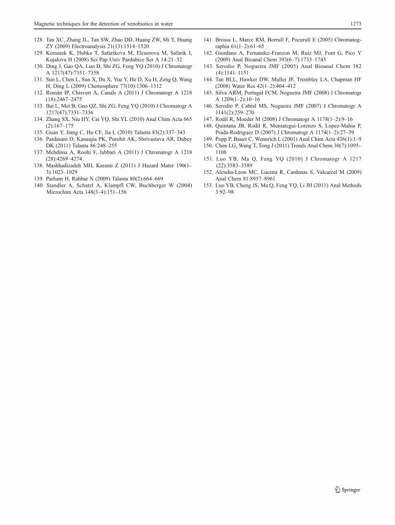
128. Tan XC, Zhang JL, Tan SW, Zhao DD, Huang ZW, Mi Y, HuangZY (2009) Electroanalysis 21(13):1514–1520
129. Komarek K, Hubka T, Safarikova M, Elcnerova M, Safarik I,Kujalova H (2008) Sci Pap Univ Pardubice Ser A 14:21–32
130. Ding J, Gao QA, Luo D, Shi ZG, Feng YQ (2010) J ChromatogrA 1217(47):7351–7358
131. Sun L, Chen L, Sun X, Du X, Yue Y, He D, Xu H, Zeng Q, WangH, Ding L (2009) Chemosphere 77(10):1306–1312
132. Román IP, Chisvert A, Canals A (2011) J Chromatogr A 1218(18):2467–2475
133. Bai L, Mei B, Guo QZ, Shi ZG, Feng YQ (2010) J Chromatogr A1217(47):7331–7336
134. Zhang SX, Niu HY, Cai YQ, Shi YL (2010) Anal Chim Acta 665(2):167–175
135. Guan Y, Jiang C, Hu CF, Jia L (2010) Talanta 83(2):337–343136. Pardasani D, Kanaujia PK, Purohit AK, Shrivastava AR, Dubey
DK (2011) Talanta 86:248–255137. Mehdinia A, Roohi F, Jabbari A (2011) J Chromatogr A 1218
(28):4269–4274138. Mashhadizadeh MH, Karami Z (2011) J Hazard Mater 190(1–
3):1023–1029139. Parham H, Rahbar N (2009) Talanta 80(2):664–669140. Standler A, Schatzl A, Klampfl CW, Buchberger W (2004)
Microchim Acta 148(3–4):151–156
141. Brossa L, Marce RM, Borrull F, Pocurull E (2005) Chromatog-raphia 61(1–2):61–65
142. Giordano A, Fernandez-Franzon M, Ruiz MJ, Font G, Pico Y(2009) Anal Bioanal Chem 393(6–7):1733–1743
143. Serodio P, Nogueira JMF (2005) Anal Bioanal Chem 382(4):1141–1151
144. Tan BLL, Hawker DW, Muller JF, Tremblay LA, Chapman HF(2008) Water Res 42(1–2):404–412
145. Silva ARM, Portugal FCM, Nogueira JMF (2008) J ChromatogrA 1209(1–2):10–16
146. Serodio P, Cabral MS, Nogueira JMF (2007) J Chromatogr A1141(2):259–270
147. Rodil R, Moeder M (2008) J Chromatogr A 1178(1–2):9–16148. Quintana JB, Rodil R, Muniategui-Lorenzo S, Lopez-Mahia P,
Prada-Rodriguez D (2007) J Chromatogr A 1174(1–2):27–39149. Popp P, Bauer C, Wennrich L (2001) Anal Chim Acta 436(1):1–9150. Chen LG, Wang T, Tong J (2011) Trends Anal Chem 30(7):1095–
1108151. Luo YB, Ma Q, Feng YQ (2010) J Chromatogr A 1217
(22):3583–3589152. Alcudia-Leon MC, Lucena R, Cardenas S, Valcarcel M (2009)
Anal Chem 81:8957–8961153. Luo YB, Cheng JS, Ma Q, Feng YQ, Li JH (2011) Anal Methods
3:92–98
Magnetic techniques for the detection of xenobiotics in water 1273