Nuclear Magnetic Resonance (NMR) Magnetic Resonance Imaging (MRI)

Magnetic Resonance Imaging Gerald R. Aben, MD, FACR Associate Professor Radiology College of Osteopathic Medicine
6/12/2012 1 DEPARTMENT OF RADIOLOGY

MRI
6/12/2012 2 DEPARTMENT OF RADIOLOGY

Magnetic Resonance Imaging • Tube shaped machine
• Uses a magnetic field and radio frequency waves • Body chemical composition and tissue structure contribute to form image contrast
• Computer generated image
• Special terms • High signal (white or bright), Low signal (dark grey or dark), Isointense (equal density)
6/12/2012 DEPARTMENT OF RADIOLOGY 3

Magnetic Resonance Imaging • Advantages:
• No ionizing radiation (no x-rays)
• Sensitive to slight chemical difference
• Three dimensional study
• Capable of showing fine anatomic structure
• Angiography can be done non-invasively
6/12/2012 DEPARTMENT OF RADIOLOGY 4

3-T MRI scanning
6/12/2012 DEPARTMENT OF RADIOLOGY 5

MRI with Patient
6/12/2012 DEPARTMENT OF RADIOLOGY 6

MRI Principles
• Hydrogen ions in the magnetic field of the MRI machine align with a small net dipole(1)
• Application of a radio frequency causes ions to move to a higher energy state(2-3) (Blue arrow) 6/12/2012 DEPARTMENT OF RADIOLOGY 7

MRI Principles
• When the frequency is removed the ions begin to return to prior state releasing energy(4 – 5)
• Resulting radiofrequency is measured and localized to create an image(Changing bl )
6/12/2012 DEPARTMENT OF RADIOLOGY 8

MRI Sequences
• T1- Weighted – short TR short TE – Fat Bright
• T2- Weighted – long TR, long TE – Water Bright
• Proton Density- Weighted or balanced imaging – long TR, Short TE
6/12/2012 DEPARTMENT OF RADIOLOGY 9
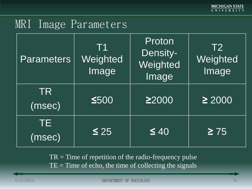
MRI Image Parameters
Parameters T1
Weighted Image
Proton Density-Weighted
Image
T2 Weighted
Image
TR (msec) ≤500 ≥2000 ≥ 2000
TE (msec) ≤ 25 ≤ 40 ≥ 75
6/12/2012 DEPARTMENT OF RADIOLOGY 10
TR = Time of repetition of the radio-frequency pulse TE = Time of echo, the time of collecting the signals

T1-Weighted
Proton Density-Weighted (balanced)
T2-Weighted
CSF Dark gray or Black Dark gray “White” or
light
White Matter
“White” or light Dark gray Dark gray
Gray Matter Gray Light gray Dark gray
Signal Intensity of Certain Tissues
6/12/2012 DEPARTMENT OF RADIOLOGY 11
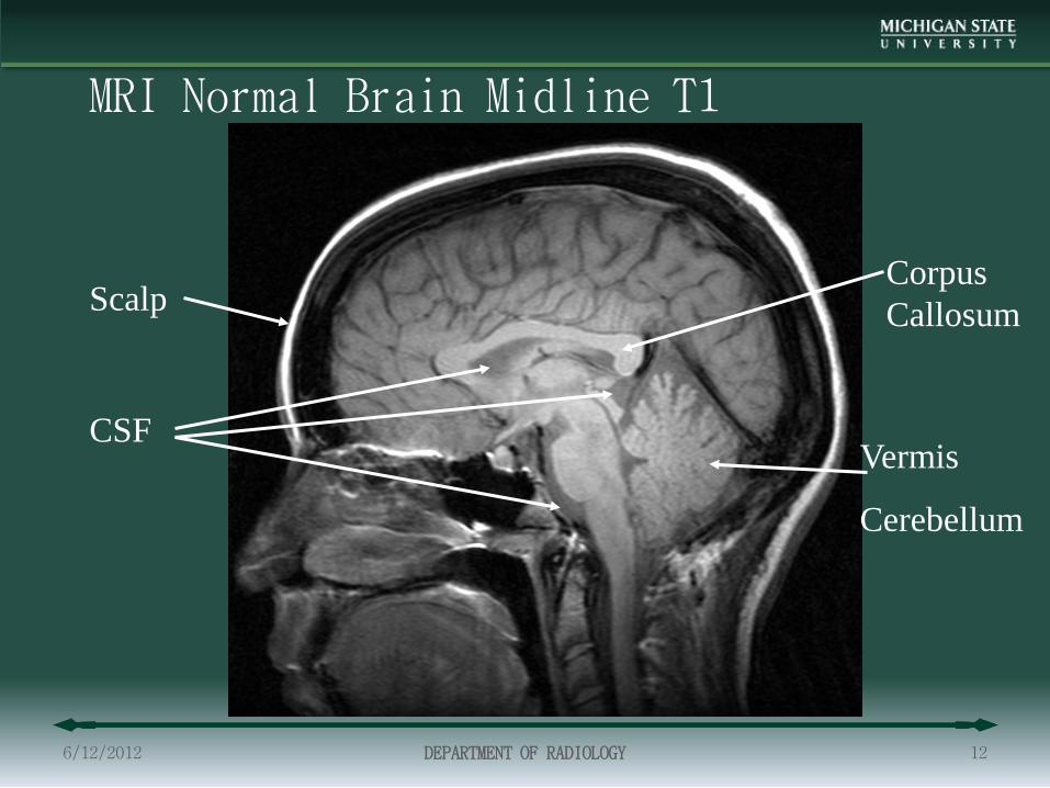
MRI Normal Brain Midline T1
6/12/2012 DEPARTMENT OF RADIOLOGY 12
Corpus Callosum
Vermis
Cerebellum
Scalp
CSF

Brain MRI Normal Axial T1
• Ventricles
• White Matter
• Grey Matter
• Scalp fat
6/12/2012 DEPARTMENT OF RADIOLOGY 13

Brain MRI Normal Flair
• T-2 Sequence
• Special technique suppresses CSF signal
• Allows better evaluation of areas adjacent to CSF spaces
• Excellent for evaluation of Multiple Sclerosis
6/12/2012 DEPARTMENT OF RADIOLOGY 14

Brain MRI Normal T2
6/12/2012 DEPARTMENT OF RADIOLOGY 15

Brain Coronal T1
6/12/2012 DEPARTMENT OF RADIOLOGY 16
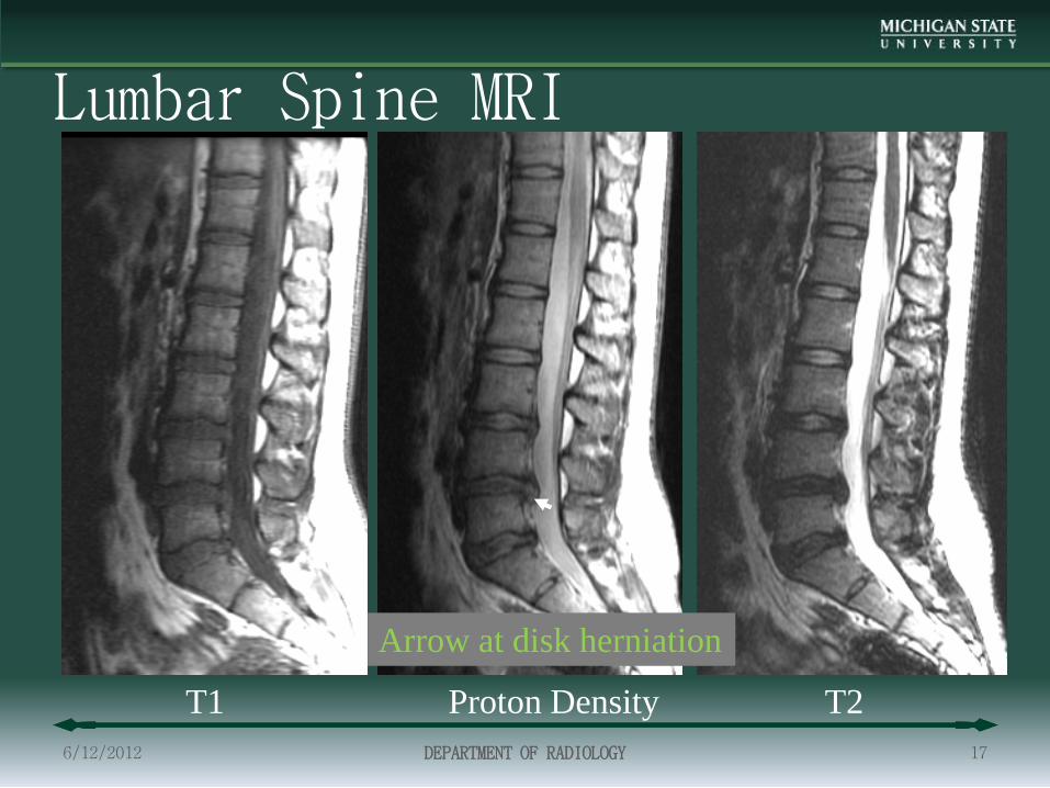
Lumbar Spine MRI
6/12/2012 DEPARTMENT OF RADIOLOGY 17
T1 Proton Density T2
Arrow at disk herniation
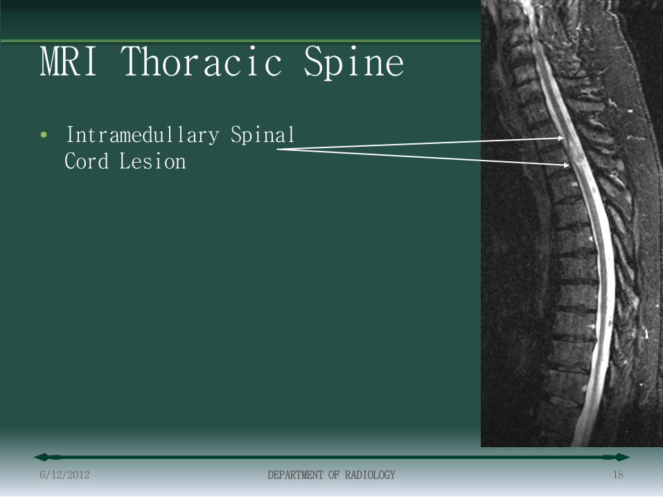
MRI Thoracic Spine
• Intramedullary Spinal Cord Lesion
6/12/2012 DEPARTMENT OF RADIOLOGY 18

MRI Thorax
6/12/2012 DEPARTMENT OF RADIOLOGY 19
Cardiac MRI
6/12/2012 DEPARTMENT OF RADIOLOGY 20
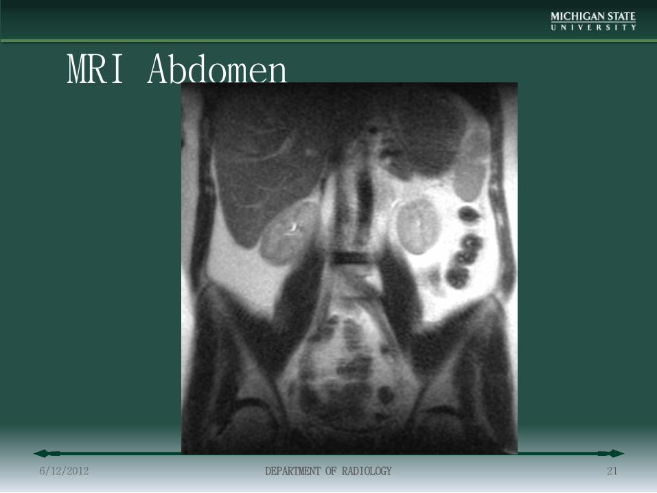
MRI Abdomen
6/12/2012 DEPARTMENT OF RADIOLOGY 21

MR Angiogram Cerebral
6/12/2012 DEPARTMENT OF RADIOLOGY 22

MRI into the Future
6/12/2012 DEPARTMENT OF RADIOLOGY 23

Cardiac Function and Perfusion • Advanced Cardiac evaluation
– Perfusion
– Function
6/12/2012 DEPARTMENT OF RADIOLOGY 24

CAD Enhanced Prostate MRI
6/12/2012 DEPARTMENT OF RADIOLOGY 25
Courtesy of Dr. Kevin DeMarco
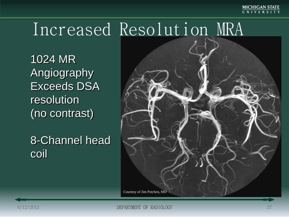
1024 MR Angiography Exceeds DSA resolution (no contrast) 8-Channel head coil
Increased Resolution MRA
6/12/2012 DEPARTMENT OF RADIOLOGY 27
Courtesy of Jim Potchen, MD
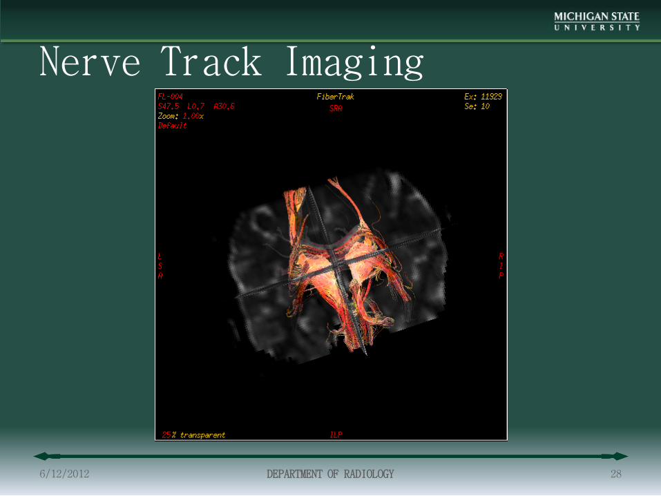
Nerve Track Imaging
6/12/2012 DEPARTMENT OF RADIOLOGY 28

Brain Functional Imaging-Listening to Brahms
Music Therapy Professor Musically Naive Engineer Courtesy of Jim Potchen, MD
6/12/2012 DEPARTMENT OF RADIOLOGY 29

Brain Functional Imaging Listening to Someone Reading Prose
Music Professor
Engineer Courtesy of Jim Potchen, MD
6/12/2012 DEPARTMENT OF RADIOLOGY 30