Deviant Functional Magnetic Resonance Imaging Patterns of Brain
256256International Journal of Scientific Study | August 2017 | Vol 5 | Issue 5
Magnetic Resonance Imaging Evaluation of Brain in Developmental Delay ChildrenRamya Kalaiarasan1, L Ashvind2, F Abu Backer Sulaiman3, L Uma Devi4, B Ashraf Ahmed5
1Post-graduate Student, Department of Radiology, Chettinad Hospital and Research Centre, Chennai, Tamil Nadu, India, 2Assistant Professor, Department of Paediatrics, Chettinad Hospital and Research Centre, Chennai, Tamil Nadu, India, 3Professor, Department of Radiology, Chettinad Hospital and Research Centre, Chennai, Tamil Nadu, India, 4Professor and Head, Department of Paediatrics, Chettinad Hospital and Research Centre, Chennai, Tamil Nadu, India, 5Associate Professor, Department of Radiology, Chettinad Hospital and Research Centre, Chennai, Tamil Nadu, India
delay (more than 2 standard deviations below the mean)in one or more developmental domains.2 Cognitive and motor development observed in infants and children are a reflection of postnatal brain development. Myelination and synaptogenesis are considered the biological correlates of this developmental process and have been studied extensively. Any delay in neurodevelopment is likely to have a biological correlate.Brain MRI is one of the major investigation of these patients and based on previous studies, about 60% of cases have abnormal findings in MRI.3
Prevalence of developmental delay in children has been reported 5-10%.4 MR imaging is an important part of the comprehensive evaluation of children with developmental delay, as many specific etiologic and pathophysiologic conditions that lead to developmental delay can be detected easily..5,6 Aim of the study is to know the most common MRI brain findings in children with global developmental delay and prevalence of normal and abnormal findings in patients in global developmental delay.
INTRODUCTION
Developmental in its broadest sense encompasses physical and mental growth that leads to the anatomical, physiological and behavioural changes that occur throughout childhood.For most paediatricians, child development relates to the changes in children’s ability to move,perform fine movements with their hands,communicates, learns new knowledge, self-care and interact with others.
Development is a dynamic process that is determined by interaction of genetic, biological and environmental factors.1 Developmental delay is defined as significant
Original Article
AbstractIntroduction: Developmental delay is termed as gross or significant delay in more than one developmental domains.Aetiology of the developmental delay is the wide spectrum. MRI is the best modality to evaluate such children. Other than early diagnosis and treatment, helps in counselling parents regarding their outcome and risk of recurrence in the siblings.
Materials and Methods: An observational and descriptive study of MRI brain in 100 paediatric patients with developmental delay for the duration of 2 years from December 2014-December 2016.
Results: Normal MRI Findings were seen in 36% cases and abnormal findings were seen in 64% (64 cases). Further the etiological factors as been classified as traumatic/neurovascular (30%), congenital/developmental (12%), metabolic/degenerative (8%), neoplastic (4%) and non-specific (10%). Most common features observed in the study were PVL, gliosis with volume loss, thinning of corpus calosum. Common anatomical abnormalities are corpus callosum agenesis and nodular heterotopia.
Conclusion: MRI brain study is an effective tool in identifying causative factor in developmental delay children with high yielding results.
Key words: Magnetic resonance imaging, Developmental delay, Brain
Access this article online
www.ijss-sn.com
Month of Submission : 06-2017 Month of Peer Review : 07-2017 Month of Acceptance : 08-2017 Month of Publishing : 08-2017
Corresponding Author: Dr. L. Ashvind, Department of Paediatrics, Chettinad Hospital and Research Centre, Chennai - 603 103, Tamil nadu, India. Phone: +91-9994679902. E-mail: [email protected]
Print ISSN: 2321-6379Online ISSN: 2321-595X
DOI: 10.17354/ijss/2017/436
Kalaiarasan, et al.: MRI Evaluation of Developmental Delay Children
257257 International Journal of Scientific Study | August 2017 | Vol 5 | Issue 5
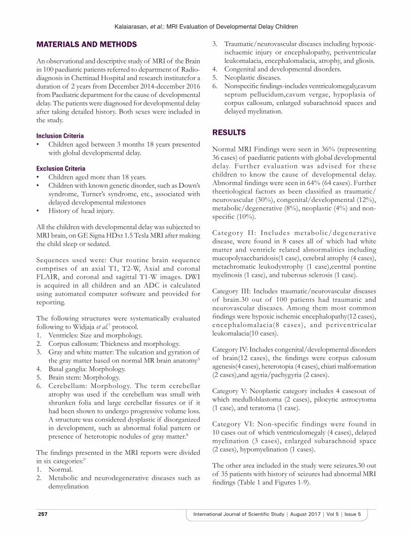
MATERIALS AND METHODS
An observational and descriptive study of MRI of the Brain in 100 paediatric patients referred to department of Radio-diagnosis in Chettinad Hospital and research institutefor a duration of 2 years from December 2014-december 2016 from Paediatric department for the cause of developmental delay. The patients were diagnosed for developmental delay after taking detailed history. Both sexes were included in the study.
Inclusion Criteria• Children aged between 3 months 18 years presented
with global developmental delay.
Exclusion Criteria• Children aged more than 18 years.• Children with known genetic disorder, such as Down’s
syndrome, Turner’s syndrome, etc., associated with delayed developmental milestones
• History of head injury.
All the children with developmental delay was subjected to MRI brain, on GE Signa HDxt 1.5 Tesla MRI after making the child sleep or sedated.
Sequences used were: Our routine brain sequence comprises of an axial T1, T2-W, Axial and coronal FLAIR, and coronal and sagittal T1-W images. DWI is acquired in all children and an ADC is calculated using automated computer software and provided for reporting.
The following structures were systematically evaluated following to Widjaja et al.7 protocol.1. Ventricles: Size and morphology.2. Corpus callosum: Thickness and morphology.3. Gray and white matter: The sulcation and gyration of
the gray matter based on normal MR brain anatomy.5
4. Basal ganglia: Morphology.5. Brain stem: Morphology.6. Cerebellum: Morphology. The term cerebellar
atrophy was used if the cerebellum was small with shrunken folia and large cerebellar fissures or if it had been shown to undergo progressive volume loss. A structure was considered dysplastic if disorganized in development, such as abnormal folial pattern or presence of heterotopic nodules of gray matter.8
The findings presented in the MRI reports were divided in six categories:91. Normal.2. Metabolic and neurodegenerative diseases such as
demyelination
3. Traumatic/neurovascular diseases including hypoxic-ischaemic injury or encephalopathy, periventricular leukomalacia, encephalomalacia, atrophy, and gliosis.
4. Congenital and developmental disorders.5. Neoplastic diseases.6. Nonspecific findings-includes ventriculomegaly,cavum
septum pellucidum,cavum vergae, hypoplasia of corpus callosum, enlarged subarachnoid spaces and delayed myelination.
RESULTS
Normal MRI Findings were seen in 36% (representing 36 cases) of paediatric patients with global developmental delay. Further evaluation was advised for these children to know the cause of developmental delay. Abnormal findings were seen in 64% (64 cases). Further theetiological factors as been classified as traumatic/neurovascular (30%), congenital/developmental (12%), metabolic/degenerative (8%), neoplastic (4%) and non-specific (10%).
Category II : Includes metabol ic/degenerat ive disease, were found in 8 cases all of which had white matter and ventricle related abnormalities including mucopolysaccharidosis(1 case), cerebral atrophy (4 cases), metachromatic leukodystrophy (1 case),central pontine myelinosis (1 case), and tuberous sclerosis (1 case).
Category III: Includes traumatic/neurovascular diseases of brain.30 out of 100 patients had traumatic and neurovascular diseases. Among them most common findings were hypoxic ischemic encephalopathy(12 cases), encephalomalacia(8 cases) , and periventricular leukomalacia(10 cases).
Category IV: Includes congenital/developmental disorders of brain(12 cases), the findings were corpus calosum agenesis(4 cases), heterotopia (4 cases), chiari malformation (2 cases),and agyria/pachygyria (2 cases).
Category V: Neoplastic category includes 4 casesout of which medulloblastoma (2 cases), pilocytic astrocytoma (1 case), and teratoma (1 case).
Category VI: Non-specific findings were found in 10 cases out of which ventriculomegaly (4 cases), delayed myelination (3 cases), enlarged subarachnoid space (2 cases), hypomyelination (1 cases).
The other area included in the study were seizures.30 out of 35 patients with history of seizures had abnormal MRI findings (Table 1 and Figures 1-9).

Kalaiarasan, et al.: MRI Evaluation of Developmental Delay Children
258258International Journal of Scientific Study | August 2017 | Vol 5 | Issue 5
DISCUSSION
Evaluation of MRI brain was done in 100 paediatric patients with developmental delay of age group three months to 18 years. The proportions of children having abnormal MRI findings in our study of 100 cases could get a definitive diagnostic yield of 64% (64 cases). Similar yield of abnormal MRI has been reported by Momen et al.,3
Figure 1: The Clinical presentation of study population with normal and abnormal MRI
Figure 2: Percentage classification of different etiological factors
Figure 3: (a and b) Lissencephaly with band heterotopia. Axial T2WI shows agyria/pachygyria complex, band of GM
isointense area with in b/l cerebral white matter hyperintensity-representating band heterotopia
a b
Figure 4: (a and b) Axial T1/T2WI shows periventricular subependymal nodules in GM representating subependymal
GM heterotopia
a b
Figure 5: (a and b) T2 FLAIR hyperintensity noted in bilateral peritrigonal region-representating b/l periventricular
leukomalacia
a b
Figure 6: (a and b) Axial T1/T2WI shows encephalomalacic changes with surrounding gliosis noted in b/l frontal and temporal lobes with exvacuo dilatation of b/l frontal and
temporal horns of lateral ventricle
a b
Table 1: Clinical presentation of study population with normal and abnormal MRIParameter Number
Abnormal MRI (N=64)
Normal MRI (N=36)
Clinical presentationOnly developmental delay 34 31Developmental delay with seizures 30 5MRI: Magnetic resonance imaging

Kalaiarasan, et al.: MRI Evaluation of Developmental Delay Children
259259 International Journal of Scientific Study | August 2017 | Vol 5 | Issue 5
Shevell et al.,4 and Widjaja et al.,7 who had a yield of 58.6%, 65.5%, and 84% respectively.
Prevalence of developmental delay in children has been reported 5-10%.4 The determination of cause is important for a number of reasons including prognostication, surveillance and prevention of secondary disability, potential
treatment, and appropriate genetic counselling.8 Apart from clinical history, physical examination, chromosomal analysis and biochemical testing, neuroimaging plays an important role in the etiologic profiling of these developmentally delayed children.Neuroimaging as an second line investigation in patients with developmental delay yields an high variable results from 9-80%.However yield of result increases with specific problems such as microcephaly, focal neurological deficit, seizure disorder.10
After evaluating the MRI findings, according to the findings we divided them into various etiological factors as described above (Table 2). Momen et al.,3 has classified their MRI findings into aetiological categories; in which Traumatic/Neurovascular Diseases (Hypoxic Ischemic Brain Injury) ranked the highest similar to our study followed by congenital/developmental anamolies(12%).The parents of children with congenital/developmental anamolies had consangious marriage and religious belief when proper history was taken. A study done by Momen et al., reported that in their study there was slightly higher incidence of congenital and developmental disease as a cause of developmental delay which could be explained by the religious beliefs that these patients follow, of not terminating the pregnancy in antenatally diagnosed abnormality.
Our present study included 8 cases of metabolic and neurodegenerative, 4 cases of neoplastic origin and nonspecific findings includes 10 cases. A study conducted by Moes et al.,11 also observed similar incidence of Degenerative/Metabolic Diseases causing global developmental delay.
MR imaging is an important part of the comprehensive evaluation of children with developmental delay, as many specific etiologic and pathophysiologic conditions that lead to developmental delay can be detected easily..5,6,12
CONCLUSION
MRI brain study is an effective tool in identifying causative factor in developmental delay children with
Table 2: Aetiological classification based on MRI findingsMRI findings N (%)Normal study 36 (36)Metabolic and neurodegenerative 8 (8)Traumatic/neurovascular diseases 30 (30)Congenital and developmental 12 (12)Neoplastic 4 (4)Non specific 10 (10)Total 100 (100)MRI: Magnetic resonance imaging
Figure 7: Medulloblastoma, axial T2WI shows midline intra-axial mass arising from cerebellum with obstructive hydrocephalus
Figure 8: Axial T1WI shows dilated cavum septum pellucidum, cavum vergae, cavum vellum interpositum cyst
Figure 9: (a and b) Axial T1/T2WI shows gliotic changes with volume loss in bilateral lentiform nucleus
a b
Kalaiarasan, et al.: MRI Evaluation of Developmental Delay Children
260260International Journal of Scientific Study | August 2017 | Vol 5 | Issue 5
high yielding results and should be considered as a second line investigation in children’s with developmental delay.Developmental delay have variety of causative factors which can be identified on MRI and aids the clinician for proper diagnosis,treatment and counselling of the parents.By using advanced MRI techniques such as diffusion tensor imaging, MR spectroscopy, Functional MRI and Tractography helps to yield high results particularly in patients with structurally normal brain.
REFERENCES
1. Children with neurodevelopmental disabilities edited by Arnab Seal, Gillian Robinson, Anne M Kelly, Jane Williams.2013:23.
2. BattagliaA, Carey JC, Diagnostic evaluation of Developmental delay/Mental retardation: An overview. American Journal of Medical Genetics part C: Semianrsin Medical Genetics.2013:117(1);3-14.
3. Momen AA, Jelodar G, Dehdashti H. Brain Magnetic Resonance Imaging Findings in Developmentally Delayed Children. International Journal of Paediatrics. 2011 Article ID 386984.
4. M. Shevell, S. Ashwal, D. Donley et al., “Practice parameter: Evaluation of the child with global developmental delay: Report of the quality standards subcommittee of the American Academy of Neurology and The Practice Committee of the Child Neurology Society,” Neurology, vol. 60, no. 3, pp. 367-380, 2003.
5. Investigation of global developmental delay.McDonald L, Rennie A, Tolmie J, Galloway P, McWilliam R, Arch Dis Child. 2006;91:701-5.
6. Walters AV. Developmental Delay Causes and Investigations. ACNR. 2010;10:32-34.
7. Widjaja E, Nilsson D,Blaser S,Raybaud C.White matter abnormalities in children with idiopathic developmental delay.Acta Radiol.2008;49(5):589-95.
8. Sandeep P, Barkovich AJ. Analysis and Classification of CerebellarMalformations. AJNR. 2002;23:1074–87.
9. Williams HJ. Imaging the child with developmental delay. Imaging. 2004;16(2):174–85.
10. McDonald L, Rennie A, Tolmie J, Galloway P, McWilliam R. Investigation of global developmental delay. Archives of Disease in Childhood. 2006;91:701-05.
11. Moes P, Schilmoeller K, Schilmoeller G. Physical, motor, sensory and developmental features associated with agenesis of the corpus callosum. Child Care Health Dev. 2009;35(5):656-72.
12. Harbord MG, Finn JP, Hall-Craggs MA, Robb SA, Kendall BE, Boyd SG. Myelination patterns on magnetic resonance of children with developmental delay. Dev Med Child Neurol. 1990;32(4):295-303.
How to cite this article: Kalaiarasan R, Ashvind L, Sulaiman ABF, Devi UL, Ahmed AB. Magnetic Resonance Imaging Evaluation of Brain in Developmental Delay Children. Int J Sci Stud 2017;5(5):256-260.
Source of Support: Nil, Conflict of Interest: None declared.