Magnetic nanomaterials as contrast agents for MRI
37
Review Magnetic nanomaterials as contrast agents for MRI Sofia Caspani 1 , Ricardo Magalhães 1 , João P. Araújo 1 and Celia T. Sousa 1, * 1 IFIMUP and Dep. Física e Astronomia, Faculdade de Ciências Universidade do Porto, Rua do Campo Alegre 687, 4169-007 Porto, Portugal * Correspondence: [email protected] Abstract: Magnetic Resonance Imaging (MRI) is a powerful, non-invasive and nondestructive tool, capable of providing three-dimensional (3D) images of living organisms. The use of magnetic contrast agents has allowed clinical researchers and analysts to enormously increase the sensitivity and specificity of MRI since these substances change the intrinsic properties of the tissues within a living body, increasing the information present in the images. The advances in nanotechnology and materials science as well as the research of new magnetic effects have been the driving forces that propel the use of magnetic nanostructures as promising alternatives to the commercial contrast agents used in MRI. This review discusses the principles associated with the use of contrast agents in MRI as well as the most recent reports focused on nanostructured contrast agents. The potential applications of gadolinium (Gd) and manganese Mn-based nanomaterials and iron oxide nanoparticles in this imaging technique are discussed as wel;, from their magnetic behavior to the mainly used materials and nanoarchitectures. Then, it is also addressed the recent efforts made to develop new types of contrast agents based on synthetic antiferromagnetic and high-aspect ratio nanostructures. Furthermore, the application of these materials in theragnosis, either as contrast agents and controlled drug release, contrast agents and thermal therapy or contrast agents and radiosensitizers, is also presented. Keywords: nanomaterials, iron oxide nanoparticles, magnetic nanodiscs, synthetic antiferromagnetic nanostructures, nanowires, contrast agents, MRI, theragnosis. Contents 1. Introduction 2. T1 and T2 contrast agents 3. Magnetic properties 4. Iron oxide nanoparticles 5. Gd and Mn-based nanomaterials 6. Synthetic antiferromagnetic nanostructures 7. High-aspect ratio nanowires 8. Theragnostic 9. Prospects and conclusions 1. Introduction Magnetic Resonance Imaging (MRI) is a non-invasive powerful diagnostic technique used in medical science. This technique has several advantages, including extreme imaging flexibility, non- Preprints (www.preprints.org) | NOT PEER-REVIEWED | Posted: 28 April 2020 doi:10.20944/preprints202004.0490.v1 © 2020 by the author(s). Distributed under a Creative Commons CC BY license. Peer-reviewed version available at Materials 2020, 13; doi:10.3390/ma13112586
Transcript of Magnetic nanomaterials as contrast agents for MRI

Review
Magnetic nanomaterials as contrast agents for MRI
Sofia Caspani1, Ricardo Magalhães1, João P. Araújo1 and Celia T. Sousa1,*
1 IFIMUP and Dep. Física e Astronomia, Faculdade de Ciências Universidade do Porto, Rua do Campo
Alegre 687, 4169-007 Porto, Portugal
* Correspondence: [email protected]
Abstract:
Magnetic Resonance Imaging (MRI) is a powerful, non-invasive and nondestructive tool, capable of
providing three-dimensional (3D) images of living organisms. The use of magnetic contrast agents
has allowed clinical researchers and analysts to enormously increase the sensitivity and specificity
of MRI since these substances change the intrinsic properties of the tissues within a living body,
increasing the information present in the images. The advances in nanotechnology and materials
science as well as the research of new magnetic effects have been the driving forces that propel the
use of magnetic nanostructures as promising alternatives to the commercial contrast agents used in
MRI. This review discusses the principles associated with the use of contrast agents in MRI as well
as the most recent reports focused on nanostructured contrast agents. The potential applications of
gadolinium (Gd) and manganese Mn-based nanomaterials and iron oxide nanoparticles in this
imaging technique are discussed as wel;, from their magnetic behavior to the mainly used materials
and nanoarchitectures. Then, it is also addressed the recent efforts made to develop new types of
contrast agents based on synthetic antiferromagnetic and high-aspect ratio nanostructures.
Furthermore, the application of these materials in theragnosis, either as contrast agents and
controlled drug release, contrast agents and thermal therapy or contrast agents and radiosensitizers,
is also presented.
Keywords: nanomaterials, iron oxide nanoparticles, magnetic nanodiscs, synthetic
antiferromagnetic nanostructures, nanowires, contrast agents, MRI, theragnosis.
Contents
1. Introduction
2. T1 and T2 contrast agents
3. Magnetic properties
4. Iron oxide nanoparticles
5. Gd and Mn-based nanomaterials
6. Synthetic antiferromagnetic nanostructures
7. High-aspect ratio nanowires
8. Theragnostic
9. Prospects and conclusions
1. Introduction
Magnetic Resonance Imaging (MRI) is a non-invasive powerful diagnostic technique used in
medical science. This technique has several advantages, including extreme imaging flexibility, non-
Preprints (www.preprints.org) | NOT PEER-REVIEWED | Posted: 28 April 2020 doi:10.20944/preprints202004.0490.v1
© 2020 by the author(s). Distributed under a Creative Commons CC BY license.
Peer-reviewed version available at Materials 2020, 13; doi:10.3390/ma13112586

2 of 37
ionizing radiation, patient harmlessness, high patient acceptance, high-resolution images with an
excellent soft tissue contrast, provision of physiological parameters and acquisition of unique clinical
information. As compared to other imaging modalities, the main advantage associated with this
approach is its high spatial resolution, whereas its major drawback is the limited sensitivity of its
probes [1]. Furthermore, over the last decades, numerous attempts have been made to improve the
MRI sensitivity and facilitate biological as well as the functional information-rich imaging by the use
of magnetic nanoparticles (NPs) and/or magnetic ions [2].
Gadolinium(III)-based contrast agents (GBCAs) are one of the most successful examples of MRI
contrast agents. About 40% of MRI scans are performed with GBCAs and in the case of neuro MRI
exams GBCAs are used in about 60% of them [3]. However, GBCAs have raised various toxicity
concerns namely associated with a devastating and potentially fatal condition called nephrogenic
systemic fibrosis. Also some fraction of the administrated GBCAs can remain in the organism for long
periods, usually in form of Gd(III) [4]. Superparamagnetic iron oxide nanoparticles (SPIONs) have
been developed and approved as viable alternatives to GBCAs. Such particles have various
advantages, namely, biocompatibility, ability to be metabolized, relatively high saturation magnetic
moments, and easy surface functionalization [5]. However, these contrast agents were not
commercially successful [3]. This can possibly be attributed to the fact that the dimensions of such
nanoparticles are restricted by the superparamagnetic regime, which limits the magnetic moment of
each particle, and, through simulations, it is verified that the ideal particle size for MRI contrast
agents surpasses such superparamagnetic threshold [6]. Among several nanomaterials that can be
found in the literature with different shapes and compositions, magnetic nanostructures (MNS), in
particularly nanodiscs and nanowires, are promising alternatives to SPIONs due to their larger
magnetic moments that are not restricted by the superparamagnetic limit [7]. Also, MNS are a
promising system for theragnostic since they can be used as contrast agents and at the same time
generate a localized heating inside the body with the use of an external alternating current (AC)
magnetic field or be used for controlled drug delivery, photodynamic therapy, and neutron capture
therapy [8] and [9].
This review is focused on the recent advances in magnetic nanoparticles as contrast enhancing
agents in MRI. Consequently, first we will discuss the principles of MRI regarding the use of T1 and
T2 contrast agents, addressing, simultaneously, the contrast agents most commonly used in the
clinical practice. Then, we explore the most recent efforts made to develop new types of contrast
agents based on MNS: SPIONs, nanodiscs, synthetic antiferromagnets and high aspect ratio
nanowires. Furthermore, the use of these nanostructures in both cancer diagnosis and therapeutics
will also be discussed.
2. T1 and T2 contrast agents
MRI contrast agents enhance image quality by reducing the relaxation times of the nearby water
protons and, consequently, changing the signal intensity of the water present in body tissues that
contain the agent [10]. An MRI contrast agent normally shortens the rates of all the relaxation
processes, however each substance predominantly influences one of them, therefore contrast agents
that mainly shorten the relaxation time of the longitudinal component of the magnetization are called
T1 or positive contrast agents, while the T2, or negative, contrast agents mainly reduce the relaxation
time of the transverse component of the magnetization [2]. In general, two parameters are primarily
used to evaluate the behavior of a contrast agent: longitudinal relaxivity (r1) and relaxivity ratio, i.e.
transversal relaxivity (r2)/longitudinal relaxivity (r1). Here, the value of r1 indicates the signal
enhancement potential of a contrast agent, while the r2/r1 ratio is an indicator of the suitability of a
Preprints (www.preprints.org) | NOT PEER-REVIEWED | Posted: 28 April 2020 doi:10.20944/preprints202004.0490.v1
Peer-reviewed version available at Materials 2020, 13; doi:10.3390/ma13112586

3 of 37
contrast agent for positive (T1) or negative (T2) contrast. In general, T1 contrast agents have a lower
r2/r1 ratio (<5) while T2 contrast agents have a larger r2/r1 ratio (>10) [11].
T1 contrast agents
The longitudinal relaxation reflects the energy loss from the spin system to its surroundings (lattice)
and represents the realignment process of the longitudinal component of the magnetization with the
external magnetic field. When a patient is submitted to a strong external magnetic field (B0), the
hydrogen nuclei, which are randomly oriented in the absence of the field, adopt one of two possible
orientations: parallel or antiparallel to the external field. The energy difference between these two
states is very small and originates a net magnetization vector (Mz) that does not produce any
measurable signal due to its static equilibrium state. To obtain information from the spins, a
radiofrequency (RF) pulse at the Larmor frequency, i.e. the frequency at which the nuclei freely
precess about B0, must be applied. Through this interaction it becomes possible to identify two
relaxation processes, resulting from the application of a pulse that causes Mz to flip 90° from the
positive z-axis to the transverse. After the RF transmitter is switched off, each individual magnetic
moment will begin to precess about B0 at their own Larmor frequency and the equilibrium state will
be sought. This means that the transversal magnetization will decay over time, due to the dephasing
of the magnetic moments, originating a decreasing signal, called free induction decay (FID), which
oscillates at the Larmor frequency, and the longitudinal component of the magnetization will return
to its initial maximum value along the direction of B0 [12]. In this context, the T1 relaxation time
provides a measure of how fast the net magnetization vector returns to its initial state parallel to B0.
This parameter is defined as the time required for Mz to recover to approximately 63% of its
equilibrium value after the application of an RF pulse, as represented in Figure 1. Consequently, the
MRI image can be improved by reducing the T1 relaxation time, which originates a bright contrast in
the acquired pictures. This can be achieved by using positive contrast agents, such as paramagnetic
ions or materials.
Complexes of gadolinium (Gd(III)), manganese (Mn(II)) and iron (Fe (III)) are the most used
paramagnetic T1 contrast agents in MRI. Gd(III) has 7 unpaired electrons in the 4f subshell, whereas
Figure 1: T1 relaxation process. Diagram showing the process of T1 relaxation after the application of a 90° RF pulse to
a system at equilibrium. The z component of the net magnetization, M,z is reduced to zero, but then recovers gradually back
to its equilibrium value if no further RF pulses are applied.
Preprints (www.preprints.org) | NOT PEER-REVIEWED | Posted: 28 April 2020 doi:10.20944/preprints202004.0490.v1
Peer-reviewed version available at Materials 2020, 13; doi:10.3390/ma13112586

4 of 37
Mn(II) and Fe(III) contains 5 unpaired electrons in the valence d orbitals. Nevertheless, all of them
present high magnetic moments, large longitudinal electronic relaxation times (∼10−8 s), and no
magnetization in the absence of an external magnetic field [13, 3, 10, 14]. There are more transition
metals and lanthanide metals with unpaired electrons, but for the metal to be effective as a relaxation
agent the electron spin-relaxation time must match the Larmor frequency of the protons [2].
Additionally, the main problem associated with paramagnetic heavy metal ions in their native form
is their toxicity [15]. Free Gd (Gd(III)), for instance, is very toxic and must be administered in its stable
form to prevent the release of the metal ion in vivo. As a result, several types of GBCAs have been
developed to satisfy these conditions [13, 3]. Of all the potential metal complexes that could be
imagined, discrete Gd(III) chelates have been, so far, the most successful paramagnetic contrast
agents so far and clearly dominate the contrast agents used in the clinic. Clinically used GBCAs can
be categorised into three groups: extracellular fluid (ECF) agents, blood pool contrast agents (BPCAs)
and organ-specific agents [10]. All GBCAs utilize an octadentate polyaminopolycarboxylato-based
ligand and have a ninth coordination site available for water ligation. As an example, the
commercially approved ECF GBCAs T1 contrast agents, for instance, are resumed in Table 1 [3, 13]
ECF agent (trade name) ECF agent (chemical
code)
ECF agent (generic
name)
Approval date
Dotarem, Clariscan Gd-DOTA gadoterate meglumine 1989 Europe
2013 United States
ProHance Gd-HPDO3A gadoteridol 1992
Gadovist (Europe)
Gadavist (United States)
Gd-DO3A-butrol gadobutrol 1998 Europe
2011 United States
Magnevist
Gd-DTPA gadopentetate
dimeglumine
1988
Omniscan Gd-DTPA-BMA gadodiamide 1993
Optimark Gd-DTPA-BMEA gadoversetamide 1999
Multihance Gd-BOPTA gadobenate
dimeglumine
2004
To further reduce the toxicity of the free metal ions and have contrast agents that cross the blood
brain barrier (BBB), the paramagnetic contrast agent research has focused on the development of
nanostructured materials over the last few years [14]. Paramagnetic NPs present several advantages,
such as the tuneability of size and shape, when compared to the contrast agents involving free metal
ions, therefore many different approaches have been used to develop paramagnetic NPs for MRI. In
general, the development of these NPs can be divided in two main classes, which are either the
formation of nanoparticles with the paramagnetic ion incorporated into the nanostructured
framework, such as 𝑮𝒅𝟐𝑶𝟑, 𝑴𝒏𝟑𝑶𝟒, 𝑫𝒚𝟐𝑶𝟑, 𝑴𝒏𝑶 [16, 17, 18, 19, 20], or post-functionalisation of the
NPs with lanthanide coordination complexes. This last approach has been developed together with
Table 1: commercially approved ECF GBCAs T1 contrast agents
Preprints (www.preprints.org) | NOT PEER-REVIEWED | Posted: 28 April 2020 doi:10.20944/preprints202004.0490.v1
Peer-reviewed version available at Materials 2020, 13; doi:10.3390/ma13112586

5 of 37
a number of supporting nanoparticle scaffolds (silica, gold, micelles and semiconducting quantum
dots), which allow a subsequent doping with pentetic acid (DTPA), dodecane tetraacetic acid (DOTA),
or derivates [21, 22, 23, 24].
As the most successful inorganic metal developed in the context of nanomedicine so far, iron-
based nanomedicine has been vastly approved in the medical realm, and recent efforts have been
focused on the development of T1 iron-based MRI [25]. Iron oxide NPs have been explored for
decades due to their magnetic properties, biocompatibility and targeting potential. In contrast to the
Gd-based contrast agents, iron oxide NPs are typically negative, i.e. T2, contrast agents (e.g.
superparamagnetic iron oxide NPs). These types of contrast agents are associated with low resolution
and background interference, caused by body fluids and voids. Therefore, to overcome this problem,
several studies have been carried out, leading to the development of ultrasmall iron oxide (USIO)
NPs [26, 27, 28, 29, 30, 5, 25, 31, 32] and magnetic nanowires (NWs) [28, 33, 34, 35, 36]. The T1
enhancement of USIO-NPs and NWs has been attributed to several possible factors, such as increased
surface area, suppressed magnetization and surface effects on magnetization. Additionally, when the
size of the nanoparticles is too small, their magnetization can be easily flipped by thermal energy.
Under this condition, their behaviour is paramagnetic [37, 38, 39, 40].
Besides the structures mentioned above, other types of structures have been accessed, such as
stealth rare earth oxide nanodiscs [41], linear arrays of magnetite nanoparticles [42] and
antiferromagnetic compounds [43, 18, 44].
T2 contrast agents
The transverse relaxation depends on the spins precession frequency around B0 and is defined
by the T2 relaxation time. This parameter represents the time interval during which the transverse
magnetization decreases to approximately 37% of its initial value, as presented in Figure 2. Initially,
after the excitation by the RF pulse, the spins precess completely in phase. But, as time passes, the
observed signal starts to decrease, since the spins begin to dephase, due to small differences in the
Figure 2: Transverse (T2 and T2*) relaxation processes. A diagram showing the process of transverse relaxation
after a 90° rf pulse is applied at equilibrium. Initially the transverse magnetization (red arrow) has a maximum
amplitude as the population of spin magnetic moments rotate in phase. The amplitude of the net transverse
magnetization (and therefore the detected signal) decays. The resultant decaying signal is known as the Free Induction
Decay (FID).
Figure 2: Transverse (T2 and T2*) relaxation processes. A diagram showing the process of transverse relaxation
after the application of a 90° RF pulse to a system at equilibrium. Initially, the transverse magnetization (red arrow) has
a maximum amplitude as the population of magnetic moments rotates in phase towards the xy plane. Then, the RF
pulse is turned off and, subsequently, the amplitude of the net transverse magnetization (and therefore the detected
signal) decays. The resultant decaying signal is known as the Free Induction Decay (FID).
Preprints (www.preprints.org) | NOT PEER-REVIEWED | Posted: 28 April 2020 doi:10.20944/preprints202004.0490.v1
Peer-reviewed version available at Materials 2020, 13; doi:10.3390/ma13112586
6 of 37
Larmor frequency induced by the spin-spin interactions and the local magnetic environment of each
proton, causing the T2 relaxation, figure 2 [45]. However, in that same figure it is possible to verify
that the real signal decays faster than the prediction based on the T2 relaxation time. In fact, it is
observed a faster exponential decrease as a function of a T2* time constant, which takes into account
not only the intrinsic effects associated with T2, but also the dephasing resulting from extrinsic
magnetic inhomogeneities, such as defects within the main static magnetic field, B0, or susceptibility
differences between adjacent tissues. [46]
In this context, superparamagnetic iron oxides nanoparticles (SPIONs) have been developed as a
viable alternative to the Gd(III)-complexes, and are widely studied by several authors [47, 48, 10, 3,
39, 49, 50, 51, 52, 53]. These nanostructures have various advantages, such as biocompatibility, ability
to be metabolized, relatively high saturation magnetic moments and ease of surface functionalization
[54]. Recently, it was also demonstrated that several types of nanoparticles were able to cross the BBB,
increasing the possibility of early diagnosis of several diseases in the brain [55].
Nevertheless, the dimension of such nanoparticles is restricted by the superparamagnetic limit,
which implies a maximum diameter of per particle in order to maintain zero remanence, which is a
fundamental property since it prevents the particles’ aggregation in the absence of a magnetic field.
For this reason, the magnetic moment of each particle is limited and, the ideal particle size for 𝑻𝟐
MRI contrasts agents (20 nm [6]) usually surpasses the superparamagnetic limit [56]. Consequentially,
to overcome these limitations, several authors have studied various alternatives namely high aspect-
ratio ferromagnetic NPs [36, 57] and synthetic antiferromagnetic (SAF) nanostructures [58].
3. Magnetic properties
The unique properties of NPs, , derive from the fact that these nanoscale magnets have high
surface-to-volume ratios [59, 60, 61]. It has been demonstrated, in several studies, that saturation
magnetization increases linearly with size until it reaches the bulk value. While the correlation
between magnetization (M) and shape is not as direct, the effect of geometry on magnetic properties
continues to be evaluated for biomedical applications. Based on the response of the intrinsic NP
magnetic dipole and the net magnetization in the presence and absence of an applied magnetic field,
NPs are typically classified as being either diamagnetic, paramagnetic, ferromagnetic, ferrimagnetic
and antiferromagnetic [59, 60].
Paramagnetic contrast agents
Paramagnetic contrast agents involve metal ions that have unpaired electrons, since materials
whose atomic magnetic moments are uncoupled display paramagnetism [14, 37, 10, 59, 60]. The
unpaired free electrons produce magnetic dipoles randomly aligned at equilibrium state, presenting
an average magnetic moment equal to zero. Thus, paramagnetic materials have moments with no
long-range order, as dipoles are aligned only upon the application of an external magnetic field, and
they possess a small positive magnetic susceptibility [59]. Regarding their MRI application,
paramagnetic nanomaterilas present several advantages over traditional coordination complexes, for
instance their composition, size and shape are readily tuneable. The magnetic characteristics are
improved by geometric local density effects rendering markedly higher T1 and/or T2 relaxometric
values than the corresponding coordination complexes. In addition, pharmacokinetics enables a
longer blood circulation time [14].
In addition, it has been stated that paramagnetic properties can also arise from dimensional
confinement. In the regime of a single magnetic domain (i.e. superparamagnetism), when the size of
the particle is below a critical value, typically 5 nm [26, 27, 28, 29, 30, 5, 25, 31, 32], the magnetization
Preprints (www.preprints.org) | NOT PEER-REVIEWED | Posted: 28 April 2020 doi:10.20944/preprints202004.0490.v1
Peer-reviewed version available at Materials 2020, 13; doi:10.3390/ma13112586

7 of 37
can be easily flipped by the thermal energy. This leads to a T1 improvement that can be attributed to
various aspects, such as the suppression of the magnetization, , the increased surface iron center
exposure, surface effects and water diffusion [37].
Superparamagnetic nanoparticles
Superparamagnetism in SPIONs originates from the paramagnetic iron centers and is
characterized by the presence of a large magnetic moment when applying an external magnetic field.
[47]. In this case, all the magnetic moment in a particle compose a single domain, free to fluctuate in
response to the thermal energy, while the individual atomic moments maintain their ordered state
relative to each other. [62] This happens when the sample volume is reduced below a critical value,
in which it costs more energy to create a domain wall than to support the external magnetostatic
energy (stray field) of the single domain state. The magnetic anisotropy energy per particle, which is
responsible for holding the magnetic moments along a certain direction, can be expressed as follows:
𝐄(𝛉) = 𝐊𝐞𝐟𝐟𝐕𝐬𝐞𝐧𝟐(𝛉)
where V is the particle volume, 𝐊𝐞𝐟𝐟 the anisotropy constant and 𝜃 is the angle between the
magnetization and the easy axis. The energy barrier 𝐊𝐞𝐟𝐟𝑉 separates the two energetically equivalent
easy directions of magnetization. With decreasing particle size, it is reached a point where the thermal
energy, 𝐤𝐁𝑇, exceeds the energy barrier 𝐊𝐞𝐟𝐟𝑉 and the magnetization is easily flipped. As a result,
for 𝐊𝐞𝐟𝐟𝑉 < 𝐤𝐁𝑇, the system behaves like a paramagnet, where instead of atomic magnetic moments,
there is now a giant (super) moment inside each particle. This system is named a superparamagnet
[56, 48, 37]. Such system has no hysteresis and the data of different temperatures superimpose onto
a universal curve of M versus H/T [63].
The relaxation time of the moment 𝜏, is given by the Néel-Brown expression reported below; where
𝐤𝐁 is the Boltzmann’ constant, and 𝛕𝟎≈𝟏𝟎−𝟗s.
𝛕 = 𝛕𝟎𝐞𝐱𝐩(𝐊𝐞𝐟𝐟𝐕
𝐤𝐁𝐓)
If the particle magnetic moment reverses at times shorter than the experimental time scales, the
system is in a superparamagnetic state, if not, it is in the so-called blocked state [56, 48, 37, 62, 64],
as presented in Figure 3.
Synthetic antiferromagnetic
Figure 3: (a) Schematic of the energy barrier (EB) required for the magnetization of a nanoparticle to flip between
the parallel and antiparallel orientations along the easy axis. (b) Illustration of particles in a (i) quasi-stable blocked and
(ii) an unblocked freely rotating state
Preprints (www.preprints.org) | NOT PEER-REVIEWED | Posted: 28 April 2020 doi:10.20944/preprints202004.0490.v1
Peer-reviewed version available at Materials 2020, 13; doi:10.3390/ma13112586

8 of 37
SAFs are a novel type of magnetic nanoparticles; their structure consists mainly in two
ferromagnetic layers separated by a nonmagnetic one. The nomenclature of ‘synthetic
antiferromagnetic’ refers to the anti-parallel alignment of the ferromagnetic layers, which then results
in the near zero remanence at low fields [65]. The coupling between two ferromagnetic layers can be
of two forms: magnetostatic or by interlayer exchange coupling. The first one strongly depends on
the aspect ratio of the structure, while the second one depends on the material and the number of
atomic layers [66]; Moreover, an oscillatory dependence on the thickness of the spacer has been found
[67, 68]. Furthermore, SAFs are nanostructures optimized to have negligible remanence, low
susceptibility around zero field and a distinct, tuneable, switch to full magnetization, which allows
high saturation magnetization values at low applied fields [65, 58, 69].
High aspect ratio nanowires
The unusual properties of nanowires (NWs) arises from their high-density of electronic states,
enhanced surface scattering of electrons and photons, high surface to volume ratio and high aspect
ratio. In comparison with others low-dimensional systems, NWs have two quantum-confined and
one unconfined direction that allows to tune their magnetic properties, such as the orientation of the
magnetic easy axis, Curie temperature, coercivity, saturation field, saturation magnetization and
remanence magnetization [7]. Moreover, in NWs with multiple segments along their length, an
antiferromagnetic coupling can be induced by controlling the separation between the magnetic layers
[70] Their magnetic properties can be modified by changing the diameter, chemical composition
and thickness of the segmented layers. NWs often appear as alternatives to the spherical NPs, as this
geometry translates into intrinsic anisotropy properties that cause them to interact differently. [71, 72,
73, 74]. Moreover, they are characterized by increased surface to volume ratio and higher magnetic
moments, originated from a prevalent shape anisotropy, which make them attractive for several
biomedical biomedical applications such as contrast agents in MRI [36].
4. Iron Oxide Nanoparticles
NPs are spherical nanostructures with a size between 1 and 100 nanometers, being comparable to
biomolecules [75, 76]. Furthermore, they present unique physical, as well as chemical, properties,
which arise from the fact that a great proportion of their atoms is present on the nanoarchitecture
surface [75, 77]. Those distinct attributes, alongside the reduced size, have made these
nanoformations a widely studied material in biomedicine, particularly as diagnostic, theranostic, or
therapeutic tools [77, 78]. Nevertheless, only a few elements can be used for such applications due to
toxicity problems [76, 79]. Within this context, iron oxide NPs have demonstrated a great potential,
especially as MRI contrast agents, since they possess low toxicity, biodegradability, chemical stability
under physiological conditions, and a fast response when an external magnetic field is applied [76,
49]. Consequently, various authors have been studying the use of these nanostructures in the context
of that medical imaging technique.
An example is the work from Hobson et al. [80], where SPIONs have been investigated as T2 contrast
agents. Particularly, here the goal was to improve the contrast produced by those NPs in T2-weighted
MRI. Therefore, 5 nm spherical nanoarchitectures have been fabricated via high temperature thermal
decomposition, coated with oleic acid, and then agglomerated inside a self-assembling polymer
(chitosan amphiphile) through physical means without cross-linking, forming raspberry SPIONs
(Figure 4). After the synthesis process, it has been verified that these nanostructures were colloidally
stable within various biomedical liquids. Afterwards, the MR relaxivities of single, as well as
clustered, NPs have been measured, having been noticed an increase on their spin-spin (r2) to spin-
lattice (r1) relaxation ratio (r2/r1) from 3.0 to 79.1 when grouping occurred, originating, therefore, a
better negative contrast. Furthermore, the aggregated nanoarchitectures have been intravenously
Preprints (www.preprints.org) | NOT PEER-REVIEWED | Posted: 28 April 2020 doi:10.20944/preprints202004.0490.v1
Peer-reviewed version available at Materials 2020, 13; doi:10.3390/ma13112586
9 of 37
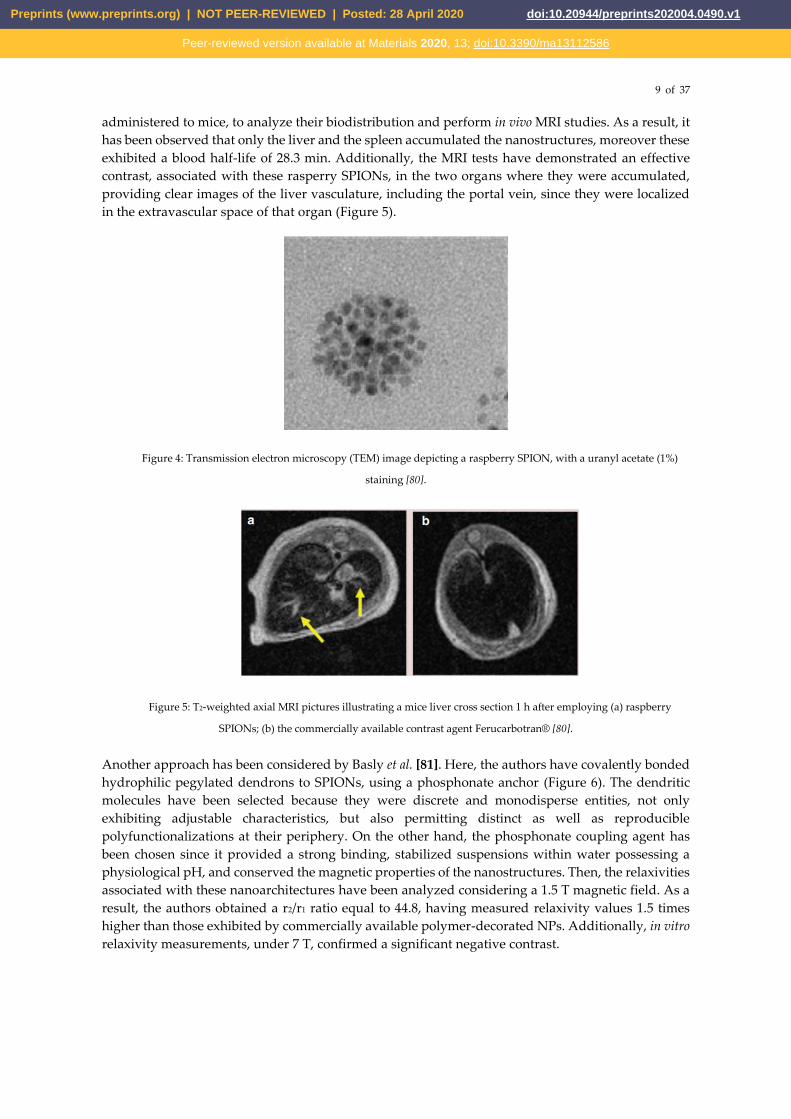
administered to mice, to analyze their biodistribution and perform in vivo MRI studies. As a result, it
has been observed that only the liver and the spleen accumulated the nanostructures, moreover these
exhibited a blood half-life of 28.3 min. Additionally, the MRI tests have demonstrated an effective
contrast, associated with these rasperry SPIONs, in the two organs where they were accumulated,
providing clear images of the liver vasculature, including the portal vein, since they were localized
in the extravascular space of that organ (Figure 5).
Figure 4: Transmission electron microscopy (TEM) image depicting a raspberry SPION, with a uranyl acetate (1%)
staining [80].
Figure 5: T2-weighted axial MRI pictures illustrating a mice liver cross section 1 h after employing (a) raspberry
SPIONs; (b) the commercially available contrast agent Ferucarbotran® [80].
Another approach has been considered by Basly et al. [81]. Here, the authors have covalently bonded
hydrophilic pegylated dendrons to SPIONs, using a phosphonate anchor (Figure 6). The dendritic
molecules have been selected because they were discrete and monodisperse entities, not only
exhibiting adjustable characteristics, but also permitting distinct as well as reproducible
polyfunctionalizations at their periphery. On the other hand, the phosphonate coupling agent has
been chosen since it provided a strong binding, stabilized suspensions within water possessing a
physiological pH, and conserved the magnetic properties of the nanostructures. Then, the relaxivities
associated with these nanoarchitectures have been analyzed considering a 1.5 T magnetic field. As a
result, the authors obtained a r2/r1 ratio equal to 44.8, having measured relaxivity values 1.5 times
higher than those exhibited by commercially available polymer-decorated NPs. Additionally, in vitro
relaxivity measurements, under 7 T, confirmed a significant negative contrast.
Preprints (www.preprints.org) | NOT PEER-REVIEWED | Posted: 28 April 2020 doi:10.20944/preprints202004.0490.v1
Peer-reviewed version available at Materials 2020, 13; doi:10.3390/ma13112586

10 of 37
Figure 6: Illustration representing a hydrophilic pegylated dendrons covalently bonded to an iron oxide nanoparticle,
via a phosphonate anchor [81].
Xie et al. [82] have performed a different study, where a MRI contrast agent for identifying brain
gliomas in vivo, i.e. lactoferrin-conjugated SPIONs (LfSPIONs), has been developed. After
synthesizing such NPs, the authors have examined their physical, chemical, and magnetic properties,
as well as their interaction with glioma cells. As a result, it has been verified a hydrodynamic diameter
equal to ∼ 75 nm, a 51 emu/g Fe saturation magnetization, plus a T2 relaxivity of 75.6 mM-1s-1, for
these spherical nanostructures. Additionally, an in vitro study, considering a rat glioma cell line (C6),
revealed that the Lf-SPIONs originated MR images possessing a better T2 contrast than the one
produced by SPIONs. Furthermore, an in vivo investigation has been performed using rat models
together with the developed NPs. It has been noticed a considerably improved contrast, between the
tumor and the neighboring normal tissues, on T2-weighted brain glioma MR images, until 48 h after
the Lf-SPIONs administration (Figure 7). Following such time period, a histochemical analysis
hasallowed the observation of those nanostructures around the vascular region of the lesion tissue
slices. Moreover, real-time polymerase chain reaction (RT-PCR) plus Western Blot have been
employed in the brain tumor tissues. These techniques have allowed the authors to confirm a larger
expression level associated with Lf receptors, when compared against normal tissues from the same
organ. Consequently, these results have indicated that Lf-SPIONs are suitable T2 MRI contrast agents
for brain glioma, presenting high selectivity and sensitivity.
Figure 7 - In vivo T2-weighted MR images of rats’ brain possessing C6 gliomas, acquired 48 h after the administration
of SPIONs; Lf-SPIONs [82].
In a different work, Gonzalez-Rodriguez et al. [83] have fabricated biomcompatible SPIONs
conjugated with graphene oxide (GO-SPIONs). These spherical nanostructures have presented a
mean size of 250 nm and have demonstrated the ability to be used towards magnetic targeted therapy,
fluorescence imaging, cancer detection via optical pH-sensing, anticancer drug delivery, as well as
MRI contrast agents. Cytotoxicity assays have revealed a reduced cell death resulting from the
Preprints (www.preprints.org) | NOT PEER-REVIEWED | Posted: 28 April 2020 doi:10.20944/preprints202004.0490.v1
Peer-reviewed version available at Materials 2020, 13; doi:10.3390/ma13112586

11 of 37
nanoparticles internalization, at a 15 g/mL concentration. Furthermore, relaxivity measurements
have indicated a r2/r1 ratio of ∼ 10.7 for the GO-SPIONs, being considerably higher than the one
exhibited by free SPIONs (∼ 2.3). Consequently, this suggested that the graphene oxide conjugated
SPIONs have had the potential to be employed as T2 MRI contrast agents. Additionally, the authors
have successfully distinguished cancer cells from healthy ones in vitro through the ratios of emission
intensity associated with NPs, since they presented fluorescence in the visible range that depended
on the medium pH (Figure 8). Concerning drug delivery, it has been achieved a successful
fluorescence-tracked intracellular delivery of hydrophobic doxorubicin non-covalently conjugated
with GO, by applying an external magnetic field. This have resulted in a 2.5-fold efficacy
enhancement, when compared against the free drug at reduced concentrations, becoming possible to
reduce the drug dose required for reaching an identical therapeutic effect.
Figure 8 - Pictures representing the GO-SPIONs emission in green (550 nm) and red (635 nm) in healthy HEK-293 versus
cancer HeLa and MCF-7 cells [83].
Also considering SPIONs, Sulek et al. [84] have fabricated a contrast agent by a non-covalent
functionalization of those nanoparticles with peptide amphiphile molecules, which provided water
solubility and improved their biocompatibility (Figure 9). Then, after production, the nanocomplexes
relaxivity has been assessed under a 3.0 T magnetic field, having been observed a r2/r1 ratio as high
as 111.55, being a much larger value than that of commercially available SPIONs. Furthermore, in
vitro incubation experiments using fibroblasts (NIH 3T3) have revealed that these functionalized NPs
were, in fact, highly biocompatible. Moreover, it has been observed that such spherical
nanostructures were located on the cell membrane or matrix. Additionally, the hydrophilic peptide
sequence located at the SPIONs surface, which has supplied stability as well as bioactivity within
aqueous conditions, could be changed so as to target them towards specific tissues.
A different T2 contrast agent, i.e. multifunctional polymeric-coated multicore NPs (bioferrofluids),
has been investigated by Ali et al. [85] .These spherical nanostructures have consisted in various
maghemite NPs involved with a hydrophilic polymer (polyethylene glycol, PEG, acrylate).
Furthermore, their uptake and toxicity in the liver of mice has been assessed through MRI together
with histological techniques. Then, the obtained outcomes have been compared against those
acquired when employing commercially available Endorem magnetic fluids, under identical
Preprints (www.preprints.org) | NOT PEER-REVIEWED | Posted: 28 April 2020 doi:10.20944/preprints202004.0490.v1
Peer-reviewed version available at Materials 2020, 13; doi:10.3390/ma13112586

12 of 37
experimental circumstances. As a result, it has beenverified that the r2/r1 ratio for the bioferrofluids
synthesized by the authors was equal to 184, while for Endorem such parameter exhibited a value of
54.02. Additionally, these NPs not only exhibited a smaller blood circulation period, but also have
demonstrated to be efficient reticuloendothelial system agents, since they remained in the liver tissue.
Moreover, it has been observed that those bioferrofluids stayed in such organ for a longer time
interval than Endorem. Nevertheless, no perceptible histological lesions in the examined liver were
caused by the two contrast agents analyzed, over a time interval of 60 days after-administration.
Another type of contrast agent has been analyzed by Zhang et al. [86]. This nanomaterial has consisted
in SPIONs coated with polyethylenimine (PEI), which were obtained through photochemistry, and
whose surface was modified by poly(ethylene glycol) methyl ether (MPEG), MPEG-PEI-SPIONs
(Figure 9). Then, the physical properties, stability, as well as MRI feasibility of these NPs have been
assessed. It has been verified that they possessed a hydrodynamic size equal to 34 nm. Furthermore,
their coating has been checked through a Fourier transform infrared spectrometer, having been
determined a 31% and 12% proportion of PEI and MPEG, respectively, in the MPEG-PEI-SPIONs.
Additionally, magnetic measurements showed a superparamagnetic behavior, as well as 46 emu/g
saturation magnetization, for these nanoarchitectures. Furthermore, a stability test has indicated that
MPEG-PEI considerably enhanced the spherical nanostructures stability. Moreover, relaxation
measurements have demonstrated similar r2 values for PEI-SPIONs and MPEG-PEI-SPIONs.
Additionally, T2-weighted MR images using MPEG-PEI-SPIONs have revealed a considerable
improvement of the MR signal, as the concentration of those NPs in water got higher. Consequently,
this indicated that these spherical nanostructures have been able to produce large magnetic field
gradients near their surface.
Figure 9: Schematic representation of the MPEG-PEI-SPIONs fabrication process [86].
Figure 10: T2-weighted MR images of MPEG-PEI-SPIONs considering different concentrations, namely (a) 0.063; (b)
0.125; (c) 0.250; (d) as well as 0.500 mg/mL [86].
Yue-Jian et al. [87] have addressed a novel contrast agent consisting in antifouling PEG-coated
SPIONs. Here, monodisperse oleic acid-coated SPIONs have been synthesized via thermal
decomposition of iron oleate. Then, the self-assembly occurring between those spherical
Preprints (www.preprints.org) | NOT PEER-REVIEWED | Posted: 28 April 2020 doi:10.20944/preprints202004.0490.v1
Peer-reviewed version available at Materials 2020, 13; doi:10.3390/ma13112586

13 of 37
nanostructures and the PEG-lipid conjugates in water. It has been observed, through dynamic light
scattering, that the PEG-coated SPIONs were stable within water for a pH from 3 unto 10 and at
sodium chloride concentrations up until 0.3 M. Furthermore, their incubation with a cell culture
medium possessing 10% fetal bovine serum, which simulated the in vivo plasma, has confirmed such
stability, not having been noticed changes in the NPs dimensions after a 24 h time period. These
results have pointed out an absence of protein adsorption upon their surface. Moreover, in vitro
relaxation measurements have indicated a greater r2 for these spherical nanoarchitectures than that
of the commercially available contrast agent Feridex IV, suggesting, therefore, that a better contrast
could be created by these PEG-coated SPIONs.
Several authors have also gained interest for the use of iron oxide NPs as T1 contrast agents, since in
the clinical practice the typically employed positive contrast agents are Gd complexes, which, as
previously referred, pose health risks to the patients [88, 10, 89, 5]. Within this context, Wei et al. [90]
have investigated zwitterion-coated SPIONs (ZES-SPIONs), possessing inorganic cores with a size of
∼ 3 nm as well as an ultrathin hydrophilic shell (∼ 1 nm). As a result, it has been verified that these
NPs presented a r2/r1 ratio equal to 2.0, being a value lower than the one associated with other SPION-
based positive contrast agents, nevertheless it was within a factor of 2 to that exhibited by Gd-based
chelates. Additionally, in vivo MRI has been performed on mice injected with ZES-SPIONs, to assess
their preclinical potential as T1 contrast agents for MRI and MR angiography. These tests have
revealed a contrast power, associated with those NPs, that was sufficiently high for their use in the
considered applications. Moreover, it has been observed an efficient renal clearance of the ZES-
SPIONs and, by measuring once again their r2/r1 ratio after excretion, the authors have verified that
the MR contrast power of those NPs was kept largely unmodified under physiological conditions.
Figure 11: T1-weighted MR angiography, at 7 T, of a mouse (a) 4 min; (b) 12 min; and (c) 20 min, after the injection of
ZES-SPIONs [90].
Yin et al. [91] have achieved a T1 contrast in MRI by employing SPIONs, with diameters between 11
and 22 nm, in a ultra-low feld (ULF) MRI system, which applied a ∼ 0.13 mT magnetic field, at room
temperature. This approach has allowed improving the positive contrast created by such NPs,
because under these conditions their relaxation times were similar to the proton Larmor precession
period, originating a great increase of the r1 value. Additionally, their r2 was lowered, since the
magnetic moments, present in the SPIONs, were not saturated at this field magnitude. As a result, a
r1 as high as 615 mM-1s-1 has been obtained for Zn0.3Fe2.7O4 NPs, coated with silicon dioxide, and
possessing a size of 18 nm, being a value 100 times larger than that of typical commercial Gd-based
positive contrast agents under large magnetic fields, i.e. 1.5 and 3.0 T. Furthermore, the authors have
verified a linear dependence of r1 on the imaginary part of the magnetic AC mass susceptibility, at
5.56 kHz, i.e. the proton resonance frequency, for all the studied cases. This result has been justified
by the NPs magnetic fluctuations, associated with Brownian motion or Néel relaxation. As a
Preprints (www.preprints.org) | NOT PEER-REVIEWED | Posted: 28 April 2020 doi:10.20944/preprints202004.0490.v1
Peer-reviewed version available at Materials 2020, 13; doi:10.3390/ma13112586
14 of 37
conclusion, various benefits have been observed for this approach, namely adjustable magnetic
susceptibility in SPIONs, improved signal, shorter imaging times, as well as the use of biocompatible
substances.
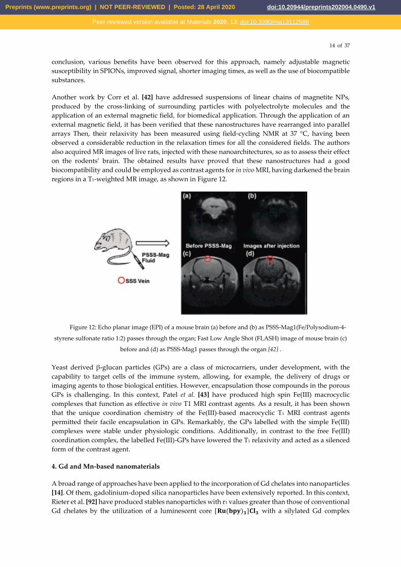
Another work by Corr et al. [42] have addressed suspensions of linear chains of magnetite NPs,
produced by the cross-linking of surrounding particles with polyelectrolyte molecules and the
application of an external magnetic field, for biomedical application. Through the application of an
external magnetic field, it has been verified that these nanostructures have rearranged into parallel
arrays Then, their relaxivity has been measured using field-cycling NMR at 37 °C, having been
observed a considerable reduction in the relaxation times for all the considered fields. The authors
also acquired MR images of live rats, injected with these nanoarchitectures, so as to assess their effect
on the rodents’ brain. The obtained results have proved that these nanostructures had a good
biocompatibility and could be employed as contrast agents for in vivo MRI, having darkened the brain
regions in a T1-weighted MR image, as shown in Figure 12.
Figure 12: Echo planar image (EPI) of a mouse brain (a) before and (b) as PSSS-Mag1(Fe/Polysodium-4-
styrene sulfonate ratio 1:2) passes through the organ; Fast Low Angle Shot (FLASH) image of mouse brain (c)
before and (d) as PSSS-Mag1 passes through the organ [42] .
Yeast derived β-glucan particles (GPs) are a class of microcarriers, under development, with the
capability to target cells of the immune system, allowing, for example, the delivery of drugs or
imaging agents to those biological entities. However, encapsulation those compounds in the porous
GPs is challenging. In this context, Patel et al. [43] have produced high spin Fe(III) macrocyclic
complexes that function as effective in vivo T1 MRI contrast agents. As a result, it has been shown
that the unique coordination chemistry of the Fe(III)-based macrocyclic T1 MRI contrast agents
permitted their facile encapsulation in GPs. Remarkably, the GPs labelled with the simple Fe(III)
complexes were stable under physiologic conditions. Additionally, in contrast to the free Fe(III)
coordination complex, the labelled Fe(III)-GPs have lowered the T1 relaxivity and acted as a silenced
form of the contrast agent.
4. Gd and Mn-based nanomaterials
A broad range of approaches have been applied to the incorporation of Gd chelates into nanoparticles
[14]. Of them, gadolinium-doped silica nanoparticles have been extensively reported. In this context,
Rieter et al. [92] have produced stables nanoparticles with r1 values greater than those of conventional
Gd chelates by the utilization of a luminescent core [𝐑𝐮(𝐛𝐩𝐲)𝟑]𝐂𝐥𝟑 with a silylated Gd complex
Preprints (www.preprints.org) | NOT PEER-REVIEWED | Posted: 28 April 2020 doi:10.20944/preprints202004.0490.v1
Peer-reviewed version available at Materials 2020, 13; doi:10.3390/ma13112586

15 of 37
coating. Another work has demonstrated that the location of the Gd chelate within mesoporous silica
nanoparticles (MSNs) greatly influences its relaxometric properties. The highest relaxivities have
been specifically reported to occur when synthesis occurs by a long delay co-condensation process,
leading to a r1 value of 33.6 ± 1.3 𝐦𝐌−𝟏𝐬−𝟏, which is higher than any previously reported Gd-DOTA
silica NPs and 20 times larger than free Gd-DOTA [93]. Then, these particles were biotinylated
showing a large relaxivity that was kept after the external biomodification, but presenting reversibly
gateable on subsequent protein recognition [94]. Graphene oxide (GO) has also been used as a
scaffold to integrate Gd-DOTA moieties. A study by Zhang et al. [95] reported that GO has been first
pegylated, functionalised with DOTA, and then metallated with Gd(III). These nanoparticles have
presented a large r1 value of 14.2 𝐦𝐌−𝟏𝐬−𝟏measured at 11.7 T. Some other strategies to incorporate
Gd in several types of nanoparticles have also been reported, namely by grafting Gd(III) in detonation
nanodiamond (DND) [96] and melanin-dots (M-dots) loaded with Gd (II) [97]
Gadolinium oxides are the most utilised alternatives to Gd chelates, where it has been found that
decreasing particle diameter resulted in a progressive trend towards higher relaxivities. For instance,
Park et al. [17] have shown that the highest relaxivities were obtained for NPs synthesised with an
average diameter, d, of ~ 1–2.5 nm, as presented in Figure 13. As a result, high contrast in in vivo T1
images of the brain tumour of a rat have been observed. The large r1 has been discussed in terms of
the big surface to volume ratio of the ultrasmall gadolinium oxide nanoparticles, coupled with the
cooperative induction of surface Gd(III) ions for the longitudinal relaxation of a water proton. It
should be noted, however, that ultrasmall 𝑮𝒅𝟐𝑶𝟑 NPs have been found to form deposits in the brain
and, consequently, there is a compromise between limiting the toxicity of the particles and
maximising imaging potency. Yin et al. [98] have produced silica nanoparticles with a 𝑮𝒅𝟐𝑶𝟑
nanoshell of varying thicknesses. By systematically changing the thickness of the silica shell, the
variations in relaxivity values could be investigated and it was demonstrated that a thinner shell
resulted in larger r1 values. Furthermore, the core-shelled nanoparticles showed negligible
nanotoxicity. The enhanced signals in in vivo tumour-targeted MRI indicated that ultrathin
gadolinium oxide nanoshells may function as a potential candidate for advanced positive contrast
agents in further clinical applications.
Figure 13: Reproductions of r1. The functions are labeled as G (Gaussian), L (Lorentzian), and LN (log-normal)
[17].
Preprints (www.preprints.org) | NOT PEER-REVIEWED | Posted: 28 April 2020 doi:10.20944/preprints202004.0490.v1
Peer-reviewed version available at Materials 2020, 13; doi:10.3390/ma13112586

16 of 37
Dual T1- and T2-weighted MRI agents were also reported by Zeng et al.. Such authors fabricated
biocompatible gadolinium hybrid iron oxide (GdIO) nanocomposites with hydrodynamic size
between 120 and 150 nm. These nanocomposites exhibited both superparamagnetic and
paramagnetic properties, with unsaturated magnetic moments of 33.5 emu g−1 at 5 T. The GdIO
samples exhibited high contrast ability for both T1 and T2-weighted MR imaging, with r1 value of
70.10 ± 3.65 mM−1 s−1 (based on Gd) and an r2 value equal to 173.55 ± 6.48 mM−1 s−1 (based on Fe). In
vivo studies showed that the GdIO nanocomposites were able to achieve the brain tissues and
translocate into neurons [99].
Bailey et al. [41] have reported the fabrication of 𝑹𝑬𝟐𝑶𝟑-based nanodiscs, with diameters ranging
from 10 to 14 nm; RE stands for Gd, dysprosium (Dy) or ytterbium (Yb) passivated with a
biocompatible polymer (Poly(acrylicacid) grafted with short methoxy-terminated polyethylene
oxides). Here, their suitability as MRI contrast agents has been analysed. The relaxation times of such
nanostructures, measured at 37 °C (body temperature) in a magnetic field of 1.41 T, have been
compared to the reported values for their spherical counterparts or small molecule chelates, based on
the DTPA ligand. The authors have also performed an MR scan of a phantom for all the considered
contrast agents, using 𝑻𝟏 weighted sequences, having been found that 𝑮𝒅𝟐𝑶𝟑 nanodiscs were more
suitable as contrast agents compared than the commercially available Gd-DTPA, due to their higher
relaxivities [100] . This factor should increase the efficiency of in vivo targeted imaging schemes, since
it becomes possible to get a high amount of proton relaxation without requiring multiple small
molecules in contact with the imaging target. Besides this benefit, it has been verified that these
𝑮𝒅𝟐𝑶𝟑 nanodiscs were suitable as 𝑻𝟏 contrast agents. Also, no significant cytotoxic effects have been
observed for the polymer coated 𝑮𝒅𝟐𝑶𝟑and 𝑫𝒚𝟐𝑶𝟑 nanoarchitectures, on a cell line derived from a
human cervical cancer (HeLa).
Singh et al. [20] also reported the suitability of polyethylene glycol (PEG) coated 𝑮𝒅𝟐𝑶𝟑
paramagnetic nanodiscs and PEG coated Gd doped iron oxide (GdIO) superparamagnetic
cubic/spherical-shaped nanoparticles, with different dimensions, as MRI contrast agents. In this case,
the relaxivities of the different nanoarchitectures have been measured with a 7 T MR scanner and it
has been showed that smaller sized nanostructures (<5 nm) were the more effective 𝑻𝟏 contrast
agents, as presented in figure 14.
Figure 14: T1 relaxation rate as a function of concentration measured for (a) Gd2O3 nanodiscs of different diameters and (b)
GdIO NPs of spherical (9 nm and 6 nm) and cubic (4 nm) shapes [20].
(a) (b)
Preprints (www.preprints.org) | NOT PEER-REVIEWED | Posted: 28 April 2020 doi:10.20944/preprints202004.0490.v1
Peer-reviewed version available at Materials 2020, 13; doi:10.3390/ma13112586

17 of 37
Besides Gd-based nanoparticles, Mn has also been extensively researched as a possible T1
contrast agent with reduced toxicity (compared to that of Gadolinium), but it possesses low native r1
relaxivities. Nevertheless, it has shown promising results as dual modal imaging agent, as presented
in Figure 5 [14]. Much effort has been invested in increasing and biocompatibility and r1 by using
derived nanoparticulate systems. For instance, PEG-functionalised Mn3O4 nanoparticles have been
encapsulated in a mesoporous, biocompatible carbon framework. Then, it was demonstrated that the
agent displayed significant contrast enhancement in T1-weighted images. The carbon encapsulation
and surface modification rendered biocompatibility and water solubility to the nanosystem.
Furthermore, the porous network ensured water accessibility for the nanoparticles, making them
helpful in interpretation of MRI scans [101].
Neves et al. [44] have addressed Mn oxide (MnO) NPs (average size of ~ 20 nm) coated with
carboxymethyl-dextran. Despite not having performed an in vivo study, the authors have considered
such nanostructures adequate as T1 contrast agents, due to their significant longitudinal relaxivity,
measured on a clinical 3.0 T MRI scanner. Moreover, it has been observed that such NPs presented
no in vitro cytotoxicity for healthy cells at concentrations lower than 25 µg/ml, however for HeLa cells
a notable toxicity has been observed, even at low concentrations of NPs (5 µg/ml).
Manganese ferrite nanoparticles (Fe3O4@MnIO) have also exhibited remarkably higher longitudinal
relaxivity than their counterpart iron oxide NPs [102]. Here, the authors found a r1 value of 33.8 mM-
1s-1 for Fe3O4@MnIO and a r2 equal to 306.3 mM-1s-1. The increased T1 relaxivity was attributed to the
extended electronic relaxation time and the increased number of unpaired electrons due to the Fe
substitution by Mn ions. Additionally, in vivo results indicated that these nanoparticles could achieve
in vivo contrast imaging with acceptable biocompatibility, with the dosage of 1 mg/kg, however the
systemic toxicity evaluation was unclear.
5. Synthetic antiferromagnetic nanostructures
More recently, antiferromagnetic nanoarchitectures have also been investigated as potential T1
contrast agents by different authors. Namely, Na et al. [18] fabricated antiferromagnetic MnO
nanoparticles of sizes between 7 and 25 nm, coated with a PEG-phospholipid shell. The relaxivity of
such particles has been measured in a 3.0 T human clinical scanner and their in vivo performance as
MRI contrast agents has been analyzed in a mouse. The obtained results have indicated that these
NPs were suitable as T1 contrast agents, having demonstrated no significant toxicity, for a MnO
concentration lower than 0.82 mM, in eight human cell lines originating from different tissues.
Furthermore, by conjugating them with a tumour-specific antibody, it has been possible to selectively
improve the contrast of breast cancer cells located in a mouse’s metastatic brain tumour, which has
been intravenously injected with the functionalized nanoparticles through T1-weighted MRI.
Liuet et al. [103] have also fabricated spherical nanostructures exhibiting antiferromagnetic properties
towards the same application. These nanoarchitectures were glutathione-functionalized iron-oxide
nanoparticles, having been produced at room temperature and in aqueous-phase, by a facile, highly
efficient, as well as eco-friendly, one-step reduction process, using
tetrakis(hydroxymethyl)phosphonium chloride as the reducing agent. After the synthesization
Preprints (www.preprints.org) | NOT PEER-REVIEWED | Posted: 28 April 2020 doi:10.20944/preprints202004.0490.v1
Peer-reviewed version available at Materials 2020, 13; doi:10.3390/ma13112586

18 of 37
procedure, the nanostructures characterization has revealed a diameter of 3.72 ± 0.12 nm, an r2 equal
to 8.28 mM-1s-1, a 2.28 r2/r1 ratio, a reduced magnetization, plus an adequate water dispersion.
Additionally, through their incubation with HeLa cells, it has been observed that they were
biocompatible and improved the acquired MR signal intensity, in T1-weighted sequences, as the iron
content inside the considered biological entities increased. Moreover, the fabricated nanoparticles
havealso been injected into mice as well as rat models, so as to not only study their in vivo circulation
and metabolic path, but also to analyze the contrast created by them in MR images, under those
conditions. As a result, it has been observed that the nanostructures escaped the hepatic
reticuloendothelial system and, subsequently, were expelled from the body via the urinary system,
enabling, therefore, the realization of a renal function assessment. This course led to a long circulation
period in the vasculature, allowing a strong improvement, in T1-weighted MRI, of the vascular
resolution at the internal carotid artery and superior sagittal sinus, which are locations where the
thrombus identification is essential for diagnosing a stroke (figure 15). Additionally, various T1- as
well as T2-weighted MR images of a rat’s kidney injected with the produced nanostructures, allowed
a detailed visualization of the cortical-medullary anatomy and renal physiological functions.
Figure 15: T1 –weighted MR images of a mice s brain, before (T1-pre) and after (T1-post) the injection of the glutathione-
functionalized iron-oxide nanoparticles [103].
In a different work, Peng et al. [43] have investigated another T1 contrast agent type, known as
antiferromagnetic-iron oxide-hydroxide nanocolloids, which possessed a diameter of 2-3 nm. Such
nanostructures have been prepared in the mesopores of worm-like mesoporous silica. Then, the
relaxation times have been measured at 40 °C using a 0.47 T Minispec spectrometer. As a result, it
has been verified that these nanoparticles not only had the lowest r2/r1 ratio reported, until 2013, for
iron-based colloidal T1 contrast agents, but also possessed a considerably large longitudinal relaxivity.
Additionally, the acquired MR images have shown that such nanocolloids were a superior T1 contrast
agent, in both in vitro (HeLa cells) and in vivo (rat and mouse) MRI, when compared to ultrasmall
iron oxide nanoparticles. Furthermore, these nanocolloids also demonstrated a high level of
biocompatibility and biodegradability.
In addition to the previously mentioned nanoarchitectures, (SAF nanostructures have also been
studied as potential contrast agents for MRI. For example, Roosbroeck et al. [58] have fabricated
phospholipid-coated, disc-shaped, and multilayered [Au(10 nm)/Ni80Fe(5 nm)20/Au(2.5
nm)/Ni80Fe(5 nm)20/Au(10 nm)] SAF nanoarchitectures, with diameters ranging from 89.8 nm to
523.2 nm, using a colloidal lithography technique. The magnetic characterization of these nanodiscs
has indicated a very low remanence value, which is necessary to prevent particle agglomeration, as
well as a high magnetization, making them adequate for biomedical applications. Then, these
nanostructures have been evaluated as T2 contrast agents, as indicated in Figure 16, having shown
improved relaxivities, at 24.85 °C in a 9.4 T magnetic field, when compared to SPIONs, especially the
smallest particles with a diameter of 90 nm. The authors have also carried out an in vitro MRI study,
using an ovarian cancer cell line (SKOV3), confirming the increased T2 relaxation for cells marked
with such nanostructures.
Preprints (www.preprints.org) | NOT PEER-REVIEWED | Posted: 28 April 2020 doi:10.20944/preprints202004.0490.v1
Peer-reviewed version available at Materials 2020, 13; doi:10.3390/ma13112586

19 of 37
Figure 16: Theoretical (black lines) and measured (points) r2 values of [Au(10 nm)/NiFe(10 nm)/Au(2.5 nm)/NiFe(10
nm)/Au(10 nm)] SAF-NPs as function of SAF-NP diameter. The reference theoretical values for spherical NiFe particles are
represented in gray [58].
6. High-aspect ratio nanowires
Nanowires (NWs) have also been addressed by some reports in the context of this biomedical
application. For example, Bañobre-López et al. [57] evaluated the relaxivity properties of poly-acrylic
acid (PAA)-coated Ni ferromagnetic NWs characterized by longitudinal magnetic anisotropy, in a
colloidally stable water dispersion. This dispersion has been produced through a process of pulsed
electrodeposition of Ni/Gold (Au) multilayer nanowires inside a porous alumina at room-
temperature, followed by the template removal and chemical etching of the Au layer in a two-step
acidic etching. The relaxation times of these nanostructures, which have presented a monodisperse
average diameter and length of ~36 nm and ~600 nm, respectively, have been measured using a
relaxometer operated at 60 MHz and under 37 °C for two magnetic fields, namely 1.41 T and 3.0 T.
In both situations, the obtained results have indicated that these nanostructures were efficient as T2
contrast agents, as clearly visible in Figure 17. The contrast effect of the PAA-coated Ni nanowires
has been verified by performing an MR scan of a phantom at a magnetic field of 3 T.
Shore et al. [36] also studied nanowires for MRI application. Specifically, Fe and segmented Fe/Au
nanowires, with various lengths and diameters, have been fabricated by template-assisted
electrodeposition. These nanostructures have been coated with compounds, namely Dop-PEG and/or
SH-PEG-COOH, which allowed the binding of biological molecules to the nanowires in order to
target specific cells. The magnetic characterization of both nanostructures has shown that the Fe/Au
nanowires exhibited a larger saturation magnetization. Since Fe layers are thinner than the diameter,
these nanostructures were easily magnetized in the direction perpendicular to the long axis of the
nanostructure, than in the Fe nanowires. The relaxivity properties of the fabricated nanowires have
been measured at 25 °C in a 1.5 T magnetic field and the obtained results have been compared against
those of Fe and Fe-Au nanoparticles. As a result, it has beenverified that the Fe nanowires with a
length of 0.7 µm and a diameter of 110 nm, coated with Dop-PEG, were the best suited as T1 contrast
agents. On the other hand, Fe-Au nanowires with a length of 1 µm and a diameter of 32.8 nm, coated
with SH-PEG-COOH and Dop-PEG, were the most appropriate as T2 contrast agents, being
comparable to commercial Fe oxide nanoparticles. The authors also performed an MR scan of some
samples containing Fe and Fe-Au nanowires, at a magnetic field of 9.4 T, in order to confirm the
contrast caused by the nanostructures in the image.
Preprints (www.preprints.org) | NOT PEER-REVIEWED | Posted: 28 April 2020 doi:10.20944/preprints202004.0490.v1
Peer-reviewed version available at Materials 2020, 13; doi:10.3390/ma13112586

20 of 37
Figure 17: T2 map associated with PAA-coated Ni nanowires, acquired at 3 T and under 37 ºC [57].
A different type of nanowires for this biomedical application has been investigated by Leung et
al. [104] .These nanostructures, made from Mn-Fe, have been synthesized through ligand-induced
self-organization of Mn–Fe oxide nanoparticles. Then, via TEM, it has been observed that they
possessed a mean diameter equal to 35 nm and, on average were 1 μm long. Furthermore, the
nanowires elemental content has been verified by inductively coupled plasma-optical emission
spectroscopy (ICP-OES), as well as through energy-dispersive X-ray (EDX) spectroscopy, having
been determined a Fe percentage of ∼40.65. Moreover, their influence in the T2 relaxation time has
been assessed using a 1.5 T MRI system. As a result, it has been noticed that these nanoarchitectures
have considerably decreased the MRI signal, when concentrations of 100 μg/mL or 10 μg/mL have
been considered, which demonstrated their potential as T2 contrast agents. Moreover, the cell
labelling efficiency of these nanostructures was assessed by incubating them with a macrophage cell
line (RAW264.7). After this process, it has been observed an effective Mn-Fe nanowires incorporation
into the considered biological entities.
Also considering the improvement of the negative contrast in MRI, Martínez-Banderas et al. [105]
have fabricated distinct one-dimensional nanostructures, which were composed by an iron core
together with an iron oxide shell (Fe-FexOy core–shell NWs). Then, their r1, r2, and r2/r1 ratio have been
assessed under a 1.5 T magnetic field, having been observed that they possessed a great potential for
this application. Furthermore, the effects of various oxidation levels as well as surface coatings have
been evaluated at a 7.0 T field. As a result, it has been verified that their r2 could be adjusted by not
only the oxide shell thickness, but also coating agents. Moreover, breast cancer cells (MDA-MB-231)
labelled with Fe-FexOy core–shell NWs, which were coated with two different compounds, namely
bovine serum albumin (BSA) and (3-aminopropyl)triethoxysilane (APTES), were inserted in tissue-
mimicking phantoms. Then, T2-weighted MR images of those two cases has been performed, having
been noticed that the BSA coating improved the dispersion, as well as the cellular internalization,
while allowing an identical cell identification efficiency through MRI, when compared against APTES.
Consequently, this has permitted the use of a lower nanostructures concentration to efficiently label
the desired cells, lowering, therefore, the probability of toxic effects. Furthermore, using such coating,
the authors have been able to detect ∼25 cells/μL by employing a NW concentration equal to 0.8 μg
of Fe/mL. Moreover, cells labelled with BSA coated nanostructures were implanted inside a mouse’s
Preprints (www.preprints.org) | NOT PEER-REVIEWED | Posted: 28 April 2020 doi:10.20944/preprints202004.0490.v1
Peer-reviewed version available at Materials 2020, 13; doi:10.3390/ma13112586

21 of 37
brain and, subsequently, several T2-weighted MR images were acquired, at a 11.7 T field, considering
various time intervals post-implantation. As a result, it has been observed that these biological
entities could be identified in such organ during, at least, 40 days after insertion.
Iron oxide (Fe3O4) nanostructures with rod like morphology (nanowires with low aspect ratio),
presenting a diameter of 4–12 nm and length ranging from 30 to 70 nm, were also addressed in the
context of this biomedical application by Mohapatra et al. [106]. As a result, it was verified that
nanorods of 70 nm length showed a r2 relaxivity of 608 mM−1 s−1. Additionally, the increase of the
nanorods size led to a linear increase in their r2 relaxivity values, from 312 to 608 s-1mM-1. This linear
trend was attributed to an enhancement of the saturation magnetization and the surface area of the
nanostructures. Moreover, in vitro assays considering HeLa cells indicated that those biological
entities exhibited a normal growth in the presence of Fe3O4 nanorods, indicating an acceptable
biocompatibility without toxic effects (approximately 90 % the cells remained viable), even after
incubation with 1 mg/ml of nanorods.
7. Theragnosis Applications
Nanotechnology is a powerful approach for the development of novel nanomaterials that can be used
in both the diagnosis and treatment of illnesses, filling, for example, the biggest challenges in the
cancer therapeutics. Several types of nanostructures have attracted much attention due to their
promising applications as “theragnostic” anti-cancer agents, showing good performance in imaging
combined therapy, namely by using hyperthermia, radiotherapy, or drug delivery [102, 107]..
A study by Wang et al. [108] reported the construction of an intelligent near-infrared (NIR) light and
a tumor microenviroment (NIR/TME) dual-responsive nanocapsule for enhanced tumor
accumulation and improved therapy efficacy. The large initial size of these nanocapsules (NCs)
ensured the circulatory stability in the blood while, under irradiation of an NIR laser, the shrinkage
and decomposition of the nanocapsule in the acidic TME guaranteed intratumoral permeability of
NPs and the controllable release of doxorubicin (DOX). Interestingly, the overproduced reactive
oxygen species (ROS), by synergistic catalysis of the Fenton reaction based on Fe/FeO NCs and light
activation from indocyanine green (ICG), relieved the hypoxia for solid tumors, which is necessary
for mitigating the hypoxia-related resistance during chemo/photo- and chemodynamic therapy. As a
result of these unique properties of the nanocapsules, it was achieved an almost complete destruction
of the tumors. In addition, dual-mode MRI and fluorescence imaging provided complementary
imaging information. Hence, this study presented the design of smart nanocapsules with enhanced
tumor accumulation, highly effective therapy and diagnosis capability.
Preprints (www.preprints.org) | NOT PEER-REVIEWED | Posted: 28 April 2020 doi:10.20944/preprints202004.0490.v1
Peer-reviewed version available at Materials 2020, 13; doi:10.3390/ma13112586

22 of 37
Until now, magnetite nanostructures with designed composition and properties are the ones that
showed greater potential as theragnostic agents, due to their versatility, biocompatibility, facile
production and good magnetic performance for remote in vitro and in vivo biomedical applications,
as presented, for instance, in Figure 18 [109]. This core-shell strategy has given rise to different
configurations like single- and multi-core@shell NPs, where magnetite is located in the core
(Fe3O4@SiO2; Fe3O4@C), in the shell (gelatin-NPs@Fe3O4-NPs) or embedded in a polymer matrix
(polyester, gelatin magnetic beads). In all cases, the nanocomposites inherit a combination of
properties that ensure their multimodal capacities for simultaneous magnetic
separation/detection/targeting procedures, like contrast agents in magnetic resonance
imaging/positron emission tomography (MRI/PET), magnetic hyperthermia (MH)/drug delivery
therapeutic agents, among others [110].
Also, a study by Efremova et al. [111] reported the growing of 25 nm octahedral-shaped Fe3O4
magnetite nanocrystals on 9 nm Au seed NPs using a modified wet-chemical synthesis. These Fe3O4-
Au Janus nanoparticles exhibited bulk-like magnetic properties. Additionally, due to their high
magnetization and octahedral shape, the hybrids have shown superior in vitro and in vivo T2 relaxivity
for magnetic resonance imaging as compared to other types of Fe3O4-Au hybrids and commercial
contrast agents. These nanoparticles provided two functional surfaces for theragnostic applications.
Furthermore, for the first time, Fe3O4-Au hybrids were conjugated with two fluorescent dyes or a
combination of drug and dye, allowing the simultaneous tracking of the nanoparticle vehicle and the
drug cargo in vitro and in vivo. Here, the drug delivery to tumors and payload release has been
demonstrated in real time by intravital microscopy. Moreover, replacing the dyes with cell-specific
molecules and drugs made the Fe3O4-Au hybrids a unique all-in-one platform for theragnostics. Gold
and SPION-loaded micelles were also used for both imaging and treatment of brain tumors, serving
a theragnostic purpose as both an MRI-based contrast agent and a radiosensitizer [107]. Other
Figure 18: In vivo accumulation of Fe3O4-MSN at tumor site. (a) In vivo T2-weighted MR images (upper) and color mapped (lower) images of tumor site
before and 3 h after intravenous injection of Fe3O4-MSN (arrows indicate tumor site). (b) Confocal laser scanning microscopic images of sectioned tumor tissue
harvested 24 h after injection. Left: Red fluorescence showing Fe3O4-MSN internalized cells. Right: Merged image with 4',6'-diamino-2-fenil-indol (DAPI)
stained nuclei (blue) (scale bar ) 10 μm). From [109]
Preprints (www.preprints.org) | NOT PEER-REVIEWED | Posted: 28 April 2020 doi:10.20944/preprints202004.0490.v1
Peer-reviewed version available at Materials 2020, 13; doi:10.3390/ma13112586

23 of 37
magnetic-gold nanoarchitectures also can be found in the literature as alternatives for theragnosis
applications [112, 113].
10. Prospects and Conclusions
The progress in contrast agents for MRI is notorious. Beyond the several contrast agents approved
for use in the humans, there are a significant number of new agents in clinical and research
development. For this purpose, there is a broad variety of fabrication procedures to obtain
paramagnetic and superparamagnetic nanoparticles with a control over their composition, size, and
shape. Furthermore, the ability to integrate additional imaging modes or drugs for treatment and the
employment of specific vectors linked to the particle surface make these contrast agents yet more
promising. More recently, other exciting spin configurations appeared in the literature as potential
MRI contrast agents, namely synthetic antiferromagnets (SAFs) and high aspect ratio nanowires
(NWs). Biocompatible SAFs nanostructures have been reported with coupling between
ferromagnetic layers and their relaxivities and saturation magnetization make them promising for
this biomedical application. Also, the characteristics of magnetic NWs and segmented NWs have
been explored. As a result, it was observed that the high surface area of these nanostructures and the
high magnetic moment also shows an interesting potential for contrast enhancement. The
development and comprehension of different magnetic effects has an uttermost importance in the
pursuit of novel contrast agents in MRI. On the other hand, systematic in vivo studies are also needed
so as to understand their mechanism of action and accumulation in the different organs.
Author Contributions: R. Magalhães and S. Caspani revised the major part of the literature and written
the several sections with equivalent contributions. J.P. Araújo revised the manuscript. C.T. Sousa structured the
review, contributed to write some sections and revised the manuscript.
Acknowledgments: The authors acknowledge funding from the European Union’s Horizon 2020 research
and innovation program under the Marie Sklodowska-Curie Grant Agreement No. 734801. C.T. Sousa thanks
FCT for financial support through the Investigador FCT program (Contract No. IF/01159/2015). R. Magalhães is
grateful to the FCT SFRH/BD/148563/2019 PhD grant. This work was also supported by the Portuguese Fundação
para a Ciência e Tecnologia (FCT) and COMPETE 2020 (FEDER) under the projects POCI-01-0145-FEDER-
028676/PTDC/CTM-CTM/28676/2017,POCI-01-0145/FEDER-032257/PTDC/FIS-OTI/32257/2017, POCI-01-0145-
FEDER-031302/PTDC/FIS-MAC/31302/2017, and POCI-01-0141-FEDER-032527.
Conflicts of Interest: The authors declare no conflict of interest.
References
[1] Vadim Kuperman, “Magnetic resonance imaging-physical principles and applications”.
[2] H. Shokrollahi, “Contrast agents for MRI,” Materials Science and Engineering , vol. C 33, pp.
4485-4497, 2013.
[3] Jessica Wahsner, Eric M. Gale, Aurora Rodríguez-Rodríguez, and Peter Caravan, “Chemistry
of MRI Contrast Agents: Current Challenges and New Frontiers,” Chemical Reviews, vol.
119, pp. 957-1057, 2019.
Preprints (www.preprints.org) | NOT PEER-REVIEWED | Posted: 28 April 2020 doi:10.20944/preprints202004.0490.v1
Peer-reviewed version available at Materials 2020, 13; doi:10.3390/ma13112586

24 of 37
[4] Peter Marckmann, Lone Skov, Kristian Rossen, Anders Dupont, Mette Brimnes Damholt,
James Goya Heaf and Henrik S. Thomsen, “Nephrogenic Systemic Fibrosis: Suspected
Causative Role of Gadodiamide Used for Contrast-Enhanced Magnetic Resonance Imaging,”
Journal of the American Society of Nephrology, vol. 17, pp. 2359-2362, 2006.
[5] Y. Bao, J. A Sherwood, and Z. Suna, “Magnetic Iron Oxide Nanoparticles as T1 Contrast
Agents for Magnetic Resonance Imaging,” Journal of Materials Chemistry C, 2018.
[6] Quoc Lam Vuong, Pierre Gillis, Yves Gossuin, “Monte Carlo simulation and theory of proton
NMR transverse relaxation induced by aggregation of magnetic particles used as MRI
contrast agents,” Journal of Magnetic Resonance, vol. 212, pp. 139-148, 2011.
[7] L. Peixoto, R. Magalhães, D. Navas, S. Moraes, C. Redondo, R. Morales, J. P. Araújo, and C. T.
Sousa, “Magnetic nanostructures for emerging biomedical applications,” Applied Physics
Reviews, vol. 7, p. 011310, 2020.
[8] Yu Gao, Yi Liu and Chenjie Xu, “Magnetic Nanoparticles for Biomedical Applications: From
Diagnosis to Treatment to Regeneration,” em Engineering in Translational Medicine.
[9] Yohan Jeong, Hee Sook Hwang & Kun Na, “Theranostics and contrast agents for magnetic
resonance imaging,” Biomaterials Research, vol. 22, 2018.
[10] Yu-Dong Xiao, Ramchandra Paudel, Jun Liu, Cong Ma, Zi-Shu Zhang and Shun-ke Zhou, “MRI
contrast agents: Classification and application (Review),” International Journal of Molecular
Medicine, vol. 38, pp. 1319-1326, 2016.
[11] Tromsdorf UI, Bigall NC, Kaul MG, Bruns OT, Nikolic MS, Mollwitz B, Sperling RA, Reimer R,
Hohenberg H, Parak WJ, Förster S, Beisiegel U, Adam G, Weller H., “Size and surface effects
on the MRI relaxivity of manganese ferrite nanoparticle contrast agents,” Nano Letters, vol.
7, pp. 2422-2427, 2007.
[12] Robert-Jan M. van Geuns, Piotr A. Wielopolski, Hein G. de Bruin, Benno J. Rensing, Peter M.
A. van Ooijen, Marc Hulshoff, Matthijs Oudkerk, and Pim J. de Feyter, “Basic Principles of
Magnetic Resonance Imaging,” Progress in Cariovascular Diseases, vol. 42, pp. 149-156,
1999.
Preprints (www.preprints.org) | NOT PEER-REVIEWED | Posted: 28 April 2020 doi:10.20944/preprints202004.0490.v1
Peer-reviewed version available at Materials 2020, 13; doi:10.3390/ma13112586

25 of 37
[13] Hee-Kyung Kim, Gang Ho Lee & Yongmin Chang, “Gadolinium as an MRI contrast agent,”
Future Medicinal Chemistry, 2018.
[14] Juan Pellico, Connor M. Ellis, and Jason J. Davis, “Nanoparticle-Based Paramagnetic Contrast
Agents for Magnetic Resonance Imaging,” Hindawi, 2019.
[15] J. Ramalho, R. C. Semelka, M. Ramalho, R. H. Nunes, M. AlObaidy, and M. Castillo,
“Gadolinium-Base Contrast Agent Accumulation and Toxicity: An Update,” American J. of
Neuroradiology, vol. 37, pp. 1192-1198, 2016.
[16] F. Hu and Y. S. Zhao, “Inorganic nanoparticle-based T1 and T1/T2 magnetic resonance
contrast probes,” Nanoscale, vol. 4, p. 6235–6243, 2012.
[17] J. Y. Park, M. J. Baek,E. S. Choi, S. Woo, J. H. Kim, T. J. Kim, J. C. Jung, K. S. Chae, Y. Chang, G.
H. Lee, “Paramagnetic ultrasmall gadolinium oxide nanoparticles as advanced T1 MRI
contrast agent: account for large longitudinal relaxivity, optimal particle diameter, and in
vivo T1 MR images,” ACS nano, vol. 3, p. 3663–3669, 2009.
[18] Hyon Bin Na, Jung Hee Lee, Kwangjin An, Yong Il Park, Mihyun Park, In Su Lee, Do‐Hyun
Nam, Sung Tae Kim, Seung‐Hoon Kim, Sang‐Wook Kim, Keun‐Ho Lim, Ki‐Soo Kim, Sun‐Ok
Kim, Taeghwan Hyeon, “Development of a T1 Contrast Agent for Magnetic Resonance
Imaging Using MnO Nanoparticles,” Angewandte Chemie International Edition, vol. 46,
2007.
[19] Kashmiri Deka, Anupam Guleria, Dinesh Kumar, Jayeeta Biswas, Saurabh Lodha, Som Datta
Kaushik, Suman Dasgupta, Pritam Deb, “Mesoporous 3D carbon framework encapsulated
manganese oxide nanoparticles as biocompatible T1 MR imaging probe,” Colloids and
Surfaces A: Physicochemical and Engineering Aspects, vol. 539, pp. 229-236, 2018.
[20] Gurvinder Singh, Birgitte Hjelmeland McDonagh,Sjoerd Hak, Davide Peddis, Sulalit
Bandopadhyay, Ioanna Sandvig, Axel Sandvigef and Wilhelm R. Glommbg, “Synthesis of
gadolinium oxide nanodisks and gadolinium doped iron oxide nanoparticles for MR contrast
agents,” Journal of Materials Chemistry B, vol. 5, pp. 418-422, 2017.
Preprints (www.preprints.org) | NOT PEER-REVIEWED | Posted: 28 April 2020 doi:10.20944/preprints202004.0490.v1
Peer-reviewed version available at Materials 2020, 13; doi:10.3390/ma13112586
26 of 37
[21] X.-Y. Zheng, J. Pellico, A. A. Khrapitchev, N. R. Sibson, and J. J. Davis, “Dy-DOTA integrated
mesoporous silica nanoparticles as promising ultrahigh field magnetic resonance imaging
contrast agents,” Nanoscale, vol. 10, p. 21041–21045, 2018.
[22] I. Craciun, G. Gunkel-Grabole, A. Belluati, C. G. Palivan, and W. Meier, “Expanding the
potential of MRI contrast agents through multifunctional polymeric nanocarriers,”
Nanomedicine, vol. 12, pp. 811-817, 2017.
[23] Yuqi Yang, Shizhen Chen, Haidong Li, Yaping Zhang, Junshuai Xie, Dennis W. Hwang, Aidong
Zhang, Maili Liu and Xin Zhou, “Engineered Paramagnetic Graphene Quantum Dots with
Enhanced Relaxivity for Tumor Imaging,” Nano Letters, vol. 19, pp. 441-448, 2018.
[24] Lucio Litti, Niccolò Rivato, Giulio Fracasso, Pietro Bontempi, Elena Nicolato, Pasquina
Marzola, Alfonso Venzo, Marco Colombatti, Marina Gobbo and Moreno Meneghetti, “A
SERRS/MRI multimodal contrast agent based on naked Au nanoparticles functionalized with
a Gd(iii) loaded PEG polymer for tumor imaging and localized hyperthermia,” Nanoscale,
vol. 10, p. 1272–1278, 2018.
[25] T. Vangijzegem, D. Stanicki, S. Boutry, Q. Paternoster, L. Vander Elst, R.N. Muller, S.
Laurent1, “VSION as high field MRI T1 contrast agent: evidence of their potential as positive
contrast agent for magnetic resonance angiography,” Nanotechnology, 2018.
[26] Elena Taboada, Elisenda Rodríguez, Anna Roig, Judit Oró, Alain Roch and Robert N. Muller,
“Relaxometric and Magnetic Characterization of Ultrasmall Iron Oxide Nanoparticles with
High Magnetization. Evaluation as Potential T1 Magnetic Resonance Imaging Contrast
Agents for Molecular Imaging,” Langmuir, vol. 23, pp. 4583-4588, 2007.
[27] U. I. Tromsdorf, O. T. Bruns, S. C. Salmen, U. Beisiegel, H. Weller, “A highly effective,
nontoxic T1 MR contrast agent based on ultrasmall PEGylated iron oxide nanoparticles,”
Nano Letters, vol. 9, pp. 4434-40, 2009.
[28] B. H. Kim, N. Lee, H. Kim, K. An, Y. I. Park, Y. Choi, K. Shin, Y. Lee, S. G. Kwon, H. B. Na, J. G.
Park, T. Y. Ahn, Y. W. Kim, W. K. Moon, S. H. Choi, T. Hyeon, “Large-scale synthesis of
uniform and extremely small-sized iron oxide nanoparticles for high-resolution T1 magnetic
Preprints (www.preprints.org) | NOT PEER-REVIEWED | Posted: 28 April 2020 doi:10.20944/preprints202004.0490.v1
Peer-reviewed version available at Materials 2020, 13; doi:10.3390/ma13112586

27 of 37
resonance imaging contrast agents,” Journal of the American Chemical Society, vol. 133, pp.
12624-31, 2011.
[29] Fengqin Hu, Qiaojuan Jia, Yilin Li and Mingyuan Gao, “Facile synthesis of ultrasmall
PEGylated iron oxide nanoparticles for dual-contrast T1- and T2-weighted magnetic
resonance imaging”.
[30] Fengqin Hu, Keith W. MacRenaris, Emily A. Waters, Taiyang Liang, Elise A. Schultz-Sikma,
Amanda L. Eckermann, and Thomas J. Meade, “Ultrasmall, Water-Soluble Magnetite
Nanoparticles with High Relaxivity for Magnetic Resonance Imaging,” The Journal of
Physical Chemistry, vol. 113, p. 20855–20860, 2009.
[31] Bo Chen, Zhanhang Guo, Chunxian Guo,a Yu Mao, Zhiguo Qin, Dewen Ye, Fengchao Zang,
Zhichao Lou, Zuoheng Zhang, Mingyue Li, Yanlong Liu,f Min Ji, Jianfei Sunb and Ning Gu,
“Moderate cooling coprecipitation for extremely small iron oxide as a pH dependent T1-
MRI contrast agent,” Nanoscale, 2020.
[32] Zheyu Shen, Tianxiang Chen, Xuehua Ma, Wenzhi Ren, Zijian Zhou, Guizhi Zhu, Ariel Zhang,
Yijing Liu, Jibin Song, Zihou Li, Huimin Ruan, Wenpei Fan, Lisen Lin, JeevaMunasinghe,
Xiaoyuan Chen, Aiguo Wu, “Multifunctional Theranostic Nanoparticles Based on
Exceedingly Small Magnetic Iron Oxide Nanoparticles for T1-Weighted Magnetic Resonance
Imaging and Chemotherapy,” ACS nano, 2017.
[33] Thomas Macher, John Totenhagen, Jennifer Sherwood, Ying Qin, Demet Gurler, Mark S.
Bolding, Yuping Bao, “Ultrathin Iron Oxide Nanowhiskers as Positive Contrast Agents for
Magnetic Resonance Imaging,” Advanced Functional Materials, vol. 25, 2014.
[34] Yuping Bao, Tianlong Wen, Anna Cristina S. Samia, Amit Khandhar and Kannan M. Krishnan,
“Magnetic Nanoparticles: Material Engineering and Emerging Applications in Lithography
and Biomedicine,” Journal of Materials Science, vol. 51, p. 513–553, 2017.
[35] Ken Cham-Fai Leung and Yi-Xiang J. Wang, “Mn–Fe Nanowires Towards Cell Labeling and
Magnetic Resonance Imaging,” Nanowires Science and Technology, 2010.
[36] Daniel Shore, Sylvie L. Pailloux, Jinjin Zhang, Thomas Gage, David J. Flannigan, Michael
Garwood, Valérie C. Pierre and Bethanie J. H. Stadler, “Electrodeposited Fe and Fe–Au
Preprints (www.preprints.org) | NOT PEER-REVIEWED | Posted: 28 April 2020 doi:10.20944/preprints202004.0490.v1
Peer-reviewed version available at Materials 2020, 13; doi:10.3390/ma13112586

28 of 37
nanowires as MRI contrast agents,” Chemical Communications, vol. 52, pp. 12634-12637,
2016.
[37] Jennifer Sherwood, “SHAPE DEPENDENT IRON OXIDE NANOPARTICLES FOR SIMULTANEOUS
IMAGING AND THERAPY,” 2018.
[38] Zhen Li, Pei Wei Yi, Qiao Sun, Hao Lei, Hong Li Zhao, Zhong Hua Zhu, Sean C. Smith, Min Bo
Lan, and Gao Qing (Max) Lu, “Ultrasmall Water-Soluble and Biocompatible Magnetic Iron
Oxide Nanoparticles as Positive and Negative Dual Contrast Agents,” Advanced Functional
Materials, vol. 22, p. 2387–2393, 2012.
[39] V. K. Sharma, A. Alipour, Z. Soran-Erdem, Z. G. Aykuta and H. V. Demir, “Highly
monodisperse low-magnetization magnetite nanocubes as simultaneous T1–T2 MRI
contrast agents,” Nanoscale, vol. 7, p. 10519–10526, 2015.
[40] Guoming Huang, Hui Li, Jiahe Chen, Zhenghuan Zhao, Lijiao Yang, Xiaoqin Chi, Zhong Chen,
Xiaomin Wangb and Jinhao Gao, “Tunable T1 and T2 contrast abilities of manganese-
engineered iron oxide nanoparticles through size control,” Nanoscale, vol. 6, p. 10404,
2014.
[41] Mark J. Bailey, Rob van der Weegen, Piper J. Klemm, Suzanne L. Baker, and Brett A. Helms,
“Stealth Rare Earth Oxide Nanodiscs for Magnetic Resonance Imaging,” Advanced
Healthcare Materials, vol. 1, pp. 437-442, 2012.
[42] Serena A. Corr, Stephen J. Byrne, Renata Tekoriute, Carla J. Meledandri, Dermot F.
Brougham Marina Lynch, Christian Kerskens, Laurence O’Dwyer, and Yurii K. Gun’ko,
“Linear Assemblies of Magnetic Nanoparticles as MRI Contrast Agents,” Journal of American
Chemical Society, vol. 130, pp. 4214-4215, 2008.
[43] Yung-Kang Peng, Chien-Liang Liu, Hsieh-Chih Chen, Shang-Wei Chou, Wei-Hsuan Tseng Yu-
Jui Tseng, Chia-Cheng Kang, Jong-Kai Hsiao, and Pi-Tai Chou, “Antiferromagnetic Iron
Nanocolloids: A New Generation in Vivo T1 MRI Contrast Agent,” Journal of the American
Chemical Society, pp. 18621-18628, 2013.
[44] Herbert R. Neves, Rafael A. Bini, Jeam H. O. Barbosa, Carlos E. G. Salmon ,and Laudemir C.
Varanda, “Dextran-Coated Antiferromagnetic MnO Nanoparticles for a T 1 -MRI Contrast
Preprints (www.preprints.org) | NOT PEER-REVIEWED | Posted: 28 April 2020 doi:10.20944/preprints202004.0490.v1
Peer-reviewed version available at Materials 2020, 13; doi:10.3390/ma13112586

29 of 37
Agent with High Colloidal Stability,” Particle & Particle Systems Characterization, vol. 33, p.
167–176, 2016.
[45] Wells Mangrum, Quoc Bao Hoang, Tim J. Amrhein, Scott M. Duncan, Charles M. Maxfield,
Elmar Merkle, Allen W. Song, Duke Review of MRI Principles:Case Review Series E-Book,
Elsevier Health Sciences, 2018.
[46] M. A. Fogel, Principles and Practice of Cardiac Magnetic Resonance in Congenital Heart
Disease: Form, Function and Flow, Wiley, 2010.
[47] Tina Lam, Philippe Pouliot, Pramod K. Avti, Frédéric Lesage, Ashok K. Kakkar,
“Superparamagnetic iron oxide based nanoprobes for imaging and theranostics,” Advances
in Colloid and Interface Science, Vols. %1 de %2199-200, pp. 95-113, 2013.
[48] Nohyun Lee and Taeghwan Hyeon, “Magnetic Nanoparticles for Magnetic Resonance
Imaging Contrast Agents,” em Magnetic Nanoparticles in Biosensing and Medicine, 2019,
pp. 228-250.
[49] Joan Estelrich, María Jesús Sánchez-Martín, Maria Antònia Busquets, “Nanoparticles in
magnetic resonance imaging: from simple to dual contrast agents,” International Journal of
Nanomedicine, vol. 10, pp. 1727-1741, 2015.
[50] Tae-Hyun Shin, Jin-sil Choi, Seokhwan Yun, Il-Sun Kim, Ho-Taek Song, Youngmee KimKook In
Park, and Jinwoo Cheon, “T1 and T2 Dual-Mode MRI Contrast Agent for Enhancing Accuracy
by Engineered Nanomaterials,” ACS nano, vol. 8, p. 3393–3401, 2014.
[51] Zapotoczny, Agnieszka Szpak • Sylwia Fiejdasz • Witold PrendotaTomasz Stra˛czek •
Czesław Kapusta • Janusz SzmydMaria Nowakowska • Szczepan, “T1–T2 Dual-modal MRI
contrast agents based on superparamagnetic iron oxide nanoparticles with surface
attached gadolinium complexes,” Journal of Nanoparticle Research, vol. 16, p. 2678, 2014.
[52] Li Li, Wen Jiang, Kui Luo, Hongmei Song, Fang Lan, Yao Wu and Zhongwei Gu,
“Superparamagnetic Iron Oxide Nanoparticles as MRI contrast agents for Non-invasive Stem
Cell Labeling and Tracking,” Theranostics, vol. 3, pp. 595-615, 2013.
Preprints (www.preprints.org) | NOT PEER-REVIEWED | Posted: 28 April 2020 doi:10.20944/preprints202004.0490.v1
Peer-reviewed version available at Materials 2020, 13; doi:10.3390/ma13112586

30 of 37
[53] S. Boutry, R.N. Muller, S. Laurent, “Targeted Iron Oxide (Nano)particles Used as MRI
Contrast Agent in Small Animal Models,” em Iron Oxide Nanoparticles for Biomedical
Applications, 2018, pp. 135-158.
[54] Catherine C. Berry and Adam S. G. Curtis, “Functionalisation of magnetic nanoparticles for
applications in biomedicine,” Journal of Physics D: Applied Physics, vol. 36, pp. R198-R206,
2003.
[55] J. S. Michael, B. S. Lee, M. Zhang, J.S. Yu, “Nanotechnology for Treatment of Glioblastoma
Multiforme,” Journal of Translational Internal Medicine, vol. 6, pp. 128-133, 2018.
[56] An-Hui Lu, E. L. Salabas, and Ferdi Schuth, “Magnetic Nanoparticles: Synthesis, Protection,
Funcionalization, and Application,” Angewandte Chemie, vol. 46, pp. 1222-1244, 2007.
[57] Manuel Banobre-Lopez, Cristina Bran, Carlos Rodriguez-Abreu, Juan Gallo, Manuel Vazquez
and Jose Rivas, “A colloidally stable water dispersion of Ni nanowires as an efficient T2-MRI
contrast agent,” Journal of Material Chemistry B, vol. 5, pp. 3338-3347, 2017.
[58] Ruben Van Roosbroeck, Willem Van Roy, Tim Stakenborg, Jesse Trekker, Antoine
D'Hollander, Tom Dresselaers, Uwe Himmelreich, Jeroen Lammertyn, and Liesbet Lagae,
“Synthetic Antiferromagnetic Nanoparticles as Potential Contrast Agents in MRI,” American
Chemical Society, vol. 8, pp. 2269-2278, 2014.
[59] Arati G. Kolhatkar 1, Andrew C. Jamison 1, Dmitri Litvinov 1,2,3,*, Richard C. Willson 2,*and
T. Randall Lee 1,*, “Tuning the Magnetic Properties of Nanoparticles,” International Journal
of Molecular Science, vol. 14, pp. 15977-16009, 2013.
[60] Abolfazl Akbarzadeh, Mohamad Samiei and Soodabeh Davaran, “Magnetic nanoparticles:
preparation, physical properties, and applications in biomedicine,” Nanoscale Research
Letters, vol. 7, p. 144, 2012.
[61] Kai Wu, Diqing Su, Jinming Liu, Renata Saha, and Jian-Ping Wang, “Magnetic Nanoparticles
in Nanomedicine,” 2018.
Preprints (www.preprints.org) | NOT PEER-REVIEWED | Posted: 28 April 2020 doi:10.20944/preprints202004.0490.v1
Peer-reviewed version available at Materials 2020, 13; doi:10.3390/ma13112586

31 of 37
[62] Q. A. Pankhurst, J. Connolly, S. K. Jones and J. Dobson, “Application of magnetic
nanoparticles in biomedicine,” J. of Physics D: Applied Physics, vol. 36, pp. R167-R181, 2003.
[63] Chenjie Xu and Shouheng Sun, “Monodisperse magnetic nanoparticles for biomedical
applications,” Polymer International, vol. 56, pp. 821-826, 2007.
[64] Vanessa Fernandes Cardoso, António Francesko, Clarisse Ribeiro, Manuel Bañobre-
López,Pedro Martins,* and Senentxu Lanceros-Mendez, “Advances in Magnetic
Nanoparticles for Biomedical Applications,” Adv. Healthcare Mater., vol. 7, p. 1700845,
2008.
[65] A.L. Koh, W. Hu, R.J. Wilson, S.X. Wang & R. Sinclair, “Preparation, structural and magnetic
characterization of synthetc anti-ferromagnetic nanoparticles,” Philosophical Magazine, vol.
88, pp. 4225-4241, 2008.
[66] P. Bruno, “Theory of interlayer exchange interactions in magnetic multilayers,” J. of Physics:
Condensed Matter, vol. 11, p. 9403, 1999.
[67] P. Bruno and C. Chappert, “Oscillatory Coupling between Ferromagnetic Layers Separated
by a Nonmagnetic Metal Spacer,” Physical Review Letters, vol. 67, p. 2592, 1991.
[68] B. Dieny, J. P. Gavigan, and J. P. Rebouillat, “Magnetisation processes, hysteresis and finite-
size effects in model multilayer systems of cubic or uniaxial anisotropy with
antiferromagnetic coupling between adjacent ferromagnetic layers,” J. of Physics:
Condensed Matter, vol. 2, pp. 159-185, 1990.
[69] Wei Hu, Robert J. Wilson, Christopher M. Earhart, Ai Leen Koh, Robert Sinclair, and Shan X.
Wang, “Synthetic antiferromagnetic nanoparticles with tunable susceptibilities,” Applied
Physics , vol. 105, p. 07B508, 2009.
[70] M. Susano, M. P. Proenca, S. Moraes, C. T. Sousa and J. P. Araújo, “Tuning the magnetic
properties of multisegmented Ni/Cu electrodeposited nanowires with controllable Ni
lengths,” Nanotechnology, vol. 27, p. 335301, 2016.
Preprints (www.preprints.org) | NOT PEER-REVIEWED | Posted: 28 April 2020 doi:10.20944/preprints202004.0490.v1
Peer-reviewed version available at Materials 2020, 13; doi:10.3390/ma13112586

32 of 37
[71] Anthony S. Arrott, Bretislav Heinrich, and Amikan Aharoni, “Point Singularities and
Magnetization Reversal in Ideally soft Ferromagnetic Cylinders,” IEEE Transaction on
Magnetics, vol. 15, p. 1228, 1979.
[72] E. H. Frei, S. Shtrikman, and D. Treves, “Critical Size and Nucleation Field of Ideal
Ferromagnetic Particles,” Physical Review, vol. 106, p. 446, 1957.
[73] E. C. Stoner, F. R. S and E. P. Wohlfarth, A mechanism of magnetic hysteresis in
heterogeneous alloys.
[74] Raluca M. Fratila, Sara Rivera-Fernandez and Jeus M. de la Fuente, “Shape matters:
synthesis and biomedical applications of high aspect ratio magnetic nanomaterials,”
Nanoscale, vol. 7, p. 8233, 2015.
[75] Bashar Issa and Ihab M. Obaidat, “Magnetic Nanoparticles as MRI Contrast Agents,” em
Magnetic Resonance Imaging, 2019.
[76] Yasir Javed, Kanwal Akhtar, Hafeez Anwar, Yasir Jamil, “MRI based on iron oxide
nanoparticles contrast agents: effect of oxidation state and architecture,” Journal of
Nanoparticle Research, vol. 19, p. 366, 2017.
[77] Ibrahim Khan, Khalid Saeed, and Idrees Khan, “Nanoparticles: Properties, applications and
toxicities,” Arabian Journal of Chemistry, vol. 12, pp. 908-931, 2019.
[78] Karrina McNamara and Syed A. M. Tofail, “Nanoparticles in biomedical applications,”
Advances in Physics: X, vol. 2, pp. 54-88, 2017.
[79] Association, I. R. Managment, Pharmaceutical Sciences: Breakthroughs in Research and
Practice, 2016.
[80] Nicholas J Hobson, Xian Weng, Bernard Siow, Catarina Veiga, Marianne Ashford, Nguyen TK
Thanh, Andreas G, Schätzlein & Ijeoma F. Uchegbu, “Clustering superparamagnetic iron
oxide nanoparticles produces organ-targeted high-contrast magnetic resonance images,”
Nanomedicine, vol. 14, pp. 1135-1152, 2019.
Preprints (www.preprints.org) | NOT PEER-REVIEWED | Posted: 28 April 2020 doi:10.20944/preprints202004.0490.v1
Peer-reviewed version available at Materials 2020, 13; doi:10.3390/ma13112586

33 of 37
[81] Brice Basly, “Dendronized iron oxide nanoparticles as contrast agents for MRI,” Chemical
Communications, vol. 46, pp. 985-987, 2010.
[82] H. Xie, Y. Zhu, W. Jiang, Q. Zhou, H. Yang, N. Gu, Y. Zhang, H. Xu, H. Xu, X. Yang, “Lactoferrin-
conjugated superparamagnetic iron oxide nanoparticles as a specific MRI contrast agent for
detection of brain glioma in vivo,” Biomaterials, vol. 32, pp. 495-502, 2011.
[83] Roberto Gonzalez-Rodriguez, Elizabeth Campbell, Anton Naumov, “Multifunctional
graphene oxide/iron oxide nanoparticles for magnetic targeted drug delivery dual magnetic
resonance/fluorescence imaging and cancer sensing,” PLoS ONE, vol. 14, 2019.
[84] Selim Sulek, a Busra Mammadov, Davut I. Mahcicek, Huseyin Sozeri, Ergin Atalar, Ayse B.
Tekinaya and Mustafa O. Guler, “Peptide functionalized superparamagnetic iron oxide
nanoparticles as MRI contrast agents,” Journal of Materials Chemistry, vol. 21, pp. 15157-
15162, 2011.
[85] Lamiaa MA Ali, Pasquina Marzola, Elena Nicolato, Silvia Fiori, Marcelo de las Heras
Guillamón, Rafael Piñol, Lierni Gabilondo, Angel Millán, and Fernando Palacio, “Polymer-
coated superparamagnetic iron oxide nanoparticles as T2 contrast agent for MRI and their
uptake in liver,” Future Science OA, 2017.
[86] Yancong Zhang, Lianying Zhang, Xinfeng Song, Xiangling Gu, Hanwen Sun ,Chunhua Fu, and
Fanzong Meng, “Synthesis of Superparamagnetic Iron Oxide Nanoparticles Modified with
MPEG-PEI via Photochemistry as New MRI Contrast Agent,” Journal of nanomaterials, 2015.
[87] T. J. X. F. Z. J.-B. G. N. Z. Y.-H. D. Y. G. L. Yue-Jian, “Synthesis, self-assembly, and
characterization of PEG-coated iron oxide nanoparticles as potential MRI contrast agent,”
Drug development and industrial pharmacy, vol. 36, pp. 20101235-1244, 2010.
[88] Joel Garcia, Stephen Z. Liu, and Angelique Y. Louie, “Biological effects of MRI contrast
agents: gadolinium retention, potential mechanisms and a role for phosphorus,”
Philosophical Transactions of the Royal Society A: Mathematical, Physical and Engineering
Sciences, vol. 375, p. 20170180, 2017.
[89] Z. Dai, Advances in Nanotheranostics II: Cancer Theranostic Nanomedicine, 2016.
Preprints (www.preprints.org) | NOT PEER-REVIEWED | Posted: 28 April 2020 doi:10.20944/preprints202004.0490.v1
Peer-reviewed version available at Materials 2020, 13; doi:10.3390/ma13112586
34 of 37
[90] H. Wei, O. T. Bruns, M. G. Kaul, E. C. Hansen, M. Barch, A. Wiśniowska, O. Chen, Y. Chen, N.
Li, S. Okada, J. M. Cordero, M. Heine, C. T. Farrar, D. M. Montana, G. Adam, H. Ittrich, A.
Jasanoff, P. Nielsen, M. G. Bawendi, “Exceedingly small iron oxide nanoparticles as positive
MRI contrast agents,” Proceedings of the National Academy of Sciences of the United States
of America, vol. 114, pp. 2325-2330, 2017.
[91] X. Yin, S. E. Russek, G. Zabow, F. Sun, J. Mohapatra, K. E. Keenan, M. A. Boss, H. Zeng, J. P.
Liu, A. Viert, S. H. Liou, J. Moreland, “Large T1 contrast enhancement using
superparamagnetic nanoparticles in ultra-low field MRI,” Scientific Reports, vol. 8, p. 11863,
2018.
[92] William J. Rieter, Jason S. Kim, Kathryn M.L. Taylor, Hongyu An, Weili Lin, Teresa Tarrant,
Wenbin Lin, “Hybrid silica nanoparticles for multimodal imaging,” Angewandte Chemie -
International Edition, vol. 46, pp. 3680-3682, 2007.
[93] J. J. Davis, W-Y Huang, G-L Davies, “Location-tuned relaxivity in Gd-doped mesoporous silica
nanoparticles,” JOURNAL OF MATERIALS CHEMISTRY, vol. 22, p. 22848–22850, 2012.
[94] W. Y. Huang, G. L. Davies and J. J. Davis, “High signal contrast gating with biomodified Gd
doped mesoporous nanoparticles,” Chemical Communications, vol. 49, pp. 60-62, 2013.
[95] Mengxin Zhang, Xiaoyun Liu, Jie Huang, Lina Wang, He Shen, Yu Luo, Zhenjun Li, Hailu
Zhang, Zongwu Deng, Zhijun Zhang, “Ultrasmall graphene oxide based T1 MRI contrast
agent for in vitro and in vivo labeling of human mesenchymal stem cells,” Nanomedicine:
Nanotechnology, Biology and Medicine, vol. 14, pp. 2475-2483, 2018.
[96] Alexander M. Panich, Moti Salti, Shaul D. Goren, Elena Yudina, A. E. Aleksenskii, Alexander
Ya. Vul, Alexander I. Shames, “Gd(III)-Grafted Detonation Nanodiamonds for MRI Contrast
Enhancement,” The Journal of Physical Chemistry C, vol. 123, p. 2627−2631, 2019.
[97] L. Xu, S. H. Hong, Y. Sun, Z. Sun, K. Shou, K. Cheng, H. Chen, D. Huang, H. Xu, Z. Cheng, “Dual
T1 and T2 weighted magnetic resonance imaging based on Gd3+ loaded bioinspired
melanin dots.,” Nanomedicine, vol. 14, pp. 1743-1752, 2018.
Preprints (www.preprints.org) | NOT PEER-REVIEWED | Posted: 28 April 2020 doi:10.20944/preprints202004.0490.v1
Peer-reviewed version available at Materials 2020, 13; doi:10.3390/ma13112586

35 of 37
[98] Jinchang Yin, Deqi Chen, Yu Zhang, Chaorui Li, Lizhi Liu and Yuanzhi Shao, “MRI relaxivity
enhancement of gadolinium oxide nanoshells with a controllable shell thickness,” Physical
Chemistry Chemical Physics, vol. 20, pp. 10038-10047, 2018.
[99] Y. Zeng, L. Wang, Z. Zhou, X. Wang, Y. Zhang, J. Wang, P. Mi, G. Liu, L. Zhou, “Gadolinium
hybrid iron oxide nanocomposites for dual T1- and T2-weighted MR imaging of cell
labeling.,” Biomaterials science, vol. 5, pp. 50-56, 2016.
[100] Vincent Jacques, Stephane Dumas, Wei-Chuan SUn, Jeffry S. Troughton, Matthew T.
Greenfield and Peter Caravan, “High-Relaxivity Magnetic Resonance Imaging Contrast
Agents Part 2,” Investigative Radiology, vol. 45, pp. 613-624, 2010.
[101] Kashmiri Deka, Anupam Guleria, Dinesh Kumar, Jayeeta Biswas, Saurabh Lodha, Som Datta
Kaushik, Suman Dasgupta, Pritam Deb, “Title: Mesoporous 3D Carbon Framework
Encapsulated Manganese Oxide Nanoparticles as Biocompatible T1 MR Imaging Probe,”
Colloids and Surfaces A: Physicochemical Engineering Aspects, vol. 539, pp. 229-236, 2018.
[102] Zhenghuan Zhao, Chengjie Sun, Jianfeng Bao, Lijiao Yang, Ruixue Wei, Jingliang Cheng,
Hongyu Lina and Jinhao Gao, “Surface manganese substitution in magnetite nanocrystals
enhances T1 contrast ability by increasing electron spin relaxation,” Journal of Materials
Chemistry B, vol. 6, pp. 401-413, 2018.
[103] Chien‐Liang Liu, Yung‐Kang Peng, Shang‐Wei Chou, Wei‐Hsuan Tseng, Yu‐Jui Tseng, Hsieh‐
Chih Chen, Jong‐Kai Hsiao, Pi‐Tai Chou, “One‐Step, Room‐Temperature Synthesis of
Glutathione‐Capped Iron‐Oxide Nanoparticles and their Application in In Vivo T1‐Weighted
Magnetic Resonance Imaging,” Small, vol. 10, pp. 3962-3969, 2014.
[104] Ken Cham-Fai Leung, Yi-Xiang J. Wang, Hao-Hao Wang, Chun-Pong Chak, “Novel one-
dimensional Mn—Fe oxide nanowires for cell labeling and magnetic resonance imaging,”
International Conference on Information Technology and Applications in Biomedicine, pp.
193-195, 2008.
[105] Aldo Isaac Martínez-Banderas, Antonio Aires, Sandra Plaza-García, Lorena Colás, Julián A.
Moreno, Timothy Ravasi, Jasmeen S. Merzaban, Pedro Ramos-Cabrer, Aitziber L.
Preprints (www.preprints.org) | NOT PEER-REVIEWED | Posted: 28 April 2020 doi:10.20944/preprints202004.0490.v1
Peer-reviewed version available at Materials 2020, 13; doi:10.3390/ma13112586

36 of 37
Cortajarena & Jürgen Kosel, “Magnetic core–shell nanowires as MRI contrast agents for cell
tracking,” Journal of Nanobiotechnology, vol. 18, pp. 1-12, 2020.
[106] Jeotikanta Mohapatra, Arijit Mitra, Himanshu Tyagi, D. Bahadur and M. Aslam, “Iron oxide
nanorods as high-performance magnetic resonance imaging contrast agents,” Nanoscale,
vol. 7, pp. 9174-9184, 2015.
[107] Sun L., Joh D.Y., Al-Zaki A., Stangl M., Murty S., Davis J.J., Baumann B.C., Alonso-Basanta M.,
Kaol G.D., Tsourkas A., Dorsey J.F., “Theranostic Application of Mixed Gold and
Superparamagnetic Iron Oxide Nanoparticle Micelles in Glioblastoma Multiforme,” Journal
of Biomedical Nanotechnology, vol. 12, pp. 347-356, 2016.
[108] Zhiyi Wang, Yanmin Ju, Zeeshan Ali, Hui Yin, Fugeng Sheng, Jian Lin, Baodui Wang &
Yanglong Hou, “Near-infrared light and tumor microenvironment dual responsive size-
switchable nanocapsules for multimodal tumor theranostics,” Nature Communications, vol.
10, 2019.
[109] Ji Eun Lee, Nohyun Lee, Hyoungsu Kim, Jaeyun Kim, Seung Hong Choi, Jeong Hyun Kim,
Taeho Kim, In Chan Song, Seung Pyo Park, Woo Kyung Moon, and Taeghwan Hyeon,
“Uniform Mesoporous Dye-Doped Silica Nanoparticles Decorated with Multiple Magnetite
Nanocrystals for Simultaneous Enhanced Magnetic Resonance Imaging, Fluorescence
Imaging, and Drug Delivery,” Journal of American Chemical Society, vol. 132, p. 552–557,
2010.
[110] Yolanda Piñeiro, Manuel González Gómez, Lisandra de Castro Alves, Angela Arnosa Prieto,
Pelayo García Acevedo, Román Seco Gudiña, Julieta Puig, Carmen Teijeiro, Susana Yáñez
Vilar and José Rivas, “Hybrid Nanostructured Magnetite Nanoparticles: From Bio-Detection
and Theragnostics to Regenerative Medicine,” magnetochemistry, vol. 6, 2020.
[111] Maria V. Efremova, Victor A. Naumenko, Marina Spasova, Anastasiia S. Garanina, ,Maxim A.
Abakumov, Anastasia D. Blokhina, Pavel A. Melnikov, Alexandra O. Prelovskaya,Markus
Heidelmann, Zi-An Li, Zheng Ma, Igor V. Shchetinin2 et. al, “Magnetite-Gold nanohybrids as
ideal all-in-one platforms for theranostics”.
Preprints (www.preprints.org) | NOT PEER-REVIEWED | Posted: 28 April 2020 doi:10.20944/preprints202004.0490.v1
Peer-reviewed version available at Materials 2020, 13; doi:10.3390/ma13112586

37 of 37
[112] C. T. Sousa, D. C. Leitao, J. Ventura, P. B. Tavares, J. P. Araújo, “A versatile synthesis method
of dendrites-free segmented nanowires with a precise size control,” Nanoscale Research
Letters, vol. 7, p. 168, 2012.
[113] Ana Espinosa, Jelena Kolosnjaj‐Tabi, Ali Abou‐Hassan, Anouchka Plan Sangnier, Alberto
Curcio, Amanda K. A. Silva, Riccardo Di Corato, Sophie Neveu, Teresa Pellegrino, Luis M. Liz‐
Marzán, Claire Wilhelm, “Magnetic (Hyper)Thermia or Photothermia? Progressive
Comparison of Iron Oxide and Gold Nanoparticles Heating in Water, in Cells, and In Vivo,”
Advanced Functional Materials, vol. 28, p. 1803660, 2018.
Preprints (www.preprints.org) | NOT PEER-REVIEWED | Posted: 28 April 2020 doi:10.20944/preprints202004.0490.v1
Peer-reviewed version available at Materials 2020, 13; doi:10.3390/ma13112586