
MACRO-ORGANIZATION AND STRUCTURAL FLEXIBILITY · PDF fileMACRO-ORGANIZATION AND STRUCTURAL...
118
MACRO-ORGANIZATION AND STRUCTURAL FLEXIBILITY OF THE PHOTOSYNTHETIC PIGMENT SYSTEM IN DIATOMS Ph.D. thesis Milán Szabó Supervisor: Dr. Győző Garab Doctoral School in Biology University of Szeged Institute of Plant Biology Biological Research Center Hungarian Academy of Sciences 2011 Szeged
Transcript of MACRO-ORGANIZATION AND STRUCTURAL FLEXIBILITY · PDF fileMACRO-ORGANIZATION AND STRUCTURAL...

MACRO-ORGANIZATION AND STRUCTURAL
FLEXIBILITY OF THE PHOTOSYNTHETIC PIGMENT
SYSTEM IN DIATOMS
Ph.D. thesis
Milán Szabó
Supervisor: Dr. Győző Garab
Doctoral School in Biology
University of Szeged
Institute of Plant Biology
Biological Research Center
Hungarian Academy of Sciences
2011
Szeged

2
TABLE OF CONTENTS
LIST OF ABBREVIATIONS..........................................................................................4 1. INTRODUCTION........................................................................................................5
1.1. General introduction ...........................................................................................5
1.2. The organization of the photosynthetic apparatus in organisms performing
oxygenic photosynthesis........................................................................................6
1.3 Special features of the photosynthetic apparatus of diatoms............................10
1.4. Photosynthetic light-harvesting.........................................................................11
1.4.1. Photosynthetic pigments........................................................................12
1.4.2. The structure and organization of the FCP complexes...........................14
1.5. Photoprotection mechanisms and operation of the diadinoxanthin-cycle .......17
1.6. Principles of circular dichroism (CD) spectroscopy.........................................20
1.7. Macro-organization of the light-harvesting antenna system............................23
2. AIMS ..........................................................................................................................27 3. MATERIALS AND METHODS ...............................................................................29
3.1. Sample preparation ...........................................................................................29
3.1.1. Culturing the diatom species .................................................................29
3.1.2. Isolation of thylakoid membranes and pigment-protein complexes ........30
3.1.3 Preparation of sucrose density gradient .................................................31
3.1.4. Breaking up the intact cells by using ultrasound....................................31
3.1.5. Determination of the chlorophyll content ..............................................32
3.2. Measurements...................................................................................................32
3.2.1. Absorption spectroscopy .......................................................................32
3.2.2. Circular dichroism spectroscopy...........................................................33
3.2.3. Linear dichroism spectroscopy..............................................................33
3.2.4. Fluorescence spectroscopy....................................................................35
3.2.5. Measurement of electrochromic absorbance changes............................35
3.2.6. Measurement of the chlorophyll fluorescence transients and the
determination of the photosynthetic parameters ........................................36
3.2.7. Determination of the pigment composition by high performance liquid
chromatography (HPLC)...........................................................................37
3.2.8. Transmission electron microscopy.........................................................39
4. RESULTS...................................................................................................................40 4.1. CD signals in the diatom Phaeodactylum tricornutum ......................................40

3
4.2. Assignment of the psi-type CD signal to the multilamellar
membrane system ...............................................................................................44
4.3. The structural flexibility of the chiral macrodomains .....................................48
4.3.1. Temperature-induced CD changes ........................................................49
4.3.2. Light-induced CD changes....................................................................50
4.3.3. Effects of the osmotic pressure and Mg-ions on the
chiral macrodomains.................................................................................56
4.4. Macro-organization of pigment-protein complexes
in Cyclotella meneghiniana .................................................................................63
4.5. Functional heterogeneity of the fucoxanthins and FCP complexes
in diatom cells .....................................................................................................68
4.5.1 Spectroscopic indications of heterogeneity of fucoxanthin in P.
tricornutum ...............................................................................................68
4.5.2 The effect of growth light intensity to the heterogeneity of fucoxanthin in
P. tricornutum ...........................................................................................72
4.5.3 Heterogeneity of fucoxanthin molecules in C. meneghiniana ..................79
5. DISCUSSION.............................................................................................................82 5.1. The psi-type CD signal is associated with the multilamellar order of the
thylakoid membranes in intact cells...................................................................82
5.2. Isolated FCP complexes do not assemble into chiral macrodomains ..............83
5.3. The chiral macrodomain organization of the pigments can be partially
retained in isolated thylakoid membranes.........................................................83
5.4. Structural flexibility of the chiral macrodomains ............................................86
5.5. Structurally flexible chiral macrodomains also in C. meneghiniana ...............89
5.6. The fucoxanthin molecules and the FCPs are spectrally and functionally
heterogeneous .....................................................................................................89
6. CONCLUSIONS ........................................................................................................92
7. ACKNOWLEDGEMENTS .......................................................................................94
8. REFERENCES...........................................................................................................95
A DOLGOZAT ÖSSZEFOGLALÁSA .......................................................................108
SUMMARY OF THE THESIS ...................................................................................113
PUBLICATIONS.........................................................................................................118

4
LIST OF ABBREVIATIONS
CD circular dichroism
Chl a chlorophyll a
Chl c chlorophyll c
DCMU 3-(3,4-dichlorophenyl)-1,1-dimethylurea
DDE diadinoxanthin-deepoxidase
Ddx diadinoxanthin
DM n-dodecyl--D-maltoside
DTT dithiothreitol
Dtx diatoxanthin
FCP fucoxanthin-chlorophyll a/c binding protein
Fx fucoxanthin
HPLC high performance liquid chromatography
LD linear dichroism
LHC light-harvesting complex
NPQ non-photochemical chlorophyll fluorescence quenching
PAM pulse amplitude modulated chlorophyll fluorometer
PAR photosynthetically active radiation
PFD photon flux density
PSI photosystem I
PSII photosystem II
psi-type CD polymerization or salt-induced circular dichroism signal
qE energy dependent component of the non-photochemical quenching
TEM transmission electron microscopy
5
1. INTRODUCTION
1.1. General introduction
The efficient collection, utilization and storage of the energy of the sunlight are
essential for the existence of the terrestrial and aquatic organisms, which all are parts of the
food chain and thereby the Biosphere. Photosynthesis is a process in plants, algae and
certain bacteria during which the photon energy of the sunlight is transformed to chemical
energy and the produced energy potential gradient creates the conditions for the formation
of biomolecules necessary for energy storage in the metabolic processes on cellular and
food webs on ecological levels. This organic material – in the form of biomass and fossil
remains - also serves as important energy source for the mankind.
Terrestrial plants and the autotrophic algal species of the aquatic communities, the
phytoplanktons, realize the photosynthetic primary production – the fixation of carbon
dioxide (CO2) to organic molecules - on the Earth. The global net primary production is
about 105 petagrams (1 Pg=1x10¹⁵ g) (Pg) of carbon annually to which marine organisms
contribute by about 46% (Field et al. 1998). The phytoplankton species populate the upper
layers of the open oceans or the coastal waters, the euphotic zone. This zone is well
illuminated; therefore phytoplankton species can perform efficient photosynthesis if
sufficient amount of nutrients is available. The biomass of the phytoplankton is less than
1% of the biomass of all photosynthesizing organisms, yet they contribute by about 45% of
the global primary production. This fact can be explained not only by the efficiency of
photosynthetic processes but also the very fast turnover time of the plant organic matter in
oceans compared to the lands (Falkowski et al. 1998; Field et al. 1998).
In contrast to terrestrial plants, whose nearly all species belong to the group “green
plants” (Embryophyta), phytoplanktons consist of various, phylogenetically diverse
eukaryotic algae and prokaryotic bacteria (Falkowski et al. 2004). Diatoms
(Bacillariophyta) are unicellular eukaryotic algae and are classified in the Phylum
Stramenopiles, and in terms of its number of species (>105) it is one of the most abundant
taxonomical group (Adl et al. 2005). Due to their specific “bloom and bust” lifecycle,
diatoms play essential role in the regulation of atmospheric CO2 concentration. When
conditions are favorable, they start the rapid growth phase (the bloom) and their biomass
becomes dominating in the phytoplankton communities. When nutrients become depleted,
the diatom cells sink to the deeper layers, from which they either return to the euphotic

6
zone through vertical mixing and start a new bloom cycle or they irreversibly sink to the
deep waters supporting thereby organic material to the deep marine organisms and
sequester the fixed CO2 in large scale from the atmosphere and euphotic zone to the deep
sea (Dugdale and Wilkerson, 1998; Smetacek, 1985). The ecological success of the
diatoms is also explained by the fact that they utilize silicate for the synthesis of cell wall
components. Silicate is hardly utilized by other algal species, therefore in silicate rich
regions diatoms can represent 70% of the phytoplankton community (Egge and Aksnes,
1992). By using silicate, diatoms synthesize unique cell wall called the frustule. The algal
cells are contained within the frustules, which is split into two asymmetric valves. The
morphology of the frustule is specific to each species. It is estimated, that the synthesis of
the silicate-based cell wall requires less energy as compared e.g. to cellulose-based cell
wall (Raven, 1983). It is also suggested that silica within the diatom cell wall is an
effective pH buffering agent (Milligan and Morel, 2002).
1.2. The organization of the photosynthetic apparatus in organisms performing oxygenic photosynthesis
The photosynthetic processes of higher plants and eukaryotic algae occur in the
chloroplasts. Evolutionary, the chloroplast became a plant cell organelle through an
endosymbiotic event, during which a non-photosynthesizing eukaryotic organism engulfed
a cyanobacterium. The chloroplasts contain the aqueous matrix called stroma and the large,
expanded membrane system, the thylakoid membranes.
The photosynthetic pigment-protein complexes, the components of the photosynthetic
electron transport chain and proton transport processes are embedded in the thylakoid
membrane bilayers. The main components of the photosynthetic electron transport chain
are photosystem II (PSII), the cytochrome b6/f complex (cyt b6/f), photosystem I (PSI) and
the ATP-synthase. Between PSII and cyt b6/f the mobile plastoquinone (PQ) serves as
electron carrier. The electron transfer processes in the thylakoid membranes of higher
plants are summarized in Fig. 1.

7
Figure 1. A detailed m
odel of the major protein com
plexes involved in photosynthesis (July 2010 update) including structural information on the organisation
of the protein complexes involved in electron (e
-) and proton (H+) transport w
ithin the thylakoid mem
brane of green plants. The electron transport scheme is based
on the literature, numerous review
s / text book figures - updated by J. Nield since 1996.

8
Photosystems are divided into two main structural and functional units: the antenna
complexes, which bind the major parts of the light-harvesting pigments and where the light
harvesting processes occur, and the core complexes, where the photochemical reactions
and the initiation of the electron transport processes take place. The photons are absorbed
mainly by the antenna complexes of the photosystems and the excitation energy is
transferred rapidly to the reaction centers’ special Chl molecules within the core complexes
where the charge separation occurs. After charge separation, rapid electron-transfer
processes create a stabile charge-separated state. The oxidized primary donor, the Chl+
molecule absorbing at around 680 nm in PSII (P680) is re-reduced by a tyrosine residue,
the TyrZ and the TyrZ+ is re-reduced by the Mn-cluster of the water oxidizing complex
(WOC). The most important innovation of oxygen-producing photosynthesis is that its
definitive electron donor is water, the most abundant chemical compound on Earth. The
electron originating from the photo-oxidation of P680 will reduce the primary electron
acceptor, the pheophytine (Pheo), from which - through the primary and secondary
quinone acceptor molecules, the D2 protein-bound QA and the D1 protein-bound QB,
respectively - the electron is transferred to the plastoquinone (PQ) pool. The PQ pool is
oxidized by the cyt b6/f complex, which in the next step then reduces the plastocyanine
(PC) or a soluble cytochrome c (cyt c) from which the electrons are transferred to PSI. In
PSI, the primary photochemical reaction involves the oxidation of the Chl in the P700
reaction center thus producing P700+. The liberated electron is transported towards NADP+
through the primary acceptor A0 (a Chl a molecule), A1 (a phylloquinone molecule),
ferredoxin (Fd) and the enzyme Fd-NADP+-oxidoreductase (FNR), which then catalyzes
the reduction of NADP+ to NADPH. The vectorial electron transport chain is coupled to
proton transfer processes, which generate a transmembrane electrochemical potential
gradient.
The transmembrane subunits of the core complexes (the intrinsic proteins) of PSII are
highly conserved among the photosynthesizing organisms. The main transmembrane
subunits are PsbA (D1) and PsbD (D2) proteins which constitute the photochemical
reaction centers and bind 6 chlorophyll a (Chl a) and 2 pheophytine (Pheo) molecules, and
the PsbB (CP47) and PsbC (CP43) proteins, which serve as core light-harvesting antennae
and bind 16 and 14 Chl molecules, respectively (Ferreira et al. 2004; Guskov et al. 2009).
In addition, 20 membrane-spanning subunits were identified, whose function is not
completely known (Guskov et al. 2009). The PSII subunits localized in the lumenal side
(the extrinsic proteins) form the WOC and show large variations between the

9
photosynthesizing organisms. The PsbO protein ensures the activity and stability of WOC
and it can be found in all organisms. The other subunits responsible for the availability of
Cl- and Ca2+ cofactors for the water oxidation are disperse in different taxonomical groups.
The structure of PSI core complex is also highly conserved in different photosynthesizing
organisms. In higher plants, PSI is a large membrane protein complex consisting of 14
subunits. The PsaA and PsaB proteins form the reaction center of PSI. Three extrinsic
subunits (PsaC, PsaD, and PsaE) and several small intrinsic subunits can be found in the
core of PSI. The PSI core binds about 100 Chl a and 20 -carotene in higher plants (Ben-
Shem et al. 2003).
The composition, structure and organization of antenna complexes of the photosystems
display large variations in the photosynthesizing organisms. In higher plants, both PSI and
PSII possess their own peripheral light-harvesting antenna complexes (LHCs), the LHCI
and LHCII, respectively. The core complexes of the photosystems and the LHC complexes
are assembled to supercomplexes. The core complexes of PSII and the attached peripheral
LHCII are located in the stacked granal thylakoid membranes, while the core complexes of
PSI and the attached peripheral light harvesting complexes reside in the unstacked stroma
lamellae (Andersson and Anderson, 1980; Anderson et al. 2008; Chow et al. 2005; Dekker
and Boekema, 2005; Garab and Mustárdy, 1999; Mustárdy and Garab, 2003).
The PSI-LHCI supercomplex consists of the PSI core and four LHCI monomers
composed of Lhca1-4 polypeptides. The PSII-LHCII supercomplex consist of a dimeric
PSII core, the monomeric “minor” antenna complexes, which are called Lhcb6 (CP24),
Lhcb5 (CP26) and Lhcb4 (CP29), and the most abundant „major” LHCII complexes,
which are trimeric and composed of various combinations of the Lhcb1-3 proteins (Dekker
and Boekema 2005). The PSII-LHCII supercomplexes are also denoted as C2S2M2
supercomplex, where C means the PSII core, S and M mean the LHCII trimers strongly or
moderately bound to cores, respectively, and the subscript indicates the number of the
components. The PSII-LHCII supercomplexes are assembled into megacomplexes, which
probably represent the native associations of PSII in thylakoid membranes and form well
ordered semi-crystalline layers (Boekema et al. 1999; Boekema et al. 2000). LHCII trimers
are also arranged into LHC-only macrodomains, lacking PSII core complexes. These are
usually heptamers, and probably represent a naturally occurring aggregation state of part of
the LHCII trimers in the thylakoid membranes (Dekker et al. 1999).

10
1.3 Special features of the photosynthetic apparatus of diatoms
Diatoms evolved through secondary endosymbiotic processes, where a non-
photosynthesizing eukaryote engulfed a photosynthesizing eukaryotic alga. Most probably
in the case of diatoms this endosymbiont was a red algal species (Oudot-Le Secq et al.
2007). Diatom cells contain either few small, or one large chloroplast (Lavaud, 2007). The
ultrastructure of chloroplasts displays several peculiar properties as compared to those of
higher plants (Fig. 2).
Figure 2. Electron micrograph of the diatom Phaeodactylum tricornutum (Lavaud et al. 2007) showing
the nucleus (N), the mitochondria (m) and the cell-wall silica valves (v). The chloroplast contains bands of
three thylakoids (t) surrounded by an inner ‘girdle stack’ of three thylakoids (g) surrounded by a four
membrane envelope (e). It contains one pyrenoid (p).
The envelope membrane consists of four bilayers, which is a remnant of secondary
endosymbiotic processes (Wilhelm et al. 2006). The outermost layer is tightly bound to
endoplasmatic reticulum, forming thereby chloroplast-endoplasmatic reticulum (Bouck et
al. 1965; Kilian et al. 2005). The thylakoid membranes of diatoms are not differentiated
into granal and stromal lamellae, i.e. the granal stacking is absent. The thylakoid
membranes are arranged into groups of three loosely stacked lamellae which span through

11
the whole length of the chloroplast (Gibbs, 1962; Pyszniak and Gibbs 1992). The thylakoid
membranes within the chloroplast are surrounded by a so called girdle lamella, which also
contains three loosely stacked thylakoid membranes (Gibbs, 1962; Murata et al. 1979).
The main components and organization of the electron transport chain of diatoms are
highly similar to that of higher plants and are described in Chapter 1.2. However, several
differences exist. PSII reaction centers isolated from diatoms contain different extrinsic
proteins, the PsbU, PsbV, PsbQ` proteins and a novel protein Psb31 (whose function is not
known yet), as compared to higher plants, which contain the PsbP and PsbQ proteins
(Okumura et al. 2008). Diatoms do not contain PC, instead cyt c is the mobile electron
carrier between cyt b6/f and PSI (Böhme and Kunert, 1980, Sandmann et al. 1983).
However, a more recent study reports that the diatom species Thalassiosira oceanica
possesses the copper-containing PC (Peers et al. 2006). Beyond PsaA and PsaB, the PSI
core of diatoms contains PsaE, PsaL and PsaM as specific PSI proteins (Lepetit et al. 2007;
Ikeda et al. 2008). PsaM is supposed to play a role in the trimerization of the PSI complex
in cyanobacteria, however the sequence similarity is only 50% (Veith et al. 2007). The
polypeptide composition of the PSI core is also varying between the diatom species
(Berkaloff et al. 1990; Ikeda et al. 2008). It has been shown that PSI of diatoms exists in
monomeric form (Veith et al. 2007), in contrast to cyanobacteria, where PSI is trimeric.
The lateral distribution of the pigment-protein complexes in diatoms also displays
several differences as compared to higher plants. In contrast to higher plants, in the diatom
thylakoid membranes the distribution of PSI and PSII is homogeneous, no lateral
heterogeneity can be observed; however, in some cases in the outer, stromal side the PSI is
more abundant (Pyszniak and Gibbs, 1992). The organization of the light-harvesting
pigment protein complexes also exhibit differences as compared to LHCs of higher plants.
For diatoms, the fucoxanthin-chlorophyll protein (FCP) complexes are the main light-
harvesting antennae. The distribution of the FCP complexes is also homogeneous on both
the lumenal and stromal side of the thylakoid membranes (Pyszniak and Gibbs, 1992) and
they transfer excitation energy equally to PSI and PSII (Owens, 1986b). For detailed
description of FCP complexes, see Chapter 1.4.2 below.
1.4. Photosynthetic light-harvesting
In aquatic ecosystems the light conditions considerably differ from that of terrestrial
habitats. In aquatic habitats the most important factor which exerts a strong impact of

12
photosynthetic efficiency is the random and large-scale changes in the incident light
intensity. Certain factors, like the wave flicker effect (the sudden, large scale changes in
irradiation at the air-water interface due to the focusing/defocusing effect of waves), clouds
and vertical mixing concern the intensity of the irradiance, in addition, the depth of the
water affects the spectral distribution of the incident light (Falkowski and Chen, 2003). All
these factors affect the photoacclimation and chromatic adaptation processes of
photosynthesizing organism, which may result in altered expression of LHC proteins, state
transition, pigment composition and energy dissipation via xanthophyll cycle (see below).
1.4.1. Photosynthetic pigments
The first step of the photosynthetic processes is the absorption of a photon by a
photosynthetic pigment. Similarly to the majority of photosynthesizing organisms, diatoms
contain Chl a, which plays a central role in the photochemical energy conversion. Chl a is
a magnesium containing chlorin ring, to which a phytol chain is esterified at pyrrole ring
IV.
Figure 3. Chemical structure of the different chlorophylls (Larkum, 2003).
Chlorophyll b (Chl b) is the main accessory Chl in higher plants and green algae. Chl b
differs only in one functional group from Chl a (Fig. 3), which confers a slightly lower
lipophylic character and shifts of the major absorption bands in the red and blue towards
the green. Diatoms contain chlorophyll c (Chl c) instead of Chl b. Chl c does not possess

13
aliphatic phytol chain and is based on the porphyrin ring rather than a chlorin ring. This
changes the absorption spectrum to produce a strong Soret (blue) absorption band in
comparison with a weak band in the red at approximately 630 nm. Different forms of Chl c
exist; in diatoms the most abundant are Chl c1 and c2. These two Chl c forms exhibit small
structural differences, Chl cl has an ethyl group present at position C4 in ring of the Chl
macrocycle, whereas Chl c2 has a vinyl group in this position (Larkum, 2003).
In diatoms, carotenoids play crucial role in light harvesting and in photoprotection.
Carotenoids contain a linear polyene chain, with C2h symmetry (Fig. 4). The conjugation
length and the type of the functional groups attached to the ionone rings terminating the
polyene chain largely determine the absorption properties of the carotenoids (Frank et al.
1996, Zigmantas et al. 2004). Carotenoids generally exhibit intense absorption between
400 and 500 nm. The electronic transition occurs from the S0 energy level directly to S2
energy level, because the S0-S1 transition is quantum-mechanically forbidden. S2 state
relaxes to S1 state in subpicosecond timescale and S1 relaxes to ground state in a
nonradiative way. The main light-harvesting carotenoid in diatoms is fucoxanthin (Fx). Fx
is an unusually asymmetric molecule, which possess an allene group at one of the ionone
rings. This molecular structure lends to Fx unique spectral properties, absorbing light in
extremely wide spectral range, e.g. between 460 and 570 nm (Goedheer, 1970; Kirk, 1977;
Zigmantas et al. 2004).
Figure 4. Structure of the main carotenoids of diatoms (Lohr et al. 1999).

14
Other carotenoids occurring in diatoms are the diadinoxanthin (Ddx), diatoxanthin (Dtx)
and -carotene. Ddx and Dtx are also asymmetric molecules, containing an acetylenic
group at one of the ionone rings. Although Ddx plays role in light harvesting (Lavaud et al.
2003; Wilhelm et al. 1990), its significance in this process is not as crucial as Fx`s. The
Ddx is a major constituent of the diadinoxanthin-cycle in diatoms together with Dtx, which
plays central role in the photoprotection mechanisms. Ddx can be deepoxidized to Dtx
during illumination with strong light (Stransky and Hager, 1970). In diatoms, the Ddx pool
is heterogeneous. The major part of the Ddx is convertible to Dtx during the Ddx cycle.
However, there is a minor fraction bound to pigment-protein complexes whose turnover is
very low and thus plays no direct role in the Ddx cycle (Lohr and Wilhelm, 2001). In
diatoms different xanthophylls like viola- anthera- and zeaxanthin could also be identified
(Lohr et al. 1999; Lohr and Wilhelm, 2001). However, these carotenoids accumulate only
under specific conditions, e.g. during illumination with strong light for long time, and they
are not involved in the xanthophyll cycle. Moreover, it has been shown that violaxanthin
(Vx) can be either a direct or an indirect (through the formation of Ddx) precursor of Dtx
(Lavaud et al. 2004; Lohr et al. 1999, 2001; Olaizola et al. 1994,).
The amount of carotenoids relative to the amount of Chl is much higher in diatoms
than in higher plants; the Fx:Chl a ratio is 1:1 and the Chl a:Fx:Chl c = 4:4:1 in the FCP of
Cyclotella meneghiniana (Papagiannakis et al. 2005) in contrast to the LHCII of higher
plants where the ratio of Chl a:lutein = 4:1 (Liu et al. 2004). Resonance Raman
spectroscopy investigations revealed the presence of 5-6 Fx molecules in one FCP
monomer (Premvardhan et al. 2009). Most recent resonance Raman spectroscopy
investigations along with the sequence analysis of the Chl binding amino acids show the
presence of 8 Chl a, 8 Fx and 2 Chl c2 molecules per FCP monomer (Premvardhan et al.
2010).
1.4.2. The structure and organization of the FCP complexes
The structure of the LHCII of higher plants is known at near atomic resolution (Liu et
al. 2004). In contrast to LHCII, the high-resolution molecular structure of the FCP is not
determined yet. In contrast to the scarce information about the structure, the genes
encoding the individual polypeptides are characterized thoroughly. The fcp genes were
shown to display high sequence homology to lhc genes, therefore the fcp genes belong to
the lhc superfamily (Bhaya and Grossmann 1993; Eppard and Rhiel 1998). They are

15
classified into four major groups. The first group represents the genes encoding the
“major” FCP proteins; these are the fcp1-5 in Cyclotella cryptica and fcpA-F in
Phaeodactylum tricornutum. The genes classified into the second group show strong
homology to lhca genes encoding the LHC I proteins in red algae. This gene is the fcp4 in
C. cryptica and Thalassiosira pseudonana (Armbrust et al. 2004; Eppard et al. 2000a). The
third group represents those genes, which show strong homology to genes encoding the
LI818 protein, which is a light-inducible protein in Chlamydomonas reinhardtii and
belongs to lhc gene superfamily. Fcp6, 7 and 12 in C. cryptica belong to this group
(Eppard and Rhiel, 2000b). Genes belonging to the fourth group display high sequence
similarity to the ELIP (early light inducible proteins) family; these proteins are also
members of the LHC superfamily and their expressions are upregulated during strong light
illumination. Therefore, they probably play a role in the photoprotection mechanisms
activated by excess irradiation. From polypeptide sequence analysis of the FCP complex,
three transmembrane helices were predicted (Eppard and Rhiel, 1998). In helices 1 and 3
high homology was found to LHC polypeptides (Green and Durnford, 1996). The FCPs are
in general smaller than the proteins of LHC of higher plants; the molecular weight of FCP
polypeptides is in the range of 17-23 kDa (Caron et al. 1988; Friedman and Alberte, 1987;
Fawley and Grossmann, 1986).
The model of the FCP complex of diatoms is depicted on Fig. 5.
Figure 5. The model of the FCP complex (Wilhelm et al. 2006).
16
Similarly to the Chl a and Chl b binding sites, which are conserved in different species
of higher plants and green algae, the Chl c binding sites are conserved in large extent
between different algal species (Eppard and Rhiel 1998). The localization of Chl molecules
within the FCP complexes of diatoms has been mapped: the fact that the excitation energy
transfer between Chl c and Chl a is nearly 100% indicate that Chl c molecules are arranged
in close vicinity of Chl a molecules (Papagiannakis et al. 2005; Premvardhan et al. 2010).
The Chl a and Chl c molecules are localized on helices 1 and 3 (Eppard and Rhiel, 1998).
Less information is available on the localization of the carotenoid molecules. Two of the
eight Fx molecules are supposed to be similarly arranged as the central luteins in LHCII, in
the close vicinity of the Chl a molecules, and probably play a role in stabilizing the FCP
complex (Premvardhan et al 2010). The same arrangement was found in brown algae
(Pascal et al. 1998). The localization of the other Fx molecules is not determined yet; it is
speculated that they are situated at the periphery of the complex, close to helix 2
(Premvardhan et al. 2010). The heterogeneity of the Fx molecules within FCP complexes
is also corroborated by Stark spectroscopy measurements of isolated FCPs (Premvardhan
et al. 2008). Recently, Raman spectroscopy measurements revealed the structure of Fx
molecules in FCP complexes; it was found that Fx molecules possess typical planar all-
trans configuration in the protein. Some of the Fx molecules exhibited specific, elongated
S-shaped planar configuration which is assumed to favour energy transfer efficiency
(Premvardhan et al. 2009).
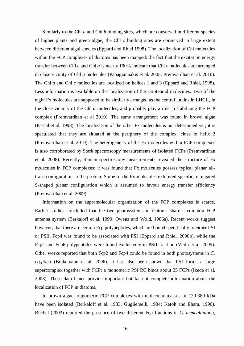
Information on the supramolecular organization of the FCP complexes is scarce.
Earlier studies concluded that the two photosystems in diatoms share a common FCP
antenna system (Berkaloff et al. 1990; Owens and Wold, 1986a). Recent works suggest
however, that there are certain Fcp polypeptides, which are bound specifically to either PSI
or PSII. Fcp4 was found to be associated with PSI (Eppard and Rhiel, 2000b), while the
Fcp2 and Fcp6 polypeptides were found exclusively in PSII fraction (Veith et al. 2009).
Other works reported that both Fcp2 and Fcp4 could be found in both photosystems in C.
cryptica (Brakemann et al. 2006). It has also been shown that PSI forms a large
supercomplex together with FCP: a monomeric PSI RC binds about 25 FCPs (Ikeda et al.
2008). These data hence provide important but far not complete information about the
localization of FCP in diatoms.
In brown algae, oligomeric FCP complexes with molecular masses of 120-380 kDa
have been isolated (Berkaloff et al. 1983; Gugliemelli, 1984; Katoh and Ehara, 1990).
Büchel (2003) reported the presence of two different Fcp fractions in C. meneghiniana,

17
which were different in molecular weight and oligomeric status. Furthermore, Beer et al.
(2006) identified the subunits of the higher oligomer FCPb and the trimer FCPa in C.
meneghiniana. FCPa possesses subunits encoded by fcp1-3 and fcp 6/7, while FCPb is
composed of the fcp5 gene products. Both FCPa and FCPb were intact with regard to
excitation energy transfer to Chl a (Beer et al. 2006). Earlier works reported isolation of
one major and one minor Chl a/c antenna fraction (Owens and Wold, 1986a). Recently,
FCP sub-fractions have also been isolated from different species exhibiting various
pigment compositions. One fraction is termed LHCF-F and contains Chl a/c and Fx but
lacks Ddx, while the other fraction is termed LHCF-D and contains Chl a, Fx and Ddx but
is nearly depleted in Chl c. While LHCF-F contains polypeptides in the range of FCPs,
LHCF-D shows smaller polypeptides (10-15 kDa) in addition to FCPs. A similar DD
enriched sub-fraction has been reported in C. meneghiniana (Beer et al. 2006), this
complex shows a high degree of de-epoxidation under high light exposure.
1.5. Photoprotection mechanisms and operation of the diadinoxanthin-cycle
Upon absorption of light, Chl a molecules of the light-harvesting antenna system
become excited to the singlet-state 1Chl*, the energy of which can be de-excited to ground
state in several pathways (Fig. 6).
Figure 6. Possible fates of excited Chl. When Chl absorbs light it is excited from its ground state to its
singlet excited state, 1Chl*. From there it has several ways to relax back to the ground state. It can relax by
emitting light, seen as fluorescence (1). Its excitation can be used to fuel photosynthetic reactions (2), or it
can de-excite by dissipating heat (3); both of these mechanisms reduce the amount of fluorescence. They are
therefore referred to as photochemical quenching (qP) and non-photochemical quenching (NPQ) of Chl
fluorescence. Last, 1Chl* can, by intersystem crossing, produce 3Chl* (4), which in turn is able to produce 1O2
*, a very reactive oxygen species (Müller et al. 2001).

18
Upon increasing ambient light intensity, at a point photochemistry attains a steady state
level and becomes light-saturated. Further increase in the light intensity will cause excess
excitation pressure on the reaction center Chl-s, the probability of the formation of 3Chl*
increases and the yield of Chl a fluorescence, originating from PSII also increases. The
existence of 3Chl* can be critical for plants, because it is able to transfer energy to ground
state O2 to generate singlet oxygen (1O2*), which is a very harmful reactive oxygen species
(ROS) and is able to damage lipids, proteins etc., causing thereby a decrease in the rate of
photosynthesis. Photosynthesizing organisms are able to keep the formation of 3Chl* at low
level through several fast photoprotective mechanisms. The quenching of Chl a
fluorescence is one of the most important mechanism and can be performed by the
operation of the photochemical reactions (qP, photochemical quenching). However, at light
intensities, which are beyond the capacity of photochemical quenching (i.e. the capacity of
the photosynthetic electron transport chain) other, non-photochemical quenching (NPQ)
processes dominate the photoprotection. In higher plants and green algae, NPQ can be
divided into three components (Horton et al. 1996; Müller et al. 2001): i) qE, energy-
dependent quenching, which is regulated by the build-up of a transthylakoid ΔpH and the
operation of the xanthophyll cycle, ii) qT, the state-transition quenching, which allows
reallocation of part of the energy absorbed from PSII to PSI and iii) qI, the photoinhibitory
quenching.
In diatoms, the qE component should play the main role in NPQ, since for qT no
evidence could be found (Owens, 1986b). Recently, it was observed that the qI component
of NPQ increased significantly upon prolonged (>60 min) light-stress in T. pseudonana
suggesting the existence of a photoinhibitory-quenching component in diatoms (Zhu and
Green 2010). The NPQ mechanisms are partially mediated by the xanthophyll cycle (XC).
During the operation of the XC, enzymatic deepoxidation/epoxidation of xanthophyll
carotenoids as a function of light intensity occurs. In higher plants and green algae, the XC
is called violaxanthin-antheraxanthin-zeaxanthin (VAZ) cycle. The cycle consists of inter-
conversion of three xanthophylls, the di-epoxide violaxanthin (Vx), the mono-epoxide
antheraxanthin (Ax) and the de-epoxidized zeaxanthin (Zx). The operation and regulation
of VAZ cycle is known in details in higher plants and green algae and is reviewed in
several papers (e.g. Yamamoto et al. 2004; Lavaud, 2007). The de-epoxidation/epoxidation
events are performed by two enzymes, a Vx de-epoxidase (VDE) and a Zx epoxidase
(ZEP). VDE is located on the lumenal side and its optimal activity is at around pH 5-6,
thus it is activated when the thylakoid lumen becomes acidic due to the build-up of the

19
proton gradient. ZEP is localized on the stroma side, its pH optimum is at around 7.5. VDE
requires the acidic form of ascorbate while ZEP requires the NADPH, H+ and O2 as
cofactors. The direction and activity of the two competing enzymatic reactions of the VAZ
cycle depends on the build-up of the transthylakoid proton gradient and subsequent change
in the lumenal and stromal pH. When the light intensity decreases, the de-epoxidation
becomes weaker and finally stops, while the inverse epoxidation reaction becomes
dominant. Hence, the accumulation of the de-epoxidized xanthophylls Zx depends on the
activity of the two enzymes, which indirectly depends (via the change in pH and
availability of the cofactors) on the light intensity.
Diatoms possess the diadinoxanthin-cycle, where Ddx is deepoxidized in one step to
Dtx by the enzyme diadinoxanthin-deepoxidase (DDE), while Dtx can be epoxidized back
to Ddx also in one step by Dtx epoxidase (DEP). The comparison of the VAZ of higher
plants and Ddx cycle of diatoms is depicted in Fig. 7.
Figure 7. Comparison of the VAZ and Ddx cycle (Wilhelm et al. 2006).
The regulation of the Ddx cycle displays several differences compared to VAZ cycle. The
pH optimum for DDE is significantly higher (Jakob and Wilhelm, 2001), which means that
already a weak lumen acidification is able to induce the DDE enzyme. It also has been
shown that – compared to VDE - DDE requires lower ascorbate concentrations (Grouneva
et al. 2006) and lower monogalactosyl-diacylglycerol (MGDG) concentrations (Goss et al.
2005) for the full activity of Ddx de-epoxidation. The DEP enzyme requires the same
cofactors as ZEP, but the regulation of the epoxidation reaction also displays peculiar
characteristics – it is fully inactivated during strong light illumination, therefore no
competitive epoxidation reaction under excess light conditions occurs (Goss et al. 2006).

20
This pH “lock” ensures the fast and efficient deepoxidation of Ddx. Other unusual
property of the epoxidation reaction is that it is not functional in complete darkness, but
very efficient and rapid on low light intensities (Goss et al. 2006; Mewes and Richter,
2002). The impairment of the epoxidation reaction is thought to occur due to shortage of
NADPH (Goss et al. 2006), which is a consequence of different regulation mechanisms of
alternative electron transport pathways and chlororespiration in different diatom species
(Grouneva et al. 2009).
The correlation between qE and the XC has been studied thoroughly in higher plants
and green algae. In diatoms, correlation could also be found between qE and Ddx de-
epoxidation (Lavaud, 2002a; Olaizola et al. 1994; Ting and Owens, 1993). Here the values
for qE are 4-5 times higher as compared to those measured in higher plants (Lavaud et al.
2002; Ruban et al. 2004). The qE seems to be more tightly associated with the presence of
the de-epoxidized xanthophyll, the Dtx compared to Zx in higher plants (Lavaud et al.
2002; Ruban et al. 2004). However, recently more qE components could be identified,
which were not strictly dependent on the presence of Dtx but rather on the presence of pH
(Grouneva et al. 2008; Lavaud et al. 2006).
qE regulation depends on many factors and proceeds within the thylakoid membrane.
XC plays a central, albeit not an exclusive role in the formation of NPQ. The recent
allosteric conformational change model provides explanation about the relationship of XC
and the conformational change of LHCII in the kinetics of the NPQ in higher plants
(Horton et al. 2008). In diatoms, the correlation between conformational changes of FCP
complexes and the quenching of fluorescence is far less understood. It has been estimated
that in C. meneghiniana the FCPa and FCPb complexes exhibit significant fluorescence
quenching which was dependent on pH but only if the complexes were aggregated in
vitro (Gundermann and Büchel, 2008). Thus, the relationship of fluorescence quenching
and the state of aggregation of the FCP in vivo still remains to be clarified.
1.6. Principles of circular dichroism (CD) spectroscopy
CD spectroscopy is a non-invasive method for investigating pigment-pigment
interaction even in as highly complex systems as intact cells in vivo. CD is the extinction
difference of the left and right circularly polarized light at a given wavelength, which is

21
composed of differential absorption (AL-AR) and (if present) differential light scattering
(SL-SR) of the sample and can be described as
CD = (AL - AR) + (SL-SR)
CD signals originate from the structural asymmetry (more precisely the chirality) or
from asymmetric interactions of the molecules (Woody, 1985).
CD signals can originate from molecular systems of different complexity. In Chl
containing systems, the physical origin of CD signals was reviewed by Garab (1996) and
Garab and Amerongen (2009).
Figure 8. Circular-dichroism spectra exhibited by the thylakoid pigments at different levels of structural
hierarchy. The acetonic extract - yielding intrinsic CD (for easier comparison, the signal is multiplied by a
factor 5), pea thylakoid membranes suspended in low salt, hypotonic medium dominated by the sum of the
excitonic bands, and the same membranes suspended in isotonic medium in the presence of Mg ions (Garab
and Amerongen, 2009).
Chiral molecules exhibit optical activity, called intrinsic CD. It can be described by the
rotational strength of the given electronic transition, which is the scalar product of the
electric and magnetic dipole moments. Chlorophylls are planar ring-structured molecules;
therefore their electric and magnetic dipole moments are nearly perpendicular to each

22
other, which results weak CD signals. The band shapes of the intrinsic CD are identical
with those of the absorption bands; however, their sign can be positive or negative,
determined by the handedness of the molecule. In photosynthetic systems, the intrinsic CD
signals are weak, because of the symmetry of the pigment molecules; for 1 absorbance
unit, it is in the range of some 10-5 intensities (cf. Fig. 8).
When chlorophylls are parts of a complex system it can happen that certain charged or
aromatic amino acids of neighboring proteins interact with them inducing e.g. twisting in
the chlorophyll molecule. In this case, the intensity and sign of the CD signal may deviate
from the intrinsic CD to a large extent and this makes the interpretation of CD signals
difficult. This induced chirality is rare in tetrapyrrole molecules, but it has been observed
e.g. in Chl a/c-containing organisms (Büchel and Garab, 1997).
In the case of excitonic interaction of two or more molecules – when they are close
enough to each other for dipole-dipole interaction, but still sufficiently apart for the
electrons remaining localized on each of the molecules – a conservative band structure can
be observed. That means that a positive and negative CD band can be observed the areas of
which gives zero in the energy spectrum (de Voe, 1965). This is called exciton-coupled CD
signal or excitonic CD. The excitonic CD originates from the fact that the polarization of
the light changes while passing (through) the excitonically interacting molecules, which
have a fixed position and orientation with respect to each other. In photosynthetic systems
the magnitude of absorption is typically an order of magnitude higher than the intrinsic CD
of the same pigment molecules. Excitonic CD can be observed in pigment-protein
complexes or small Chl aggregates, where the pigments are localized close to each other,
therefore are capable of participating in short-range dipole-dipole interactions. Excitonic
CD is often used for fingerprinting isolated pigment-protein complexes and carry
information on pigment-pigment interactions. However, due to the complexity of the
analysis of CD spectra only a few detailed models are available which interpret CD in
terms of high resolution molecular structures. Recently, such model was presented on
isolated LHCII trimers and monomers (Georgakopoulou et al. 2007).
In aggregates with sizes commeasurable with the wavelength of the visible light and
with high chromophore density, the intensity and wavelength positions of the conservative
excitonic bands may change and new “anomalous” CD bands with non-conservative band
structure appear which means that the intensity of the negative or positive component of a
bandpair can vary independently from each other. These signals are called psi (polymer or
salt-induced) type bands since they were originally observed in complex macromolecules

23
and aggregates e.g. in condensed chromatin and DNA molecules. In aggregates, exhibiting
psi-type CD not only the short-range dipole-dipole interactions, but also the long-range,
radiative couplings have to be taken into account. It is true that in small aggregates the
excitonic interactions are stronger than the radiative and intermedier interactions, but the
strength display r-3 dependence (r is the distance between the dipoles), while the radiation
and intermediate coupling mechanisms between the dipoles exhibit r-1 and r-2
dependencies, respectively; therefore these latter interactions are much more effective for
large distances than the dipole-dipole interactions. Psi-type CD can also be observed in
non-absorbing regions, which originate from circular differential scattering and thus can
provide useful information about the size of the aggregate (Garab et al. 1988a). The
intensity of the psi-type bands depends on the extent of the long-range chiral order, the
domain size and the direction of the chiral order (handedness) (Keller and Bustamante,
1986; Kim et al. 1986). Photosynthetic pigments are able to form large chiral aggregates,
more precisely, chiral macrodomains exhibiting intense psi-type CD signals (Fig. 8. bold
line), which are given rise by the presence of long-range chiral organization or macro-
organization of pigments, possibly extending the pigment interactions to the thylakoid
membrane system.
1.7. Macro-organization of the light-harvesting antenna system
Granal thylakoid membranes contain the majority of PSII-LHCII supercomplexes. It
has been shown by electron-microscopic studies that PSII-LHCII supercomplexes are
arranged into well ordered semi-crystalline layers, which are proposed to be the native
organization of PSII in higher plants (Boekema et al. 1999, 2000). In LHCII the density of
the chromophore is high and the existence of the LHCII trimers in ordered layers gives
chance for strong pigment-pigment interactions and extended aggregates, called chiral
macroaggregates or macrodomains. These large aggregates can serve the basis for long-
distance migration of the excitation energy, which might be important in energy supply for
the reaction centers and its down-regulation via nonphotochemical quenching (Garab and
van Amerongen, 2009). By using polarized light spectroscopic – mainly CD - and
microscopic methods it has been shown that in granal thylakoid membranes the pigments
are arranged in densely packed chirally ordered, large (200-400 nm in diameter)
macrodomains exhibiting large psi-type CD signals (for detailed explanation for physical

24
origin of CD signals see Chapter 1.6) (Barzda et al. 1994; Finzi et al. 1989; Garab et al.
1988a).
The role of PSII-LHCII in macrodomain organization could be revealed by using
antenna mutants; it has been shown that the absence of minor antenna complex CP24 is
accompanied by the decrease or even the disappearance of psi-type CD signal (Kovács et
al. 2006). Moreover, it was found that the amount of PsbS protein – which is an important
regulator in the interaction of LHCII with PSII in granal thylakoid membranes – correlated
with the psi-type CD signal (Kiss et al. 2008). Psi-type CD signal can be found not only in
granal thylakoid membranes but also in tightly stacked lamellar aggregates of isolated
LHCIIs, which possess well defined three-dimensional structure. In contrast, disordered
aggregates without well-defined structure display no intense psi-type CD bands (Simidjiev
et al. 1997).
In higher plants, structural flexibility of the photosynthetic thylakoid membranes and
pigment-protein complexes are important in short-term acclimation to environmental
changes, such as temperature and/or illumination (Anderson and Andersson, 1988). The
macro-organization level of hierarchic assemblies like granal thylakoid membranes react
most readily to perturbations; this might be important for adjusting the functions without
significantly altering the structure and composition of the constituents (Garab and van
Amerongen, 2009).
The chirally organized macrodomains of the photosynthetic membranes have been
shown to undergo gross (up to 80-90%) light induced reversible structural changes, which
can be detected in the major psi-type CD bands. This suggests that pigments not only
display long-range interactions but the macrodomains with long-range order also possess
the ability for fast, large-scale but reversible structural transitions. These structural
modifications were extensively investigated in higher plant species in the past decades.
Isolated thylakoid membranes exhibit pronounced light-induced CD changes, which were
correlated with the functionality of linear electron transport chain (Gregory, 1975; Faludi-
Dániel et al. 1984). However, later studies explored that the light-induced changes are
largely independent from the photochemical activity of the thylakoid membranes, although
they were sensitive to the inhibitors of qE component of non-photochemical quenching
(Istokovics et al. 1997). This finding is also in accordance with the fact that the amplitude
of light-induced CD changes exhibited linear correlation with the light intensity, even
above the intensities, which saturated the linear electron transport (Barzda et al. 1996).
Moreover, it has been shown that light-induced CD changes could be observed not only in

25
isolated thylakoids but also in aggregated LHCII preparations (Barzda et al. 1996).
Illumination with strong light affected the chiral macrodomains, as it was shown by the
reversible decrease of the intensity of the psi-type CD signals.
It has been shown that the chirally organized macrodomains in isolated thylakoid
membranes are also susceptible to elevated temperatures. This can be concluded from the
decrease of the psi-type CD signals between 40 and 50 C, while the excitonic CD signals
persist even at 65-70 C. These data have revealed that the thermal stability of the chiral
macrodomains is considerably lower than that of the pigment- protein complexes (Cseh et
al. 2000). Thylakoid membranes preilluminated with strong actinic light exhibited even
higher sensitivity: the psi-type signal at 689 nm decreased already at around 30 °C. In
contrast, the excitonic interactions were essentially not affected by preillumination,
suggesting that the majority of pigment-protein complexes suffered no detectable light-
induced changes.
For explaining the above data, a biological thermo-optic effect was suggested.
According to this hypothesis, fast local thermal transients (local T-jumps) occur due to
dissipated excitation energy, which cause elementary structural changes. These changes
can exist if we assume that the thermal instability is inherent, „built-in” in the structure.
The mechanism of the heat dissipation was revealed experimentally by ultrafast transient
spectroscopy. Local heating in LHCII trimers was induced by exciton-exciton annihilation
resulting in a fast nonradiative relaxation with a simultaneous conversion of the excitation
energy into heat; the time course of the heat transient was monitored by recording the
ultrafast changes in the absorbance at 684 nm (Gulbinas et al. 2006). Thermal transients
can readily induce structural changes in intact thylakoids between 45 and 55 °C (Cseh et
al. 2000). It has also been shown that thermo-optic mechanism plays role in light-induced
monomerization of LHCII trimers not only in isolated complexes, but also in isolated
thylakoid membranes and whole leaves (Garab et al. 2002).
It was found that by increasing the sorbitol concentration in the suspension of isolated
thylakoid membranes the amplitudes of the 688 nm and 515 nm CD bands increase, while
the CD band at 674 nm was sensitive only to little extent. However, by increasing the
concentration of Mg2+ ions, the intensity of the 674 nm band became larger, while the CD
bands at 688 and 515 nm were essentially insensitive to this treatment. Based on these
results, it was concluded that the organization of the macrodomains is regulated by two
main external factors: i) the screening of divalent cations which facilitates the stacking of

26
the membranes and ii) the osmotic pressure of the medium which influence the lateral
packing density of the chromophores (Garab et al. 1991).
All of these structural modifications upon changes in the environmental factors indicate
that flexibility of the photosynthetic membranes plays an important role in fine adjusting
the photosynthetic light harvesting functions. In organisms (e.g. in diatoms), which
experience extreme and sudden changes in the environmental conditions the structural
flexibility is thought to be crucial in the regulation of the photoprotective mechanisms. The
investigation of the changes on structural and supramolecular level are particularly
important to obtain a complete picture about the protective mechanisms against surplus
light in diatoms together with the previous knowledge gained by characterization of e.g.
electron transfer processes and quenching of excess excitation energy.

27
2. AIMS
Diatoms are one of the most abundant taxonomical group of algae, which play essential
role in the regulation of atmospheric CO2 concentration. Diatoms in their natural habitat
are well known for their ability to survive large fluctuations in light intensity, e.g. in
mixing waters, and thus evolved short-term light adaptation mechanisms to fine-tune their
photosynthetic functions to rapidly changing conditions. This evidently requires a highly
organized but structurally flexible light harvesting antenna system. This motivated us to
obtain detailed information on the structure and flexibility of the pigment system on
different hierarchical levels, particularly on the macro-organization.
It has been well established that in higher plants and green algae the pigment-protein
complexes are arranged into structurally flexible chirally organized macrodomains, and
similar macro-organizations have been identified in some Chl a/b/c- and Chl a/c-
containing organisms. However, the macrodomain organization and structural flexibility of
pigment-protein complexes of diatoms, have not been investigated before.
Therefore our main goal was to obtain information about the molecular and
supramolecular organization of the light-harvesting system of diatoms, and to characterize
their structural flexibility.
During my PhD work, the following aims and goals were addressed:
I. To conduct systematic study on the (macro-)organization of the pigment-protein
complexes in P. tricornutum and C. meneghiniana at different levels of structural
complexity. By using mainly CD spectroscopy, my aim was to provide information
about the pigment-pigment interactions in isolated complexes and thylakoid
membranes and about the macro-organization of the complexes, i.e., about the
chiral macrodomains in whole cells.
II. To examine the structural stability and flexibility of the pigment-protein complexes in
thylakoid membranes by measuring their thermal and light stabilities, as well as the
ability of the chiral macrodomains to undergo light and temperature induced
reorganizations; further, to examine the effect of changes in the ionic strength and
osmotic pressure of the medium on intact cells and isolated thylakoid membranes.
It was also the aim of the present work to establish correlations of these
reorganizations with functional parameters, such as the maximum quantum yield of
28
PSII (measured as Fv/Fm), the non-photochemical quenching (NPQ) of the Chl a
fluorescence, and the Ddx cycle.
III. To gain information about the orientation and local environments of carotenoids,
especially of the Fx molecules, by analyzing the flash-induced electrochromic
absorbance changes of intact P. tricornutum and C. meneghiniana cells. Further, to
investigate the structural and functional heterogeneity of Fx molecules and of the
FCPs by using linear dichroism, low temperature fluorescence excitation and
emission spectroscopy and different growth conditions, in order to study the role of
heterogeneity in excitation energy supply to the two photosystems.

29
3. MATERIALS AND METHODS
3.1. Sample preparation
3.1.1. Culturing the diatom species
The diatom Phaeodactylum tricornutum (SAG, 1090-1a, Göttingen) was grown in
ASP-2 medium according to Provasoli (1957).
The composition of the ASP-2 medium, pH=7.7:
K2HPO4*3H2O 100 mg/l
H3BO3 10 mg/l
MgSO4*7H2O 2000 mg/l
KCl 1600 mg/l
NaNO3 1000 mg/l
TRIS 1000 mg/l
NaCl 5000 mg/l
CaCl2 400 mg/l
Na-EDTA 30.2 mg/l
Trace elements:
FeCl3*6H2O 3.3 mg/l
CoCl2*6H2O 0.012 mg/l
CuCl2*H2O 0.003 mg/l
Na2MoO4*H2O 0.006 mg/l
ZnCl2 0.3 mg/l
MnCl2*2H2O 3.3 mg/l
Cyclotella meneghiniana (SAG, 1020-1a) was also grown in ASP-2 medium (Provasoli,
1957) but here the medium contained in addition 212 mg/l Na2SiO3 * 5H2O.

30
Diatoms were grown in 250-1000 ml Erlenmeyer flasks containing 100-400 ml ASP-2
medium in sterile batch cultures by applying continuous shaking with 100-110 rpm.
Illumination was provided by using fluorescent tubes (TL-D 18W/33-640, PHILIPS) with
a photon flux density (PFD) of 40 mol photons m-2 s-1 photosynthetically active radiation
(PAR) („normal light”) in a dark/light period of 16 h/8 h. For certain experiments cells
were also grown on either low light (LL) or high light (HL) intensities with PFD values of
10-15 and 180-200 mol photons m-2 s-1, respectively. When growing HL cells, a halogen
lamp (PAR 38 230V 120W, Tungsram) was used. The temperature of the growth chamber
was set to 19 °C.
3.1.2. Isolation of thylakoid membranes and pigment-protein complexes
The following buffers were applied for the isolation of thylakoid membranes and
pigment-protein complexes:
Buffer „A”:
10 mM MES, pH=6.5
2 mM KCl
5 mM Na-EDTA
1 M Sorbitol
Buffer „B”:
10 mM MES, pH=6.5
2 mM KCl
5 mM Na-EDTA
Depending on the experiment, the buffers also contained 1-25 mM MgCl2. Before
isolation, the cells were adapted to low light intensities (~10 mol photons m-2 s-1) for
about 30 min. Cells from the exponential growth phase with chlorophyll concentration of
4-5 µg/ml were harvested by centrifugation (4.000 g, 5 min). The following steps were
carried out at 4 ºC in dim light. The pelleted cells were resuspended in 20-25 ml isolation
buffer ‘A’, then disrupted in a French pressure cell (Thermo Scientific) at a pressure of
12.500 psi (8.62x10⁷ Pa), which was sufficient for breaking up about 90% of the cells.

31
Unbroken cells were pelleted by centrifugation (1.000 g, 10 min) and resuspended in about
20 ml of isolation buffer ‘A’ again. The suspension was subjected to the French-press for a
second time using the same conditions. After centrifugation (1.000 g, 10 min) the
supernatant was merged with the supernatant of the first centrifugation and centrifuged at
40.000 g for 20 min. The pelleted thylakoids were resuspended in 1-5 ml of isolation
buffer ’B’.
For solubilization of the membranes, equal amounts of the isolated thylakoids,
corresponding to 0.5-1 mg Chl, were centrifuged at 40.000 g for 20 min. The pelleted
thylakoid membranes were solubilized in n-dodecyl -D-maltoside (DM). The detergent
from the 10% stock (w/v) was added to the membrane fraction with the ratio of
DM/Chl=20:1. The solubilization was performed in complete darkness on ice bath for 20
min by using continuous stirring. The solubilized membranes were centrifuged at 40.000 g
for 20 min and the supernatant was loaded onto a continuous sucrose gradient (0 to 0.7 M
sucrose, supplemented with 0.03 % DM). Separation was carried out by ultracentrifugation
(Sorvall, UltraPro 80) using a swing-out rotor (Sorvall, TH-641) at 110.000 g for 18 h at
4 ºC.
3.1.3 Preparation of sucrose density gradient
Continuous sucrose gradient was prepared by using a gradient mixer peristaltic pump.
To prepare 11 ml gradient, 5.5 ml buffer „B” containing 0.03% (v/v) DM was pipetted into
the front tube and 5.5 ml buffer „B” containing 0.7 M sucrose and 0.03% (v/v) DM was
pipetted into the rear tube. The connection between the two tubes was blocked by a valve
during the filling up process. After starting the peristaltic pump the valve was opened
which allowed the mixing of the two solutions. To provide homogeneous mixing, magnetic
stirrer was applied in the front tube. The gradient was prepared in ultracentrifuge tubes.
3.1.4. Breaking up the intact cells by using ultrasound
Sonication of intact cells was performed in a Branson Sonifier 450 on ice with a 10 s
sonication – 30 s cooling cycle. The same sample was sonicated for different time periods
in the range of 10 s – 180 s in ASP-2 medium.

32
3.1.5. Determination of the chlorophyll content
The chlorophyll content was determined spectrophotometrically in acetonic pigment-
extracts. 3-5 ml cell suspension was centrifuged (4.000 g, 5 min). The pellet was
resuspended in 90% acetone and in the presence of glass beads it was homogenized for 2x1
min at 4 ºC with a homogenizer (Mini-BeadbeaterTM, Biospec Products). The
homogenizate was centrifuged (10.000 g, 2 min) and the supernatant was taken for
chlorophyll content determination. In the case of isolated thylakoids or pigment-protein
complexes, pigments were extracted also in 90% acetone, centrifuged (10.000g, 2 min),
and the supernatant was taken for chlorophyll content determination. The absorption of the
acetonic extract was measured in split beam spectrophotometer (Shimadzu UV-1601) at
630 and 664 nm with a pathlength of 1 cm. The absorption was set to 0 at 750 nm and 90%
acetone was applied in the reference cuvette. The calculation of the concentration of Chl a
and c was performed according to Jeffrey and Humprey (1975):
Chl a = 11.47*E664
– 0.4*E630
[mg/l] Chl c = 24.34*E
630 – 3.73*E
664 [mg/l]
where E is the extinction at the given wavelength. The obtained values were corrected by
the applied dilution factor.
3.2. Measurements
3.2.1. Absorption spectroscopy
Absorbance spectra of intact cells were recorded with a Shimadzu UV-3000
spectrophotometer, in split beam mode, in the wavelength range of 400 – 750 nm. The
optical pathlength was 1 cm and the bandwidth was set to 2 nm. In order to minimize the
spectral artifacts caused by light scattering of turbid cell suspensions, the spectra were
measured in a sample holder designed correcting for scattering: the cuvette was placed as
close as possible to the photomultiplier tube, in front of which a quartz diffuser was placed.
The reference cell contained ASP-2 medium. All absorption spectra were measured at
room temperature.

33
3.2.2. Circular dichroism spectroscopy
CD spectra were measured with a Jobin-Yvon CD6 or a Jasco J-815 dichrograph in the
wavelength range of 400-750 nm. The optical pathlength was 1 cm, the cuvette was placed
5 cm from the photomultiplier tube. The spectra were recorded in 1 nm steps with
integration time of 0.3-1 s and a bandwidth of 2 nm. The CD spectra are plotted in
absorbance units. The CD spectra of the samples were corrected with the CD spectrum of
the respective buffer.
The measurements of temperature-dependent CD-changes were performed in a
thermostated sample holder. The samples were preincubated at different temperatures in
the range of 20-55 °C in 5 °C steps for 10 min and then measured at the same temperature.
The measurements of the light-induced CD changes were performed in the following
ways:
i) To follow the spectral changes caused by illumination, the CD spectrum of the dark-
adapted cell suspension was measured at first, then the sample was illuminated with strong
actinic white light (800 mol photons m-2 s-1 PFD) for 2-30 min, and then the light-treated
sample spectrum was recorded.
ii) Kinetics of the light-induced CD-changes at a given wavelength was measured in
the presence of 7% Ficoll to avoid sedimentation of the dark-adapted cells during the
measurement. The monochromator was set to the desired wavelength and the time-course
measurement was started with an integration time of 1 s. After recording the baseline for 3-
4 min in the dark, illumination was performed with a side-illumination attachment (Barzda
et al. 1996). The photomultiplier was protected with crossing filters against the actinic light
from the exciting beam. CD-changes were induced by blue light of 800 µmol photons m-2
s-1 PFD.
3.2.3. Linear dichroism spectroscopy
Linear dichroism (LD) spectroscopy provides information about the orientation of the
transition dipoles with respect to a laboratory-fixed coordinate system, in which the sample
is macroscopically aligned. LD is a differential absorption of two orthogonal linearly
polarized beams:
LD = A A

34
In order to obtain appropriate LD signal on macroscopic scale, the samples of interest must
be aligned, because in randomly oriented particles the LD signal vanishes, even if the
chromophores within the sample possess significant intrinsic anisotropy. Photosynthetic
systems, such as isolated chloroplasts or thylakoid membranes can be oriented by using gel
squeezing method (Abdourakhmanov, 1979) without significant loss of the physiological
and structural properties of the embedded particles (Ganago et al. 1983), however in some
cases – especially in complex systems – acrylamide caused deterioration, e.g. the chiral
macrodomain organization of the chloroplasts became reduced and the membranes lost
their ability to retain the energized state (Osváth et al. 1994). Strong magnetic field can
also be used for aligning the samples in ‘regular’ suspensions used also for functional
studies. With magnetic field nearly 100% orientation can be achieved, i.e. all chloroplasts
can be aligned in a way that their thylakoid membranes tend to orient perpendicular to the
magnetic field. Whereas, particles possessing large diamagnetic susceptibility, e.g.
chloroplasts or thylakoid membranes can only be effectively oriented with magnetic field
strength of 0.5-0.7 T, smaller particles (isolated pigment-protein complexes) cannot be
aligned in the same field, because their diamagnetic susceptibility anisotropy is too small.
Linear dichroism (LD) spectra were measured at room temperature in a Jobin-Yvon
CD6 dichrograph, equipped with LD boards, in the wavelength range of 400-730 nm with
a bandwidth of 2 nm in 1 nm steps. The optical pathlength was 1 cm. The cuvette
containing the sample was placed 5 cm from the photomultiplier tube. Cells or thylakoid
membranes, isolated from P. tricornutum or C. meneghiniana were oriented with the gel-
squeezing method. The samples were embedded in 5% polyacrylamide gel, which was
prepared using 0.416 ml 30% (w/v) acrylamide and 0.8% (w/w) bis-acrylamide solution,
50 µl 10% (w/v) ammonium persulfate, 2.03 ml sample diluted with the appropriate buffer
and 5 µl N,N,N',N'-tetramethyl-ethane-1,2-diamine (TEMED). The solution was
polymerized in a plastic cube. After the polymerization was complete, the gel was removed
from the plastic cube and was squeezed to half the original width (M=2, where M is the
squeezing factor) in a squeezing cuvette. The samples were measured in the direction
perpendicular to the squeezing direction (“edge” alignment). The LD spectra are plotted in
absorbance units.
The orientation angles were calculated according to Garab (1996). The parameter
related to the orientation angle is the order parameter (S) what can be obtained as:
S=LD/3A
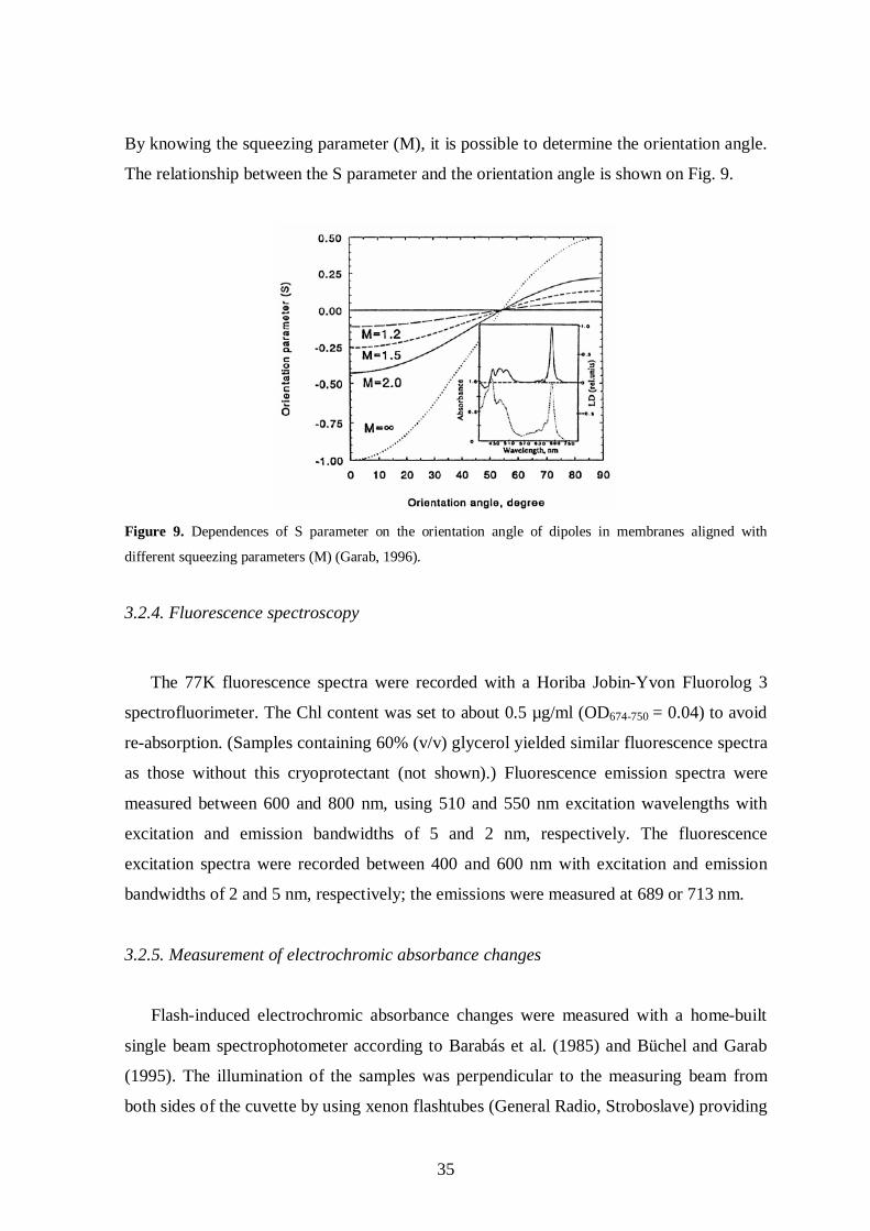
35
By knowing the squeezing parameter (M), it is possible to determine the orientation angle.
The relationship between the S parameter and the orientation angle is shown on Fig. 9.
Figure 9. Dependences of S parameter on the orientation angle of dipoles in membranes aligned with
different squeezing parameters (M) (Garab, 1996).
3.2.4. Fluorescence spectroscopy
The 77K fluorescence spectra were recorded with a Horiba Jobin-Yvon Fluorolog 3
spectrofluorimeter. The Chl content was set to about 0.5 µg/ml (OD674-750 = 0.04) to avoid
re-absorption. (Samples containing 60% (v/v) glycerol yielded similar fluorescence spectra
as those without this cryoprotectant (not shown).) Fluorescence emission spectra were
measured between 600 and 800 nm, using 510 and 550 nm excitation wavelengths with
excitation and emission bandwidths of 5 and 2 nm, respectively. The fluorescence
excitation spectra were recorded between 400 and 600 nm with excitation and emission
bandwidths of 2 and 5 nm, respectively; the emissions were measured at 689 or 713 nm.
3.2.5. Measurement of electrochromic absorbance changes
Flash-induced electrochromic absorbance changes were measured with a home-built
single beam spectrophotometer according to Barabás et al. (1985) and Büchel and Garab
(1995). The illumination of the samples was perpendicular to the measuring beam from
both sides of the cuvette by using xenon flashtubes (General Radio, Stroboslave) providing

36
single turnover flashes of 3 µs lengths. The frequency of the flashes was 1 s-1. The optical
pathlength was 1 cm in the direction of the measuring beam and 0.5 cm in the direction of
the actinic flashlight. The actinic flashes were passed through a Schott RG 630-2 mm filter.
The wavelength of the measuring light was selected manually by using a Zeiss SPM 2
monochromator (bandwidth 5 nm) and after passing the sample the light was led to an EMI
9558 B photomultiplier protected by a Corning 4-96 filter. The flash-induced absorbance
transients were recorded and averaged with the aid of a computer-controlled digital
oscilloscope (TEK 2224, Tektronix) and a differential amplifier (TM503B, Tektronix).
During the measurements the synchronization of the flash lamps and the oscilloscope was
performed using a digital timer (Mikro-GMK). To increase the signal-to-noise (S/N) ratio,
32-64 kinetic traces were averaged. To determine the transient spectra of the
electrochromic absorbance changes, kinetic traces were measured in 5 nm steps between
470 and 570 nm. The electrochromic signal at a given wavelength was calculated from the
kinetic traces where the signal amplitude at the time point of the flash was subtracted from
the signal amplitude at 5 ms after the flash. The measurements were performed in the
presence of 7% Ficoll to avoid rapid sedimentation of cells during the measurement. The
electrochromic absorbance changes are expressed in -I/I units.
In order to reveal the absorbance bands of the electrochromically shifted pigments, the
transient spectra were fitted with the first derivatives of Gaussians according to the
equation
dAdxfffffffff@ k B
x @x0
h3fffffffffffffffffffB 2pwwwwwwwwwwwwwwwwwwwwwwwwwwwwwwwwwwwww
f g
B e@
x @x0
b c2
2 B h 2ffffffffffffffffffffffffffffffffffffff
where A=absorbance, x0 = frequency at peak position, h=halfbandwidth, k=amplitude of
the Gaussian
Based on the fitted values of x0, h and k, the absorbance spectrum of the shifted
pigments were reconstituted by using Gaussian function.
3.2.6. Measurement of the chlorophyll fluorescence transients and the determination of the
photosynthetic parameters
Room temperature fluorescence measurements were performed using a PAM 101 Chl
fluorometer (Walz, Effeltrich, Germany) equipped with a Clark-type oxygen electrode as a

37
sample holder. The algal cell cultures and isolated thylakoids were used at Chl content of
15 and 20 µg/ml, respectively. The fluorescence parameters F0 and Fm were recorded after
45 min low light adaptation (20 mol photons m-2 s-1). (F0 and Fm are the minimum and
maximum fluorescence yield, respectively, in low light-adapted state.) The light intensity
of saturating flashes was 4.000 mol photons m-2 s-1. The maximum quantum yield of PSII
photochemistry of thylakoids in reaction buffers in the absence or presence of sorbitol and
MgCl2 was calculated as Fv/Fm, where Fv = Fm-F0, called variable fluorescence. F0 and Fm
were determined after a 5 min incubation in the reaction medium, 50 mM HEPES-KOH
pH=7.5, which was complemented with either or both MgCl2 (1 - 25 mM) and sorbitol
(350 mM). During the NPQ measurements saturating light flashes (4.000 mol photons
m-2 s-1) with duration of 700 ms were applied with 1 min intervals. The actinic light
intensity was adjusted to 700 µmol photons m-2 s-1. In the case of isolated thylakoid
membranes, 40 mM ascorbate was added as a co-substrate for DDE, to enable the
conversion of Ddx to Dtx, and 200 µM methyl-viologen was added as electron acceptor.
NPQ was calculated after 10 min actinic light illumination using the Stern-Volmer data
treatment (NPQ = Fm/Fm’-1) according to Bilger and Björkman (1990). (Fm’ is the
maximum fluorescence yield in actinic-light adapted state.)
3.2.7. Determination of the pigment composition by high performance liquid
chromatography (HPLC)
In the case of intact cells, pigments were isolated by filtering 10 ml of algal cell
suspension. After removal of the ASP-2 medium by using a vacuum pump, 1 ml of HPLC
medium (90% methanol/0.2 M ammonium acetate (90/10, v/v) and 10% ethylacetate) and
a small quantity of glass beads were added. The cells were disrupted in a cell homogenizer
(Braun, FRG), centrifuged, and the supernatant was taken for HPLC analysis. In the case
of thylakoid membranes, pigments were extracted by addition of 500 µl ethylacetate, 500
µl distilled water, and a spatula tip of NaCl to 250 µl of the different thylakoid samples.
The samples were stirred several times to transfer the pigments into the organic phase and
afterward centrifuged for 2 min at 20,000g. The organic phases were collected and the
pigments dried in a gentle N2 stream. The dried pigments were dissolved in HPLC medium.
Pigments were analyzed on a Waters 600 MS chromatography system equipped with a
Waters 717 autosampler and a Waters 996 photodiode array detector (Waters Millipore,

38
FRG) recording absorption spectra between 350 and 700 nm. A wide-pore ET 250/4
Nucleosil 300-5 C18 column (Macherey-Nagel, FRG) was used for separation. Eluents and
gradient programs were as described in Kraay, 1992. For the separation of pigments the
following eluents were used:
Eluent A: 85% Methanol/15% 0.5 M Ammonium-acetate (in Aqua dest.)
Eluent B: 90% Acetonitrile/10 % H2O
Eluent C: 100% Ethylacetate
The flow-rate in the column was set to 0.8 ml/min and during the separation the eluents
were degassed with helium. The following eluent compositions in relation to timescale
were used for separation:
Pigments were quantified according to Lohr and Wilhelm, 2001 and Wilhelm, 1995. The
parameters to determine the concentration of the given pigments are:
The concentration of a given pigment was obtained as:
n = A/(conversion factor*molecular mass), where A is the integrated area of the
absorption.
Time (min) % Eluent A % Eluent B % Eluent C 0 60 40 0 2 0 100 0 7 0 80 20
17 0 50 50 21 0 30 70
28,5 0 30 70 29,5 0 100 0 30,5 60 40 0
Pigment Wavelength for integration (nm)
Conversion factor (area/µg pigment)
Molecular mass in µg/mol
Chl c 440 22536980 610000000 Fx 440 10709573 659000000
Ddx 480 14103018 583000000 Dtx 480 12773475 566000000
β-Car 480 15238466 537000000 Chl a 430 7272969 894000000

39
3.2.8. Transmission electron microscopy
Cells and isolated thylakoid membranes were visualized by Zeiss 902 electron
microscope. The samples were pelleted by centrifugation at 5.000 g for 10 min in
Eppendorf tubes. The pellet was fixed with 2% glutaraldehyde diluted with 0.1 M
phosphate buffer (pH = 7.2) for 3 hours in the case of cells and 1 hour in the case of
isolated thylakoids. Glutaraldehyde was washed out by overnight incubation in 0.1 M
phosphate buffer. Samples were postfixed with 1% osmium-tetroxide diluted in 0.1 M
phosphate buffer for 2 hours. After short (~1 min) washing with 0.1 M phosphate buffer,
samples were gradually dehydrated in 30, 50, 70, 90, 96 and 100% ethanol for 10-15 min
at each step. The incubation step in 100% ethanol was repeated 3 times. After that, samples
were incubated in propylene oxide for 10-15 min 2 times and in 1:1 ratio of propylene
oxide:Araldite for overnight. Samples were transferred to pure Araldite resin and incubated
for 24 h. The Eppendorf tubes containing the samples embedded in the resin were placed to
thermostated chamber and the resin was hardened for 48-72 h at 56 °C. After trimming the
hardened resin blocks, ultrathin sections with a thickness of 70-90 nm were cut with
ultramicrotome (Leica Ultracut UCT). Sections were taken up onto 400 mesh copper grids.
For contrasting the specimen, sections were stained with 2% uranyl acetate for 45 min,
washed with distillated water and stained with lead citrate for 15-20 min and washed with
distillated water.

40
4. RESULTS
4.1. CD signals in the diatom Phaeodactylum tricornutum
The aim of the present chapter is to record and describe the CD signals of the diatom P.
tricornutum originating from pigment molecules. P. tricornutum is a typical pennate
species possessing bilateral cell symmetry and one large chloroplast per cell whose
genome is completely sequenced (Oudot-Le Secq et al. 2007). This species is extensively
used as a laboratory organism in various fields of diatom research.
We have started our investigations with the examination and comparison of the
pigment-pigment interactions in hierarchically different pigment organizations prior
assigning certain bands to structural and macro-organizational features.
First, the CD spectra of free pigment molecules were measured, which we considered
as the simplest organizational level in the pigment system, because pigments are detached
from the proteins and thus the CD signatures of single molecules can be recorded. Then,
CD spectra on isolated pigment-protein complexes were recorded, where the pigments are
bound to proteins, which give a chance for the presence of pigment-pigment interactions.
The next organizational level to be examined was the isolated thylakoid membranes, where
the pigment-protein complexes are embedded in the membrane. Finally, we have measured
CD spectrum of intact cells, which can be considered as the in vivo state and the most
complex organization of the pigment system.
The CD spectra of pigment extracts, isolated thylakoid membranes and intact cells of
P. tricornutum are depicted in Fig. 10.

41
Figure 10. CD spectra if intact cells, isolated thylakoid membranes and pigment extracts of
Phaeodactylum tricornutum. The spectra are plotted for identical Chl content of the samples (20 µg/ml). For
better comparison, the signal of pigment extract was multiplied by a factor of 5.
Free pigment molecules extracted in 90% acetone exhibit weak CD signals at (-)663
and (+)434 nm. The very weak intensity of these bands and the lack of the conservative
bands structure show that these CD bands can indeed be assigned as intrinsic signals.
Then we have characterized the CD signals of isolated pigment-protein complexes. By
using sucrose gradient centrifugation, the main pigment-protein fractions were separated
from solubilized thylakoid membranes (Fig. 11a). The polypeptide and pigment
composition of the pigment-protein complexes were determined earlier and are described
by Lepetit et al. (2007). In my work, the pigment-pigment interactions revealed by CD
spectroscopy were in the focus of interest.
In isolated FCP of P. tricornutum, characteristic CD bands could be observed at (-)679
nm, at (-)475 nm and at (+)445 nm (Fig. 11b). The negative band at 679 nm – although
exhibits large intensity – cannot be considered as excitonic CD band, because the
conservative split signal is missing. This indicates that between the Qy dipoles of the Chl a
molecules no excitonic interactions exist. Similar bands have been identified at around 679
400 450 500 550 600 650 700 750-1.0
-0.5
0.0
0.5
1.0
intact cells thylakoid membranes pigment extract x5
CD
(10-3
)
Wavelength (nm)

42
nm in other Chl a/c-containing organisms (Büchel and Garab, 1997; Mimuro et al. 1990).
The (-)679 nm band has been proposed to originate from a small fraction of Chl a
molecules, which exhibit induced chirality due to the twisting of the molecules in the
protein environment (Büchel and Garab, 1997). It was proposed earlier that the strong
excitonic bandpair at (+)445/(-)478 nm originates from Chl a/Fx interactions (Büchel,
2003). However, recent data indicate that this split signal can have a different origin, from
a pair of blue-shifted Fx molecules (Premvardhan et al. 2009), additional contributions
might come from Chl c2:Chl a excitonic interactions (Premvardhan et al. 2008). Isolated
FCP contains only substoichiometric Ddx (or Dtx), and appear not to contribute to the CD
signals.
Isolated photosystems exhibit a pronounced split excitonic CD bandpair with the
maxima at (+)676 and (-)690 nm, which indicates strong excitonic coupling between the
Qy dipoles of the Chl a molecules. In the blue region of the photosystem CD spectra, the
signals at (-)430 nm and (+)445 nm probably also originate from Chl a excitonic
interactions (Fig. 11b). Weak CD signals can be observed at all organization levels of
chromophores at around 623 nm, which – taking into account their spectral positions –
most probably originate from Chl c.
400 450 500 550 600 650 700 750-1.0
-0.5
0.0
0.5
1.0
1.5
2.0
FCP PSII PSI
CD
(10-3
)
Wavelength (nm)
(b)
Figure 11. Separation of FCP, PSII and PSI pigment-protein complexes by sucrose density gradient
ultracentrifugation (a), and CD spectra of the same fractions (b). Spectra are plotted to the same Chl
content (20 g/ml).
FCP
PSII PSI
(a)

43
During the past few years, many efforts have been made to isolate FCP complexes in
their native state. In our work, we attempted to isolate oligomeric FCP fractions by using
lower detergent concentration in order to keep the complexes as intact as possible. After
solubilization of the thylakoid membranes with DM/Chl ratio of 5 instead of 20, a
significantly different sucrose density gradient pattern could be observed (Fig. 12a). At
DM/Chl ratio of 5, a new band (named FCPo) appeared at higher sucrose gradient density.
Here the PSII and PSI fractions could not be separated due to the mild solubilization, only
one PS fraction could be observed which was dominated by PSI, as suggested by its
fluorescence emission spectrum. Gel-filtration chromatography analysis indicated that
FCPo possesses higher degree of oligomerization compared to FCP; the molecular mass of
FCPo was estimated to be 440 kDa, whereas the molecular mass of FCP is 230 kDa
(Lepetit et al. 2007). The protein composition of FCP and FCPo was almost identical.
FCPo contained a small amount of PSII proteins, which indicates that FCPo and PSII did
not separate completely on the sucrose gradient (Lepetit et al. 2007). Both FCP and FCPo
exhibited perfect excitation energy transfer, as concluded form low-temperature
fluorescence spectroscopy measurements. In my work, the investigation of the pigment-
pigment interactions was in the focus of interest as studied by CD spectroscopy. The CD
spectra of FCP and FCPo are depicted on Fig 12b.
Figure 12. The position of the pigment-protein fractions on the sucrose gradient using different
detergent (DM) to Chl ratios during solubilization (a). CD spectra of the isolated FCP and FCPo complexes
(b). The Chl content was adjusted to the same value, 20 g/ml.
(a)
400 450 500 550 600 650 700 750
-0.5
0.0
0.5
1.0 FCPo FCP
CD
(10-3
)
Wavelength (nm)
(b)

44
The CD spectra of the FCPo and FCP are very similar to each other; small differences
in the Soret region probably originate from small alterations in the excitonic interactions.
In isolated thylakoid membranes, characteristic CD bands could be found at around
(+)445 nm, (-)478 nm and (-)679 nm (Fig. 10). The band pair at (+)445/(-)478 nm is most
likely given rise by excitonic interactions involving Chl a, Chl c and carotenoids, mainly
Fx, similarly to isolated FCP as it was described above. The (-)679 nm band cannot be
considered as excitonic CD bands, because the conservative band structure is missing,
instead, it originates from the induced chirality of Chl molecules as it is described above in
the case of FCP.
Intact cells exhibit an intense CD band at around (+)698 nm (Fig. 10). The intensity of
this band is larger than the intrinsic and excitonic CD signals, moreover, it is associated
with strong differential scattering. The intensity of the 698 nm band decreases or even
disappears in thylakoid membranes and varies independently from the (-)679 nm band,
therefore the excitonic property of this band can be ruled out. Intact cells exhibited
excitonic bands at (+)445 nm and (-)478 nm, however, surprisingly this bandpair was
much weaker than in isolated FCPs and thylakoids. This might be caused by scattering
artifacts in intact cells, or by an overlapping strong and broad psi-type band in the same
region.
4.2. Assignment of the psi-type CD signal to the multilamellar membrane system
The assignment of the main CD bands with psi-type features, anomalous band shape
and long differential scattering tail, are based on the observations described in the previous
chapter. In particular, the (+)698 nm band, observed in whole cells but not in isolated
thylakoids, has been shown to exhibit non-excitonic features. In contrast, the (-)679 nm
band persisted both in isolated thylakoid membranes and in whole cells. In order to prove
that these features originate from a macro-organization of the complexes and to find the
structural entity which carries these features, a method was necessary, by the aid of which
we could follow the gradual spectroscopic changes along with the ultrastructural changes.
To this end, we applied ultrasonic disintegration of intact cells, which was performed for
different time periods, and the changes in the CD spectra were checked after every
sonication step. From the same sample aliquots were taken for electron microscopy.
Sonication led to gradual diminution of the large (+)698 nm signal, while the negative

45
band in the red did not decrease considerably, although a 4 nm wavelength shift was
visible (Fig. 13a).
Figure 13. Effect of sonication on the CD spectra of Phaeodactylum tricornutum cells (a). The same cell
suspension was sonicated for 10, 60, 90 and 180 s. Chlorophyll concentration, 15 µg/ml. Difference CD
spectra of sonicated-minus-control samples (b).
400 450 500 550 600 650 700 750
-1.2
-0.8
-0.4
0.0
0.4
0.8
1.2
1.6
control 10 s 60 s 90 s 180 s
CD
(10-3
)
Wavelength (nm)
(a)
400 450 500 550 600 650 700 750
-1.0
-0.5
0.0
0.5
1.0
10s-control 60s-control 90s-control 180s-control
CD
(10-3
)
Wavelength (nm)
(b)

46
Interestingly, after the 10 s sonication step, a small increase occurred both in the
(+)698 and (-)679 nm signal. The reason of this change is unknown, the probable
explanation is that upon short-term sonication, some changes in the optical properties of
cell wall or organelles occurred, and thereby some scattering artifacts caused by the cell
walls had been eliminated. Upon sonication, the intensity of the (+)445/(-)478 nm band
pair gradually increased, the excitonic interactions became more expressed (see also the
difference spectra in Fig. 13b). The equal changes in the intensity of the (+)445/(-)478 nm
band pair confirm the excitonic property of these CD signals, while the decrease of the
large 698 nm band intensity independently from the (-)679 nm band rules out the excitonic
property of these CD signals and refers to psi-type feature suggesting the presence of the
chirally organized macrodomains of the Chl a molecules (Fig. 13b).
By using electron microscopy, we attempted to visualize the structural changes caused by
sonication (Fig. 14).

47
Figure 14. CD
spectra and electron-micrographs of Phaeodactylum
tricornutum cells sonicated for the indicated tim
e intervals. Scale bars: 0.6 m.
The CD
spectra were m
easured on the same sam
ple, which w
as prepared for EM studies.
( )

48
Fig. 14 shows that in the control, non-sonicated cells the thylakoid membranes are
arranged in loosely stacked, multilamellar membrane system within the chloroplast. After
sonication for 10 s, the regular, multilamellar system of the thylakoids did not change
considerably, but it is visible that some vacuoles appeared within the cell. These changes in
the cell ultrastructure might influence the differential light scattering of left and right
circularly polarized beams and thereby some changes in CD spectra might occur. After 60s
sonication, drastical changes in cell ultrastructure and considerable loss of the
multilamellar organization of thylakoid membranes could not be observed and the psi-type
signal at 698 nm decreased but did not disappear completely. Sonication for longer time
periods caused the complete destruction of the cell ultrastructure and lamellarity of the
thylakoids with a concomitant loss of the psi-type signals. The CD spectrum of the
completely broken cells is similar to that of isolated thylakoid membranes. These results
indicate that the presence of the psi-type CD signal correlates with the presence of the
multilamellar organization of the thylakoid membranes.
4.3. The structural flexibility of the chiral macrodomains
In diatoms, the structural flexibility of photosynthetic membranes and pigment protein
complexes under variations in the environmental conditions has not been investigated yet.
Chiral macrodomains are capable undergoing transient and reversible structural changes,
which indicate a flexible organization of the chromophores (Chapter 1.7). It has been
shown in Chapter 4.2 that the chromophores are arranged in chiral macrodomains,
reflected by the presence of psi-type CD signal at around 698 nm. In the present chapter
our aim was to examine the changes in the macrodomain organization caused by different
environmental factors, such as illumination, temperature, changes in osmotic pressure and
ionic strength and tested the reversibility of the changes, which indicates the existence of a
flexible system.

49
4.3.1. Temperature-induced CD changes
Ambient temperature affects considerably both the physiological functions and
structural features of the thylakoid membranes. Higher plants exhibited temperature-
dependent CD changes, where the chiral macrodomains proved to be the most sensitive to
elevated temperatures. To test the effect of the elevated temperatures on the pigment
interactions and macrodomain structure in diatoms, P. tricornutum cells were subjected to
heat treatment in the range of 20-55 ºC. The spectral changes caused by the heat treatment
are depicted on Fig. 15.
Figure 15. The effect of elevated temperatures on the CD spectra of P. tricornutum cells. The cells were
incubated consecutively for 10 min at the indicated temperature and measured at the same temperature. Inset,
temperature dependence of the CD signal at 698 nm, 679 nm and at 450 nm). Chlorophyll content, 15 µg/ml.
400 450 500 550 600 650 700 750
-0.5
0.0
0.5
1.0
20 °C 25 °C 30 °C 35 °C 40 °C 45 °C 50 °C
CD
(10-3
)
Wavelength (nm)
-0.4
0
0.4
0.8
1.2
20 25 30 35 40 45 50 55
Temperature (ºC)
CD
(rel
. val
ues)
450-470
679-750
698-750

50
While the (+)450 and (-)679 nm bands mostly preserved their intensities, a continuous,
large decrease in the intensity of the (+)698 nm could be observed upon increasing the
temperature up to 45 ºC. Above 45 ºC, all characteristic CD bands related to intra- or
intermolecular interactions disappeared. The inset in Fig. 15 shows the relative changes of
the indicated bands as a function of the temperature. By increasing the temperature up to
45 ºC, 60 % of the original intensity of the (+)698 nm signal and 20% of the original
intensity of the (-)679 nm band was lost, while at 450 nm only little changes could be seen.
Thus, the (+)698 nm signal proved to be the most sensitive to elevated temperatures, while
the excitonic bands at around 450 nm and the (-)679 nm band displayed higher stability.
Above 45 ºC a steep decrease in the intensities of all bands occurred, which indicates the
loss of all pigment-pigment interactions inside the protein complexes or their small
aggregates. The highest temperature sensitivity could be observed in the case of the (+)698
nm band and it changed independently from the (-)679 nm band. This again points to the
psi-type feature of the (+)698 nm band, moreover it indicates in the same time that chiral
macrodomains are more susceptible to elevated temperatures than the excitonic CD bands.
These data are in good agreement with the data obtained for the temperature-
susceptibilities in higher plant thylakoid membranes (Cseh et al. 2000; Dobrikova et al.
2003).
4.3.2. Light-induced CD changes
Upon illumination with strong actinic light, a pronounced increase in the amplitude of
the (+)698 nm signal and a small increase in the amplitude of the (-)679 nm signal could be
observed, while the other regions of the spectra remained essentially unchanged (Fig. 16).

51
400 450 500 550 600 650 700 750
-0.8
-0.4
0.0
0.4
0.8
1.2
dark light light-dark
CD
(10-3
)
Wavelength (nm)
Figure 16. CD spectra of dark-adapted and preilluminated P. tricornutum cells and the difference
spectrum of illuminated-minus-dark-adapted cells. The measurements were performed at room temperature;
the light intensity of illumination was 800 µmol photons m-2 s-1; Chl content, 15 µg/ml.
In order to determine the extent and the possible ability for recovery of the light-
induced CD changes, the kinetics of the changes were also examined at different
wavelengths. The most pronounced changes in intact cells illuminated for several minutes
occurred at 698 nm. After illumination for 100 s the increase in the signal intensity levelled
off. After switching off the illumination, the signal decreased to its original value; this
recovery took about 200 s (Fig. 17).

52
Figure 17. Time course of the light-induced CD-changes at 698 nm. Closed bars, dark periods; open bar,
illumination period. The measurements were performed at room temperature; the light intensity of
illumination was 800 µmol photons m-2 s-1; Chl content, 15 µg/ml.
The kinetics of the light-induced CD changes can be characterized by fitting the initial
phase and calculating the rate of the changes. The magnitude of the changes is given as
ΔCD/CD values, where the CD changes upon illumination (ΔCD) is normalized on the
initial CD values of the dark-adapted state.
By using these parameters, the light-intensity dependence of the light-induced CD
changes was measured. By increasing the illumination intensity, the ΔCD/CD values and
the rate increased, however, above 450 µmol photons m-2 s-1 no further change could be
observed (Table I).
Intensity (mol photons m-2s-1) CD/CD Rate (CD/t, s-1)
40 0.12 5x10⁻⁷
100 0.18 8x10⁻⁷
450 0.36 2.8x10⁻⁶
700 0.3 2.5x10⁻⁶
Table I. Light intensity dependence of the light-induced CD changes at 698 nm. n=3, SD20%.
0 100 200 300 400
0.4
0.5
0.6
0.7
CD
698 (1
0-3)
Time (s)

53
To reveal the possible correlations between the light-induced CD changes and the
operation of the photosynthetic electron and proton transport and photochemical activity,
different inhibitors were applied during the measurements of the kinetics of the light-
induced CD changes (Table II).
CD/CD Rate (CD/t, s-1)
Control 0.33 3.5x10⁻⁶
DCMU 0.23 3.8x10⁻⁶
NH4Cl 0.36 3.1x10⁻⁶
DTT 0.17 1.2x10⁻⁶
Table II. Effect of different inhibitors and uncouplers on the light-induced CD changes. Applied
concentration of the chemicals: 3-(3,4-dichlorophenyl)-1,1-dimethylurea (DCMU): 20 M, dithiothreitol
(DTT): 5 mM, ammonium chloride (NH4Cl): 5 mM. The intensity of the illumination was 800 µmol photons
m-2 s-1 during the experiment. n=3, SD20%.
DCMU inhibits the photosynthetic electron transport by displacing QB from its binding
site on the D1 protein and thereby the photochemistry becomes arrested (Velthuys, 1981).
It has been shown that isolated thylakoid membranes of higher plants exhibit reduced CD
in the presence of DCMU. However, it is also established that the activity of the electron
transport is not mandatory for the structural changes, rather it plays a “conditioning” role
in this process (Istokovics et al. 1997). In diatoms, DCMU also inhibited to some extent
the amplitude of the light-induced CD changes and the rate was even increased to little
extent. This result indicates that in P. tricornutum the inhibition of linear electron transport
does not affect the light-induced structural changes. This is in a good agreement with the
NPQ analyses, where it was found that in the presence of DCMU the NPQ kinetics were
not affected by DCMU (Grouneva et al. 2009).
To test the possible involvement of the proton gradient in the light-induced CD
changes, NH4Cl was applied. The uncoupler NH4Cl dissipates the proton gradient, which is
formed during the operation of the vectorial photosynthetic electron transport. NH4Cl
inhibited the qE component of NPQ in P. tricornutum (Ruban et al. 2004). It has been
shown earlier that NH4Cl inhibits the light-induced CD changes in higher plants (Garab et
al. 1988). However, later it was found that nigericin (a Na+/H+ ionophore) dissipated the
proton gradient at low concentrations, but the CD was not affected (Istokovics et al.

54
1997). NH4Cl was applied on P. tricornutum cells to examine whether the dissipation of
the proton gradient inhibits the light-induced structural changes. NH4Cl did not cause any
significant inhibition of either the magnitude or the rate of the light-induced CD change.
Since we have seen no inhibition of light-induced CD changes by NH4Cl, while it has been
shown that this compound blocks the NPQ, a clear correlation between the presence of
pH and structural reorganizations cannot be revealed at present. Another chemical
compound, which dissipates the proton-gradient and thus blocks the NPQ is the nigericin.
However, it could not be used in our experiments, because it has been shown earlier that it
does not penetrate the cell wall of diatoms (Ruban et al. 2004).
The violaxanthin or diadinoxanthin deepoxidase inhibitor, dithiothreitol (DTT)
suppresses the deepoxidation reaction step of the xanthophyll-cycle, and thereby the
formation of the xanthophylls responsible for or participating in the qE component of NPQ
is blocked (Goss et al. 2006). DTT suppressed significantly the light-induced CD changes
in P. tricornutum (Table II), which suggests a correlation between the operation of the
xanthophyll cycle and structural reorganizations. However, DTT probably also affects
metabolic processes other than the xanthophyll cycle. Therefore, further experiments are
necessary to clarify the connection between the light-induced changes and the NPQ
processes in diatoms.
In diatoms, light intensity during growth influences the size of chloroplast and the
amount of thylakoid membranes. It has been shown that high light intensities cause a
decrease in the chloroplast size and the amount of the thylakoid membranes, however the
membrane ultrastructure, i.e. the bands of three did not change with light intensity (Janssen
et al. 2001, Moisan et al. 1999, 2006). It has also been observed that the amount of PSI and
PSII increases considerably in the low light grown cells, also the cell volume increased in
P. tricornutum (Friedman and Alberte, 1986). In another diatom species, Skeletonema
costatum, the amount of PSI decreased while the amount of PSII increased by decreasing
the growth light intensity (Falkowski et al. 1981). In the present work, the macrodomain
organization of the cells grown on HL and LL intensities was investigated (Fig. 18).

55
Figure 18. CD spectra of HL and LL cells. The samples were adjusted to the same Qy absorbance of Chl
a, OD673-750 ≈ 0.5.
Cells grown on different light intensities exhibited remarkably different CD spectra
(Fig. 18). LL cells exhibited weaker excitonic bands in the blue region compared to HL
cells. Moreover, LL cells displayed significantly larger psi-type bands at 698 nm and a less
intense signal at 679 nm. These differences might be caused by a higher degree of stacking
of the thylakoid membranes. This hypothesis was checked by electron microscopy of intact
HL and LL cells (Fig. 19).
Figure 19. Electron micrograph of P. tricornutum cells grown either on LL (a) or on HL (b) intensities.
Scale bar: 0.6 m.
(b) (a)
400 450 500 550 600 650 700 750
-1.0
-0.5
0.0
0.5
1.0
1.5
2.0
HL LL
CD
(10-3
)
Wavelength (nm)

56
The thylakoid membrane ultrastructure displayed no significant differences between
HL and LL cells as it is visible on the electron micrographs (Fig. 19), which is in
agreement with previous observations (Janssen et al. 2001), thus the changes in the psi-
type CD signal caused by the light intensity during growth cannot be explained by the
changes in the thylakoid membrane ultrastructure.
In order to examine the structural flexibility of chiral macrodomains in cells grown on
different light intensities, the extent of the light-induced changes was measured (Table III).
CD/CD Rate (CD/t, s-1)
HL 0.33 3.9x10⁻⁶
LL 0.05 1.3x10⁻⁶
Table III. Light-induced CD changes at 698 nm in P. tricornutum cells grown either on LL or HL. The
intensity of the illumination was 800 µmol photons m-2 s-1. n=3, SD20%.
Cells grown on LL exhibited a much smaller CD/CD ratio compared to cells grown
on HL. The rate of the light-induced CD changes is also significantly higher in HL cells
than in LL cells. This result indicates that in LL cells the light-induced structural changes
are restricted compared to HL cells, despite the fact that in LL cells a more pronounced
psi-type CD signal at 698 nm can be observed than in HL cells (Fig. 18).
4.3.3. Effects of the osmotic pressure and Mg-ions on the chiral macrodomains
We examined the role of divalent cations and osmolarity on the formation of chiral
macrodomains and the relation of the structural changes to photosynthetic functions. As it
is shown in Fig. 20a, upon increasing the sorbitol concentration in the medium, a large
decrease in the psi-type CD signal occurred.

57
Figure 20. Dependence of the CD spectra of intact Phaeodactylum tricornutum cells on the osmotic
pressure of the medium (a). Inset, the spectral region between 650 and 750 nm. The cells were suspended in
the culture medium and in the same medium supplemented with different concentrations of sorbitol. The
“recovery” spectrum was recorded on cells suspended in the culture medium following their treatment with
600 mM sorbitol. Chlorophyll content, 15 µg/ml. Electron micrographs of P. tricornutum cells in the absence
(b) and presence (c) of 600 mM sorbitol. Scale bar: 0.6 m.
400 450 500 550 600 650 700 750-0.8
-0.4
0.0
0.4
0.8
control sorbitol 200 mM sorbitol 400 mM sorbitol 600 mM recovery
CD
(10-3
)
Wavelength (nm)
(a)
(c)
650 675 700 725 750
-0.6
-0.3
0.0
0.3
0.6
CD
(10-3
)
Wavelength (nm)
(b)

58
When the cells were resuspended in the normal culture medium again, the psi-type CD
at 698 nm could be restored. It was interesting to observe that the reversible modulation of
the CD signals was not confined to the psi-type CD, but also affected, albeit to lesser
extent, the excitonic interactions in the Soret. It was also interesting to note that an increase
in the sorbitol concentration resulted in a red-shift of the (-)679 nm band, which was also
reversed after resuspending the cells in the culture medium (Fig 20a, inset). In order to test
the effect of increasing the osmotic pressure in the medium on the thylakoid membrane
ultrastructure, untreated and sorbitol-treated intact cells were analyzed by TEM. On the
electron micrographs no significant changes could be observed in the lamellarity of the
thylakoid membrane system (Fig. 20b and c). Nonetheless, the fact that the CD changes
caused by increased osmotic pressure could be reversed and no destruction in the thylakoid
membrane system was visible indicates that the chiral macrodomains are capable to exhibit
physiologically relevant structural reorganizations upon changing the ambient osmotic
pressure.
It was found previously that divalent cations (i.e., Mg2+) were not able to influence the
CD bands in P. meiringensis; this was attributed to the lack of stacked thylakoid
membranes in these algae (Büchel and Garab, 1997). To test the effect of magnesium-ions
on the psi-type CD signal of the thylakoid membranes, we isolated membranes in two
different ways, in the presence or in the absence of 5 mM MgCl2 (Fig. 21).

59
When thylakoids were isolated without MgCl2 their CD spectrum in the red region
lacked the (+)698 nm band, characteristic of the CD spectra of intact cells. In the presence
of MgCl2 during the isolation, the macrodomain organization of the thylakoid membranes
was better preserved, since the (+)698 nm band appeared in the CD spectra, although a
large part of its intensity was lost as compared to intact cells (compare Fig. 21 to Fig. 10).
It is to be noted that the bandshape of the (-)679 nm band changed its shape and position in
the presence of MgCl2 compared to thylakoids isolated in the absence of this cation; part of
these changes might have been caused by the partial restoration of the psi-type signals but
cations probably also induced alterations in the excitonic interactions. In general, the shape
of the CD spectrum of thylakoids in the presence of MgCl2 resembles more the spectrum
of intact cells in the red region (difference spectrum in Fig. 21) than in its absence, thus
indicating a more intact chiral macrodomain organization in the presence of Mg2+.
Figure 21. CD spectra of thylakoid membranes isolated from P. tricornutum in the absence and
presence of 5 mM MgCl2 and the corresponding difference spectrum.Chl content, 20 g/ml.
400 450 500 550 600 650 700 750
-0.8
-0.4
0.0
0.4
0.8
CD
(10-3
)
Wavelength (nm)
0 mM MgCl2 5 mM MgCl
2
5 mM-0 mM MgCl2

60
In another set of experiments we isolated thylakoid membranes in the absence of
MgCl2 and administered the cations or the osmoticum afterwards. After isolation,
thylakoids were suspended in HEPES buffers containing i) no addition, ii) 25 mM MgCl2,
and iii) 25 mM MgCl2 + 350 mM sorbitol (Fig. 22).
Figure 22. CD spectra of isolated thylakoid membranes suspended in different reaction media (a), and
the corresponding difference spectra (b). Thylakoids were suspended for 5 min in 50 mM HEPES, 50 mM
HEPES + 25 mM MgCl2 and 50 mM HEPES + 25 mM MgCl2 + 350 mM sorbitol, as indicated. Chl content,
20 µg/ml.
As shown in Figure 22a, adding Mg2+ changed the CD spectra, but there were no
further changes upon adding sorbitol after Mg2+: the negative CD in the region between
500 and 550 nm became more negative, as did the (-)679 nm band. In the region at around
698 nm a small increase appeared, which was completely absent in thylakoids incubated in
the HEPES buffer only. This peak is better visualized in the difference spectra on Fig. 22b.
This band, which appeared upon adding Mg2+ to the isolated thylakoid membranes
strongly resembles the band found at the same wavelength in the CD spectra of intact cells.
This indicates that Mg2+ was able partially restore the native structure of the thylakoid
membrane, i.e., the macrodomain organization of the complexes.
In higher plants, divalent cations play important role in membrane stacking. By this
means, it prevents the ‘spillover’ of excitation energy from PSII to PSI, which depends on
the spatial separation of the two photosystems (for a recent review see Chow et al. 2005).
The lateral separation of PSII and PSI leads to the increase of the chloroplasts fluorescence
400 450 500 550 600 650 700 750-1.2
-0.8
-0.4
0.0
0.4
0.8 HEPES HEPES+MgCl2 HEPES+Sorbitol+MgCl2
CD
(10-3
)
Wavelength (nm)
(a)
400 450 500 550 600 650 700 750-0.4
-0.3
-0.2
-0.1
0.0
0.1
0.2
(HEPES+MgCl2)-HEPES (HEPES+MgCl2+sorbitol)-HEPES
CD
(10-3
)
Wavelength (nm)
(b)

61
(Telfer et al. 1976). Mg-ions have also been shown to increase the maximum quantum
yield of PSII and its capacity for the NPQ (Noctor et al. 1993; Rees and Horton, 1990).
Along with the CD measurements, Fv/Fm and NPQ values were also measured on
thylakoid membranes treated as described above.
Fo Fm Fv/Fm NPQ
HEPES 0.20 0.23 0.17 0.03
HEPES+MgCl2 0.17 0.27 0.37 0.19
HEPES+MgCl2+Sorbitol 0.17 0.28 0.39 0.26
Intact cells 0.12 0.38 0.69 1.13
Table IV. Chlorophyll fluorescence parameters of intact cells and isolated thylakoid membranes of
Phaeodactylum tricornutum suspended in different media. The membranes with a chlorophyll content of 20
µg/ml were suspended for 5 min in 50 mM HEPES-containing buffers supplemented with 25 mM MgCl2 and
350 mM sorbitol. The light intensity of the saturating pulse and the actinic light illumination were 4.000 and
700 mol photons m-2 s-1, respectively. n=5, SD<10%.
As shown in Table IV, Chl fluorescence parameters of the thylakoid membranes of P.
tricornutum were also sensitive to Mg2+. In general, the Fv/Fm values were considerably
lower in isolated thylakoid membranes than in intact cells, indicating structural
deteriorations during isolation, an observation consistent with the CD data. Addition of
Mg2+ caused an increase in Fv/Fm by about 35%, while F0 decreased by about 12%,
indicating a partially restored association of FCP with the reaction center complexes. The
additional presence of sorbitol led to a further small increase of the maximum quantum
yield (measured as Fv/Fm) and NPQ.
We also tested the effect of MgCl2 on NPQ. The overall NPQ in isolated thylakoids
was always considerably lower than in intact cells, again evidently due to impairments
caused by the isolation procedure (Table IV). Isolated thylakoid membranes of diatoms
easily lose their whole chain electron transport activity; no matter how carefully they were
prepared and what isolation method was used (Martinson et al. 1998, Jakob and Goss,
personal communication). The magnitude of NPQ increased significantly in thylakoids in
the presence of Mg2+. Again, sorbitol had only little effect. We also tested the de-
epoxidation efficiency of thylakoid membranes by using HPLC. The de-epoxidation ratio

62
([Dtx]/[Dtx]+[Ddx]) was measured in thylakoid membranes suspended in HEPES buffer
only and in HEPES buffer containing 25 mM MgCl2 (Fig. 23).
0.070
0.075
0.080
0.085
0.090
0.095
0.100
0min 5min 15min
Dtx
/(Dtx
+Ddx
)
HEPES
+MgCl2
+MgCl2+Sorbitol
Figure 23. De-epoxidation state of isolated thylakoid membranes suspended in HEPES buffer only and
in HEPES buffer containing 25 mM MgCl2 and 25 mM MgCl2 + 350 mM Sorbitol. Thylakoid membranes
were illuminated for 0, 5 and 15 min with white light of 800 mol photons m-2 s-1 PFD.
It can be seen that the ability of thylakoid membranes to accumulate Dtx is
considerably higher in the presence of Mg2+ ions and is even more expressed if sorbitol is
also present.
Hence, it can be concluded that Mg-ions not only restore, at least partly, the
macrostructure of the thylakoid membranes but also enhance the light-harvesting and
quenching capacity of the membranes. Interestingly, not only Mg2+ plays a major role in
restoring the macrodomain organization; sorbitol also exerts positive effects on the Chl
fluorescence parameters related to PSII photochemistry, NPQ and Ddx cycle activities
(Table IV and Fig. 23).
The above data, correlations between the CD and Chl fluorescence parameters, show
that, similar to plants, the fully functional state of the entire apparatus can only be granted
with the unperturbed macro-organization of the complexes. Conversely, it can be inferred
that reorganizations reflected by changes in the psi-type CD bands play an important role
in regulatory processes; a similar conclusion has been reached for higher plant thylakoid
membranes.

63
4.4. Macro-organization of pigment-protein complexes in Cyclotella meneghiniana
We have shown in the previous chapters that the pigment molecules are arranged into
chirally organized macrodomains in P. tricornutum cells, while in isolated thylakoid
membranes and in pigment-protein complexes mostly only the excitonic couplings were
observed. It was an important aim of the present study to investigate the pigment
interactions in another diatom species and answer the question whether the chiral macro-
organization is a unique feature in this diatom or can be found in other diatoms as well.
Therefore we investigated Cyclotella meneghiniana, which is a centric diatom with a
radial cell symmetry. A difference as compared to P. tricornutum that these cells contain
several small chloroplasts per cell, but otherwise the thylakoid membrane organization
within these chloroplasts (i.e. the group of three stacked membranes) is similar to P.
tricornutum.
For C. meneghiniana, the CD spectra were also measured on intact cells, isolated
thylakoid membranes, and FCP complexes. In intact cells, a large psi-type signal could be
observed at around (+)694 nm, which is lost upon the isolation of thylakoid membranes
(Fig. 24). In intact cells and FCP complexes a strong CD band could be observed at around
(-)675 nm; in isolated thylakoid membranes this band was red-shifted to about 680 nm. In
isolated thylakoid membranes and FCP from C. meneghiniana the (-)470 nm band is
broader as compared to that of the same preparations from P. tricornutum. The Qy band of
Chl a in isolated FCP from C. meneghiniana is more complex as compared to the same
band in P. tricornutum preparations, here a broad band can be observed at around 673 nm.
This negative band was identified earlier as CD signature of trimeric and oligomeric FCPs
(Büchel, 2003).

64
400 450 500 550 600 650 700 750-1.0
-0.5
0.0
0.5
1.0
1.5
CD
(10-3
)
Wavelength (nm)
cells thylakoids FCP
Figure 24. CD spectra of intact cells, isolated thylakoid membranes and FCP complexes of Cyclotella
meneghiniana. The spectra were recorded at identical Chl contents (20 µg/ml).
Thus, it can be concluded that pigments are arranged into chiral macrodomains also in C.
meneghiniana.
The structural flexibility of the macroaggregates was also tested in this diatom species.
Upon illumination of intact cells with strong white light, the amplitude of the (+)694 and -
to a lesser extent - the (-)675 nm band increased, while the excitonic bands remained
essentially unchanged (Fig. 25).

65
Figure 25. CD spectra of dark-adapted and preilluminated C. meneghiniana cells and the difference CD
spectrum of illuminated-minus-dark-adapted cells. The measurements were performed at room temperature;
the light intensity of illumination was 800 µmol photons m-2 s-1; Chl content, 20 µg/ml.
The effect of elevated temperatures was also tested on intact C. meneghiniana cells
(Fig. 26).
400 450 500 550 600 650 700 750-1.5
-1.0
-0.5
0.0
0.5
1.0
1.5
2.0
dark light light-dark
CD
(10-3
)
Wavelength (nm)
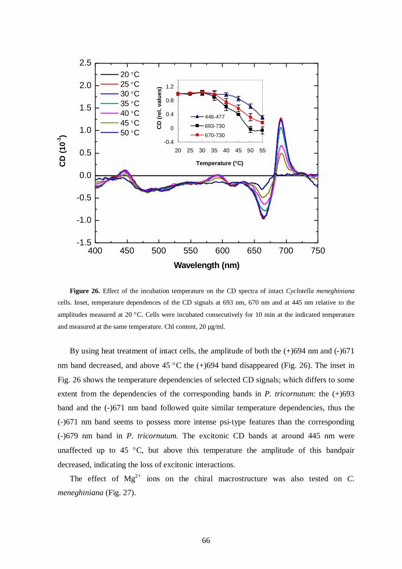
66
Figure 26. Effect of the incubation temperature on the CD spectra of intact Cyclotella meneghiniana
cells. Inset, temperature dependences of the CD signals at 693 nm, 670 nm and at 445 nm relative to the
amplitudes measured at 20 C. Cells were incubated consecutively for 10 min at the indicated temperature
and measured at the same temperature. Chl content, 20 µg/ml.
By using heat treatment of intact cells, the amplitude of both the (+)694 nm and (-)671
nm band decreased, and above 45 C the (+)694 band disappeared (Fig. 26). The inset in
Fig. 26 shows the temperature dependencies of selected CD signals; which differs to some
extent from the dependencies of the corresponding bands in P. tricornutum: the (+)693
band and the (-)671 nm band followed quite similar temperature dependencies, thus the
(-)671 nm band seems to possess more intense psi-type features than the corresponding
(-)679 nm band in P. tricornutum. The excitonic CD bands at around 445 nm were
unaffected up to 45 C, but above this temperature the amplitude of this bandpair
decreased, indicating the loss of excitonic interactions.
The effect of Mg2+ ions on the chiral macrostructure was also tested on C.
meneghiniana (Fig. 27).
400 450 500 550 600 650 700 750-1.5
-1.0
-0.5
0.0
0.5
1.0
1.5
2.0
2.5
20 C 25 C 30 C 35 C 40 C 45 C 50 C
CD
(10-3
)
Wavelength (nm)
-0.4
0
0.4
0.8
1.2
20 25 30 35 40 45 50 55
Temperature (°C)C
D (r
el. v
alue
s)
446-477
693-730
670-730
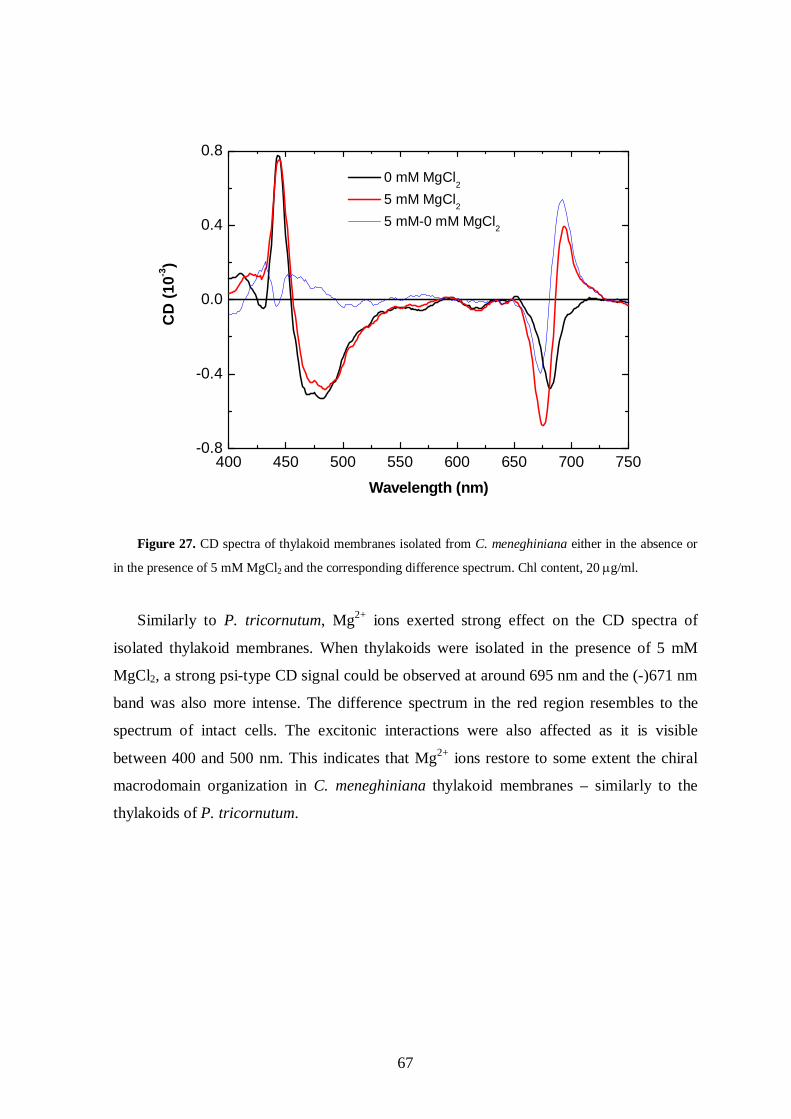
67
400 450 500 550 600 650 700 750-0.8
-0.4
0.0
0.4
0.8
CD
(10-3
)
Wavelength (nm)
0 mM MgCl2
5 mM MgCl2 5 mM-0 mM MgCl2
Figure 27. CD spectra of thylakoid membranes isolated from C. meneghiniana either in the absence or
in the presence of 5 mM MgCl2 and the corresponding difference spectrum. Chl content, 20 g/ml.
Similarly to P. tricornutum, Mg2+ ions exerted strong effect on the CD spectra of
isolated thylakoid membranes. When thylakoids were isolated in the presence of 5 mM
MgCl2, a strong psi-type CD signal could be observed at around 695 nm and the (-)671 nm
band was also more intense. The difference spectrum in the red region resembles to the
spectrum of intact cells. The excitonic interactions were also affected as it is visible
between 400 and 500 nm. This indicates that Mg2+ ions restore to some extent the chiral
macrodomain organization in C. meneghiniana thylakoid membranes – similarly to the
thylakoids of P. tricornutum.

68
4.5. Functional heterogeneity of the fucoxanthins and FCP complexes in diatom cells
4.5.1 Spectroscopic indications of heterogeneity of fucoxanthin in P. tricornutum
Pigment-pigment interactions can be well characterized by using CD spectroscopy,
however it gives only little information about the microenvironment of the interacting
pigments. To gain further information about the orientation and the local environment of
pigment molecules absorbing in the green `window`, we analyzed the flash-induced
electrochromic absorbance changes of intact cells of P. tricornutum between 470 and 570
nm. Upon the excitation of photosynthetic membranes with a single turnover saturating
flash, a uniform transmembrane electrical field of 105 V/cm magnitude is built up (Kakitani
et al. 1982). This field induces an almost homogeneous shift of the absorption bands of the
so-called field-indicating pigment molecules; the magnitude of the shift is linearly
proportional to the field strength (Cramer and Crofts, 1982; Junge, 1977). The primary
factor, which governs the shift, is the interaction of the ground and excited state of the
pigment molecule with the external field. The frequency shift is given as:
where denotes the frequency shift (cm-1) and and denotes the difference between
ground- and excited-state dipole moments and polarizabilities, respectively and F is the
electric field vector (Kakitani et al. 1982). Carotenoid molecules, which do not possess
permanent dipole moment, respond quadratically to the electric field, while molecules
having permanent dipole moment, exhibit linear absorption shift field dependencies. Thus,
different pigment molecules do not respond equally to the electric field and only certain
fraction of molecules exhibit characteristic, large electrochromic absorbance changes.
Flash-induced electrochromic absorbance changes were measured between 470 and
570 nm in 5 nm steps. The kinetics of typical flash-induced absorbance transients at
characteristic wavelengths are depicted in Fig. 28 (insets), which show the transient
spectrum calculated from the kinetic transients of the electrochromic absorbance changes.

69
Figure 28. Transient spectra of flash-induced absorbance changes, dominated by electrochromic
changes, in dark-adapted intact cells of Phaeodactylum tricornutum 5 ms after the exciting flash. Flash-
induced electrochromic absorbance transients at the indicated wavelengths (insets).
The transient spectra revealed a major band-shift with a bandpair at (+)565/(-)535 nm
and a minor band-shift with a bandpair at (+)515/(-)485 nm. In a recent study, with the aid
of absorption and Stark spectroscopy on FCP complexes isolated from Cyclotella
meneghiniana, three main Fx forms, Fxblue, Fxgreen and Fxred with wavelength positions at
around 465, 515 and 545 nm, respectively, were identified. The lower-energy Fxred and
Fxgreen forms exhibited large changes in static dipole moment on photon absorption, of
about 40 and 15 D, respectively (Premvardhan et al. 2008). The above described
terminology for the different Fx pools is used also in the present work, because these
different forms are spectrally well discernible, according to their wavelength position. The
strong electrochromic response of these Fx molecules shows that they probably interact
with Chl molecules, thereby lending a dipole moment to this molecule. In higher plants,
the strongest electrochromic response, at around 515 nm, is attributed to lutein/Chl b
interactions (Sewe and Reich, 1977). Earlier, we hypothesized that the long-wavelength
absorbing, field-indicating lutein/Chl b pigment pair in the LHCII might be replaced by a
similar Fx/Chl c pair in the FCP (Szabó et al. 2008, see in ’Publications’). However, recent
460 480 500 520 540 560 580-0.004
-0.002
0.000
0.002
0.004
0.006
0.008
-I/I
Wavelength (nm)
-20 0 20 40 60 80-0.003
-0.002
-0.001
0.000
0.001
-I/I
Time (ms)
535 nm
-20 0 20 40 60 80
0.000
0.005
0.010 560 nm
Time (ms)
-I/I

70
results showed that strong interaction occur between Fx molecules rather than between Fx
and Chl molecules (Premvardhan et al. 2008). These data also show that, similarly to
higher plants, purple bacteria and Chl a/c-containing algae (Büchel and Garab, 1995; de
Grooth et al. 1980; Joliot and Joliot, 1989; Kakitani et al. 1982), the electrochromic
absorbance changes in P. tricornutum originate from a minor fraction of pigment
molecules.
In our work, spectroscopic properties of the different Fx molecules have also been
characterized. Intact P. tricornutum cells exhibit long absorption shoulder between 480 and
560 nm originating from different Fx pools (Fig. 29). Fxred molecules exhibit red-shifted
and weak absorbance band at around 545 nm, and display only weak CD signal in this
region (see e.g. Fig. 18). It is interesting to note that a similar, weak, CD-silent long-
wavelength (~535 nm) absorbing band was identified in a marine green alga, and was
assigned to originate from a new electronic excited state, Sx between S1 and S2, of
siphonaxanthin, which appears to transfer to a specific Chl a molecule(s) (Akimoto et al.
2004, 2007). This state arises only in pigment-protein complexes, probably due to a
specific interaction with amino acids (Akimoto et al. 2007), and resembles the long
wavelength absorbance band of Fx in diatoms (Gillbro et al. 1993).
Fluorescence excitation spectra revealed that both short and long wavelength forms
exhibit efficient energy transfer to Chl a, where the energy transfer efficiency of Fxred is
especially remarkable, since it displays much weaker absorption (Fig. 29). This result is in
accordance with earlier data of energy transfer efficiencies measured on isolated FCP
(Papagiannakis et al. 2005).

71
Figure 29. Fluorescence excitation and absorption spectra of P. tricornutum cells. Fluorescence was
recorded at 689 nm. For better comparison, the spectra are normalized at 438 nm.
Linear dichroism measurements of intact P. tricornutum cells revealed major bands
peaking at 425 nm, 550 nm and 695 nm (Fig. 30).
Figure 30. Linear dichroism spectrum of P. tricornutum cells aligned by gel squeezing, OD673-750 = 0.35.
400 450 500 5500.2
0.4
0.6
0.8
1.0
Fluo
resc
ence
inte
nsity
or a
bsor
banc
e (r
el. u
nits
)
Wavelength (nm)
Fluorescence excitation Absorption
400 450 500 550 600 650 700
-4
0
4
8
12
16
LD (1
0-3)
Wavelength (nm)

72
The 425 and 695 nm bands originate from Chl a. Minor bands at around 463 and 600-
630 nm, originating from Chl c, at around 495 nm, originating probably from Ddx and the
bands at 510 nm and 545 nm, originating from Fx could also be observed.
4.5.2 The effect of growth light intensity to the heterogeneity of fucoxanthin in P.
tricornutum
It was also the aim of the present work to characterize the heterogeneity of the Fx
molecules in intact cells grown on different light conditions. Different light conditions
during growth can modify the pigment composition and Fcp polypeptide composition of
diatom cells. From the point of view of the efficiency of the excitation energy transfer, the
microenvironment and orientation of carotenoid molecules are determinant factors, which
are also supposed to change with the growth light intensity.
Absorption spectra were measured on HL and LL cells (Fig. 31). Both HL and LL cells
exhibit absorption peaks at 673, 633 and 437 nm, and shoulders at 460 and 490 nm. The
position and shape of the unusually broad and complex bands between 490 and 570 nm,
originating mostly from Fx, did not differ substantially between HL and LL cells. In HL
cells, the more intense absorption bands between 400 and 500 nm indicate that the
photoprotective carotenoids, diadino- and diatoxanthin accumulated.
400 450 500 550 600 650 700 750
0.00
0.05
0.10
0.15
0.20
0.25
0.30
HL LL
Abs
orba
nce
Wavelength (nm)
Figure 31. Absorption spectra of P. tricornutum cells grown on HL or LL intensities. Samples were
adjusted to the same Qy absorption of Chl a.
.

73
In order to obtain information on the local environment and polarizabilities of
carotenoids within the thylakoid membranes of intact diatom cells grown at different light
intensities, electrochromic absorbance changes were measured on HL and LL cells. Strong
electrochromic signal at around (+)565 and (-)535 and a less intense signal at (+)515 and
(-)485 nm could be observed in both LL and HL cells. The peak positions did not vary with
the growth light conditions, however, the amplitudes of the bands exhibited considerable
differences. In LL cells, the longer wavelength absorbance transient (565/535 nm) was
more intense, while the shorter wavelength band (515/485 nm) did not change
considerably compared to HL cells (Fig. 32a). The ordinate of the transient spectra are
shifted to zero at 470 nm, to correct for an observed baseline shift. The origin of the
baseline anomaly is unknown; it has been shown earlier that the electrochromic absorbance
changes can be accompanied by scattering transients, which are physically correlated to the
absorption changes (Garab et al. 1979).
The light-induced electrochromic absorbance transient spectra are composed of the first
derivatives of the absorbance bands of the participating electronic transitions (Büchel and
Garab, 1995). This specificity can be used to identify the field-indicating pigment
molecules and determine their concentrations. The information is essentially the same that
can be obtained from Stark spectroscopy using external field on randomly oriented protein
complexes (Boxer, 2009). The measured points at the indicated wavelengths together with
the fitting curves are depicted in Fig. 32a.

74
Figure 32. Electrochromic absorbance transients of HL (open squares) and LL (closed squares) cells of
P. tricornutum (a), and absorbance bands of the field-sensitive pigments obtained from the fits of the
transients (b) of HL and LL cells. The data points, 5 ms after the exciting flashes, were obtained from kinetic
traces after averaging 32 transients, the repetition rate of the flashes was 1 s-1. In the transient spectra, the
data points were fitted with first derivatives of Gaussians of HL and LL cells. Samples were adjusted to the
same Chl a Qy absorption, OD673-750 ≈ 1.
The parameters obtained from fitting with first derivatives of Gaussians are shown in Table
V. These parameters were used to reconstitute the absorbance spectrum of shifted
pigments, which are shown in Fig. 32b.
HL (565/535) HL(515/485) LL(565/535) LL(515/485)
x0 5500.6 5011.3 5500.35 5011.5
k -1.70.21 -0.810.9 -50.33 -0.850.8
h 11.10.8 11.71.4 13.40.4 11.51.6
Table V. Parameters obtained from mathematical fit with first derivatives of gaussians of the measured
spectra of fast absorbance changes of HL and LL cells. The parameters x0, k and h are defined in Chapter
3.2.5.
The electrochromic transients in LL and HL cells differed significantly from each other
(Fig. 32a): LL cells contained significantly larger contributions from Fxred than HL cells.
As determined from the calculated absorption spectra (Fig. 32b), in HL cells the intensity
of the Fxred band was 2.2 ± 0.2 times larger than that of Fxgreen. In LL cells, the Fxred/Fxgreen
ratio was 5.8 ± 0.7. Assuming no change in the electrostatic properties of the two forms,
460 480 500 520 540 560 580
-0.005
0.000
0.005
0.010
LL measured HL measured HL fitted LL fitted
-I/I
Wavelength (nm)
510 nm 560 nm
(a)
460 480 500 520 540 560 580
0.00
0.02
0.04
0.06
0.08
0.10
0.12
0.14
LL HL
Abs
orba
nce
(a.u
.)
Wavelength (nm)
500 nm 550 nm
(b)

75
this difference must originate from an accumulation of Fxred in LL cells. This means that
Fxred and Fxgreen originate from different FCPs. In other words, the fact that the ratio of the
two electrochromic bands varies in a broad range in HL and LL cells indicates that the two
Fx forms are bound to different FCPs. At the given signal to noise ratio, no other
electrochromic band could be identified, suggesting that the differences between the
ground- and excited-state dipole moments of all other pigment molecules absorbing
between 470 and 570 nm are significantly weaker. Neither was there a contribution from
Ddx or Dtx, although the amount of these xanthophylls was 3-4 times higher in HL cells
than in LL cells, calculated on the basis of the same Chl a content. The Fx content on the
same basis was also found to remain unchanged, as verified by HPLC (Fig. 33).
0
200
400
600
800
Chl c Fx Ddx Dtx ß car
mM
pig
men
t / M
Chl
a
HLLL
Figure 33. Pigment content of HL and LL P. tricornutum cells determined by HPLC.
This is in a good agreement with the results of Lepetit et al. (2010). They also found
that by increasing the light intensity during growth only the Ddx cycle pigments
accumulated, while the amount of Fx and Chl c remained unchanged both in P.
tricornutum and C. meneghiniana.
In order to obtain information on the orientation of Fx molecules in cells grown either
on HL or LL light, LD measurements were performed. The two Fx forms were clearly
discernible in the LD spectra of both HL and LL cells as it is shown in Fig. 34.

76
Figure 34. Linear dichroism spectra of HL and LL cells oriented by gel squeezing method (a) and by
magnetic field of 0.5 T (b). The Chl contents of the two samples were equal, OD673-750 = 0.35.
Both in HL and LL cells Fxred at around 550 nm exhibited an intense signal, and in HL
cells a stronger band appeared at around 515 nm. Essentially the same information could
be obtained on intact cells oriented either by gel squeezing method (Fig. 34a) or by strong
magnetic field (Fig. 34b). These results also show that intact P. tricornutum cells can be
well oriented in magnetic field.
The characteristic LD bands at 550 and 515 nm could also be observed in the case of
isolated thylakoid membranes isolated from HL and LL cells (Fig. 35). The LD spectra of
HL and LL thylakoids were quite similar, significant changes could only be observed
between 400 and 500 nm, which may result from the different Ddx and Dtx composition of
HL and LL cells (shown in Fig. 33). In the case of thylakoid membranes the relative
intensity of LD bands at 550 nm compared to 515 nm is lower than in the case of intact
cells. Similar differences in the 515 and 550 nm band could also be observed earlier by
comparing the LD spectra of intact cells and isolated thylakoids of P. tricornutum grown
on normal light intensities (Hiller and Breton, 1992).
475 500 525 550 575 600 625-2
-1
0
1
2
3
LL HL
LD (1
0-3)
Wavelength (nm)
(a)
475 500 525 550 575 600 625-2
-1
0
1
2
3
4
LD (1
0-3)
Wavelength (nm)
LL HL
(b)

77
400 450 500 550 600 650 700
-80
-40
0
40
80
120
LD (1
0-3)
Wavelength (nm)
LL HL
Figure 35. Linear dichroism spectra of isolated thylakoid membranes from HL and LL cells of P.
tricornutum. Thylakoid membranes were oriented by gel-squeezing method. The Chl contents of the two
samples were equal, OD673-750 = 0.4.
Using the squeezing parameter M=2 and the calculated order parameter (Garab, 1996),
the orientation angle of the transition dipole of Fxred at 550 nm in membranes isolated from
LL cells was found to be 29° with respect to the membrane plane. In comparison, the
orientation angle of the Qy transition of Chl a was calculated to be 17° in the same
membranes. In contrast to the large signal at 550 nm, the LD signal at 500 nm was weaker,
showing that the transition dipoles at around 500 nm, with a calculated 34-36° orientation
angle, i.e. tend to tilt out somewhat more from the membrane plane than those at 550 nm.
The LD spectrum most clearly indicates the presence of the so-called Fxgreen being quite
different from Fxred, with the former exhibiting a much weaker signal.
In order to obtain information about the functional significance of the heterogeneity of
the Fx forms and the FCPs, low-temperature fluorescence excitation and emission spectra
were recorded. The excitation spectra revealed that energy transfer to Chl a occurs from
both the short- and long-wavelength Fx forms (Fig. 36a and b).

78
Figure 36. Low temperature (77 K) fluorescence excitation (a and b) and emission (c and d) spectra of
LL (a and c) and HL (b and d) P. tricornutum cells. The fluorescence excitation spectra, normalized at 438
nm, were recorded for the 689 nm and the 713 nm bands. For the emission spectra, normalized at 713 nm, the
cells were excited at 510 nm or at 550 nm. The room temperature absorbance spectra of LL (a) and HL (b)
cells are also normalized to the 438 nm band of the excitation spectra. (The wavelength for normalization,
438 nm, was selected because of the maximum of Chl a absorbance and virtually no overlap with the two Fx
forms in question, i.e. with Fxred and Fxgreen).
The excitation spectra also show that Fxgreen, absorbing at around 500 nm, supplies
energy about equally to the Chl a molecules that emit at 689 and 713 nm. In contrast, the
fluorescence emission was stronger at 689 nm compared to 713 nm for excitation into the
520 – 560 nm range, in the Fxred-absorbing region, in both LL (Fig. 36a) and HL (Fig. 36b)
cells. Fluorescence emission spectra (Fig. 36c and d) indicate that upon excitation at 550
nm, both LL and HL cells exhibit somewhat stronger fluorescence emission at 689 nm,
600 650 700 750 800
0
10
20
30
40
510 nm 550 nm
Fluo
resc
ence
inte
nsity
(cps
x 1
04 )
Wavelength (nm)
(d)
HL
600 650 700 750 800
0
10
20
30
40
510 nm 550 nm
Fluo
resc
ence
inte
nsity
(cps
x 1
04 )
Wavelength (nm)
(c)
LL
480 500 520 540 560 580 600
0.2
0.4
0.6
0.8
1.0
689 nm 713 nm absorption
Fluo
resc
ence
or a
bsor
banc
e (r
el. u
nits
)
Wavelength (nm)
(a)
LL
480 500 520 540 560 580 600
0.2
0.4
0.6
0.8
1.0
689 nm 713 nm absorption
Fluo
resc
ence
or a
bsor
banc
e (r
el. u
nits
)
Wavelength (nm)
(b)
HL

79
indicating that Fxred favours energy transfer to F689 (PSII). In contrast, excitation at 510 nm
of both LL and HL cells exhibit less intense fluorescence at 689 nm, indicating that Fxgreen
favours energy transfer to F713 (PSI) or has no preference for ET to either one of the PSs.
4.5.3 Heterogeneity of fucoxanthin molecules in C. meneghiniana
The electrochromic transient spectrum was measured also in C. meneghiniana. Typical
kinetics of the flash-induced absorbance changes can be seen in the inset of Fig. 37. Like in
P. tricornutum, two main electrochromic bands could be observed on the transient
spectrum: a smaller band at (+)535 and (-)485 nm and a more intense one at (+)565 and
(-)545 nm. The longer wavelength band is red-shifted compared to the respective band in
P. tricornutum cells.
Figure 37. Transient spectrum, induced by single turnover saturating flashes, in intact cells of Cyclotella
meneghiniana. The data points, 5 ms after the exciting flashes, were obtained from kinetic traces after
averaging 32 transients, the repetition rate of the flashes was 1 s-1. A typical trace, recorded at 560 nm, is
shown in inset. The transient spectrum was fitted with the sum of first derivative of two Gaussians (solid
line) and a constant – tentatively assigned to scattering transient.
460 480 500 520 540 560 580-0.001
0.000
0.001
0.002
0.003
-20 0 20 40 60 80-0.001
0.000
0.001
0.002
0.003
-I/I
Time (ms)
-I/I
Wavelength (nm)

80
The measured datapoints could be fitted reasonably well with the first derivative of
Gaussians indicating the electrochromic nature of the Fx molecules also in C.
meneghiniana cells. Similarly to P. tricornutum, an unknown shift of the transient spectra
– probably due to scattering transients – could also be observed.
We attempted to record the LD spectra of intact cells and isolated thylakoid
membranes of C. meneghiniana. However, we found that intact cells could not be aligned
either by gel squeezing or by magnetic field, therefore no valuable LD spectra could be
recorded on this level. This is probably because of the lack the longitudinal symmetry of
these cells and the presence of several chloroplasts with different orientations. We were
able, however, to measure the LD signature of isolated thylakoid membranes. Essentially
similar LD spectra were obtained in C. meneghiniana thylakoid membranes, showing that
the orientation of Fxred and Fxgreen are essentially the same as in P. tricornutum (Fig. 38).
Figure 38. Linear dichroism spectra of isolated thylakoid membranes of P. tricornutum and C.
meneghiniana. Thylakoid membranes were oriented by gel-squeezing method. The measured LD of C.
meneghiniana was somewhat weaker, 0.09 at 679 nm, but for better comparison the two spectra are
normalized to their red maxima.
400 450 500 550 600 650 700
-50
0
50
100
C. meneghiniana P. tricornutum
LD (1
0-3)
Wavelength (nm)

81
The fluorescence excitation and emission spectra were measured on intact cells in order
to obtain information about the excitation energy transfer efficiency of different Fx pools
(Fig. 39).
Figure 39. Low temperature (77 K) fluorescence excitation (a) and emission (b) spectra of Cyclotella
meneghiniana cells. The fluorescence excitation spectra, normalized at 438 nm, were recorded for the
689 nm and the 713 nm bands. For the emission spectra, normalized at 713 nm, the cells were excited at
510 nm and at 550 nm. The room temperature absorbance spectrum of the cells (a) is also normalized to the
438 nm band of the excitation spectra.
The fluorescence excitation spectra indicate that Fxred transfers excitation energy to Chl
a somewhat more efficiently that Fxgreen, as reflected by the stronger band between 520 and
555 nm (Fig. 39a). Fluorescence emission spectra exhibited a characteristic peak at 689 nm
and a band at 713 nm (Fig. 39b). The 713 nm band was much weaker than in P.
tricornutum. The 689 nm band was more intense upon excitation at 550 nm compared to
the excitation at 510 nm.
400 450 500 550 600
0.2
0.4
0.6
0.8
1.0
689 nm 713 nm absorption
Fluo
resc
ence
or a
bsor
banc
e (r
el. u
nits
)
Wavelength (nm)
(a)
600 650 700 750 800
0
5
10
15
20
25
510 nm 550 nm
Fluo
resc
ence
inte
nsity
(cps
x 1
04 )
Wavelength (nm)
(b)

82
5. DISCUSSION
The regulation of light-harvesting antennae on the supramolecular level is important in
the adjustment of light-harvesting efficiency and adaptation to various stress conditions in
all photosynthesizing organisms. It has been shown in the past decades that in higher plants
the photosynthetic pigments are assembled into structurally flexible chirally-organized
macrodomains, in mature granal thylakoid membranes. In contrast, there is no systematic
study on the supramolecular organization of pigments on different organizational levels in
diatoms.
5.1. The psi-type CD signal is associated with the multilamellar order of the thylakoid membranes in intact cells
I have shown in Chapter 4.2 that intact cells of P. tricornutum exhibited psi-type CD
signatures at around 698 nm. Upon isolation of thylakoid membranes or disruption of cells
by sonication, the psi-type features decreased to large extent, while the excitonic
interactions were not affected, suggesting that Chl a molecules are arranged into chiral
macrodomains, exhibiting long-range interactions. The chiral macrodomains, in
accordance with the theory of psi-type CD for large ordered 3 dimensional arrays (Garab
1996; Keller and Bustamante, 1986), appeared to be associated with a multilamellar
thylakoid membrane system in the cell. This was shown by TEM studies – the CD changes
induced by sonication could be well correlated with the changes in the thylakoid
membrane ultrastructure. Similar CD changes have been reported in other Chl a/c-
containing algae upon disruption of intact cells or isolated chloroplasts (Büchel and Garab
1997; Goss et al. 2000). In Mantoniella squamata, much weaker psi-type CD signals are
present in intact cells compared to higher plant chloroplasts, and no psi-type CD could be
identified in the isolated LHC and thylakoid membranes (Goss et al. 2000). Intact cells and
isolated chloroplasts of Pleurochloris meiringensis possess psi-type CD signals that are
diminished when the chloroplasts are disrupted; it has also been found that the intensity of
the psi-type signal changes with the light intensity during growth (Büchel and Garab,
1997). The sensitivity of (+)698 nm band, and possibly also the disappearance of the broad
band in the blue, further corroborated our conclusion on the origin of this (these) band(s) in
chirally organized macrodomains. The results presented in this work about the macro-

83
organization of chromophores in diatoms are thus in good agreement with other studies on
other Chl a/c-containing algal species (Büchel and Garab, 1997; Goss et al. 2000).
The main difference between the organization of thylakoid membranes in higher plants
and diatoms can be found in the lateral heterogeneity of the pigment-protein
supercomplexes. Granal thylakoid membranes of higher plants, which are arranged in
stacked multilamellar membrane system enriched in PSII-LHCII supercomplexes form
large ordered chiral domains exhibiting psi-type CD signals (reviewed e.g. in Garab and
Mustárdy, 1999). The thylakoid membranes of diatoms are also arranged into – loosely
stacked - multilamellar membrane system; however, here we cannot speak about granal
and stromal separation and lateral heterogeneity of PSII and PSI supercomplexes, thus the
question arises what is the nature of the psi-type CD signals detected in P. tricornutum. To
answer this question, we examined the (macro-)organization of the pigment system in
vitro, in isolated FCP complexes and in isolated thylakoid membranes.
5.2. Isolated FCP complexes do not assemble into chiral macrodomains
In Chapter 4.1 I have shown that the CD spectra of isolated FCP complexes are
dominated by excitonic interactions, and the psi-type CD signal is absent. However, the
question arises whether the FCP complex isolated in this way represents the native state of
the antenna system of P. tricornutum. By applying milder solubilization we have shown
that it is possible to separate FCP complexes with different oligomeric states (Lepetit et al.
2007). The CD spectra of trimeric FCP and higher oligomeric FCPo presented in Fig. 12
are very similar to each other and lack psi-type signal, indicating that the isolated
oligomeric antenna of P. tricornutum does not form chirally organized macrodomains or
large aggregates in contrast to LHCII of higher plants. Nevertheless, the existence of
somewhat stronger excitonic interactions in FCPo than in FCP suggests that FCPo
represents a more native state of the light-harvesting antenna in P. tricornutum.
5.3. The chiral macrodomain organization of the pigments can be partially retained in isolated thylakoid membranes
In granal thylakoid membranes two external factors influence the long-range chiral
order of the chromophores: i) electrostatic screening of the negatively charged
polypeptides by divalent cations, which is required for the lateral separation of the two
photosystems and the stacking of membranes, ii) the osmotic pressure of the medium,

84
which influences mainly the lateral packing density of the complexes (Barzda et al. 1994;
Garab et al. 1991). It has been shown in higher plants that the absence of the divalent
cations from the incubation medium resulted in a dramatic decrease of the heat-stability of
isolated thylakoid membranes. The same phenomenon could be observed when isolated
thylakoids were (re)suspended in hypotonic medium, however, the heat stability was
preserved to more extent in this case (Cseh et al. 2000). This shows that the electrostatic
interactions and divalent cations, i.e. electrostatic screening play crucial role in membrane
stacking and the self-assembly of grana (Chow et al. 2005; Kiss et al. 2008; Barber, 1982),
along with the lateral separation od PSII and PSI complexes and the formation of LHCII-
enriched macrodomains within the thylakoids, which must precede the stacking (Barzda et
al. 1994, 1996; Mustárdy et al. 2008). The role of membrane stacking is not confined to the
maintaining of the structure of the grana membranes, but it is also essential in ensuring the
physiological functions, e.g. preventing the spillover of excitation energy from PSII to PSI
(Chow et al. 2005; Trissl and Wilhelm, 1993) and providing the photoprotective
mechanisms, i.e. NPQ (Horton and Ruban, 2005). The stimulating effect of Mg2+ on NPQ
was first shown by Noctor et al. (1993) and Rees and Horton (1990). Earlier electron
microscopic and CD spectroscopic studies have provided evidence that incubation of
thylakoids of higher plants in sorbitol and MgCl2-containing media stabilizes grana
stacking, whereas in buffers without sorbitol and MgCl2 significant unstacking of grana
membranes takes place (reviewed by Garab and Mustárdy, 1999). It has been shown that
grana stacking is essential for the formation of NPQ, especially in its xanthophyll-cycle
component (Goss et al. 2007). Disturbances in the macro-organization of the PSII antenna
lead to a strong reduction of NPQ (Kovács et al. 2006).
The role of electrostatic screening by divalent cations and the ambient osmolarity in the
regulation of the stacking of the thylakoids have been scarcely investigated diatoms. Due
to the lack of the detailed structural information about the pigment-protein complexes and
their interactions in the thylakoid membranes, the exact association of two membrane
layers and thus the mechanism of the stacking is not known in diatoms, and in general, in
Chl a/c-containing organisms.
It has been found that changes in the osmolarity dramatically affect the psi-type CD
signals in Pleurochloris meiringensis, while the ionic composition had no influence on
these bands (Büchel and Garab, 1997). Similar studies on the role of physicochemical
factors in the modification of thylakoid membrane stacking in diatoms could not be found
in the literature. Moreover, there is no information available about the correlation between

85
membrane stacking and the parameters of chlorophyll fluorescence. In diatoms, no lateral
heterogeneity could be observed, but they exhibit strong and efficient NPQ. The NPQ is
strictly related to the operation of the diadinoxanthin-cycle in these organisms (Goss et al.
2006; Lavaud et al. 2002), although recently some components of NPQ were found to be
independent of the operation of the xanthophylls cycle (Lavaud et al. 2006; Grouneva et al.
2008).
In Chapter 4.3.3 the role of Mg2+ in the macro-organization of the complexes was
tested on isolated thylakoid membranes. We have shown that in the presence of Mg2+ ions
the psi-type CD signal could be significantly preserved, pointing to the important role of
divalent cations in the stacking of thylakoid membranes in P. tricornutum. Thylakoid
membranes isolated in the absence of MgCl2 lost the psi-type CD signal, however it could
be restored to some extent by resuspending the membranes in buffer containing MgCl2.
Thus, in isolated membranes psi-type CD signal could also be observed and the CD
spectrum resembled the spectrum of intact cells. We have also shown that the osmotic
pressure plays no or only minor role in this process. This situation is similar to granal
thylakoid membranes, where it has also been shown that divalent cations are the main
factors controlling the macro-organization of thylakoid membranes (Garab et al. 1991). In
Chapter 4.3.3 correlations were revealed between the Mg2+-assisted macro-organization
and some functional parameters, Fv/Fm, NPQ and the operation of Ddx cycle.
These results provide evidence for the existence of chiral macrodomains in thylakoid
membranes under physiologically relevant conditions. At present, it is not possible to
assign the chiral macrodomains to exact structures and stacking mechanism in the
thylakoids of diatoms. However, our results provide evidence for the first time that well
ordered, three dimensional network of the thylakoid membranes of P. tricornutum exhibit
psi-type CD signals. It also must be pointed out, that parallel running membrane sheets
themselves, i.e., without a chiral organization of the complexes, cannot give rise to psi-type
CD, as e.g. in bundle sheath chloroplasts (Faludi-Dániel et al. 1973) or in the case of CP24
depleted Arabidopsis plants (Kovács et al. 2006). Preliminary measurements of small-
angle neutron scattering (SANS) - which provide information about the changes in the
repeat distances of the multilamellar thylakoid membrane system – indicate that in P.
tricornutum cells the thylakoid membranes are indeed organized into highly ordered
multilamellar system with well defined repeat distances.

86
5.4. Structural flexibility of the chiral macrodomains
It has been known for a long time that in higher plants the chirally organized
macrodomains exhibit remarkable reversible structural flexibility (Barzda et al. 1996, Cseh
et al. 2005, Garab et al. 1988b, Holm et al. 2005), which provides the photosynthetic
membranes enhanced photoprotection capability on the supramolecular level. The
photoprotective mechanisms in relation with electron transport and xanthophyll cycle
activity, the role of pH are examined in details in diatoms using various techniques based
mainly on fluorescence measurements (reviewed in Wilhelm et al. 2006 and Lavaud,
2007). However, little is known about the structural modifications of thylakoid membranes
and light harvesting antennae behind the well characterized quenching mechanisms. There
are studies available on isolated FCP complexes which exhibit markedly different
fluorescence yield in different aggregation states (e.g. Gundermann et al. 2008), however,
the physiological role of aggregation of FCP is not elucidated yet. Miloslavina et al. (2009)
provided the first evidence that under NPQ conditions part of the FCP complexes detaches
from PSII reaction center and becomes aggregated and the mechanism of aggregation is
similar to that of observed in vitro.
The amplitude of the psi-type CD signal could be modulated by certain environmental
factors, showing that the macrodomains exhibit considerable structural flexibility.
We intended to examine the structural rearrangements of the chiral macrodomains
caused by changes in different environmental factors, such as heat, illumination with
strong light, light intensity during growth and changes in osmotic pressure and ionic
strength.
I have shown in Chapter 4.3.1 that the psi-type CD bands in P. tricornutum cells
exhibit increased sensitivity to elevated temperatures compared to the excitonic bands. In
particular, the (+)698 nm psi-type band is the most susceptible, but the negative CD band
at around 679 nm also displays higher sensitivity to elevated temperatures than the
excitonic bands. These data suggest that the macro-organization can be prone to thermo-
optically inducible reorganizations. However, presently we have no evidence for such an
origin of the reorganizations in intact cells of P. tricornutum. Preliminary results indicate
that the CD changes caused by elevated temperatures are reversible.
Intact cells subjected to illumination with strong actinic light exhibited reversible light-
induced CD changes. Upon illumination, the amplitude of the main psi-type CD bands
increased, while the excitonic CD bands remained essentially unchanged (Fig. 16). The

87
fact that diatoms exhibit increase in the intensity of the psi-type CD band might be
explained by the aggregation of individual pigment-protein complexes, like the FCP
complex, or changes in the repeat distances of the thylakoid stacks. The time kinetics of
the light induced CD changes at 698 nm is commensurable with the kinetics of the
induction and relaxation of NPQ (Ruban et al. 2004, Goss et al. 2006). Moreover, the
xanthophyll cycle inhibitor DTT inhibited the light-induced CD changes, which suggests
the involvement of Ddx-deepoxidation in the structural changes. It has been found in
higher plants that Zx acts as an allosteric activator for qE and the conformational changes
of LHCII (Horton et al. 2008), which could also be true for the Dtx and conformational
changes in the antenna system of diatoms. However, the proton gradient uncoupler NH4Cl
did not affect significantly the light-induced CD changes in P. tricornutum, therefore at
present it is not clear how the NPQ and the changes in the macrodomain order are
correlated. Further experiments are needed to clarify the relationship of the structural
changes and NPQ in diatoms. Nevertheless, these results are in a good accordance with
SANS measurements, which give information about the changes in the repeat distances of
the multilamellar thylakoid membrane system in vivo. In these measurements, P.
tricornutum cells exhibited significant and reversible light-induced changes in the center
position of the Bragg peak in the scattering profile, indicating that changes in the repeat
distances occurred. These data will be presented in the PhD dissertation of Gergely Nagy.
NH4Cl did not influence significantly the SANS changes either, which is in accordance
with CD measurements.
The light intensity during growth also affected significantly the intensity of the psi-type
bands in P. tricornutum as it has been demonstrated in Chapter 4.3.2; LL exhibited more
intense psi-type bands than HL cells. In LL cells, however, the kinetics and amplitude of
the light-induced CD changes were much smaller than in HL cells. These results suggest
that in LL cells the structural flexibility of the chiral macrodomains are less expressed,
which might be correlated with the less effective photoprotective capability of cells grown
on lower light intensities. Nonetheless, the large psi-type signal suggests the presence of
large ordered arrays of antenna complexes, which may be advantageous for the enhanced
light-harvesting capability.
We have observed that the intensity (+)698 nm band decreased reversibly by increasing
the ambient sorbitol concentration. These results may indicate that variations in the
osmotic pressure affect the lumenal space and/or the interthylakoidal space, and thus the
repeat distances in the multilamellar membrane system. Indeed, the average repeat

88
distances, determined from SANS data, have revealed shrinking up to 1-2 nm; these
changes could not be discerned by TEM. It is interesting to note that the light-induced CD
changes increase while the sorbitol-induced changes decrease the psi-type CD. Under the
same conditions, as reflected by SANS, the repeat distance increase and decrease,
respectively; data which can help in elucidation of the origin of the psi-type signal in
diatoms. The changes indicate alterations in the supramolecular array of the complexes,
evidently via or associated with the changes in the membrane ultrastructure, but during the
osmotically-induced changes excitonic interactions appear to be affected. The spectral
changes observed upon changing the ambient sorbitol concentration are peculiar. In higher
plants, strong psi-type CD signal is observed if the isolated thylakoid membranes are
suspended in sorbitol+MgCl2 containing buffer. In the presence of MgCl2 but in the
absence of sorbitol, the psi-type signal at (-)676 nm decreased the most, which is due to
partial destacking of the membranes which prevents the disassembly of the chiral
macrodomains (Cseh et al. 2000). In diatoms the situation is completely different; the psi-
type signal decreases by increasing the sorbitol concentration. In cellular systems, the
increase in the ambient osmolarity causes water loss from the cells (hypertonic media). The
loss in cellular water content causes a decreased hydration of the membranes which may
result in a concomitant “membrane-crowding” within the chloroplast. This may affect the
macro-organization and even the excitonic interactions. It is important to emphasize,
however, that the CD changes induced by osmolarity are reversible.
CD spectra of pigments at different organizational levels contain a large amount of
information. In a complex system, e.g. in intact cells or isolated thylakoid membranes, the
CD signals originating from different types of interactions are superimposed onto each
other, which make the interpretation of CD signals extremely difficult. Nevertheless, CD
data provide important information on the changes in pigment-pigment interactions e.g. in
intact cells subjected to different treatments as described in Chapter 4.3. Although a deep
analysis of CD spectra would require detailed quantum-mechanical calculations, such as
attempted only on structures with atomic resolution (Georgakopoulou et al. 2006, 2007),
mathematical deconvolution, e.g. using singular value decomposition and using the
measured spectra of isolated FCP and thylakoid membranes might provide further insights
on the nature of structural modifications caused by environmental factors.

89
5.5. Structurally flexible chiral macrodomains also in C. meneghiniana
In Chapter 4.4 we examined a taxonomically different diatom, C. meneghiniana.
Essentially the same psi-type CD bands at around 698 nm could be found in these cells, as
in P. tricornutum. This band was also absent in isolated FCP. Isolated thylakoid
membranes partially preserved the psi-type CD signal when Mg2+ ions were applied during
isolation. The chiral macrodomains exhibited remarkable structural flexibility when
elevated temperatures and illumination with strong light were applied, thus – similarly to
P. tricornutum – pigments are arranged into structurally flexible chiral macrodomains.
5.6. The fucoxanthin molecules and the FCPs are spectrally and functionally heterogeneous
Earlier data obtained from fluorescence spectroscopy suggest that the Fx population is
not homogeneous in diatoms, one part of the molecules transfers the energy more
efficiently to Chl a (the red-shifted form) while the other part displays only poor energy
transfer efficiency (the blue-shifted form) (Papagiannakis et al. 2005). Ultrafast transient
spectroscopy studies indicate that the energy transfer pathways within the isolated FCP are
also heterogeneous (Papagiannakis et al. 2005; Gildenhoff et al. 2010). To gain more
information about the microenvironment, more precisely about the charge-transfer
properties of Fx and the changes in static dipole moments, Stark-spectroscopy
measurements were performed earlier on isolated FCP complexes (Premvardhan et al.
2008). Stark-effect, a homogeneous bandshifts in the absorption spectrum, was induced by
external electric field. The magnitude of the bandshift (only a few Ångstroms) depends on
the difference between the ground- and excited state dipole moments and polarizabilities,
therefore Stark signals provide unique information on the microenvironment of the given
molecule (reviewed by Boxer, 2009). It has been shown in Chapter 4.5.1 that P.
tricornutum cells two main electrochromic absorbance band-shifts can be identified, with
positive/negative peaks at around 565/535 nm and 515/485 nm (Fig. 28) that overlap the 0-
0 and 0-0/0-1 bands of Fxred and Fxgreen, respectively (Premvardhan et al. 2008). The
occurrence of the electrochromic absorbance transients in this diatom is consistent with the
Stark signal of isolated FCP in the external field. The energetic locations of the Fx species
that give rise to the two electrochromic transitions in P. tricornutum cell are close to the 0-
0 bands of Fxred and Fxgreen, respectively, previously identified in the FCP complexes
isolated from C. meneghiniana. In this context, it is important to note that we have found

90
essentially the same electrochromic transients in C. meneghiniana cells, where Fxred and
Fxgreen were found at 553 and 502 nm, respectively.
To interpret the role of Fx in the excitation energy transfer processes, the pigment-
pigment interactions and the orientation of the pigments in the membranes must also be
taken into account. Isolated thylakoid membranes of algae containing Chl a/c-type antenna
systems exhibit excitonic CD bands in the 400-500 nm spectral range which is a sum
spectrum of Chl a, Chl c and carotenoids (Goss et al. 2000; Katoh, 1992; Mimuro et al.
1990; Büchel and Garab, 1997; Büchel, 2003). In isolated thylakoid membranes of brown
algae the (-)478 nm band originates from the S2 energy level of Fx, while the broad
shoulder between 478 and 525 nm originates probably from the S1 level of Fx (Katoh,
1992). Isolated Fx molecules, similarly to other carotenoids, exhibit no CD bands,
however, upon binding to the proteins they acquire CD signals due to induced chirality and
strong excitonic coupling to Chl a, thereby contribute to the excitonic signals at (+)445
nm/(-)478 nm. Moreover, protein bound Fx molecules also undergo bathochromic shift,
and this phenomenon also contributes to the long CD shoulder observed between 478 and
525 nm (Katoh, 1992).
The CD band between 470 and 530 nm have been shown to originate mostly from Fx
molecules, however the broad band with a long shoulder above 500 nm suggests that the
Fx interactions are not uniform. In both HL and LL cells, no CD signatures are associated
with the field-sensitive Fx forms, which are essentially CD silent as in isolated FCP
(Büchel, 2003). Nevertheless, it is worth mentioning that LL cells exhibit a more intense
psi-type band at (+)698 nm than HL cells, which is most probably due to the accumulation
of the light harvesting complexes in LL membranes. Similar data were obtained in another
Chl a/c-containing organism, Pleurochloris meiringensis (Büchel and Garab, 1997). It
must be noted here, that two different types of light sources were used for growing the
cells under HL and LL conditions (see Chapter 3.1.1), thus, it cannot be excluded that
minor variations in the spectral composition of the growth light source caused chromatic
adaptation of the cells, in addition to the adaptation to the light intensity. However, this
does not affect our conclusion that the spectral heterogeneity of Fx varies upon changing
the growth conditions, a finding that strongly suggests that the heterogeneity originates
from a heterogeneity of FCP complexes. The orientations of the Fx transition dipole
moments relative to the membrane plane can be used to understand the nature of the
electrochromic signals in the transmembrane field: larger signals are expected not only on
the basis of the intrinsic dipolar properties of the pigments but also when the pigments are

91
aligned to have their static dipole moments, , (close to) parallel to the transmembrane
field, F. The large electrochromic transient, proportional to Fcos (is the angle between
and F), from Fxred therefore indicates that its static dipole moment must be close to
parallel to the trans-membrane field vector (F). Notably, the angle between the transition
dipole and the change in static-dipole moments was measured to be ~ 20o in solution
(Premvardhan et al. 2008) which in conjunction with the LD data, an orientation angle of
29° of the transition dipole with respect to the membrane plane, would mean that the Fxred
could be oriented at ~ 49° with respect to the membrane plane; hence, ~ 41° between and
F, which can yield more than 70% of the maximum possible electrochromic response to
the trans-membrane field. For Fxgreen this value can be as high as 83 %. It must also be
noted that the angle between the transition dipole and static dipole moment could be even
larger in the protein for Fxred if it has a planar ‘S’ shape conformation, as proposed in
Premvardhan et al. (2009).
The data presented in Chapters 4.5.1 and 4.5.2 on the heterogeneity of the Fx
molecules and of the FCP pool are in reasonable agreement with biochemical analyses.
The presence of different FCP pools was indicated by Western blot analysis, showing that
the FCPs of the antenna are different from the FCPs connected to the photosystems
(Brakemann et al. 2006, Veith et al. 2009). In addition, the FCP composition of the antenna
varied if the algae were grown under LL or HL conditions (Beer et al. 2006). It has also
been found that the expression of the individual FCP proteins displays heterogeneous
response to the HL intensities (Becker and Rhiel, 2006; Brakemann et al. 2006).
Heterogeneity of FCPs can also be seen in the ultrafast fluorescence decay kinetics
(Miloslavina et al. 2009), which showed that two different pools of FCPs are responsible
for the generation of steady-state non-photochemical fluorescence quenching (NPQ) both
in P. tricornutum and C. meneghiniana. One pool of FCPs, detached from the
photosystems, forms a fluorescence-quenching site, whereas the second quenching site
consists of FCPs that remain in contact with the PSII core complex (Miloslavina et al.
2009). (NPQ at this site furthermore depends on the presence of the de-epoxidized
xanthophyll cycle pigment Dtx.) The heterogeneous lipid distribution in the thylakoid
membranes of diatoms also supports the existence of different FCP pools and
macrodomains of the pigment-protein complexes (Goss et al. 2009), although no
variability of stacked to exposed membranes is observed by electron microscopy (Pyszniak
and Gibbs, 1992).

92
6. CONCLUSIONS
I investigated the molecular organization of the main light harvesting antenna
complexes, the fucoxanthin-chlorophyll protein (FCP) in isolated trimeric and higher
oligomeric forms and the (macro-)organization of complexes in isolated thylakoid
membranes and in whole cells of two taxonomically different diatom species,
Phaeodactylum tricornutum and Cyclotella meneghiniana. Information on the molecular
architectures and their structural flexibilities were obtained mainly from circular dichroism
spectroscopy and other spectroscopic techniques, including low temperature fluorescence,
linear dichroism and flash-spectrometry, as well as from electron microscopy and
biochemical analyses.
The results of my work can be summarized as follows:
I. In whole cells, the pigment-protein complexes are arranged into chiral
macrodomains, as reflected by the presence of intense psi-type CD at around
698 nm, which is associated with the multilamellar organization of the
membranes. The psi-type CD is superimposed on excitonic and intrinsic CD
bands, which originate mainly from the major light harvesting antenna
complexes, the FCP complexes and the pigment molecules, respectively. In the
presence of Mg2+ ions, the chiral macrodomain organization could partially be
retained in isolated thylakoid membranes; this macro-organization proved to be
important with regard to photosynthetic functions, such as Fv/Fm, NPQ and the
deepoxidation ratio. Our data also show, in accordance with literature data, that
FCP in vivo is found predominantly in oligomeric forms.
II. The chiral macrodomains exhibit remarkable structural flexibility. They are
very sensitive to variations of the ambient temperature, depend on the light
intensity during growth, reversibly respond to changes in the osmotic pressure,
and are capable of undergoing rapid, reversible light-induced reorganizations.
III. Fucoxanthin (Fx), the main light harvesting carotenoid in diatoms exists in
different spectral forms due to its occurrence in different microenvironments, as
revealed by electrochromic absorption changes induced by single turnover
saturating flashes on intact cells. Two main electric field-sensitive Fx
populations were found, which could be assigned as Fxgreen and Fxred, forms
identified earlier by Stark-spectroscopy on isolated FCP complexes. The

93
heterogeneity of the Fx molecules in vivo manifests itself in small but well
discernible differences in the energy transfer pathways toward the two types of
reaction centers, thus indicating the role of heterogeneity at the level of FCPs
associated with the two photosystems.

94
7. ACKNOWLEDGEMENTS
I wish to thank to my supervisor, Dr. Győző Garab for his great support in all aspects
of my research carrier and his help in the theoretical and practical work during my PhD
studies.
I wish to thank to the former and the present directors of Biological Research Centre,
Prof. Dénes Dudits and Prof. Pál Ormos, and to the director of the Institute of Plant
Biology, Dr. Imre Vass, for supporting our work in Biological Research Center.
I wish to thank to Prof. Christian Wilhelm and Dr. Reimund Goss to the possibility to
perform part of my research work at the University of Leipzig and their excellent guidance.
Special thanks to Dr. Bernard Lepetit, who supported me a great help in the laboratory
work and a great company.
I wish to thank to all my colleagues in our research group for all the technical and
personal help and for the fruitful discussions. I wish to thank to my friends in BRC for
their great company during the past years.
My warmest thanks to my family for their support during all my research carrier and
encouragements and to my bride Fruzsina for her motivation and help in all aspects of my
personal life.
95
8. REFERENCES
Abdourakhmanov I, Ganago AO, Erokhin YuE, Solov`ev A, Chugunov V (1979) Orientation and linear dichroism of the reaction centers from Rhodopseudomonas sphaeroides R-26. Biochimica et Biophysica Acta 546, 183-186. Adl SM, Simpson AGB, Farmer MA, Andersen RA, Anderson OR, Barta JR, Browser SS, Brugerolle G, Fensome RA, Fredericq S, James TY, Karpov S, Kugrens P, Krug J, Lane CE, Lewis LA, Lodge J, Lynn DH, Mann DG, McCourt RM, Mendoza L, Moestrup O, Mozley-Standridge SE, Nerad TA, Shearer CA, Smirnov AV, Spiegel FW, Taylor MFJR (2005) The new higher level classification of eukaryotes with emphasis on the taxonomy of protists. Journal of Eukaryotic Microbiology 52, 399-451. Akimoto S, Yamazaki I, Murakami A, Takaichi S, Mimuro M (2004) Ultrafast excitation relaxation dynamics and energy transfer in the siphonaxanthin-containing green alga Codium fragile. Chemical Physics Letters 390, 45-49. Akimoto S, Tomo T, Naitoh Y, Otomo A, Murakami A, Mimuro M (2007) Identification of a new excited state responsible for the in vivo unique absorbtion band of siphonaxanthin in the green alga Codium fragile. Journal of Physical Chemistry B 111, 9179-9181. Andersson B, Anderson JM (1980) Lateral Heterogeneity in the Distribution of Chlorophyll-Protein Complexes of the Thylakoid Membranes of Spinach-Chloroplasts. Biochimica et Biophysica Acta 593, 427-440. Anderson JM, Andersson B (1988) The dynamic photosynthetic membrane and regulation of solar-energy conversion. Trends Biochemical Sciences 13, 351-355. Anderson JM, Chow WS, De Las Rivas J (2008) Dynamic flexibility in the structure and function of photosystem II in higher plant thylakoid membranes: the grana enigma. Photosynthesis Research 98, 575-587. Armbrust EV, Berges JA, Bowler C, Green BR, Martinez D, Putnam, NH, Zhou S, Allen AE, Apt KE, Bechner M, Brzezinski MA, Chaal BK, Chiovitti A, Davis AK, Demarest MS, Detter JC, Glavina T, Goodstein D, Hadi MZ, Hellsten U, Hildebrand M, Jenkins BD, Jurka J, Kapitonov VV, Kröger N, Lau WWY, Lane TW, Larimer FW, Lippmeier JC, Lucas S, Medina M, Montsant A, Obornik M, Parker MS, Palenik B, Pazour GJ, Richardson PM, Rynearson TA, Saito MA, Schwartz DC, Thamatrakoln K, Valentin K, Vardi A, Wilkerson FP, Rokhsar DS (2004) The genome of the diatom Thalassiosira pseudonana: Ecology, evolution and metabolism. Science 306, 79-86 Barabás K, Zimányi L, Garab G (1985) Kinetics of the Flash-Induced Electrochromic Absorbency Change in the Presence of Background Illumination - Turnover Rate of the Electron-Transport .1. Isolated Intact Chloroplasts. Journal of Bioenergetics and Biomembranes 17, 349-364.

96
Barber J (1982) Influence of surface-charges on thylakoid structure and function. Annual Review of Plant Physiology and Plant Molecular Biology 33, 261–295. Barzda V, Mustárdy L, Garab G (1994) Size Dependency of Circular-Dichroism in Macroaggregates of Photosynthetic Pigment-Protein Complexes. Biochemistry 33, 10837-10841.
Barzda V, Istokovics A, Simidjiev I, Garab G (1996) Structural flexibility of chiral macroaggregates of light-harvesting chlorophyll a/b pigment-protein complexes. Light-induced reversible structural changes associated with energy dissipation. Biochemistry 35, 8981-8985.
Becker F, Rhiel E (2006) Immuno-electron microscopic quantification of the fucoxanthin chlorophyll a/c binding polypeptides Fcp2, Fcp4, and Fcp6 of Cyclotella cryptica grown under low- and high-light intensities. International Microbiology 9, 29-36. Beer A, Gundermann K, Beckmann J, Büchel C (2006) Subunit composition and pigmentation of fucoxanthin-chlorophyll proteins in diatoms: Evidence for a subunit involved in diadinoxanthin and diatoxanthin binding. Biochemistry 45, 13046-13053. Ben-Shem A, Frolow F, Nelson N (2003) Crystal structure of plant photosystem I. Nature 426, 630-635. Berkaloff C, Duval J-C, Hauswirth N, Rousseau B (1983) Freeze fracture study of thylakoids of Fucus serratus. Journal of Phycology 19, 96-100. Berkaloff C, Caron L, Rousseau B (1990) Subunit organization of PSI particles from brown algae and diatoms: polypeptide and pigments analysis. Photosynthesis Research 23, 181-193. Bhaya D, Grossman AR (1993) Characterization of gene clusters encoding the fucoxanthin chlorophyll proteins of the diatom Phaeodactylum tricornutum. Nucleic Acids Research 21, 4458-4466. Bilger W, Björkmann O (1990) Role of the xanthophyll cycle in photoprotection elucidated by measurements of light-induced absorbance changes, fluorescence and photosynthesis in leaves of Hedera canariensis. Photosynthesis Research 25, 173-185. Boekema EJ, van Roon H, van Breemen JFL, Dekker JP (1999) Supramolecular organization of photosystem II and its light-harvesting antenna in partially solubilized photosystem II membranes. European Journal of Biochemistry 266, 444-452. Boekema EJ, van Breemen JFL, van Roon H, Dekker JP (2000) Arrangement of photosystem II supercomplexes in crystalline macrodomains within the thylakoid membrane of green plant chloroplasts. Journal of Molecular Biology 301, 1123-1133. Bouck GB (1965) Fine structure and organelle associations in brown algae. Journal of Cell Biology 26, 523–537.

97
Böhme H, Kunert KJ (1980) Photoreactions of cytochromes in algal chloroplasts. European Journal of Biochemistry 106, 329—336. Boxer SG (2009) Stark Realities. Journal of Physical Chemistry B 113, 2972-2983. Brakemann T, Schlörmann W, Marquardt J, Nolte M, Rhiel E (2006) Association of fucoxanthin chlorophyll a/c-binding polypeptides with photosystems and phosphorylationin the centric diatom Cyclotella cryptica. Protist 157, 463–475. Büchel C, Garab G (1995) Electrochromic Absorbency Changes in the Chlorophyll-C-Containing Alga Pleurochloris-Meiringensis (Xanthophyceae). Photosynthesis Research 43, 49-56. Büchel C, Garab G (1997) Organization of the pigment molecules in the chlorophyll a/c light-harvesting complex of Pleurochloris meiringensis (Xanthophyceae). Characterization with circular dichroism and absorbance spectroscopy. Journal of Photochemistry and Photobiology B-Biology 37, 118-124. Büchel C (2003) Fucoxanthin-chlorophyll proteins in diatoms: 18 and 19 kDa subunits assemble into different oligomeric states. Biochemistry-US 42, 13027-13034. Caron L, Remy R, and Berkaloff C (1988) Polypeptide composition of light-harvesting complexes from some brown algae and diatoms. FEBS Letters 229, 11-15. Cramer W, Crofts AR (1982) in: Govindjee (ed) Photosynthesis: Energy Conversion by Plants and Bacteria, Academic Press, Inc. New York, pp 387-467. Cseh Z, Rajagopal S, Tsonev T, Busheva M, Papp E, Garab G (2000) Thermooptic effect in chloroplast thylakoid membranes. Thermal and light stability of pigment arrays with different levels of structural complexity. Biochemistry 39, 15250-15257. Chow WS, Kim EH, Horton P, Anderson JM (2005) Granal stacking of thylakoid membranes in higher plant chloroplasts: the physicochemical forces at work and the functional consequences that ensue. Photochemical & Photobiological Sciences 4, 1081-1090. de Grooth BG, van Gorkom HJ, Meiburg RF (1980) Electrochromic absorbance changes in spinach chloroplasts induced by an external electrical field. Biochimica et Biophysica Acta 589, 299-314. Dekker JP, Boekema EJ (2005) Supramolecular organization of thylakoid membrane proteins in green plants. Biochimica et Biophysica Acta-Bioenergetics 1706, 12-39. Dekker JP, Roon van H, Boekema EJ (1999) Heptameric association of light-harvesting complex II trimers in partially solubilized photosystem II membranes. FEBS Letters 449, 211– 214. DeVoe H (1965) Optical properties of molecular aggregates. II. Classical theory of the refraction, absorption, and optical activity of solutions and crystals. Journal of Chemical Physics 43, 3199-3208.

98
Dobrikova AG, Várkonyi Z, Krumova SB, Kovács L, Kostov GK, Todinova SJ, Busheva MC, Taneva SG, Garab G (2003) Structural Rearrangements in chloroplast thylakoid membranes revealed by differential scanning calorimetry and circular dichroism spectroscopy. Thermo-optic effect. Biochemistry 42, 11272-11280. Dugdale RC, Wilkerson FP (1998) Silicate regulation of new production in the equatorial Pacific upwelling. Nature 391, 270-273. Egge JK, Aksnes DL (1992) Silicate as regulating nutrient in phytoplankton competition. Marine Ecology Progress Series 83, 281-289. Enami I, Okumura A, Nagao R, Suzuki T, Iwai M, Shen JR (2008) Structures and functions of the extrinsic proteins of photosystem II from different species. Photosynthesis Research 98, 349–363. Eppard M, Rhiel E (1998) The genes encoding light-harvesting subunits of Cyclotella cryptica (Bacillariophyceae) constitute a complex and heterogenous family. Molecular and General Genetics 260, 335-345. Eppard M, Krumbein WE, von Haeseler A, Rhiel E (2000a) Characterization of fcp4 and fcp12, two additional genes encoding light harvesting proteins of Cyclotella cryptica (Bacillariophyceae) and phylogenetic analysis of this complex gene family. Plant Biology 2, 283-289. Eppard M, Rhiel E (2000b) Investigations on gene copy number, introns and chromosomal arrangement of genes encoding the fucoxanthin chlorophyll a/c-binding proteins of the centric diatom Cyclotella cryptica. Protist 151, 27-39. Falkowski PG, Barber RT, Smetacek V (1998) Biogeochemical controls and feedbacks on ocean primary production. Science 281, 200-206. Falkowski PG, Owens TG, Ley AC, Mauzerall DC (1981) Effects of Growth Irradiance Levels on the Ratio of Reaction Centers in 2 Species of Marine-Phytoplankton. Plant Physiology 68, 969-973. Falkowski PG, Chen Y (2003) Photoacclimation of Light Harvesting Systems in Eukaryotic Algae. In: Green BR and Parson WW (eds) Light-Harvesting Antennas in Photosynthesis, Advances in Photosynthesis, Vol. 13, Kluwer Academic Publishers, Dordrecht/Boston/London, pp 423-447 Falkowski PG, Katz ME, Knoll AH, Quigg A, Raven JA, Schofield O, Taylor FJR (2004) The evolution of modern eukaryotic phytoplankton. Science 305, 354-360. Faludi-Dániel Á, Demeter S, Garay AS (1973) Circular dichroism spectra of granal and agranal chloroplasts of maize. Plant Physiology 52, 54-56. Fawley MW, Grossmann AR (1986) Polypeptides of a light-harvesting complex of the diatom Phaeodactylum tricornutum are synthesized in the cytoplasm of the cell as precursors. Plant Physiology 81, 149-155.

99
Ferreira KN, Iverson TM, Maghlaoui K, Barber J, Iwata S (2004) Architecture of the photosynthetic oxygen-evolving center. Science 303, 1831– 1838. Field CB, Behrenfeld MJ, Randerson JT, Falkowski P (1998) Primary production of the biosphere: Integrating terrestrial and oceanic components. Science 281, 237-240. Frank HA, Cogdell RJ (1996) Carotenoids in photosynthesis. Photochemistry and Photobiology 63, 257-264. Friedman AL, Alberte RS (1986) Biogenesis and light regulation of the major light harvesting chlorophyll-protein of diatoms. Plant Physiology 80, 43-51. Friedman AL, Alberte RS (1987) Phylogenetic distribution of the major diatom light-harvesting pigment-protein determined by immunological methods. Journal of Phycology 23, 427-433. Ganago AO, Garab G, Faludi-Dániel Á (1983) Polarization Fluorescence of Chloroplasts Oriented in Polyacrylamide-Gel - Experiment and Theoretical-Analysis. Molecular Biology 17, 987-994. Garab G, Paillotin G, Joliot P (1979) Flash-induced scattering transients in the 10 s-5s time range between 450 and 540 nm with Chlorella cells. Biochimica et Biophysica Acta 545, 445-453. Garab G, Wells S, Finzi L, Bustamante C (1988a) Helically Organized Macroaggregates of Pigment Protein Complexes in Chloroplasts - Evidence from Circular Intensity Differential Scattering. Biochemistry 27, 5839-5843. Garab G, Leegood RC, Walker DA, Sutherland JC, Hind G (1988b) Reversible Changes in Macroorganization of the Light-Harvesting Chlorophyll A/B Pigment Protein Complex Detected by Circular-Dichroism. Biochemistry 27, 2430-2434. Garab G, Kieleczawa J, Sutherland JC, Bustamante C, Hind G (1991) Organization of Pigment Protein Complexes Into Macrodomains in the Thylakoid Membranes of Wild-Type and Chlorophyll-B-Less Mutant of Barley As Revealed by Circular-Dichroism. Photochemistry and Photobiology 54, 273-281. Garab G (1996) Linear and Circular Dichroism. In: Amesz J and Hoff AJ (eds) Biophysical Techniques in Photosynthesis, Advances in Photosynthesis, Vol. 3, Kluwer Academic Publishers, Dordrecht/Boston/London, pp 11-40. Garab G, Mustardy L (1999) Role of LHCII-containing macrodomains in the structure, function and dynamics of grana. Australian Journal of Plant Physiology 26, 649-658. Garab G, van Amerongen H (2009) Linear dichroism and circular dichroism in photosynthesis research. Photosynthesis Research 101, 135-146. Georgakopoulou S, van Grondelle R, van der Zwan G (2006) Explaining the visible and near-infrared circular dichroism spectra of light-harvesting 1 complexes from purple bacteria: A modeling study. Journal of Physical Chemistry B 110, 3344-3353.

100
Georgakopoulou S, van der Zwan G, Bassi R, van Grondelle R, van Amerongen H, Croce R (2007) Understanding the changes in the circular dichroism of light harvesting complex II upon varying its pigment composition and organization. Biochemistry 46, 4745-4754. Gibbs S (1962) The ultrastructure of the chloroplasts of algae. Journal of Ultrastructure Research 7, 418-435. Gildenhoff N, Amarie S, Gundermann K, Beer A, Büchel C, Wachtveitl J (2010) Oligomerization and pigmentation dependent excitation energy transfer in fucoxanthin-chlorophyll proteins. Biochimica et Biophysica Acta-Bioenergetics 1797, 543-549. Gillbro T, Andersson PO, Liu RSH, Asato AE, Takaishi S, Cogdell RJ (1993) Location of the carotenoid 2A(G)-state and its role in photosynthesis. Photochemistry and Photobiology 57, 44-48. Goedheer JC (1970) On the pigment system of brown algae. Photosynthetica 4, 97-106. Goss R, Wilhelm C, Garab G (2000) Organization of the pigment molecules in the chlorophyll a/b/c containing alga Mantoniella squamata (Prasinophyceae) studied by means of absorption, circular and linear dichroism spectroscopy. Biochimica et Biophysica Acta-Bioenergetics 1457, 190-199. Goss R, Lohr M, Latowski D, Grzyb J, Vieler A, Wilhelm C, Strzalka K (2005) Role of hexagonal structure-forming lipids in diadinoxanthin and violaxanthin solubilization and de-epoxidation. Biochemistry 44, 4028-4036. Goss R, Pinto EA, Wilhelm C, Richter M (2006) The importance of a highly active and Delta pH-regulated diatoxanthin epoxidase for the regulation of the PSII antenna function in diadinoxanthin cycle containing algae. Journal of Plant Physiology 163, 1008-1021. Goss R, Oroszi S, Wilhelm C (2007) The importance of grana stacking for xanthophyll cycle-dependent NPQ in the thylakoid membranes of higher plants. Physiologia Plantarum 131, 496-507. Goss R, Nerlich J, Lepetit B, Schaller S, Vieler A, Wilhem C (2009) The lipid dependence of diadinoxanthin de-epoxidation presents new evidence for a macrodomain organization of the diatom thylakoid membrane. Journal of Plant Physiology 166, 1839-1854. Green BR, Durnford DG (1996) The chlorophyll-carotenoid proteins of oxygenic photosynthesis. Annual Review of Plant Physiology and Plant Molecular Biology 47, 685-714. Gregory RPF (1975) Evidence That Circular Dichroic Chlorophyll Forms a-682 And a-710 Are Oriented At Right Angles To Thylakoid Membranes Of Whole Chloroplasts, And That Circular-Dichroism Is Light-Dependent. Biochemical Journal 148, 487-497.

101
Grouneva I, Jakob T, Wilhelm C, Goss R (2006) Influence of ascorbate and pH on the activity of the xanthophyll cycle-enzyme diadinoxanthin de-epoxidase. Physiologia Plantarum 126, 205-211. Grouneva I, Jakob T, Wilhelm C, Goss R (2008) A new multicomponent NPQ mechanism in the diatom Cyclotella meneghiniana. Plant and Cell Physiology 49, 1217-1225. Grouneva I, Jakob T, Wilhelm C, Goss R (2009) The regulation of xanthophyll cycle activity and of non-photochemical fluorescence quenching by two alternative electron flows in the diatoms Phaeodactylum tricornutum and Cyclotella meneghiniana. Biochimica et Biophysica Acta-Bioenergetics 1787, 929-938. Guglielmi G, Lavaud J, Rousseau B, Etienne A-L, Houmard J, Ruban AV (2005) The light-harvesting antenna of the diatom Phaeodactylum tricornutum Evidence for a diadinoxanthin-binding subcomplex. FEBS Journal 272, 4339-4348. Gugliemelli LA (1984) Isolation and characterization of pigment-protein particles from the light-harvesting complex of Phaeodactylum tricornutum. Biochimica et Biophysica Acta 766, 45-50. Gulbinas V, Karpicz R, Garab G, Valkunas L (2006) Nonequilibrium heating in LHCII complexes monitored by ultrafast absorbance transients. Biochemistry 45, 9559-9565. Gundermann K, Büchel C (2008) The fluorescence yield of the trimeric fucoxanthin-chlorophyll-protein FCPa in the diatom Cyclotella meneghiniana is dependent on the amount of bound diatoxanthin. Photosynthesis Research 95, 229-235. Guskov A, Kern J, Gabdulkhakov A, Broser M, Zouni A, Saenger W (2009) Cyanobacterial photosystem II at 2.9-angstrom resolution and the role of quinones, lipids, channels and chloride. Nature Structural and Molecular Biology 16, 334-342. Hiller RG, Breton J (1992) A Linear Dichroism Study of Photosynthetic Pigment Organization in 2 Fucoxanthin-Containing Algae. Biochimica et Biophysica Acta 1102, 365-370. Holm JK, Várkonyi Z, Kovács L, Posselt D, Garab G (2005) Thermo-optically induced reorganizations in the main light harvesting antenna of plants. II. Indications for the role of LHCII-only macrodomains in thylakoids. Photosynthesis Research 86, 275-282. Horton P, Ruban AV, Walters RG (1996) Regulation of light harvesting in green plants. Annual Review of Plant Physiology and Plant Molecular Biology 47, 655-684. Horton P, Ruban AV (2005) Molecular design of the photosystem II light-harvesting antenna: photosynthesis and photoprotection. Journal of Experimental Botany 56, 365–373. Horton P, Johnson MP, Perez-Bueno ML, Kiss AZ, Ruban AV (2008) Photosynthetic acclimation: Does the dynamic structure and macro-organisation of photosystem II in

102
higher plant grana membranes regulate light harvesting states? FEBS Journal 275, 1069-1079. Ikeda Y, Komura M, Watanabe M, Minami C, Koike H, Itoh S, Kashino Y, Satoh K (2008) Photosystem I complexes associated with fucoxanthin-chlorophyll-binding proteins from a marine centric diatom, Chaetoceros gracilis. Biochimica et Biophysica Acta 1777, 351-361. Istokovics A, Simidjiev I, Lajkó F, Garab G (1997) Characterization of the light induced reversible changes in the chiral macroorganization of the chromophores in chloroplast thylakoid membranes. Temperature dependence and effect of inhibitors. Photosynthesis Research 54, 45-53. Jakob T, Wilhelm C (2001) Unusual pH-dependence of diadinoxanthin deepoxidase activation causes chlororespiratory induced accumulation of diatoxanthin in the diatom Phaeodactylum tricornutum. Journal of Plant Physiology 158, 383-390. Janssen M, Bathke L, Marquardt J, Krumbein WE, Rhiel E (2001) Changes in the photosynthetic apparatus of diatoms in response to low and high light intensities. International Microbiology 4, 27-33. Jeffrey SW, Humphrey GF (1975) New spectrophotometric equations for determining chlorophylls a, b, c1 and c2 in higher plants, algae and natural phytoplankton. Biochemie und Physiologie der Pflanzen 167, 191-194. Joliot P, Joliot A (1989) Characterization of linear and quadratic electrochromic probes in Chlorella sorokiniana and Chlamydomonas reinhardtii. Biochimica et Biophysica Acta 975, 355-360. Junge W (1977) Membrane-potentials in photosynthesis. Annual Review of Plant Physiology and Plant Molecular Biology 28, 503-536. Kakitani T, Honig B, Crofts AR (1982) Theoretical studies of the electrochromic response of carotenoids in photosynthetic membranes. Biophysical Journal 39, 57-63. Katoh T, Ehara T (1990) Supramolecular assembly of fucoxanthinchlorophyll-protein complexes isolated from a brown alga, Petanolia fascia - Electron microscopic studies. Plant and Cell Physiology 31, 439-447. Katoh T (1992) S1 state of fucoxanthin involved in energy transfer to chlorophyll a in the light-harversing proteins of brown algae. In: Murata N (ed.) Research in Photosynthesis. Proceedings of the IXth International Congress on Photosynthesis, Nagoya, Japan, September 1992, Vol. 1. Kluwer Academic Publishers, Dordrecht/Boston/London, p 227. Keller D, Bustamante C (1986) Theory of the interaction of light with large inhomogeneous molecular aggregates. 2 . Psi-type circular-dichroism. Journal of Chemical Physics 84, 2972-2980.

103
Kilian O, Kroth PG (2005) Identification and characterization of a new conserved motif within the presequence of proteins targeted into complex diatom plastids. Plant Journal 41, 175-183. Kirk JTO (1977) Thermal dissociation of fucoxanthin protein binding in pigment complexes from chloroplasts of Hormosira (Phaeophyta). Plant Science Letters 9, 373-380. Kiss AZ, Ruban AV, Horton P (2008) The PsbS protein controls the organization of the photosystem II antenna in higher plant thylakoid membranes. Journal of Biological Chemistry 283, 3972-3978. Kraay GW, Zapata M, Veldhuis MJW (1992) Separation of chlorophylls c1, c2, and c3 of marine phytoplancton by reversed-phase-C18-high-performance liquid chromatography. Journal of Phycology 28, 708-712. Kovács L, Damkjaer J, Kereiche S, Ilioaia C, Ruban AV, Boekema EJ, Jansson S, Horton P (2006) Lack of the light-harvesting complex CP24 affects the structure and function of the grana membranes of higher plant chloroplasts. Plant Cell 18, 3106-3120. Larkum AWD (2003) Light-Harvesting Systems in Algae. In: Larkum AWD, Douglas SE and Raven JA (eds) Photosynthesis in Algae, Kluwer Academic Publishers Dordrecht/Boston/London, pp 277-304. Lavaud J, Rousseau B, Etienne A-L (2003) Enrichment of the light-harvesting complex in diadinoxanthin and implications for the nonphotochemical fluorescence quenching in diatoms. Biochemistry 42, 5802-5808. Lavaud J, Rousseau B, van Gorkom H, Etienne A-L (2002) Influence of the diadinoxanthin pool size on photoprotection in the marine planktonic diatom Phaeodactylum tricornutum. Plant Physiology 129, 1398-1406. Lavaud J, Kroth PG (2006) In Diatoms, the Transthylakoid Proton Gradient Regulates the Photoprotective Non-photochemical Fluorescence Quenching Beyond its Control on the Xanthophyll Cycle. Plant and Cell Physiology 47, 1010–1016. Lavaud J (2007) Fast Regulation of Photosynthesis in Diatoms: Mechanisms, Evolution and Ecophysiology. Functional Plant Science and Biotechnology 1, 267-287. Lepetit B, Volke D, Szabó M, Hoffmann R, Garab G, Wilhelm C, Goss R (2007) Spectroscopic and molecular characterization of the oligomeric antenna of the diatom Phaeodactylum tricornutum. Biochemistry 46, 9813-9822. Lepetit B, Volke D, Gilbert M, Wilhelm C, Goss R (2010) Evidence for the existence of one antenna-associated, lipid-dissolved, and two protein-bound pools of diadinoxanthin cycle pigments in diatoms. Plant Physiology. Published online, DOI:10.1104/pp.110.166454 Liu Z, Yan H, Wang K, Kuang T, Zhang J, Gui L, An X, Chang W (2004) Crystal structure of spinach light-harvesting complex at 2.72 Å resolution. Nature 428: 287-292.

104
Lohr M, Wilhelm C (1999) Algae displaying the diadinoxanthin cycle also possess the violaxanthin cycle. Proceedings of the National Academy of Sciences USA 96, 8784-8789 Lohr M, Wilhelm C (2001) Xanthophyll synthesis in diatoms: quantification of putative intermediates and comparison of pigment conversion kinetics with rate constants derived from a model. Planta 212, 382-391. Macpherson AN, Hiller RG (2003) Light-harvesting systems in chlorophyll c containing algae. In: Green BR and Parson WW (eds) Light-Harvesting Antennas in Photosynthesis, Kluwer Academic Publishers, Dordrecht, The Netherlands, pp 325-352. Martinson TA, Ikeuchi M, Plumey FG (1998) Oxygen evolving diatom thylakoid membranes. Biochimica et Biophysica Acta 1409, 72-83. Mewes H, Richter M (2002) Supplementary ultraviolet-B radiation induces a rapid reversal of the diadinoxanthin in the strong light-exposed diatom Phaeodactylum tricornutum. Plant Physiology 130, 1527–1535. Milligan A-J, Morel F-M-M (2002) A proton buffering role for silica in diatoms. Science 297, 1848-1850. Miloslavina Y, Grouneva I, Lambrev PH, Lepetit B, Goss R, Wilhelm C, Holzwarth AR (2009) Ultrafast fluorescence study on the location and mechanism of non-photochemical quenching in diatoms. Biochimica et Biophysica Acta 1787,1189-1197. Mimuro M, Katoh T, Kawai H (1990) Spatial Arrangement of Pigments and Their Interaction in the Fucoxanthin-Chlorophyll a/c Protein Assembly (Fcpa) Isolated from the Brown Alga Dictyota dichotoma - Analysis by Means of Polarized Spectroscopy. Biochimica et Biophysica Acta 1015, 450-456. Moisan TA, Mitchell BG (1999) Photophysiological acclimation of Phaeocystis antarctica Karsten under light limitation. Limnology and Oceanography 44, 247-258. Moisan TA, Ellisman MH, Buitenhuys CW, Sosinsky GE (2006) Differences in chloroplast ultrastructure of Phaeocystis antarctica in low and high light. Marine Biology 149, 1281-1290. Murata N, Kume N, Okada Y, Hori T (1979) Preparation of girdle lamella-containing chloroplasts from the diatom Phaeodactylum tricornutum. Plant and Cell Physiology 20, 1047-1053. Mustárdy L, Garab G (2003) Granum revisited. A three-dimensional model - where things fall into place. Trends in Plant Science 8, 117-122. Mustárdy L, Buttle K, Steinbach G, Garab G (2008) The Three-Dimensional Network of the Thylakoid Membranes in Plants: Quasihelical Model of the Granum-Stroma Assembly. Plant Cell 20, 2552-2557.

105
Müller P, Li X-P, Niyogi KK (2001) Non-photochemical quenching. A response to excess light energy. Plant Physiology 125, 1558-1566. Noctor G, Ruban AV, Horton P (1993) Modulation of Delta-Ph-Dependent Nonphotochemical Quenching of Chlorophyll Fluorescence in Spinach-Chloroplasts. Biochimica et Biophysica Acta 1183, 339-344. Okumura A, Nagao R, Suzuki T, Yamagoe S, Iwai M, Nakazato K, Enami I (2008) A novel protein in Photosystem II of a diatom Chaetoceros gracilis is one of the extrinsic proteins located on lumenal side and directly associates with PSII core components. Biochimica et Biophysica Acta 1777, 1545–1551. Olaizola M, Laroche J, Kolber Z, Falkowski PG (1994) Non-photochemical fluorescence quenching and the diadinoxanthin cycle in a marine diatom. Photosynthesis Research 41, 357-370. Osváth S, Meszéna G, Barzda V, Garab G (1994) Trapping Magnetically Oriented Chloroplast Thylakoid Membranes in Gels for Electric Measurements. Journal of Photochemistry and Photobiology B-Biology 26, 287-292. Oudot-Le Secq M-P, Grimwood J, Shapiro H, Armbrust EV, Bowler C, Green BR (2007) Chloroplast genomes of the diatoms Phaeodactylum tricornutum and Thalassiosira pseudonana: comparison with other plastid genome of the red lineage. Molecular Genetics and Genomics 277, 427-439. Owens TG, Wold ER (1986a) Light-harvesting function in the diatom Phaeodactylum tricornutum. I. Isolation and characterization of pigment-protein complexes. Plant Physiology 80, 732-738. Owens TG (1986b) Light-harvesting function in the diatom Phaeodactylum tricornutum, II: Distribution of excitation energy between the photosystems. Plant Physiology 80, 739-746. Papagiannakis E, van Stokkum IHM, Fey H, Büchel C, van Grondelle R (2005) Spectroscopic charaterization of the excitation energy transfer in the fucoxanthin-chlorophyll protein of diatoms. Photosynthesis Research 86, 241-250. Pascal AA, Caron L, Rousseau B, Lapouge K, Duval JC, Robert B (1998) Resonance Raman spectroscopy of a light-harvesting protein from the brown alga Laminaria saccharina. Biochemistry 37, 2450-2457. Peers G, Price NM (2006) Copper-containing plastocyanin used for electron transport by an oceanic diatom. Nature 441, 341-344. Premvardhan L, Sandberg DJ, Fey H, Birge RR, Buchel C, van Grondelle R (2008) The charge-transfer properties of the S-2 state of fucoxanthin in solution and in fucoxanthin chlorophyll-a/c(2) protein (FCP) based on stark spectroscopy and molecular-orbital theory. Journal of Physical Chemistry B 112, 11838-11853.

106
Premvardhan L, Bordes L, Beer A, Buchel C, Robert B (2009) Carotenoid Structures and Environments in Trimeric and Oligomeric Fucoxanthin Chlorophyll a/c(2) Proteins from Resonance Raman Spectroscopy. Journal of Physical Chemistry B 113, 12565-12574. Provasoli L, McLaughlin JJA, Droop MR (1957) The development of artificial media for marine algae. Archives of Mikrobiology 25, 392-428. Pyszniac AM, Gibbs SP (1992) Immunocytochemical localization of photosystem I and the fucoxanthin-chlorophyll a/c light-harvesting complex in the diatom Phaeodactylum tricornutum. Protoplasma 166, 208-217. Raven JA (1983) The transport and function of silicon in plants. Biological Reviews 58, 179-207. Rees D, Horton P (1990) The mechanism of changes in photosystem II efficiency in spinach thylakoids. Biochimica et Biophysica Acta 1016, 219–227. Ruban VA, Lavaud J, Rousseau B, Guglielmi G, Horton P, Etienne A-L (2004) The super-excess energy dissipation in diatom algae: comparative analysis with higher plants. Photosynthesis Research 82, 165-175. Sandmann G, Reck H, Kessler E, Böger P (1983) Distribution of plastocyanin and soluble plastidic cytochrome c in various classes of algae. Archives of Microbiology 134, 23–27. Sewe KV, Reich R (1977) Effect of molecular polarization on electrochromism of carotenoids. 2. Lutein-chlorophyll complexes – origin of field-indicating absorption change at 520 nm in membranes of photosynthesis. Zeitschrift für Naturforschung C 32, 161-171. Simidjiev I, Barzda V, Mustárdy L, Garab G (1997) Isolation of lamellar aggregates of the light-harvesting chlorophyll a/b protein complex of photosystem II with long-range chiral order and structural flexibility. Analytical Biochemistry 250, 169-175. Smetacek VS (1985) Role of sinking in diatom life-history cycles: Ecological, evolutionary and geological significance. Marine Biology 84, 239-251. Stransky H, Hager A (1970) Das Carotinoidmuster und die Verbreitung des lichtinduzierten Xanthophyllcyclus in verschiedenen Algenklassen V. Einzelne Vertreter der Cryptophyceae,Euglenophyceae, Bacillariophyceae, Chrysophyceae und Phaeophyceae. Archives of Microbiology 73, 77-89. Telfer A, Nicolson J, Barber J (1976) Cation control of chloroplast structure and chlorophyll a fluorescence yield and its relevance to the intact chloroplast. FEBS Letters 65, 77-83. Ting CS, Owens TG (1993) Photochemical and non-photochemical fluorescence quenching processes in the diatom Phaeodactylum tricornutum. Plant Physiology 101, 1323-1330.

107
Trissl HW, Wilhelm C (1993) Why do thylakoid membranes of higher plants form grana stacks? Trends in Biochemical Sciences 18, 415-419. Veith T, Büchel C (2007) The monomeric photosystem I-complex of the diatom Phaeodactylum tricornutum binds specific fucoxanthin chlorophyll proteins (FCPs) as light-harvesting complexes. Biochimica et Biophysica Acta-Bioenergetics 1767, 1428-1435. Veith T, Brauns J, Weisheit W, Mittag M, Büchel C (2009) Identification of a specific fucoxanthin-chlorophyll protein in the light harvesting complex of photosystem I in the diatom Cyclotella meneghiniana. Biochimica et Biophysica Acta-Bioenergetics 1787, 905-912. Velthuys BR (1981) Electron-dependent competition between plastoquinone and inhibitors for binding to photosystem-II. FEBS Letters 126, 277-281. Wilhelm C (1990) The biochemistry and physiology of light-harvesting processes in chlorophyll b- and chlorophyll c-containing algae. Plant Physiology and Biochemistry 28, 293-30. Wilhelm C, Volkmar P, Lohman C, Becker A, Meyer M (1995) The HPLC-aided pigment analysis of phytoplankton cells as a powerful tool in water quality control. Aqua (London) 44, 132-141. Wilhelm C, Büchel C, Fisahn J, Goss R, Jakob T, LaRoche J, Lavaud J, Lohr M, Riebesell U, Stehfest K, Valentin K, Kroth PG (2006) The regulation of carbon and nutrient assimilation in diatoms is significantly different from green algae. Protist 157, 91-124. Woody RW (1985) Circular dichroism of peptides. In: Hraby VJ (ed) The Peptides, Academic Press, New York, pp 15-114. Yamamoto HY, Bugos RC, Hieber AD (2004) Biochemistry and Molecular Biology of the Xanthophyll Cycle. In: Frank HA, Young AJ, Britton G, Cogdell RJ (eds) The Photochemistry of Carotenoids, Advances in Photosynthesis, Vol. 8, Kluwer Academic Publishers, Dordrecht/Boston/London, pp 293-303. Zhu S-H, Green BR (2010) Photoprotection in the diatom Thalassiosira pseudonana: Role of LI818-like proteins in response to high light stress. Biochimica et Biophysica Acta 1797, 1449-1457. Zigmantas D, Hiller RG, Sharples FP, Frank HA, Sundstrom V, Polivka T (2004) Effect of a conjugated carbonyl group on the photophysical properties of carotenoids Physical Chemistry Chemical Physics 6, 3009-3016.

108
A DOLGOZAT ÖSSZEFOGLALÁSA
A kovamoszatok döntő szerepet játszanak Földünkön az éves elsődleges nettó
fotoszintetikus produkcióban. Ezen élőlények biomassza tömege a Föld fotoszintetizáló
szervezeteinek kevesebb, mint 1%-a, mégis az elsődleges éves produkció mintegy 25%-
ához járulnak hozzá (Field és mtsai 1998). Speciális életciklusuknak köszönhetően a légkör
CO2 koncentrációjának szabályozásában kiemelkedő jelentőségűek, mindazonáltal ennek
következtében igen gyorsan és erősen változó környezeti tényezőknek vannak kitéve, mely
hatékony fotoprotektív mechanizmusok kifejlődését tette szükségessé.
A kovamoszatok fotoszintetikus apparátusa számos tekintetben különbözik a
magasabbrendű növényekétől. A kloroplasztisz burkolómembránja négy membránból épül
fel, amely a másodlagos endoszimbiózis eredménye. Szemben a magasabbrendű
növényekkel, a tilakoid membránok nem különülnek el gránum, illetve sztróma
tilakoidokká. A tilakoid membránok hármasával szerveződött membránkötegekként
húzódnak végig a kloroplasztisz egész hosszában. Az elektrontranszportlánc komponenseit
alkotó fotoszintetikus pigment-protein komplexek laterális elrendeződésében éles
különbségek figyelhetők meg a magasabbrendű növényekhez képest. Magasabbrendű
növényekben a kettes típusú fotokémiai rendszer (PSII), illetve a hozzá tartozó
fénybegyűjtő pigment-protein antenna komplex (LHCII) a szorosan tapadt gránum
tilakoidokban, míg az egyes típusú fotokémiai rendszer (PSI) és a hozzátartozó
fénybegyűjtő komplex (LHCI) a nem tapadt sztróma tilakoidokban található meg (Chow és
mtsai 2005; Dekker és mtsai 2005; Mustárdy és Garab, 2003). Ezzel szemben
kovamoszatokban a PSII és PSI membránon belüli elrendeződése szinte teljesen homogén,
nincsen laterális szegregáció (Pyszniak és Gibbs, 1992). A fő fénybegyűjtő komplexek a
fukoxantin-klorofill proteinek (FCP), amelyek fénybegyűjtő antennaként szolgálnak mind
a PSI és a PSII számára. Kovamoszatokban a klorofill a mellett klorofill c található, míg a
karotenoidok közül a fukoxantin, ß-karotin, diadino- és diatoxantin fordul elő, mely utóbbi
kettő a diadinoxantin (xantofill)-ciklus komponensei.
A kovamoszatok FCP komplexének szerkezetéről nem áll rendelkezésre atomi
részletességű információ. Továbbá a pigment-protein komplexek szupramolekuláris
szerveződése alig ismert. Magasabbrendű növényekben a PSII-LHCII szuperkomplexek
királisan rendezett makrodoménekbe szerveződnek, amelyet cirkuláris dikroizmus (CD)
spekroszkópia segítségével sikerült kimutatni (Barzda és mtsai 1994; Garab, 1996). A
cirkuláris dikroizmus (CD) spektroszkópia egy nem invazív vizsgálati módszer, amely

109
különösen alkalmas – többek között – biológiai rendszerek térszerkezeti viszonyainak
vizsgálatára, illetve a rendszerekben végbemenő szerkezetváltozások nyomonkövetésére.
Hierarchikusan szervezett rendszerekben a CD különböző molekuláris szerkezetekből
származik. A molekulák belső aszimmetriájából, kiralitásából származó CD jel az
intrinzikus CD jel, amely megfigyelhető pl. szabad klorofill molekulák esetén, a
fotoszintetikus szervezetek többségében elhanyagolható nagyságú. Kettő vagy több
molekula exciton kölcsönhatása esetén (amely megvalósul pl. izolált pigment-protein
komplexekben) jellegzetes exciton CD sávpárokat figyelhetünk meg, melyek konzervatív
sávszerkezettel jellemezhetőek. Nagyméretű, királisan rendezett komplex rendszerekben az
exciton CD sávoknál egy nagyságrenddel nagyobb intenzitású CD sávok figyelhetőek meg.
Ezek a sávok nem-konzervatív sávszerkezettel jellemezhetőek, és az abszorpciós sávokon
kívül is megjelenhetnek CD jelek, melyek a differenciális fényszórásból erednek. Ezeket a
sávokat psi-típusú (polymer or salt-induced) sávoknak nevezzük.
A kloroplasztisz gránum tilakoid membránjaiban és a LHCII makroaggregátumokban a
kromofórok denzitása és rendezettsége igen nagy, köztük erős, hosszútávú kölcsönhatások
jöhetnek létre, amely jellegzetes psi-típusú CD sávok kialakulásához vezet. A komplexek
ilyen nagyfokú rendezettségének számos fiziológiai jelentősége lehet: lehetővé teszi
hosszú távolságokra is a hatékony gerjesztési energiavándorlást és biztosíthatja a
fotoszintetikus fénybegyűjtés finomszabályozását.
Kimutatták, hogy a királisan rendezett makrodomének fény- és hő-indukált reverzibilis
szerkezetváltozásokat mutatnak (Barzda és mtsai 1996; Garab és mtsai 1988). Ezek a
néhány perces időskálán lejátszódó változások kizárólag a hosszú távú rendezettséget
érintették, és nem voltak hatással a pigment-protein komplexek belső szerkezetére utaló
exciton sávokra.
A kovamoszatok pigmentjeinek makroszerveződését korábban nem vizsgálták. A
fotoszintetikus pigment-protein komplexek fehérje, illetve pigmentösszetételéről az utóbbi
években jelentős kísérleti eredmények születtek. Jelentős ismertekkel rendelkezünk
továbbá az extra fényenergia okozta károsodások kivédésére szolgáló mechanizmusok
működéséről is élettani illetve biokémiai szinten. Azonban a fényenergia hasznosítás és a
fotoprotekció folyamatainak teljes megértéshez elengedhetetlen a szerkezeti háttér
feltérképezése, illetve annak megismerése, hogy létezik-e olyan hosszú távú rendezettség a
pigmentmolekulák esetén, amely képes gyorsan, reverzibilisen követni a gyorsan változó
környezeti tényezők okozta hatásokat a fotoszintetikus elektrontranszportlánc
komponenseinek károsodása illetve hatékonyságának csökkentése nélkül.
110
Munkám során a következő célokat tűztem ki: i) a pigment-protein komplexek
makroszerveződésének átfogó vizsgálata különböző szerveződési szinteken, szabad
pigment molekulákon, izolált tilakoid membránokban és FCP komplexekben, valamint
intakt sejtekben Phaeodactylum tricornutum és Cyclotella meneghiniana kovamoszat
fajokban ii) a makrodomének szerkezeti flexibilitásának vizsgálata különböző környezeti
tényezők – hőmérséklet, beeső fényintenzitás, nevelési fényintenzitás, közeg ozmotikus
nyomása és ionerőssége -megváltoztatása mellett; a szerkezeti változások korreláltatása a
fotoszintetikus paraméterek változásaival, illetve a Ddx ciklus működésével iii) a
karotenoidok, különösen a fukoxantin (Fx) mikrokörnyezetének és kölcsönhatásainak,
valamint heterogenitásának vizsgálata.
A P. tricornutum kovamoszat fajban a következő CD sávok figyelhetőek meg
különböző szerveződési szinteken: gyenge intrinzikus CD jelekkel rendelkeznek az
acetonnal extrahált szabad pigment molekulák, ezek hozzájárulása a CD-sávokhoz
általában diatomokban is elhanyagolható. Intakt sejtekben és izolált tilakoid
membránokban jellegzetes CD sávok találhatóak. A (+)445/(-)478 nm-nél látható sávpár
exction sávoknak tekinthető, mely a klorofill a, c, és a Fx molekulák CD jeleiből
származik. A 679 nm-es sáv – annak ellenére, hogy jóval erősebb, mint az intrinzikus CD
jel – nem tekinthető exciton sávnak, mivel nem mutat konzervatív sávszerkezetet.
Valószínűleg egy indukált intrinzikus sávnak tekinthető, mely a klorofill a molekulák
fehérje környezet-indukálta csavarodásából eredhet, és más algafajokból származó klorofill
a/c-t tartalmazó antenna rendszereknél is megfigyelhető (Büchel és Garab 1997). Egész
sejtek erős CD sávokkal rendelkeznek 698 nm környékén. Ez a sáv nagy intenzitással, 700
nm felett differenciális fényszórással rendelkezik, valamint eltűnik a sejtek feltörésekor,
így nem-konzervatív, psi-típusú CD sávnak tekinthető, mely királisan rendezett
makrodoménekből ered.
Cukorgrádiens ultracentrifugálás segítségével elkülönítettük a kovamoszatok főbb
pigment-protein komplexeit detergenssel szolubilizált tilakoid membránokból. A detergens
koncentrációtól függően lehetséges volt trimer, illetve oligomer állapotú, FCP és FCPo
komplexek izolálása (Lepetit et al. 2007). Izolált FCP és FCPo komplexek CD spektruma
egymáshoz igen hasonló, és egyik szerveződési szinten sem figyelhető meg psi-típusú CD,
így az FCPo frakció önmagában nem képez királis makrodoméneket.
A (+)698 nm-nél megfigyelhető CD sáv psi-típusú természetét igazoltuk oly módon,
hogy a sejteket fokozatosan törtük fel ultrahangos szonikálással és követtük a spektrális
változásokat. A sejtek feltörése során megfigyelhető volt a 698 nm-es sáv fokozatos

111
eltűnése, míg a (-)679 nm-es sáv intenzitása jóval kevésbé változott. Érdekes módon a
(+)445/(-)478 nm-es sávpár intenzitása fokozatosan nőtt a sejtek szétesésével. Erre
magyarázat lehet az, hogy jelen lehet sejtek differenciális fényszórásából eredő artefaktum.
A szonikált sejteket egyúttal transzmissziós elektronmikroszkópos vizsgálatnak is
alávetettem, mely kimutatta, hogy psi-típusú CD sáv csak akkor van jelen, ha a tilakoid
membránok rendezett, multilamelláris rétegekbe rendeződnek.
A továbbiakban arra kerestünk információt, hogy a királis makrodomének képesek-e
különböző környezeti tényezők hatására indukált szerkezetváltozásokra. Hőkezelés során a
psi-típusú CD sáv sokkal érzékenyebbnek bizonyult, mint az exciton sávok; már
alacsonyabb hőmérsékleteknél is jelentősen csökkent a 698 nm-es sáv intenzitása, míg a
(+)445/(-)478 nm-es sávpár nem változott 45 °C-ig, mely hőmérséklet felett eltűnt, jelezve
ezzel a pigment-protein komplexek szétesését.
Egész sejtekben megfigyeltem fényindukált CD változásokat is erős fénnyel történő
megvilágítás hatására főként (+)698 nm-en és kismértékben (-)679 nm-en, míg az egyéb
spektrális régiók érzéketlennek mutatkoztak a fénykezelésre. 698 nm-en időkinetikai
méréseket is végeztem, melyek kimutatták, hogy a fényindukált szerkezetváltozások
reverzibilisek. Különböző fénykörülmények alkalmazásával nevelt P. tricornutum sejtek
CD spektruma jelentősen különbözött; alacsony fényintenzitáson (LL) nőtt sejtek jóval
erősebb CD sávokkal rendelkeztek 698 nm-nél, míg a 679 nm-es sáv intenzitása gyengébb
volt, mint magas fényintenzitáson (HL) nőtt sejtek esetén.
A közeg ozmotikus nyomásának emelésével a psi-típusú CD sáv intenzitása jelentősen
csökkent, valamint az exciton sávokban is megfigyelhetőek voltak kisebb változások. Ezek
a változások szintén reverzibilisnek bizonyultak, továbbá elektronmikroszkópos felvételek
alapján elmondható, hogy a tilakoid membránok ultrastruktúrája sem deformálódott, így
kijelenthető, hogy a psi-típusú CD sáv reverzibilis változásai fiziológiás jelentősséggel
bírnak.
Izolált tilakoid membránokban a psi-típusú CD sáv eltűnik. Kísérleteink során azonban
azt tapasztaltuk, hogy a MgCl2 jelenlétében izolált tilakoid membránok jelentős mértékben
képesek megtartani a makrodomén szerveződést, melyre utal a psi-típusú CD sáv
viszonylag nagy intenzitása. Amennyiben a tilakoid membránokat MgCl2 hiányában
izoláltuk, a psi-típusú CD sáv eltűnt, azonban részben visszaállítható volt MgCl2-ot
tartalmazó puffer segítségével. MgCl2 kezelés hatására az Fv/Fm és NPQ paraméterek és a
de-epoxidációs arány is jóval magasabbak voltak, mint Mg2+ ionok hiányában, így a Mg2+

112
ionok nemcsak a membránok királis makroszerveződésére, hanem ezzel párhuzamosan
fotoszintetikus aktivitására is hatással voltak.
A pigmentek királis makroszerveződése megfigyelhető volt egy másik kovamoszat faj
sejtjeiben, Cyclotella meneginiana-ban is. Izolált tilakoid membránokban a 694 nm-nél
megfigyelhető psi-típusú CD sáv intenzitása lecsökken Mg2+ ionok jelenlétében illetve
teljesen eltűnik annak hiányában. A psi-típusú CD sáv intakt sejtekben sokkal
érzékenyebbnek bizonyult erős fénnyel történő megvilágítás, illetve hőkezelés hatására,
mint az exciton sávok. Ezek alapján elmondható, hogy a pigmentek királis
makroszerveződése nem korlátozódik a P. tricornutum-ra, hanem valószínűleg általános
jelenség kovamoszatokban.
A pigment-pigment kölcsönhatások jól tanulmányozhatóak CD spektroszkópia
segítségével, azonban az adott pigment fénybegyűjtésben betöltött szerepének teljes
megértéséhez szükséges a lokális környezetének, membránban történő orientációjának
ismerete. Ennek felderítésére villanófény-indukálta elektrokróm abszorpció változás-
méréseket végeztünk 470 és 570 nm között intakt P. tricornutum sejteken, amely
információt ad az adott pigmentek (az elsődleges töltésszétválás során felépülő) elektromos
tér hatására bekövetkező abszorpcióváltozásairól; ez a tulajdonság pedig a Fx molekulák
dipólmomentumával áll kapcsolatban. P. tricornutum-ban két fő elektrokróm jelet
azonosítottunk 515 és 565 nm-nél, melyekhez negatív sávok társultak 485, illetve 535 nm-
nél. Ezek az elektrokróm sávpárok arra utalnak, hogy a Fx molekulák kölcsönhatásba
kerülhetnek a klorofill molekulákkal, ezáltal megnő az átmeneti dipólmomentumuk.
Korábbi munkákból ismert, hogy a Fx molekulák strukturálisan és funkcionálisan
heterogén csoportokat alkotnak C. meneginiana-ból izolált FCP komplexekben
(Papagiannakis és mtsai 2005; Premvardhan és mtsai 2008, 2009). Ezeket a különböző Fx
formákat Fxgreen és Fxred névvel látták el spektrális pozíciójuknak megfelelően
(Premvardhan és mtsai 2008). Az elektrokróm tranziens spektrumban megfigyelhető
565/535 nm-nél megfigyelhető sávpár a Fxred, míg a 515/485 nm-es sávpár a Fxgreen
analógjaként értelmezhető.
Megállapítottuk, hogy a növesztés során alkalmazott különböző megvilágítási
körülmények befolyásolják a különböző Fx formák arányát; LL sejtekben jóval nagyobb
mennyiségű Fxred található, mint HL sejtekben, míg a Fxgreen mennyisége nem változik
jelentősen. Mélyhőmérsékletű fluoreszcencia spektroszkópia segítségével kimutattuk, hogy
Fxred kissé hatékonyabb kl a-ra történő gerjesztési energiatranszfert mutat, mint a Fxgreen,
ez igaz mind HL mind pedig LL sejtek esetén. Izolált tilakoidokon mért LD spektrumok

113
alapján azt találtuk, hogy a Fxred molekulák kisebb orientációs szöget zárnak be a membrán
síkjával, mint Fxgreen molekulák. Hasonló eredményeket kaptunk C. meneghiniana esetén
is, ami arra utal, hogy a Fx molekulák spektrális és funkcionális heterogenitása általános
jelenség lehet kovamoszatokban.
SUMMARY OF THE THESIS
Diatoms play a determinant role in the photosynthetic primary production in the Earth.
The biomass of the diatom species is less than 1% of the total biomass of the
photosynthesizing organism, still they contribute by about 25% to the total primary
production (Field et al. 1998). Due to their specific life cycle, they are also important in the
regulation of the atmospheric CO2 concentration.
The photosynthetic apparatus of diatoms displays several differences compared to
higher plants. The envelope membrane of the chloroplast consists of four membranes,
which is a remnant of secondary endosymbiotic processes. In contrast to higher plants, the
thylakoid membranes are not differentiated into granum and stroma lamellae. The
thylakoids are arranged into groups of three stacked membranes, which span the whole
length of the chloroplasts. The lateral organization of photosynthetic pigment-protein
complexes is also different compared to higher plants. In higher plants, the photosystem II
(PSII) and its accessory light-harvesting complex (LHCII) is located in the stacked granal
thylakoid membranes, while photosystem I (PSI) with its accessory light-harvesting
antenna (LHCI) can be found in the non-stacked stroma thylakoids (Chow et al. 2005;
Dekker et al. 2005; Mustárdy and Garab, 2003). In contrast, in diatoms the localization of
PSII and PSI in the membrane is homogeneous, no lateral heterogeneity could be observed
(Pyszniak and Gibbs, 1992). The main light-harvesting complexes of diatoms are the
fucoxanthin-chlorophyll protein (FCP) complexes, which serve as accessory antennae for
both PSI and PSII. Diatoms possess Chl a and Chl c, and their main light harvesting
carotenoid is the fucoxanthin (Fx). The photoprotective carotenoids are the diadino- and
diatoxanthin (Ddx and Dtx), which are the components of the diadinoxanthin cycle.
The atomic resolution structure of FCP complexes is not known at present, and our
knowledge about the supramolecular organization of the pigment-protein complexes is also
rudimentary. In higher plants it is well known that PSII-LHCII supercomplexes are
arranged into chirally ordered macrodomains in the granum, as it has been shown by

114
circular dichroism (CD) spectroscopy (Barzda et al. 1994; Garab, 1996). CD spectroscopy
is a powerful, non-invasive method for the investigation of structural properties of complex
biological systems and to monitor their structural changes. In the case of single molecules
(e.g. Chl molecules) intrinsic CD signals can be observed, which originate from their
inherent asymmetry and chirality. In the case of excitonic interactions of two or more
molecules (which occurs e.g. in isolated pigment-protein complexes), excitonic CD signals
can be observed, which can be characterized with a conservative band structure. In large,
chirally ordered macroaggregates, anomalous CD signals with large intensity and non-
conservative band structure can be observed, and it can be associated with differential
light-scattering. These CD bands are considered as polymer or salt-induced (psi) type
signals. In granal thylakoid membranes the density of pigments is very high and long range
interactions exist, which exhibit psi-type CD signals. This highly ordered structure have
physiological significance: it might help in the efficient excitation energy transfer for large
distances and can ensure the fine regulation of light-harvesting processes.
It has been shown earlier that these chiral macrodomains are capable for light- and
heat-induced reversible structural changes (Garab et al. 1988). These changes occurred in
minutes timescale and could be observed only in the long-range order, while the excitonic
interactions were not affected.
The macro-organization of pigment molecules in diatoms has never been investigated.
There are several studies available about the protein and pigment composition of the
complexes. Moreover, the photoprotective mechanisms are also characterized in details in
physiological and biochemical levels. However, in order to fully understand the light-
harvesting processes, it is important to reveal the long-range organization of the pigments,
which is able to follow rapidly the sudden changes in the different environmental factors.
In my PhD work, the following aims were addressed: i) to conduct systematic study on
the macro-organization of pigments in different levels of structural complexity, on free
pigment molecules, on isolated pigment-protein complexes and thylakoid membranes, and
on intact cells of Phaeodactylum tricornutum and Cyclotella meneghiniana ii) to
investigate the effect of different environmental factors (temperature, strong illumination,
different light conditions during growth, osmotic pressure and ionic composition) on the
macrodomains and to correlate these possible changes with physiological parameters iii) to
investigate the microenvironment, the interactions and heterogeneity of Fx, the most
important light-harvesting carotenoid, both in Phaeodactylum tricornutum and Cyclotella
meneghiniana.

115
In P. tricornutum, the following CD signals could be observed. Weak intrinsic signals
could be detected in extracted pigment molecules. Isolated thylakoid membranes exhibited
a CD bandpair at (+)445/(-)478 nm, which is an excitonic CD signal and originates from
Chl a, c and carotenoids, mainly from Fx. At 679 nm a negative band could be observed
which cannot be considered as excitonic band, because it does not exhibit conservative
band structure. It is most probably a strong intrinsic band originating from Chl molecules,
which are twisted due to the protein environment. Similar bands could be identified in
another Chl a/c-containing organism (Büchel and Garab, 1997).
Intact cells exhibit large CD bands at around 698 nm. This band is associated with
differential scattering and disappears when cells are disrupted, thus it can be considered as
psi-type signal originating from chiral macrodomains.
By using sucrose gradient centrifugation we separated the main pigment-protein
complexes from solubilized thylakoid membranes. Depending on the detergent
concentration, it was possible to separate trimeric and oligomeric FCP complexes (FCP
and FCPo, respectively). The CD spectra of FCP and FCPo are very similar to each other,
and psi-type CD signal could be observed in neither of them, thus even FCPo does not
form chiral macroaggregates.
The psi-type property of the large band at (+)698 nm could be verified by using
sonication of the cells. Upon breaking up the cells, the intensity of the (+)698 nm band
decreases and finally disappears, while the (-)679 nm band changes only to little extent.
Interestingly, the intensity of the (+)445/(-)478 nm bandpair increased, which can be
explained by a scattering artifact or the presence of a strong band in this region. The
transmission electron microscopic pictures of control and sonicated cells show that the psi-
type band exists only if the thylakoid membranes are arranged into multilamellar system.
In the followings, we intended to investigate the structural flexibility of chiral
macrodomains. By using heat treatment of cells, the psi-type band proved to be much more
sensitive than the excitonic bands; the intensity of the psi-type CD signal decreased already
at lower temperatures, while the excitonic bands remained unchanged up to 45 °C. Above
this temperature the excitonic CD signals disappeared, indicating the disassembly of
pigment-protein complexes.
Light-induced CD changes could also be observed in intact cells. The (+)698 nm band
and – to lesser extent – the (-)679 nm band displayed changes, while the other spectral
changes remained essentially unchanged. The kinetical measurements of the light-induced
changes showed that the (+)698 nm band the structural changes are rapid and reversible.

116
Intact cells grown on different light conditions displayed remarkably different CD spectra.
Cells grown on low light (LL) exhibited much larger psi-type signal and weaker signal at
(-)679 than cells grown on high light (HL).
The increase in the ambient osmotic pressure the intensity of the psi-type CD signal
decreased, and also the excitonic interactions changed to some extent. These changes were
also reversible, moreover, no significant changes could be observed on the electron
micrographs of control and treated cells, thus the changes in chiral macrodomains occurred
without the loss of the regular membrane system.
In isolated thylakoid membranes the psi-type CD signal disappeared. However, when
cells were isolated in the presence of MgCl2, the psi-type signal could be well retained. The
psi-type signal could be partially restored when thylakoids (isolated in the absence of
MgCl2) were incubated in MgCl2-containing buffers. Along with the psi-type signal, the
functional parameters Fv/Fm and NPQ values and the deepoxidation ratio were also
significantly higher in the presence of Mg2+ ions.
The chiral macro-organization of the pigments were also observed in another diatom
species, Cyclotella meneghiniana. The intensity of the psi-type band at around 694 nm
decreased in thylakoid membranes, however, in the presence of MgCl2 it could be retained
to a large extent. The psi-type band was sensitive to illumination with strong light and heat
treatment, while the excitonic bands were unaffected by these treatments. Thus, it can be
concluded that the macro-organization of the pigments can be observed also in another
diatom species.
The pigment-pigment interactions can be well characterized by using CD spectroscopy,
however, in order to clarify the role of a given pigment in the light-harvesting processes, it
is important to obtain information about its microenvironment and orientation in the
thylakoid membranes. To doing this, we measured the flash-induced electrochromic
absorbance changes between 470 and 570 nm on intact P. tricornutum cells, which gives
information about the absorption changes (which is related to the changes in the dipole
moments of the pigment) caused by the electric field due to primary charge separation.
In P. tricornutum two main electrochromic signals were identified at 515 and 565 nm
which were accompanied by negative bands at 485 and 535 nm, respectively. These
electrochromic bandpairs suggest that Fx molecules interact with Chl molecules, thereby
gaining large dipole moments.
It has been shown earlier that Fx molecules exhibit structural and functional
heterogeneity in FCP complexes isolated from C. meneghiniana (Papagiannakis et al.

117
2005; Premvardhan et al. 2008, 2009). These different Fx forms were assigned as Fxgreen
and Fxred according to their spectral positions (Premvardhan et al. 2008). In our work, the
electrochromic bandpairs at 565/535 nm and at 515/485 nm can be interpreted as the
analogues of Fxred and Fxgreen, respectively.
We found that the light conditions during growth affects the ratio of the different Fx
forms; LL grown cells accumulate Fxred compared to HL grown cells, while the amount of
Fxgreen did not change considerably.
Low temperature fluorescence spectroscopy measurements revealed that Fxred displays
a somewhat more efficient excitation energy transfer to Chl a than Fxgreen, which was true
for both HL and LL cells. We have also established that Fxred molecules possess smaller
orientation angle respect to the membrane plane than Fxgreen, which also contribute to the
enhanced excitation energy transfer to Chl a.
Similar results were obtained in the case of C. meneghiniana, which suggests that
heterogeneity of Fx molecules is a general feature in diatoms.

118
PUBLICATIONS
*Szabó M, Premvardhan L, Lepetit B, Goss R, Wilhelm C and Garab G (2010) Functional heterogeneity of the fucoxanthins and fucoxanthin-chlorophyll proteins in diatom cells revealed by their electrochromic response and fluorescence and linear dichroism spectra. Chemical Physics 373: 110-114 IF: 2.277 *Szabó M, Lepetit B, Goss R, Wilhelm C, Mustárdy L and Garab G (2008) Structurally flexible macro-organization of the pigment–protein complexes of the diatom Phaeodactylum tricornutum. Photosynthesis Research 95: 237-245 IF: 2.681 Lepetit B, Volke D, Szabó M, Hoffmann R, Garab G, Wilhelm C and Goss R (2008) The Oligomeric Antenna of the Diatom P. tricornutum – Localisation of Diadinoxanthin Cycle Pigments. In: Allen JF, Gantt E, Golbeck JH and Osmond B (eds), Photosynthesis. Energy from the Sun, pp 283-286, Springer Lepetit B, Volke D, Szabó M, Hoffmann R, Garab G, Wilhelm C and Goss R (2007) Spectroscopic and Molecular Characterization of the Oligomeric Antenna of the Diatom Phaeodactylum tricornutum. Biochemistry 46: 9813-9822 IF: 3.368 * These publications are the basis of the present Ph.D. thesis