
M Journal of Microbial & Biochemical Technology · Datura stramonium organs, and successfully...
12
Volume 8(1): 030-041 (2016) - 30 J Microb Biochem Technol ISSN: 1948-5948 JMBT, an open access journal Aydi Ben Abdallah et al., J Microb Biochem Technol 2016, 8:1 DOI: 10.4172/1948-5948.1000259 Research Article Open Access Endophytic Bacteria from Datura stramonium for Fusarium Wilt Suppression and Tomato Growth Promotion Rania Aydi Ben Abdallah 1,2* , Hayfa Jabnoun-Khiareddine 2 , Ahlem Nefzi 2,3 , Sonia Mokni-Tlili 4 and Mejda Daami-Remadi 2 1 National Agronomic Institute of Tunisia, University of Carthage, Tunisia 2 UR13AGR09- Integrated Horticultural Production in the Tunisian Centre-East, Regional Center of Research on Horticulture and Organic Agriculture, University of Sousse, Tunisia 3 Faculty of Sciences of Bizerte, University of Carthage, Tunisia 4 Biology Department, Science College in Abha of Girls, King Khalid University; Laboratory Waste Water Treatment, Research Center and Water Technology, Borj Cedria, Tunisia *Corresponding author: Aydi Ben Abdallah R, UR13AGR09- Integrated Horticultural Production in the Tunisian Centre-East, Regional Center of Research on Horticulture and Organic Agriculture, University of Sousse, Tunisia, Tel: +216 73 368 125; E-mail: [email protected] Received January 06, 2016; Accepted January 20, 2016; Published January 27, 2016 Citation: Aydi Ben Abdallah R, Jabnoun-Khiareddine H, Nefzi A, Mokni-Tlili S, Daami-Remadi M (2016) Endophytic Bacteria from Datura stramonium for Fusarium Wilt Suppression and Tomato Growth Promotion. J Microb Biochem Technol 8: 030-041. doi:10.4172/1948-5948.1000259 Copyright: © 2016 Aydi Ben Abdallah R, et al. This is an open-access article distributed under the terms of the Creative Commons Attribution License, which permits unrestricted use, distribution, and reproduction in any medium, provided the original author and source are credited. Keywords: Biocontrol; Endophytic bacteria; Fusarium oxysporum f. sp. lycopersici; Datura stramonium; Tomato growth; Wilt severity Introduction Tomato (Lycopersicon esculentum Mill.) is one of the most widely grown vegetable crops in the world and constitutes a major agricultural industry; it is the second mostly consumed vegetable aſter potato. However, this crop is highly susceptible to many serious diseases caused by fungi, bacteria and viruses [1]. Fusarium oxysporum f. sp. lycopersici (Sacc.) W.C. Snyder and H.N. Hans (FOL) is one of the most important pathogens infecting tomato. Diseased plants exhibit yellowing and wilting of the foliage, vascular discoloration, stunting and eventual death of the whole plant [2]. Fusarium wilt severity depends on regional cultural practices. is pathogen is responsible for important crop losses both in open field and protected crops [3]. e control of Fusarium wilt of tomato is so difficult due to internal pathogen progress within vascular tissues and to the limited effectiveness of fungicides. Moreover, the survival of its resting structures, i.e., chlamydospores in the soil for many years without a host, limited the suppressive effect of crop rotation [4]. In addition, resistance of tomato cultivars to races 1 and 2 of FOL, widely exploited as an effective and safe alternative for controlling disease, was countered by the emergence of the race 3 of the pathogen in several countries. erefore, research studies have been more focused on sources of genetic resistance to this new race and safe alternatives for effective pathogen control [5]. Given the endogenous progress of the pathogen within plant tissues, the use of endophytic microorganisms (fungi or bacteria) could better limit the disease [6,7]. Endophytic bacteria are ubiquitous in most plant species and either remain localized at their entry points or spread to other plant parts. Indeed, they have been isolated from stems, roots, flowers, leaves, fruits and seeds [8]. Since their first isolation, more than 200 bacterial genera belonging to 16 phyla have been reported as endophytes. Endophytic bacteria may be culturable and unculturable on culture media [9]. ey have been recognized several benefits for their host plants, as growth- promoting and biocontrol agents [10], and are known to colonize plant tissues without causing harmful effects [8]. erefore, searching for new potent endophytic bacteria is a way of controlling plant diseases while preserving plant health and environment. Wild indigenous plants are better suited to extreme biotic and abiotic soil conditions. us, they may be a potential source for isolation of beneficial endophytic microorganisms [11]. Several previous studies have shown that wild Solanaceae species such as Datura stramonium may be useful as potential sources of bioactive molecules with various medicinal and pharmacological properties [12,13], antifungal, antibacterial and insecticidal activities [14,15]. Recently, D. stramonium was used as natural source of endophytic fungi such as Abstract Ten nonpathogenic bacterial isolates, recovered from Datura stramonium organs, and successfully colonizing the internal stem tissues of tomato cv. Rio Grande were screened for their ability to suppress tomato Fusarium wilt, caused by Fusaium oxysporum f. sp. lycopersici (FOL), and to enhance tomato growth. S37 and S40 isolates were found to be the most effective in decreasing leaf yellowing by 94-88% and the vascular browning extent by 96-95%, respectively, as compared to FOL-inoculated and untreated control. A significant enhancement of growth parameters was recorded on tomato plants inoculated or not with the pathogen. The two bioactive isolates were morphologically and biochemically characterized and identified using 16S rDNA sequencing genes as Stenotrophomonas maltophilia str. S37 and Bacillus mojavensis str. S40. Screened in vitro for their antifungal activity toward FOL, these strains led to 43.8 and 39% decrease in pathogen radial growth and to the formation of an inhibition zone of about 11.37 and 12.12 mm in diameter, respectively. S. maltophilia str. S37 and B. mojavensis str. S40 were found to be chitinase-, protease- and pectinase-producing strains but only S. maltophilia str. S37 was able to produce the volatile metabolite hydrogen cyanide. Indole-3-acetic acid production, phosphate solubilizing ability and pectinase activity were investigated for elucidating their plant growth promoting traits and their endophytic colonization ability. To our knowledge, this is the first report on endophytic bacteria from D. stramonium exhibiting Fusarium wilt suppression potential and plant growth-promoting ability on tomato. J o u r n a l o f M i c r o b i a l & B i o c h e m i c a l T e c h n o l o g y ISSN: 1948-5948 Journal of Microbial & Biochemical Technology
Transcript of M Journal of Microbial & Biochemical Technology · Datura stramonium organs, and successfully...

Volume 8(1): 030-041 (2016) - 30J Microb Biochem Technol ISSN: 1948-5948 JMBT, an open access journal
Aydi Ben Abdallah et al., J Microb Biochem Technol 2016, 8:1 DOI: 10.4172/1948-5948.1000259
Research Article Open Access
Endophytic Bacteria from Datura stramonium for Fusarium Wilt Suppression and Tomato Growth PromotionRania Aydi Ben Abdallah1,2*, Hayfa Jabnoun-Khiareddine2, Ahlem Nefzi2,3, Sonia Mokni-Tlili4 and Mejda Daami-Remadi2
1National Agronomic Institute of Tunisia, University of Carthage, Tunisia2UR13AGR09- Integrated Horticultural Production in the Tunisian Centre-East, Regional Center of Research on Horticulture and Organic Agriculture, University of Sousse, Tunisia3Faculty of Sciences of Bizerte, University of Carthage, Tunisia4Biology Department, Science College in Abha of Girls, King Khalid University; Laboratory Waste Water Treatment, Research Center and Water Technology, Borj Cedria, Tunisia
*Corresponding author: Aydi Ben Abdallah R, UR13AGR09- IntegratedHorticultural Production in the Tunisian Centre-East, Regional Center of Research on Horticulture and Organic Agriculture, University of Sousse, Tunisia, Tel: +21673 368 125; E-mail: [email protected]
Received January 06, 2016; Accepted January 20, 2016; Published January 27, 2016
Citation: Aydi Ben Abdallah R, Jabnoun-Khiareddine H, Nefzi A, Mokni-Tlili S, Daami-Remadi M (2016) Endophytic Bacteria from Datura stramonium for Fusarium Wilt Suppression and Tomato Growth Promotion. J Microb Biochem Technol 8: 030-041. doi:10.4172/1948-5948.1000259
Copyright: © 2016 Aydi Ben Abdallah R, et al. This is an open-access article distributed under the terms of the Creative Commons Attribution License, which permits unrestricted use, distribution, and reproduction in any medium, provided the original author and source are credited.
Keywords: Biocontrol; Endophytic bacteria; Fusarium oxysporum f.sp. lycopersici; Datura stramonium; Tomato growth; Wilt severity
IntroductionTomato (Lycopersicon esculentum Mill.) is one of the most widely
grown vegetable crops in the world and constitutes a major agricultural industry; it is the second mostly consumed vegetable after potato. However, this crop is highly susceptible to many serious diseases caused by fungi, bacteria and viruses [1]. Fusarium oxysporum f. sp. lycopersici (Sacc.) W.C. Snyder and H.N. Hans (FOL) is one of the most important pathogens infecting tomato. Diseased plants exhibit yellowing and wilting of the foliage, vascular discoloration, stunting and eventual death of the whole plant [2]. Fusarium wilt severity depends on regional cultural practices. This pathogen is responsible for important crop losses both in open field and protected crops [3].
The control of Fusarium wilt of tomato is so difficult due to internal pathogen progress within vascular tissues and to the limited effectiveness of fungicides. Moreover, the survival of its resting structures, i.e., chlamydospores in the soil for many years without a host, limited the suppressive effect of crop rotation [4]. In addition, resistance of tomato cultivars to races 1 and 2 of FOL, widely exploited as an effective and safe alternative for controlling disease, was countered by the emergence of the race 3 of the pathogen in several countries. Therefore, research studies have been more focused on sources of genetic resistance to this new race and safe alternatives for effective pathogen control [5].
Given the endogenous progress of the pathogen within plant tissues, the use of endophytic microorganisms (fungi or bacteria) could better limit the disease [6,7].
Endophytic bacteria are ubiquitous in most plant species and either remain localized at their entry points or spread to other plant parts.
Indeed, they have been isolated from stems, roots, flowers, leaves, fruits and seeds [8]. Since their first isolation, more than 200 bacterial genera belonging to 16 phyla have been reported as endophytes. Endophytic bacteria may be culturable and unculturable on culture media [9]. They have been recognized several benefits for their host plants, as growth-promoting and biocontrol agents [10], and are known to colonize plant tissues without causing harmful effects [8].
Therefore, searching for new potent endophytic bacteria is a way of controlling plant diseases while preserving plant health and environment. Wild indigenous plants are better suited to extreme biotic and abiotic soil conditions. Thus, they may be a potential source for isolation of beneficial endophytic microorganisms [11]. Several previous studies have shown that wild Solanaceae species such as Datura stramonium may be useful as potential sources of bioactive molecules with various medicinal and pharmacological properties [12,13], antifungal, antibacterial and insecticidal activities [14,15]. Recently, D. stramonium was used as natural source of endophytic fungi such as
AbstractTen nonpathogenic bacterial isolates, recovered from Datura stramonium organs, and successfully colonizing
the internal stem tissues of tomato cv. Rio Grande were screened for their ability to suppress tomato Fusarium wilt, caused by Fusaium oxysporum f. sp. lycopersici (FOL), and to enhance tomato growth. S37 and S40 isolates were found to be the most effective in decreasing leaf yellowing by 94-88% and the vascular browning extent by 96-95%, respectively, as compared to FOL-inoculated and untreated control. A significant enhancement of growth parameters was recorded on tomato plants inoculated or not with the pathogen. The two bioactive isolates were morphologically and biochemically characterized and identified using 16S rDNA sequencing genes as Stenotrophomonas maltophilia str. S37 and Bacillus mojavensis str. S40. Screened in vitro for their antifungal activity toward FOL, these strains led to 43.8 and 39% decrease in pathogen radial growth and to the formation of an inhibition zone of about 11.37 and 12.12 mm in diameter, respectively. S. maltophilia str. S37 and B. mojavensis str. S40 were found to be chitinase-, protease- and pectinase-producing strains but only S. maltophilia str. S37 was able to produce the volatile metabolite hydrogen cyanide. Indole-3-acetic acid production, phosphate solubilizing ability and pectinase activity were investigated for elucidating their plant growth promoting traits and their endophytic colonization ability. To our knowledge, this is the first report on endophytic bacteria from D. stramonium exhibiting Fusarium wilt suppression potential and plant growth-promoting ability on tomato.
Jour
nal o
f Micr
obial & Biochemical Technology
ISSN: 1948-5948
Journal ofMicrobial & Biochemical Technology

Citation: Aydi Ben Abdallah R, Jabnoun-Khiareddine H, Nefzi A, Mokni-Tlili S, Daami-Remadi M (2016) Endophytic Bacteria from Datura stramonium for Fusarium Wilt Suppression and Tomato Growth Promotion. J Microb Biochem Technol 8: 030-041. doi:10.4172/1948-5948.1000259
Volume 8(1): 030-041 (2016) - 31J Microb Biochem Technol ISSN: 1948-5948 JMBT, an open access journal
Aspergillus pulvinus, A. terreus, A. flavus, Aspergillus sp. and Curvularia sp. exhibiting antibacterial activity against human pathogen bacteria i.e. Escherichia coli, Pseudomonas aeruginosa, Klebsiella pneumoniae and Salmonella typhi [7]. Endophytic Streptomyces spp. isolated from D. stramonium were shown to be active against various human pathogen bacteria such as Bacillus subtilis, Staphylococcus aureus, Enterococcus faecalis, and fungi namely A. niger, A. flavus and Trycophyton rubrum [11]. Other wild Solanaceae species such as Nicotiana glauca and Solanum trilobatum have already been successfully used as natural sources of beneficial root- and leaf-associated bacteria [16,17]. These bacteria inhibit pathogen growth via production of antibiotics, cell wall-degrading enzymes, competition for nutrients and minerals and/or inducing systemic resistance [18]. As plant-growth promoting bacteria (PGPB), endophytic bacteria can also enhance plant growth by activating a number of similar mechanisms, including indole-3-acetic acid (IAA) production, phosphate solubilization activity, siderophores production and nitrogen fixation [19].
This study is carried out to identify the culturable endophytic bacteria isolated from surface-sterilized tissues of a wild Solanaceous species, Datura stramonium, and to test their in vitro antifungal activity against FOL, and their Fusarium wilt suppression and plant growth-stimulating abilities.
Materials and MethodPlant material preparation
Tomato cv. Rio Grande seedlings were used in the bioassays. This cultivar is known to be susceptible to FOL races 2 and 3 [20]. Seedlings were cultured in alveolus plates (7 × 7 cm) filled with previously sterilized peat (Floragard Vertriebs GmbH für gartenbau, Oldenburg) until reaching the two-true-leaf growth stage.
Pathogen isolation and culture
Tomato plants showing typical leaf damage signs of Fusarium wilt and vascular discoloration were sampled for pathogen isolation. Stem sections (3-5 cm in length) were rinsed thoroughly with tap water. After surface-disinfesting in sodium hypochlorite solution (5%) for 2 min, stem pieces (1 cm in length) were rinsed three times with sterile-distilled water (SDW), dried on sterile filter paper and plated onto Potato Dextrose Agar (PDA) medium amended with streptomycin sulphate (300 mg/L) (w/v). Fungal cultures were incubated for 7 days at 25°C. Fusarium growing colonies was cleaned up by successive sub-culturing and cultures were further purified by single-spore isolation. Pathogenicity of F. oxysporum isolates collected was confirmed on tomato cv. Rio Grande seedlings fulfilling Koch’s postulates. The most aggressive isolate of F. oxysporm f. sp. lycopersici (FOL) was selected for in vitro and in vivo antagonism assays.
Endophytic bacteria isolation and culture
Apparently healthy wild D. stramonium plants, growing spontaneously nearby tomato fields with a history of severe soil-borne diseases, were used for isolation of endophytic bacteria. Different organs (stems, leaves, flowers and roots) were sampled on April 2013 from Chott-Mariem, Tunisia.
Five samples (5 cm in length) of stems, leaves, flowers and/or roots were individually disinfected by soaking in 70% ethanol for 1 min, immersion in 1% sodium hypochlorite for 10 min then in 70% ethanol for 30 s. They were rinsed three times in SDW and air-dried on sterile filter papers. Twenty pieces (1 cm in length) from each sample were
aseptically transferred on Nutrient Agar (NA) medium and incubated at 25°C for 48 h. The efficiency of the surface disinfection process, known to be a key factor for successful isolation of endophytic bacteria, was checked according to Hallmann et al. [8]. For each sampled organ, bacterial colonies exhibiting morphological diversity were picked separately onto NA medium. Isolates collected and their isolation sources were given in Table 1. Before being used in the different bioassays, stored cultures in Nutrient Broth supplemented with 40% glycerol at -20°C were subcultured onto NA and incubated at 25°C for 48 h.
Endophytic colonization ability
Bacterial isolates collected were transferred to NA amended with 100 µg/mL (w/v) of streptomycin sulphate and 100 µg/mL (w/v) of rifampicin [21]. Only bacterial isolates exhibiting resistance to the both antibiotics were selected and the wild type isolate was inoculated to tomato cv. Rio Grande seedlings (two-true-leaf stage) by root dipping 30 min into bacterial cell suspensions adjusted at 108 cells/mL [22]. Cell suspensions were prepared by scraping bacterial colonies, growing in NA for 48 h, in SDW. The control plants were dipped in SDW only.
Inoculated and uninoculated control seedlings were transplanted into individual pots (12.5 cm × 14.5 cm) containing commercialized peat. Each treatment was repeated five times (five plants per isolate). After 60 days of culture under greenhouse conditions, a re-isolation of bacterial isolates from the internal tissues of tomato stems was carried out on NA supplemented with streptomycin sulphate and rifampicin. Stems were disinfected and cut as described above then drilled by exerting physical pressure using a sterile pincer. The liquid exuding from the cut stem surfaces was collected and streaked directly on NA amended with antibiotics (100 µg/mL) (w/v). After incubation at 25°C for 48 h, bacterial colonies similar to the wild type ones were considered as endophytes.
Hypersensitivity test
Hypersensitivity test of endophytic isolates was performed on tobacco plants. Bacterial cell suspension (~108 cells/ml, 10 µl) was injected to tobacco leaves using a microsyringe. Uninoculated control leaves were challenged with SDW only (negative control). Tobacco plants (inoculated and uninoculated) were incubated at room temperature for 24 h. Isolates inducing the formation of chlorotic and/or necrotic zones on inoculated leaf areas were considered as pathogenic and they were excluded from further antagonism screening bioassays [23].
Assessment of Fusarium wilt suppression ability
Bacterial isolates were applied to tomato cv. Rio Grande seedlings (two-true-leaf stage) by drenching the culture substrate with 25 ml of
Table 1: Bacterial isolates collected from surface sterilized organs of Datura stramonium plants sampled in Chott-Mariem (Sousse, N35°56'20.451''; E10°33'32.028''), Tunisia on April 2013.
Bacterial isolates OrganS36 StemS37 StemS38 FlowerS39 FlowerS40 FlowerS41 LeafS43 RootS44 RootS45 RootS55 Root
Citation: Aydi Ben Abdallah R, Jabnoun-Khiareddine H, Nefzi A, Mokni-Tlili S, Daami-Remadi M (2016) Endophytic Bacteria from Datura stramonium for Fusarium Wilt Suppression and Tomato Growth Promotion. J Microb Biochem Technol 8: 030-041. doi:10.4172/1948-5948.1000259
Volume 8(1): 030-041 (2016) - 32J Microb Biochem Technol ISSN: 1948-5948 JMBT, an open access journal
a water bacterial cell suspension containing 108 cells/mL, prepared by scraping bacterial colonies in SDW [24]. Challenge inoculation with FOL was performed 6 days post-bacterial treatment by a soil drench with 25 ml of a conidial suspension (106 conidia/mL) [22]. This method of inoculation was used to avoid any trauma of tomato plants following root injury. Uninoculated control seedlings were treated with SDW only (negative control). The positive control plants were inoculated with FOL and treated with SDW. Each treatment was repeated five times (five plants per individual treatment).
The parameters, recorded 60 days following pathogen challenge, were the disease severity, the plant height, the vascular browning extent (from collar), the fresh weight of the whole plant and the frequency of FOL re-isolation (percentage of pathogen colonization of stem fragments) on PDA.
Disease severity ratings were performed using the following 0-4 scale where 0=no symptoms (healthy leaves in the whole plant); 1=<25% of leaves with yellowing and/or necrosis; 2=26-50% of leaves with yellowing and/or necrosis; 3=51-75% of leaves with yellowing and/or necrosis; 4=76-100% of leaves with yellowing and/or necrosis [25].
The isolation frequency of the pathogen was calculated using following formula: IF (%) =f/F × 100, where f=number of stem fragments showing pathogen growing colonies and F=total number of stem fragments plated on PDA [3].
The most efficient bacterial isolates in reducing tomato Fusarium wilt severity were selected for further characterization and identification.
Assessment of plant growth-promoting ability
Endophytic bacterial isolates were also tested in vivo for their stimulating effects of plant growth. Bacterial suspensions (~108 cells/mL), prepared by scraping bacterial colonies in SDW, were applied on tomato cv. Rio Grande seedlings (two-true-leaf stage) by soaking roots for 30 min [26]. Control seedlings were soaked with SDW only. Treated and control seedlings were potted in sterile peat, grown in a greenhouse for about 60 days, and watered when needed with tap water. Each individual treatment was repeated five times. At the end of the experiment, tomato plants were uprooted and washed for removing adhering peat. Root and aerial parts length and fresh weight were recorded for all tomato plants.
Morphological and biochemical characterization of bioactive bacterial isolates
Colonies of the bioactive bacterial isolates were observed macroscopically and characterized based on their size, shape, margin, elevation, texture, opacity, consistency and pigmentation on NA medium. Morphology, mobility and Gram's staining were performed using light microscopy [27]. Bioactive isolates were also characterized using conventional biochemical tests such as catalase, Red of Methyl (RM), Vosges Proskauer (VP), mannitol, lecithinase, urease, indole, tryptophane desaminase, simmons citrate, hydrogen sulfide, gas production, attack carbohydrates, nitrate reductase, lysine decarboxylase and pyocyanin on King A according to Schaad et al. [28] protocols.
Molecular identification of bioactive bacterial isolates
Molecular characterization of bioactive bacterial isolates was performed after extraction of genomic DNA according to Chen and Kuo [29] for the Gram- bacteria and to van Soolingen et al. [30] for the Gram +ve bacteria. The 16S rDNA was amplified using the universal
eubacterial primers 27f (5'-AGAGTTTGATC(A/C)TGGCTCAG-3') and 1492r (5'-TACGG(C/T)TACCTTGTTACGACTT-3') [3]. Amplifications were carried out in Thermal Cycler (CS Cleaver, Scientific Ltd., TC 32/80). The cycling conditions were as follows: one denaturing cycle at 94°C for 4 min, followed by 40 denaturing cycles at 94°C for 30 s, annealing at 45°C for 30 s, and polymerization at 72°C for 45 s. The amplification was terminated with an extension cycle at 72°C for 7 min. The homology of the 16S rDNA sequence of a given isolate was performed using BLAST program from GenBank database (http: www.ncbi.nlm.gov/BLAST/). Alignment of sequences was performed using the ClustalX (1.81). The two bioactive culturable endophytic bacteria sequences, belonging to 10 isolates, were submitted to GenBank and assigned the following accession numbers: KR818085 and KP993207.
Assessment of the antifungal activity of the bioactive bacterial isolates
The antagonistic potential of the selected bacterial isolates against FOL was evaluated using the streak method on PDA. Bacterial suspension (~108 cells/mL) were streaked across the center of the Petri plate (9 cm in diameter) and perpendicularly to the first streak. Four agar plugs (6 mm in diameter) cut from the actively growing edge of a 7 day-old cultures of FOL were placed at each side of the tested bacterial isolate. Control plates were streaked with SDW only [31]. After 4 days of incubation at 25°C, colony diameter of FOL was noted. The mycelial growth inhibition rate of the pathogen (IR) was calculated using the formula of Tiru et al. [32] as follows: IR % = [(C2-C1) /C2] × 100 where C2: colony diameter of pathogen in control plates and C1: colony diameter of pathogen co-cultured with bacterial isolate.
The antifungal activity of the selected isolates was also evaluated on PDA using the disc diffusion method. The pathogen was incorporated into PDA and the bacterial treatments were applied as 20 µl droplets of bacterial suspension (~108 cells/mL) deposited on Whatman No. 1. filter paper discs (6 mm in diameter) using four discs per plate. For control plates, four filter paper were treated with the same volume of SDW [4]. Each individual treatment was repeated four times. After incubation at 25°C for 4 days, the diameter of the inhibition zone was noted.
Assessment of enzymatic activity of the bioactive bacterial isolates
Protease activity: The bioactive bacterial isolates were checked for protease production using the spot inoculation technique onto skim milk agar (3%) (v/v) medium. Petri plates containing skim milk agar only were used as untreated control. Experiments were performed in triplicate. Bacterial cultures were incubated at 28 ± 2°C for 48 h. The proteolytic activity was detected by the formation of a clear zone around the bacterial spots and the diameter of this zone was measured [32].
Chitinase activity: The chitinolytic activity of the bioactive bacterial isolates was checked onto culture medium containing fine powdered chitin (MP Biomedicals, LLC, IIIKrich, France). Chitinase production was performed by streaking individually bacterial suspensions (~108 cells/mL) on sterilized chitin-agar medium (0.5% w/v). Petri plates containing chitin-agar medium only were used as control for comparison. Experiments were performed in triplicate. All plates were incubated at 28 ± 2°C for 72 h until chitin clearing zones could be seen around bacterial colonies [32].
Pectinase activity: Pectinase production was assessed by individually streaking bacterial suspensions (~108 cells/mL) onto NA

Citation: Aydi Ben Abdallah R, Jabnoun-Khiareddine H, Nefzi A, Mokni-Tlili S, Daami-Remadi M (2016) Endophytic Bacteria from Datura stramonium for Fusarium Wilt Suppression and Tomato Growth Promotion. J Microb Biochem Technol 8: 030-041. doi:10.4172/1948-5948.1000259
Volume 8(1): 030-041 (2016) - 33J Microb Biochem Technol ISSN: 1948-5948 JMBT, an open access journal
supplemented with pectin (5 g/L) (w/v). Petri plates containing NA supplemented with pectin only were used as control. Experiments were performed in triplicate. After 48 h of incubation at 28 ± 2°C, plates were checked for presence or absence of a clear zone around colonies [32].
Pectinolytic activity was determined by measuring the amount of reducing sugars liberated from pectin using dinitrosalicylic acid (DNS) solution [33]. Bacterial isolates were grown in minimal medium supplemented with pectin (1%) (w/v) and cethyltrimethylammonium bromide (CTAB) (1%) (v/v). Cultures were incubated at 28 ± 2°C on a rotary shaker at 150 rpm for 72 h. Every 24 h, 1-mL of the bacterial culture was centrifuged in 13000 rpm for 10 min. The supernatant was used as enzyme source. The reaction mixture contained 50 µL of the supernatant, 450 µL of 50 mM phosphate buffer (pH 7.0) and 500 µL of pectin (1%) (w/v). The reaction mixture was incubated at 28 ± 2°C for 30 min and the reaction was terminated by adding 1-mL of DNS solution. The negative control contained 500 µL of 50 mM phosphate buffer (pH 7.0), 500 µL of pectin (1%) (w/v) and 1-mL of DNS solution added after incubation. The positive control contained 450 µL of 50 mM phosphate buffer (pH 7.0), 500 µL of pectin (1%) (w/v) and 50 µL of the supernatant and 1-mL of DNS solution added after incubation. The color of the end product was developed by boiling at 100°C for 15 min. Each treatment was replicated three times. The reducing sugar concentration was determined by optical density at 540 nm using a spectrophotometer (UV-VIS DualBeam Model 2700). The polygalacturonic acid was used as standard calibration. The concentration of polygalacturonic acid was derived from a standard curve, prepared by a dilutions series of polygalaturonic acid (1%) (w/v) in 50 mM phosphate buffer (pH 7.0). Specific activity (U=1 unit of pectinase) was defined as the amount of the enzyme that released 1 µmol of polygalacturonic acid per mg of protein per min. The pectinolytic activity was calculated using the following formula: Activity (U / mL) = (([RS] × RV) /Z) × 106 × (1/t) × (1/EV), where [RS]: reducing sugar concentration (g/L), RV: reactional volume (10-3 L), t: timing of reaction (30 min), EV: enzyme solution volume (0.5 mL), Z: molar mass of polygalacturonic acid liberated [34].
Hydrogen cyanide (HCN) production ability: The hydrogen cyanide (HCN) was detected qualitatively according to Lorck [35]. Bioactive bacterial isolates were individually inoculated on Petri plates containing NA supplemented with glycine (4.4 g/L) (w/v). Sterile filter paper discs (9 cm in diameter, Whatman No. 1) soaked in picric alkaline solution (2% sodium carbonate in 0.5% picric acid) were placed in the lid of each Petri dish. Uninoculated control plates were used for comparison. Plates were sealed with parafilm and incubated at 25°C for 4 days. Color change from yellow to light-reddish brown indicates positive HCN production by the isolate tested.
Phosphate solubilization ability: Phosphate solubilization was evaluated qualitatively according to Katzenlson and Bose [36] with some modifications. An agar plug (6 mm in diameter) containing the bacterial isolate colony, previously grown on NA during 48 h, was putted onto Pikovskaya medium. Uninoculated plates were used as control. Experiments were performed in triplicate. After 7 days of incubation at 28 ± 2°C, clear zones formed around colonies, due to the utilization of tricalcium phosphate present in the medium, were measured.
Indole-3-acetic acid (IAA) production ability: The ability of bioactive bacterial isolates to produce indole-3-acetic acid (IAA) was checked using the colorimetric method described by Glickmann and Dessaux [37] with some modifications. This method consisted of inoculating bacterial isolate into 2 mL of Luria-Bertani Broth (LB)
medium supplemented with L-tryptophan (50 µg/mL) (w/v). 2 mL of Salkwoski’s reagent and 2-3 drops of orthophosphoric acid were added to 1 mL of the culture supernatant. Uninoculated growth medium was used as negative control. The development of red color indicates positive IAA production. Absorbance was measured daily at 530 nm. The concentration of IAA was determined and compared to a standard curve prepared from IAA dilution series at 100 µg/mL (w/v) in LB medium.
Statistical analysis
Data were subjected to a one-way analysis of variance (ANOVA) using Statistical Package for the Social Sciences (SPSS) software for Windows version 16.0. For the antifungal activity tests conducted in vitro, each individual treatment was repeated four times. The other in vitro tests (proteolytic, chitinolytic and pectinolytic activity, hydrogen cyanide, indole-3-acetic acid production and phosphate solubilization) were carried out in triplicate experiments. For all the tests conducted in vivo, each individual treatment was replicated five times. Each experiment was repeated twice. Means were separated using Student-Newman-Keuls test at P ≤ 0.05. Correlations between Fusarium wilt severity and plant growth parameters were analyzed using bivariate Pearson’s test at P ≤ 0.01.
ResultsEndophytic ability of culturable bacterial isolates from Datura stramonium organs
Ten (10) culturable bacterial isolates, selected among many ones based on their macromorphological diversity on NA, were recovered from different organs of D. stramonium plants. The number of isolates, exhibiting distinct morphological traits on NA, seemed to be higher in roots (40% of isolates) than in flowers (30%), stems (20%) and leaves (10%). These 10 selected isolates were found to be resistant to streptomycin and rifampicin and were successfully re-isolated from the internal stem tissues of tomato cv. Rio Grande plants.
Hypersensitivity reaction
The hypersensitivity test performed onto tobacco plants revealed, after 24 h of incubation, that all the inoculated leaves remained healthy. No hypersensitive reaction (HR) (chlorotic or necrotic zone) was detected on the leaf areas as compared to the uninoculated control leaves. Thus, the ten isolates tested were found to be non-pathogenic and were selected for further screenings.
Antifungal potential of endophytic bacterial isolates towards Fusarium wilt disease
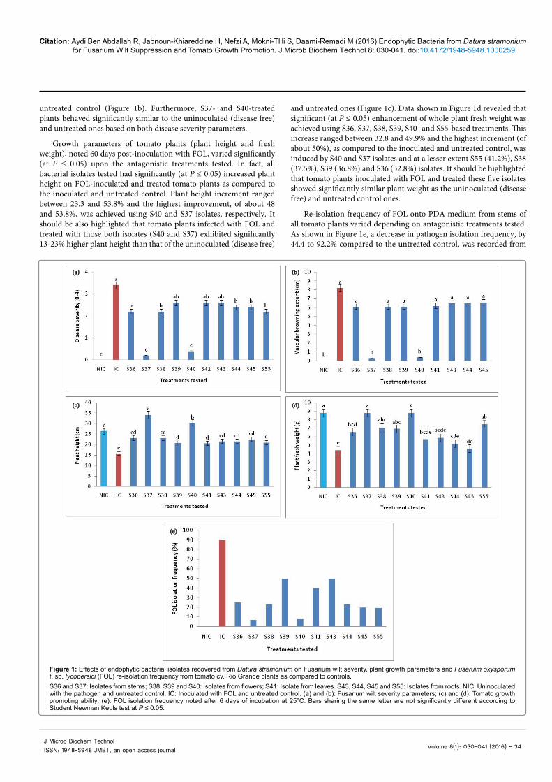
ANOVA analysis revealed that Fusarium wilt severity, noted on tomato plants 60 days post-inoculation with FOL, varied significantly (at P ≤ 0.05) depending on antagonistic treatments tested. In fact, disease severity rate varied between 0.2 and 2.6 on tomato plants challenged with FOL and treated with the endophytic isolates while the highest disease severity (3.4) was noted on FOL-inoculated and untreated control plants. A significant (at P ≤ 0.05) decrease by 29.4 to 94.1% in Fusarium wilt severity on tomato plants already challenged with FOL, as compared to the inoculated and untreated control, was achieved using the isolates S36, S37, S38, S40, S44, S45 and S55 (Figure 1a). S37 and S40 isolates were found to be the most effective in suppressing the yellowing and wilt symptoms by 94.1 and 88.2%, respectively (Figure 1a), and in reducing the vascular browning extent by 96.3 and 95.1%, respectively, as compared to the inoculated and
Citation: Aydi Ben Abdallah R, Jabnoun-Khiareddine H, Nefzi A, Mokni-Tlili S, Daami-Remadi M (2016) Endophytic Bacteria from Datura stramonium for Fusarium Wilt Suppression and Tomato Growth Promotion. J Microb Biochem Technol 8: 030-041. doi:10.4172/1948-5948.1000259
Volume 8(1): 030-041 (2016) - 34J Microb Biochem Technol ISSN: 1948-5948 JMBT, an open access journal
untreated control (Figure 1b). Furthermore, S37- and S40-treated plants behaved significantly similar to the uninoculated (disease free) and untreated ones based on both disease severity parameters.
Growth parameters of tomato plants (plant height and fresh weight), noted 60 days post-inoculation with FOL, varied significantly (at P ≤ 0.05) upon the antagonistic treatments tested. In fact, all bacterial isolates tested had significantly (at P ≤ 0.05) increased plant height on FOL-inoculated and treated tomato plants as compared to the inoculated and untreated control. Plant height increment ranged between 23.3 and 53.8% and the highest improvement, of about 48 and 53.8%, was achieved using S40 and S37 isolates, respectively. It should be also highlighted that tomato plants infected with FOL and treated with those both isolates (S40 and S37) exhibited significantly 13-23% higher plant height than that of the uninoculated (disease free)
and untreated ones (Figure 1c). Data shown in Figure 1d revealed that significant (at P ≤ 0.05) enhancement of whole plant fresh weight was achieved using S36, S37, S38, S39, S40- and S55-based treatments. This increase ranged between 32.8 and 49.9% and the highest increment (of about 50%), as compared to the inoculated and untreated control, was induced by S40 and S37 isolates and at a lesser extent S55 (41.2%), S38 (37.5%), S39 (36.8%) and S36 (32.8%) isolates. It should be highlighted that tomato plants inoculated with FOL and treated these five isolates showed significantly similar plant weight as the uninoculated (disease free) and untreated control ones.
Re-isolation frequency of FOL onto PDA medium from stems of all tomato plants varied depending on antagonistic treatments tested. As shown in Figure 1e, a decrease in pathogen isolation frequency, by 44.4 to 92.2% compared to the untreated control, was recorded from
(a) (b)
(c) (d)
(e)
S36 and S37: Isolates from stems; S38, S39 and S40: Isolates from flowers; S41: Isolate from leaves. S43, S44, S45 and S55: Isolates from roots. NIC: Uninoculated with the pathogen and untreated control. IC: Inoculated with FOL and untreated control. (a) and (b): Fusarium wilt severity parameters; (c) and (d): Tomato growth promoting ability; (e): FOL isolation frequency noted after 6 days of incubation at 25°C. Bars sharing the same letter are not significantly different according to Student Newman Keuls test at P ≤ 0.05.
Figure 1: Effects of endophytic bacterial isolates recovered from Datura stramonium on Fusarium wilt severity, plant growth parameters and Fusaruim oxysporum f. sp. lycopersici (FOL) re-isolation frequency from tomato cv. Rio Grande plants as compared to controls.
Citation: Aydi Ben Abdallah R, Jabnoun-Khiareddine H, Nefzi A, Mokni-Tlili S, Daami-Remadi M (2016) Endophytic Bacteria from Datura stramonium for Fusarium Wilt Suppression and Tomato Growth Promotion. J Microb Biochem Technol 8: 030-041. doi:10.4172/1948-5948.1000259
Volume 8(1): 030-041 (2016) - 35J Microb Biochem Technol ISSN: 1948-5948 JMBT, an open access journal
tomato plants already infected with FOL and treated with all isolates tested. The highest decrease in re-isolation frequency, of about 91.1 and 92.2%, was achieved using S40 and S37 isolates, respectively.
Correlation between Fusarium wilt severity and plant growth parameters
Pearson’s correlation analysis revealed that plant height was significantly and negatively related to the leaf damage rate (r=-0.907; P=4.7687 E-5) and the vascular browning extent (r=-0.896; P=8.0266 E-5) indicating that increased Fusarium wilt severity led to plant stunting. Also, the plant fresh weight was significantly and negatively related to Fusarium wilt severity scores (r=-0.875; P=1.9301 E-4) and the vascular browning extent (r=-0.857; P=3.6853 E-4).
Similar trend was noted between FOL re-isolation frequency and plant growth parameters where significant and negative correlations were also detected between FOL re-isolation frequency, plant height (r=-0.767; P=0.0036), and whole plant fresh weight (r=-0.638; P=0.0143). Pathogen isolation frequency was positively related to Fusarium wilt scores (r=0.800; P=0.0017) and the vascular browning extent (r=0.702; P=0.0109).
This analysis indicated that the reduced Fusarium wilt severity, recorded on tomato plants and achieved using endophytic bacteria-based treatments, was related to the decrease in FOL colonization potential leading consequently to the registered plant growth promotion.
Plant growth-promoting ability of the endophytic bacterial isolates
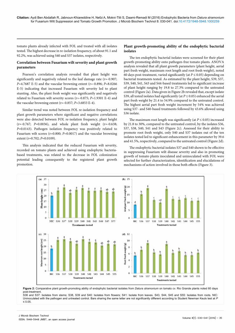
The ten endophytic bacterial isolates were screened for their plant growth-promoting ability onto pathogen-free tomato plants. ANOVA analysis revealed that all plant growth parameters (plant height, aerial part fresh weight, maximum root length and root fresh weight), noted 60 days post-treatment, varied significantly (at P ≤ 0.05) depending on bacterial treatments tested. As estimated by the plant height, S39, S37, S39, S40, S41, S43 and S44-based treatments led to significant increase of plant height ranging by 19.8 to 27.3% compared to the untreated control (Figure 2a). Data given in Figure 2b revealed that, except isolate S39, all tested isolates had significantly (at P ≤ 0.05) enhanced the aerial part fresh weight by 21.4 to 54.9% compared to the untreated control. The highest aerial part fresh weight increment by 54% was achieved using S37- and S40-based treatments followed by 43.6% allowed using S36 isolate.
The maximum root length was significantly (at P ≤ 0.05) increased by 21.8 to 30%, compared to the untreated control, by the isolates S36, S37, S38, S40, S41 and S43 (Figure 2c). Assessed for their ability to promote root fresh weight, only S40 and S37 isolates out of the ten isolates tested led to significant enhancement in this parameter by 39.6 and 41.5%, respectively, compared to the untreated control (Figure 2d).
The endophytic bacterial isolates S37 and S40 shown to be effective in suppressing Fusarium wilt disease severity and also in promoting growth of tomato plants inoculated and uninoculated with FOL were selected for further characterization, identification and elucidations of mechanisms of action involved in those both effects (Figure 3).
Figure 2: Comparative plant growth-promoting ability of endophytic bacterial isolates from Datura stramonium on tomato cv. Rio Grande plants noted 60 days post-treatment. S36 and S37: Isolates from stems; S38, S39 and S40: Isolates from flowers; S41: Isolate from leaves. S43, S44, S45 and S55: Isolates from roots. NIC: Uninoculated with the pathogen and untreated control. Bars sharing the same letter are not significantly different according to Student Newman Keuls test at P ≤ 0.05.

Citation: Aydi Ben Abdallah R, Jabnoun-Khiareddine H, Nefzi A, Mokni-Tlili S, Daami-Remadi M (2016) Endophytic Bacteria from Datura stramonium for Fusarium Wilt Suppression and Tomato Growth Promotion. J Microb Biochem Technol 8: 030-041. doi:10.4172/1948-5948.1000259
Volume 8(1): 030-041 (2016) - 36J Microb Biochem Technol ISSN: 1948-5948 JMBT, an open access journal
Characterization and identification of the two bioactive bacterial isolates
Two bioactive bacterial isolates S37 and S40 were morphologically and biochemically characterized and identified using 16S rDNA sequencing gene. In fact, the colony morphology of the isolate S37 showed a circular form with an entire margin and raised elevation, rough surface and cream color on NA medium. Microscopically, S37 cells were rod-shaped and motile. S37 was a Gram negative strain. This culturable isolate was able to produce catalase, nitrate reductase, indole by tryptophanase and hydrogen sulfide. Simmons citrate and mannitol were used by S37 colonies as a carbon source. The isolate S37 cannot produce urease, lechitinase, lysine decarboxylase, tryptophane desaminase and pyocyanin on King A medium. S37 cannot ferment glucose through the mixed acid (MR-) but by using the glycol butylene path (VP +). Blast analysis of sequenced 16S rDNA gene homology and the phylogenetic analysis based on neighbor joining (NJ) method with 1000 bootstrap sampling revealed that the isolate S37 belonged with 100% of similarity to Stenotrophomonas maltophilia strain RPS 4 (Table 2 and Figure 4). The accession number of S. maltophilia str. S18 was KR818085 (Table 2).
Macroscopically, S40 colonies showed an irregular form with a curly margin and a humped elevation, rough surface and white color on NA medium. Microscopically, S40 isolate was rod-shaped and motile bacteria. S40 was a Gram positive strain. This culturable isolate was able to produce the catalase, indole by tryptophanase, nitrate reductase, and tryptophane desaminase. S40 used simmons citrate and mannitol as carbon sources. The isolate S40 cannot ferment glucose through the mixed acid (MR-) but by using the glycol butylene path (VP +). S40 cannot produce urease, hydrogen sulfide, lechitinase, lysine decarboxylase, and pyocyanin on King A medium. Blast analysis of sequenced 16S rDNA gene homology and the phylogenetic analysis based on NJ method with 1000 bootstrap sampling revealed that the isolate S40 belonged with 99% of similarity to Bacillus mojavensis strain CEN2 (Table 2 and Figure 4). The accession number for the partial 16S rDNA gene of B. mojavensis str. S40 was KR818086 (Table 2).
Antifungal activity of Stenotrophomonas maltophilia str. S37 and Bacillus mojavensis str. S40 toward Fusarium oxysporum f. sp. lycopersici
ANOVA revealed a significant (at P ≤ 0.05) variation in the colony diameter of FOL depending on antagonistic treatments tested. In fact,
as shown in Table 3, the two endophytic bacteria, tested using the streak method, induced a significant decrease in the mycelial growth of FOL, noted after 4 days of incubation at 25°C as compared to the untreated control. Pathogen growth was inhibited by 43.8 and 39% with S. maltophilia str. S37 and B. mojavensis str. S40, respectively. Furthermore, tested using the disc diffusion method on PDA medium, S37 and S40 isolates formed an inhibition zone against FOL of about 11.37 to 12.12 mm, respectively (Table 3 and Figure 5). This indicates their ability to produce diffusible bioactive substances with antifungal potential against FOL.
Enzymatic activities of Stenotrophomonas maltophilia str. S37 and Bacillus mojavensis str. S40
Tested on agar medium supplemented with chitin, S37 and S40 isolates led to the formation of clear zones around colonies after 72 h of incubation at 28 ± 2°C. This indicates that S. maltophilia str. S37 and B. mojavensis str. S40 are chitinase-producing strains.
Transferred on milk agar medium, clear zones were formed around S. maltophilia str. S37 colonies (21.67 mm in diameter) and B. mojavensis str. S40 colonies (25.67 mm in diameter) incubated for 48 h at 28 ± 2°C indicating that they are protease-producing strains.
The two endophytic isolates cultured onto pectin agar medium formed clear zones around colonies after 48 h of incubation at 28 ± 2°C. Thus, S. maltophilia str. S37 and B. mojavensis str. S40 were found to be pectinase-producing strains.
HCN production potential
Only the isolate S. maltophilia str. S37 induced a modification on the filter paper color (light-reddish color) as compared to the untreated control (yellow) after 4 days of incubation at 25°C. Thus, this isolate was able to produce HCN on NA medium amended with glycin while B. mojavensis str. S40 did not.
Plant growth-promoting abilities of Stenotrophomonas maltophilia str. S37 and Bacillus mojavensis str. S40
Results shown in Figure 6 indicate the significant ability of the selected endophytic isolates, B. mojavensis str. S40 and S. maltophilia str. S37, to produce the indole-3-acetic acid (IAA) involved in tomato growth stimulation after 48 h of incubation. However, after 24 h of incubation, only S. maltophilia str. S37 was able to produce significantly higher IAA amount as compared to B. mojavensis str. S40. In fact, this
Figure 3: Effect of endophytic bacteria (S37 and S40 isolates) from Datura stramonium on Fusarium wilt severity and growth promotion of tomato cv. Rio Grande plants compared to the untreated controls. NIC: Uninoculated with the pathogen and untreated control. IC: Inoculated with FOL and untreated control. S37: Isolate from stems. S40: Isolate from flowers.

Citation: Aydi Ben Abdallah R, Jabnoun-Khiareddine H, Nefzi A, Mokni-Tlili S, Daami-Remadi M (2016) Endophytic Bacteria from Datura stramonium for Fusarium Wilt Suppression and Tomato Growth Promotion. J Microb Biochem Technol 8: 030-041. doi:10.4172/1948-5948.1000259
Volume 8(1): 030-041 (2016) - 37J Microb Biochem Technol ISSN: 1948-5948 JMBT, an open access journal
production of IAA, recorded at 28 ± 2°C, raised from 1.58-3.64 µg/mL after 24 h of incubation to 21.42-21.9 µg/mL after 48 h.
The two bioactive isolates S40 and S37 were also able to solubilize the phosphate as indicated by the formation of clear zones of about 13.50 and 13.83 mm in diameter around colonies, respectively.
Data given in Figure 7 indicated that only S. maltophilia str. S37 exhibited a significant pectinase activity upon the period of incubation compared to the control. In fact, S. maltophilia str. S37 pectinase activity increased from 2.7 U/mL at after 24 h of incubation to 19.56 U/
mL after 72 h whereas that of B. mojavensis str. S40 yielded 2.94 U/mL only after 72 h of incubation.
DiscussionBiological control of Fusarium wilt of tomato using epiphytic
bacteria has been extensively investigated [3,38,39]. In previous studies, endophytic bacteria were used for the control of vascular pathogens such as F. oxysporum f. sp. vasinfectum on cotton plants [21] and Verticillium dahliae on oilseed rape [40], eggplants and potato plants [41]. However, the present investigation focused on potential use of
Bacillus_subtilis_Gy11
S40
Bacillus_mojavensis_CEN2
Bacillus_licheniformis_262ZY2
Brevibacterium_halotolerans_U2
S37
Stenotrophomonas_maltophilia_R
Uncultured_bacterium_clone_Y1
Gamma_proteobacterium_JAUIB930.02
Figure 4: Neighbor-joining phylogenetic tree of partial 16S rDNA sequences of the two bioactive endophytic bacterial isolates from Datura stramonium and their closest phylogenetic relatives. The nucleotide sequences used of representative strains were obtained from Genbank database under the following accession numbers: JQ308605 (Stenotrophomonas maltophilia strain RPS 4), DQ983424 (Gamma proteobacterium strain JAUIB93), KJ736837 (Uncultured bacterium clone Y1), KF731834 (Bacillus mojavensis strain CEN2), KT345642 (Brevibacterium halotolerans strain U277), KF831384 (Bacillus licheniformis strain 262ZY2), KP876486 (Bacillus subtilis strain Gy11) and for the bacterial isolates tested: KR818085 (S37) and KR818086 (S40). The tree topology was constructed using ClustalX (1.81).
Bacterial treatments Colony diameter (cm) and growth inhibition of FOL (%) Inhibition zone (mm)Untreated control 3.71 a 0 b
Stenotrophomonas maltophilia str. S37 ( KR818085) 2.08 b (43.8) 11.37 aBacillus mojavensis str. S40 ( KR818086) 2.26 b (39) 12.12 a
Table 3: Antifungal activity of Stenotrophomonas maltophilia str. S37 and Bacillus mojavensis str. S40 toward Fusarium oxysporum f. sp. lycopersici noted after 4 days of incubation at 25°C.For each column, values followed by the same letter are not significantly different according to Student Newman Keuls test at P ≤ 0.05. Values in parenthesis indicate the percentage (in %) of the mycelial growth inhibition of Fusarium oxysporum f. sp. lycopersici as compared to the untreated control.
Figure 5: Inhibition on the mycelial growth of Fusaruim oxysporum f. sp. lycopercisi by Stenotrophomonas maltophilia str. S37 (KR818085) and Bacillus mojavensis str. S40 (KR818086) noted after 4 days of incubation at 25°C on Potato Dextrose Agar medium as compared to the untreated controls. (a): Streak method; (b): Disc diffusion method.
Phylogenetic group Isolate Accession number Most related species Sequence homology (%)Xanthomonadaceae S37 KR818085 RPS 4, Stenotrophomonas maltophilia 100
Bacillaceae S40 KR818086 CEN2, Bacillus mojavensis 99
Table 2: Identification of the bioactive endophytic bacterial isolates recovered from Datura stramonium by 16S rDNA sequencing genes.

Citation: Aydi Ben Abdallah R, Jabnoun-Khiareddine H, Nefzi A, Mokni-Tlili S, Daami-Remadi M (2016) Endophytic Bacteria from Datura stramonium for Fusarium Wilt Suppression and Tomato Growth Promotion. J Microb Biochem Technol 8: 030-041. doi:10.4172/1948-5948.1000259
Volume 8(1): 030-041 (2016) - 38J Microb Biochem Technol ISSN: 1948-5948 JMBT, an open access journal
endophytic bacteria for controlling tomato Fusarium wilt. The native wild Solanaceae D. stramonium from Tunisia (Chott-Mariem, Sousse) was used for the isolation of endophytic bacteria. Aqueous extracts [42], methanol extracts [43], petroleum ether, hexane, chloroform and ethanol extracts [15] from D. stramonium were extensively exploited for their antifungal activity against plant pathogenic fungi. However, few data were available on D. stramonium use as potent source of isolation of fungal [21,44] and bacterial biocontrol agents [11].
The main objective of this work was on the assessment of the ability of D. stramonium endophytic bacteria to suppress Fusarium wilt and to improve plant growth. Ten non-pathogenic bacteria were collected and artificially transformed onto NA media amended with streptomycin and
rifampicin for obtaining double-resistant mutants. Their endophytic progress within tomato stems was confirmed according to Chen et al. [21] procedure using streptomycin- and rifampicin- resistant mutants.
The ten bacterial isolates recovered from D. stramonium and exhibiting endophytic behavior on tomato plants, were assessed for their ability to control Fusarium wilt. The results from the current study clearly demonstrated the strong inhibitory effect of disease severity displayed by S37 and S40 isolates, originally recovered from D. stramonium stems and flowers, leading to more than 88% decrease in leaf damage index and 95% in vascular browning extent, respectively. This result is in agreement with previous findings reporting tomato Fusarium wilt severity decrease by at least 75% induced by two
ba b b
a a
05
10152025
Control S37 S40 Control S37 S40
24 48
Indo
le-3
-ace
tic a
cid
(µg/
ml)
Bacterial treatments / Incubation period (h)Figure 6: Indole-3-acetic acid production by the bioactive endophytic bacterial recovered from Datura stramonium and noted after 24 and 48 h of incubation at 28 ± 2°C in Luria-Broth medium. S37: Stenotrophomonas maltophilia str. S37 (KR818085) isolated from stems; S40: Bacillus mojavensis str. S40 (KR818086) isolated from flowers. For each incubation period, bars sharing the same letter are not significantly different according to Student Newman Keuls test at P ≤ 0.05.
ba
ab b
a
b b
a
b
0
5
10
15
20
25
Control S37 S40 Control S37 S40 Control S37 S40
24 48 72
Pect
inas
e ac
tivity
(U/m
l)
Bacterial treatments / Incubation period (h)Figure 7: Pectinase activity of bioactive endophytic bacterial isolates recovered from Datura stramonium noted after 24, 48 and 72 h of incubation at 28 ± 2°C in minimum medium broth amended with pectin (1%) (w/v) and under continuous shaking at 150 rpm. S37: Stenotrophomonas maltophilia str. S37 (KR818085) isolated from stems; S40: Bacillus mojavensis str. S40 (KR818086) isolated from flowers. For each incubation period, bars sharing the same letter are not significantly different according to Student Newman Keuls test at P ≤ 0.05.
Citation: Aydi Ben Abdallah R, Jabnoun-Khiareddine H, Nefzi A, Mokni-Tlili S, Daami-Remadi M (2016) Endophytic Bacteria from Datura stramonium for Fusarium Wilt Suppression and Tomato Growth Promotion. J Microb Biochem Technol 8: 030-041. doi:10.4172/1948-5948.1000259
Volume 8(1): 030-041 (2016) - 39J Microb Biochem Technol ISSN: 1948-5948 JMBT, an open access journal
unidentified endophytic isolates PA and PF, issued from healthy wild and cultivated oilseed rape plants [24]. Native S37 and S40 isolates were also able to enhance plant growth on tomato plants inoculated or not with FOL. Furthermore, Fusarium wilt severity decrease was shown to be related to the registered increment in all plant growth parameters. In fact, as reported in previous studies, endophytic bacteria may exhibit antifungal potential directly toward fungal pathogens and may act indirectly through the promotion of plant growth [45]. Similar growth enhancements were also achieved using B. mojavensis for treatment of maize plants grown in presence of F. verticillioides pathogenic isolates [6]. PGPB potential was also investigated by using endophytic B. amyloliquefaciens, isolated from soybean roots, leading to enhanced soybean yield [46].
The two endophytic isolates found to be bioactive in suppressing disease and in improving growth were identified by 16S rDNA sequencing as Stenotrophomonas maltophilia str. S37 (KR818085) and B. mojavensis str. S40 (KR818086). It should be mentioned that the genus Bacillus was predominant in the associated phyllosphere bacterial community recovered from healthy N. glauca plants [17] where B. endophyticus, B. megaterium, B. niacini, B. simplex and B. stratosphericus were frequently isolated. Culturable Bacillus spp. isolated from wild Solanaceae such as S. trilobatum [16], S. melongena and S. torvum [47] were not tested against the causative agent of tomato Fusarium wilt, among the pathogenic fungi used, nor for their plant growth promoting ability onto tomato plants. However, B. brevis str. B2 and B. subtilis (strs. B5, B7 and B8), issued from tomato rhizosphere, were previously used as plant growth-promoting agents in tomato plants [48]. Hence, to our knowledge, this is the first report of endophytic B. mojavensis recovered from D. stramonium flowers and exhibiting both antifungal activity and growth-promoting potential onto tomato seedlings.
The second bioactive endophytic bacterial species associated to D. stramonium stems was identified in the present study as S. maltophilia. This species has been detected in coffee seeds and has been successfully used for the control of leaf spot (Bipolaris sorokininana) on tall fescue [49], and has also been reported in potatoes [50]. Endophytic S. maltophilia strains are also isolated from cotton roots and stems [51] and are also recovered from rice roots among other bacteria belonging to the genera Achromobacter, Burkholderia, Delftia, Enterobacter, Pseudomonas, Planomicrobium, Methylobacterium, Acinetobacter, and Pantoea [52]. To our knowledge, this is the first report of endophytic S. maltophilia str. S37 from D. stramonium stems acting as FOL disease-suppressive and as tomato growth-promoting agent.
In the present study, S. maltophilia str. S37 and B. mojavensis str. S40, evaluated in vitro for their antagonistic potential toward FOL, had reduced pathogen mycelial growth and formed an inhibition zone. Similar inhibitory effects are reported by Nandhini et al. [53] and Patel et al. [27] using endophytic Pseudomonas aeruginosa str. HR7, Pseudomonas sp. str. TEP3, Bacillus sp. str. TEB6, Klebsiella sp. str. TEK1 and Citrobacter sp. issued from cultivated tomato for F. oxysporum biocontrol. According to Bacon and Hinton [54], endophytic B. mojavensis strains are able to reduce growth of F. moniliforme with or without formation of an inhibition zone depending on strains but strong fungal mycelium lysis is frequently observed in the confrontation zone. Antimicrobial substances produced by S. maltophilia str. UPMKB9 could inhibit spore germination of F. oxysporum and Colletotrichum gloeosporioides and affect their hyphal morphology [55].
The mechanism of action deployed in vitro by these endophytic
bacteria seemed to be mainly via allelochemicals. In fact, S. maltophilia str. S37 and B. mojavensis str. S40 were found to be chitinase-, protease- and pectinase-producing strains as shown on chitin agar medium, milk agar medium and pectin agar medium, respectively. Therefore, the mechanism of action displayed by these both bioactive strains toward FOL and elucidated in the current study is through production of hydrolytic enzymes. S. maltophilia plays important roles in agricultural production as a plant growth-promoting bacterium, which could suppress disease development by secretion of antibiotics such as maltophilin, xanthobaccin which have antifungal activity, but inactive against bacteria [56,57]. Also, ability of production of extracellular enzymes such as protease [58] and chitinase [49], and potential root colonization [58] are also reported. Endophytic Bacillus spp. (B. mojavensis, B. subtilis, B. pumilis, B. amyloliquefaciens) are commonly known as lipopeptide antibiotics [59] and/or hydrolytic enzymes producing strains [6]. Furthermore, the production of HCN is involved in the effective inhibition of pathogen. Our strain S. maltophilia str. S37 from D. stramonium was potentially able to produce HCN in NA amended with glycin. However, S. maltophilia TEM56 and PM22 strains isolated from Amaranthus hybridusand and Cucurbita maxima, respectively were negative for HCN production [60].
S. maltophilia str. S37 and B. mojavensis str. S40, selected in the current study, were also valued for their plant growth-promoting ability through production of indole-3-acetic acid (IAA), phosphate solubilization and pectinase activity. In fact, IAA production ability of S. maltophilia str. S37 and B. mojavensis str. S40 from D. stramonium, of about 21 µg/mL, seemed to be higher than that reported for B. thuringiensis (4 mg/L), B. arbutinivorans (5 mg/L), and B. subtilis (5 mg/L) but lesser than that produced by B. megaterium (>50 mg/L) [45]. Also, this IAA amount (21 µg/mL) is higher than that secreted by the endophytic S. maltophilia str. TEM56 isolated from Amaranthus hybridus and S. maltophilia str. PM22 isolated from Cucurbita maxima, 0.32 and 0.49 mg/L, respectively [60].
Among mechanisms involved in plant growth promotion, enhanced mineral uptake through solubilization of bound soil iron and phosphorus and plant supply with siderophores and nitrogen are widely reported [61]. In the current investigation, phosphate solubilization ability was detected, based on Pikovskaya agar test, for S. maltophilia str. S37 and B. mojavensis str. S40. Similar findings were reported for endophytic several B. velezensis, B. mojavensis, and B. methylotrophicus strains [6] but B. subtilis subsp. subtilis str. M9V1r did not exhibit phosphatase activity [61]. The endophytic S. maltophilia str. PM22 is able to produce phosphatase but S. maltophilia str. TEM56 fail to solubilize phosphate [60]. The phosphatase activity displayed by S. maltophilia str. S37 and B. mojavensis str. S40, expressed by the formation of a clear zone of about 13 mm around colonies in Pikovskaya medium, was also higher than that reported for unidentified endophytic bacterial isolates HR11, HR12 and HR18 where clear zone diameters vary between 8 to 11 mm but less than those induced by HR7, HR9, HR1, HR3, HR17 isolates showing diameters ranging from 17 to 31 mm [27].
Both selected strains S. maltophilia str. S37 and B. mojavensis str. S40, exhibiting endophytic colonization ability, were found to be pectinase-producing strains and only S. maltophilia str. S37 displayed a significant pectinase activity upon the period of incubation compared to the control. In fact, hydrolytic enzymes such as pectinases and cellulases may play a role in the mechanisms by which endophytic bacteria penetrate into and persist in the host plant as reported by

Citation: Aydi Ben Abdallah R, Jabnoun-Khiareddine H, Nefzi A, Mokni-Tlili S, Daami-Remadi M (2016) Endophytic Bacteria from Datura stramonium for Fusarium Wilt Suppression and Tomato Growth Promotion. J Microb Biochem Technol 8: 030-041. doi:10.4172/1948-5948.1000259
Volume 8(1): 030-041 (2016) - 40J Microb Biochem Technol ISSN: 1948-5948 JMBT, an open access journal
Hallmann et al. [8]. It should be also mentioned that these enzymes act normally as virulence factors for plant pathogenic bacteria but in case of endophytic microorganisms, they might play a role in invasion of host plants by endophytes as demonstrated for S. maltophilia [50], B. cereus, B. subtilis and B. stearothermophilus [62].
ConclusionThe screening of the endophytic bacteria isolated from surface-
sterilized tissues of D. stramonium with antifungal potential towards FOL led to the selection of two promising biocontrol agents useful for Fusarium wilt control and enhancement of tomato growth. Their benefits as bioferlizer were clearly demonstrated onto uninoculated and inoculated tomato plants with the pathogen. Hence, D. stramonium was firstly reported in the current study as a potential source of potent endophytic bacteria, acting both as biocontrol agents and as biofertilizers, and identified as S. maltophilia str. S37 (KR818085) and B. mojavensis str. S40 (KR818086).
Testing the antifungal activity of their cell-free cultures and organic extracts in vivo may give additional information on their effects on Fusarium wilt suppressive ability and tomato growth stimulation.
References
1. Olaniyi JO, Akanbi WB, Adejumo TA, Akande OG (2010) Growth, fruit yield and nutritional quality of tomato varieties. Afr J Food Sci 4: 398-402.
2. McGovern RJ (2015) Management of tomato diseases caused by Fusarium oxysporum. Crop Prot 73: 78-92.
3. Moretti M, Gilardi G, Gullino ML, Garibaldi A (2008) Biological control potential of Achromobacter xylosoxydans for suppressing Fusarium wilt of tomato. Int J Bot 4: 369-375.
4. Vethavalli S, Sudha SS (2012) In vitro and in silico studies on biocontrol agent of bacterial strains against Fusarium oxysporum f. sp. lycopersici. Res Biotechnol 3: 22-31.
5. Reis A, Giordano LB, Lopes CA, Boiteux LS (2004) Novel sources of multiple resistances to three races of Fusarium oxysporum f. sp. lycopersici in Lycopersicon germplasm. Crop Breed Appl Biotechnol 4: 495-502.
6. Kalai-Grami L, Saidi S, Bachkouel S, Ben Slimene I, Mnari-Hattab M, et al. (2014) Isolation and characterization of putative endophytic bacteria antagonistic to Phoma tracheiphila and Verticillium albo-atrum. Appl Biochem Biotechnol 174: 365-375.
7. Mahdi T, Mohamed I, Yagi S (2014) Endophytic fungal communities associated with ethno-medicinal plants from Sudan and their antimicrobial and antioxidant prospective. J. Forest Products and Industries 3: 248-256.
8. Hallmann J, Quadt-Hallmann A, Mahaffee WF, Kloepper JW (1997) Bacterial endophytes in agricultural crops. Can J Microbiol 43: 895-914.
9. Berg G, Hallmann J (2006) Control of plant pathogenic fungi with bacterial endophytes: Microbial root endophytes, E-Publishing Springer, Berlin.
10. Gaiero JR, McCall CA, Thompson KA, Day NJ, Best AS, et al. (2013) Inside the root microbiome: bacterial root endophytes and plant growth promotion. Am J Bot 100: 1738-1750.
11. Nimal Christhudas IVS, Praveen Kumar P, Agastian P (2012) Antimicrobial activity and HPLC analysis of tropane alkaloids in Streptomyces spp. isolated from Datura stramonium L. Asian Journal of Pharmaceutical and Clinical Research 5: 278-282.
12. Soni P, Siddiqui AA, Dwivedi J, Soni V (2012) Pharmacological properties of Datura stramonium L. as a potential medicinal tree: an overview. Asian Pac J Trop Biomed 2: 1002-1008.
13. Gaire BP, Subedi L (2013) A review on the pharmacological and toxicological aspects of Datura stramonium L. J Integr Med 11: 73-79.
14. Jalander V, Gachande BD (2012) Effect of aqueous leaf extracts of Datura sp. against two plant pathogenic fungi. International Journal of Food, Agriculture and Veterinary Sciences 2: 131-134.
15. Girmay S (2015) Preliminary phytochemical screening and in vitro antimicrobial activity of Datura stramonium leaves extracts collected from Eastern Ethiopia. Int Res J Biol Sci 4: 55-59.
16. Bhuvaneswari S, Madhavan S, Panneerselvam A (2013) Enumertion of endophytic bacteria from Solanum trilobatum L. World J Pharm Res 3: 2270-2279.
17. Izhaki I, Fridman S, Gerchman Y, Halpern M (2013) Variability of bacterial community composition on leaves between and within plant species. Curr Microbiol 66: 227-235.
18. Lugtenberg B, Malfanova N, Kamilova F, Berg G (2013) Microbial control of plant diseases: Molecular Microbial Ecology of the Rhizosphere, Wiley-Blackwell.
19. Malfanova N, Lugtenberg B, Berg G (2013) Bacterial endophytes: who and where, and what are they doing there? Molecular Microbial Ecology of the Rhizosphere, Wiley-Blackwell.
20. Barker SJ, Edmonds-Tibbett TL, Forsyth LM, Klingler JP, Toussaint JP, et al. (2005) Root infection of the reduced mycorrhizal colonization (rmc) mutant of tomato reveals genetic interaction between symbiosis and parasitism. Physiol Mol Plant Pathol 67: 277-283.
21. Chen C, Bauske EM, Musson G, Rodríguez-Kábana R, Kloepper JW (1995) Biological control of Fusarium wilt on cotton by use of endophytic bacteria. Biol Control 5: 83-91.
22. Fakhouri W, Buchenauer H (2002) Characteristics of fluorescent pseudomonas isolates towards controlling of tomato wilt caused by Fusarium oxysporum f. sp. lycopersici. J Plant Dis Prot 110: 143-156.
23. Nawangsih AA, Damayanti I, Wiyono S, Kartika JG (2011) Selection and characterization of endophytic bacteria as biocontrol agents of tomato bacterial wilt disease. J Biosci 18: 66-70.
24. Nejad P, Johnson PA (2000) Endophytic bacteria induce growth promotion and wilt disease suppression in oilseed rape and tomato. Biol Control 18: 208-215.
25. Amini J (2009) Physiological race of Fusarium oxysporum f. sp. lycopersici in Kurdistan province of Iran and reaction of some tomato cultivars to race 1 of pathogen. Plant Pathol J 8: 68-73.
26. Botta AL, Santacecilia A, Ercole C, Cacchio P, Del Gallo M (2013) In vitro and in vivo inoculation of four endophytic bacteria on Lycopersicon esculentum. N Biotechnol 30: 666-674.
27. Patel HA, Patel RK, Khristi SK, Parikh K, Rajendran G (2012) Isolation and characterization of bacterial endophytes from Lycopersicon esculentum plant and their plant growth promoting characteristics. Nepal J Biotechnol 2: 37-52.
28. Schaad NW, Jones JB, Chun W (2001) Laboratory guide for identification of plant pathogenic bacteria. (3 rdedn), St. Paul, USA.
29. Chen WP, Kuo TT (1993) A simple and rapid method for the preparation of gram-negative bacterial genomic DNA. Nucleic Acids Res 21: 2260.
30. van Soolingen D, de Haas PE, Hermans PW, van Embden JD (1994) DNA fingerprinting of Mycobacterium tuberculosis. Methods Enzymol 235: 196-205.
31. Sadfi N, Chérif M, Fliss I, Boudabbous A, Antoun H (2001) Evaluation of bacterial isolates from salty soils and Bacillus thuringiensis strains for the biocontrol of Fusarium dry rot of potato tubers. J Plant Pathol 83: 101-118.
32. Tiru M, Muleta D, Bercha G, Adugna G (2013) Antagonistic effect of rhizobacteria against coffee wilts disease caused by Gibberella xylarioides. Asian J Plant Pathol 7: 109-122.
33. Miller GL (1959) Use of dinitrosalicyIic acid reagent for determination of reducing sugar. Anal Chem 131: 426-428.
34. Mokni-Tlili S, Belguith H, Hassen A, Gargouri A (2011) Studies on the ecology of actinomycetes in an agricultural soil amended with organics residues: II. Assessment of enzymatic activities of Actinomycetales isolates. World J Microbiol Biotechnol 27: 2251-2259.
35. Lorck H (1948) Production of hydrocyanic acid by bacteria. Physiol Plant 1: 142-146.
36. KATZNELSON H, BOSE B (1959) Metabolic activity and phosphate-dissolving capability of bacterial isolates from wheat roots, rhizosphere, and non-rhizosphere soil. Can J Microbiol 5: 79-85.
37. Glickmann E, Dessaux Y (1995) A critical examination of the specificity of
Citation: Aydi Ben Abdallah R, Jabnoun-Khiareddine H, Nefzi A, Mokni-Tlili S, Daami-Remadi M (2016) Endophytic Bacteria from Datura stramonium for Fusarium Wilt Suppression and Tomato Growth Promotion. J Microb Biochem Technol 8: 030-041. doi:10.4172/1948-5948.1000259
Volume 8(1): 030-041 (2016) - 41J Microb Biochem Technol ISSN: 1948-5948 JMBT, an open access journal
the salkowski reagent for indolic compounds produced by phytopathogenic bacteria. Appl Environ Microbiol 61: 793-796.
38. Monda EO (2002) Biological control of Fusarium wilt of tomato. J Trop Microbiol 1: 74-78.
39. Akköprü A, Demir S (2005) Biological control of Fusarium wilt in tomato caused by Fusarium oxysporum f. sp. lycopersici by AMF Glomus intraradices andsome rhizobacteria. J Phytopathol 153: 544-550.
40. Alström S (2001) Characteristics of bacteria from oilseed rape in relation totheir biocontrol activity against Verticillium dahliae. J Phytopathol 149: 57-64.
41. Eleftherios CT, Dimitrios IT, Sotirios ET, Polymnia PA, Panayiotis K (2004)Selection and screening of endorhizosphere bacteria from solarized soils asbiocontrol agents against Verticillium dahliae of Solanaceous hosts. Eur J Plant Pathol 110: 35-44.
42. Arzoo K, Biswas SK, Rajik M (2012) Biochemical evidences of defenceresponse in tomato against Fusarium wilt induced by plant extracts. PlantPathol J 11: 42-50.
43. Usha K, Singh B, Praseetha P, Deepa N, Agarwal DK, et al. (2009) Antifungalactivity of Datura stramonium, Calotropis gigantea and Azadirachta indicaagainst Fusarium mangiferae and floral malformation in mango. Eur J Plant Pathol 124: 637-657.
44. Sun J, Awakawa T, Noguchi H, Abe I (2012) Induced production of mycotoxinsin an endophytic fungus from the medicinal plant Datura stramonium L. BioorgMed Chem Lett 22: 6397-6400.
45. Wang S, Wang W, Jin Z, Du B, Ding Y, et al. (2013) Screening and diversity ofplant growth promoting endophytic bacteria from peanut. Afr J Microbiol Res7: 875-884.
46. Sharma SK, Ramesh A, Johri BN (2013) Isolation and characterization of plant growth-promoting Bacillus amyloliquefaciens Strain sks_bnj_1 and its influence on rhizosphere soil properties and nutrition of soybean (Glycine max L. Merrill). J Virol Microbiol 2013: 1-19.
47. Achari GA, Ramesh R (2014) Diversity, biocontrol, and plant growth promoting abilities of xylem residing bacteria from solanaceous crops. Int J Microbiol2014: 296521.
48. Algam SA, Guan-lin X, Coosemans J (2005) Delivery methods for introducingendophytic Bacillus into tomato and their effect on growth promotion andsuppression of tomato wilt. Plant Pathol J 4: 69-74.
49. Zhang Z, Yuen GY (1999) Biological Control of Bipolaris sorokiniana on TallFescue by Stenotrophomonas maltophilia Strain C3. Phytopathology 89: 817-822.
50. Garbeva P, Overbeek LS, Vuurde JW, Elsas JD (2001) Analysis of Endophytic Bacterial Communities of Potato by Plating and Denaturing Gradient GelElectrophoresis (DGGE) of 16S rDNA Based PCR Fragments. Microb Ecol 41: 369-383.
51. McInroy JA, Kloepper JW (1995) Population dynamics of endophytic bacteria in field-grown sweet corn and cotton. Can J Microbiol 41: 895-901.
52. Sun L, Qiu F, Zhang X, Dai X, Dong X, et al. (2008) Endophytic bacterialdiversity in rice (Oryza sativa L.) roots estimated by 16S rDNA sequence analysis. Microb Ecol 55: 415-424.
53. Nandhini S, Sendhilvel V, Babu S (2012) Endophytic bacteria from tomato and their efficacy against Fusarium oxysporum f. sp. lycopersici, the wilt pathogen.J Biopesticides 5: 178-185.
54. Bacon CW, Hinton DM (2002) Endophytic and biological control potential ofBacillus mojavensis and related species. Biol Control 2: 274-284.
55. Farhana MSN, Bivi MR, Khairulmazmi A (2011) Effect of carbon sourceson bacterial production of metabolites against Fusarium oxysporum andColletotrichum gloeosporioides. Int J Agric Biol 13: 1-8.
56. Jakobi M, Winkelmann G, Kaiser D, Kempler C, Jung G, et al. (1996)Maltophilin: a new antifungal compound produced by Stenotrophomonas maltophilia R3089. J Antibiot (Tokyo) 49: 1101-1104.
57. Nakayama T, Homma Y, Hashidoko Y, Mizutani J, Tahara S (1999) Possiblerole of xanthobaccins produced by Stenotrophomonas sp. strain SB-K88 insuppression of sugar beet damping-off disease. Appl Environ Microbiol 65:4334-4339.
58. Dunne C, Crowley JJ, Moenne-Loccoz Y, Dowling DN, de Bruijn FJ, et al. (1997) Biological control of Pythium ultimum by Stenotrophomonas maltophilia W81 is mediated by an extracellular proteolytic activity. Microbiology 143: 3921-3931.
59. Bacon CW, Hinton DM (2011) Bacillus mojavensis: Its endophytic nature,the surfactins, and their role in the plant response to infection by Fusarium verticillioides: Bacteria in agrobiology: Plant growth responses, Springer-Verlag, Berlin Heidelberg.
60. Ngoma L, Esau B, Babalola OO (2013) Isolation and characterization ofbeneficial indigenous endophytic bacteria for plant growth promoting activity in Molelwane Farm, Mafikeng, South Africa. Afr J Biotechnol 12: 4105-4114.
61. Ngamau CN, Matiru VN, Tani A, Muthuri CW (2012) Isolation and identification of endophytic bacteria of bananas (Musa spp.) in Kenya and their potential asbiofertilizers for sustainable banana production. Afr J Microbiol Res 6: 6414-6422
62. Torimiro N, Okonji RE (2013) A comparative study of pectinolytic enzymeproduction by Bacillus species. Afr J Biotechnol 12: 6498-6503.


![Green ynhei of ile nanopaicle mediated by adiionally ed ... · Cyperus rotundus Wholeplant UV,FTIR,SEM,EDX 20.5 ± 9.6 446 [52] Datura stramonium Leaf UV,FTIR,TEM,XRD 18 444 [53]](https://static.fdocuments.us/doc/165x107/608bfb2bfdf4bc75ae03d113/green-ynhei-of-ile-nanopaicle-mediated-by-adiionally-ed-cyperus-rotundus-wholeplant.jpg)

