Løysingsforslag eksamen TFY4315 2013 -...
13
TFY 4315 Biophysics of Ionizing Radiation Examination paper 04.06.2013 The examination paper and solution proposal are worked out by Anne Beate Langeland Marthinsen and Tore Lindmo Solution proposal Problem 1 a) Why does radiation with for example x-rays on a human have much higher biological effect than equal amount of energy given in form of heat or mechanical energy? The energy of x-rays absorbed in living material (humans) is deposited unevenly in discrete packets. The energy in a beam of x-rays is quantized into large individual packets, each of which is big enough to break a chemical bond (Double strand breaks in DNA) initiating a chain of events culminating in a biological change. The biological effect of radiation is determined not by the total amount of energy absorbed, but by the energy of each individual energy packet. Increasing the dose of x-rays will increase the number of high energy packets. For low doses of x-rays fewer energy packets are absorbed, but those absorbed may have enough energy to damage DNA locally. Energy in the form of heat and mechanical energy is absorbed uniformly and evenly, and much greater quantities of energy are required to produce damage in living things. Examples: Total body irradiation of x-rays with 4 Gy is lethal in 50% of individuals. The dose represents absorption of energy of about the same level as energy delivered in a sip of hot coffee, or lifting a person 0.4m. b) Define what we mean by Linear energy transfer (LET) Linear energy transfer (LET) is the energy transferred pr. unit length of the track. The LET (L) of a charged particle in medium is the quotient: L = dE/dl
Transcript of Løysingsforslag eksamen TFY4315 2013 -...

TFY 4315 Biophysics of Ionizing Radiation Examination paper 04.06.2013 The examination paper and solution proposal are worked out by Anne Beate Langeland Marthinsen and Tore Lindmo
Solution proposal
Problem 1
a) Why does radiation with for example x-rays on a human have much higher biological effect than equal amount of energy given in form of heat or mechanical energy?
The energy of x-rays absorbed in living material (humans) is deposited unevenly in discrete packets. The energy in a beam of x-rays is quantized into large individual packets, each of which is big enough to break a chemical bond (Double strand breaks in DNA) initiating a chain of events culminating in a biological change. The biological effect of radiation is determined not by the total amount of energy absorbed, but by the energy of each individual energy packet. Increasing the dose of x-rays will increase the number of high energy packets. For low doses of x-rays fewer energy packets are absorbed, but those absorbed may have enough energy to damage DNA locally. Energy in the form of heat and mechanical energy is absorbed uniformly and evenly, and much greater quantities of energy are required to produce damage in living things. Examples: Total body irradiation of x-rays with 4 Gy is lethal in 50% of individuals. The dose represents absorption of energy of about the same level as energy delivered in a sip of hot coffee, or lifting a person 0.4m. b) Define what we mean by Linear energy transfer (LET)
Linear energy transfer (LET) is the energy transferred pr. unit length of the track. The LET (L) of a charged particle in medium is the quotient:
L = dE/dl

dE = average energy locally imparted to the medium by a charged particle of specified energy in traversing a distance of dl
Unit: eV / m
c) For a given type of radiation; what happens to LET when the radiation energy increases?
The density of ionization decreases, giving lower LET, as the radiation energy increase.
Problem 2
a) An individual is exposed to radiation. Define by introducing radiation and tissue weighting factors what is ment by:
Absorbed dose
Equivalent dose
Effective dose Indicate the units used for each type of dose.
Absorbed dose = Energy absorbed pr. unit mass of tissue Physical quantity Unit: Grey (Gy) (= J/kg) Equivalent dose: The same absorbed dose of different types of radiation may have different biological effects. Adjusting the absorbed dose (for each tissue (organ) irradiated) with a Radiation weighting factor (WR) (appropriate to the type and energy of the radiation) gives the equivalent dose with comparable data for biological effects: (A radiation weighting factor is an estimate of the effectiveness per unit dose of the given radiation relative to low-LET standard). (Equivalent dose)T = ∑ RT x WR for each tissue (organ) (T) (Absorbed dose)RT = absorbed dose of radiation type R in tissue (organ) T Unit: Sievert (Sv) Effective dose = The sum of all the weighted equivalent doses in all tissues or organs irradiated:
Effective dose = ∑ ∑ RT x WR) x WT
= ∑ T x WT (Absorbed dose)RT = absorbed dose of radiation type R in tissue (organ) T

WR = Radiation weighting factor WT = Tissue weighting factor Unit: Sievert The effective dose is the whole body dose of X-rays that would have to be delivered to produce the same stochastic risk as the partial body dose that actually was delivered.
• Provides an easy assessment of overall risk • Makes comparison of risks simpler
Effective dose is in principle nonmeasurable b) What are the radiation weighting factors for
Photons
Electrons
Protons
α-particles
Neutrons
Radiation weighting factors (WR):
Photons: 1
Electrons: 1
Protons: 2
α-particles: 20
Neutrons: A continuous curve as a function of neutron energy.
:
c) In radiation therapy patients can be given total doses which are much larger than lethal doses corresponding to acute radiation syndrome (ARS). Why is that tolerated? Give estimates of total doses associated with:
curative external photon radiation therapy
acute radiation syndrome
In radiotherapy generally smaller parts of the body are irradiated (not the whole body). When fractionating of the radiation, with enough time for repair of sublethal damage between each fraction, the total dose tolerated is much higher than corresponding tolerated dose given in one fraction.
Acute radiation syndrome is associated with radiation of the whole organism.
Common total doses corresponding to curative external photon radiation therapy:
2 Gy x 25 – 35 = 50 – 70 Gy
Total doses given to the whole body associated with acute radiation syndrome (lethal): 3 – 5 Gy
Problem 3
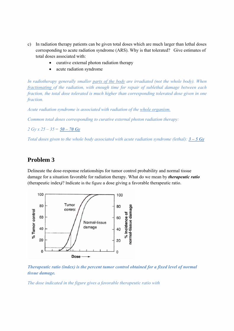
Delineate the dose-response relationships for tumor control probability and normal tissue damage for a situation favorable for radiation therapy. What do we mean by therapeutic ratio (therapeutic index)? Indicate in the figure a dose giving a favorable therapeutic ratio.
Therapeutic ratio (index) is the percent tumor control obtained for a fixed level of normal tissue damage.
The dose indicated in the figure gives a favorable therapeutic ratio with
30 % probability of tumor control
5 % incidence of normal tissue damage
How can the therapeutic ratio be manipulated to give a therapeutic gain?
Therapeutic ratio can be manipulated by:
• Time – fractionation regimens
• Radiation sensitizers
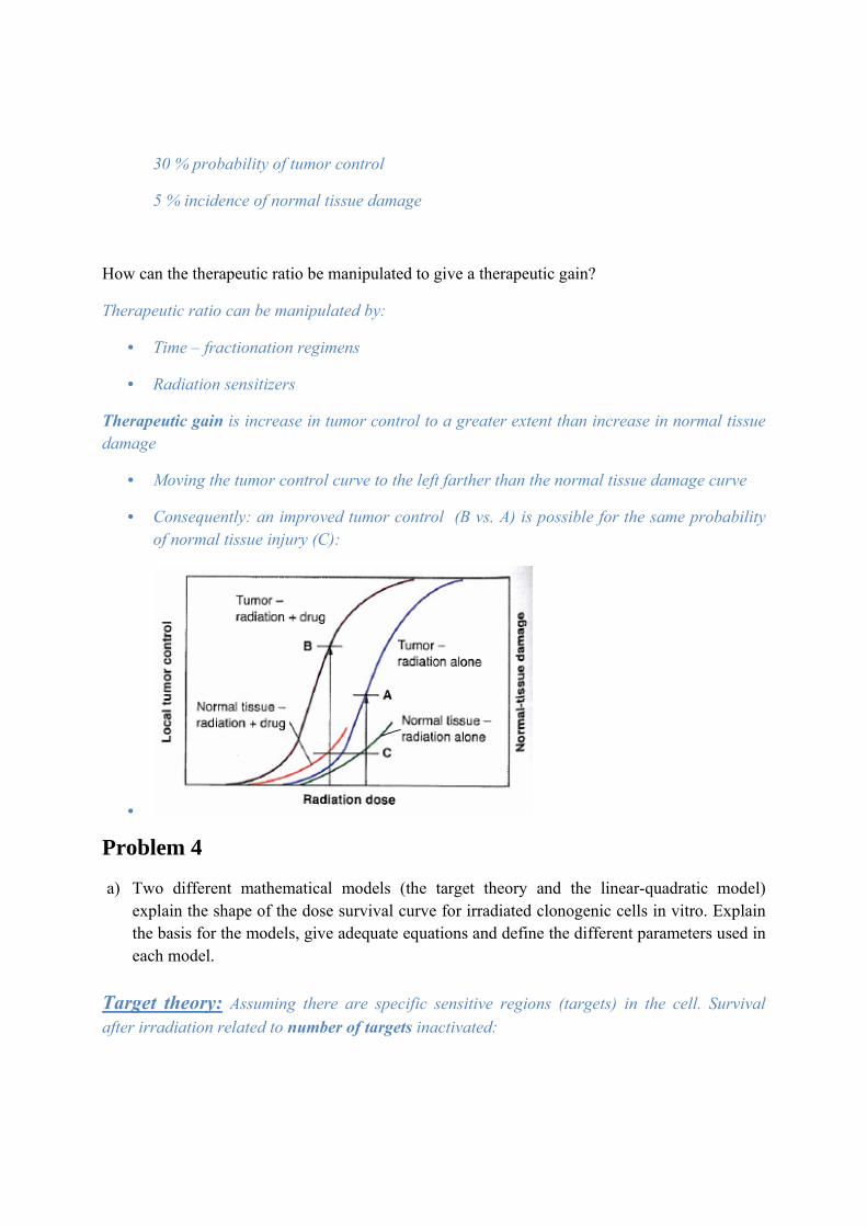
Therapeutic gain is increase in tumor control to a greater extent than increase in normal tissue damage
• Moving the tumor control curve to the left farther than the normal tissue damage curve
• Consequently: an improved tumor control (B vs. A) is possible for the same probability of normal tissue injury (C):
•
Problem 4
a) Two different mathematical models (the target theory and the linear-quadratic model) explain the shape of the dose survival curve for irradiated clonogenic cells in vitro. Explain the basis for the models, give adequate equations and define the different parameters used in each model.
Target theory: Assuming there are specific sensitive regions (targets) in the cell. Survival after irradiation related to number of targets inactivated:

• Single target – single hit inactivation:
– One hit by radiation on a single sensitive target would lead to cell death
– Exponential survival curve (straight line in semilogarithmic plot (survival vs. dose))
Poisson statistics:
P (survival) = P (0 hits) = /
P (survival) = S = survival fraction of cells
D= radiation dose
D0 = dose given on average one hit pr. target
• Multi target – single hit inactivation
– n independent targets must be inactivated (each by a single hit) for the cell to be inactivated
– Survival curve with a shoulder in semilogarithmic plots (survival vs. dose)
Poisson statistics:
P (0 hits on a specific target) = /
P (specific target inactivated) = 1- /
n targets in the cell:
P (all n targets inactivated) = (1- / )n
P (survival) = P (not all targets inactivated) = 1-(1- / )n
The equation for the tangent of the survival curve is dP/dD. The limit for large values of D is
n /
This curve crosses the D=0, at the «surviving fraction» n (see figure)
The quasithreshold dose Dq is the dose representing surviving fraction 1 on this tangent:
ln(n) = Dq/D0

T
The lin
Cell sur
S = surv
D = del
α and β
Two com
The theory c
near quad
rvival can ge
vival fractio
livered dose
are constan
mponents to
– α-co
– β-coletha
•
can be expa
dratic theo
enerally be
S =
on of cells
e
nts
o cell killing
omponent: P
omponent: Pal))
• (dual ratwo sepa
anded to mu
ory
described b
g by radiatio
Proportiona
Proportiona
adiation actarate breaks
ulti hit inacti
by:
on:
al to dose (i
al to the sq
tion – somes)
ivation
(irreparable
quare of the
e chromosom
– lethal)
e dose (rep
me aberrati
pairable – p
ions are the
potentially
e result of

•
The components of cell killing that are proportional to D and D2 are equal when:
αD = βD2 or D = α/β
The linear and quadratic contribution to cell killing are equal at a dose equal to the ratio α/β
The linear-quadratic model is most used the latest years. Why?
Problems with the target theory:
• Specific radiation targets have not been identified for mammalian cells
• Predicts flat response for low radiation (not supported by experimental data)
– The multi target model can be modified by adding an additional single target component. This type of curve predicts cell killing in the low dose region occurs almost linearly, implicating no sparing of damage should occur, which is usually not found experimentally.
– A way of overcoming this limitation is to use a multi-target component as the initial slope, - making the model very complicated (with 4 parameters)……
For exponential survival curves (straight line in semi logarithmic plot) the target theory (single hit – single target) is useful. For common survival curves with shoulders the target theory must be adjusted with extra parameters to give good fit to the observed shape of the curves. A biological explanation of the theory has not been well described.

b) α/β values in the linear-quadratic model are easily found from dose survival relationships in clonogenic assays. How can we infer α/β value from multifraction experiments in nonclonogenic systems (as scoring of radiation effects on animal skin)?
Multifraction experiment with defined functional end points (example: Scoring of mouse foot or pig skin reactions). Doses that result in the same skin reaction delivered as single dose or fractionated regimens must be determined experimentally.
Assumptions:
The dose-response relationship is represented adequately by the LQ formula
S =
Each dose in the fractionated regimen produces the same biologic effect
Full repair of sublethal damage takes place between dose fractions, but no cell proliferation occurs.
The total dose D divided into fractions of dose :
S = ( )n
-
=
For a given level of injury (equal S):
Plotting (1/ ) against :
Straight line
Intercept ordinate: - /
Slope: - / In general S is not known, but the ratio of the intercept to slope gives estimate of α/β

c) Give representative α/β values for early and late responding tissues.
Early responding tissue
• α/β Larger ≈ about 10 Gy (≈ 5 – 20 Gy)
Late responding tissue
• α/β Smaller ≈ about 2 Gy ( ≈ 1 – 4 Gy)
d) A patient receives external photon radiation therapy to a tumor with a known α/β value. A total dose D with fraction doses d is given. An equivalent total dose corresponding to fraction doses of 2 Gy (EQD2) can be written:
EQD2 = D ( /
/)
Show how this equation is derived from the basic BED (biologically effective dose) formula.
Biologically effective dose (BED) for fractionated radiotherapy is defined by:
BED = D (1+ /
)
The BED of a radiation regimen with fraction dose d and total dose D can be converted to equivalent total dose in 2 Gy fractions (EQD2) by:

BED = D (1+ /
) = EQD2 (1+ /
)
Rewritten:
EQD2 = D ( /
/)
Problem 5
Radiation with photons is considered as standard therapy for cancer treatment. Neutrons, protons or heavier ions are alternative radiation modalities in cancer therapy. Give an overview of biological gains and disadvantages based on the individual radiation characteristics seen for each of these treatment modalities compared to the standard radiation therapy.
Neutrons
Characteristics:
– High LET radiation – Depth dose similar to photons – Straighter dose survival curve than photons
Advantages:
– Reduced oxygen enhancement ratio (OER) vs. photons. May overcome the relative radioresistance of some tumors because of hypoxia (seen for photon radiation)
– Relative biologic effectiveness (RBE) larger for slow growing tumors Disadvantages:
Straighter survival curve implies: – Loses the differential sparing effect between tumor and late responding normal
tissue characteristic for photons o Little or no repair of sublethal damage and potentially lethal damage o Unacceptable late damage to normal tissue in many cases o Higher incidence of radiation induced secondary cancer
– Less variation of sensitivity through cell cycle
o Cells in normal tissue spend less time in the radiosensitive phase (G2/M) than do cycling tumor cells. The relative sparing of normal tissue is lost by use of neutrons

There has been some use of neutrons for radiation therapy from 1930, but neutrons have never become mainstream in radiotherapy and are not in use today.
Protons Characteristics:
– Charged particles – Dose delivered in an intense burst of ionization (the Bragg peak) – Mostly low LET (High LET in the Bragg peak)
Advantages
– Superior physical dose distribution – Sparsely ionizing, except for a very short region at the end of the particles’
range, just before they stop – precisely confine the high dose region to the tumor volume – minimize the dose to surrounding normal tissue
– Radiobiological properties (RBE and OER) similar to photon radiation – For the same tumor dose, protons will deliver a lower dose to a smaller volume of
normal tissue than high energy photons.
Proton therapy is a favorable treatment modality especially for tumors located near radiation sensitive critical organs.
Heavy ions
– Charged particles (mostly used: carbon-ions) – Dose delivered in an intense burst of ionization (the Bragg peak) – Higher LET
Advantages:
• Lower OAR vs. photons. May overcome the relative radioresistance of some tumors because of hypoxia (seen for photon radiation)
• Favorable depth dose distribution
• High cell killing effectiveness
Disadvantages;
• Loss of repair capacity (due to higher LET)
– Dose fractionation would no longer result in a differential sparing of normal tissue

• Smaller variation in radiosensitivity with phase of cell cycle
Hypofractionated radiotherapy (one or two large fractions) is used