Lymphoproliferative disorders
112
Dr Tai Al Akawy Senior Pediatrician at Alexandria University Children’s Hospital LYMPHOPROLIFERATIVE DISORDERS 19/02/2022 1
-
Upload
tai-alakawy -
Category
Health & Medicine
-
view
148 -
download
0
Transcript of Lymphoproliferative disorders
Dr Tai Al Akawy
Senior Pediatrician at
Alexandria University Children’s Hospital
LYMPHOPROLIFERATIVE DISORDERS
03/05/2023 1

BASED ON
03/05/2023 2
LYMPHOPROLIFERATIVE DISORDERS
Mean
Uncontrolled hyperplasia of lymphoid tissues
An abnormal overgrowth of the lymphatic system that is similar in many ways to lymphomas
03/05/2023 3
THEY ARE
• Heterogenous group of diseases that range from reactive polyclonal hyperplasia ( immunologic disorders )
To
• True monoclonal ( malignant ) diseases
03/05/2023 4
Lymphoproliferative diseases associated with immune deficiency in children
• The immunodeficient state predisposes a patient not only to infectious diseases but also to cancer, in particular cancer of the immune system
03/05/2023 5

• Patients with various forms of immune deficiency have an increased risk , especially , for malignant lymphomas
03/05/2023 6

LYMPHOPROLIFERATION IN IMMUNODEFICIENT PATIENTS
• forms a spectrum from benign-appearing polymorphic, polyclonal processes
to
• monomorphic, monoclonal processes with morphologic features of large cell or Burkitt lymphoma
03/05/2023 7

03/05/2023 8
These lymphoproliferations are, in most cases, driven by the Epstein-Barr virus (EBV)
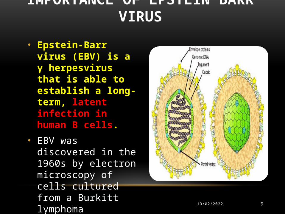
• Epstein-Barr virus (EBV) is a γ herpesvirus that is able to establish a long-term, latent infection in human B cells.
• EBV was discovered in the 1960s by electron microscopy of cells cultured from a Burkitt lymphoma
IMPORTANCE OF EPSTEIN BARR VIRUS
903/05/2023

EBV
10
• Once infected, a lifelong carrier state develops.
• Low grade virus replication and shedding can be demonstrated in the epithelial cells of the pharynx of all seropositive individuals.
• EBV is able to immortalize B-lymphocytes in vitro and in vivo
03/05/2023
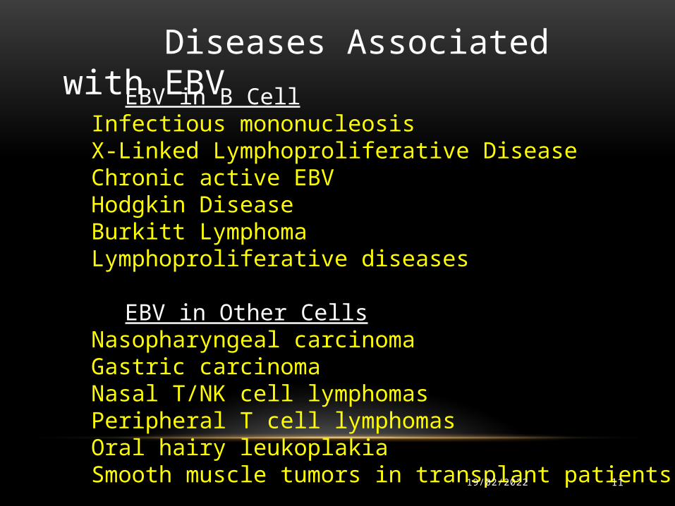
EBV in B CellInfectious mononucleosisX-Linked Lymphoproliferative DiseaseChronic active EBVHodgkin Disease Burkitt LymphomaLymphoproliferative diseases
EBV in Other Cells
Nasopharyngeal carcinomaGastric carcinomaNasal T/NK cell lymphomasPeripheral T cell lymphomasOral hairy leukoplakiaSmooth muscle tumors in transplant patients
Diseases Associated with EBV
03/05/2023 11

12
MOLECULAR CONFIGURATION OF EPSTEIN BARR VIRUS
03/05/2023
EBV genome is enclosed in a nuclear capsid surrounded by a glycoprotein envelope
13
MOLECULAR BIOLOGY • Viral capsid antigens (VCAs) are found in replicating cells.• EBV early antigens (EAs) consist of >15 proteins coded by genes
distributed throughout the genome.• EBV nuclear antigen (EBNA) corresponds to six proteins found in
the nucleus of an EBV-infected cell.
03/05/2023
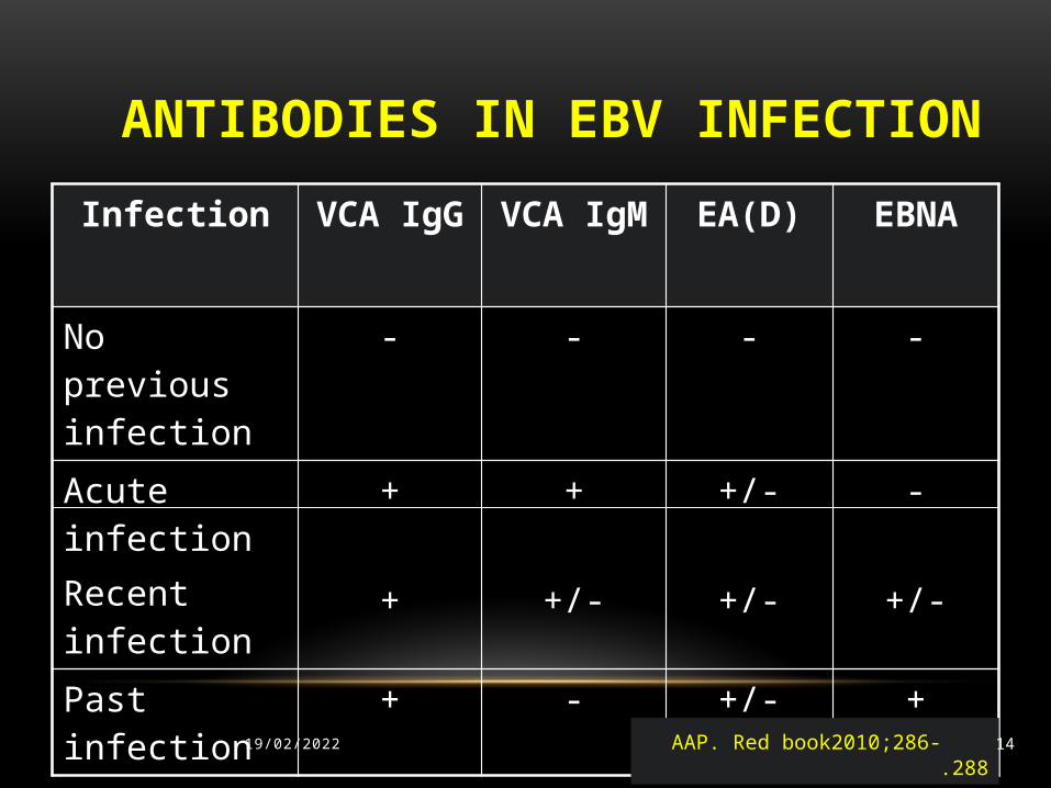
ANTIBODIES IN EBV INFECTIONInfection VCA IgG VCA
IgMEA(D) EBNA
No previous infection
- - - -
Acute infectionRecent infection
+
+
+
+/-
+/-
+/-
-
+/-
Past infection
+ - +/- +
AAP. Red book2010;286-288.03/05/2023 14
15
LATENCY• Latently infected B cells are the primary reservoir of EBV in the body
• >100 gene may be expressed during active viral replication, only 11 are expressed during viral latency.
• Latency ( the virus limits cytotoxic T-cell recognition of EBV-infected cells) .
03/05/2023
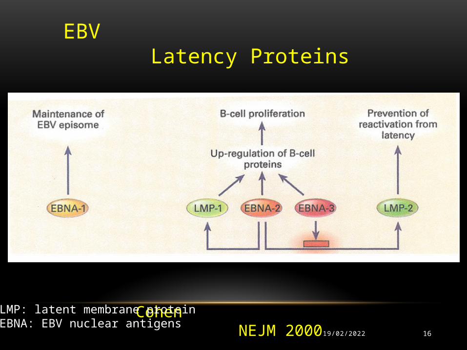
EBV Latency Proteins
Cohen NEJM 200003/05/2023 16
LMP: latent membrane protein EBNA: EBV nuclear antigens
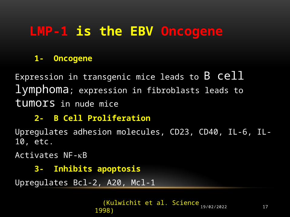
1- Oncogene
Expression in transgenic mice leads to B cell lymphoma; expression
in fibroblasts leads to tumors in nude mice
2- B Cell Proliferation Upregulates adhesion molecules, CD23, CD40, IL-6, IL-10, etc.
Activates NF-B
3- Inhibits apoptosis Upregulates Bcl-2, A20, Mcl-1
LMP-1 is the EBV Oncogene
(Kulwichit et al. Science 1998)03/05/2023 17
EBV
• EBV is widely disseminated. It is estimated that 95% of world’s population is exposed to the virus, which makes it the most ubiquitous virus known to man
• EBV is only a minor problem for immuno-competent persons, but it can become a major one for immunologically compromised patients
03/05/2023 18
Ubiquitous : present everywhere

LYMPHOPROLIFERATIVE DISORDERS
03/05/2023 19
• X-linked lymphoproliferative disorder• The autoimmune lymphoproliferative syndrome ( ALPS)
• Castleman disease (CD)
• Posttransplant lymphoproliferative disease (PTLD)

X-linked lymphoproliferative disorder (DUNCAN DISEASE)
1 in 100,0000 Age of onset: 2.5 yrs old, older reported
Unique predisposition to uncontrolled infection with Epstein Barr virus
EBV induces:- fatal/severe infectious mononucleosis- Secondary agammaglobulinemia- Lymphoma- Bone marrow failure

DUNCAN DISEASE
03/05/2023 21
• Defect in SAP- interferes with NK and CD8+ CTL function
• The prognosis is poor
• The nature of the lymphoproliferation might change from a polyclonal to a monoclonal process (more aggressive )

CLONALITY ASSESSMENT
• Clonality can be detected by immunohistochemical analysis of immunoglobulins
• The most reliable method for clonality assessment is by using polymerase chain reaction to detect the complete spectrum of possible rearrangements
03/05/2023 22

03/05/2023 23
Autoimmune lymphoproliferative syndrome
(ALPS)
Canale-Smith syndrome

HISTORY
• Canale and Smith reported five cases with lymphadenopathy, splenomegaly, and autoimmune cytopenias in 1967.
• In 1995, Rieux-Laucat et al. and Fisher et al. documented that this novel disorder was associated with inherited mutations in the Fas gene.
• FAS : cell surface death receptor
(TNF receptor superfamily, member 6; apoptosis stimulating fragment)
03/05/2023 24
2004,
• New mutations in other intermediates , in the Fas signaling pathway, such as Fas ligand (FasL) gene mutation and caspase gene mutations ( 8 or 10)
03/05/2023 25
ALPS ETIOLOGY
• ALPS , is the first human disease in which the etiology has been attributed to a primary defect in apoptosis
• The etiology has been attributed to dysregulation of lymphocyte homeostasis due to a primary defect in Fas-induced apoptosis
03/05/2023 26

APOPTOSIS DYSREGULATION• Dramatically illustrated in the autoimmune lymphoproliferative
syndrome (ALPS) of childhood
• ALPS is the result of dominant inheritance of a mutated gene, TNFRSF6, which encodes the transmembrane protein Fas (also known as CD95), a major mediator of lymphocyte apoptosis
03/05/2023 27

03/05/2023 28
Caspase-activated deoxyribonuclease (CAD) and its inhibitor (I-CAD)

03/05/2023 29

03/05/2023 30
Structure of the FAS gene with mutations associated with ALPS-FAS

GENOTYPE-BASED ALPS CLASSIFICATION
• Type Ia : Mutation on TNFRSF6 (Fas) gene
• Type Ib : Mutation on TNFSF6 (FasL) gene
• Type II : Mutation on caspase 8 or 10 genes
• Type III : No known mutation
03/05/2023 31

ALPS
• FAS mutations are either somatic or germ-line
• Patients with germline mutation of the intracellular domain of Fas have the highest risk of developing lymphoma
14 times greater for non-Hodgkin’s lymphoma and
51 times greater for Hodgkin’s lymphoma
03/05/2023 32

ALPS IS DEFINED BY THE TRIAD OF
1- Chronic , non-malignant enlargement of lymph nodes and spleen;
2- Increased number of double negative T cells (DNTs) ,
that lack both the CD4 and CD8 surface molecules but , express the α/β T cell receptors (α/β+ DNTs);
3- Impaired lymphocyte apoptosis in vitro
03/05/2023 33
ALPS, CLINICAL FEATURES
• The median age at presentation is 24 months
• Prominent non-malignant lymphadenopathy often accompanied by splenomegaly (in some cases with hepatomegaly)
And
• Autoimmune cytopenias of one or more lineages
03/05/2023 34
ALPS
• Non-hematological autoimmune diseases can also occur in association with ALPS, including ,
glomerulonephritis, uveitis, Guillain- Barré syndrome, autoimmune liver disease, urticaria and arthritis
03/05/2023 35
ALPS
• Several cytokine abnormalities have been found in patients with ALPS, the most striking of which is a significantly elevated IL-10
03/05/2023 36

OVEREXPRESSION OF IL-10
• May be involved in the proliferation of autoimmune B cells
and
• May cause the persistence and activation of malignant and autoimmune cell clones.
03/05/2023 37

DIFFERENTIAL DIAGNOSIS
Children with
• Generalized lymphadenopathy,
• Splenomegaly,
and
• Autoimmune multilineage cytopenias represents
a diagnostic challenge
03/05/2023 38
Clinical and laboratory features overlap with and may manifest as
other childhood diseases
ALPS
03/05/2023 39

AS
• Systemic infections,
• collagen vascular diseases,
• lymphoma,
• Evans syndrome
• 1ry immunological disorders:
Common variable immunodeficiency, Wiskott-Aldrich syndrome, IL-2 receptor α-chain deficiency
03/05/2023 40

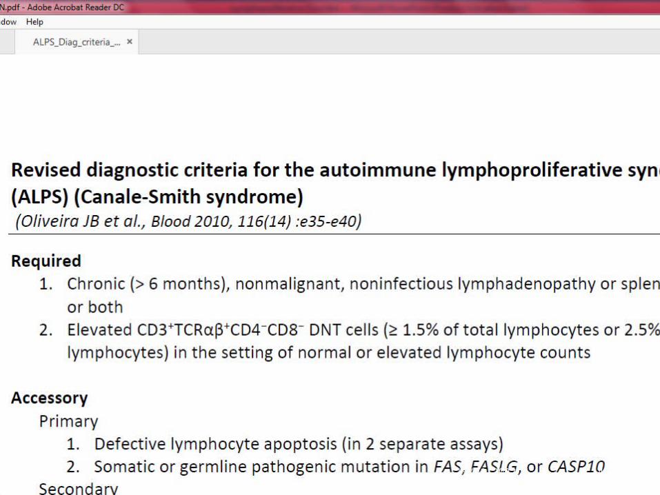
(Oliveira JB et al., Blood 2010, 116(14) :e35-e40)
REVISED DIAGNOSTIC CRITERIA FOR THE AUTOIMMUNE LYMPHOPROLIFERATIVE SYNDROME (ALPS)
(CANALE-SMITH SYNDROME)
03/05/2023 41
(Oliveira JB et al., Blood 2010, 116(14) :e35-e40 )
REQUIRED
1. Chronic (> 6 months), nonmalignant, noninfectious lymphadenopathy or splenomegaly or both
2. Elevated CD3+TCRαβ+ CD4−CD8− DNT cells in the setting of normal or elevated lymphocyte counts
03/05/2023 42

ACCESSORY• Primary 1. Defective lymphocyte apoptosis (in 2 separate assays)
2. Somatic or germline mutation in FAS, FASL, or CASP10
03/05/2023 43

ACCESSORY
Secondary 1. Elevated plasma sFASL levels (>200 pg/mL) OR elevated plasma interleukin-10 levels (>20 pg/mL) OR elevated serum vitamin B12 levels (> 1500 ng/L) OR elevated plasma interleukin-18 levels > 500 pg/mL
2. Typical immunohistological findings as reviewed by an experienced hematopathologist
03/05/2023 44
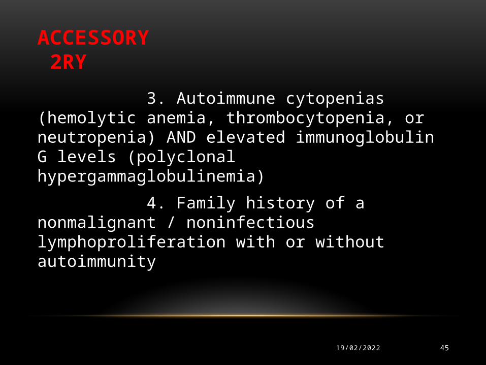
ACCESSORY 2RY
3. Autoimmune cytopenias (hemolytic anemia, thrombocytopenia, or neutropenia) AND elevated immunoglobulin G levels (polyclonal hypergammaglobulinemia)
4. Family history of a nonmalignant / noninfectious lymphoproliferation with or without autoimmunity
03/05/2023 45
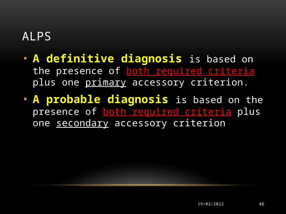
ALPS
• A definitive diagnosis is based on the presence of both required criteria plus one primary accessory criterion.
• A probable diagnosis is based on the presence of both required criteria plus one secondary accessory criterion
03/05/2023 46
03/05/2023 47

03/05/2023 48

03/05/2023 49

ALPSTreatment modalities for ALPS are directed at
• The chronic and persistent lymphoproliferation
• Autoimmunity, and
• Malignancies
03/05/2023 50
ALPS• Lymphoproliferation does respond to corticosteroids and other
immunosuppressants like azathioprine , cyclosporine or mycophenolate mofetil
03/05/2023 51

ALPSThe treatment of all autoimmune manifestations is the same as in
patients without ALPS
• Autoimmune cytopathies respond well to corticosteroids• Immune thrombocytopenia is less sensitive to intravenous
immunoglobulin (IVIG) therapy than conventional ITP
03/05/2023 52

ALPS• Some ALPS patients with chronic neutropenia and recurrent
infections benefit from thrice weekly, low dose (1-2 μg/kg/d) recombinant granulocyte colony stimulating factor
03/05/2023 53
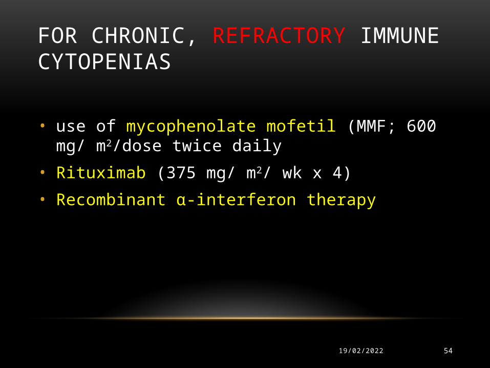
FOR CHRONIC, REFRACTORY IMMUNE CYTOPENIAS
• use of mycophenolate mofetil (MMF; 600 mg/ m2/dose twice daily
• Rituximab (375 mg/ m2/ wk x 4)
• Recombinant α-interferon therapy
03/05/2023 54

ALPS , PROGNOSIS
• Despite the often impressive lymphadenopathy and splenomegaly, the prognosis with regard to lymphoproliferation is good
• Most patients demonstrate regression of lymphoproliferation
03/05/2023 55

THE MAJOR DETERMINANTS OF MORBIDITY AND MORTALITY IN ALPS ARE
• The severity of the autoimmune disease
• Hypersplenism
• Postsplenectomy-related sepsis
• Development of lymphoma
03/05/2023 56
Careful long-term surveillance in patients with mutations of the Fas
protein is especially required
03/05/2023 57

REFERENCES• *Rieux-Laucat F, Le Deist F, Hivroz C, Roberts IA, Debatin KM, Fischer A, de Villartay JP.
Mutations in Fas associated
with human lymphoproliferative syndrome and autoimmunity. Science 2005;268:1347-9.
• *Drappa J, Vaishnaw AK, Sullivan KE, Chu JL, Elkon KB. Fas gene mutations in the Canale-Smith syndrome, an inherited lymphoproliferative disorder associated with autoimmunity. N Engl J Med 2006;335:1643-9.
• *Straus SE, Sneller M, Lenardo MJ, Puck JM, Strober W. An inherited disorder of lymphocyte apoptosis: the autoimmune lymphoproliferative syndrome. Ann Intern Med 2009;130:591-601.
• *Rieux-Laucat F, Fischer A, Deist FL. Cell-death signaling and human disease. Curr Opin Immunol 2003;15:325-31.
• *Alvarado CS, Straus SE, Li S, Dale JK, Mann K, Le A, Lauer SJ. Autoimmune lymphoproliferative syndrome: a cause of chronic splenomegaly, lymphadenopathy, and cytopenias in children-report on diagnosis and management of five patients. Pediatr Blood Cancer 2014;43:164-9.
03/05/2023 58

REFERENCES• *Bleesing JJ, Brown MR, Novicio C, Guarraia D, Dale JK, Straus SE, Fleisher TA. A composite
picture of TcR alpha/beta(+) CD4(-)CD8(-) T Cells (alpha/beta- DNTCs) in humans with autoimmune lymphoproliferative syndrome. Clin Immunol 2012;104:21-30.
• *Clementi R, Dagna L, Dianzani U, Dupre L, Dianzani I, Ponzoni M, Cometa A, Chiocchetti A, Sabbadini MG, Rugarli C, Ciceri F, Maccario R, Locatelli F, Danesino C, Ferrarini M, Bregni M. Inherited perforin and Fas mutations in a patient with autoimmune lymphoproliferative syndrome and lymphoma. N Engl J Med 2004;351:1419-24.
• *Ceretelli S, Petrini M, Galimberti S, Testi C, Frizzera G. Interferon-alpha activity in a case of severe autoimmune lymphoproliferative disease. Ann Hematol 2011;80:49-52
• *Heelan BT, Tormey V, Amlot P, Payne E, Mehta A, Webster AD. Effect of anti-CD20 (rituximab) on resistant thrombocytopenia in autoimmune lymphoproliferative syndrome. Br J Haematol 2012;118:1078-81.
• *Rao VK, Dugan F, Dale JK, Davis J, Tretler J, Hurley JK, Fleisher T, Puck J, Straus SE. Use of mycophenolate mofetil for chronic, refractory immune cytopenias in children with autoimmune lymphoproliferative syndrome. Br J Haematol 2005;129:534-8.
03/05/2023 59

REFERENCES
03/05/2023 60
• *Fisher GH, Rosenberg FJ, Straus SE, et al. Dominant interfering Fas gene mutations impair apoptosis in a human autoimmune lymphoproliferative syndrome. Cell 1995;81(6):935-946.
• *Rieux-Laucat F, Le Deist F, Hivroz C, et al. Mutations in Fas associated with human lymphoproliferative syndrome and autoimmunity. Science 1995;268(5215):1347-1349.
• *Oliveira JB, Fleisher T. Autoimmune lymphoproliferative syndrome. Curr Opin Allergy Clin Immunol 2004;4:497-503.
• *Rieux-Laucat F, Deist FL, Fischer A. Autoimmune lymphoproliferative syndromes: genetic defects of apoptosis pathways. Cell Death Differentiation 2013;10:124-33.
• *Carter LB, Procter JL, Dale JK, Straus SE, Cantilena CC. Description of serologic features in autoimmune lymphoproliferative syndrome. Transfusion 2006;40:943-48
• *Rao VK, Carrasquillo JA, Dale JK, Bacharach SL, Whatley M, Dugan F, Tretler J, Fleisher T, Puck JM, Wilson W, Jaffe ES, Avila N, Chen CC, Straus SE. Fluorodeoxyglucose positron emission tomography (FDG-PET) for monitoring lymphadenopathy in the autoimmune lymphoproliferative syndrome (ALPS). Am J Hematol 2010 ;8:81-85.
• *Worth A, Thrasher AJ, Gaspar HB. Autoimmune lymphoproliferative syndrome: molecular basis of disease and clinical phenotype. Br J Haematol 2006;133:124-40.
03/05/2023 61
a rare disease of lymph nodes and other lymphatic tissues
CASTLEMAN DISEASE (CD)

CASTLEMAN DISEASE (CD)
03/05/2023 62
• It is also known as giant lymph node hyperplasia, and angiofollicular lymph node hyperplasia
• It was first described by Dr. Benjamin Castleman in the 1950s.
• CD is not a cancer. Instead, it is a lymphoproliferative disorder.
TYPES OF CASTLEMAN DISEASE
03/05/2023 63
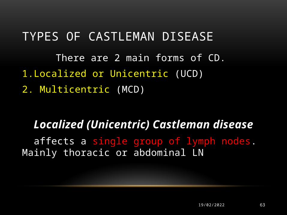
There are 2 main forms of CD.
1.Localized or Unicentric (UCD)
2. Multicentric (MCD)
Localized (Unicentric) Castleman disease affects a single group of lymph nodes. Mainly thoracic or abdominal LN
MULTICENTRIC CD
03/05/2023 64
• affects more than a single group of lymph nodes
• It can also affect other lymphoid tissue
• It is more serious than the localized type, particularly in people with HIV infection.
MICROSCOPIC SUBTYPES OF CASTLEMAN DISEASE
03/05/2023 65
• The hyaline vascular type – It is most common. It tends to be localized, but in rare cases it is multicentric
• The plasma cell type – It is slightly more likely to be multicentric, but it is sometimes localized
• The mixed subtype -- It shows areas of both types. It occurs less often.
• In choosing treatments, the microscopic type is less important than whether the disease is localized or multicentric .

Effaced architecture of the LN , increased angiogenesis in castlemans disease , HE stain 4 X
Normal architecture of LN

RISK FACTOR
03/05/2023 67
• Infection with HIV virus
• Infection with HHV-8
• Age : Younger patients are more likely to have the localized form . Adults and those with HIV and HHSV-8 infection are more likely to have the multicentric form.
PATHOGENESIS
03/05/2023 68
• Dysregulated and overproduced IL-6, particularly in patients with MCD, stimulates the production of acute phase reactants in the liver,
• resulting in constitutional symptoms, including fever, sweats, and fatigue, and
• laboratory abnormalities, such as anemia, elevated inflammatory markers, hypergammaglobulinemia, and hypoalbuminemia.

CASTLEMAN DISEASE
03/05/2023 69
• IL-6 also stimulates B-cell proliferation and induces the expression of vascular endothelial growth factor and increased angiogenesis.
• The activation of the IL-6 receptor further results in the activation of the Janus kinase–signal transducers and the activation of transcription pathway and the mitogen-activated protein kinase cascade, which enhances B-cell proliferation and survival

CASTLEMAN DISEASE
03/05/2023 70
• HIV-associated MCD, is usually associated with HHV-8 infection
• Patients with HHV-8-positive MCD,
infected cells express a viral analog of IL-6 (vIL-6), which likely contributes to the pathogenesis of this significant subset of
Castleman disease
PATHOLOGICAL DIAGNOSIS
03/05/2023 71
• Castleman disease is a pathological diagnosis made by excisional biopsy of affected lymph node.
• In cases of deeper or less accessible disease, core needle biopsy is preferred to fine needle aspiration, because fine needle aspirations are insensitive for both UCD and MCD.

Atrophic germinal centers , HE stain
40X
Normal germinal center, containing larger
lymphocytes undergoing activation

Vascular proliferation ,20 X, increased angiogenesis

Vascular proliferation 40 X

STAGING
03/05/2023 75
The goals of the staging and pretreatment evaluation in Castleman disease are to
• (1) determine whether the patient has unicentric or multicentric disease
• (2) identify patients with systemic inflammatory manifestations of Castleman disease,
• (3) assess for the presence of HIV, as well as associated conditions and malignancies.

THE INITIAL LABORATORY EVALUATION
03/05/2023 76
• Complete blood count
• Inflammatory markers (erythrocyte sedimentation rate and C-reactive protein)
• Complete metabolic panel, and albumin
• HIV testing should be performed in all patients
• Plasma HHV-8 DNA levels (a helpful biomarker, both to support the diagnosis of MCD and to monitor disease activity and response to therapy)
CASTLEMAN DISEASE
03/05/2023 77
• Levels of cytokines, most notably IL-6 and IL-10 are not recommended to be routinely measured.
• Computed tomography of the chest, abdomen, and pelvis should be obtained at the time of diagnosis to assess for adenopathy and splenomegaly and to assess resectability in patients with UCD

MANAGEMENT
03/05/2023 78
• The optimal therapy for UCD is surgical resection
• Radiation therapy in patients who are not candidate for surgical excision.
The natural history of MCD is variable
Some patients may present with indolent disease and very slow progression over months to years,
while others will experience an acute and fulminant disease that can be fatal within weeks

TREATMENT OPTIONS FOR MCD
03/05/2023 79
• Antiretroviral Therapy : for HIV-associated MCD
• Glucocorticoids
• Cytotoxic Chemotherapy : single-agent chemotherapy in the treatment of MCD,
etoposide, vinblastine, cyclophosphamide, chlorambucil
Single-agent chemotherapies are often administered at doses and schedules routinely used to treat patients with lymphoma

TREATMENT OPTIONS FOR MCD
03/05/2023 80
• Rituximab: (the current mainstay therapy), is highly active as monotherapy in MCD.
• Anti-Interleukin 6 Therapy: Siltuximab and tocilizumab are monoclonal antibodies targeting IL-6 and its receptor (IL-6R), respectively, (new additions to the treatment)
• Antiherpesvirus Therapy: have been explored as therapy for HIV-associated MCD , given the pathogenetic link with HHV-8

POEMS SYNDROME
03/05/2023 81
(Polyneuropathy, Organomegaly, Endocrinopathy, M-protein, and Skin changes)
• MCD is present in 15% to 25% of patients with POEMS syndrome (Castle-POEMS)
• MCD is included as a major criterion for the diagnosis of POEMS syndrome

POEMS SYNDROME
03/05/2023 82
• Clinical features include
hepatosplenomegaly, lymphadenopathy, endocrinopathy, skin changes, osteosclerotic bone lesions, elevated levels of vascular endothelial growth factor and elevated protein in the cerebrospinal fluid.

03/05/2023 83

REFERENCES
03/05/2023 84
• *Stebbing J, Adams C, Sanitt A, et al. Plasma HHV8 DNA predicts relapse in individuals with HIV-associated multicentric Castleman disease. Blood. 2011;118(2):271-275
• * . Newsom-Davis T, Bower M, Wildfire A, et al. Resolution of AIDS-related Castleman’s disease with anti-CD20 monoclonal antibodies is associated with declining IL-6 and TNF-alpha levels. Leuk Lymphoma. 2004;45(9):1939-1941.
• * Bower M, Veraitch O, Szydlo R, et al. Cytokine changes during rituximab therapy in HIV-associated multicentric Castleman disease. Blood. 2009;113(19):4521-4524.
• * Carbone A, De Paoli P, Gloghini A, et al. KSHV-associated multicentric Castleman disease: A tangle of different entities requiring multitarget treatment strategies. Int J Cancer. 2014.
• * . Matthiesen C, Ramgopol R, Seavey J, et al. Intensity modulated radiation therapy (IMRT) for the treatment of unicentric Castlemans disease: a case report and review of the use of radiotherapy in the literature. Radiol Oncol. 2012;46(3):265-270

03/05/2023 85
POSTTRANSPLANT LYMPHOPROLIFERATIVE DISEASE (PTLD)

POSTTRANSPLANT LYMPHOPROLIFERATIVE DISEASE (PTLD)
03/05/2023 86
• is a well-recognized complication of both solid organ transplantation (SOT) and allogeneic hematopoietic stem cell transplantation (HSCT).

PTLD
03/05/2023 87
• In most cases, PTLD is associated with (EBV) infection of B cells, either as a consequence of reactivation of the virus post-transplantation or from primary EBV infection

PTLD
03/05/2023 88
• In cases of primary infection,
EBV may be acquired from the donor graft or, less commonly, from environmental exposure.

PTLD
03/05/2023 89
• While T-cell lymphoproliferative disorders can also occur after SOT and HSCT, the vast majority are B-cell proliferations.
• PTLD is identified by having a high index of suspicion in the appropriate clinical setting

PTLD
03/05/2023 90
• The diagnosis is made by histopathological evidence of lymphoproliferation, commonly with the presence of EBV DNA, RNA, detected in tissues
• Most cases of PTLD occur within the first post-transplant year.

FREQUENCY A.Parker etal, BJH 149; 675 (2010)

03/05/2023 92
The more intense the immunosuppression used, the greater the risk of PTLD and the earlier it tends to
occur
PTLD

THE 2008 (WHO) CLASSIFICATION SYSTEM
03/05/2023 93
• 4 major histopathologic subtypes of PTLD:
(1) early hyperplastic lesions,
(2) polymorphic lesions
(3) monomorphic lesions, and
(4) classic Hodgkin-type lymphomas

Jaffe et al -2001
A
BC
D
PATHOGENESIS - PTLD • B cell proliferation induced by EBV infection• Cytotoxic T cells keep EBV-infected B cells in check
Anti T cell Rx or T cell depletion is therefore a risk factor for PTLD
• EBV-driven polyclonal proliferations leading to EBV(+) or EBV(-) lymphomas of predominantly B-cell or less often T-cell type
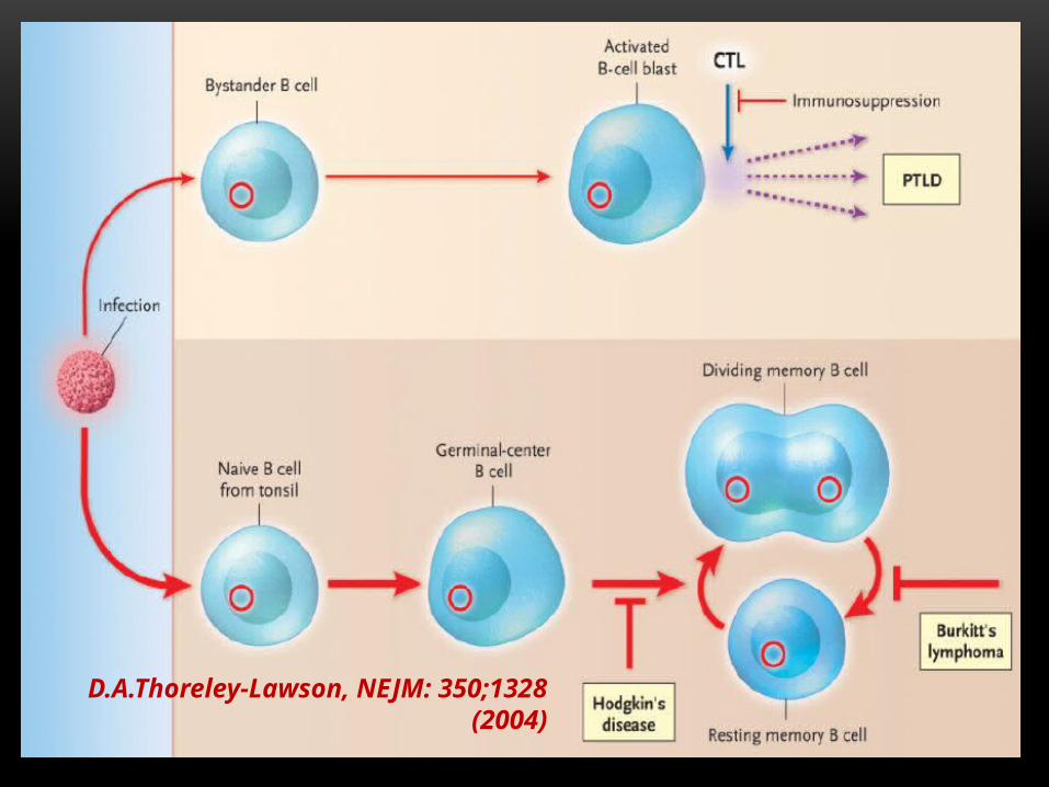
D.A.Thoreley-Lawson, NEJM: 350;1328 (2004)

EBV-negative PTLD
03/05/2023 98
- Present much later
(median 50-60mo vs 6-10 mo)
- Monomorphic
- Poor outcomes , poor response to therapy
- Increasing in frequency

PTLD DIAGNOSIS
03/05/2023 99
• EBV viral load
• Imaging
• Tissue biopsy (excisional node biopsy) -Confirm EBV positivity by immunostaining LMP1 – latent membrane protein 1
EBER- EBV-encoded RNA
-Histological grade
-Immunophenotyping (CD 20)
-Cytogenetics
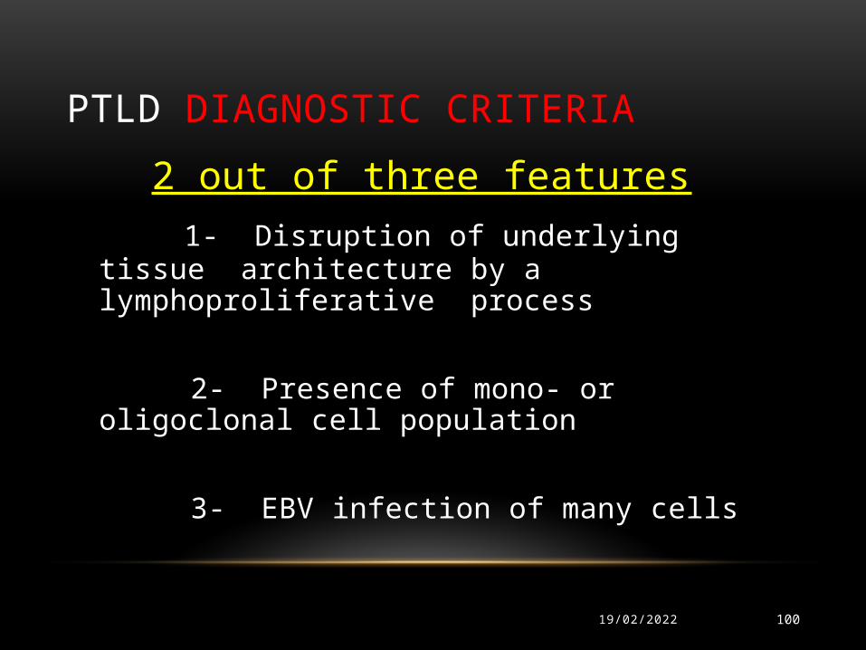
PTLD DIAGNOSTIC CRITERIA
03/05/2023 100
2 out of three features 1- Disruption of underlying tissue architecture by a
lymphoproliferative process
2- Presence of mono- or oligoclonal cell population
3- EBV infection of many cells

PTLD MANAGEMENT
03/05/2023 101
1) REDUCTION OF IMMUNOSUPPRESSION2) Antiviral agents (Ganciclovir, Acyclovir,Maribavir)
3) Surgery and Radiotherapy (localized)
4) Rituximab
5) Rituximab + Chemotherapy
6) EBV Directed cytotoxic T lymphocytes (CTL) – in clinical trials

PTLD
03/05/2023 102
• The cornerstone of initial management of PTLD is reduction or withdrawal of immunosuppression, which may reverse the lymphoproliferative process
• This potential for reversibility with reduction of immunosuppression distinguishes PTLD from neoplastic lymphoproliferative disorders

BUT,
03/05/2023 103
• Reduction of immunosuppression also carries the risk of inducing allograft dysfunction or loss and is not always feasible

PTLD CNS RIS LOCALISED
RADIO MULTIFOCAL RADIOOR CHEMO OR LOCAL EXCN LOW HIGH RISK RISK
RITUX RITUX + CHEMORIS: reduce immuno-suppression

03/05/2023 105

POST-TRANSPLANT EBV – MONITORING
03/05/2023 106
AT LEAST WEEKLY FOR 3 MONTHS
• LONGER MONITORING FOR:
- GVHD
- PREVIOUS EBV REACTIVATION
J.Styczynski etal, BMT 43; 757 (2009)

PTLD PREVENTION
03/05/2023 107
1) IV ganciclovir to high-risk pts for at least 100 days
2) Oral acyclovir in low risk patients

MEDICOLEGAL ISSUES
03/05/2023 108
Transplantation and the accompanying immunosuppression, put patients at risk for potentially
fatal infection and malignancy. Transplant candidates must be fully informed of these risks as part of the
consent process pre-transplantation.
REFERENCES
03/05/2023 109
• Green M, Webber S. Posttransplantation lymphoproliferative disorders. Pediatr Clin North Am. 2003 Dec. 50(6):1471-91.
• Swerdlow SH, Webber SA, Chadburn A, Ferry JA. Posttransplant lymphoproliferative disorders. Swerdlow SH, Campo E, Harris NL, Jaffe ES. WHO classification of tumors of haemotopoietic and lymphoid tissue. IARC, Lyon; 2008. 343-350.
• Glotz D, Chapman JR, Dharnidharka VR, Hanto DW, Castro MC, Hirsch HH, et al. The Seville Expert Workshop for Progress in Posttransplant Lymphoproliferative Disorders. Transplantation. 2012 Sep 18.
• Shaknovich R, Basso K, Bhagat G, et al. Identification of rare Epstein-Barr virus infected memory B cells and plasma cells in non-monomorphic post-transplant lymphoproliferative disorders and the signature of viral signaling. Haematologica. 2006 Oct. 91(10):1313-20.
• D'Antiga L, Del Rizzo M, Mengoli C, Cillo U, Guariso G, Zancan L. Sustained Epstein-Barr virus detection in paediatric liver transplantation. Insights into the occurrence of late PTLD. Liver Transpl. 2007 Mar. 13(3):343-8.
REFERENCES
03/05/2023 110
• Swinnen LJ, LeBlanc M, Grogan TM, Gordon LI, Stiff PJ, Miller AM. Prospective study of sequential reduction in immunosuppression, interferon alpha-2B, and chemotherapy for posttransplantation lymphoproliferative disorder. Transplantation. 2008 Jul 27. 86(2):215-22.
• Schubert S, Renner C, Hammer M, Abdul-Khaliq H, Lehmkuhl HB, Berger F. Relationship of immunosuppression to Epstein-Barr viral load and lymphoproliferative disease in pediatric heart transplant patients. J Heart Lung Transplant. 2008 Jan. 27(1):100-5.
• Dharnidharka VR, Lamb KE, Gregg JA, Meier-Kriesche HU. Associations between EBV serostatus and organ transplant type in PTLD risk: an analysis of the SRTR National Registry Data in the United States. Am J Transplant. 2012 Apr. 12(4):976-83.
• Jamali FR, Otrock ZK, Soweid AM, Al-Awar GN, Mahfouz RA, Haidar GR. An overview of the pathogenesis and natural history of post-transplant T-cell lymphoma. Leuk Lymphoma. 2007 Jun. 48(6):1237-41.

REFERENCES
03/05/2023 111
• Cruz RJ Jr, Ramachandra S, Sasatomi E, Dimartini A, de Vera M, Fontes P, et al. Surgical management of gastrointestinal posttransplant lymphoproliferative disorders in liver transplant recipients. Transplantation. 2012 Aug 27. 94(4):417-23.
• Becker YT, Samaniego-Picota M, Sollinger HW. The emerging role of rituximab in organ transplantation. Transpl Int. 2006 Aug. 19(8):621-8.
• Taj MM, Messahel B, Mycroft J, Pritchard-Jones K, Baker A, Height S. Efficacy and tolerability of high-dose methotrexate in central nervous system positive or relapsed lymphoproliferative disease following liver transplant in children. Br J Haematol. 2008 Jan. 140(2):191-6.
• Bollard CM, Rooney CM, Heslop HE. T-cell therapy in the treatment of post-transplant lymphoproliferative disease. Nat Rev Clin Oncol. 2012 Sep. 9(9):510-9.
• Gross TG, Bucuvalas JC, Park JR, Greiner TC, Hinrich SH, Kaufman SS. Low-dose chemotherapy for Epstein-Barr virus-positive post-transplantation lymphoproliferative disease in children after solid organ transplantation. J Clin Oncol. 2005 Sep 20. 23(27):6481-8.

THANK YOU
03/05/2023 112


![Lymphoproliferative disorders in inflammatory bowel ... · transplantation lymphoproliferative disorders (PTLD), which can develop due to both primary and secondary immunosuppression[6].](https://static.fdocuments.us/doc/165x107/5f0addb37e708231d42db993/lymphoproliferative-disorders-in-inflammatory-bowel-transplantation-lymphoproliferative.jpg)