Lymphoma Final - Handout.ppt - 2.pdf• Brain MRI and LP . 7 Ann Arbor Staging System ...
41
1 Kami Maddocks, MD Associate Professor of Clinical Medicine Division of Hematology Department of Internal Medicine The Ohio State University Wexner Medical Center The James Cancer Hospital and Solove Research Institute Aggressive Lymphomas Lymphoma Non-Hodgkin’s Lymphoma Low Grade/Indolent Lymphoma High Grade/ Aggressive Lymphoma Hodgkin Disease 5 subtypes
-
Upload
truongtram -
Category
Documents
-
view
216 -
download
1
Transcript of Lymphoma Final - Handout.ppt - 2.pdf• Brain MRI and LP . 7 Ann Arbor Staging System ...

1
Kami Maddocks, MDAssociate Professor of Clinical Medicine
Division of HematologyDepartment of Internal Medicine
The Ohio State University Wexner Medical CenterThe James Cancer Hospital and
Solove Research Institute
Aggressive Lymphomas
Lymphoma
Non-Hodgkin’s Lymphoma
Low Grade/Indolent Lymphoma
High Grade/ Aggressive Lymphoma
Hodgkin Disease
5 subtypes

2
Cellular Origin of Non-Hodgkin’s Lymphoma
(U.S. and Europe)-------------------------------------
Cellular Origin of Non-Hodgkin’s Lymphoma
(U.S. and Europe)-------------------------------------
Cancer statistics, 2018Cancer statistics, 2018
CA: A Cancer Journal for Clinicians4 JAN 2018 DOI: 10.3322/caac.21442 http://onlinelibrary.wiley.com/doi/10.3322/caac.21442/full#caac21442-fig-0001

3
Non-Hodgkin’s Lymphoma
Non-Hodgkin’s Lymphoma
• 4% of all cancers• 7th most common cancer in men & women• ~ 74,680 people (41,730 males and 32,950
females) diagnosed• ~ 19,910 people will die of this disease (11,510
and 8,400)• Risk of developing NHL is 1 in 47• One of the more common cancers among
children, teens and young adults but risk of developing NHL increases throughout life with half the patients ≥ 65 years of age
Hodgkin DiseaseHodgkin Disease• ~ 8,500 new cases (3,660 in females and
4,840 in males)
• ~ 1,050 deaths (430 females, 620 males)
• Most common in early adulthood but bimodal
• Peak patients aged 15-34
• Risk rises again > 55 years old
• ~ 10-15% of cases diagnosed in children and teenagers
• 5-year relative survival rate for all comers is 86%
4
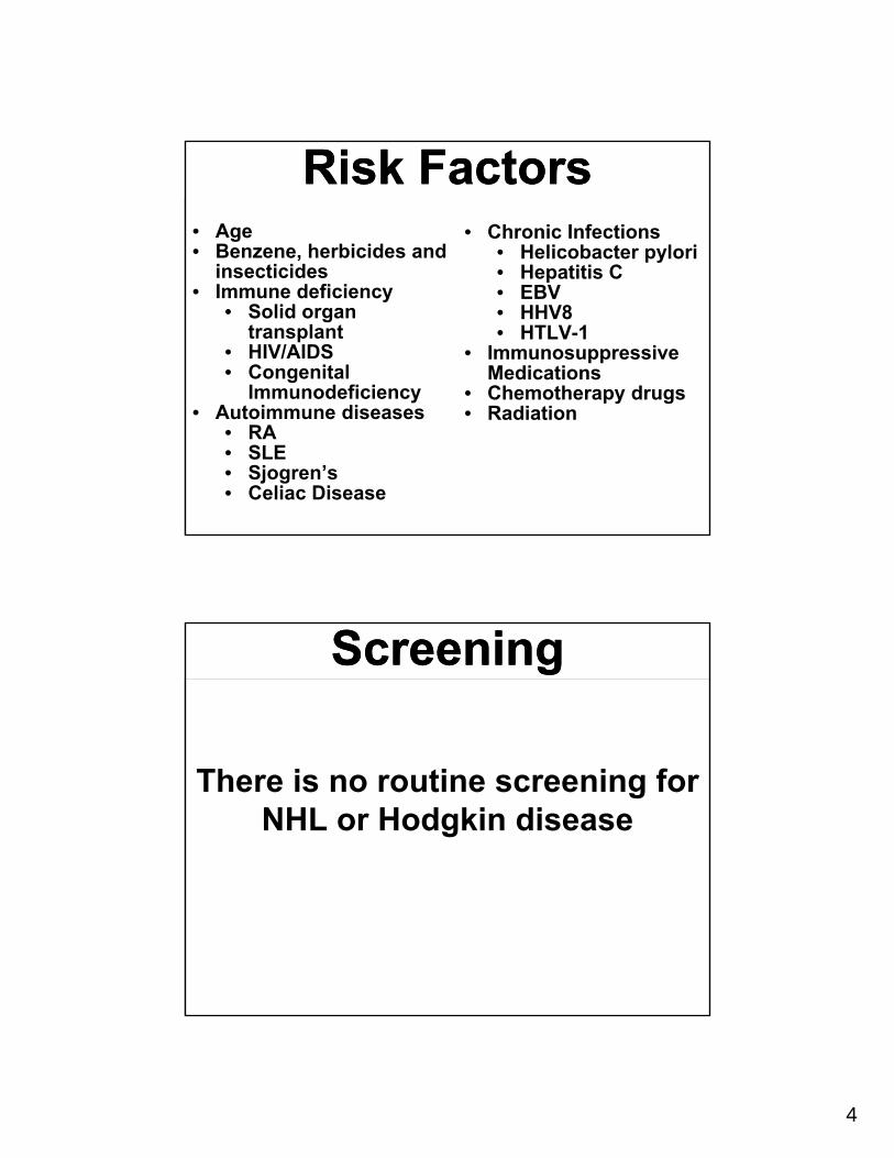
Risk Factors Risk Factors • Age • Benzene, herbicides and
insecticides• Immune deficiency
• Solid organ transplant
• HIV/AIDS• Congenital
Immunodeficiency• Autoimmune diseases
• RA• SLE• Sjogren’s• Celiac Disease
• Chronic Infections• Helicobacter pylori• Hepatitis C • EBV• HHV8• HTLV-1
• Immunosuppressive Medications
• Chemotherapy drugs• Radiation
Screening Screening
There is no routine screening for NHL or Hodgkin disease

5
Patient Presentations Patient Presentations • Enlargement of a lymph node
• Symptoms from bulky lymphadenopathy
• Dependent on location – pain, dyspnea, early satiety, renal dysfunction
• Abnormal blood counts
• Some patients present with “B” symptoms
• Fevers, night sweats, weight loss
• Pruritus in Hodgkin Disease
How is Lymphoma Diagnosed?
How is Lymphoma Diagnosed?
• An excisional biopsy is ideal
• Fine needle aspirations are generally non-diagnostic• Even if indicative of lymphoma, not
necessarily of subtype
• A core biopsy often does not obtain enough tissue for all diagnostic studies
• A bone marrow biopsy and aspirate can help make the diagnosis and is used in staging
6
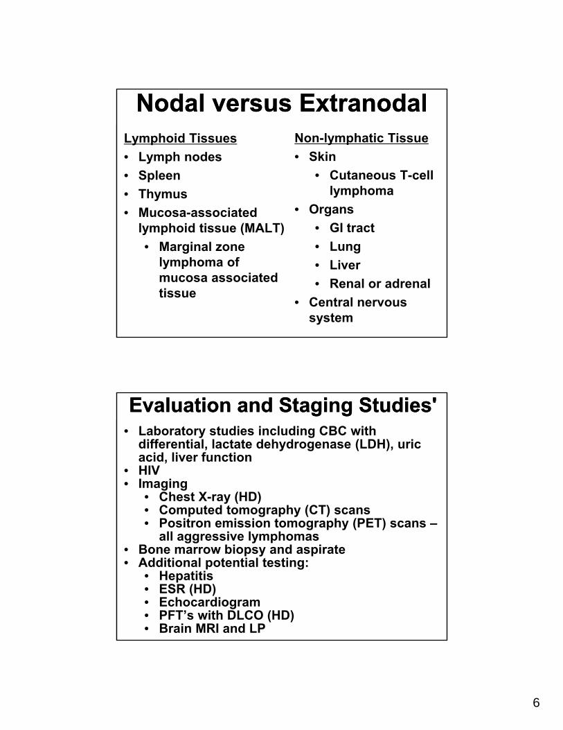
Nodal versus ExtranodalNodal versus ExtranodalLymphoid Tissues
• Lymph nodes
• Spleen
• Thymus
• Mucosa-associated lymphoid tissue (MALT)
• Marginal zone lymphoma of mucosa associated tissue
Non-lymphatic Tissue
• Skin
• Cutaneous T-cell lymphoma
• Organs
• GI tract
• Lung
• Liver
• Renal or adrenal
• Central nervous system
Evaluation and Staging Studies'Evaluation and Staging Studies'• Laboratory studies including CBC with
differential, lactate dehydrogenase (LDH), uric acid, liver function
• HIV• Imaging
• Chest X-ray (HD)• Computed tomography (CT) scans• Positron emission tomography (PET) scans –
all aggressive lymphomas• Bone marrow biopsy and aspirate• Additional potential testing:
• Hepatitis • ESR (HD)• Echocardiogram • PFT’s with DLCO (HD)• Brain MRI and LP
7
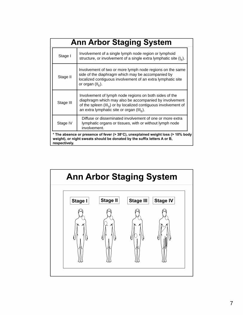
Ann Arbor Staging System
Stage IInvolvement of a single lymph node region or lymphoidstructure, or involvement of a single extra lymphatic site (IE).
Stage II
Involvement of two or more lymph node regions on the sameside of the diaphragm which may be accompanied bylocalized contiguous involvement of an extra lymphatic site or organ (IIE).
Stage III
Involvement of lymph node regions on both sides of the diaphragm which may also be accompanied by involvement of the spleen (IIIS) or by localized contiguous involvement ofan extra lymphatic site or organ (IIIE).
Stage IVDiffuse or disseminated involvement of one or more extralymphatic organs or tissues, with or without lymph node involvement.
* The absence or presence of fever (> 38C), unexplained weight loss (> 10% body weight), or night sweats should be donated by the suffix letters A or B, respectively.
Stage I Stage II Stage III Stage IV
Ann Arbor Staging System

8
SubtypesSubtypes
• Non-Hodgkin’s Lymphoma
• Diffuse Large B Cell Lymphoma
• Burkitt Lymphoma
• Mantle Cell Lymphoma
• Peripheral T Cell Lymphoma
• Hodgkin Lymphoma
Diffuse Large B-cell NHLDiffuse Large B-cell NHL
• Epidemiology• Most common, constitutes 30-40% of adult NHL• Usually presents in middle aged and older
adults, male predominance• Median age 7th decade
• Clinical Features• 30-40% of cases present with early stage
disease• Usually symptomatic and not incidental• Symptoms noticed over weeks to a few months• 1/3 cases arise from low grade process

9
• Rapid growth (weeks) enlarging tumor • Usually rapidly enlarging, symptomatic mass
at a single nodal or extranodal site• bulky palpable mass(es)
• thoracic and abdominal adenopathy without superficial lymph nodes
• Extranodal extension or involvement common• Most common extranodal site is
gastrointestinal region, but CNS, bone, thyroid, testis, lung, kidney, and liver involvement all occur
• Symptoms• Dependent on site of disease involvement• “B” symptoms (fevers, sweats, weight loss)
Diffuse Large B-cell NHLDiffuse Large B-cell NHL
• Laboratory abnormalities• Cytopenias if disease involvement of BM or
enlarged spleen• Elevated lactate dehydrogenase• Elevated uric acid• Spontaneous tumor lysis
Diffuse Large B-cell NHLDiffuse Large B-cell NHL

10
Hodgkin PresentationHodgkin Presentation• Painless adenopathy
• 75% neck, left > right• 25% axillary• 10% inguinal and iliac
• Rarely, alcohol ingestion can induce pain in node
• Pruritus very common• Can predate lymphadenopathy by months,
can delay diagnosis as patient travels to and from dermatologists
• Intense, refractory to topical and oral antihistamines
Hodgkin PresentationHodgkin Presentation• Cough, SOB, DOE in patients with mediastinal
disease• Hemoptysis rarely
• Usually tracks • Subdiaphragmatic presentations uncommon• 1/3 have B-symptoms (fever, NS, weight loss)
• Frequently cause of FUO in older males, very difficult to diagnose in these patients as they often have stage IV disease and small nodes

11
TreatmentTreatment• Chemotherapy based
• R-CHOP standard chemotherapy in most diffuse large B cell lymphoma• Rituximab, cyclophosphamide, doxorubicin,
vincristine and prednisone• ABVD standard chemotherapy in Hodgkin
Disease• doxorubicin, bleomycin, vinblastine and
dacarbazine • More aggressive/intensive regimens used in
some of the other rare but more aggressive lymphomas
• XRT in select cases• Early/limited stage Diffuse Large B cell
lymphoma• Select cases of Early Stage HD• Bulky Hodgkin Disease
ToxicitiesToxicities• R-CHOP
• Infusion reactions, cytopenias, infections, need for transfusion, GI toxicity (nausea/vomiting/diarrhea/constipation/mucositis), alopecia, anorexia, peripheral neuropathy, cardiotoxicity, small risk of secondary malignancy including acute leukemia
• ABVD• Cytopenias, infections, alopecia, muscle
cramps, GI toxicity (nausea/vomiting/diarrhea/constipation/mucositis), phlebitis, peripheral neuropathy, cardiotoxicity, pulmonary toxicity
• Radiation• Dependent on location – local symptoms• Fatigue, cytopenias, skin burn/irritation• CAD or CHF (HD – mediastinal)• Secondary malignancies

12
Lymphoma Emergencies Lymphoma Emergencies
• Presentation• Cord Compression
• SVC Syndrome
• CNS Lymphoma
• Spontaneous Tumor Lysis
• Treatment • Neutropenic Fever
Infectious ComplicationsInfectious Complications
• Most common cause of morbidity and mortality in the cancer patient
• Neutropenia major risk factor for infection
• Hematologic diseases associated with inherent immune defects

13
NeutropeniaNeutropenia• Chemotherapy induced, bone marrow
involvement and heavily pretreated
• Correlation between neutrophil count (both duration and rate of decline) and frequency and severity of infection
• 20% patients ANC ≤ 100 bloodstream infection
• Infectious complications combination of neutropenia, disruption of mucosal barriers, microbial flora shifts
Neutropenic FeverNeutropenic Fever• Fever
• Single oral temperature 38.3 C (101) or higher• 38.0 C (100.4) or higher for an hour
• Neutropenia• ANC less than 500/mcL• ANC less than 1000/mcL with a predicted
decline in the next 48 hours• Weakness, hypotension, syncope, pain, localized
symptoms• Mortality highest among
• Initial neutrophil counts ≤ 100• Prolonged neutropenia (≥ 7 days)• Delay in treatment with broad spectrum
antibiotics

14
• 2/3 patients with fever have occult infection
• Bacteria most common early pathogen
• Coag negative staph, S. aureus, viridans strep, entrerococci
• E. coli, klebsiella, enterobacter, pseudomonas
• HSV, RSV, influenza
• Antibiotic-resistant bacteria, yeast, other fungi, virus common causes of subsequent infections
• Candida, aspergillus
Neutropenic Fever
Evaluation and TreatmentEvaluation and Treatment• Exam with localizing signs (vascular access, mucosal
membranes)• CBC with differential, Chemistry Panel, Liver Function,
Urinalysis/culture, Blood cultures, Chest x-ray• Intravenous antibiotic therapy
• IV broad spectrum monotherapy usually sufficient• Anti-pseudomonal cephalosporin (cefepime or
ceftazidime)• Piperacillin/tazobactam• Imipenem/cilastatin or meropenem
• Vancomycin consideration• Apparent catheter related infections• Gram-positive bacteremia• Known colonization with MRSA• Soft tissue infection• Clinically unstable
• Double gram-negative coverage• High risk for pseudomonas infections
• Clinically unstable

15
• Persistent febrile neutropenia• Anti-fungal coverage after 4 days• CT scans to investigate for source
• Treatment duration dependent on infection but should continue until ANC recovers to ≥ 500
• Removal of vascular access• Immediate in unstable patient• Fungal or mycobacterial bloodstream
infections• S. aureus, P. aeruginosa and VRE strongly
consider
Evaluation and Treatment
Follow-upFollow-up
• Hematologic follow up every 3 months for 2 years, every 4-6 months from years 3-5 and then yearly dependent on patient preference
• Physical exam and blood tests (CBC, LDH in NHL, sedimentation rate in HD if initially elevated
16
SurvivorSurvivor
“ An individual is considered a cancer survivor from the time of diagnosis,
through the balance of his or her life”
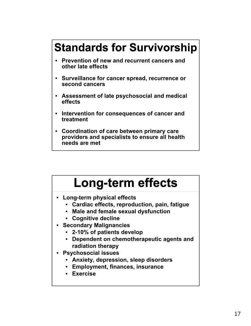
SurvivorshipSurvivorship• Many survivors experience physical and/or
psychosocial effects of cancer and its treatment
• Some evident during anti-cancer therapy (long-term effects)
• Some manifest months or years after therapy (late effects)• Recent review suggests 50% of survivors
suffer from late effects of cancer therapy
• Incidence increases with longer follow up time
• May occur less with new therapies and standards
17
Standards for SurvivorshipStandards for Survivorship• Prevention of new and recurrent cancers and
other late effects
• Surveillance for cancer spread, recurrence or second cancers
• Assessment of late psychosocial and medical effects
• Intervention for consequences of cancer and treatment
• Coordination of care between primary care providers and specialists to ensure all health needs are met
Long-term effectsLong-term effects• Long-term physical effects
• Cardiac effects, reproduction, pain, fatigue• Male and female sexual dysfunction• Cognitive decline
• Secondary Malignancies• 2-10% of patients develop• Dependent on chemotherapeutic agents and
radiation therapy • Psychosocial issues
• Anxiety, depression, sleep disorders• Employment, finances, insurance• Exercise

18
Secondary MalignanciesSecondary Malignancies• Multifactorial (decreased immune surveillance,
treatment related, virally mediated)
• Solid tumors most common• XRT > combined modality > chemotherapy alone
• Breast and lung cancer most common (esp in HD)
• Risk of secondary acute leukemia in both HD and NHL and risk of secondary NHL in HD• Higher risk with chemotherapy
• Alkylating agents
• Highest risk 5-10 years
• Topoisomerase II inhibitors, anthracyclines
• Highest risk 2-3 years
Screening for second malignancyScreening for second malignancy• AGE APPROPRIATE SCREENING and SKIN
EXAMS• Referral to dermatology
• Colonoscopy, prostate, mammogram, PAP smear
• SMOKING CESSATION
• Yearly chest imaging in patients with history of smoking or chest irradiation
• Annual breast screening 5-8 years after completion of therapy or age 40 if history of chest or axillary radiation
• MRI for those receiving between the ages of 10-30
19
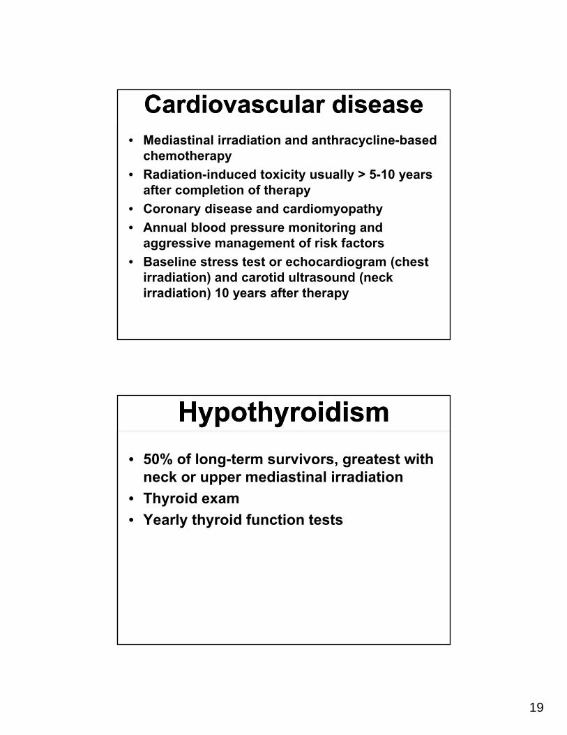
Cardiovascular diseaseCardiovascular disease• Mediastinal irradiation and anthracycline-based
chemotherapy
• Radiation-induced toxicity usually > 5-10 years after completion of therapy
• Coronary disease and cardiomyopathy
• Annual blood pressure monitoring and aggressive management of risk factors
• Baseline stress test or echocardiogram (chest irradiation) and carotid ultrasound (neck irradiation) 10 years after therapy
HypothyroidismHypothyroidism
• 50% of long-term survivors, greatest with neck or upper mediastinal irradiation
• Thyroid exam
• Yearly thyroid function tests

20
MyelosuppressionMyelosuppression
• Usually resolves with time from therapy
• Some delayed myelosuppression
• Concern if new cytopenias develop
• Appropriate immunizations and prevention of infection
ResourcesResources
• Leukemia and Lymphoma Society
• Lymphoma Research Foundation

21
Stem cell transplant in aggressive lymphomasStem cell transplant in aggressive lymphomas
How do you define “cured”?
How do you define “cured”?

22
Beth Christian, MDAssociate Professor of Clinical Internal Medicine
Division of HematologyThe Ohio State University Wexner Medical Center
Indolent Non-Hodgkin Lymphoma
Indolent lymphomaIndolent lymphoma• A heterogeneous group of malignancies
derived from mature B lymphocytes
• Overview the common subtypes
• Follicular lymphoma
• Marginal zone lymphoma, 3 varieties
• Lymphoplasmacytic lymphoma
23
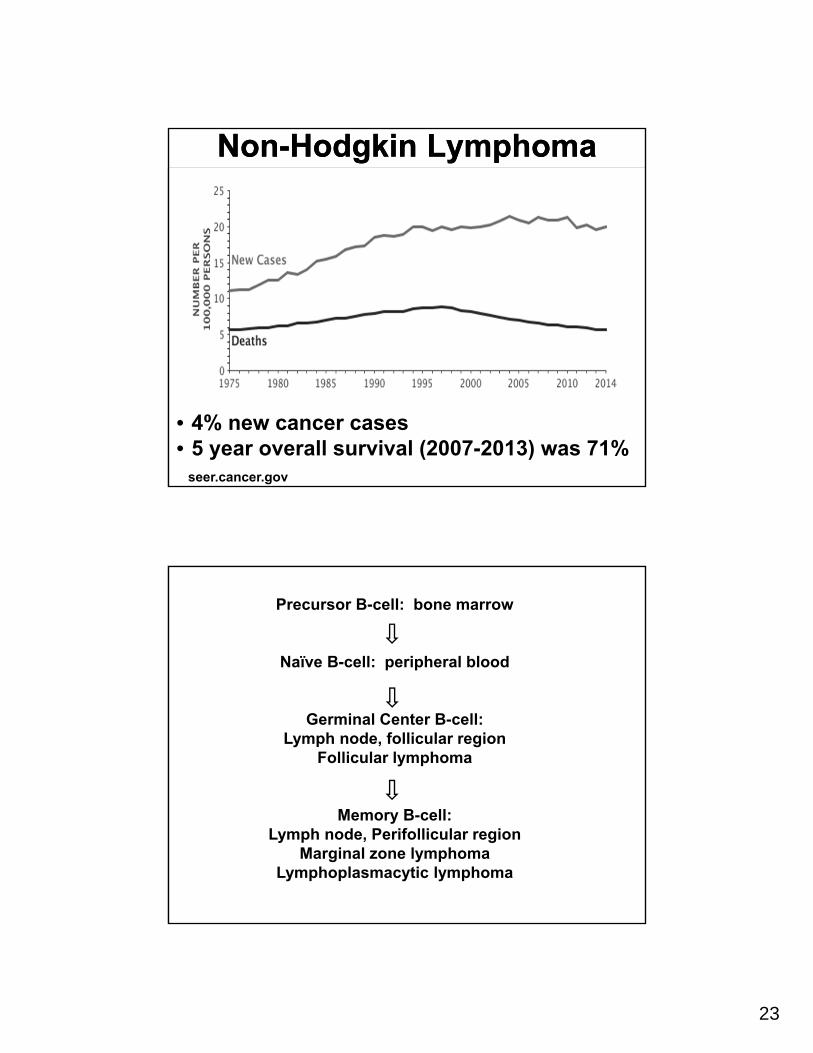
Non-Hodgkin LymphomaNon-Hodgkin Lymphoma
seer.cancer.gov
• 4% new cancer cases• 5 year overall survival (2007-2013) was 71%
Precursor B-cell: bone marrow
Naïve B-cell: peripheral blood
Germinal Center B-cell: Lymph node, follicular region
Follicular lymphoma
Memory B-cell: Lymph node, Perifollicular region
Marginal zone lymphomaLymphoplasmacytic lymphoma

24
Pathologic DiagnosisPathologic Diagnosis
• Morphology• Appearance of the malignant cells• Pattern of involvement within a lymph
node• Immunophenotype or pattern of antigens
expressed on the cell surface• Flow cytometry • Immunohistochemistry
• Cytogenetics
Indolent LymphomaIndolent Lymphoma• Characterized by slow growth over years
• Severe symptoms are uncommon
• Advanced stage indolent lymphoma is not cured with standard therapy
• Therapy for advanced stage indolent lymphoma is given with the goal of treating or preventing complications of the disease
25
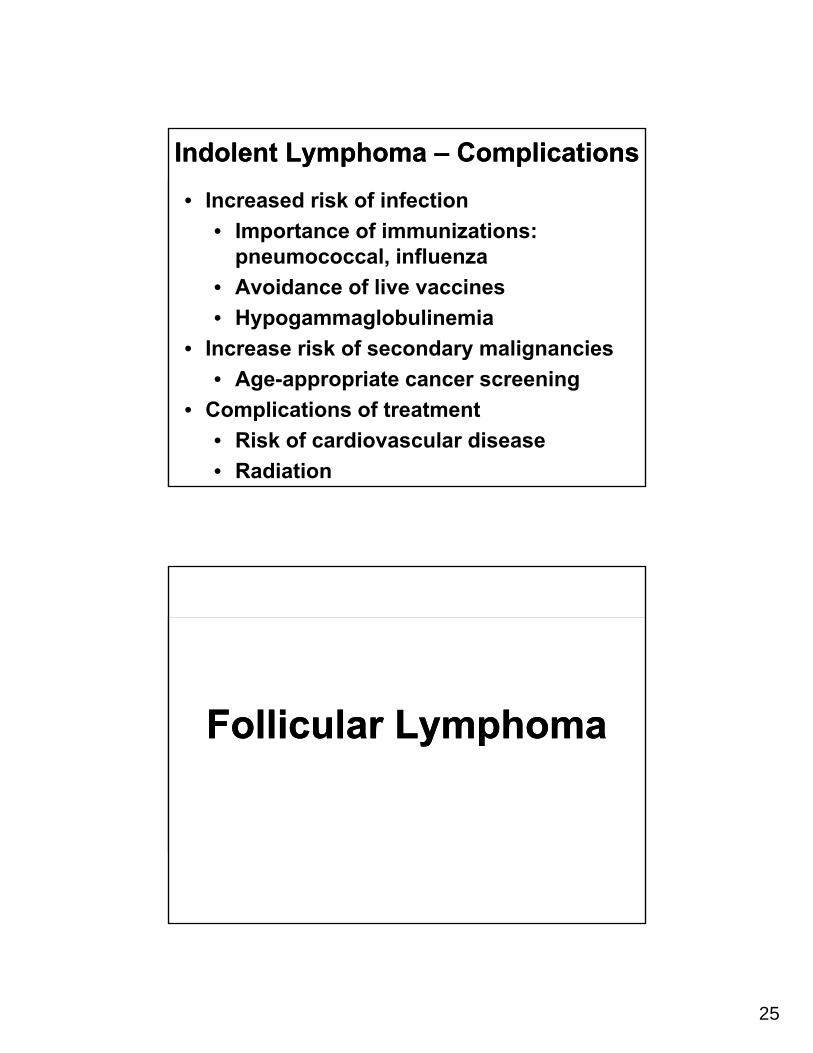
Indolent Lymphoma – ComplicationsIndolent Lymphoma – Complications
• Increased risk of infection
• Importance of immunizations: pneumococcal, influenza
• Avoidance of live vaccines
• Hypogammaglobulinemia
• Increase risk of secondary malignancies
• Age-appropriate cancer screening
• Complications of treatment
• Risk of cardiovascular disease
• Radiation
Follicular LymphomaFollicular Lymphoma
26
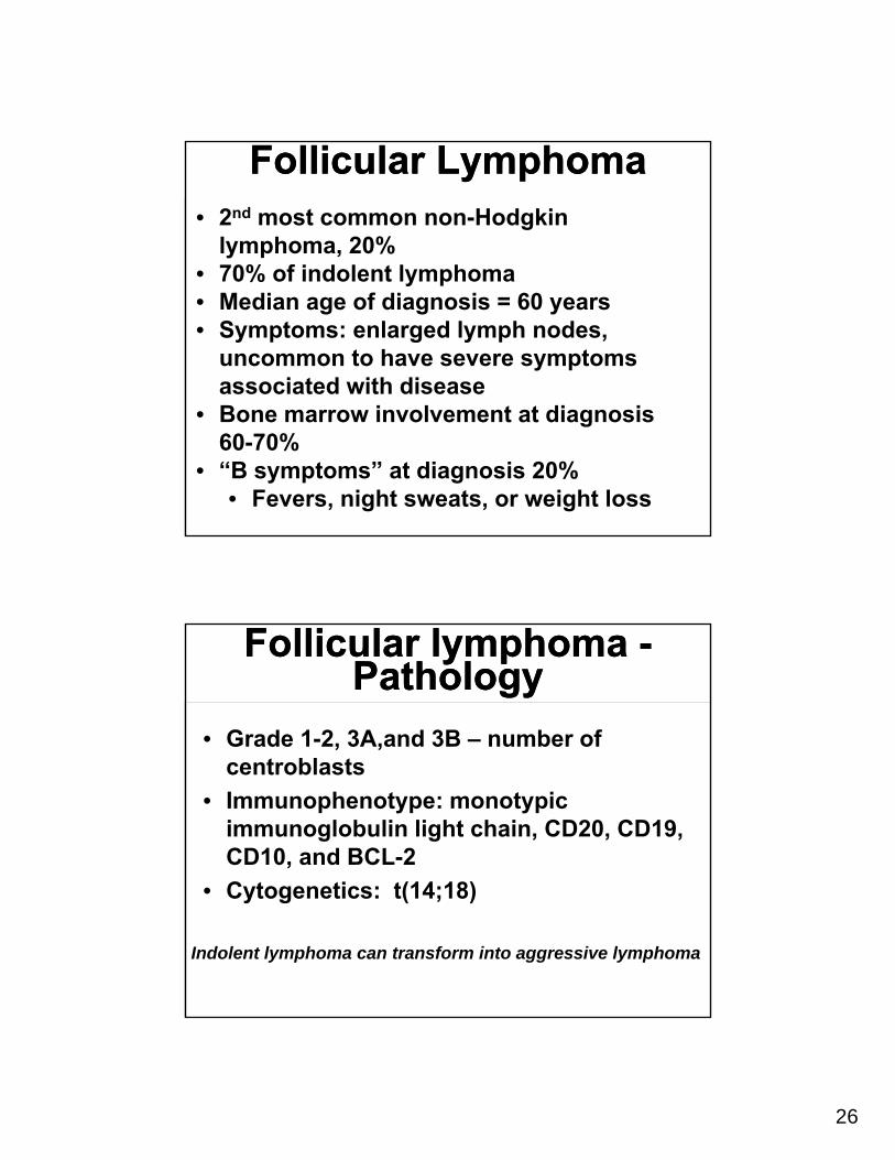
Follicular LymphomaFollicular Lymphoma• 2nd most common non-Hodgkin
lymphoma, 20%• 70% of indolent lymphoma• Median age of diagnosis = 60 years• Symptoms: enlarged lymph nodes,
uncommon to have severe symptoms associated with disease
• Bone marrow involvement at diagnosis 60-70%
• “B symptoms” at diagnosis 20%• Fevers, night sweats, or weight loss
Follicular lymphoma -Pathology
Follicular lymphoma -Pathology
• Grade 1-2, 3A,and 3B – number of centroblasts
• Immunophenotype: monotypic immunoglobulin light chain, CD20, CD19, CD10, and BCL-2
• Cytogenetics: t(14;18)
Indolent lymphoma can transform into aggressive lymphoma

27
Follicular Lymphoma – TreatmentFollicular Lymphoma – Treatment
Treatment decisions are primarily made to improve the quality of a patient’s life:
Questions:
1. How can life be made better with treatment?
2. Is the benefit of treatment greater than the side effects?
3. What treatment is the right one at this point in the patient’s course?
Follicular Lymphoma -Treatment
Follicular Lymphoma -Treatment
• Early Stage – radiation or observation
• Advanced Stage
“Watch and Wait” – Criteria for treatment
• Involvement of ≥3 nodal sites, each with a diameter of ≥3 cm
• Any nodal or extranodal tumor mass with a diameter of ≥7 cm
• B symptoms
• Splenomegaly
• Pleural effusions or peritoneal ascites
• Cytopenias or leukemic involvement
28
Treatment Milestones in Follicular Lymphoma
FDA Approvals
Treatment Milestones in Follicular Lymphoma
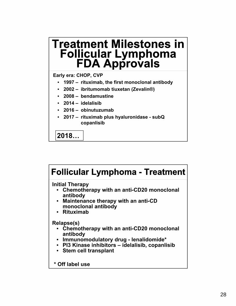
FDA ApprovalsEarly era: CHOP, CVP
• 1997 – rituximab, the first monoclonal antibody
• 2002 – ibritumomab tiuxetan (Zevalin®)
• 2008 – bendamustine
• 2014 – idelalisib
• 2016 – obinutuzumab
• 2017 – rituximab plus hyaluronidase - subQ copanlisib
2018…
Follicular Lymphoma - TreatmentFollicular Lymphoma - TreatmentInitial Therapy
• Chemotherapy with an anti-CD20 monoclonal antibody
• Maintenance therapy with an anti-CD monoclonal antibody
• Rituximab
Relapse(s)• Chemotherapy with an anti-CD20 monoclonal
antibody• Immunomodulatory drug - lenalidomide*• PI3 Kinase inhibitors – idelalisib, copanlisib• Stem cell transplant
* Off label use

29
RituximabRituximab• First monoclonal antibody approved for
treatment
• Chimeric – both mouse and human components
• Binds to CD20 on B-cells, normal and malignant
• Antibody-dependent cell-mediated cytotoxicity
• Natural killer cells recognize the antibody bound to the cancer cell and release cytokines and cytotoxic granules
• Complement-dependent cytotoxicity
• The antibody activates complement and the membrane attack complex destroys the cell
• Depletes B-cells for up to 6 months
RituximabRituximab• A major advance in the treatment of follicular
lymphoma • First targeted treatment• Side effects
• Infusion reactions• Rash, potentially severe• Increased risk for infections• Hepatitis B reactivation
• Model for the development of all monoclonal antibody therapy• Obinutuzumab

30
Follicular LymphomaFollicular Lymphoma• The prognosis of follicular lymphoma
continues to improve
• A chronic disease, requiring intermittent treatment
• Median overall survival ~ 20 years
• Allogeneic stem cell transplant is a potentially treatment
• Clinical Trials
Marginal Zone LymphomaMarginal Zone Lymphoma
MALT, gastricMALT, non-gastricSplenic MZLNodal MZL
31
Marginal Zone LymphomaMarginal Zone Lymphoma
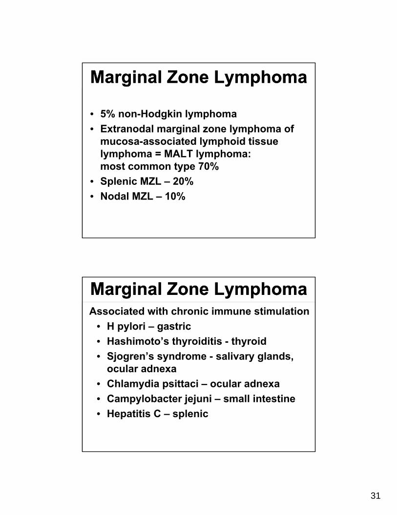
• 5% non-Hodgkin lymphoma
• Extranodal marginal zone lymphoma of mucosa-associated lymphoid tissue lymphoma = MALT lymphoma: most common type 70%
• Splenic MZL – 20%
• Nodal MZL – 10%
Marginal Zone LymphomaMarginal Zone LymphomaAssociated with chronic immune stimulation
• H pylori – gastric
• Hashimoto’s thyroiditis - thyroid
• Sjogren’s syndrome - salivary glands, ocular adnexa
• Chlamydia psittaci – ocular adnexa
• Campylobacter jejuni – small intestine
• Hepatitis C – splenic
32
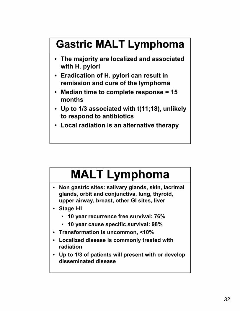
Gastric MALT LymphomaGastric MALT Lymphoma• The majority are localized and associated
with H. pylori
• Eradication of H. pylori can result in remission and cure of the lymphoma
• Median time to complete response = 15 months
• Up to 1/3 associated with t(11;18), unlikely to respond to antibiotics
• Local radiation is an alternative therapy
MALT LymphomaMALT Lymphoma• Non gastric sites: salivary glands, skin, lacrimal
glands, orbit and conjunctiva, lung, thyroid, upper airway, breast, other GI sites, liver
• Stage I-II
• 10 year recurrence free survival: 76%
• 10 year cause specific survival: 98%
• Transformation is uncommon, <10%
• Localized disease is commonly treated with radiation
• Up to 1/3 of patients will present with or develop disseminated disease
33
Splenic Marginal Zone Lymphoma
Splenic Marginal Zone Lymphoma
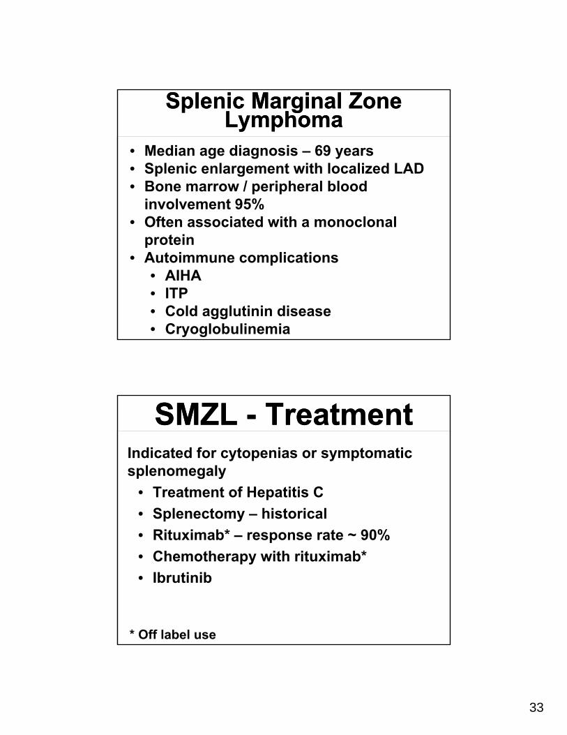
• Median age diagnosis – 69 years• Splenic enlargement with localized LAD• Bone marrow / peripheral blood
involvement 95%• Often associated with a monoclonal
protein• Autoimmune complications
• AIHA• ITP• Cold agglutinin disease• Cryoglobulinemia
SMZL - TreatmentSMZL - TreatmentIndicated for cytopenias or symptomatic splenomegaly
• Treatment of Hepatitis C
• Splenectomy – historical
• Rituximab* – response rate ~ 90%
• Chemotherapy with rituximab*
• Ibrutinib
* Off label use

34
Marginal Zone LymphomaMarginal Zone Lymphoma
• Ibrutinib – the first and only FDA approved therapy (2017)
• Bruton’s tyrosine kinase inhibitor
• Side effects
• Increased bleeding risk
• Diarrhea
• Rash
• Atrial fibrillation
• Infection
Nodal MZLNodal MZL
• 1% of NHL, 10% of MZL
• Median age of diagnosis: 50-64 years
• Bone marrow involvement in approximately 1/3
• Association with Hepatitis C
• Transformation occurs in approximately 15%
35
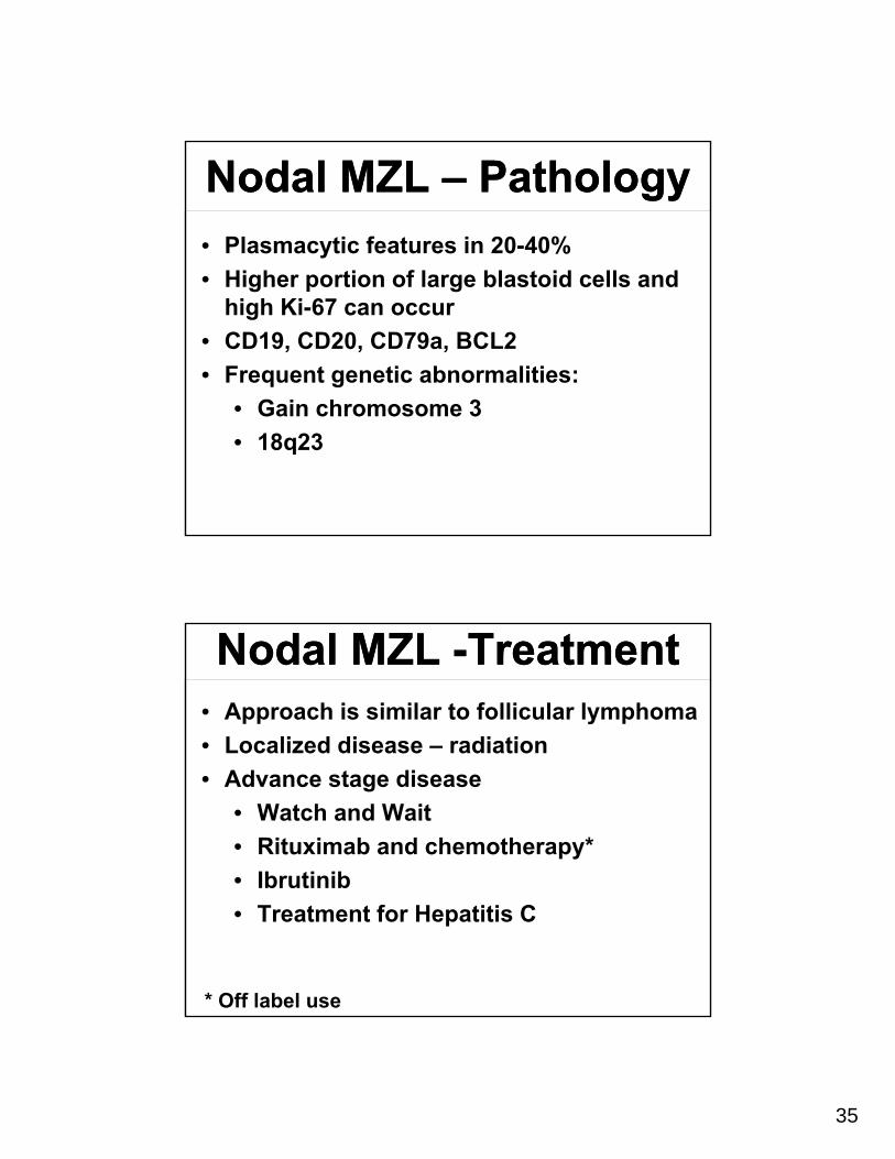
Nodal MZL – PathologyNodal MZL – Pathology
• Plasmacytic features in 20-40%
• Higher portion of large blastoid cells and high Ki-67 can occur
• CD19, CD20, CD79a, BCL2
• Frequent genetic abnormalities:
• Gain chromosome 3
• 18q23
Nodal MZL -TreatmentNodal MZL -Treatment• Approach is similar to follicular lymphoma
• Localized disease – radiation
• Advance stage disease
• Watch and Wait
• Rituximab and chemotherapy*
• Ibrutinib
• Treatment for Hepatitis C
* Off label use

36
Lymphoplasmacytic Lymphoma
Lymphoplasmacytic Lymphoma
Waldenström macroglobulinemia
Lymphoplasmacytic lymphoma &Waldenström macroglobulinemia (WM)
Lymphoplasmacytic lymphoma &Waldenström macroglobulinemia (WM)
• 1% of Non-Hodgkin Lymphoma
• Median age diagnosis: 73 years
• Male:Female 1.6:1
• Lymphoplasmacytic lymphoma with an IgM monoclonal protein = Waldenström macroglobulinemia
• Non-IgM associated LPL is very rare
• Familial association

37
WM - PathologyWM - PathologyImmunophenotype
• sIgM, CD19, CD20, CD22, CD79
• Up to 20% can express CD5, CD10, or CD23
Cytogenetics
• Chromosome 6q deletions in up to half of patients
• MYD88 somatic mutations are present in >90%
WM – Signs/SymptomsWM – Signs/Symptoms
• Anemia – most common presentation
• Elevated total protein and a monoclonal protein
• Bone marrow involvement is very common
• Lymphadenopathy: 15%
• Splenomegaly: 10%
• AIHA, ITP
• Cryglobulinemia, Cold agglutinin disease

38
WM – Signs/SymptomsWM – Signs/Symptoms• Neuropathy
• 20% patients at presentation• distal, symmetric, and slowly progressive
sensorimotor peripheral neuropathy causing paresthesias and weakness
• Antibodies: Anti-myelin-associated glycoprotein and GM1 ganglioside
• Renal complications• Direct infiltration• Immune mediated glomerulonephritis
• Amyloid• Skin involvement / vasculitis• Bing Neel Syndrome = CNS involvement
WM – Signs/SymptomsWM – Signs/SymptomsIgM
• Pentamer and the largest antibody• Accumulation of IgM can result in
increased blood viscosity• IgM ~ 6000 mg/dL but can vary
• There is no definitive viscosity level where symptoms occur but typically ~ 4 cP
• Use caution with PRBC transfusions –increasing hematocrit will increase viscosity
39
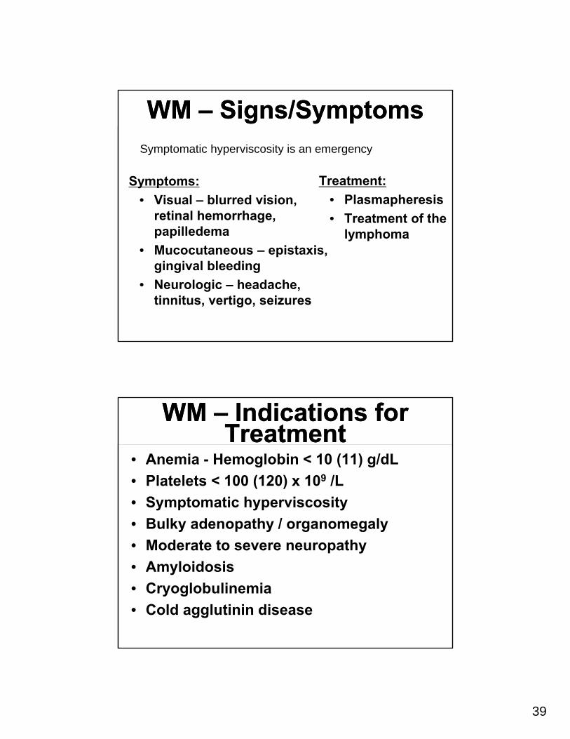
WM – Signs/SymptomsWM – Signs/SymptomsSymptomatic hyperviscosity is an emergency
Symptoms:
• Visual – blurred vision, retinal hemorrhage, papilledema
• Mucocutaneous – epistaxis, gingival bleeding
• Neurologic – headache, tinnitus, vertigo, seizures
Treatment:
• Plasmapheresis
• Treatment of the lymphoma
WM – Indications for Treatment
WM – Indications for Treatment
• Anemia - Hemoglobin < 10 (11) g/dL
• Platelets < 100 (120) x 109 /L
• Symptomatic hyperviscosity
• Bulky adenopathy / organomegaly
• Moderate to severe neuropathy
• Amyloidosis
• Cryoglobulinemia
• Cold agglutinin disease
40
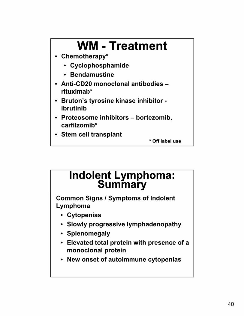
WM - TreatmentWM - Treatment• Chemotherapy*
• Cyclophosphamide
• Bendamustine
• Anti-CD20 monoclonal antibodies –rituximab*
• Bruton’s tyrosine kinase inhibitor -ibrutinib
• Proteosome inhibitors – bortezomib, carfilzomib*
• Stem cell transplant* Off label use
Indolent Lymphoma: Summary
Indolent Lymphoma: Summary
Common Signs / Symptoms of Indolent Lymphoma
• Cytopenias
• Slowly progressive lymphadenopathy
• Splenomegaly
• Elevated total protein with presence of a monoclonal protein
• New onset of autoimmune cytopenias

41
Indolent Lymphoma: SummaryIndolent Lymphoma: Summary• The mainstay of therapy is to only treat
when needed
• The prognosis of indolent lymphomas is very good and improving over time
• We are less reliant on traditional chemotherapy and have increasing options for treatment
• Clinical trials – new options are on the horizon
• Allogeneic stem cell transplant offers a potentially curative option


![2. HODGKIN'S & NON-HODGKIN'S LYMPHOMA [A] Diagnosis and · PDF file · 2014-07-31HODGKIN'S & NON-HODGKIN'S LYMPHOMA [A] Diagnosis and Staging ... unstained BM or peripheral blood](https://static.fdocuments.us/doc/165x107/5aa8642b7f8b9a72188b8bcf/2-hodgkins-non-hodgkins-lymphoma-a-diagnosis-and-2014-07-31hodgkins-non-hodgkins.jpg)


![Immunosuppressant Medications Final - Handout.ppt. 2000;47:291-298 DMARDs ... Microsoft PowerPoint - Immunosuppressant Medications Final - Handout.ppt [Compatibility Mode] ...](https://static.fdocuments.us/doc/165x107/5afd1d6e7f8b9a444f8d00a7/immunosuppressant-medications-final-200047291-298-dmards-microsoft-powerpoint.jpg)