Lymphoma - Alberta Health Services · Medical journal articles were searched using Medline (1950 to...
152
CLINICAL PRACTICE GUIDELINE LYHE-002 Version 12 LYMPHOMA Effective Date: September 2019 The recommendations contained in this guideline are a consensus of the Alberta Provincial Hematology Tumour Team synthesis of currently accepted approaches to management, derived from a review of relevant scientific literature. Clinicians applying these guidelines should, in consultation with the patient, use independent medical judgment in the context of individual clinical circumstances to direct care.
Transcript of Lymphoma - Alberta Health Services · Medical journal articles were searched using Medline (1950 to...

CLINICAL PRACTICE GUIDELINE LYHE-002Version 12
LYMPHOMA
Effective Date: September 2019
The recommendations contained in this guideline are a consensus of the Alberta Provincial Hematology TumourTeam synthesis of currently accepted approaches to management, derived from a review of relevant scientific
literature. Clinicians applying these guidelines should, in consultation with the patient, use independent medicaljudgment in the context of individual clinical circumstances to direct care.

CLINICAL PRACTICE GUIDELINE LYHE-002
Version 11
Page 2 of 5
Table of Contents
Background
Guideline Questions
Development and Revision History
Search Strategy
Target Population
Discussion
I. Diagnosis and Pathologic Classifications
II. Staging
III. Treatment of Non-Hodgkin Lymphomas
IV. Cutaneous Lymphoma
V. Hodgkin Lymphoma
VI. HDCT and Hematopoietic Stem Cell Transplantation for Lymphoma
VII. Supportive Care in the Treatment of Lymphoma
VIII. Follow-Up Care in the Treatment of Lymphoma
Glossary of Abbreviations
Dissemination
Maintenance
Conflict of Interest
Appendix A (Chemotherapy Regimens)
Appendix B: General Radiotherapy Guidelines
Appendix C: Prognostic Models
Appendix D: Lymphoma Response Criteria
Appendix E: New Lymphoma Patient Data Sheet
Appendix F: Ideal Body Weight

CLINICAL PRACTICE GUIDELINE LYHE-002
Version 11
Page 3 of 5
BACKGROUND Lymphomas encompass a group of lymphoproliferative malignant diseases that originate from T- and B-cells in the lymphatic system. Traditionally, lymphomas have been subcategorized into two groups: Hodgkin lymphoma and non-Hodgkin lymphoma. It is now known however, that Hodgkin lymphoma is simply one of the numerous varieties of lymphoma, and that non-Hodgkin lymphoma is a fairly meaningless term, representing all of the other subtypes of this disease. Non-Hodgkin lymphoma involves a heterogeneous group of over 40 lymphoproliferative malignancies with diverse patterns of behaviours and responses to treatments. Non-Hodgkin lymphoma is much less predictable than Hodgkin lymphoma and prognosis depends on the histologic type, stage, and treatment. In Canadian males and females, the incidence rates for non-Hodgkin lymphoma showed a marked increase by approximately 50% between 1978 and the late 1990s, but have since stabilized.1 Mortality rates have followed a similar pattern. The clearest risk factor for the disease is immunosuppression associated with HIV infection, or medications used to prevent rejection in organ transplantation. Other factors that increase risk of non-Hodgkin lymphoma are poorly understood but may include occupational exposures to pesticides, herbicides, and dioxins, as well as chronic immune stimulation associated with autoimmune disorders (e.g. thyroiditis, Sjogren’s Syndrome, SLE) or infections (e.g. Helicobacter pylori gastritis, hepatitis C virus).2 In 2015, it is estimated that 8200 new cases of non-Hodgkin lymphoma will be diagnosed in Canada, and 2650 deaths will occur, making non-Hodgkin lymphoma the sixth most common cause of cancer-related death in Canada.3
Hodgkin lymphoma is a malignancy characterized histopathologically by the presence of Reed-Sternberg cells in the appropriate cellular background. Although rare, Hodgkin lymphoma is one of the best-characterized malignancies of the lymphatic system and one of the most readily curable forms of malignant disease.2 The incidence rate has remained fairly steady over time, it is estimated that approximately 1000 new cases of Hodgkin lymphoma are diagnosed in Canada each year.3 It is important to note that lymphoma also represents the most commonly diagnosed non-epithelial cancers in adolescents and young adults in Canada. Between 1992 and 2005, 5577 new cases of Hodgkin and non-Hodgkin lymphoma were diagnosed in Canadians aged 15-29 years.1 The following guidelines do not address lymphoma in the pediatric or adolescent populations. GUIDELINE QUESTIONS • What are the diagnostic criteria for the most common lymphomas? • What are the staging and re-staging procedures for Hodgkin and non-Hodgkin lymphomas? • What are the recommended treatment and management options for Hodgkin and non-Hodgkin
lymphomas? • What are the recommended follow-up procedures for patients with malignant Hodgkin and non-
Hodgkin lymphoma? DEVELOPMENT AND REVISION HISTORY This updated guideline was reviewed and endorsed by the Alberta Provincial Hematology Tumour Team. Members of this team include hematologists, medical oncologists, radiation oncologists, surgical oncologists, nurses, nurse-practitioners, hematopathologists, and pharmacists. Updated evidence was selected and reviewed by members from the Alberta Provincial Hematology Tumour Team and a Knowledge Management Specialist from the Guideline Resource Unit. The draft guideline was circulated to all tumour team members for comment and approval, and all comments were reviewed by the tumour team lead and incorporated into the final version of the guideline, where appropriate. A detailed

CLINICAL PRACTICE GUIDELINE LYHE-002
Version 11
Page 4 of 5
description of the methodology followed during the guideline development and updating process can be found in the Guideline Resource Unit Handbook. The original guideline was developed in March 2006 and was revised on the following dates: May 2007, June 2009, November 2009 January 2011, December 2011, September 2012, April 2013, December 2014, December 2015, February 2016 and April 2016. SEARCH STRATEGY Medical journal articles were searched using Medline (1950 to October Week 1, 2015), EMBASE (1980 to October Week 1, 2015), Cochrane Database of Systematic Reviews (3rd Quarter, 2015), and PubMed electronic databases. An updated review of the relevant existing practice guidelines for lymphoma was also conducted by accessing the websites of the National Comprehensive Cancer Network (NCCN), Cancer Care Ontario (CCO), the British Columbia Cancer Agency (BCCA), the European Society for Medical Oncology (ESMO), and the British Committee for Standards in Haematology. TARGET POPULATION The following guidelines apply to adults over 18 years of age. Different principles may apply to pediatric and adolescent patients.

CLINICAL PRACTICE GUIDELINE LYHE-002Version 11
Page 5 of 5
REFERENCES
1. Canadian Cancer Society’s Steering Committee. Canadian Cancer Statistics 2009. Special Topic: Cancer in
Adolescents and Young Adults. April 2009; Available at: http://www.cancer.ca/Canada-
wide/About%20cancer/Cancer%20statistics/~/media/CCS/Canada%20wide/Files%20List/English%20files%20h
eading/pdf%20not%20in%20publications%20section/Stats%202009E%20Special%20Topics.ashx. Accessed
October 17, 2011.
2. Marcus R. Lymphoma: pathology, diagnosis, and treatment. 14th ed. New York: Cambridge University Press;
2007.
3. Canadian Cancer Society’s Steering Committee on Cancer Statistics. Canadian Cancer Statistics 2011. May 2011;
Available at: http://www.cancer.ca/Canada-
wide/About%20cancer/~/media/CCS/Canada%20wide/Files%20List/English%20files%20heading/PDF%20-
%20Policy%20-%20Canadian%20Cancer%20Statistics%20-
%20English/Canadian%20Cancer%20Statistics%202011%20-%20English.ashx. Accessed October 17, 2011.

CLINICAL PRACTICE GUIDELINE LYHE-002Version 12
I. Diagnosis and Pathologic Classification
Page 1 of 5
DISCUSSION
I. DIAGNOSIS AND PATHOLOGIC CLASSIFICATION1-6
An excisional lymph node biopsy of the largest regionally involved lymph node is the optimal specimen forinitial diagnostic assessment. Similarly, a sizable biopsy from the organ of origin in extranodal lymphomasis also suitable. Compelling clinical contraindications to an open biopsy should be present beforeconsidering any other options. A careful clinical examination or radiological investigations for moreaccessible or palpable pathologic adenopathy could be useful in decision making prior to opting for alesser diagnostic specimen. Fine needle aspirate biopsies are inadequate for the initial diagnosis oflymphoma. These latter specimens may provide adequate material for evaluating possible relapse,clarification of staging at questionable sites and as a source of additional specimen where required forfurther special testing or research. Occasionally,a generous core needle biopsy comprising many coresamples with sufficient material to perform the appropriate ancillary techniques required for diagnosticassessment (immunohistochemistry, flow cytometry, PCR for IgH and TCR gene rearrangements, andFISH for major translocations) may supply adequate tissue, in cases when a lymph node is not easilyaccessible for excisional or incisional biopsy. A reference lymphoma pathologist should confirm lymphomadiagnoses in each and every case. This is particularly important in cases when only a core needle biopsyis available, and whenever requested by the treating clinician.
Table 1 describes the histologic subclassification of the malignant lymphomas, and is an adaptation of the most recent WHO classification6. This classification is based on the light microscopic interpretationcomplemented by special stains, immunophenotyping, cytogenetics and other ancillary information asavailable. The specific lymphomas are divided into three major groups, according to the degree of clinicalaggressiveness, for treatment planning. All B-cell lymphomas should be immuno-phenotyped to determineif they are CD20 positive.

CLINICAL PRACTICE GUIDELINE LYHE-002Version 12
I. Diagnosis and Pathologic Classification
Page 2 of 5
Table 1. Lymphoma classification6.
B-cell T-cell
Ind
ole
nt
Follicular, grades 1-2, 3aSmall lymphocytic Lymphoma/Chronic Lymphocytic LeukemiaMarginal zone, extranodal (MALT)Splenic marginal zoneMarginal zone, nodal (monocytoid B-cell)Lymphoplasmacytic (Waldenström’s macroglobulinemia)Primary cutaneous, follicle centreHairy cell leukemiaNodular lymphocyte predominant Hodgkin LymphomaMantle cell (can be aggressive)
Mycosis fungoides /Sezary syndromePrimary cutaneous, CD30+Primary cutaneous perioheral T-cell lymphoma PTCL,CD30-T-cell large granular lymphocytic leukemia
Ag
gre
ss
ive
Diffuse large B-cell
o T-cell/histocyte-rich DLBCL
o Primary DLBCL of the CNS
o Primary cutaneous DLBCL, leg-type
o EBV-positive DLBCL of the elderlyDLBCL associated with chronic inflammationLymphomatoid granulomatosisPrimary mediastinal large B-cellIntravascular large B-cellALK positive large B-cellPlasmablastic lymphomaLBCL in HHV8-associated Castleman diseasePrimary effusion lymphomaFollicular grade 3b (large cell)Classical Hodgkin lymphoma
⇒ Nodular sclerosis
⇒ Mixed cellularity
⇒ Lymphocyte rich
⇒ Lymphocyte depleted
Peripheral T-cell, unspecifiedAngioimmunoblastic (AITL. formerly AILD)Enteropathy associated T-cellHepatosplenic T-cellSubcutaneous panniculitis-likeAnaplastic large cell (CD30+) ALK+Anaplastic large cell (CD30+) ALK-Extranodal NK/T-cell, nasal type
Sp
ec
ial
Burkitt lymphomaIntermediate between DLBCL and BLIntermediate between DLBCL and Hodgkin lymphomaB lymphoblastic leukemia/lymphomaB prolymphocytic leukemiaLymphomas associated with HIV infectionLymphomas associated with primary immune disordersPost-transplant lymphoproliferative disorders (PTLD)
o Plasmacytic hyperplasia and infectiousmononucleosis-like PTLD
o Polymorphic PTLD
o Monomorphic PTLD
o Classical Hodgkin-type PTLDOther iatrogenic immunodeficiency-associated lymphomas
T lymphoblastic leukemia/lymphomaAdult T-cell leukemia/lymphoma (ATLL)T prolymphocytic leukemia

CLINICAL PRACTICE GUIDELINE LYHE-002Version 12
I. Diagnosis and Pathologic Classification
Page 3 of 5
Required Immunohistochemical and Ancillary Testing for Lymphoma
In general, guidelines for using the various ancillary methods, includingimmunohistochemical andfluorescence in situ hybridization (FISH) testing as outlined in the most recent version of the World HealthOrganization Classification of Tumours of Haematopoietic and Lymphoid Tissues should be followed so asto confirm a specific diagnosis and provide necessary prognostic and/or predictive information6. Inaddition, the following are recommended by the Alberta Provincial Hematology Tumour Team7,8:
1. Classical Hodgkin Lymphoma: The immunohistochemical panel may includeCD45/CD3/CD20/CD30/CD15/ PAX5/MUM1 and should be selected on a case by case basis at thediscretion of the hematopathologist. EBV studies by in situ hybridization (EBER) may be considered ifdifficulty exists diagnostically, as most cases of the mixed-cellularity subtype of classical Hodgkinlymphoma are EBER positive.
2. Diffuse Large B-Cell Lymphoma (DLBCL):• Immunohistochemical (IHC) panels to distinguish between Activated B Cell (ABC) type and
Germinal Centre B-cell (GCB) cell of origin (COO) types have limitations (regardless of whichalgorithm is employed)when compared to gene expression profiling8,9. However, GCB vs non-GCBCOO by IHC does correlate with survival rates following RCHOP chemotherapy, and therefore addsprognostic information when managing DLBCL. The Alberta hematopathologists currently use asimple algorithm published by Hans et al, requiring IHC stains for CD10, BCL6 and MUM1, in whichCD10+ or BCL6+/ MUM1- cases are designated as GCB COO, whereas cases negative fornegative/BCL6+/MUM1+ phenotype are considered to have a non-GCB COO.
• EBER and CD5 expression confer worse prognosis, and may be used to identify various clinical-pathological entities with distinct implications. Determining CD5 expression should be considered onall DLBCL cases. EBER should be performed in patients with immune suppression relatedlymphomas, or those who possibly have EBV-related DLBCL (consider past the age of 50)10
• Rearrangments of the C-MYC gene as determined by FISH, especially in association with BCL2and/or BCL6 (so called "double hit" or "triple hit" disease) are associated with very poor outcomesfollowing R-CHOP therapy, as well as high rates of central nervous system relapse. Patients with adouble-hit or triple-hit lymphoma under age 70 years should receive more aggressive therapy andpossibly stem cell transplantation. Though it represents approximately only 5-10% of DLBCLcases11, it is very important to recognize these patients, and therefore, MYC rearrangement testingby FISH is to be performed on all patients younger than 70 y.o. with the appropriate lymphomahistology, i.e. DLBCL or lymphoma that are so called "unclassifiable" with intermediate morphologicalfeatures between DLBCL and Burkitt. If MYC is rearranged, the case should also undergo BCL2 andBCL6 rearrangement testing by FISH. MYC and BCL2 test results are required within 2 weeks ofdiagnoses for all new patients within the appropriate diagnostic category and age group. FISHtesting may also be performed in select instances at the discretion of the reportinghematopathologist if such studies are deemed diagnostically useful.
• Immunohistochemical studies cannot be used as a surrogate for MYC rearrangement.• However, the detection of MYC and BCL2 concurrent overexpression by IHC in so-called “dual
expressor” DLBCL, identifies a numerically significant subset of the DLBCL with potentially similaraggressive behavior compared to double-hit lymphoma cases, but representing a distinct group ofpatients (more often an ABC subtype as opposed to double hit DLBCL which are usually GCB). Thisgroup is also associated with a high rate of CNS relapse11. Therefore, provided adequatebenchmarks and interpretation standards can be established for reproducibility, IHC for MYC andBCL2 expression should also be strongly considered on all DLBCL cases9,12.

CLINICAL PRACTICE GUIDELINE LYHE-002Version 12
I. Diagnosis and Pathologic Classification
Page 4 of 5
3. Follicular Lymphoma: must document grade (1-2, 3a or 3b), because all grade 3b should receive R-CHOP rather than other chemotherapy regimens. Also, if a diffuse pattern is present, this should bespecified and a relative proportion noted, as outlined in the WHO Classification.
4. Peripheral T-Cell Lymphoma: cytotoxic T-cell markers (CD8/CD57/Granzyme B) correlate with poorprognosis and should be considered. Notably, however, peripheral T cell lymphomas are not classifiedon the basis of these phenotypic markers. EBV studies by in situ hybridization (EBER) should beperformed in cases where angioimmunoblastic T cell lymphoma (AITL) and extranodal T/NK celllymphoma, nasal type enter in the differential diagnosis.
5. Mantle Cell Lymphoma: Evidence of CyclinD1 deregulation confirmed by IHC (positive staining forCyclinD1) and/or FISH (positive for t(11;14)) is needed to confirm the diagnosis, provided othermorphophenotypic findings are consistent with the diagnosis. Poor prognostic features must bementioned in the report, including blastoid and pleomorphic morphologic variants. The proliferationindex as measured by Ki67 or Mib-1 (used to calculate MIPI score) is to be reported. In cases where itis difficult to differentiate MCL from CLL, flow cytometry for CD200 and IHC for SOX11 may beperformed13. For patients who are deemed transplant-eligible (i.e. age <65 and fit for intensivetherapy), TP53 mutational testing should be performed at time of diagnosis to identify high-risk patientsmore appropriate for allogeneic stem cell transplantation14.

CLINICAL PRACTICE GUIDELINE LYHE-002Version 12
I. Diagnosis and Pathologic Classification
Page 5 of 5
REFERENCES
1. Harris NL, Jaffe ES, Stein H, Banks PM, Chan JK, Cleary ML, et al. A revised European-American classification oflymphoid neoplasms: a proposal from the International Lymphoma Study Group. Blood 1994 Sep 1;84(5):1361-1392.
2. Armitage JO, Weisenburger DD. New approach to classifying non-Hodgkin's lymphomas: clinical features of themajor histologic subtypes. Non-Hodgkin's Lymphoma Classification Project. J Clin Oncol 1998 Aug;16(8):2780-2795.
3. A clinical evaluation of the International Lymphoma Study Group classification of non-Hodgkin's lymphoma. TheNon-Hodgkin's Lymphoma Classification Project. Blood 1997 Jun 1;89(11):3909-3918.
4. International Non-Hodgkin's Lymphoma Prognostic Factors Project. A predictive model for aggressive non-Hodgkin'slymphoma. N Engl J Med 1993 Sep 30;329(14):987-994.
5. Armitage JO. Treatment of non-Hodgkin's lymphoma. N Engl J Med 1993 Apr 8;328(14):1023-1030.6. Swerdlow SH, Campo E, Harris NL, Jaffe ES, Pileri SA, Stein H. World Health Organization Classification of Tumours
of Haematopoietic and Lymphoid Tissues. Lyon, France: International Agency for Research on Cancer (iARC);2008.
7. Meyer PN, Fu K, Greiner TC, Smith LM, Delabie J, Gascoyne RD, et al. Immunohistochemical methods for predictingcell of origin and survival in patients with diffuse large B-cell lymphoma treated with rituximab. J Clin Oncol 2011Jan 10;29(2):200-207.
8. Gutierrez-Garcia G, Cardesa-Salzmann T, Climent F, Gonzalez-Barca E, Mercadal S, Mate JL, et al. Gene-expression profiling and not immunophenotypic algorithms predicts prognosis in patients with diffuse large B-celllymphoma treated with immunochemotherapy. Blood 2011 May 5;117(18):4836-4843.
9. Savage KJ, Sehn LH, Villa D, Kansara RR, Mottok A, Ennishi D, et al. The Impact of Concurrent MYC BCL2 ProteinExpression on the Risk of Secondary Central Nervous System Relapse in Diffuse Large B-Cell Lymphoma(DLBCL). Blood 2014;124(21):495.
10. Jain P, Fayad LE, Rosenwald A, Young KH, O'Brien S. Recent advances in de novo CD5+ diffuse large B celllymphoma. Am J Hematol 2013 Sep;88(9):798-802.
11. Petrich AM, Nabhan C, Smith SM. MYC-associated and double-hit lymphomas: a review of pathobiology,prognosis, and therapeutic approaches. Cancer 2014 Dec 15;120(24):3884-3895.
12. Hu S, Xu-Monette ZY, Tzankov A, Green T, Wu L, Balasubramanyam A, et al. MYC/BCL2 protein coexpressioncontributes to the inferior survival of activated B-cell subtype of diffuse large B-cell lymphoma and demonstrateshigh-risk gene expression signatures: a report from The International DLBCL Rituximab-CHOP ConsortiumProgram. Blood 2013 May 16;121(20):31; quiz 4250.
13. Wasik AM, Priebe V, Lord M, Jeppsson-Ahlberg A, Christensson B, Sander B. Flow cytometric analysis of SOX11:a new diagnostic method for distinguishing B-cell chronic lymphocytic leukemia/small lymphocytic lymphoma frommantle cell lymphoma. Leuk Lymphoma 2015 May;56(5):1425-1431.
14. Eskelund CW, Dahl C, Hansen JW, Westman M, Kolstad A, Pedersen LB, et al. TP53 mutations identify youngermantle cell lymphoma patients who do not benefit from intensive chemoimmunotherapy. Blood 2017 Oct26;130(17):1903-1910.

CLINICAL PRACTICE GUIDELINE LYHE-002Version 12
II. Staging
Page 1 of 3
II. STAGING1-12
Mandatory Staging Procedures• Pathology review whenever possible (essential for core needle biopsies)• Complete history and physical examination stating ECOG Performance Score, B symptoms• CBC & differential, creatinine, electrolytes, Alk P, ALT, LDH, bilirubin, total protein, albumin, calcium• Hepatitis B Surface Antigen, Hepatitis B Surface Antibody, and Hepatitis B Core Antibody must be done
prior to initiating chemo/immunotherapy. Patients who are Hepatitis B Surface Antigen positive, andthose who are Hepatitis B Core Antibody positive with detectable HBV DNA by Q-PCR should receivesuppressive therapy with entacavir or tenofovir. Those who are Hepatitis B Core Antibody positive andHepatitis B Surface Antibody negative and have no detectable HBV DNA, should undergo serial Q-PCRtesting q1-2mo for HBV DNA.
• ESR (for early stage Hodgkin lymphoma)• Beta-2-microglobulin• Serum protein electrophoresis and quantitative IgG, IgA, and IgM for indolent B-cell lymphomas• Pregnancy test: if at risk• Bone marrow aspiration and 2cm biopsy (BMasp/bx) with flow cytometry for patients with indolent B-cell
and a marrow biopsy (without flow cytometry) for aggressive T-cell non-Hodgkin lymphomas. BMasp/bxis not required for Hodgkin lymphoma or DLBCL if a staging PET/CT is performed.
• FDG-PET and Diagnostic CT NeckChestAbdomenPelvis for FDG-avid, nodal lymphomas, whichincludes all histologies except chronic lymphocytic leukemia/small lymphocytic lymphoma,lymphoplasmacytic lymphoma, mycosis fungoides, and marginal zone NHLs (unless there is a suspicionof aggressive transformation). Nodal lymphomas that are not FDG avid should have a staging diagnosticCT scan of NCAP. PET-CT is especially important for patients who otherwise have non-bulky, stage I-IIA lymphoma, and are being considered for involved field radiation (IFRT) following abbreviated (or no)chemotherapy. PET/CT is not necessarily required for Follicular Lymphoma if the results will not changemanagement, particularly for a patient who will likely undergo watchful waiting.
Table 1. Selected non-routine tests and required presentationTest Required Presentation/Condition
CSF and MRI Brain with gadBrain, intraocular, epidural, testicular, paranasal sinus, kidney, adrenal, or symptomsreferable to CNS or nerve roots. Consider for elevated LDH, ECOG 2-4, and >1 ENS.
ENT exam Suprahyoid cervical lymph node or stomach
UGI & SBFT Waldeyer’s ring involvement
Ophthalmologic (slit lamp) exam Primary brain lymphoma
HIV serologyIf any HIV risk factors.Lymphomas with unusual presentations or aggressiveness including Primary CNS.
Cardio-oncology imaging (MR orEchocardiogram)
All patients who are planned to receive anthracycline or high dose chemotherapy (esp, > 50years of age, or with history of hypertension or cardiopulmonary disease)
Pulmonary function tests if bleomycin chemotherapy is planned
Table 2. Staging systemStage Description
Stage I Single lymph node region (I) or one extralymphatic organ (IE)
Stage IITwo or more lymph node regions, same side of the diaphragm (II), or local extralymphatic extension plus lymphnodes, same side of the diaphragm (IIE)
Stage III Lymph node regions on both sides of the diaphragm either alone (III) or with local extra-lymphatic extension (IIIE)

CLINICAL PRACTICE GUIDELINE LYHE-002Version 12
II. Staging
Page 2 of 3
Stage IV
Diffuse involvement of one or more extralymphatic organs or sites
• A: No B symptoms
• B: at least one of the following: unexplained weight loss >10% baseline within 6 months of staging,unexplained fever >38°C, or drenching night sweats
For treatment planning, patients are divided into two groups by stage:
1. Limited Stage: Non-bulky stage IA(E) or IIA(E) (< 3 adjacent lymph node regions)2. Advanced Stage:
• Stage II involving >3 or non-adjacent lymph node regions• or stage III or IV• or B symptoms• or bulky tumour mass (> 10cm)
Restaging Schedule
1. The following are to be performed prior to each chemotherapy treatment:• Clinical parameters: brief history and physical examination, toxicity notation, ECOG status• Bloodwork:
o CBC/differential/plateleto also consider EP/creatinine and LFTs
2. Requirements for CT scanning of chest/ abdomen/ pelvis:• Routine CT scanning:
o after 3 months (4 cycles) of therapy and again after completion of all therapy for Non-Hodgkin Lymphomas
o if a residual mass is seen on the CT after completion of all therapy, then repeat aPET/CT for aggressive lymphoma to determine partial or complete remission.
o a repeat CT scan should be considered 6-12 months post-treatment; otherwise, nofurther routine CT scans are required
o Hodgkin lymphoma patients should undergo a PET/CT after 2 cycles ABVD (rather thanCT after 4 cycles) as outlined below in the Hodgkin Lymphoma treatment guidelines.
• Other requirements for CT scanning:o as indicated to investigate clinical signs or symptoms, or abnormal laboratory tests
3. Bone marrow aspirate & biopsy (with sample sent for flow cytometry):• Repeat for transplant-eligible patients with aggressive histology lymphomas who otherwise are in
complete remission after completion of chemotherapy, if marrow was positive at diagnosis
4. PET/CT Imaging:• Assessment of residual radiographic or clinical abnormalities of uncertain significance at the time of
re-staging following completion of therapy.• Hodgkin lymphoma patients should undergo a PET/CT after 2 cycles ABVD (rather than CT after 4
cycles) as outlined below in the Hodgkin Lymphoma treatment guidelines.
Table 3. PET result significance and treatment recommendations.
PET Result Final Response Treatment Recommendation
Negative Complete Observation
Positive Partial Consider biopsy, IFRT, or HDCT/ASCT versus observation

CLINICAL PRACTICE GUIDELINE LYHE-002Version 12
II. Staging
Page 3 of 3
REFERENCES
1. Cheson BD. Staging and evaluation of the patient with lymphoma. Hematol Oncol Clin North Am 2008
Oct;22(5):825-37, vii-viii PubMed ID 18954739.
2. Cheson BD. New staging and response criteria for non-Hodgkin lymphoma and Hodgkin lymphoma. Radiol Clin
North Am 2008 Mar;46(2):213-23, vii PubMed ID 18619377.
3. Cheson BD, Horning SJ, Coiffier B, Shipp MA, Fisher RI, Connors JM, et al. Report of an international workshop to
standardize response criteria for non-Hodgkin's lymphomas. NCI Sponsored International Working Group. J Clin
Oncol 1999 Apr;17(4):1244 PubMed ID 10561185.
4. Cheson BD, Pfistner B, Juweid ME, Gascoyne RD, Specht L, Horning SJ, et al. Revised response criteria for
malignant lymphoma. J Clin Oncol 2007 Feb 10;25(5):579-586 PubMed ID 17242396.
5. Brepoels L, Stroobants S, De Wever W, Spaepen K, Vandenberghe P, Thomas J, et al. Hodgkin lymphoma:
Response assessment by revised International Workshop Criteria. Leuk Lymphoma 2007 Aug;48(8):1539-1547
PubMed ID 17701585.
6. Juweid ME, Stroobants S, Hoekstra OS, Mottaghy FM, Dietlein M, Guermazi A, et al. Use of positron emission
tomography for response assessment of lymphoma: consensus of the Imaging Subcommittee of International
Harmonization Project in Lymphoma. J Clin Oncol 2007 Feb 10;25(5):571-578 PubMed ID 17242397.
7. van Besien K, Ha CS, Murphy S, McLaughlin P, Rodriguez A, Amin K, et al. Risk factors, treatment, and outcome
of central nervous system recurrence in adults with intermediate-grade and immunoblastic lymphoma. Blood
1998 Feb 15;91(4):1178-1184 PubMed ID 9454747.
8. Naughton MJ, Hess JL, Zutter MM, Bartlett NL. Bone marrow staging in patients with non-Hodgkin's lymphoma: is
flow cytometry a useful test? Cancer 1998 Mar 15;82(6):1154-1159 PubMed ID 9506363.
9. Johnson PW, Whelan J, Longhurst S, Stepniewska K, Matthews J, Amess J, et al. Beta-2 microglobulin: a
prognostic factor in diffuse aggressive non-Hodgkin's lymphomas. Br J Cancer 1993 Apr;67(4):792-797
PubMed ID 8471438.
10. Litam P, Swan F, Cabanillas F, Tucker SL, McLaughlin P, Hagemeister FB, et al. Prognostic value of serum beta-
2 microglobulin in low-grade lymphoma. Ann Intern Med 1991 May 15;114(10):855-860 PubMed ID 2014946.
11. Kobe C, Dietlein M, Franklin J, Markova J, Lohri A, Amthauer H, et al. Positron emission tomography has a high
negative predictive value for progression or early relapse for patients with residual disease after first-line
chemotherapy in advanced-stage Hodgkin lymphoma. Blood 2008 Nov 15;112(10):3989-3994 PubMed ID
18757777.
12. Cheson BD, Fisher RI, Barrington SF, Cavalli F, Schwartz LH, Zucca E, et al. Recommendations for Initial
Evaluation, Staging, and Response Assessment of Hodgkin and Non-Hodgkin Lymphoma: The Lugano
Classification. J Clin Oncol 2014 Aug 11 PubMed ID 25113753.

CLINICAL PRACTICE GUIDELINE LYHE-002Version 12
III. Treatment of Non-Hodgkin Lymphomas
Page 1 of 37
III. TREATMENT OF NON-HODGKIN LYMPHOMAS1-49
Treatment of non-Hodgkin lymphomas is based on histologic subtype, extent of disease, and age of thepatient. In the case of discordant (2 separate sites of disease with differing types of lymphoma),composite (1 site of disease with 2 discrete types of lmphoma at that site), or transformed (a secondlymphoma developing out of a background of previously known lymphoma) lymphoma, treatment must bedirected at the most aggressive phase of the disease. Approaches outlined for aggressive lymphomas aregenerally applicable to both B- and T-cell types. However, treatments for lymphomas presenting at specialsites, poor prognosis lymphomas in younger patients, and lymphomas arising in association withimmunodeficiency (HIV, post-organ transplant) are outlined in the section titled “Special Problems inLymphoma Management” below.
Diffuse Large B-Cell Lymphoma (DLBCL)4,45-47,50-52
Table 1. Initial therapy of DLBCL/aggressive CD20+ lymphomas without MYC Rearrangement by FISH.
Stage# RiskFactors*
Treatment**
Limited, andbulk <7 cm
0
• R-CHOP x 4 cycles if CR by PET/CT 14-21days after 4th cycle.
• R-CHOP x6 with IFRT (30-35Gy) if PR by PET/CT after 4th cycle RCHOP52
• RCHOP x3 plus IFRT if patients unable to tolerate more than 3 cycles RCHOP
Limited, andbulk <7 cm
1-4• R-CHOP x6 cycles with no IFRT if CR by PET/CT 14-21d after 4th cycle RCHOP
• R-CHOP x 6 cycles plus IFRT (30-35Gy) if only PR by PET/CT after 4th cycle RCHOP
Advanced***,or limited stagewith bulk ≥7 cm
0-3 orage>65 yrs
• R-CHOP x 6 cycles possibly followed by IFRT (30-35Gy) to site of prior bulk if no CR byPET/CT 21-28d after 6th cycle RCHOP****
Advanced***4-5 andage <70 yrs
• Acceptable alternatives:
• R-CHOP x 6, then high-dose therapy/ASCT if no CR or relapse, or
• R-CHOP x4-6 then high-dose chemotherapy/ASCT in first remission.
• Especially recommended if MYC/BCL2 dual protein expression or PET+ afterRCHOPx4.
• IFRT (30-35Gy) to site of prior bulk disease if no CR to chemotherapy****
• Consider CNS prophylaxis with high-dose IV methotrexate as described later in guidelines
* IPI Risk Factors for Limited Stage: increased LDH, stage II, ECOG performance status 2-4, age>60 years.
*IPI Risk Factors for Advanced Stage: increased LDH, stage III/IV, >1 Extranodal Site, ECOG 2-4, age>60 years.
**R-CEOP should be used for DLBCL patients who have prior cardiac disease and reduced left ventricular ejection fraction. As presented by the BC Cancer Agency at the ASH 2009 Meeting (abstract 408), R-CEOP (etoposide 50mg/m2 IV day1 and 100mg/m2 po days 2-3) resulted in a 5 year TTP of 57% for 81 patients with DLBCL.
***For patients >age 60 years, 3-7 days of prednisone 100mg/day pre-R-CHOP as well as G-CSF prophylaxis are recommended to decrease toxicity.
Important: Patients who present with masses >10cm or bone involvement (esp. stage I-II) should be considered for radiation oncology consultation, even if CR to RCHOP chemotherapy by PET/CT.
Prophylactic intrathecal chemotherapy has not been proven to decrease meningeal or parenchymal brainrelapse of lymphoma in well-designed studies. Due to the lack of proven benefit, intrathecal chemotherapycan not be recommended even in high risk situations where the risk of CNS relapse is approximately 10%or higher. Also, primary CNS and intraocular lymphomas do not require intrathecal chemotherapy as long

CLINICAL PRACTICE GUIDELINE LYHE-002Version 12
III. Treatment of Non-Hodgkin Lymphomas
Page 2 of 37
as they are treated with IV high-dose methotrexate-based regimens (discussed in “Special Problems inLymphoma Management” section).
HDCT/ASCT as Part of Initial Therapy for DLBCLRandomized phase 3 trials have not proven an OS benefit for first remission consolidation with ASCTcompared to RCHOP alone for aaIPI=2-3 DLBCL patients. Most recently, Chiapella et al. (2017) evaluatedRituximab-dose-dense chemotherapy with or without HDCT/ASCT in 412 patients with aaIPI=2-3 DLBCL(DLCL04), and reported improved PFS but not OS with ASCT consolidation53. This is similar to the USintergroup/NCIC study reported by Stiff PJ et al. (2013)54, however, in the latter study, patients who hadaaIPI=3 experienced statistically significant improvements in 2yr PFS (75% vs 43%) as well as OS (82% vs64%) with ASCT compared to RCHOP alone, respectively. aaIPI does not adequately identify poor prognosisDLBCL in young patients, as evidenced by the OS of 75-80% for aaIPI=2 patients in the RCHOP-only armsof the US intergroup trial and the Italian DLCL04 trial. This is supported by unpublished retrospective Albertapopulation data from a 2013 analysis, wherin 112 HIV-, CNS- patients 18-65yo with IPI=3-5DLBCLexperienced 5yr OS of 68% with ASCT (n=37) vs 56% without ASCT (n=75), however, including 166IPI=2-5 patients, the OS difference was not significantly different with (n=46) or without (n=120) ASCT (72%vs 64%). Newer methods of identifying poor prognosis DLBCL patients include the use of interim or finalPET+ response to RCHOP, as well as cell of origin (COO) GCB vs non-GCB, and MYC/BCL2 expression.Ennishi et al. (2017) reported very poor outcomes (5yr TTP <30%) for GCB DLBCL patients associated withhigh IPI scores and BCL2 translocations, as well as ABC DLBCL associated with high IPI scores and BCL2gain/expression55. In addition, several investigators have reported very low salvage rates for the use of ASCTfor relapsed/refractory MYC/BCL2 dual protein expression DLBCL. However, determining COO by IHCalgorithms is unreliable, and COO by nanostring Lymph2Cx GEP is not currently funded. Unpublished datafor 237 patients aged 18-65 years with IPI=3-5 DLBCL treated in Alberta from 2006-2017 found a 5 yearoverall survival rate of 81% for 100 IPI=3 patients but only 63% for 137 IPI=4-5 patients. Only a minority hadfirst remission ASCT consolidation therapy. This local real world experience suggests that 40% of IPI=4-5DLBCL patients are not cured by induction RCHOP or subsequent salvage therapy with ASCT forrelapsed/refractory disease. The Positron Emission Tomography–Guided Therapy of Aggressive Non-Hodgkin Lymphomas (PETAL) study [Ulrich D¨uhrsen, J Clin Oncol 36:2024-2034. 2018] reported 5yr event-free survival from the day of negative vs positive interim PET scanning (change SUVmax 66%) as follows:80% vs 40% for IPI=0-1, 60% vs 40% for IPI=2, 60% vs 30% for IPI=3, and 40% vs 10% for IPI=4-5. Inconclusion, patients who present with DLBCL and IPI=4-5 are reasonably treated with ASCT as first remission consolidation after 4-6 cycles RCHOP induction therapy, especially those who also have: 1) MYC and BCL2 protein expression by IHC; or 2) PET+ after 4-6 cycles RCHOP (particularly asdetermined by change in SUVmax <66% from baseline).
Recommendations for CNS Prophylaxis23,48,49,56
For DLBCL, factors associated with high risk (>10%) for relapse in the central nervous system include 4-6of the following factors: 1) Age >60 years, 2) elevated LDH, 3) ECOG=2-4, 4) Stage 3-4, 5) >1 extranodalsite of involvement, and 6) kidney or adrenal involvement. For such high risk patients, CNS prophylaxisshould involve high dose intravenous methotrexate 3.5g/m2 x 3 doses mid-cycle (~day15) of R-CHOP orR-CHOEP cycles 2, 4, 6. This is particularly the case for patients with 4-6 of the above risk factors whoalso have DLBCL pathology demonstrating non-GCB cell of origin (eg. CD10- and BCL6- or MUM1+), ordual expression of MYC+ and BCL2+ by immunohistochemistry, where the risk of CNS relapse is 15-20%,as well as those with double hit lymphoma (MYC and BCL2 mutations/rearrangements by FISH). Theother high risk presentation is that of testicular lymphoma where CNS prophylaxis should involve highdose intravenous methotrexate 3.5g/m2 every 14-28 days x 2-3 doses after completion of all 6 cycles of R-CHOP. The overall chance of cure and patient co-morbidities should be considered before proceeding

CLINICAL PRACTICE GUIDELINE LYHE-002Version 12
III. Treatment of Non-Hodgkin Lymphomas
Page 3 of 37
with methotrexate. For example, high risk IPI DLBCL in patients over age 70 years is associated with lowprogression-free survival rates, and poor tolerance of methotrexate, so CNS prophylaxis is probably notappropriate.
Treatment of relapsed DLBCL. All patients younger than 65-70 years of age who experience diseasepersistence or progression after initial RCHOP chemotherapy should be considered for high dose salvagetherapy with autologous stem cell transplantation (SCT). These patients should be referred to the BMTclinic as soon as possible, or a transplant physician should be contacted directly to discuss managementdecisions. Often these patients will require special salvage therapy recommendations that maynecessitate management by the transplant program in a hospital setting (e.g., R-DICEP or R-MICE).Potential transplant candidates should receive rituximab with the salvage chemotherapy to maximize thechance of response, and in-vivo purge blood of tumour cells. Other patients who are not transplantcandidates could receive conventional salvage therapy regimens such as DHAP, ICE, GDP, CEPP orMEP. Amongst these options, GDP is generally preferred because it can be given on an outpatient basis.Prognosis of relapsed DLBCL patients who do not undergo high-dose chemotherapy (HDCT) and SCT isextremely poor, with median survival rates of less than 6 months. Palliation is the main goal for non-transplant candidates. Involved field radiotherapy (IFRT) to symptomatic sites may also benefit thesepalliative patients. Third-line chemotherapy for relapsed DLBCL is rarely of benefit. If done, there hasusually been a definite response to second line therapy, with disease control during and for a few monthsafter the second-line treatment finished. Some palliative patients at or beyond second relapse may havesymptomatic benefit from prednisone alone, or low dose daily oral chemotherapy with chlorambucil0.1mg/kg/day or etoposide 50mg/day, or combination oral therapy such as PEPC.
Secondary CNS Lymphoma:57-60
Selected patients with CNS relapse/progression may be candidates for aggressive therapy as outlined inAppendix A, subheading VIII. One of 3 induction regimens is recommended for transplant-eligible patientsand one of two options for transplant in-eligible patients, based on presentation:
1) Isolated CNS lymphoma: HDMTX-based induction then RDHAP for stem cell mobilization andcollection, then R-TBuM/ASCT for transplant eligible (table A) or HDMTX/AraC then Ifosfamide fortransplant ineligible (table D).
2) Early Systemic and CNS lymphoma (prior to completing RCHOP x6): RCHOP and HDMTX x4cycles then RDHAP for stem cell mobilization and collection, then R-TBuM/ASCT for transplanteligible (table B) or RCHOP/MTX followed by AraC then ifosfamide in transplant ineligible (table E).
3) Late relapse (prior RCHOP x6) with systemic and CNS lymphoma: HDMTX-Ifosfamide-etopside x2then RDHAP for stem cell mobilization and collection, then R-TBuM/ASCT for transplant eligible(table C) or palliation for transplant ineligible (table F)
Unfortunately, most patients with secondary CNS lymphoma experience poor response to salvagetherapy, including high dose methotrexate/cytarabine-based regimens. These patients who are unfit toreceive or do not respond to high dose methotrexate/cytarabine-based therapy are best managed withpalliative intent, including possible use of intrathecal chemotherapy or palliative cranial radiotherapy.

CLINICAL PRACTICE GUIDELINE LYHE-002
Version 12 III. Treatment of Non-Hodgkin Lymphomas
Page 4 of 37
Figure 1. Treatment algorithm for diffuse large B-cell lymphoma with no double hit (MYC/BCL2 mutations)
Limited Stage STAGE Advanced Stage (Stage III-IV), or
Stage I-II and Limited stage with bulk ≥ 7 cm, or No B symptoms B symptoms No Bulk ≥ 7 cm
RCHOPx4 IPI score and age (co-morbid health) - PET - PET + mIPI=0 mIPI=1-4 IPI=0-3 or IPI=4-5 Age >70yrs Age <70yrs Observation RCHOPx2 RT if bone RT if bone R-CHOP x 2 plus IFRT R-CHOP x 6 ± IFRT* RCHOPx 6
(ASCT PET+ post-RCHOPx4-6) ±IFRT*
± IV HDMTX
No CR or RELAPSE CNS prophylaxis Yes Probably Transplant Eligible No -Age <70 years, ECOG 0-2 R-DICEP or -LVEF >45%, PFTs >50% predicted Palliative Rx or clinical trial R-GDP or -no active infection or cirrhosis decreased GDP
CEPP or PEPC or
IFRT PR/CR (<10cm masses) NR/PD
High Dose Therapy/ASCT
Modified IPI (mIPI) score: stage II, age >60 years, ECOG 2-4, elevated LDH
*IFRT 30-35 Gy if localized PET+ residual

CLINICAL PRACTICE GUIDELINE LYHE-002
Version 12 III. Treatment of Non-Hodgkin Lymphomas
Page 5 of 37
Treatment of special DLBCL entities.RW.ERROR - Unable to find reference:4761 Double Hit Lymphoma with MYC and BCL2 mutations/rearrangements by FISH: The largest multicentre retrospective analysis of 311 double hit lymphoma patients reported an OS rate of <50% if IPI=2-5 vs 65% for IPI=0-1, and >80% if IPI=0 (Petrich AM, Gandhi M, Jovanovic B: Blood 2014;124:2354-61). In addition, the OS rate was approximately 90% for 39 patients who achieve CR following induction chemotherapy and then underwent SCT compared to 60% for 112 patients who achieved CR but did not receive SCT. Although this numerical difference was not statistically significant (p=0.1), it was very clinically significant, indicating that the study was underpowered to draw any meaningful conclusions regarding the role of ASCT consolidation. More recently, Landsburg et al. (2017) reported outcomes of 159 patients with Double-Hit Lymphoma who achieve CR following induction therapy. This study demonstrated that PFS and OS were superior with an intensive regimen relative to RCHOP, and that ASCT only improve outcomes for patients who initially received RCHOP, but not an intensive regimen.RW.ERROR - Unable to find
reference:15321 These studies suggest that DHL patients treated with RCHOP should be considered for ASCT consolidation, esp with IPI=2-5 at diagnosis, however other patients who achieve CR after an intensive induction regimen (such as DA-EPOCH-R or R-CODOXM/IVAC) probably should not receive ASCT consolidation. Due to the lack of prospective randomized controlled studies, however, it is impossible to determine if the optimal approach involves RCHOP induction followed by ASCT or an intensive induction chemotherapy regimen. Alberta recommendations for special DLBCL entities: 1. DLBCL with MYC translocated by FISH
• MYC-rearranged DLBCL (or Intermediate Between DLBCL and Burkitt Lymphoma) but no translocation of BCL2 or BCL6: R-CHOP x 6 cycles for most patients. However, for the poor prognosis situation of MYC mutated and age <70 years and IPI 4-5: R-CHOP x4 then RDHAP or RDICEP x1, then HDCT/ASCT. Alternatively R-CODOX-M/IVAC or DA-EPOCH-R should be considered
• MYC rearranged and BCL2 or BCL6 rearranged (DOUBLE HIT) or BCL2 and BCL6 rearranged (TRIPLE HIT). IPI=0-1:
o RCHOP or with HDMTX after cycles 2,4,6, or o DA-EPOCH-R
• IPI=2-5: Options include: A. RCHOP with HDMTX after cycles 2 (±4) then RDICEPx1 then HDCT/ASCT using CNS
penetrating regimen with either R-BuMel/ASCT or R-MelTBI/ASCT (not BEAM)
• Note: it is difficult to mobilize autologous blood stem cells after multiple cycles of intensive chemotherapy + G-CSF (RCODOXM/IVAC), particularly for older patients. Therefore, if the goal is to proceed to transplant, then RCHOPx4 + HDMTXx2 is generally preferred for patients >60 years, or those who received prior chemotherapy for indolent lymphoma in the past and now have transformed disease.
B. DA-EPOCH-R or R-CODOX-M/IVAC
2. Intermediate Between DLBCL and Hodgkin Lymphoma: • R-CHOP x 6 cycles for most patients • RCHOP followed by ASCT if high risk factors are present (IPI=3-5)
Primary Mediastinal B-Cell Lymphoma Primary mediastinal B-cell lymphoma (PMBCL) of thymic origin represents 6-10% of all DLBCLs, and most commonly affects young adults (median age ~35), women more than men61. It frequently is associated with a

CLINICAL PRACTICE GUIDELINE LYHE-002
Version 12 III. Treatment of Non-Hodgkin Lymphomas
Page 6 of 37
bulky mediastinal mass that directly extends into extranodal thoracic tissues such as pleura, pericardium and chest wall, but rarely involves the marrow or intra-abdomial sites. Overall, PMBCL is associated with a better prognosis than other DLBCLs, including GCB DLBCLs. The IPI score tends not to work well for PMBCL because most patients are young with fairly well preserved performance status, and have elevated LDH. Therefore, limited vs advanced stage, and number extranodal sites (esp pleural effusions) tend to be the only factors that subdivide patients into excellent vs good prognosis. Likewise, because most patients have a very good prognosis, interim or end of treatment restaging PET imaging is associated with very high negative predictive value, but relatively low positive predictive value62. Therefore, a positive restaging PET scan should probably not be used alone to guide further therapy. Treatment of PMBCL with RCHOP +/- IFRT is associated with cure rates of approximately 75% and overall survival rates of 90%. Phase II studies have reported that intensifying chemotherapy (eg. dose adjusted EPOCH-R) maintains excellent outcomes while avoiding IFRT, but there are no phase III randomized controlled trials that prove DA-EPOCH-R is superior to RCHOP, or even that IFRT after RCHOP improves survival rates relative to RCHOP alone. The latter is studied in the ongoing IELSG-37 clinical trial. A large retrospective study from 11 centres compared outcomes of 132 PMBCL patients treated with R-CHOP (n=56) or with dose-adjusted R-EPOCH (n=76), and found similar survival rates of approximately 90% with both regimens63. The prospective phase III CALGB/Alliance 50303 study randomized 464 DLBCL (including ~6% PMBCL) patients to RCHOP or DA-EPOCH-R, and found no difference in EFS or OS between regimens, although there was substantially more toxicitiy with DA-EPOCH-R. Unpublished retrospective real world data for 50 consecutive patients in Alberta treated with RCHOP from 2005-2017 found a long-term overall survival rate of approximately 90% regardless of limited (n=33) or advanced (n=17) stage at diagnosis, and regardless of treatment with (n=30) or without (n=20) IFRT. The OS rate was 100% for the 13 limited stage patients treated with RCHOP alone, without IFRT. In conclusion, available evidence supports the use of RCHOP for patients with PMBCL, and does not support the use of DA-EPOCH-R. In view of the long term risk of secondary malignancy and premature heart disease from IFRT in young patients, IFRT should probably be restricted to those with bulky masses >10cm at diagnosis that do not respond well to chemotherapy (eg. < 50% response and could also be considered for patients with positive end of treatment PET/CT).
0 60 120 1800
10
20
30
40
50
60
70
80
90
100
PMBCL in Alberta 2005-2017Treated with RCHOP +/- RT (n=50)
Months
% O
S
Stage 1-2 (n=33)
Stage 3-4 (n=17)
0 60 120 1800
10
20
30
40
50
60
70
80
90
100
PMBCL in Alberta 2005-2017Treated with RCHOP +/- RT (n=50)
Months
% O
S All 50 pts
RT (n=30)
noRT (n=20)5yr OS 89%5yr OS noRT 87%5yr OS RT 91%

CLINICAL PRACTICE GUIDELINE LYHE-002
Version 12 III. Treatment of Non-Hodgkin Lymphomas
Page 7 of 37
Follicular Lymphoma64-109 Throughout the following suggested treatment approach, three over-riding principles should be considered: 1. These are guidelines only. This disease often carries a long, incurable, remitting/relapsing natural
history and, therefore, several treatment approaches are reasonable. 2. The mere presence of disease does not alone imply the need for treatment. 3. If therapy is required for predominantly localized disease, IFRT should be considered in lieu of
systemic pharmacological treatment as long as the radiotherapy can be done with minimal early or delayed side-effects (e.g., xerostomia, severe nausea/vomiting) and without eliminating future treatment options (e.g., should not radiate ≥25% bone marrow). Figure 2 outlines the treatment algorithm for follicular lymphoma.
Figure 2. Treatment algorithm for follicular lymphoma.
STAGE
Stage IA or contiguous stage IIA Advanced stage disease (Stage III/IV, B symptoms, or bulky mass > 10cm)
IFRT 24Gy/12 – 30Gy/20 or Consider observation if disease in Chest, abdo, or pelvis
Indications for Systemic Therapy (any 1 of the following):
o Patient symptoms (eg. fever, night sweats, weight loss, malaise, pain, nausea) o Significant lymphadenopathy: > 7cm mass, ≥3 sites and ≥3 cm, rapidly progressive o Splenomegaly > 6cm below costal margin or hypersplenism or pain o Impending organ compromise (compression, pleural/pericardial effusions, ascites) o Cytopenias secondary to bone marrow infiltration o Progressive disease after ≥1 year follow-up (clinically or on CT). o Patient preference because of anxiety and poor quality of life without treatment
No Yes
Observe (or arrange follow-up) Grade 1,2,3a B-R x 6 Grade 3b Serious co-morbidity clinical assessments q3-6 months R-CHOP x 6 limited life expectancy and CT at 1 year after diagnosis then if PR/CR chlorambucil p.o. or
(“watchful waiting”) rituximab q3 months x 2 years fludarabine p.o.
Initial therapy of stage IA and contiguous stage IIA. IFRT 24Gy/12-30Gy/20 fractions is recommended for newly diagnosed patients with peripheral stage IA or contiguous non-bulky stage IIA follicular lymphoma, even if the patient is asymptomatic.
Initial therapy of advanced stage disease (stage III/IV, B symptoms, or bulky stage I/II). Indications for systemic therapy (usually stage III/IV or bulky stage I/II) include any one of the following: • Patient symptoms (fever, night sweats, weight loss, malaise, pain, nausea) • Significant lymphadenopathy (> 7 cm mass, > 3 sites and > 3cm, rapidly progressive) • Splenomegaly > 6 cm below costal margin, or hypersplenism, or pain • Impending organ compromise (compression, pleural/pericardial effusions, ascites)

CLINICAL PRACTICE GUIDELINE LYHE-002
Version 12 III. Treatment of Non-Hodgkin Lymphomas
Page 8 of 37
• Cytopenias secondary to bone marrow infiltration • Progressive disease after >1 year follow-up, clinically or by CT imaging • Patient preference because of anxiety and poor quality of life without treatment
For patients who do not have any of the above indications for therapy, the recommended approach is to observe with (or arrange) follow-up clinical assessments every 3-6 months (“watchful waiting”), and a CT CAP 1 year after diagnosis. For patients not meeting treatment criteria 1 year after diagnosis, another CT 2 years after diagnosis could be considered. Patients who have progressive disease on follow-up should generally be treated, even if they do not fulfill any of the other indications for therapy. A retrospective study of 238 Alberta follicular lymphoma patients managed with watchful waiting found that 24% developed transformed disease or significant organ dysfunction (at a median of 30months) prior to initiating initial therapy, and these patients had inferior survival rates compared to other patients requiring therapy who were initially managed with watchful waiting (10 yr OS 67.9% vs 85.7%, HR 3.000 (95%CI 1.696-7.126), p=0.0007). These watchful waiting patients did not undergo routine follow-up CT scans at 1 or 2 years to identify progression. It is possible that these adverse outcomes might have been avoided with closer monitoring by CT imaging and earlier initiation of chemoimmunotherapy110.
For grades 1,2,3a follicular lymphoma who have an indication for therapy, the recommended therapy involves 6 cycles of B-R (bendamustine-rituximab) chemotherapy, followed in responding patients by 2 years of maintenance rituximab (375mg/m2 IV single dose every 3 months for total of eight doses). In patients with previously untreated indolent lymphoma, B-R can be considered as a prefered first-line treatment approach to R-CHOP because of increased progression-free survival and fewer side-effects. Patients who have limited life-expectancy from serious co-morbid illness, or who do not want intravenous therapy, may be treated with oral chlorambucil or fludarabine monotherapy. The recently reported GALLIUM clinical trial investigated the value of obinutuzumab in combination with chemotherapy followed by maintenance therapy compared to standard therapy with rituximab chemo-immunotherapy plus maintenance in the firstline treatment of follicular lymphoma. The study demonstrates superiority of obinutuzumab over rituximab in terms of PFS (3-year PFS was 81.9% (95%CI: 77.9-85.2%) vs. 77.9% (95%CI: 73.8-81.4%), respectively, HR: 0.71 (95%CI: 0.54-0.93), p=0.014) with acceptable increased toxicity with the use of obinutuzumab (74.6% vs 67.8% of patients experienced a grade ≥3 toxicity, respectively). However, the study is reported with short follow-up (median 34.5 months) and as such, demonstrates no clear OS advantage to the replacement of rituximab with obinutuzumab (p=0.210). Based on the lack of an OS advantage and the greater cost of obinutuzumab (particularly when compared to currently available subcutaneous rituximab), longer follow-up is required before considering the replacement of rituximab with obinutuzumab in frontline therapy for FL111. For grade 3b follicular lymphoma or DLBCL with areas of follicular lymphoma, R-CHOP should be used. Rituximab maintenance has not been proven effective following R-CHOP therapy for large B-cell lymphoma, and therefore is not recommended.
Therapy of relapsed disease. Therapeutic recommendations for recurrent follicular lymphoma need to be individualized, and no one recommendation is suitable for all patients. Numerous factors need to be taken into consideration before recommending therapy for recurrent follicular lymphoma, including: • Patient Factors: Age, co-morbidity, symptoms, short vs. long-term goals, preservation of future options,
reimbursement/ability to pay for expensive treatments, acceptance of risks/toxicities of treatment option relative to potential benefit (RR, PFS, OS).
• Disease Factors: Stage, sites of involvement, grade, transformation, prior therapy, time from prior therapy (disease-free interval).

CLINICAL PRACTICE GUIDELINE LYHE-002
Version 12 III. Treatment of Non-Hodgkin Lymphomas
Page 9 of 37
For example, previously healthy patients younger than 70 years who relapse within 2 years of initial chemotherapy have a median life expectancy of <5 years, and are best managed with HDCT/ autologous SCT. HDCT/SCT maximizes the length of disease control for all patients less than 70 years, regardless of length of initial remission, and as such is a reasonable treatment option for those who accept potential risks/toxicities. Therefore, patients younger than 70 years without serious co-morbid disease, and who respond to salvage therapy should be considered for high dose chemotherapy and autologous (relapse 1-2) or allogeneic stem cell transplantation (relapse 3). A large retrospective study of consecutively treated relapsed follicular lymphoma patients in Alberta and BC reported 5 year overall survival rates following relapse of ~90% for those who received ASCT vs ~60% for those who did not receive ASCT. This marked difference in survival retained significance in multivariate as well as instrumental variable analyses112. Conversely, some patients may be best managed by repeating their initial treatment regimen, especially if they achieved an initial remission greater than 5 years. Other patients should be changed to a second line standard-dose chemotherapy regimen (bendamustine, chlorambucil, CVP, fludarabine, etoposide, CEPP, GDP, FND, PEC, or MEP). For patients who have rituximab, it is reasonable to re-treat with rituximab alone or with chemotherapy as long as the patient attained at least a 6 month remission to prior rituximab-based therapy. Rituximab maintenance should only be used once in the course of a patient’s disease (first remission or first relapse). Palliative, symptomatic care (possibly including palliative IFRT 4Gy/2 fractions) is usually the best option for patients who were refractory to their 2 most recent treatment regimens, those with CNS involvement, or those with an ECOG score of 3-4. A phase 3, open-label, two-arm parallel, randomized trial (GADOLIN), compared obinutuzumab and bendamustine followed by obinutuzumab maintenance to bendamustine alone in patients with rituximab-refractory, indolent non-Hodgkin lymphoma (failure to respond or progress during or within 6 months of a rituximab containing regimen). The primary outcome was PFS, and other outcomes included OS, overall response, duration of response, quality of life, and adverse events. In the subgroup of patients with follicular lymphoma, the median PFS was 25.3 months in patients treated with obinutuzumab plus bendamustine versus 14 months in patients treated with bendamustine alone (HR[95%CI]: 0.52[0.39,0.69]; p<0.0001). From the April 2016 data cut-off, median OS for obinutuzumab plus bendamustine was not estimable (NE) and median OS for bendamustine alone was 53.9 months (40.9 to NE) (HR[95%CI: 0.58[0.39.0.86]; p=0.0061). While there was no significant advantage reported for patients with other subtypes of iNHL, this was deemed to be based purely on the small numbers in other subgroups. Based on these results, it is recommended that obinutuzumab chemo-immunotherapy be considered in patients with rituximab-refractory iNHL. While the study used bendamustine as a chemotherapy backbone, few patients on the study had received bendamustine as their frontline therapy. Given current practice to use BR for the frontline treatment of FL and the fact that there is no biological reason that the same clinical benefit of obinutuzumab would not be seen in combination with other chemotherapies, alternate NHL chemotherapy backbones could be considered for patients deemed inappropriate for bendamustine retreatment. While there was a higher frequency of serious adverse events in the obinutuzumab plus bendamustine arm, many of these were infusion-related reactions which can be safely managed. Relatively frequent infections were also noted so prophylactic antibiotics and antivirals should be considered, especially when obinutuzumab is combined with bendamustine. Another option to consider for rituximab-refractory relapsed FL patients is radioimmunotherapy with 90Y-ibritumomab tiuxetan (Zevalin). This option, however, requires Director’s Privilege approval, and is not currently listed on the Alberta Cancer Drug Benefit List for funding. In a small study of 57 patients with rituximab-refractory FL (median 4 prior therapies), the overall response rate to 90Y-ibritumomab tiuxetan

CLINICAL PRACTICE GUIDELINE LYHE-002 Version 12
III. Treatment of Non-Hodgkin Lymphomas
Page 10 of 37
was 74% (CR 15%) and median duration of response of 8.7months. There may be small subset of patients (10-15%) who achieve long-term PFS following 90Y-ibritumomab tiuxetan90,113.
Indolent Lymphomas (Excluding Follicular Histology)1,114-122 Indolent lymphomas should generally be treated similarly to follicular grade 1-2 lymphomas.
Recurrent CD20+ indolent B-cell lymphomas should be considered for rituximab therapy alone (375mg/m2 weekly x 4) or rituximab plus chemotherapy (B-R, R-fludarabine, R-FC, R-FND, R-CVP), or chemotherapy alone (chlorambucil, fludarabine, etoposide, CEPP, GDP, FND, PEC, or MEP). Patients less than 70 years of age without serious co-morbid disease, and who respond to salvage therapy could be considered for high dose chemotherapy and autologous or allogeneic stem cell transplantation.
Table 2. Treatment of Indolent Lymphomas114
Stage Treatment
Limited IFRT (24Gy/12 - 30Gy/20)
Advanced
Asymptomatic: observation until treatment indication Symptomatic: • majority should receive B-R, then rituximab maintenance• alternatives in special situations include IFRT, fludarabine, or chlorambucil
Splenic Marginal Zone Lymphoma
Splenic marginal zone lymphoma is an uncommon type of non-Hodgkin lymphoma characterized by splenomegaly, cytopenias, lymphocytosis, and less commonly lymphadenopathy. Revised diagnostic criteria now specify the typical blood and bone marrow findings of SMZL and splenic biopsy is not usually required to establish the diagnosis123. It is still reasonable, however, to proceed with splenectomy if the cause of splenomegaly is not determined following peripheral blood and bone marrow evaluation.
The disease course is indolent and many patients can be managed expectantly until symptomatic splenomegaly or pronounced cytopenias develop. SMZL prognostic scoring systems have been described, with low hemoglobin, low platelets, elevated lactate dehydrogenase and extra-hilar lymphadenopathy as adverse markers124.
In rare cases, SMZL has been associated with hepatitis C infection (HCV), so all patients should be screened at diagnosis. Those who are HCV+ should first be offered HCV-directed therapy, as the lymphoma may regress avoiding the immediate need for further therapy125,126. Splenectomy has otherwise been the standard approach to treat SMZL for over two decades127. The role of splenectomy as frontline treatment of SMZL is now controversial128,129. One large SEER database review found no improvement on overall survival or lymphoma specific survival following splenectomy130. On the other hand, a recent single centre registry review suggested that splenectomy remains superior over chemotherapy with improved overall survival (61 vs 42%) and failure free survival (39 vs 13%) at 10 years131. However, almost half of the patients in the chemotherapy arm were treated in the pre-rituximab era which may have skewed the results in favour of splenectomy. Risks posed by splenectomy include operative morbidity and mortality, particularly in the elderly, or those with multiple comorbidities. However, surgical outcomes are improving at experienced centres. The risk of infection with encapsulated organisms is a serious concern, but may be mitigated with timely vaccination and long-term antibiotic prophylaxis132.

CLINICAL PRACTICE GUIDELINE LYHE-002 Version 12
III. Treatment of Non-Hodgkin Lymphomas
Page 11 of 37
Monotherapy with rituximab has recently emerged as a non-operative alternative129,133 with reports suggesting survival outcomes similar to historical patients treated with splenectomy. Chemo-immunotherapy such as rituximab-bendamustine (BR) is also a rational approach for SMZL given the recent favourable results of a large scale RCT of iNHL, including marginal zone histology108.
Although existing evidence is inadequate to conclude which treatment approach is superior, we propose the following strategy for managing SMZL:
1. Rituximab monotherapy is recommended as frontline therapy for most patients. A standardregimen is rituximab 375mg/m2once weekly for 4 weeks, followed by a response assessment 4-6weeks later.
a. Those achieving at least a partial response, defined by conventional response criteria123,should subsequently receive maintenance rituximab (375mg/m2 every 3 months for 2years).
b. Non-responders or those with progressive disease should proceed with either:i. Splenectomy if the spleen is the major site of disease orii. BR for those with additional nodal disease, extensive bone marrow involvement, or
non-operative candidates, then followed by maintenance rituximab (375mg/m2 every3 months for 2 years)
2. Select patients who require a splenectomy to establish the diagnosis and have no bone marrow,peripheral blood, or nodal involvement, do not require maintenance rituximab and may simply beobserved.
Lymphoplasmacytic Lymphoma (LPL)
Diagnostic criteria for Waldenström macroglobulinemia (WM): • IgM monoclonal gammopathy of any concentration• Bone marrow infiltration by small lymphocytes showing plasmacytoid/plasma cell differentiation, usually
with intertrabecular pattern of bone marrow infiltration• LPL immunophenotype:
o surface IgM+ CD5- CD10- CD19+ CD20+ CD22+ CD23- CD25+ CD27+ FMC7+ CD103- CD138-
• Recent findings documented a strong association between WM and the MYD88 L265P variant, whichmight serve as an additional tool to diagnose WM and to separate it from other entities such as multiplemyeloma, monoclonal gammopathy of undetermined significance, splenic marginal zone lymphoma andMALT lymphoma
Diagnostic approach to confirm a suspected case of WM: 1. Serum protein electrophoresis with immunofixation: to characterize the type of light and heavy chains.2. 24-Hour urine for protein electrophoresis: 40%-80% have detectable Bence Jones proteinuria.3. Serum B2-microglobulin: for prognostic evaluation.4. Bone marrow biopsy: intratrabecular monoclonal lymphoplasmacytic infiltrate, ranging from
predominantly lymphocytic to lymphoplasmacytic to overt plasma cells.5. CT of the abdomen and pelvis: to detect organomegaly and lymphadenopathy (skeletal surveys and
bone scans are not necessary in absence of symptoms).6. Blood or plasma viscosity: if signs and symptoms of hyperviscosity syndrome (HVS) or IgM> 50 g/L.7. Direct antiglobulin test and cold agglutinin titre if positive.

CLINICAL PRACTICE GUIDELINE LYHE-002
Version 12 III. Treatment of Non-Hodgkin Lymphomas
Page 12 of 37
8. Cryoglobulins.
IgM monoclonal protein response assessment in WM122. Serum IgM monoclonal protein should be measured by serum protein electrophoresis. The use of nephelometry to determine total serum IgM should be discouraged because this method is unreliable, especially when the levels of monoclonal protein are high. The presence of cryoglobulin or cold agglutinin may affect determination of IgM; therefore, testing of cryoglobulin and cold agglutinin at baseline should be considered, and if present, serum samples should be reevaluated at 37°C to ensure accurate and consistent determination of the monoclonal protein levels.
Hyperviscosity syndrome (HVS) in LPL. Symptoms and signs of hyperviscosity include spontaneous bleeding, neurological symptoms and retinopathy. Patients with HVS have an expanded plasma volume and cardiac failure may also occur. There are several published reports demonstrating the efficacy of plasmapheresis in HVS although randomised data are lacking. There is not, however, a simple linear relationship between paraprotein concentration and either plasma viscosity, whole blood viscosity or symptoms. An increase in IgM concentration from 20 to 30 g/L results in an increase in plasma viscosity of <2 centipoise (cP) but an increase from 40 to 50 g/L increases the plasma viscosity by around 5 cP. Indeed, a 1-volume plasma exchange results in a 35-40% decrease in IgM concentration but in up to a 60% reduction in plasma viscosity. In patients with WM the actual plasma volume may exceed that calculated and, given the data above, a 1–1.5 volume exchange is therefore advisable. General treatment guidelines for LPL/WM122. The usual indications for starting patients with LPL/WM on active therapy consist of clinical evidence of adverse effects of the paraprotein (HVS with neurological or ocular disturbance, peripheral neuropathy, amyloidosis, symptomatic cryoglobulinemia), symptomatic anemia (Hb<100g/L..beware of pseudo-anemia from hemodilution), platelets <100, progression to high-grade lymphoma, significant adenopathy or organomegaly, or constitutional symptoms.
• Plasmapheresis: 1-2 procedures, exchanging 1-1.5 calculated plasma volumes, are advised for the treatment of HVS in WM, followed by chemotherapy to prevent paraprotein re-accumulation. In patients who are drug-resistant, plasmapheresis may be indicated for long-term management. Although there are few studies that consider the role of plasma exchange in the treatment of cryoglobulinemia, there is a clear rationale for its use. The treatment room should be warm and blood warmers used in the cell separator circuit to prevent precipitation during the procedure.
• Chemotherapy: The most common initial chemotherapy for LPL is B-R (Bendamustine-Rituximab) followed by rituximab maintenance, similar to other indolent B-cell lymphomas. For patients who do not tolerate B-R, CDR (Cyclophosphamide, Decadron, Rituximab) or Bortezomib-based therapy (eg. R-Bortezomib, R-CyBorD) should be considered. Rituximab is active in the treatment of WM but associated with the risk of transient exacerbation of disease-related complications and should be used with caution in patients with symptoms of hyperviscosity and/or IgM levels >40 g/L. In patient with hyperviscosity and/or IgM levels >40 g/L, it is advised to hold rituximab for cycle 1, and start rituximab with cycle 2 chemotherapy. In retrospective studies, purine analogue therapy is associated with higher rates of prolonged cytopenias, infections, secondary MDS/AML, and transformation to large cell lymphoma when compared to therapy with alkylating agents. Autologous SCT is used with increasing frequency for LPL, and as such, purine analogues and chlorambucil should be avoided as initial therapy for transplant-eligible patients to prevent risk of blood mobilization failure in the future.
• Second-line therapy commonly involves a Bortezomib-based regimen (eg. R-Bortezomib, R-CyBorD). Purine analogues (Fludarabine) are usually reserved for multiply relapsed disease.
• Non-chemotherapy options for multiply relapsed patients may involve Ibrutinib, Everolimus, or Thalidomide. Among these options, Ibrutinib is the most effective and least toxic, and is considered the

CLINICAL PRACTICE GUIDELINE LYHE-002
Version 12 III. Treatment of Non-Hodgkin Lymphomas
Page 13 of 37
option of choice. In a study of 31 multiply relapsed, rituximab refractory patients, the response rate to ibrutinib was 90% and 18mo PFS was 86%134.
• High-dose therapy supported by autologous SCT has a role in the management of selected patients with WM who have chemosensitive primary induction failure or relapsed disease (preferably first or second relapse). Autologous stem cell collection is often not possible for patients who have received more than 4 months of prior chlorambucil or purine analogue (fludarabine or 2-CDA) therapy. Re-induction therapy prior to ASCT can usually be achieved with R-CyBorD (Cyclophosphamide, Bortezomib, Dexamethasone). As with other indolent lymphomas, allogeneic SCT should be considered at second or third relapse, before the disease develops absolute chemoresistance. Allogeneic transplantation is rarely done prior to autologous SCT for patients in first or second relapse.
Hairy Cell Leukemia
Hairy cell leukemia (HCL) and HCL variant (HCL‐V) are mature lymphoid B‐cell disorders, characterized by the identification of hairy cells and a specific genetic profile. Diagnosis of HCL is based on morphological evidence of hairy cells, immunophenotypic positivity for CD11C, CD103, CD123, and CD25
expression and the presence of BRAF V600E somatic mutation. BRAF‐V600E has not been identified in other B‐cell chronic lymphoproliferative disorders except very rarely so the mutation is now considered as the molecular hallmark of the disease. Absence of the BRAF gene mutation is reported in approximately 10% of patients with HCL and appears to constitute a subgroup with a poor prognosis135. Patients with asymptomatic HCL may be managed with active observation (watch & wait strategy). Symptomatic patients should be treated with symptoms commonly derived from cytopenias or splenomegaly. Most guidelines agree that even asymptomatic patients with marked cytopenias should be treated including at least one of the following: hemoglobin < 110 mg/dL, platelet count <100 000/µL, or an absolute neutrophil count <1000/µL.
In the first‐line setting, purine analogs (cladribine or pentostatin) have been demonstrated to result in long overall survival. No randomized trials have been performed in HCL with no studies to suggest superiority of either drug but cladribine is available in Canada and is the most frequently used drug worldwide for HCL. Early studies used continuous intravenous dosing over 7 days136 but more recent studies (non-comparative) have investigated subcutaneous dosing over 5 days and demonstrate excellent responses137. The recommended dose of cladribine is 0.1-0.14mg/kg daily for 5-7 days. We recommend sc dosing for convenience and reduced infusion times. Infection prophylaxis is recommended as with other purine analogues (PJP and viral prophylaxis for 6-12 months) and patients with active infections should have control of infection prior to therapy initiation if possible.
For relapsed HCL, cladribine can result in a second durable remission however, synergy has been demonstrated with rituximab such that we recommend combination therapy with rituximab and cladribine for relapsed disease138. Rituximab should be provided at a weekly dose of 375mg/m2 x 8 weeks and can be provided concurrently with cladribine. Given the proven equivalence of sc rituximab in other CD20 positive lymphoproliferative disorders, we recommend sc dosing for both cladribine and rituximab. Careful attention for and prophylaxis against infection is recommended. Given the importance of BRAF V600E in this disease, BRAF inhibitors have been investigated in relapsed patients with high response rates. Low dose vemurafenib at 240mg twice daily was reported to result in complete remissions in 40% of patients. Unfortunately, results do not appear durable after drug discontinuation and retreatment or chronic treatment may be required139. We recommend BRAF inhibition for patients who are refractory to cladribine (relapse < 24 months) or relapse after cladribine + rituximab140. Vemurafenib has also been used

CLINICAL PRACTICE GUIDELINE LYHE-002
Version 12 III. Treatment of Non-Hodgkin Lymphomas
Page 14 of 37
successfully in previously untreated patients with active infections as a bridge to cladribine therapy during treatment of the infection.
Special Lymphomas
These diagnoses sometimes constitute an oncologic emergency. Treatment may require intensive high dose chemotherapy with central nervous system prophylaxis, and may need to begin within 48 hours, whether staging is complete or not. Patients should be seen for consultation at a major referral centre and may require complicated high dose chemotherapy regimens. Acceptable treatment approaches for some of the entities are outlined below.
Mantle cell lymphoma108,109,141-156 Characteristics of mantle cell lymphoma include: male predominance, median age approximately 65 years, advanced stage with multiple extranodal sites (marrow, blood, and intestinal tract), relative chemoresistance, no evidence for curability following R-CHOP chemotherapy, median time to relapse after initial chemotherapy of 12-18 months and median survival following RCHOP induction of 3-5 years. Significant improvements in PFS over RCHOP alone have been demonstrated with the addition of high dose cytarabine to RCHOP-like regimen induction followed by high dose therapy and ASCT for transplant eligible patients, and for B-R induction for transplant ineligible patients, as well as for prolonged rituximab maintenance after completing initial chemotherapy. Recommendation regarding Watchful Waiting for MCL: Although most patients with MCL have relatively aggressive disease, and even those asymptomatic patients initially managed with watchful waiting have median times to first systemic therapy of 11-12 months, a small proprotion of patients can be manged expectantly for over 5 years157,158. Features suggestive of indolent MCL include leukemic non-nodal presentations, predominantly hypermutated immunoglobulin heavy chain variable regions, non complex karyotypes and absence of SOX11 expression by immunohistochemistry159. Occasionally, nodal MCL can also follow an indolent course157,158. Prognostic indices such as the MIPI have not been shown to identify indolent MCL157. Poor prognostic features associated with shorter survivals and response durations, for which expectant management is not appropriate, include high burden nodal disease, Ki-67 positivity >20-30%, blastoid histology, p53 or Notch1 mutation, gene expression profiling and altered microRNA signature160. No prospective randomized trials, or properly designed retrospective comparative effectiveness research studies have compared immediate treatment versus watch-and-wait for MCL patients without clear indications for therapy. Poorly designed retrospective studies suggest similar survival outcomes to immediate therapy, however these studies were biased because patients were selected for watchful waiting based upon better prognostic factors (eg. younger age, better performance status) and did not routinely administer immediate aggressive therapy according to current standards to all patients in the control groups157,158. Propsective randomized trials have demonstrated that more aggressive therapy improves PFS and OS rates relative to less aggressive therapy for MCL. Extrapolating these data to the hypothetical question of aggressive therapy vs no immediate therapy leads to the logical conclusion that immediate therapy is likely the superior approach for most MCL patients. Given the lack of high quality evidence from properly conducted comparative studies to prove the W&W is non-inferior to immediate therapy, W&W should only be considered for patients who present with all of the following features:

CLINICAL PRACTICE GUIDELINE LYHE-002
Version 12 III. Treatment of Non-Hodgkin Lymphomas
Page 15 of 37
1) Non-nodal disease such as CLL-like presentation (lymphocytosis without associated cytopenias) or stage IAE marginal zone-like presentation. Patients presenting with nodal disease should generally receive immediate chemo-immunotherapy as indicated in treatment sections below unless they have significant co-morbidity that will limit life-expectancy, low tumor burden, and meet other criteria listed in this section below.
2) No disease-related symptoms 3) No adverse pathology features such as blastoid variant, Ki67>20% of cells, or complex cytogenetic
changes. Other adverse features include SOX11 expression and complex cytogenetic changes, however, SOX11 immunohistochemistry is not currently available in Alberta.
4) Patient consent to forgo immediate therapy despite knowledge of demonstrated survival benefits of aggressive vs less aggressive therapy. Patient agreement to surveillance disease monitoring.
Treatment – Transplant Eligible Patients (Age <65yrs) The accepted standard of care for newly diagnosed MCL patients <65 years of age without major co-morbidities involves chemoimmunotherapy followed by high dose therapy with ASCT and then 3 years of rituximab maintenance administered every 2 months. Progression free and overall survival benefit has been established in a prospective randomized trial for patients treated with myeloablative radiochemotherapy followed by autologous stem cell transplant in first remission as consolidation after CHOP-like chemotherapy153,154. This strategy was compared to interferon alpha maintenance and demonstrated a 22mo improvement in progression free survival and 20.5mo improvement in overall survival with ASCT. These benefits of ASCT were seen in patients who had low risk MIPI, and who attained CR after RCHOP induction. The addition of high dose cytarabine with rituximab (RC) to RCHOP-like regimens is associated with improved rates of CR, PFS, and OS relative to RCHOP alone. This is supported by studies from the GELA and the European MCL Network with R-CHOP/R-DHAP induction prior to ASCT (RCHOP-21 x 3 followed by R-DHAP x3 , or alternating RCHOP/RDHAP x 6 cycles)155, as well as the Nordic regimen published as a phase II trial involving RCHOP-21 alternating with Ara-C [3gm2 for patients under age 60 years or 2g/m2 for patients over 60 years, repeated every 12 hours for a total of 4 doses], for a total of 6 cycles, then ASCT161. Given the superiority of BR over RCHOP in terms of efficacy and tolerability in patients with MCL, a phase 2 study of BR and RC induction for transplant-eligible patients was conducted and demonstrated a favorable safety profile as well as efficacy (with CR 96% and 93% MRD negativity after ASCT)162. A pooled analysis of 89 patients who received BR/RC induction chemotherapy prior to ASCT demonstrated a high transplant rate (89%), and durable remissions (5-yr PFS 80% and OS 85%) thus confirming that BR/RC is an excellent choice for induction therapy in MCL163. TP53 mutation is an uncommon (11%) but significant poor prognostic finding in patients with MCL, highly associated with blastoid morphology, Ki-67 >30%, and high risk MIPI164. Unfortunately, intensified standard-of-care regimens for younger patients with MCL do not overcome the deleterious effects of TP53 mutations, with a median OS of 1.8 years, compared to 12.7 years for TP53-unmutated (p,0.0001)164. Response to ibrutinib is also less favorable in patients with mutated versus wild-type TP53, with median PFS of only 4 months165. As such, all patients who are transplant-eligible should undergo TP53 mutational testing, with allogeneic SCT preferred over autologous SCT for patients with available donors. Allogeneic SCT should also be considered in patients with relapsed disease or with high risk MIPI scores at diagnosis and significant blood/marrow involvement, especially with blastoid morphology. Although maintenance rituximab has been shown to improve PFS and OS (4 year OS 87% vs. 63%) in the elderly population (age > 60) after induction with R-CHOP166, the role of rituximab maintenance after

CLINICAL PRACTICE GUIDELINE LYHE-002
Version 12 III. Treatment of Non-Hodgkin Lymphomas
Page 16 of 37
ASCT for younger patients was uncertain until results of the phase III trial (LyMa) were reported.[152] In the LyMa trial, 299 patients <66years of age with mantle cell lymphoma recieved 4 courses of R-DHAP followed by R-BEAM/ASCT (patients who did not achieve at least PR after R-DHAP could receive 4 additional courses of R-CHOP to facilitate ASCT) and 240 responders were then randomly assigined to receive 3 years of rituximab maintanence therapy (375 mg/m2, one injection every two months) or watch and wait. The median follow-up from randomization after transplantation was 50.2 months (range, 46.4 to 54.2). Starting from randomization, the rate of event-free survival at 4 years was 79% (95% confidence interval [CI], 70 to 86) in the rituximab group versus 61% (95% CI, 51 to 70) in the observation group (P=0.001). The rate of progression-free survival at 4 years was 83% (95% CI, 73 to 88) in the rituximab group versus 64% (95% CI, 55 to 73) in the observation group (P<0.001). The rate of overall survival was 89% (95% CI, 81 to 94) in the rituximab group versus 80% (95% CI, 72 to 88) in the observation group (P=0.04). According to a Cox regression unadjusted analysis, the rate of overall survival at 4 years was higher in the rituximab group than in the observation group (hazard ratio for death, 0.50; 95% CI, 0.26 to 0.99; P=0.04)167. Further analysis of the LyMa group found that minimal residual disease (determined through Q-PCR for clonal Ig gene rearrangements on bone marrow and/or peripheral blood) is an early predictor of PFS in younger mantle cell lymphoma patients. The group reported 72% and 79% of patients in the watch and wait arm and 59% and 80% in the rituximab arm were negative for minimal residual disease by bone marrow and peripheral blood, respectively. The estimated 3 year PFS for MRD positive/watch and wait, negative/ watch and wait, positive/rituximab, negative/rituximab patients, according to BM and PB MRD status were: 61.6% (95%CI: 35.4-79.8), 83.9% (95%CI: 73.5-93.4), 80% (95%CI: 50-93.1), vs 92.8% ((95%CI: )81.6-97.3), respectively (p=0.0027)168. In support of the LyMa trial, a retrospective review of 72 patients previously enrollend in a phase II trial showed a progression free survival benefit in patients who recieved maintenance Rituximab vs those who did not (2 year PFS 90% vs. 65%)169. Treatment – Transplant Ineligible Patients (Age >60-65yrs) For patients with mantle cell lymphoma over 60-65 years of age, B-R induction x6 cycles followed by rituximab maintenance q2mo until progression is the standard of care. Results from a recently published open-label, multicentre, randomized, phase 3 non-inferiority trial found a significant benefit for progression-free survival in patients with mantle cell lymphoma treated with B-R versus R-CHOP (HR 0.61, 95%CI 0.42-0.87, p=0.0072)108. The recently completed European Mantle Cell Lymphoma Elderly trial reported the results of different maintenance therapy regimens for patients older than 60 years of age with stage III-IV mantle cell lymphoma who were not eligible for HDCT. Initially, patients were randomized to 8 cycles of 3 weekly R-CHOP or 6 cycles of 4 weekly R-FC. Patients in complete or partial remission were then randomized to maintenance with rituximab 375 mg/m2 every 2 months or interferon-α 2a or 2b; both were continued until progression. After a median follow-up of 30 months, rituximab maintenance was associated with a singnificantly longer remission duration compared to interferon maintenance (51 vs. 24 months; HR=0.56, 95% CI 0.36-0.88; p=0.0117). While there was no difference in overall survival between the two groups, a subcohort of patients treated with R-CHOP appeared to show an advantage in 3-year overall survival with rituximab versus interferon maintenance (85% vs. 70%, p=0.0375). Grade III-IV hematologic toxicity was higher in the patients treated with interferon. The investigators concluded that R-CHOP induction followed by rituximab therapy should be the standard of care for elderly patients with mantle cell lymphoma. The rare patient who has stage I-IIA, non-bulky mantle cell lymphoma could be considered for B-R + IFRT, or even IFRT alone if they are older than 70 years of age or have significant co-morbidities.

CLINICAL PRACTICE GUIDELINE LYHE-002
Version 12 III. Treatment of Non-Hodgkin Lymphomas
Page 17 of 37
Summary Initial Treatment Recommendations for Mantle Cell Lymphoma: Watchful waiting should only be considered for patients who present with all of the following features:
1) Non-nodal disease such as CLL-like presentation (lymphocytosis without associated cytopenias) or stage IAE marginal zone-like presentation. Patients presenting with nodal disease should generally receive immediate chemo-immunotherapy as indicated in treatment sections below unless they have significant co-morbidity that will limit life-expectancy, low tumor burden, and meet other criteria listed in this section below.
2) No disease-related symptoms 3) No adverse pathology features such as blastoid variant, Ki67>20% of cells, or complex cytogenetic
changes. Other adverse features include SOX11 expression and complex cytogenetic changes, however, SOX11 immunohistochemistry is not currently available in Alberta.
4) Patient consent to forgo immediate therapy despite knowledge of demonstrated survival benefits of aggressive vs less aggressive therapy. Patient agreement to surveillance disease monitoring.
Treatment – Transplant Eligible Patients (Age <65yrs)
1) Induction: BRx3 cycles, followed by RCx3 cycles a. High dose cytarabine dosing
i. 2 g/m2 BID daily x2 days for age <60, CrCl >60 ml/min, and no pre-exisiting neurotoxicity
ii. 1.5 g/m2 BID for age>60, or CrCl 46-60 ml/min, or pre-exisiting neurotoxicity iii. 1 g/m2 BID for CrCl 31-45 ml/min
2) Autologous blood stem cell collection with high dose cytarabine and G-CSF mobilization
3) Consolidation: High dose therapy and ASCT
4) Maintenance rituximab 375mg/m2 IV or 1400mg sc (preferred) every 2 months x 3 years post
ASCT Treatment – Transplant Ineligible Patients (Age >60-65yrs)
1) Induction: Bendamustine-Rituximab x6 cycles 2) Rituximab maintenance q2mo until progression or for maximum 4 years
The rare patient with stage I-IIA, non-bulky mantle cell lymphoma could be considered for B-R + IFRT, or even IFRT alone if they are older than 70 years of age or have significant co-morbidities. Treatment Relapsed Mantle Cell Lymphoma There is no standard treatment for relapsed MCL but there are many options, including chemotherapy and novel agents170. In general, treatment choice should take into consideration duration of response to previous treatment.

CLINICAL PRACTICE GUIDELINE LYHE-002
Version 12 III. Treatment of Non-Hodgkin Lymphomas
Page 18 of 37
Patients who achieved 12-24 months PFS with previous chemotherapy may do well with a different noncross-resistant chemotherapy regimen (R-bendamustine or R-BAC if previous (R) CHOP, or vice versa170-172. Other treatment options include bortezomib combined with rituximab +/- chemotherapy173,174. Patients with a shorter duration of response to previous treatment should be offered a novel agent. Bruton’s tyrosine kinase (BTK) inhibitor ibrutinib has shown the most promise as a therapeutic agent for MCL and does have ministerial approval for relapsed/refractory MCL. A phase 3 trial that randomized relapsed or refractory MCL patients who previously received at least one rituximab-containing regimen showed superior PFS using ibrutinib over temsirolimus (mPFS 14.6 vs. 6.2 months, p<0.0001) but no significant advantage in OS175. The efficacy of ibrutinib is enhanced by rituximab; a phase 2 trial using ibrutinib 560 mg PO daily plus rituximab IV q weekly x 4, q month x 7, q 2 months x 8; at a median follow up of 16.5 months, ORR was 88%, 15-month PFS was 69% and 15 month OS was 83%176. Lenalidomide also has efficacy in this setting, particularly in combination with rituximab +/- chemotherapy174,177. Maintenance rituximab prolongs PFS and OS in relapsed MCL178 but has not been studied in patients that received it after first-line therapy. Allogeneic stem cell transplant has the potential to cure MCL, as is evident from a plateau in the survival curves that is often seen post transplant. Because most patients present over the age of 60 and with multiple comorbidities, allogeneic stem cell transplant is not often offered. It is suggested in relapsed or refractory disease for those patients who are young and fit, even after autologous stem cell transplant. Several retrospective reviews have looked at the outcomes of allogeneic stem cell transplant in the relapsed/refractory setting. Le Gouill et al. have shown a 2 year EFS of 50%, 2 year OS of 53% and 1- and 2-year transplant related mortality of 22% and 32% respectively179. Longer term follow up has demonstrated 6 year PFS and OS rates of 46% and 53%, respectively confirming the plateau in response that is often seen to allogeneic stem cell transplantation180. Response to chemotherapy has consistently been shown to predict both success of allogeneic stem cell transplant and transplant related mortality, with the best outcomes in those who have achieved a CR or CRu. Chronic GVHD has been shown in retrospective reviews to reduce the risk of relapse and DLI has been shown to salvage some patient who relapse or progress post allogeneic stem cell transplant, suggesting a graft-versus-tumour effect in MCL. Reduced intensity Allo SCT in the MCL setting has also been looked at retrospectively with 5 year PFS and OS rates of 14% and 37% respectively, and 1 year non relapse mortality of 18%181. Ibrutinib bridging prior to allogeneic SCT has recently been shown to improve transplant outcomes, with 2 year PFS 76% and 2-year OS 86% in one published series182. The Calgary experience suggests no difference in OS or PFS when allogeneic vs. autologous stem cell transplantation are used in front-line therapy however, in the relapsed/refractory setting, allogeneic stem cell transplantation appears to offer superior OS and PFS.

CLINICAL PRACTICE GUIDELINE LYHE-002
Version 12 III. Treatment of Non-Hodgkin Lymphomas
Page 19 of 37
Allogeneic SCT for Mantle Cell Lymphoma should be offered if the following conditions are met:
1) Chemosensitive disease (PR/CR to most recent chemotherapy) 2) ECOG 0-2 3) Disease status first remission to 2nd relapse:
a. First remission only if: TP53-mutated, blastoid variant, or high risk MIPI (full myeloablative conditioning).
b. Relapsed MCL (1st or 2nd relapse only): i. >1year following ASCT (reduced intensity conditioning for alloSCT if prior ASCT) ii. If no prior ASCT (full myeloablative conditioning pre-AlloSCT)
Lymphoblastic lymphoma183-189 Patients with lymphoblastic lymphoma require aggressive combination chemotherapy, similar to regimens used in acute lymphoblastic leukemia (ALL), involving induction, consolidation, prophylactic intrathecal chemotherapy and either maintenance therapy or first remission allogeneic SCT (occasionally autologous SCT). Refractory or relapsed patients should be considered for allogeneic SCT if not done previously.
Burkitt lymphoma190-192 Patients with classical Burkitt Lymphoma require aggressive combination chemotherapy with prophylactic intrathecal chemotherapy. Acceptable regimens such as R-CODOX-M/IVAC are described in Appendix A.
Hematopoietic Stem Cell Transplantationfor Mantle Cell Lymphoma in Calgary (n=80)
0 60 120 1800
10
20
30
40
50
60
70
80
90
100AutoSCT PR1 (n=40)
AutoSCT R/R (n=15)
AlloSCT PR1 (n=7)
AlloSCT R/R (n=18)
Months
% O
S
Mar 2014
Hematopoietic Stem Cell Transplantationfor Mantle Cell Lymphoma in Calgary (n=80)
0 60 120 1800
10
20
30
40
50
60
70
80
90
100 AutoSCT PR1 (n=40)
AutoSCT R/R (n=15)
AlloSCT PR1 (n=7)
AlloSCT R/R (n=18)
Mar 2014 Months
% P
FS

CLINICAL PRACTICE GUIDELINE LYHE-002
Version 12 III. Treatment of Non-Hodgkin Lymphomas
Page 20 of 37
First-remission autologous SCT should be considered for patients who cannot tolerate timely administration of full dose R-CODOX-M/IVAC (particularly with adverse prognostic features). Patients who do not have classical Burkitt Lymphoma (eg. Double hit DLBCL, Unclassifiable with features intermediate between DLBCL and Burkitt Lymphoma, etc) do not seem to achieve high cure rates with R-CODOX-M/IVAC, and instead should receive different induction therapy, often with first remission ASCT (see section on DLBCL above).
Special Problems in Lymphoma Management
Gastric MALT lymphoma193-201 For complete staging evaluation, patients with gastric MALT lymphoma require gastroscopy and multiple mucosal biopsies for Helicobacter pylori. Stage IAE low grade gastric MALT should be treated with omeprazole 20mg twice daily, clarithromycin 500mg twice daily and either metronidazole 500mg twice daily or amoxicillin 1000mg twice daily for one week202, or an equally effective regimen such as the Hp-PAC. After treatment with antibiotics, patients should undergo repeat gastroscopy at 3 months, then every 6 months for 2 years, then annually for 3 years. Biopsies should be taken for lymphoma and H pylori each time. One re-treatment should be tried if H pylori persists. MALT lymphoma may slowly regress over 12-18 months after H pylori eradication. If lymphoma recurs or persists more than 12-18 months after eradication of H pylori, the patient should receive upper abdominal irradiation (30 Gy/20 fractions with POP if anatomy permits, otherwise 4-5 field plan with superior portion AP/PA and inferior portion AP, R lateral and L lateral). Patients with localized MALT lymphomas are reported to have excellent clinical outcomes after moderate-dose radiation, significantly less risk of distant recurrence, and good overall survival203. Patients could also be considered for IFRT rather than H pylori therapy if the tumour is associated with t(11;18), NFkB, or nuclear bcl-10 expression. Stage IIAE or greater gastric MALT should be managed as advanced low grade lymphoma plus eradication of H pylori with antibiotics. Other histologies of gastric lymphoma should be managed as per the sections on aggressive lymphomas or follicular lymphomas above.
Testicular lymphoma196,204-206 In contrast to other patients with localized large B-cell lymphoma, patients with stage IAE or IIAE testicular lymphoma are cured less than 50% of the time using brief chemotherapy and irradiation. Thus, the recommended treatment for all stages of testicular lymphoma is a full course of chemotherapy (R-CHOP x 6 cycles). An additional problem often seen in these patients is relapse in the opposite testicle. This can be prevented by scrotal irradiation (25-30Gy/10-15 fractions). Finally, these patients are at high risk for CNS relapse. Although some experts recommend prophylactic intrathecal chemotherapy, especially for stage 3-4 disease, this has not been proven effective. Unfortunately, many of the CNS relapses occur within the brain parenchyma, and are not prevented by intrathecal chemotherapy. For this reason, CNS prophylaxis should involve high dose intravenous methotrexate 3.5g/m2 every 14-28 days x 2-3 doses after completion of all 6 cycles of R-CHOP.
Primary CNS lymphoma (PCNSL)196,207-218 Diagnosis of PCNSL is based on a biopsy of the brain lesion, or pathological examination of a vitrectomy or CSF specimen. A bone marrow biopsy and CT scan of the chest, abdomen, and pelvis is required to rule out systemic disease. Additional staging tests include CSF cytology (only if lumbar puncture is not contraindicated because of intracranial hypertension and midline shift). HIV serology should also be obtained. Treatment of PCNSL involves induction chemotherapy based upon high dose methotrexate 3.5g/m2 every 2 weeks for 4 to 5 doses. Intrathecal methotrexate has not been shown to be beneficial if high-dose

CLINICAL PRACTICE GUIDELINE LYHE-002
Version 12 III. Treatment of Non-Hodgkin Lymphomas
Page 21 of 37
methotrexate is used. In a phase II trial, 79 patients aged 18 to 75 years with ECOG 0-3 and mostly low-to-intermediate IELSG risk were randomized to treatment with high dose methotrexate plus cytarabine or high-dose methotrexate alone for 4 cycles every 3 weeks, followed by whole brain radiotherapy (WBRT)207. The investigators reported superior CR (18% vs. 46%, p=0.006), ORR (40% vs. 69%, p=0.009) and 3 year EFS (24% vs. 35%, p=0.02) for patients treated with high-dose methotrexate and cytarabine versus high-dose methotrexate alone. It is therefore recommended to use high-dose methotrexate and cytarabine during induction therapy for PCNSL207.
Whole brain radiotherapy (WBRT) has fallen out of favour for PCNSL, based in part upon high rates of severe neurotoxcity following high-dose methotrexate, and in part due to the results of the G-PCNSL-SG1 randomized controlled trial, in which 551 immunocompetent PCNSL patients (median age 63 years) were randomized to chemotherapy followed by WBRT (arms A1, B1) or chemotherapy alone (arms A2, B2) 219. 411 patients entered the post-high dose methotrexate phase, and 318 of these patients were treated per protocol. For this per protocol population, there were no differences in median OS (32.4 vs. 37.1 months, p=0.8) or median PFS (18.3 vs. 12 months, p=0.13) between the chemotherapy plus WBRT arms (A1+B1, n=154) or chemotherapy alone arms (A2+B2, n=164), respectively219. A recent study suggests neurotoxicity can be reduced by decreasing WBRT dose to 23.4Gy after CR to induction HDMTX-based chemotherapy. The 2-year PFS was 78% in these patients218.
Although patients with refractory or relapsed PCNSL typically have dismal outcomes, autologous stem cell transplantation (ASCT) has shown promising results in this setting. Soussain et al. (2001) have reported a 3-year event-free survival (EFS) rate of 53% for patients with relapsed/refractory PCNSL undergoing ASCT following high dose thiotepa, busulfan and cyclophosphamide (TBC) conditioning217. Small studies have demonstrated durable remissions with ASCT for PCNSL, however, the optimal conditioning regimen remains undefined220-223. With the knowledge of our initial encouraging experience with TBC/ASCT220, and the lack of any widely accepted standard treatment for PCNSL, TBC/ASCT consolidation was considered an acceptable option to treat consenting PCNSL patients at our centre. We treated 21 PCNSL patients aged 34-69 years (median 56) with high dose thiotepa, busulfan, cyclophosphamide (TBC), and ASCT as part of front-line therapy, without WBRT224. Patient characteristics included: Karnofsky performance status (KPS) <70% (n=17), age >60 years (n=8), deep brain involvement (n=16). Treatment-induced neurotoxicity was not observed in any of these patients. Three of the 21 patients experienced primary refractory/progressive disease during HDMTX/Ara-C induction. Eleven of 21 patients (52%) survived progression-free at a median follow-up of 60 (6-125) months post-ASCT. Causes of death included progressive PCNSL (n=4), progressive systemic lymphoma (n=1), early treatment-related mortality (transplant-related mortality [TRM], n=3), and 2 late deaths from pneumonia 3 years post-ASCT. All patients who died of TRM were over 60 years of age and had poor performance status. An American study treated 32 PCNSL patients with 5-7 cycles R-MPV, and 25 patients went on to receive TBC/ASCT. The 1-year EFS was 78%, the 2-year OS was 76%, TRM was 8% and no patient developed delayed neurotoxicity225. The role of Rituximab in treating PCNSL was evaluated in the International Extranodal Lymphoma Study Group (IELSG) 32 study226, which randomized patients with histologically-proven primary CNS lymphoma to receive a maximum of four 3-week cycles of methotrexate at 3.5 g/m2 on day 1 and cytarabine at 2 g/m2 twice daily on days 2 and 3, either alone (arm A; n = 75), in combination with 375 mg/m2 of rituximab on day -5 and 0 (arm B; n = 69), or combined with rituximab at the same dose plus 30 mg/m2 of thiotepa on day 4 (MATRIX arm; n = 75). The study was conducted at 52 locations across five countries. The median patient age was 58 years (range, 18-70) and all patients were HIV-negative. Overall, patients had an

CLINICAL PRACTICE GUIDELINE LYHE-002
Version 12 III. Treatment of Non-Hodgkin Lymphomas
Page 22 of 37
ECOG PS ≤3, with patients aged 66 to 70 years having an ECOG PS ≤2. Patient characteristics were well balanced among the study arms. Autologous stem cells were successfully collected after the second treatment course in 152 patients (94%). In the MATRIX arm C, the overall response rate was 87% (95% CI, 80-94) compared with 74% (95% CI, 64-84), and 53% (95% CI, 42-64) in arms B and A, respectively (P = .00001 for A vs C). 226As reported by Dr. Andrés Ferreri at the ASH 2016 conference (abstr 511), at a median follow-up of 40 months, the PFS rate was approximately 55% in the MATRIX arm C, 39% in arm B, and 29% in arm A, with OS rates of 63%, 46%, and 31%, respectively. Of the 219 enrolled patients, 118 (54%) patients without progressive disease (n=52) or excessive toxicity/poor mobilization/refusal (n=49) underwent a second randomization comparing consolidation with whole-brain irradiation (n=59) or ASCT (n=59). The CR rate similarly improved from 54% after induction up to 94% after either consolidation therapy, suggesting a very important role for consolidation therapy. There were no statistically significant differences in PFS after the two consolidation treatments (3yr PFS apprimately 60-70%), however, neurotoxicity rates were higher in the WBRT arm. The Anocef-Goelams PRECIS prospective randomized phase II trial evaluated high dose chemotherapy and ASCT consolidation using TBC conditioning (n=38) vs WBRT (n=38) after induction therapy (R-MBVPx2 then R-AraC x2) for PCNSL pts 18-60yo in 23 French centres, and reported 2 yr PFS rates of 86.8% vs 63.2% in favor of ASCT227. The potential benefit of rituximab with induction chemotherapy was not confirmed in different phase III trial by HOVON 105/ALLG NHL 24228, in which 119 patients in Netherlands, Australia and New Zealand were randomized to 2 cycles of induction (MTX, BCNU, teniposide, prednisone) with or without rituximab, then followed by consolidation with cytarabine and WBRT 30Gy (+10Gy boost) if <60yrs of age. This study reported non-significantly different 1 year EFS rates of 49% and 52% for rituximab vs no rituximab (ORR 87% and CR 67%). The Alberta Lymphoma Group established a provincial PCNSL Treatment Protocol in November 2011. The rationale behind the 2001 protocol included:
1) Induction chemotherapy: a. First 2 cycles: HDMTX 3.5g/m2 d1,15 with procarbazine 100mg/m2 po d1-7. This treatment
had been shown to induce response and is tolerable for patients who may be debilitated at the time of initial diagnosis of PCNSL. Cytarabine was not added to first cycle HDMTX because patients may not tolerate intensive therapy well until performance status improves.
b. Stem Cell Mobilization and Apheresis: to be done with first dose of Cytarabine because stem cells may not mobilize well after multiple cycles Cytarabine/G-CSF. Rituximab will be used in addition to Cytarabine due to reports that lymphoma cells can circulate in blood and marrow in patients with PCNSL229, and Rituximab may decrease risk of collecting contaminated autograft as has been shown for other B-cell lymphomas.
c. Final 2 Cycles will combine Cytarabine with HDMTX as done in a prior IELSG study to improve response rates and decrease frequency of primary progressive disease207.
d. Rituximab was added in 2016 for a total of 6 doses during induction to improve response 2) High Dose Chemotherapy (patients <65yo with no significant co-morbidities, KPS>60% after
induction therapy, and PCNSL not secondary to immune suppression): a. Thiotepa 300mg/m2 x2d and Busulfan 3.2mg/kg x3d without cyclophosphamide. Because
cyclophosphamide does not penetrate the blood brain barrier particularly well, its omission may decrease treatment-related mortality without decreasing cure rates compared to the previous TBC regimen.

CLINICAL PRACTICE GUIDELINE LYHE-002
Version 12 III. Treatment of Non-Hodgkin Lymphomas
Page 23 of 37
3) Ifosfamide consolidation (transplant refusal or ineligible patients): a. Ifosfamide crosses BBB approximately 30%, and gives some exposure of PCNSL to
alkylating agent therapy230,231. We recently completed a retrospective review of the outcomes of this protocol for patients treated between Nov 2011 and Dec 2017. In total, 42 patients with a median age of 61 yrs (42-82) were diagnosed with PCNSL from November 2011 – December 2017 in Alberta. Of these 42 patients, 26 patients with a median age of 56.5 years (42-63) were initially deemed to be transplant-eligible and achieved a 3 year PFS rate of 78.3%, even though only 21 (81%) actually received ASCT. Of the 5 who did not proceed to ASCT, 2 had progressive disease on induction and 2 had toxicity to induction preventing ASCT. There was no transplant-related mortality. The 3 yr PFS was 81.2% for the 21 patients who received TBu/ASCT after 2011 compared to only 54.5% for 22 historical control patients who received TBC/ASCT as part of upfront therapy for PCNSL prior to 2011 in Alberta, with respective 3 yr OS rates of 87.1% and 54.5%. Of the other 16 patients who were considered transplant-ineligible at diagnosis, their median age was 70 yrs (61-82), and only 8 were initiated on the transplant ineligible protocol (others received palliation only (n=4), WBRT alone (n=1), and single agent MTX alone (n=3). The 3 yr PFS rate for the 16 transplant ineligible patients was 0%. Recommendations: PCNSL Transplant-Eligible The above evidence suggests that transplant-eligible patients are best treated with HDMTX/AraC-based induction followed by TBu/ASCT consolidation. There also is a potentially important role for the addition of rituximab to induction chemotherapy when ASCT consolidation is used. However, the optimal number of induction chemotherapy cycles is unknown, and perhaps as soon as a patient achieves a response and is physically well, they should proceed directly to ASCT before the disease starts to progress, or cumulative toxicity from further induction therapy prevents ASCT consolidation. As such, the 2018 PCNSL guidelines have been modified to decrease the length of induction therapy prior to ASCT. We have not incorporated MATRIX induction, because the use of MATRIX may decrease the ability of patients to proceed to ASCT due to toxicity, increased likelihood of patient refusal due to treatment-fatigue, or due to poor stem cell mobilization. We believe the use of ASCT is more important than the use of MATRIX. Our real world outcomes using non-MATRIX induction and TBu/ASCT are numerically superior to those reported in the MATRIX study. Recommendations: PCNSL Transplant-Ineligible:
1) Not chemotherapy candidates due to CIRS score>6 or ECOG≥3 after dexamethasone therapy: a. palliative WBRT or b. best supportive palliative care only
2) Chemotherapy candidates with CIRS score=0-6 and ECOG 0-2: a. MATRIX x 2-4 cycles. Omit HDMTX if unable to tolerate full MATRIX due to creat
clearance <50ml/min. For patients receiving full-dose MATRIX, omit HDMTX if signs of renal dysfunction with prior cycle and reduce to once daily AraC if complications from myelosuppression. Restaging should be performed after 2 cycles of therapy. Patients who fail to achieve a radiological and/or clinical response after 2 cycles should be considered for palliation or referral for consolidation WBRT.

CLINICAL PRACTICE GUIDELINE LYHE-002
Version 12 III. Treatment of Non-Hodgkin Lymphomas
Page 24 of 37
For a detailed description of recommended PCNSL treatment regimens, please refer to Appendix A, subheading VII, sections A and B. For palliative therapy, doses of cranial radiotherapy should be 30Gy in 10-20 fractions.
Eye lymphoma.
Orbital or peri-orbital lymphoma196,232: Peri-orbital lymphoma of the bony orbit or the soft tissues in and around the orbit but outside of the globe and optic nerve should be managed as indicated in the earlier sections on aggressive lymphomas, marginal zone lymphomas or follicular lymphoma, as appropriate for the type and stage of the lymphoma. Approximately 40% of such patients have advanced disease discovered when carefully staged. In general, 25-30Gy/20 fractions radiotherapy to whole orbit/periorbital tissue is recommended for indolent peri-orbital lymphomas.
Conjunctival lymphoma196,232: Lymphoma involving the conjunctiva but not the structures within the globe or the optic nerve is usually of low grade and should be treated with 25-30Gy/20 fractions of radiotherapy. Doses, fields, and shielding specifically modified for treatment of the eye are necessary to minimize long-term complications such as xerophthalmia or cataract formation. Intra-ocular and optic nerve lymphoma196,233: • Lymphoma involving the vitreous, retina or other structures within the optic nerve or globe is usually of
large cell type and is equivalent to PCNSL. Bilateral involvement is common. Evaluation and management should be the same as for PCNSL. Acceptable treatment involves induction chemotherapy with high dose methotrexate and high dose cytarabine as described for PCNSL in Appendix A.
• Lymphoma involving the uveal structures (choroid) is a rare presentation of lymphoma, and is usually of indolent type. This disease is best managed with treatment appropriate for stage and local extent of disease.

CLINICAL PRACTICE GUIDELINE LYHE-002
Version 12 III. Treatment of Non-Hodgkin Lymphomas
Page 25 of 37
REFERENCES
1. National Comprehensive Cancer Network. Practice Guidelines in Oncology. Non-Hodgkin's Lymphomas. Version 1.2011. Available at: http://www.nccn.org/professionals/physician_gls/pdf/nhl.pdf. Accessed January 25, 2011.
2. Porcu P, Nichols CR. Evaluation and management of the "new" lymphoma entities: mantle cell lymphoma, lymphoma of mucosa-associated lymphoid tissue, anaplastic large-cell lymphoma, and primary mediastinal B-cell lymphoma. Curr Probl Cancer 1998;22(5):283-368.
3. Miller TP, Dahlberg S, Cassady JR, Adelstein DJ, Spier CM, Grogan TM, et al. Chemotherapy alone compared with chemotherapy plus radiotherapy for localized intermediate- and high-grade non-Hodgkin's lymphoma. N Engl J Med 1998 Jul 2;339(1):21-26.
4. Persky DO, Unger JM, Spier CM, Stea B, LeBlanc M, McCarty MJ, et al. Phase II study of rituximab plus three cycles of CHOP and involved-field radiotherapy for patients with limited-stage aggressive B-cell lymphoma: Southwest Oncology Group study 0014. J Clin Oncol 2008 May 10;26(14):2258-2263.
5. Shenkier TN, Voss N, Fairey R, Gascoyne RD, Hoskins P, Klasa R, et al. Brief chemotherapy and involved-region irradiation for limited-stage diffuse large-cell lymphoma: an 18-year experience from the British Columbia Cancer Agency. J Clin Oncol 2002 Jan 1;20(1):197-204.
6. Bonnet C, Fillet G, Mounier N, Ganem G, Molina TJ, Thieblemont C, et al. CHOP alone compared with CHOP plus radiotherapy for localized aggressive lymphoma in elderly patients: a study by the Groupe d'Etude des Lymphomes de l'Adulte. J Clin Oncol 2007 Mar 1;25(7):787-792.
7. Sehn LH, Savage K, Hoskins P, Klasa R, Shenkier T, Gascoyne RD, et al. Limited-stage DLBCL patients with a negative PET scan following three cycles of R-CHOP have an excellent outcome following abbreviated immuno-chemotherapy alone. Annals of Oncology 2008 June 1;19(suppl 4):iv9-iv100.
8. Fisher RI, Gaynor ER, Dahlberg S, Oken MM, Grogan TM, Mize EM, et al. Comparison of a standard regimen (CHOP) with three intensive chemotherapy regimens for advanced non-Hodgkin's lymphoma. N Engl J Med 1993 Apr 8;328(14):1002-1006.
9. Coiffier B, Lepage E, Briere J, Herbrecht R, Tilly H, Bouabdallah R, et al. CHOP chemotherapy plus rituximab compared with CHOP alone in elderly patients with diffuse large-B-cell lymphoma. N Engl J Med 2002 Jan 24;346(4):235-242.
10. Sehn LH, Berry B, Chhanabhai M, Fitzgerald C, Gill K, Hoskins P, et al. The revised International Prognostic Index (R-IPI) is a better predictor of outcome than the standard IPI for patients with diffuse large B-cell lymphoma treated with R-CHOP. Blood 2007 Mar 1;109(5):1857-1861.
11. Sehn LH, Chhanabhai M, Gill K, Hoskins P, Klasa R, Savage KJ, et al. Outcome in Patients with Diffuse Large B-Cell Lymphoma (DLBCL) Treated with CHOP-R Can Be Predicted by Stage and Serum Lactate Dehydrogenase (LDH) Level. Blood 2006;108(11):2739.
12. Pfreundschuh M, Zwick C, Zeynalova S, Duhrsen U, Pfluger KH, Vrieling T, et al. Dose-escalated CHOEP for the treatment of young patients with aggressive non-Hodgkin's lymphoma: II. Results of the randomized high-CHOEP trial of the German High-Grade Non-Hodgkin's Lymphoma Study Group (DSHNHL). Ann Oncol 2008 Mar;19(3):545-552.
13. Pfreundschuh MG, Trümper L, Ma D, Österborg A, Pettengell R, Trneny M, et al. Randomized intergroup trial of first line treatment for patients <=60 years with diffuse large B-cell non-Hodgkin's lymphoma (DLBCL) with a CHOP-like regimen with or without the anti-CD20 antibody rituximab -early stopping after the first interim analysis. JCO 2004;22(14):6500.
14. Aviles A, Delgado S, Nambo MJ, Alatriste S, Diaz-Maqueo JC. Adjuvant radiotherapy to sites of previous bulky disease in patients stage IV diffuse large cell lymphoma. Int J Radiat Oncol Biol Phys 1994 Nov 15;30(4):799-803.
15. Ferreri A, Dell'Oro S, Reni G, Ceresoli G, Cozzarini C, Ponzoni M. The role of consolidation radiotherapy in the management of stage III-IV diffuse large cell lymphoma. Ann Oncol ; (ESMO Meeting Abstracts) 1998;9(Suppl 4):Abstract 5180.
16. Yahalom J. Radiation therapy in the treatment of lymphoma. Curr Opin Oncol 1999 Sep;11(5):370-374. 17. Gianni AM, Bregni M, Siena S, Brambilla C, Di Nicola M, Lombardi F, et al. High-dose chemotherapy and
autologous bone marrow transplantation compared with MACOP-B in aggressive B-cell lymphoma. N Engl J Med 1997 May 1;336(18):1290-1297.

CLINICAL PRACTICE GUIDELINE LYHE-002
Version 12 III. Treatment of Non-Hodgkin Lymphomas
Page 26 of 37
18. Haioun C, Lepage F, Gisselbrecht C. Survival benefit of high dose therapy over sequential chemotherapy in poor risk aggressive non-Hodgkin’s lymphoma. Final analysis of the prospective LHN 87-2 protocol. A GELA study. Blood 1999;ASH Annual Meeting Abstracts);94(Suppl 1):Abstract 2711.
19. Greb A, Bohlius J, Trelle S, Schiefer D, De Souza CA, Gisselbrecht C, et al. High-dose chemotherapy with autologous stem cell support in first-line treatment of aggressive non-Hodgkin lymphoma - results of a comprehensive meta-analysis. Cancer Treat Rev 2007 Jun;33(4):338-346.
20. Philip T, Guglielmi C, Hagenbeek A, Somers R, Van der Lelie H, Bron D, et al. Autologous bone marrow transplantation as compared with salvage chemotherapy in relapses of chemotherapy-sensitive non-Hodgkin's lymphoma. N Engl J Med 1995 Dec 7;333(23):1540-1545.
21. Tarella C, Zanni M, Magni M, Benedetti F, Patti C, Barbui T, et al. Rituximab improves the efficacy of high-dose chemotherapy with autograft for high-risk follicular and diffuse large B-cell lymphoma: a multicenter Gruppo Italiano Terapie Innnovative nei linfomi survey. J Clin Oncol 2008 Jul 1;26(19):3166-3175.
22. Sieniawski M, Staak O, Glossmann JP, Reineke T, Scheuss H, Diehl V, et al. Rituximab added to an intensified salvage chemotherapy program followed by autologous stem cell transplantation improved the outcome in relapsed and refractory aggressive non-Hodgkin lymphoma. Ann Hematol 2007 Feb;86(2):107-115.
23. Kridel R, Dietrich PY. Prevention of CNS relapse in diffuse large B-cell lymphoma. Lancet Oncol 2011 Dec;12(13):1258-1266.
24. Aukema SM, Siebert R, Schuuring E, van Imhoff GW, Kluin-Nelemans HC, Boerma EJ, et al. Double-hit B-cell lymphomas. Blood 2011 Feb 24;117(8):2319-2331.
25. Smith SM, Anastasi J, Cohen KS, Godley LA. The impact of MYC expression in lymphoma biology: beyond Burkitt lymphoma. Blood Cells Mol Dis 2010 Dec 15;45(4):317-323.
26. Wu D, Wood BL, Dorer R, Fromm JR. "Double-Hit" mature B-cell lymphomas show a common immunophenotype by flow cytometry that includes decreased CD20 expression. Am J Clin Pathol 2010 Aug;134(2):258-265.
27. Snuderl M, Kolman OK, Chen YB, Hsu JJ, Ackerman AM, Dal Cin P, et al. B-cell lymphomas with concurrent IGH-BCL2 and MYC rearrangements are aggressive neoplasms with clinical and pathologic features distinct from Burkitt lymphoma and diffuse large B-cell lymphoma. Am J Surg Pathol 2010 Mar;34(3):327-340.
28. Doorduijn JK, van der Holt B, van Imhoff GW, van der Hem, K. G., Kramer MH, van Oers MH, et al. CHOP compared with CHOP plus granulocyte colony-stimulating factor in elderly patients with aggressive non-Hodgkin's lymphoma. J Clin Oncol 2003 Aug 15;21(16):3041-3050.
29. Koch P, del Valle F, Berdel WE, Willich NA, Reers B, Hiddemann W, et al. Primary gastrointestinal non-Hodgkin's lymphoma: II. Combined surgical and conservative or conservative management only in localized gastric lymphoma--results of the prospective German Multicenter Study GIT NHL 01/92. J Clin Oncol 2001 Sep 15;19(18):3874-3883.
30. Nieder C, Licht T, Andratschke N, Peschel C, Molls M. Influence of differing radiotherapy strategies on treatment results in diffuse large-cell lymphoma: a review. Cancer Treat Rev 2003 Feb;29(1):11-19.
31. Ng AK. Diffuse large B-cell lymphoma. Semin Radiat Oncol 2007 Jul;17(3):169-175. 32. Horning SJ, Weller E, Kim K, Earle JD, O'Connell MJ, Habermann TM, et al. Chemotherapy with or without
radiotherapy in limited-stage diffuse aggressive non-Hodgkin's lymphoma: Eastern Cooperative Oncology Group study 1484. J Clin Oncol 2004 Aug 1;22(15):3032-3038.
33. Aviles A, Fernandez R, Calva A, Neri N, Huerta-Guzman J, Nambo MJ. Radiotherapy versus combined therapy in early stages with bulky disease aggressive malignant lymphoma. Hematology 2003 Feb;8(1):7-10.
34. Moser EC, Kluin-Nelemans HC, Carde P, Meerwaldt JH, Tirelli U, Aleman BM, et al. Impact of involved field radiotherapy in partial response after doxorubicin-based chemotherapy for advanced aggressive non-Hodgkin's lymphoma. Int J Radiat Oncol Biol Phys 2006 Nov 15;66(4):1168-1177.
35. Bernstein SH, Unger JM, Leblanc M, Friedberg J, Miller TP, Fisher RI. Natural history of CNS relapse in patients with aggressive non-Hodgkin's lymphoma: a 20-year follow-up analysis of SWOG 8516 -- the Southwest Oncology Group. J Clin Oncol 2009 Jan 1;27(1):114-119.
36. Mourad N, Mounier N, Briere J, Raffoux E, Delmer A, Feller A, et al. Clinical, biologic, and pathologic features in 157 patients with angioimmunoblastic T-cell lymphoma treated within the Groupe d'Etude des Lymphomes de l'Adulte (GELA) trials. Blood 2008 May 1;111(9):4463-4470.

CLINICAL PRACTICE GUIDELINE LYHE-002Version 12
III. Treatment of Non-Hodgkin Lymphomas
Page 27 of 37
37. Vellenga E, van Putten WL, van 't Veer, M. B., Zijlstra JM, Fibbe WE, van Oers MH, et al. Rituximab improvesthe treatment results of DHAP-VIM-DHAP and ASCT in relapsed/progressive aggressive CD20+ NHL: a prospectiverandomized HOVON trial. Blood 2008 Jan 15;111(2):537-543.
38. Boehme V, Zeynalova S, Kloess M, Loeffler M, Kaiser U, Pfreundschuh M, et al. Incidence and risk factors ofcentral nervous system recurrence in aggressive lymphoma--a survey of 1693 patients treated in protocols of theGerman High-Grade Non-Hodgkin's Lymphoma Study Group (DSHNHL). Ann Oncol 2007 Jan;18(1):149-157.
39. Habermann TM, Weller EA, Morrison VA, Gascoyne RD, Cassileth PA, Cohn JB, et al. Rituximab-CHOPversus CHOP alone or with maintenance rituximab in older patients with diffuse large B-cell lymphoma. J Clin Oncol2006 Jul 1;24(19):3121-3127.
40. Pfreundschuh M, Trumper L, Osterborg A, Pettengell R, Trneny M, Imrie K, et al. CHOP-like chemotherapyplus rituximab versus CHOP-like chemotherapy alone in young patients with good-prognosis diffuse large-B-celllymphoma: a randomised controlled trial by the MabThera International Trial (MInT) Group. Lancet Oncol 2006May;7(5):379-391.
41. Schmitz N, Kloess M, Reiser M, Berdel WE, Metzner B, Dorken B, et al. Four versus six courses of a dose-escalated cyclophosphamide, doxorubicin, vincristine, and prednisone (CHOP) regimen plus etoposide (megaCHOEP)and autologous stem cell transplantation: early dose intensity is crucial in treating younger patients with poor prognosisaggressive lymphoma. Cancer 2006 Jan 1;106(1):136-145.
42. Hashino S, Morioka M, Irie T, Shiroshita N, Kawamura T, Suzuki S, et al. Cost benefit and clinical efficacy oflow-dose granulocyte colony-stimulating factor after standard chemotherapy in patients with non-Hodgkin's lymphoma.Int J Lab Hematol 2008 Aug;30(4):292-299.
43. Feugier P, Van Hoof A, Sebban C, Solal-Celigny P, Bouabdallah R, Ferme C, et al. Long-term results of theR-CHOP study in the treatment of elderly patients with diffuse large B-cell lymphoma: a study by the Groupe d'Etudedes Lymphomes de l'Adulte. J Clin Oncol 2005 Jun 20;23(18):4117-4126.
44. Zinzani PL, Martelli M, Poletti V, Vitolo U, Gobbi PG, Chisesi T, et al. Practice guidelines for the managementof extranodal non-Hodgkin's lymphomas of adult non-immunodeficient patients. Part I: primary lung and mediastinallymphomas. A project of the Italian Society of Hematology, the Italian Society of Experimental Hematology and theItalian Group for Bone Marrow Transplantation. Haematologica 2008 Sep;93(9):1364-1371.
45. Pfreundschuh M, Trumper L, Kloess M, Schmits R, Feller AC, Rudolph C, et al. Two-weekly or 3-weeklyCHOP chemotherapy with or without etoposide for the treatment of young patients with good-prognosis (normal LDH)aggressive lymphomas: results of the NHL-B1 trial of the DSHNHL. Blood 2004 Aug 1;104(3):626-633.
46. Schmitz N, Nickelsen M, Ziepert M, Haenel M, Borchmann P, Viardot A, et al. Aggressive Chemotherapy(CHOEP-14) and Rituximab or High-Dose Therapy (MegaCHOEP) and Rituximab for Young, High-Risk Patients withAggressive B-Cell Lymphoma: Results of the MegaCHOEP Trial of the German High — Grade Non-HodgkinLymphoma Study Group (DSHNHL). Blood 2009;114(22):404.
47. Gang AO, Strom C, Pedersen M, d'Amore F, Pedersen LM, Bukh A, et al. R-CHOEP-14 improves overallsurvival in young high-risk patients with diffuse large B-cell lymphoma compared with R-CHOP-14. A population-basedinvestigation from the Danish Lymphoma Group. Ann Oncol 2012 Jan;23(1):147-153.
48. Abramson JS, Hellmann M, Barnes JA, Hammerman P, Toomey C, Takvorian T, et al. Intravenousmethotrexate as central nervous system (CNS) prophylaxis is associated with a low risk of CNS recurrence in high-riskpatients with diffuse large B-cell lymphoma. Cancer 2010 Sep 15;116(18):4283-4290.
49. Korfel A. Prevention of central nervous system relapses in diffuse large B-cell lymphoma: which patients andhow? Curr Opin Oncol 2011 Sep;23(5):436-440.
50. Phan J, Mazloom A, Medeiros LJ, Zreik TG, Wogan C, Shihadeh F, et al. Benefit of consolidative radiationtherapy in patients with diffuse large B-cell lymphoma treated with R-CHOP chemotherapy. J Clin Oncol 2010 Sep20;28(27):4170-4176.
51. Hong J, Kim AJ, Park JS, Lee SH, Lee KC, Park J, et al. Additional rituximab-CHOP (R-CHOP) versusinvolved-field radiotherapy after a brief course of R-CHOP in limited, non-bulky diffuse large B-cell lymphoma: aretrospective analysis. Korean J Hematol 2010 Dec;45(4):253-259.
52. Lamy T, Damaj G, Soubeyran P, Gyan E, Cartron G, Bouabdallah K, et al. R-CHOP 14 with or withoutradiotherapy in nonbulky limited-stage diffuse large B-cell lymphoma. Blood 2018 Jan 11;131(2):174-181.
53. Chiappella A, Martelli M, Angelucci E, Brusamolino E, Evangelista A, Carella AM, et al. Rituximab-dose-densechemotherapy with or without high-dose chemotherapy plus autologous stem-cell transplantation in high-risk diffuse

CLINICAL PRACTICE GUIDELINE LYHE-002Version 12
III. Treatment of Non-Hodgkin Lymphomas
Page 28 of 37
large B-cell lymphoma (DLCL04): final results of a multicentre, open-label, randomised, controlled, phase 3 study.Lancet Oncol 2017 Aug;18(8):1076-1088.
54. Stiff PJ, Unger JM, Cook JR, Constine LS, Couban S, Stewart DA, et al. Autologous transplantation asconsolidation for aggressive non-Hodgkin's lymphoma. N Engl J Med 2013 Oct 31;369(18):1681-1690.
55. Ennishi D, Mottok A, Ben-Neriah S, Shulha HP, Farinha P, Chan FC, et al. Genetic profiling of MYC and BCL2in diffuse large B-cell lymphoma determines cell-of-origin-specific clinical impact. Blood 2017 May 18;129(20):2760-2770.
56. Savage KJ, Zeynalova S, Kansara RR, Nickelsen M, Villa D, Sehn LH, et al. Validation of a Prognostic Modelto Assess the Risk of CNS Disease in Patients with Aggressive B-Cell Lymphoma. Blood 2014;124(21):394.
57. Kasamon YL, Jones RJ, Piantadosi S, Ambinder RF, Abrams RA, Borowitz MJ, et al. High-dose therapy andblood or marrow transplantation for non-Hodgkin lymphoma with central nervous system involvement. Biol BloodMarrow Transplant 2005 Feb;11(2):93-100.
58. Korfel A, Elter T, Thiel E, Hanel M, Mohle R, Schroers R, et al. Phase II study of central nervous system(CNS)-directed chemotherapy including high-dose chemotherapy with autologous stem cell transplantation for CNSrelapse of aggressive lymphomas. Haematologica 2013 Mar;98(3):364-370.
59. Maziarz RT, Wang Z, Zhang MJ, Bolwell BJ, Chen AI, Fenske TS, et al. Autologous haematopoietic celltransplantation for non-Hodgkin lymphoma with secondary CNS involvement. Br J Haematol 2013 Sep;162(5):648-656.
60. Williams CD, Pearce R, Taghipour G, Green ES, Philip T, Goldstone AH. Autologous bone marrowtransplantation for patients with non-Hodgkin's lymphoma and CNS involvement: those transplanted with active CNSdisease have a poor outcome--a report by the European Bone Marrow Transplant Lymphoma Registry. J Clin Oncol1994 Nov;12(11):2415-2422.
61. Martelli M, Ferreri A, Di Rocco A, Ansuinelli M, Johnson PWM. Primary mediastinal large B-cell lymphoma.Crit Rev Oncol Hematol 2017 May;113:318-327.
62. Lazarovici J, Terroir M, Arfi-Rouche J, Michot JM, Mussot S, Florea V, et al. Poor predictive value of positiveinterim FDG-PET/CT in primary mediastinal large B-cell lymphoma. Eur J Nucl Med Mol Imaging 2017Nov;44(12):2018-2024.
63. Shah NN, Szabo A, Huntington SF, Epperla N, Reddy N, Ganguly S, et al. R-CHOP versus dose-adjusted R-EPOCH in frontline management of primary mediastinal B-cell lymphoma: a multi-centre analysis. Br J Haematol 2018Feb;180(4):534-544.
64. Tsang RW, Gospodarowicz MK. Radiation therapy for localized low-grade non-Hodgkin's lymphomas.Hematol Oncol 2005 Mar;23(1):10-17.
65. Tsang RW, Gospodarowicz MK. Low-grade non-hodgkin lymphomas. Semin Radiat Oncol 2007Jul;17(3):198-205.
66. Mac Manus MP, Hoppe RT. Is radiotherapy curative for stage I and II low-grade follicular lymphoma? Resultsof a long-term follow-up study of patients treated at Stanford University. J Clin Oncol 1996 Apr;14(4):1282-1290.
67. Advani R, Rosenberg SA, Horning SJ. Stage I and II follicular non-Hodgkin's lymphoma: long-term follow-upof no initial therapy. J Clin Oncol 2004 Apr 15;22(8):1454-1459.
68. Peterson BA, Petroni GR, Frizzera G, Barcos M, Bloomfield CD, Nissen NI, et al. Prolonged single-agentversus combination chemotherapy in indolent follicular lymphomas: a study of the cancer and leukemia group B. J ClinOncol 2003 Jan 1;21(1):5-15.
69. Brice P, Bastion Y, Lepage E, Brousse N, Haioun C, Moreau P, et al. Comparison in low-tumor-burdenfollicular lymphomas between an initial no-treatment policy, prednimustine, or interferon alfa: a randomized study fromthe Groupe d'Etude des Lymphomes Folliculaires. Groupe d'Etude des Lymphomes de l'Adulte. J Clin Oncol 1997Mar;15(3):1110-1117.
70. Solal-Celigny P, Lepage E, Brousse N, Tendler CL, Brice P, Haioun C, et al. Doxorubicin-containing regimenwith or without interferon alfa-2b for advanced follicular lymphomas: final analysis of survival and toxicity in the Grouped'Etude des Lymphomes Folliculaires 86 Trial. J Clin Oncol 1998 Jul;16(7):2332-2338.
71. Coiffier B, Neidhardt-Berard EM, Tilly H, Belanger C, Bouabdallah R, Haioun C, et al. Fludarabine alonecompared to CHVP plus interferon in elderly patients with follicular lymphoma and adverse prognostic parameters: aGELA study. Groupe d'Etudes des Lymphomes de l'Adulte. Ann Oncol 1999 Oct;10(10):1191-1197.
72. Hagenbeek A, Eghbali H, Monfardini S. Fludarabine versus conventional CVP chemotherapy in newlydiagnosed patients with stages III and IV low grade malignant non-Hodgkin’s lymphoma. Preliminary results from a

CLINICAL PRACTICE GUIDELINE LYHE-002Version 12
III. Treatment of Non-Hodgkin Lymphomas
Page 29 of 37
prospective randomized phase III clinical trial in 381 patients. Blood 1998;ASH Annual Meeting Abstracts); 92:Abstract1294.
73. Weisdorf DJ, Andersen JW, Glick JH, Oken MM. Survival after relapse of low-grade non-Hodgkin's lymphoma:implications for marrow transplantation. J Clin Oncol 1992 Jun;10(6):942-947.
74. Rohatiner AZ, Johnson PW, Price CG, Arnott SJ, Amess JA, Norton AJ, et al. Myeloablative therapy withautologous bone marrow transplantation as consolidation therapy for recurrent follicular lymphoma. J Clin Oncol 1994Jun;12(6):1177-1184.
75. Bierman PJ, Vose JM, Anderson JR, Bishop MR, Kessinger A, Armitage JO. High-dose therapy withautologous hematopoietic rescue for follicular low-grade non-Hodgkin's lymphoma. J Clin Oncol 1997 Feb;15(2):445-450.
76. van Besien K, Sobocinski KA, Rowlings PA, Murphy SC, Armitage JO, Bishop MR, et al. Allogeneic bonemarrow transplantation for low-grade lymphoma. Blood 1998 Sep 1;92(5):1832-1836.
77. McLaughlin P, Grillo-Lopez AJ, Link BK, Levy R, Czuczman MS, Williams ME, et al. Rituximab chimeric anti-CD20 monoclonal antibody therapy for relapsed indolent lymphoma: half of patients respond to a four-dose treatmentprogram. J Clin Oncol 1998 Aug;16(8):2825-2833.
78. Maloney DG, Grillo-Lopez AJ, White CA, Bodkin D, Schilder RJ, Neidhart JA, et al. IDEC-C2B8 (Rituximab)anti-CD20 monoclonal antibody therapy in patients with relapsed low-grade non-Hodgkin's lymphoma. Blood 1997 Sep15;90(6):2188-2195.
79. Davies AJ, Rohatiner AZ, Howell S, Britton KE, Owens SE, Micallef IN, et al. Tositumomab and iodine I 131tositumomab for recurrent indolent and transformed B-cell non-Hodgkin's lymphoma. J Clin Oncol 2004 Apr15;22(8):1469-1479.
80. Witzig TE, Gordon LI, Cabanillas F, Czuczman MS, Emmanouilides C, Joyce R, et al. Randomized controlledtrial of yttrium-90-labeled ibritumomab tiuxetan radioimmunotherapy versus rituximab immunotherapy for patients withrelapsed or refractory low-grade, follicular, or transformed B-cell non-Hodgkin's lymphoma. J Clin Oncol 2002 May15;20(10):2453-2463.
81. Schouten HC, Qian W, Kvaloy S, Porcellini A, Hagberg H, Johnsen HE, et al. High-dose therapy improvesprogression-free survival and survival in relapsed follicular non-Hodgkin's lymphoma: results from the randomizedEuropean CUP trial. J Clin Oncol 2003 Nov 1;21(21):3918-3927.
82. Brice P, Simon D, Bouabdallah R, Belanger C, Haioun C, Thieblemont C, et al. High-dose therapy withautologous stem-cell transplantation (ASCT) after first progression prolonged survival of follicular lymphoma patientsincluded in the prospective GELF 86 protocol. Ann Oncol 2000 Dec;11(12):1585-1590.
83. Marcus R, Imrie K, Belch A, Cunningham D, Flores E, Catalano J, et al. CVP chemotherapy plus rituximabcompared with CVP as first-line treatment for advanced follicular lymphoma. Blood 2005 Feb 15;105(4):1417-1423.
84. Marcus R, Imrie K, Solal-Celigny P, Catalano JV, Dmoszynska A, Raposo JC, et al. Phase III study of R-CVPcompared with cyclophosphamide, vincristine, and prednisone alone in patients with previously untreated advancedfollicular lymphoma. J Clin Oncol 2008 Oct 1;26(28):4579-4586.
85. Hochster H, Weller E, Gascoyne R, Ryan T, Habermann T, Gordan L. Maintenance Rituximab after CVPResults in Superior Clinical Outcome in Advanced Follicular Lymphoma (FL): Results of the E1496 Phase III Trial fromthe Eastern Cooperative Oncology Group and the Cancer and Leukemia Group B. ASH Annual Meeting Abstracts.Blood 2005 November 16;106(11):349.
86. van Oers MH, Klasa R, Marcus RE, Wolf M, Kimby E, Gascoyne RD, et al. Rituximab maintenance improvesclinical outcome of relapsed/resistant follicular non-Hodgkin lymphoma in patients both with and without rituximabduring induction: results of a prospective randomized phase 3 intergroup trial. Blood 2006 Nov 15;108(10):3295-3301.
87. van Oers MH, Van Glabbeke M, Giurgea L, Klasa R, Marcus RE, Wolf M, et al. Rituximab maintenancetreatment of relapsed/resistant follicular non-Hodgkin's lymphoma: long-term outcome of the EORTC 20981 phase IIIrandomized intergroup study. J Clin Oncol 2010 Jun 10;28(17):2853-2858.
88. Schulz H, Bohlius JF, Trelle S, Skoetz N, Reiser M, Kober T, et al. Immunochemotherapy with rituximab andoverall survival in patients with indolent or mantle cell lymphoma: a systematic review and meta-analysis. J Natl CancerInst 2007 May 2;99(9):706-714.
89. Cheung MC, Haynes AE, Meyer RM, Stevens A, Imrie KR, Members of the Hematology, Disease Site Groupof the Cancer Care Ontario Program in Evidence-Based Care. Rituximab in lymphoma: a systematic review andconsensus practice guideline from Cancer Care Ontario. Cancer Treat Rev 2007 Apr;33(2):161-176.

CLINICAL PRACTICE GUIDELINE LYHE-002Version 12
III. Treatment of Non-Hodgkin Lymphomas
Page 30 of 37
90. Witzig TE, Molina A, Gordon LI, Emmanouilides C, Schilder RJ, Flinn IW, et al. Long-term responses inpatients with recurring or refractory B-cell non-Hodgkin lymphoma treated with yttrium 90 ibritumomab tiuxetan. Cancer2007 May 1;109(9):1804-1810.
91. Morschhauser F, Radford J, Van Hoof A, Vitolo U, Soubeyran P, Tilly H, et al. Phase III trial of consolidationtherapy with yttrium-90-ibritumomab tiuxetan compared with no additional therapy after first remission in advancedfollicular lymphoma. J Clin Oncol 2008 Nov 10;26(32):5156-5164.
92. Brown JR, Feng Y, Gribben JG, Neuberg D, Fisher DC, Mauch P, et al. Long-term survival after autologousbone marrow transplantation for follicular lymphoma in first remission. Biol Blood Marrow Transplant 2007Sep;13(9):1057-1065.
93. Sebban C, Brice P, Delarue R, Haioun C, Souleau B, Mounier N, et al. Impact of rituximab and/or high-dosetherapy with autotransplant at time of relapse in patients with follicular lymphoma: a GELA study. J Clin Oncol 2008Jul 20;26(21):3614-3620.
94. Salles G, Mounier N, de Guibert S, Morschhauser F, Doyen C, Rossi JF, et al. Rituximab combined withchemotherapy and interferon in follicular lymphoma patients: results of the GELA-GOELAMS FL2000 study. Blood2008 Dec 15;112(13):4824-4831.
95. Hochster H, Weller E, Gascoyne RD, Habermann TM, Gordon LI, Ryan T, et al. Maintenance rituximab aftercyclophosphamide, vincristine, and prednisone prolongs progression-free survival in advanced indolent lymphoma:results of the randomized phase III ECOG1496 Study. J Clin Oncol 2009 Apr 1;27(10):1607-1614.
96. Gyan E, Foussard C, Bertrand P, Michenet P, Le Gouill S, Berthou C, et al. High-dose therapy followed byautologous purged stem cell transplantation and doxorubicin-based chemotherapy in patients with advanced follicularlymphoma: a randomized multicenter study by the GOELAMS with final results after a median follow-up of 9 years.Blood 2009 Jan 29;113(5):995-1001.
97. Nickenig C, Dreyling M, Hoster E, Pfreundschuh M, Trumper L, Reiser M, et al. Combined cyclophosphamide,vincristine, doxorubicin, and prednisone (CHOP) improves response rates but not survival and has lower hematologictoxicity compared with combined mitoxantrone, chlorambucil, and prednisone (MCP) in follicular and mantle celllymphomas: results of a prospective randomized trial of the German Low-Grade Lymphoma Study Group. Cancer 2006Sep 1;107(5):1014-1022.
98. Sebban C, Mounier N, Brousse N, Belanger C, Brice P, Haioun C, et al. Standard chemotherapy with interferoncompared with CHOP followed by high-dose therapy with autologous stem cell transplantation in untreated patientswith advanced follicular lymphoma: the GELF-94 randomized study from the Groupe d'Etude des Lymphomes del'Adulte (GELA). Blood 2006 Oct 15;108(8):2540-2544.
99. Ladetto M, De Marco F, Benedetti F, Vitolo U, Patti C, Rambaldi A, et al. Prospective, multicenter randomizedGITMO/IIL trial comparing intensive (R-HDS) versus conventional (CHOP-R) chemoimmunotherapy in high-riskfollicular lymphoma at diagnosis: the superior disease control of R-HDS does not translate into an overall survivaladvantage. Blood 2008 Apr 15;111(8):4004-4013.
100. Nickenig C, Dreyling M, Hoster E, Ludwig WD, Dorken B, Freund M, et al. Initial chemotherapy withmitoxantrone, chlorambucil, prednisone impairs the collection of stem cells in patients with indolent lymphomas--resultsof a randomized comparison by the German Low-Grade Lymphoma Study Group. Ann Oncol 2007 Jan;18(1):136-142.
101. Hagenbeek A, Eghbali H, Monfardini S, Vitolo U, Hoskin PJ, de Wolf-Peeters C, et al. Phase III intergroupstudy of fludarabine phosphate compared with cyclophosphamide, vincristine, and prednisone chemotherapy in newlydiagnosed patients with stage III and IV low-grade malignant Non-Hodgkin's lymphoma. J Clin Oncol 2006 Apr1;24(10):1590-1596.
102. Hiddemann W, Kneba M, Dreyling M, Schmitz N, Lengfelder E, Schmits R, et al. Frontline therapy withrituximab added to the combination of cyclophosphamide, doxorubicin, vincristine, and prednisone (CHOP) significantly improves the outcome for patients with advanced-stage follicular lymphoma compared with therapy with CHOP alone:results of a prospective randomized study of the German Low-Grade Lymphoma Study Group. Blood 2005 Dec1;106(12):3725-3732.
103. Herold M, Schulze A, Niederwieser D, Franke A, Fricke HJ, Richter P, et al. Bendamustine, vincristine andprednisone (BOP) versus cyclophosphamide, vincristine and prednisone (COP) in advanced indolent non-Hodgkin'slymphoma and mantle cell lymphoma: results of a randomised phase III trial (OSHO# 19). J Cancer Res Clin Oncol2006 Feb;132(2):105-112.

CLINICAL PRACTICE GUIDELINE LYHE-002Version 12
III. Treatment of Non-Hodgkin Lymphomas
Page 31 of 37
104. Rohatiner AZ, Nadler L, Davies AJ, Apostolidis J, Neuberg D, Matthews J, et al. Myeloablative therapy withautologous bone marrow transplantation for follicular lymphoma at the time of second or subsequent remission: long-term follow-up. J Clin Oncol 2007 Jun 20;25(18):2554-2559.
105. Salles G, Seymour JF, Offner F, Lopez-Guillermo A, Belada D, Xerri L, et al. Rituximab maintenance for 2years in patients with high tumour burden follicular lymphoma responding to rituximab plus chemotherapy (PRIMA): aphase 3, randomised controlled trial. Lancet 2011 Jan 1;377(9759):42-51.
106. Siddhartha G, Vijay P. R-CHOP versus R-CVP in the treatment of follicular lymphoma: a meta-analysis andcritical appraisal of current literature. J Hematol Oncol 2009 Mar 24;2:1-14.
107. Oliansky DM, Gordon LI, King J, Laport G, Leonard JP, McLaughlin P, et al. The role of cytotoxic therapywith hematopoietic stem cell transplantation in the treatment of follicular lymphoma: an evidence-based review. BiolBlood Marrow Transplant 2010 Apr;16(4):443-468.
108. Rummel MJ, Niederle N, Maschmeyer G, Banat GA, von Grunhagen U, Losem C, et al. Bendamustine plusrituximab versus CHOP plus rituximab as first-line treatment for patients with indolent and mantle-cell lymphomas: anopen-label, multicentre, randomised, phase 3 non-inferiority trial. Lancet 2013 Apr 6;381(9873):1203-1210.
109. Rummel MJ, Kaiser U, Balser C, Stauch MB, Brugger W, Welslau M, et al. Bendamustine Plus RituximabVersus Fludarabine Plus Rituximab In Patients with Relapsed Follicular, Indolent and Mantle Cell Lymphomas – FinalResults of the Randomized Phase III Study NHL 2–2003 on Behalf of the StiL (Study Group Indolent Lymphomas,Germany). Blood 2010;116(21):856.
110. Davies G, Ghosh S, Oh D, Manna M, Peters A, Stewart C, et al. Significant Adverse Consequences of InitialWatchful Waiting for Follicular Lymphoma Patients: A Retrospective Analysis of Patients Diagnosed in Alberta from1994-2011. Blood 2017;130:4041.
111. Marcus RE, Davies AJ, Ando K, Klapper W, Opat S, Owen CJ, et al. Obinutuzumab-Based Induction andMaintenance Prolongs Progression-Free Survival (PFS) in Patients with Previously Untreated Follicular Lymphoma:Primary Results of the Randomized Phase 3 GALLIUM Study. Blood 2016;128(22):6.
112. Oh DH, Li H, Duan Q, Villa D, Peters A, Chua N, et al. Quantifying Benefit of Autologous Transplantation forRelapsed Follicular Lymphoma Patients via Instrumental Variable Analysis. Biol Blood Marrow Transplant 2016May;22(5):941-948.
113. Witzig TE, Flinn IW, Gordon LI, Emmanouilides C, Czuczman MS, Saleh MN, et al. Treatment withibritumomab tiuxetan radioimmunotherapy in patients with rituximab-refractory follicular non-Hodgkin's lymphoma. JClin Oncol 2002 Aug 1;20(15):3262-3269.
114. Vose JM. Current approaches to the management of non-Hodgkin's lymphoma. Semin Oncol 1998Aug;25(4):483-491.
115. Dimopoulos MA, Alexanian R. Waldenstrom's macroglobulinemia. Blood 1994 Mar 15;83(6):1452-1459.116. Dimopoulos MA, Kantarjian H, Weber D, O'Brien S, Estey E, Delasalle K, et al. Primary therapy of
Waldenstrom's macroglobulinemia with 2-chlorodeoxyadenosine. J Clin Oncol 1994 Dec;12(12):2694-2698.117. Foran JM, Rohatiner AZ, Coiffier B, Barbui T, Johnson SA, Hiddemann W, et al. Multicenter phase II study
of fludarabine phosphate for patients with newly diagnosed lymphoplasmacytoid lymphoma, Waldenstrom'smacroglobulinemia, and mantle-cell lymphoma. J Clin Oncol 1999 Feb;17(2):546-553.
118. Keating MJ, O'Brien S, Lerner S, Koller C, Beran M, Robertson LE, et al. Long-term follow-up of patientswith chronic lymphocytic leukemia (CLL) receiving fludarabine regimens as initial therapy. Blood 1998 Aug15;92(4):1165-1171.
119. Buske C, Hoster E, Dreyling M, Eimermacher H, Wandt H, Metzner B, et al. The addition of rituximab tofront-line therapy with CHOP (R-CHOP) results in a higher response rate and longer time to treatment failure in patientswith lymphoplasmacytic lymphoma: results of a randomized trial of the German Low-Grade Lymphoma Study Group(GLSG). Leukemia 2009 Jan;23(1):153-161.
120. Vijay A, Gertz MA. Waldenstrom macroglobulinemia. Blood 2007 Jun 15;109(12):5096-5103.121. Dimopoulos MA, Kyle RA, Anagnostopoulos A, Treon SP. Diagnosis and management of Waldenstrom's
macroglobulinemia. J Clin Oncol 2005 Mar 1;23(7):1564-1577.122. Johnson SA, Birchall J, Luckie C, Oscier DG, Owen RG, Haemato-Oncology Task Force of the British
Committee for Standards in Haematology. Guidelines on the management of Waldenstrom macroglobulinaemia. Br JHaematol 2006 Mar;132(6):683-697.
123. Matutes E, Oscier D, Montalban C, Berger F, Callet-Bauchu E, Dogan A, et al. Splenic marginal zonelymphoma proposals for a revision of diagnostic, staging and therapeutic criteria. Leukemia 2008 Mar;22(3):487-495.

CLINICAL PRACTICE GUIDELINE LYHE-002Version 12
III. Treatment of Non-Hodgkin Lymphomas
Page 32 of 37
124. Kalpadakis C, Pangalis GA, Angelopoulou MK, Sachanas S, Kontopidou F, Moschogiannis M, et al.Validation of the simplified prognostic score for splenic marginal zone lymphoma of the Splenic Marginal ZoneLymphoma Working Group. Leuk Lymphoma 2014 Nov;55(11):2640-2642.
125. Hermine O, Lefrere F, Bronowicki JP, Mariette X, Jondeau K, Eclache-Saudreau V, et al. Regression ofsplenic lymphoma with villous lymphocytes after treatment of hepatitis C virus infection. N Engl J Med 2002 Jul11;347(2):89-94.
126. Iannitto E, Tripodo C. How I diagnose and treat splenic lymphomas. Blood 2011 Mar 3;117(9):2585-2595.127. Mulligan SP, Matutes E, Dearden C, Catovsky D. Splenic lymphoma with villous lymphocytes: natural history
and response to therapy in 50 cases. Br J Haematol 1991 Jun;78(2):206-209.128. Bennett M, Schechter GP. Treatment of splenic marginal zone lymphoma: splenectomy versus rituximab.
Semin Hematol 2010 Apr;47(2):143-147.129. Tsimberidou AM, Catovsky D, Schlette E, O'Brien S, Wierda WG, Kantarjian H, et al. Outcomes in patients
with splenic marginal zone lymphoma and marginal zone lymphoma treated with rituximab with or withoutchemotherapy or chemotherapy alone. Cancer 2006 Jul 1;107(1):125-135.
130. Olszewski AJ. Survival outcomes with and without splenectomy in splenic marginal zone lymphoma. Am JHematol 2012 Nov;87(11):119.
131. Xing KH, Kahlon A, Skinnider BF, Connors JM, Gascoyne RD, Sehn LH, et al. Outcomes in splenic marginalzone lymphoma: analysis of 107 patients treated in British Columbia. Br J Haematol 2015 May;169(4):520-527.
132. Davies JM, Lewis MP, Wimperis J, Rafi I, Ladhani S, Bolton-Maggs PH, et al. Review of guidelines for theprevention and treatment of infection in patients with an absent or dysfunctional spleen: prepared on behalf of theBritish Committee for Standards in Haematology by a working party of the Haemato-Oncology task force. Br J Haematol2011 Nov;155(3):308-317.
133. Kalpadakis C, Pangalis GA, Angelopoulou MK, Sachanas S, Kontopidou FN, Yiakoumis X, et al. Treatmentof splenic marginal zone lymphoma with rituximab monotherapy: progress report and comparison with splenectomy.Oncologist 2013;18(2):190-197.
134. Dimopoulos MA, Trotman J, Tedeschi A, Matous JV, Macdonald D, Tam C, et al. Ibrutinib for patients withrituximab-refractory Waldenstrom's macroglobulinaemia (iNNOVATE): an open-label substudy of an international,multicentre, phase 3 trial. Lancet Oncol 2017 Feb;18(2):241-250.
135. Troussard X, Cornet E. Hairy cell leukemia 2018: Update on diagnosis, risk-stratification, and treatment. AmJ Hematol 2017 Dec;92(12):1382-1390.
136. Saven A, Burian C, Koziol JA, Piro LD. Long-term follow-up of patients with hairy cell leukemia after cladribinetreatment. Blood 1998 Sep 15;92(6):1918-1926.
137. von Rohr A, Schmitz SF, Tichelli A, Hess U, Piguet D, Wernli M, et al. Treatment of hairy cell leukemia withcladribine (2-chlorodeoxyadenosine) by subcutaneous bolus injection: a phase II study. Ann Oncol 2002Oct;13(10):1641-1649.
138. Ravandi F, O'Brien S, Jorgensen J, Pierce S, Faderl S, Ferrajoli A, et al. Phase 2 study of cladribine followedby rituximab in patients with hairy cell leukemia. Blood 2011 Oct 6;118(14):3818-3823.
139. Dietrich S, Pircher A, Endris V, Peyrade F, Wendtner CM, Follows GA, et al. BRAF inhibition in hairy cellleukemia with low-dose vemurafenib. Blood 2016 Jun 9;127(23):2847-2855.
140. Grever MR, Abdel-Wahab O, Andritsos LA, Banerji V, Barrientos J, Blachly JS, et al. Consensus guidelinesfor the diagnosis and management of patients with classic hairy cell leukemia. Blood 2017 Feb 2;129(5):553-560.
141. pCODR Expert Review Committee (pERC). Treanda for indolent Non-Hodgkin Lymphoma. 2012; Availableat: https://cadth.ca/treanda-indolent-non-hodgkin-lymphoma. Accessed 06/29, 2019.
142. Teodorovic I, Pittaluga S, Kluin-Nelemans JC, Meerwaldt JH, Hagenbeek A, van Glabbeke M, et al. Efficacyof four different regimens in 64 mantle-cell lymphoma cases: clinicopathologic comparison with 498 other non-Hodgkin's lymphoma subtypes. European Organization for the Research and Treatment of Cancer LymphomaCooperative Group. J Clin Oncol 1995 Nov;13(11):2819-2826.
143. Fisher RI, Dahlberg S, Nathwani BN, Banks PM, Miller TP, Grogan TM. A clinical analysis of two indolentlymphoma entities: mantle cell lymphoma and marginal zone lymphoma (including the mucosa-associated lymphoidtissue and monocytoid B-cell subcategories): a Southwest Oncology Group study. Blood 1995 Feb 15;85(4):1075-1082.
144. Hiddemann W, Unterhalt M, Herrmann R, Woltjen HH, Kreuser ED, Trumper L, et al. Mantle-cell lymphomashave more widespread disease and a slower response to chemotherapy compared with follicle-center lymphomas:

CLINICAL PRACTICE GUIDELINE LYHE-002Version 12
III. Treatment of Non-Hodgkin Lymphomas
Page 33 of 37
results of a prospective comparative analysis of the German Low-Grade Lymphoma Study Group. J Clin Oncol 1998May;16(5):1922-1930.
145. Kroger N, Hoffknecht M, Dreger P, Kruger W, Zeller W, Krull A, et al. Long-term disease-free survival ofpatients with advanced mantle-cell lymphoma following high-dose chemotherapy. Bone Marrow Transplant 1998Jan;21(1):55-57.
146. Freedman AS, Neuberg D, Gribben JG, Mauch P, Soiffer RJ, Fisher DC, et al. High-dose chemoradiotherapyand anti-B-cell monoclonal antibody-purged autologous bone marrow transplantation in mantle-cell lymphoma: noevidence for long-term remission. J Clin Oncol 1998 Jan;16(1):13-18.
147. Khouri IF, Lee MS, Saliba RM, Jun G, Fayad L, Younes A, et al. Nonablative allogeneic stem-celltransplantation for advanced/recurrent mantle-cell lymphoma. J Clin Oncol 2003 Dec 1;21(23):4407-4412.
148. Howard OM, Gribben JG, Neuberg DS, Grossbard M, Poor C, Janicek MJ, et al. Rituximab and CHOPinduction therapy for newly diagnosed mantle-cell lymphoma: molecular complete responses are not predictive ofprogression-free survival. J Clin Oncol 2002 Mar 1;20(5):1288-1294.
149. Lenz G, Dreyling M, Hoster E, Wormann B, Duhrsen U, Metzner B, et al. Immunochemotherapy withrituximab and cyclophosphamide, doxorubicin, vincristine, and prednisone significantly improves response and time totreatment failure, but not long-term outcome in patients with previously untreated mantle cell lymphoma: results of aprospective randomized trial of the German Low Grade Lymphoma Study Group (GLSG). J Clin Oncol 2005 Mar20;23(9):1984-1992.
150. Kasamon YL, Jones RJ, Diehl LF, Nayer H, Borowitz MJ, Garrett-Mayer E, et al. Outcomes of autologousand allogeneic blood or marrow transplantation for mantle cell lymphoma. Biol Blood Marrow Transplant 2005Jan;11(1):39-46.
151. Ganti AK, Bierman PJ, Lynch JC, Bociek RG, Vose JM, Armitage JO. Hematopoietic stem cell transplantationin mantle cell lymphoma. Ann Oncol 2005 Apr;16(4):618-624.
152. Witzig TE. Current treatment approaches for mantle-cell lymphoma. J Clin Oncol 2005 Sep 10;23(26):6409-6414.
153. Dreyling M, Lenz G, Hoster E, Van Hoof A, Gisselbrecht C, Schmits R, et al. Early consolidation bymyeloablative radiochemotherapy followed by autologous stem cell transplantation in first remission significantlyprolongs progression-free survival in mantle-cell lymphoma: results of a prospective randomized trial of the EuropeanMCL Network. Blood 2005 Apr 1;105(7):2677-2684.
154. Delarue R, Haioun C, Ribrag V, Brice P, Delmer A, Tilly H, et al. RCHOP and RDHAP Followed by AutologousStem Cell Transplantation (ASCT) in Mantle Cell Lymphoma (MCL) : Final Results of a Phase II Study from the GELA.Blood 2008;112(11):581.
155. Hermine O, Hoster E, Walewski J, Ribrag V, Brousse N, Thieblemont C, et al. Alternating Courses of 3xCHOP and 3x DHAP Plus Rituximab Followed by a High Dose ARA-C Containing Myeloablative Regimen andAutologous Stem Cell Transplantation (ASCT) Is Superior to 6 Courses CHOP Plus Rituximab Followed byMyeloablative Radiochemotherapy and ASCT In Mantle Cell Lymphoma: Results of the MCL Younger Trial of theEuropean Mantle Cell Lymphoma Network (MCL net). Blood 2010;116(21):110.
156. Romaguera JE, Fayad L, Rodriguez MA, Broglio KR, Hagemeister FB, Pro B, et al. High rate of durableremissions after treatment of newly diagnosed aggressive mantle-cell lymphoma with rituximab plus hyper-CVADalternating with rituximab plus high-dose methotrexate and cytarabine. J Clin Oncol 2005 Oct 1;23(28):7013-7023.
157. Martin P, Chadburn A, Christos P, Weil K, Furman RR, Ruan J, et al. Outcome of deferred initial therapy inmantle-cell lymphoma. J Clin Oncol 2009 Mar 10;27(8):1209-1213.
158. Eve HE, Furtado MV, Hamon MD, Rule SA. Time to treatment does not influence overall survival in newlydiagnosed mantle-cell lymphoma. J Clin Oncol 2009 Nov 10;27(32):e18-90; author reply e191.
159. Fernandez V, Salamero O, Espinet B, Sole F, Royo C, Navarro A, et al. Genomic and gene expressionprofiling defines indolent forms of mantle cell lymphoma. Cancer Res 2010 Feb 15;70(4):1408-1418.
160. Dreyling, M. (On behalf of the European MCL Network). ASCT for Mantle Cell Lymphoma in First Remission.ASH 2008:a769.
161. Geisler CH, Kolstad A, Laurell A, Andersen NS, Pedersen LB, Jerkeman M, et al. Long-term progression-free survival of mantle cell lymphoma after intensive front-line immunochemotherapy with in vivo-purged stem cellrescue: a nonrandomized phase 2 multicenter study by the Nordic Lymphoma Group. Blood 2008 Oct 1;112(7):2687-2693.

CLINICAL PRACTICE GUIDELINE LYHE-002Version 12
III. Treatment of Non-Hodgkin Lymphomas
Page 34 of 37
162. Armand P, Redd R, Bsat J, Mayuram S, Giardino A, Fisher DC, et al. A phase 2 study of Rituximab-Bendamustine and Rituximab-Cytarabine for transplant-eligible patients with mantle cell lymphoma. Br J Haematol2016 Apr;173(1):89-95.
163. Merryman RW, Kahl BS, Redd RA, Bsat J, Chase ML, LaCasce AS, et al. Rituximab/Bendamustine andRituximab/Cytarabine (RB/RC) Induction Chemotherapy for Transplant-Eligible Patients with Mantle Cell Lymphoma:A Pooled Analysis of Two Phase 2 Clinical Trials and Off-Trial Experience. Blood 2018;132:145.
164. Eskelund CW, Dahl C, Hansen JW, Westman M, Kolstad A, Pedersen LB, et al. TP53 mutations identifyyounger mantle cell lymphoma patients who do not benefit from intensive chemoimmunotherapy. Blood 2017 Oct26;130(17):1903-1910.
165. Rule S, Dreyling M, Goy A, Hess G, Auer R, Kahl B, et al. Ibrutinib for the treatment of relapsed/refractorymantle cell lymphoma: extended 3.5-year follow-up from a pooled analysis. Haematologica2018:haematol.2018.205229.
166. Kluin-Nelemans HC, Hoster E, Hermine O, Walewski J, Trneny M, Geisler CH, et al. Treatment of olderpatients with mantle-cell lymphoma. N Engl J Med 2012 Aug 9;367(6):520-531.
167. Le Gouill S, Thieblemont C, Oberic L, Moreau A, Bouabdallah K, Dartigeas C, et al. Rituximab afterAutologous Stem-Cell Transplantation in Mantle-Cell Lymphoma. N Engl J Med 2017 Sep 28;377(13):1250-1260.
168. Callanan M. Lymphoma: Chemotherapy, excluding Pre-Clinical Models: Aggressive NHL - MCL, Burkitt, T-Cell. 2015(ASH):Oral Session 623.
169. Dietrich S, Weidle J, Rieger M, Meissner J, Radujkovic A, Ho AD, et al. Rituximab maintenance therapy afterautologous stem cell transplantation prolongs progression-free survival in patients with mantle cell lymphoma.Leukemia 2014 Mar;28(3):708-709.
170. Dreyling M, Campo E, Hermine O, Jerkeman M, Le Gouill S, Rule S, et al. Newly diagnosed and relapsedmantle cell lymphoma: ESMO Clinical Practice Guidelines for diagnosis, treatment and follow-up. Ann Oncol 2017 Jul1;28(suppl_4):iv6-iv71.
171. Rummel M, Kaiser U, Balser C, Stauch M, Brugger W, Welslau M, et al. Bendamustine plus rituximab versusfludarabine plus rituximab for patients with relapsed indolent and mantle-cell lymphomas: a multicentre, randomised,open-label, non-inferiority phase 3 trial. Lancet Oncol 2016 Jan;17(1):57-66.
172. Visco C, Chiappella A, Nassi L, Patti C, Ferrero S, Barbero D, et al. Rituximab, bendamustine, and low-dosecytarabine as induction therapy in elderly patients with mantle cell lymphoma: a multicentre, phase 2 trial fromFondazione Italiana Linfomi. Lancet Haematol 2017 Jan;4(1):e1-e23.
173. Robak T, Huang H, Jin J, Zhu J, Liu T, Samoilova O, et al. Bortezomib-based therapy for newly diagnosedmantle-cell lymphoma. N Engl J Med 2015 Mar 5;372(10):944-953.
174. Martin P, Ruan J, Leonard JP. The potential for chemotherapy-free strategies in mantle cell lymphoma. Blood2017 Oct 26;130(17):1881-1888.
175. Dreyling M, Jurczak W, Jerkeman M, Silva RS, Rusconi C, Trneny M, et al. Ibrutinib versus temsirolimus inpatients with relapsed or refractory mantle-cell lymphoma: an international, randomised, open-label, phase 3 study.Lancet 2016 Feb 20;387(10020):770-778.
176. Wang ML, Lee H, Chuang H, Wagner-Bartak N, Hagemeister F, Westin J, et al. Ibrutinib in combination withrituximab in relapsed or refractory mantle cell lymphoma: a single-centre, open-label, phase 2 trial. Lancet Oncol 2016Jan;17(1):48-56.
177. Wang M, Fayad L, Wagner-Bartak N, Zhang L, Hagemeister F, Neelapu SS, et al. Lenalidomide incombination with rituximab for patients with relapsed or refractory mantle-cell lymphoma: a phase 1/2 clinical trial.Lancet Oncol 2012 Jul;13(7):716-723.
178. Forstpointner R, Unterhalt M, Dreyling M, Bock HP, Repp R, Wandt H, et al. Maintenance therapy withrituximab leads to a significant prolongation of response duration after salvage therapy with a combination of rituximab,fludarabine, cyclophosphamide, and mitoxantrone (R-FCM) in patients with recurring and refractory follicular andmantle cell lymphomas: Results of a prospective randomized study of the German Low Grade Lymphoma Study Group(GLSG). Blood 2006 Dec 15;108(13):4003-4008.
179. Le Gouill S, Kroger N, Dhedin N, Nagler A, Bouabdallah K, Yakoub-Agha I, et al. Reduced-intensityconditioning allogeneic stem cell transplantation for relapsed/refractory mantle cell lymphoma: a multicenterexperience. Ann Oncol 2012 Oct;23(10):2695-2703.
180. Tam CS, Bassett R, Ledesma C, Korbling M, Alousi A, Hosing C, et al. Mature results of the M. D. AndersonCancer Center risk-adapted transplantation strategy in mantle cell lymphoma. Blood 2009 Apr 30;113(18):4144-4152.

CLINICAL PRACTICE GUIDELINE LYHE-002Version 12
III. Treatment of Non-Hodgkin Lymphomas
Page 35 of 37
181. Cook G, Smith GM, Kirkland K, Lee J, Pearce R, Thomson K, et al. Outcome following Reduced-IntensityAllogeneic Stem Cell Transplantation (RIC AlloSCT) for relapsed and refractory mantle cell lymphoma (MCL): a studyof the British Society for Blood and Marrow Transplantation. Biol Blood Marrow Transplant 2010 Oct;16(10):1419-1427.
182. Dreger P, Michallet M, Bosman P, Dietrich S, Sobh M, Boumendil A, et al. Ibrutinib for bridging to allogeneichematopoietic cell transplantation in patients with chronic lymphocytic leukemia or mantle cell lymphoma: a study bythe EBMT Chronic Malignancies and Lymphoma Working Parties. Bone Marrow Transplant 2019 Jan;54(1):44-52.
183. Picozzi VJ,Jr, Coleman CN. Lymphoblastic lymphoma. Semin Oncol 1990 Feb;17(1):96-103.184. Morel P, Lepage E, Brice P, Dupriez B, D'Agay MF, Fenaux P, et al. Prognosis and treatment of lymphoblastic
lymphoma in adults: a report on 80 patients. J Clin Oncol 1992 Jul;10(7):1078-1085.185. Santini G, Sweetenham J, Simnett S. Autologous stem cell transplantation in first remission improves
outcome in adult patients with lymphoblastic lymphoma: results from a randomized trial of the European group forblood and marrow transplantation (EBMT) and the UK lymphoma group (UKLG). Bone Marrow Transplant1998;21(European Group for Blood and Bone Marrow Transplantation Meeting Abstracts):Abstract 624.
186. Milpied N, Attal M, Gisselbrecht C. Lymphoblastic lymphoma: What is the best time for transplantation (BMT).A study from SFGM. Bone Marrow Transplant 1998;21(European Group for Blood and Bone MarrowTransplantation Meeting Abstracts):Abstract 597.
187. Sweetenham JW, Santini G, Qian W, Guelfi M, Schmitz N, Simnett S, et al. High-dose therapy andautologous stem-cell transplantation versus conventional-dose consolidation/maintenance therapy as postremissiontherapy for adult patients with lymphoblastic lymphoma: results of a randomized trial of the European Group for Bloodand Marrow Transplantation and the United Kingdom Lymphoma Group. J Clin Oncol 2001 Jun 1;19(11):2927-2936.
188. Larson RA, Dodge RK, Linker CA, Stone RM, Powell BL, Lee EJ, et al. A randomized controlled trial offilgrastim during remission induction and consolidation chemotherapy for adults with acute lymphoblastic leukemia:CALGB study 9111. Blood 1998 Sep 1;92(5):1556-1564.
189. Hoelzer D, Gokbuget N, Digel W, Faak T, Kneba M, Reutzel R, et al. Outcome of adult patients with T-lymphoblastic lymphoma treated according to protocols for acute lymphoblastic leukemia. Blood 2002 Jun15;99(12):4379-4385.
190. Magrath I, Adde M, Shad A, Venzon D, Seibel N, Gootenberg J, et al. Adults and children with small non-cleaved-cell lymphoma have a similar excellent outcome when treated with the same chemotherapy regimen. J ClinOncol 1996 Mar;14(3):925-934.
191. McMaster ML, Greer JP, Greco FA, Johnson DH, Wolff SN, Hainsworth JD. Effective treatment of small-noncleaved-cell lymphoma with high-intensity, brief-duration chemotherapy. J Clin Oncol 1991 Jun;9(6):941-946.
192. Soussain C, Patte C, Ostronoff M, Delmer A, Rigal-Huguet F, Cambier N, et al. Small noncleaved celllymphoma and leukemia in adults. A retrospective study of 65 adults treated with the LMB pediatric protocols. Blood1995 Feb 1;85(3):664-674.
193. Soll AH. Consensus conference. Medical treatment of peptic ulcer disease. Practice guidelines. PracticeParameters Committee of the American College of Gastroenterology. JAMA 1996 Feb 28;275(8):622-629.
194. Zucca E, Roggero E. Biology and treatment of MALT lymphoma: the state-of-the-art in 1996. A workshop atthe 6th International Conference on Malignant Lymphoma. Mucosa-Associated Lymphoid Tissue. Ann Oncol 1996Oct;7(8):787-792.
195. Crump M, Gospodarowicz M, Shepherd FA. Lymphoma of the gastrointestinal tract. Semin Oncol 1999Jun;26(3):324-337.
196. Connors JM. Problems in lymphoma management: special sites of presentation. Oncology (Williston Park)1998 Feb;12(2):18-5.
197. Schechter NR, Portlock CS, Yahalom J. Treatment of mucosa-associated lymphoid tissue lymphoma of thestomach with radiation alone. J Clin Oncol 1998 May;16(5):1916-1921.
198. Tsang RW, Gospodarowicz MK, Pintilie M, Wells W, Hodgson DC, Sun A, et al. Localized mucosa-associatedlymphoid tissue lymphoma treated with radiation therapy has excellent clinical outcome. J Clin Oncol 2003 Nov15;21(22):4157-4164.
199. Della Biancia C, Hunt M, Furhang E, Wu E, Yahalom J. Radiation treatment planning techniques forlymphoma of the stomach. Int J Radiat Oncol Biol Phys 2005 Jul 1;62(3):745-751.
200. Hancock BW, Qian W, Linch D, Delchier JC, Smith P, Jakupovic I, et al. Chlorambucil versus observationafter anti-Helicobacter therapy in gastric MALT lymphomas: results of the international randomised LY03 trial. Br JHaematol 2009 Feb;144(3):367-375.

CLINICAL PRACTICE GUIDELINE LYHE-002Version 12
III. Treatment of Non-Hodgkin Lymphomas
Page 36 of 37
201. Goda JS, Gospodarowicz M, Pintilie M, Wells W, Hodgson DC, Sun A, et al. Long-term outcome in localizedextranodal mucosa-associated lymphoid tissue lymphomas treated with radiotherapy. Cancer 2010 Aug15;116(16):3815-3824.
202. Lal P, Nagar YS, Kumar S, Singh S, Maria Das KJ, Narayan SL, et al. Medulloblastomas: clinical profile,treatment techniques and outcome - an institutional experience. Indian J Cancer 2002;39(3):97-105.
203. Bahr O, Hermisson M, Rona S, Rieger J, Nussbaum S, Kortvelyessy P, et al. Intravenous and orallevetiracetam in patients with a suspected primary brain tumor and symptomatic seizures undergoing neurosurgery:the HELLO trial. Acta Neurochir (Wien) 2012 Feb;154(2):22-35; discussion 235.
204. Shahab N, Doll DC. Testicular lymphoma. Semin Oncol 1999 Jun;26(3):259-269.205. Zucca E, Conconi A, Mughal TI, Sarris AH, Seymour JF, Vitolo U, et al. Patterns of outcome and prognostic
factors in primary large-cell lymphoma of the testis in a survey by the International Extranodal Lymphoma Study Group.J Clin Oncol 2003 Jan 1;21(1):20-27.
206. Zouhair A, Weber D, Belkacemi Y, Ketterer N, Dietrich PY, Villa S, et al. Outcome and patterns of failure intesticular lymphoma: a multicenter Rare Cancer Network study. Int J Radiat Oncol Biol Phys 2002 Mar 1;52(3):652-656.
207. Ferreri AJ, Reni M, Foppoli M, Martelli M, Pangalis GA, Frezzato M, et al. High-dose cytarabine plus high-dose methotrexate versus high-dose methotrexate alone in patients with primary CNS lymphoma: a randomised phase2 trial. Lancet 2009 Oct 31;374(9700):1512-1520.
208. Abrey LE, Batchelor TT, Ferreri AJ, Gospodarowicz M, Pulczynski EJ, Zucca E, et al. Report of aninternational workshop to standardize baseline evaluation and response criteria for primary CNS lymphoma. J ClinOncol 2005 Aug 1;23(22):5034-5043.
209. Sandor V, Stark-Vancs V, Pearson D, Nussenblat R, Whitcup SM, Brouwers P, et al. Phase II trial ofchemotherapy alone for primary CNS and intraocular lymphoma. J Clin Oncol 1998 Sep;16(9):3000-3006.
210. Boiardi A, Silvani A, Pozzi A, Fariselli L, Broggi G, Salmaggi A. Chemotherapy is effective as early treatmentfor primary central nervous system lymphoma. J Neurol 1999 Jan;246(1):31-37.
211. Abrey LE, DeAngelis LM, Yahalom J. Long-term survival in primary CNS lymphoma. J Clin Oncol 1998Mar;16(3):859-863.
212. Hoang-Xuan K, Taillandier L, Chinot O, Soubeyran P, Bogdhan U, Hildebrand J, et al. Chemotherapy aloneas initial treatment for primary CNS lymphoma in patients older than 60 years: a multicenter phase II study (26952) ofthe European Organization for Research and Treatment of Cancer Brain Tumor Group. J Clin Oncol 2003 Jul15;21(14):2726-2731.
213. Ferreri AJ, Blay JY, Reni M, Pasini F, Spina M, Ambrosetti A, et al. Prognostic scoring system for primaryCNS lymphomas: the International Extranodal Lymphoma Study Group experience. J Clin Oncol 2003 Jan15;21(2):266-272.
214. Ferreri AJ, Abrey LE, Blay JY, Borisch B, Hochman J, Neuwelt EA, et al. Summary statement on primarycentral nervous system lymphomas from the Eighth International Conference on Malignant Lymphoma, Lugano,Switzerland, June 12 to 15, 2002. J Clin Oncol 2003 Jun 15;21(12):2407-2414.
215. Abrey LE, Yahalom J, DeAngelis LM. Treatment for primary CNS lymphoma: the next step. J Clin Oncol2000 Sep;18(17):3144-3150.
216. Shah GD, Yahalom J, Correa DD, Lai RK, Raizer JJ, Schiff D, et al. Combined immunochemotherapy withreduced whole-brain radiotherapy for newly diagnosed primary CNS lymphoma. J Clin Oncol 2007 Oct 20;25(30):4730-4735.
217. Soussain C, Suzan F, Hoang-Xuan K, Cassoux N, Levy V, Azar N, et al. Results of intensive chemotherapyfollowed by hematopoietic stem-cell rescue in 22 patients with refractory or recurrent primary CNS lymphoma orintraocular lymphoma. J Clin Oncol 2001 Feb 1;19(3):742-749.
218. Curry RC, Correa D, Raizer JJ, Grimm SA, Lai R, Schiff D, et al. Consolidation reduced dose whole brainradiotherapy (rdWBRT) following methotrexate, rituximab, procarbazine, vincristine, cytarabine (R-MPV-A) for newlydiagnosed primary CNS lymphoma (PCNSL): Final results and long-term outcome. JCO 2012;30(15):2006.
219. Thiel E, Korfel A, Martus P, Kanz L, Griesinger F, Rauch M, et al. Should whole-brain radiotherapy beconsidered standard of care in newly diagnosed primary central nervous system lymphoma? The G-PCNSL-SG1randomized phase IV trial. JCO 2010;28(15):8008.

CLINICAL PRACTICE GUIDELINE LYHE-002Version 12
III. Treatment of Non-Hodgkin Lymphomas
Page 37 of 37
220. Cheng T, Forsyth P, Chaudhry A, Morris D, Gluck S, Russell JA, et al. High-dose thiotepa, busulfan,cyclophosphamide and ASCT without whole-brain radiotherapy for poor prognosis primary CNS lymphoma. BoneMarrow Transplant 2003 Apr;31(8):679-685.
221. Campen CJ, Tombleson RL, Green MR. High-dose chemotherapy with hematopoietic stem celltransplantation for the treatment of primary central nervous system lymphoma. J Neurooncol 2011 Feb;101(3):345-355.
222. Illerhaus G, Marks R, Ihorst G, Guttenberger R, Ostertag C, Derigs G, et al. High-dose chemotherapy withautologous stem-cell transplantation and hyperfractionated radiotherapy as first-line treatment of primary CNSlymphoma. J Clin Oncol 2006 Aug 20;24(24):3865-3870.
223. Illerhaus G, Muller F, Feuerhake F, Schafer AO, Ostertag C, Finke J. High-dose chemotherapy andautologous stem-cell transplantation without consolidating radiotherapy as first-line treatment for primary lymphoma ofthe central nervous system. Haematologica 2008 Jan;93(1):147-148.
224. Alimohamed N, Daly A, Owen C, Duggan P, Stewart DA. Upfront thiotepa, busulfan, cyclophosphamide, andautologous stem cell transplantation for primary CNS lymphoma: a single centre experience. Leuk Lymphoma 2012May;53(5):862-867.
225. Omuro AMP, Correa D, Moskowitz C, Matasar MJ, DeAngelis LM, Kaley TJ, et al. Rituximab, methotrexate(MTX), procarbazine, and vincristine (R-MPV) followed by consolidation high-dose chemotherapy (HDC) andautologous stem-cell transplant (ASCT) for newly diagnosed primary CNS lymphoma (PCNSL). JCO2012;30(15):2008.
226. Ferreri AJ, Cwynarski K, Pulczynski E. Addition of thiotepa and rituximab to antimetabolites significantlyimproves outcome in primary CNS lymphoma: The first randomization of the IELSG32 trial abstract]. ; 2015.
227. Houillier C, Taillandier L, Lamy T, Chinot O, Molucon-Chabrot C, Soubeyran P, et al. Whole BrainRadiotherapy (WBRT) Versus Intensive Chemotherapy with Haematopoietic Stem Cell Rescue (IC + HCR) for PrimaryCentral Nervous System Lymphoma (PCNSL) in Young Patients: An Intergroup Anocef-Goelams Randomized PhaseII Trial (PRECIS). Blood 2016;128(22):782.
228. Bromberg J, Issa S, Bukanina K, Minnema MC, Seute T, Durian M, et al. Effect of Rituximab in PrimaryCentral Nervous System Lymphoma - results of the Randomized Phase III HOVON 105/ALLG NHL 24 Study. Blood2017;130:582.
229. McCann KJ, Ashton-Key M, Smith K, Stevenson FK, Ottensmeier CH. Primary central nervous systemlymphoma: tumor-related clones exist in the blood and bone marrow with evidence for separate development. Blood2009 May 7;113(19):4677-4680.
230. Motl S, Zhuang Y, Waters CM, Stewart CF. Pharmacokinetic considerations in the treatment of CNS tumours.Clin Pharmacokinet 2006;45(9):871-903.
231. Wiebe VJ, Smith BR, DeGregorio MW, Rappeport JM. Pharmacology of agents used in bone marrowtransplant conditioning regimens. Crit Rev Oncol Hematol 1992 Nov;13(3):241-270.
232. Decaudin D, de Cremoux P, Vincent-Salomon A, Dendale R, Rouic LL. Ocular adnexal lymphoma: a reviewof clinicopathologic features and treatment options. Blood 2006 Sep 1;108(5):1451-1460.
233. Soussain C, Merle-Beral H, Reux I, Sutton L, Fardeau C, Gerber S, et al. A single-center study of 11 patientswith intraocular lymphoma treated with conventional chemotherapy followed by high-dose chemotherapy andautologous bone marrow transplantation in 5 cases. Leuk Lymphoma 1996 Oct;23(3-4):339-345.

CLINICAL PRACTICE GUIDELINE LYHE-002 Version 12
IV. Cutaneous Lymphomas
Page 1 of 33
IV. CUTANEOUS LYMPHOMAS1-25
Table 1. Classification criteria of primary cutaneous lymphomas (WHO 2016) Disease entity Subtype Minimum diagnostic workup Other useful diagnostic
tests
PRIMARY CUTANEOUS T-CELL LYMPHOMAS
Mycosis fungoides (MF) Classic MF*
Folliculotropic MF
Pagetoid reticulosis
Granulomatous slackskin disease
Clinico-pathological correlation supported by immunohistochemistry (CD3, CD4, CD8, CD30) and clonality by TCRr Large cell transformation (>25%) to be noted if present
IHC: CD2, CD5, CD7,PD1
DUSP22-IRF4translocations (tumorstage)1
Sezary’s syndrome (SS) Clinico-pathological correlation supported by
skin biopsy (IHC and TCRr)
blood: CD4/CD8 ratio (FC), clonalitybyTCRr or TCRVbeta chain Abs
PD-1 (IHC and FC) Blood: CD5, CD7, CD26, CCR4, CD158k, Sezary cell absolute count in blood smear
Primary cutaneous CD30+ lymphoproliferative disease
lymphomatoidpapulosis (LyP, typesA,B,C,D,E)
pcALCL (anaplasticlarge cell lymphoma)
Typical skin lesions and histopathology
IHC: CD3, CD4, CD8, CD30, ALK ,EMA IHC:CD2,3,5,CD7,CD15,
TIA-1, granzymeB,CD56, betaF1, MUM-1
FISH: 6p25rearrangement(DUSP22-IRF4)
TCRr
Subcutaneous panniculitis-like T-cell lymphoma (SPTCL)
Typical skin lesions and histopathology
IHC: CD3, CD8,CD4,TIA-1 , CD56,CD30, EBER
TCRr
IHC: granzyme B, TCR-gamma.(-)1 βF1,
EBV-associated T-cell especially extranodal NK/T cell lymphoma
nasal type angioimmunoblastic hydroa vacciniforme-like lymphoproliferative disorder
EBER by ISH
CD3,CD56,CD4,CD8,CD2,CD5,CD7
EBV antibody profile and DNA load
TCR and IgH clonality status
IHC: TIA-1, granzymeB, CD56, CD21,PD-1, CXCL13, CD10, bcl-6, CD20
Primary cutaneous acral CD8+ lymphoma
Typical skin lesions and histopathology
IHC: CD4,CD8,CD3, CD2,CD7 7
TCRr
IHC: TIA-1 granzymeB,perforin, KI67, βF1
pc CD8+ aggressive epidermotropic cytotoxic T-cell lymphoma
Typical skin lesions and histopathology
IHC: CD2, 3, 4, 5, 7, 8, 15, 30, 45RA,TIA-1, CD56, betaF1, EBER by FISH
TCRr
IHC: TCR-gamma,granzymeB, perforin
pc gamma-delta T-cell lymphoma
IHC: CD2, 3, 4, 5, 7, 8, 30, 45RA,
TIA-1, CD56, betaF1, EBER by ISH
TCRr
IHC: TCR-gamma,granzymeB,perforin
pc CD4+ small/medium cell T-cell lymphoproliferative disorder
Clinical picture,sudden
CD4, CD8,CD3, PD-1, CD30,CD7,
CD56,TIA-1, CD20
IHC: CXCL13,BCL6
*not included in formal WHO classification of pc lymphomas1 DUSP22-IRF4 translocation FISH assay is not routinely available in Alberta

CLINICAL PRACTICE GUIDELINE LYHE-002 Version 12
IV. Cutaneous Lymphomas
Page 2 of 33
pcPTL NOS IHC: CD2, 3, 4, 5, 7, 8, 30,, TIA-1, CD56, betaF1, EBER by FISH TCRr
IHC: TCR-gamma,granzymeB, CXCL13,CD10, bcl-6, CD20
PRIMARY CUTANEOUS B-CELL LYMPHOMAS
Typical skin lesions andhistopathology—R/O EBV+mucocutaneous ulcer
IHC: CD3, CD5, CD20, CD10, bcl-2,bcl-6, MUM-1, kappa/lambda
Ig rearrangement
-CD30, CD138, FOX-P1, EBER by ISH (in DLBL), Ki-67, Cyclin D1,CD79a,CD21, CD23 -MYD88L265P mutation in DLBLLT
pc follicle center lymphoma (pc FCL)
pc diffuse large B-cell lymphoma, leg type
OTHER LYMPHOMAS PRESENTING IN THE SKIN (not included in WHO2016 classification)
Intravascular B-cell lymphoma
Intravascular B-celllymphoma*
Intravascular NK/Tcell lymphoma*
CD30+ lymphoma
Variable clinical presentation; diagnosis based on histopathology and IHC
IHC: CD2, CD3, CD5, CD20, CD79a,CD10, bcl-2, bcl-6, MUM-1,kappa/lambda, CD56, CD30, betaF1,EBER1, TIA-1, granzymeB, ALK-1
Ig rearrangement
TCRr
Blastic plasmacytoid dendritic cell neoplasm (BPDCN)
This is not a mature
lymphoid neoplasm as
per the 2016 WHO
classification, but may
present with prominent
skin disease
Variable but often skin-based clinical presentation; diagnosis based on histopathology and IHC
IHC: CD2, CD3, CD7,CD5, CD4, D8,CD20, CD79a, CD56, CD123, TIA-1,TdT, CD34,TIA-1, perforin, CD117,myeloperoxidase,lysozyme
Ig rearrangement
TCRr
EBER and LMP1
granzymeB, TCL-1, CD303 TCR-gamma, βF1
Adult T-cell leukemia lymphoma
Smoldering and chronic forms are skin-presenting illnesses with mild systemic signs
CD4,CD25,CD8,CD3,CD7,CD2,CD5,
CD52,CD30
HTLV1 serology /integration status
FOXP3 by IHC
TFH lymphoma IHC: CD2, 3, 4, 5, 7, 8, 10, 30, PD-1 TCRr
IHC: ICOS, bcl-6, CXCL13, bcl
Extranodal marginal zone lymphoma of mucosa-associated lymphoid tissue
Typical skin lesions and histopatholgy
IHC: CD3, CD5, CD20, CD10, bcl-2,bcl-6, MUM-1, kappa/lambda( IHC orFISH)
Ig rearrangement
CD138, Ki-67, Cyclin D1,CD79a,CD21, CD23,CD4,CD8,PD1
Abbreviations: Pc = primary cutaneous, IHC = immunohistochemistry, TCRr = TCR rearrangement, FC = flow cytometry.

CLINICAL PRACTICE GUIDELINE LYHE-002 Version 12
IV. Cutaneous Lymphomas
Page 3 of 33
Table 2. Mycosis fungoides and Sezary’s syndrome Staging (2007 ISCL/EORTC)
Classification Description Comments
T (skin) Patch indicates any size skin lesion without significant elevation or induration whereas a plaque is elevated or indurated. Presence/absence of hypo- or hyperpigmentation, scale, crusting, poikiloderma or ulceration should be noted. Tumor indicates at least one 1-cm diameter solid or nodular lesion with evidence of depth and/or vertical growth. Note total number and volume of lesions, largest size lesion, and region of body involved.
T0 No clinically and/or histopathologically suspicious lesions
T1 T1a patch only T1b plaque +/- patch
Limited patches, papules, and/or plaques covering <10% of the skin surface.
T2 T2a patch only T2b plaque +/- patch
Patches, papules or plaques covering => 10% of the skin surface.
T3 One or more tumors (=>1-cm diameter)
T4 Confluence of erythema covering =>80% body surface area
N (lymph nodes) Abnormal peripheral lymph node indicates any palpable peripheral node that on physical examination is firm, irregular, clustered, fixed or 1.5 cm or larger in diameter. Node groups examined on physical examination include cervical, supraclavicular, epitrochlear, axillary, and inguinal. Central nodes, which are not generally amenable to pathologic assessment, are not currently considered in the nodal classification unless used to establish N3 histopathologically.
N0 No clinically abnormal peripheral lymph nodes
N1 N1a – clone negative N1b – clone positive
Clinically abnormal peripheral lymph nodes; histopathology Dutch grade 1 or NCI LN0-2.
N2 N2a – clone negative N2b – clone positive
Clinically abnormal peripheral lymph nodes; histopathology Dutch grade 2 or NCI LN3
N3 Clinically abnormal peripheral lymph nodes; histopathology Dutch grades 3-4 or NCI LN4; clone positive or negative
Nx Clinically abnormal peripheral lymph nodes; no histologic confirmation
B (peripheral blood) For blood, Sézary cells are defined as lymphocytes with hyperconvoluted cerebriform nuclei. Alternatives to Sezary cell count: (1) expanded CD4+ or CD3+ cells with CD4/CD8 ratio of 10 or more, (2) expanded CD4+ cells with abnormal immunophenotype including loss of CD7 or CD26
B0 B0a – clone negative B0b – clone positive
Absence of significant blood involvement: =<5% of peripheral blood lymphocytes are atypical (Sézary) cells
B1 B1a – clone negative B1b – clone positive
Low blood tumor burden: >5% of peripheral blood lymphocytes are atypical (Sézary) cells but does not meet the criteria of B2
B2 High blood tumor burden: _=>1000/uL Sezary cells with positive clone
M (visceral organs) For viscera, spleen and liver may be diagnosed by imaging criteria M0 No visceral organ involvement
M1 Visceral involvement (must have pathology confirmation and organ involved should be specified)

CLINICAL PRACTICE GUIDELINE LYHE-002 Version 12
IV. Cutaneous Lymphomas
Page 4 of 33
Table 3. Staging of mycosis fungoides and Sezary’s syndrome26-29
Staging procedures for Mycosis Fungiodes/Sezary Syndrome
Complete physical examination: Describe type size of skin lesions, estimate percentage of body
surface area involved, presence of palpable lymph nodes, and organomegaly
Skin biopsy: At least one biopsy required, several concurrent biopsies may be indicated
Blood tests: CBC with differential, liver function tests, creatinine, LDH. Peripheral blood flow
cytometry and molecular studies for TCR gene rearrangement in cases of suspected Sezary
Syndrome
Imaging: For MF stage IA no additional imaging techniques are necessary. For patients with MF
stage II or higher imaging including CT scan of chest, abdomen, and pelvis and/or FDG-PET scan
are recommended. Full body imaging for MF stage IB (T2N0M0) is discretionary, and simple CXR
and select U/S imaging may be adequate
Lymph node biopsy: Biopsy of enlarged (>1.5cm) or abnormal lymph node. Preference given for
nodes with abnormal uptake on FDG-PET. Excisional biopsy is preferred in cases of MF in order
to reliably discriminate dermatopathic lymphadenopathy from that involved with lymphoma
Bone marrow biopsy: Bone marrow biopsy and aspiration is not a routinely recommended
procedure in MF unless a patient has stage IV disease (B2)
Clinical Stages and 5-year Disease Specific Survival (%)
T N M B 5-year DSS (%)
IA
IB
1
2
0
0
0
0
0,1
0,1
98
89
IIA
IIB
1-2
3
1,2
0-2
0
0
0,1
0,1
89
56
IIIA
IIIB
4
4
0-2
0-2
0
0
0
1
54
48
IVA1
IVA2
IVB
1-4
1-4
1-4
0-2
3
0-3
0
0
1
2
0-2
0-2
41
23
18

CLINICAL PRACTICE GUIDELINE LYHE-002 Version 12
IV. Cutaneous Lymphomas
Page 5 of 33
Treatment of mycosis fungoides/sezary syndrome
Overview
MF at early stages (I-IIA) should preferentially be treated with skin-directed therapies (SDT) including phototherapy, topical steroids, nitrogen mustard. Treatment can be combined with biological response
modifiers (IFN-, retinoids) in cases of resistant or progressive skin disease. Local radiotherapy plays a key role in palliation and treating sanctuary sites. Total skin electron beam therapy is highly effective in T2 or T3 disease however its widespread use is limited by the availabilty of this technique. Predictably, chemotherapy leads to short remission durations and therefore should be reserved after other therapies have been tried. Its use should be limited to tumour (T3) or more advanced stages. It may be considered frontline in cases with histologic large-cell transformation and high risk features (see discussion below). Monotherapy (low-dose methotrexate, gemcitabine) is generally preferred over combination chemotherapy (e.g. CHOP) unless the patient has extensive burden of disease (nodal and extra-cutaneous and is fit to tolerate. Targeted therapies have demonstrated activity in MF/SS, and are currently reserved for the relapsed/refractory setting or in clinical trials. The optimal conditions for allogenic bone marrow transplant have not been elucidated, but may play a role in highly selected cases (see discussion below). Extracorporeal photopheresis is a unique treatment modality indicated for the treatment of erythrodermic MF/SS. Consensus recommendations for the treatment of MF/SS have recently been updated and are outlined elsewhere30. The following table intends to summarize a managment approach.
Table 4. Treatment of mycosis fungoides30-41 Therapy Mycosis Fungoides SS/E-MF Dose and potential
toxicities
Early stage disease
Advanced stage disease
Expectant policy ++ Suitable for stage I in conjunction with symptomatic treatment if required. Patient with single lesion can be considered for RT for “curative therapy”
Topical corticosteroids
++++ ++ +++ Potent steroids such as Clobetasol/betamethasone, long term use can cause side effects such as skin atrophy
PUVA +++ + +++ For patch/plaque disease.2-3 X week. Limited availability, available only in Edmonton/Calgary. Risk of skin cancer with cumulative dosing
UVB ++++ + ++ For thin patch only, as skin penetration not as deep, 2-3 x week. Risk of skin

CLINICAL PRACTICE GUIDELINE LYHE-002 Version 12
IV. Cutaneous Lymphomas
Page 6 of 33
cancer with cumulative dosing
Topical Carmustine ++ Has to be compounded. Erythema ,mostly mild but can be severe
Oral Bexarotene ++ +++ ++++ 200 to 300mg/M2, orally daily. Responses can be durable. Most common side effects are hypertriglyceridemia and hypothyroidism usually requiring treatment and have to be monitored regularly. Not available in Canada, requires SAP.
Interferon alpha ++ ++++ ++++ 3-5 MU/d or 3 x week. Difficult tolerating the drug, cytopenias, thyroid disturbance, mood changes. It can be combined with PUVA, ECP, and Retinoid.
HDACi: Vorinostat, romidepsin
+ +++ ++++ Vorinostat, 400 mg po daily, S/E diarrhea, nausea, QT prolongation, cytopenias. Not on the Formulary, only through private insurance. Romidepsin-14mg/M2 iv day1,8,15 of a 28 day cycle, QT prolongation, metabolized by CYP3A4.Limited data in combination, can be used with ECP
Oral Methotrexate + +++ +++ 20-30mg/week can be given up to 60-70 mg/week. Watch for cytopenias, liver dysfunction. Can be used in combination with ECP, PUVA, and IFN.
Localized radiotherapy
+++ +++ Localized plaques, tumors or nodules
TSEB + +++ + For widespread disease. Can be repeated but high cumulative doses associated with skin toxicity. Patient to travel to Ontario.

CLINICAL PRACTICE GUIDELINE LYHE-002 Version 12
IV. Cutaneous Lymphomas
Page 7 of 33
ECP ++ ++++ Available only in Calgary, needs IV access, which can be problematic
Alemtuzumab + ++++ Available through Clinigen on compassionate basis. Low dose 10mg three times a week, may be effective decreasing the risk of infections
Brentuximab +++ Shown to be effective with all levels of CD30 expression but responses significantly lower if CD30 expression less than 30 %.Peripheral neuropathy, limiting side effect.1.8mg/kg IV q every 3 weeks for up to 16 cycles
Single agent chemotherapy, Gemcitabine, liposomal Doxorubicin
+ ++ Beyond third line
Combination chemotherapy such as CHOP
+ Refractory Disease
Allogenic Bone marrow transplant
+ ++ Very selected cases
Clinical trials Use if available.
Staging and treatment of non-MF cutaneous lymphomas42-45
Table 5. Diagnostic workup and staging Classification Description
T T1 Solitary skin lesion
T1a: a solitary lesion with diameter <5cm
T1b: a solitary lesion with diameter >5cm
T2 Regional skin involvement (multiple lesions limited to 1 body region or 2 contiguous body regions)
T2a: skin lesions present in a <15-cm diameter circular area
T2b: skin lesions present in a >15-cm and <30-cm diametercircular area
T2c: skin lesions present in a >30-cm diameter circular area
T3 Generalized skin involvement
T3a: multiple lesions involving 2 noncontiguous body regions
T3b: multiple lesions involving 3 or more body regions
N N0 No clinical or pathologic lymph node involvement
N1 Involvement of 1 peripheral lymph node region that drains an area of current or prior skin involvement

CLINICAL PRACTICE GUIDELINE LYHE-002 Version 12
IV. Cutaneous Lymphomas
Page 8 of 33
N2 Involvement of 2 or more peripheral lymph node regions or involvement of any lymph node region that does not drain an area of current or prior skin involvement
N3 Involvement of central lymph nodes
M M0 No evidence of organ disease
M1 Extracutaneous organ disease
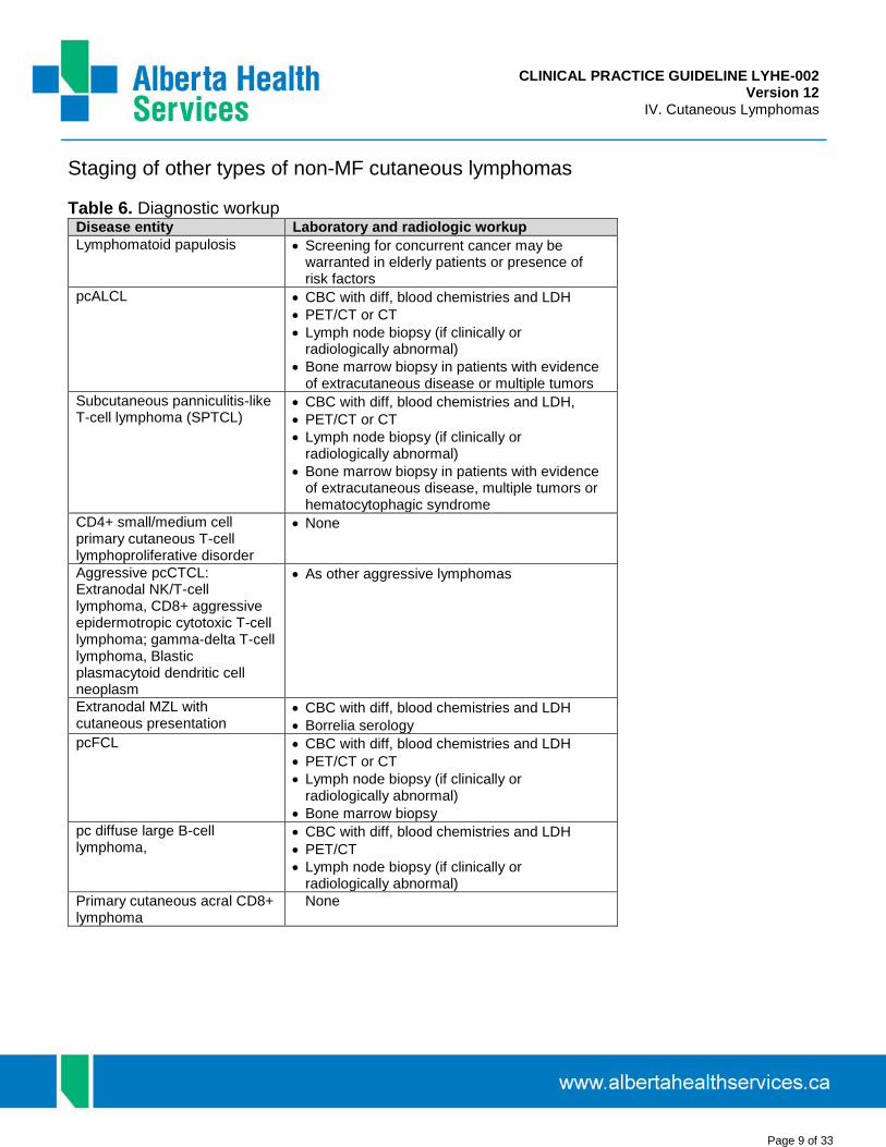
CLINICAL PRACTICE GUIDELINE LYHE-002 Version 12
IV. Cutaneous Lymphomas
Page 9 of 33
Staging of other types of non-MF cutaneous lymphomas
Table 6. Diagnostic workup Disease entity Laboratory and radiologic workup
Lymphomatoid papulosis Screening for concurrent cancer may bewarranted in elderly patients or presence ofrisk factors
pcALCL CBC with diff, blood chemistries and LDH
PET/CT or CT
Lymph node biopsy (if clinically orradiologically abnormal)
Bone marrow biopsy in patients with evidenceof extracutaneous disease or multiple tumors
Subcutaneous panniculitis-like T-cell lymphoma (SPTCL)
CBC with diff, blood chemistries and LDH,
PET/CT or CT
Lymph node biopsy (if clinically orradiologically abnormal)
Bone marrow biopsy in patients with evidenceof extracutaneous disease, multiple tumors orhematocytophagic syndrome
CD4+ small/medium cell primary cutaneous T-cell lymphoproliferative disorder
None
Aggressive pcCTCL: Extranodal NK/T-cell lymphoma, CD8+ aggressive epidermotropic cytotoxic T-cell lymphoma; gamma-delta T-cell lymphoma, Blastic plasmacytoid dendritic cell neoplasm
As other aggressive lymphomas
Extranodal MZL with cutaneous presentation
CBC with diff, blood chemistries and LDH
Borrelia serology
pcFCL CBC with diff, blood chemistries and LDH
PET/CT or CT
Lymph node biopsy (if clinically orradiologically abnormal)
Bone marrow biopsy
pc diffuse large B-cell lymphoma,
CBC with diff, blood chemistries and LDH
PET/CT
Lymph node biopsy (if clinically orradiologically abnormal)
Primary cutaneous acral CD8+ lymphoma
None

CLINICAL PRACTICE GUIDELINE LYHE-002 Version 12
IV. Cutaneous Lymphomas
Page 10 of 33
Treatment of other types of non-MF cutaneous lymphomas46-61
Table 7. Treatment of other types of cutaneous lymphomas CTCL Subtype First line treatment Second or third line
treatment
Lymphomatoid papulosis
Solitary lesion
Large/stigmitizinglesion
Multifocal
Observation Topical high potency corticosteroids
Surgical excision Local radiotherapy
Narrow band UVB Psoralen UVA light therapy Low dose MTX(5-25mg/wk)
Topical carmustine 0.2-0.4%*
Interferon alpha Isotretinoin or Alitretinoin
Primary Cutaneous ALCL
Solitary lesion
Multifocal or frequentlyrecurrent
Extracutaneousinvolvement
Surgical excision Local radiotherapy (15Gy)
Low dose MTX (5-25mg/week) maintenance
CHOP or CEOP
Isotretinoin or Alitretinoin
Interferon
Single agent chemotherapy (gemcitabine, etoposide)
Brentuximab vedotin*
Subcutaneous panniculitis-like T-cell lymphoma
Associatedhemophagocyticsyndrome
Systemic corticosteroids, alone, or in combination with methotrexate
CHOP or CEOP x 6 +/- HDT-ASCT in eligible patients
Cyclosporine §
Vorinostat¶
Local radiotherapy
Oral Bexarotene1
Primary cutaneous acral CD8+ T-cell lymphoma Provisional entity
Intralesional corticosteroids Local radiotherapy
Primary cutaneous CD4+ small/medium T-cell lymphoproliferative disorder
If tumour rapidlygrowing or > 5cm, HighKi67
Observation Topical corticosteroids Intralesional corticosteroids
Local radiotherapy
Local radiotherapy
Primary cutaneous aggressive epidermotropic CD8+ cytotoxic T-cell lymphoma or Primary cutaneous γδ T-Cell lymphoma
Multiagent chemotherapy (CHOP or CEOP) plus IFRT 30Gy/10 or 45Gy/25
Vorinostat¶
HDT/ASCT or Allogeneic stem cell transplantation in eligible candidates

CLINICAL PRACTICE GUIDELINE LYHE-002 Version 12
IV. Cutaneous Lymphomas
Page 11 of 33
Blastic plasmacytoid dendritic cell neoplasm (CD4+/CD56+ hematodermic neoplasm)
Multiagent chemotherapy (CHOP or CEOP)
Acute lymphoblastic leukemia type protocol if concurrent bone marrow involvement
Allogeneic stem cell transplantation in first remission for eligible patients
Single agent chemotheraoy (Gemcitabine)
Local radiotherapy
Primary cutaneous extranodal NK/T cell lymphoma, nasal type
Combined Modality (CHOP or CEOP plus IFRT) for localized presentation SMILE or equivalent for advanced stage
HDT-ASCT in eligible patients with relapsed/refractory
Primary Cutaneous Marginal Zone Lymphoma or Primary Cutaneous Follicle Center Lymphoma
Solitary lesion
Multifocal lesions
B. burgdorferiassociated pcMZL
Surgical excision Local radiotherapy (15-35Gy)
Observation Chlorambucil Rituximab monotherapy*
Antibiotics (cephalosporin or doxycycline)
Intralesional corticosteroids
Intralesional rituximab (5-20mg per lesion q4week x 3-6 cycles)*
Treat as systemic (R-Bendamustine x 6)
Primary cutaneous large B cell lymphoma, leg type
R-CHOP x 6 +/- IFRT
IFRT +/- rituximab monotherapy* if frail
Short term director’s privilege (STDP) required§ Short term exceptional drug therapy (STEDT) approval required
⌘ Health Canada Special Access Program required ¶ Not covered by AHS Cancer Control Drug Benefit list. Manufacturer’s reimbursement assistance program available. Dispensed through retail pharmacy
★ plication required for access. Drug not funded.

CLINICAL PRACTICE GUIDELINE LYHE-002 Version 12
IV. Cutaneous Lymphomas
Page 12 of 33
Special topics in CTCL
The role of transplantation in cutaneous lymphoma35,36,39,62-71
Existing studies of allogeneic stem cell transplantation in mycosis fungoides or sezary syndrome are limited to small, retrospective reports or case series. Autologous stem cell transplantation has not been associated with durable remissions and therefore has been largely abandoned for MF/SS. The following recommendations are based on best available outcome data and established consensus guidelines:
Patients with MF/SS should be risk-stratified using the CTCL International Consortium prognosis score. Patients with high-risk disease (3 or 4 of age>60, elevated LDH, stage IV or LCT) should be considered for allogeneic transplantation as part of second line of therapy.
• Patients with advanced stage 3 or stage 4 MF/SS who progress after more than two lines of systemictherapy should be considered for allogenic transplantation.
• Selected patients with stage 2 MF/SS or with large cell transformation may be considered forallogeneic BMT..
• Patients must meet other eligibility criteria for transplant prior to being considered. Issues such aschemosensitivity (CR or PR to last line of therapy), adequate performance status (ECOG 0-2) andpreserved organ function apply.
• TSEB before transplant may be considered prior to transplantation for improved skin control.• Transplantation in other rare and aggressive CTCL such as CD8+ epidermotropic aggressive T cell
lymphoma or primary cutaneous gamma-delta T cell lymphoma is at this time a largely experimentalapproach
• Relapses still occur after allogeneic transplants and may be treated adjustment of immunosuppression, DLIinfusion, or further skin-directed treatments. Distinguishing CTCL from transplant associated GVHD requiresmultidisciplinary expertise.
Large Cell Transformation in Mycosis Fungoides
The pathologic definition of large cell transformation in mycosis fungoides (LCT-MF) is the presence of large cells (>= 4 times the size of a small lymphocyte) in 25% of more of the dermal infiltrate or forming microscopic nodules. The cells are often CD30+ by IHC however CD30- variants are also described. It is difficult to discriminate from other subtypes of cutaneous lymphoma, including cutaneous anaplastic large cell lymphoma (cALCL) or lymphomatoid papulosis (LyP), which may also coexist with mycosis fungoides. The prognosis of LyP and cALCL is considerably more favourable than LCT-MF. Historical estimates for long-term survival with LCT-MF is less than 20%, and most series report a median survival of 2-36 months. However, a subset of patients with limited LCT-MF may follow a more indolent course. One large EORTC cohort analysis reported a median survival of 8.3 years for patients with LCT, and the authors concluded LCT is significant for disease progression but not survival outcome.26 Currently, there is a lack of prospective research to guide a standardized approach for management of LCT-MF. Most patients are treated with combination chemotherapy however it remains it is unclear which patients benefit from this approach. Several clinical and pathological characteristics in LCT-MF have been associated with poor prognosis,28,33 including advanced age (> 60 years), elevated LDH at transformation, advanced stage (III/IV), extra-cutaneous transformation, the presence of follicular mucinosis, folliculotropism, and CD30-negativity. Additional pathologic variables have been described but may not be routinely analyzable so have been omitted from these recommendations. We recommend to consider intensive chemotherapeutic strategies (monotherapy or combination in suitable fit candidates) in patients with any of the following clinical or pathologic variables associated with high risk LCT-MF. In the absence of these, we recommend treatment as per MF guidelines (see Table I).

CLINICAL PRACTICE GUIDELINE LYHE-002 Version 12
IV. Cutaneous Lymphomas
Page 13 of 33
Clinical variables for high risk LCT-MF:
advanced age (> 60 years)
elevated LDH at transformation
generalized tumours (versus solitary or regional)
advanced stage (III/IV)
extra-cutaneous transformation
Adverse Pathologic variables in LCT
absent papillary dermal involvement (assessment may be limited by provided tissues)
folliculotropism
follicular mucinosis
absence of fibrosis
CD30 expression in less than 50% of neoplastic cells
Brentuximab vedotin has activity in LCT-MF. A phase 2 study of brentuximab in a heavily pre-treated CD30+ MF/SS population, the majority of whom had LCT (30/32, 90%) showed a significant response rate of 70%.52 A subsequent prospective, randomized controlled trial of brentuximab vedotin versus physician’s choice (MTX or bexarotene) in CD30+ CTCL demonstrated a significant improvement in objective global response lasting atleast 4 months with brentuximab (56.3% versus 12.5%).57 The study included both previously treatment CD30+ MF and CD30+ ALCL. Although the histologic characteristics of the CD30+ MF patients were unreported, a proportion may have had transformed MF, as this was not an exclusion criteria. Brentuximab vedotin is indicated for previously treated CD30+ MF, and could be tried for high risk LCT-MF patients as defined above, who are either unsuitable for chemotherapy or refractory/relapsed folllowing chemotherapy.
Aggressive T-Cell Lymphomas
NK/T-cell lymphoma, nasal type:72-80 Natural killer (NK)/T-cell lymphoma, nasal type is a rare and aggressive extranodal neoplasm that almost exclusively affects Asian and South American adults in the fifth decade of life, with a male:female ratio of approximately 3:1. It typically arises in the nasal cavity or surrounding structures, such as the sinuses, palate, nasopharynx, tonsils, hypopharynx, and larynx. While the pathogenesis of NK/T-cell lymphoma, nasal type is not well understood, the Epstein-Barr virus (EBV) is implicated in almost all cases. Approximately 25% of cases show a p53 mutation; in addition, p21 over-expression is also frequent in nasal NK/T-cell lymphoma, and seems to be independent of p53 gene status.75
Hematopathological evaluation of a biopsy specimen from the site of involvement is the basis for diagnosis of nasal NK/T-cell lymphoma. The recommended immunohistochemistry panel includes:73,81 B-cell: CD20 T-lineage antigens: CD2, CD7, CD8, CD4, CD5, CD3 NK lineage markers: CD56 Cytotoxic granules (granzyme B and/or TIA-1) Ki-67 In situ hybridization for EBV-encoded RNA (EBER)

CLINICAL PRACTICE GUIDELINE LYHE-002 Version 12
IV. Cutaneous Lymphomas
Page 14 of 33
For patients with early-stage nasal NK/T-cell lymphoma, early or upfront radiotherapy (intensive regimens such as a total dose ≥ 50 Gy) plays an essential role in therapy, and has been associated with higher overall survival and complete response rates compared to chemotherapy alone.76 However, radiotherapy alone is also associated with high relapse rates. Combined modality therapy is recommended. In a phase II trial involving 30 patients treated with concurrent radiation (40–52.8 Gy) and weekly cisplatin (30mg/m2) followed by 3 cycles of VIPD chemotherapy (etoposide 100mg/m2 d1-3 + ifosfamide 1.2g/m2 d1-3 + cisplatin 33mg/m2 d1-3 + dexamethasone 40mg d1-4), Kim et al. (2009) reported an overall response rate of 83.3%, and a complete response rate of 80%.77 The 3-year progression-free and overall survival rates were 85.2% and 86.3%, respectively. While 26 patients completed all 3 cycles, there was a high rate of grade 4 neutropenia (41.4%). Similar results have been described in a phase I/II study involving 26 assessable patients treated with radiotherapy (50 Gy) and 3 courses of dexamethasone, etoposide, ifosfamide, and carboplatin (DeVIC).78 In another recently completed phase II trial, 31 patients with stage I or II disease were treated with radiotherapy 40–50.4 Gy plus cisplatin 30mg/m2 weekly, followed by 2 cycles of VIDL (etoposide 100mg/m2 d1-3, ifosfamide 1200mg/m2 d1-3, dexamethasone 40mg d1-3, and L-asparaginase 4000IU/m2.79 The overall response rate after the concurrent chemo-radiotherapy was 90%, and after VIDL was 92.6%; one-year progression-free survival was approximately 75%; lower than with VIPD, though a lower total dose of radiation was provided in this study and is postulated as the cause of the reduced PFS. Grade 3-4 leucopenia was reported in 85.1% of patients, and hepatic toxicity associated with L-asparaginase, the majority of which was grade 1 or 2, was reported in 55% of patients. Despite these results, L-asparaginase appears to be an active agent in this disease and most novel regimens incorporate L-asparaginase into treatment. One such regimen, GOLD (gemcitabine, oxaliplatin, l-asparaginase, dexamethasone) was recently reported in N=55 patients of whom 10 had stage III/IV disease. Amongst patients treated with the GOLD regimen, 91% (48/55) experienced a response, with 62% (34/55) having a complete response, and 29% (15/55) a partial response. In patients with stage I/II disease, 1-year PFS and OS were 87% and 98%, respectively. In patients with stage III/IV disease, 1-year PFS and OS were 66% and 75%, respectively82.
For patients with stage III-IV disease, complete remission rates are less than 15%, and the median overall survival is approximately 4 months.80 The recommended options for therapy include either enrollment in a clinical trial or treatment with an L-asparaginase-based combination chemotherapy regimen. The most well-studied regimen is the SMILE regimen with several small series of patients reported 83-85. While the SMILE regimen was first reported to have excellent response rates (overall, and complete in 79% and 45%, respectively) in relapsed/refractory patients, an updated study of the use of the SMILE regimen as frontline therapy for advanced stage patients reported a short median OS (12.2 months; 1-year OS was 45%) with a high rate of TRM (5 of 87 patients died of sepsis)83. While the GOLD regimen has less reported patients, the toxicity is significantly less (Grade 3-4 neutropenia of 16% compared to SMILE of 92%84 with serious infections in 4% and 31-45%83,84 of patients treated, respectively). For this reason, patients of advanced age or with comorbidities or a history of infections should be considered for therapy with GOLD for 2-4 cycles followed by SCT if possible while younger, fit patients can be treated with SMILE x 2 cycles with a goal of proceeding to SCT as consolidation. The role of allogeneic or autologous SCT is not yet well defined however, as data is limited but it is suggested when possible for advanced stage or relapsed/refractory patients.
Peripheral T-cell lymphomas (PTCL):86-94 With the exception of ALK-positive anaplastic large cell lymphoma, CHOP chemotherapy cures less than 30% of patients with PTCL. Options that may be associated with higher cure rates include CHOP x 4-6 cycles followed by HDCT/ASCT in responding patients or intensification of CHOP with etoposide (CHOEP). The German High-Grade Non-Hodgkin Lymphoma Study Group (DSHNHL) analyzed results of 343 PTCL

CLINICAL PRACTICE GUIDELINE LYHE-002 Version 12
IV. Cutaneous Lymphomas
Page 15 of 33
patients treated within their trials.95 The majority belonged to the four major T-cell lymphoma subtypes: anaplastic large cell lymphoma (ALCL), ALK-positive (n=78); ALCL, ALK-negative (n=113); peripheral T-cell lymphoma, unspecified (PTCLU; n=70); and angioimmunoblastic T-cell lymphoma (AITL; n=28). Treatment consisted of 6-8 courses of CHOP or etoposide plus (CHOEP). Three-year event-free and overall survival rates were 75.8% and 89.8% for the ALCL, ALK-positive patients, 50.0% and 67.5% for the AITL patients, 45.7% and 62.1% for the ALCL, ALK-negative patients, and 41.1% and 53.9% for the PTCLU patients. The International Prognostic Index (IPI) was effective in defining risk groups with significantly different outcomes. For patients, 60 years of age or younger with LDH levels < upper normal value, etoposide was associated with an improvement in 3-year EFS (75.4% vs. 51.0%, p=0003).86 Aviles and colleagues recently reported the results of a phase III trial involving 217 patients with PTCL unspecified.87 Patients were treated with either CMED every 14 days x 6 cycles or standard CHOP. The 10-year PFS was 70% in the CMED group versus 43% in the CHOP group (p<0.01), and the 10-year OS was 60% in the CMED group versus 34% in the CHOP group (p<0.01).87
Retrospective and prospective phase II trials support the use of SCT as part of upfront therapy for PTCL. Sieniawski and colleagues reported 5-year PFS rates of 60% for 26 patients with enteropathy associated T-cell lymphoma treated with IVE-methotrexate induction therapy followed by autologous SCT, compared to only 22% for 54 patients treated with CHOP-like therapy alone.87 Two prospective trials have also been reported. In the first, Reimer and colleagues reported results of CHOP x 4-6 cycles followed by dexabeam or ESHAP followed by CyTBI/ASCT for 83 patients (including 32 with PTCL-not otherwise specified, and 27 with angioimmunoblastic T-cell lymphoma).89 Fifty-five of the 83 patients received transplantation. In an intent-to-treat analysis, with a median follow-up time of 33 months, the estimated 3-year OS, DFS, and PFS rate were 48%, 53%, and 36%, respectively.96 In the second prospective trial, Rodriguez and colleagues from the Spanish Lymphoma and Autologous Transplantation Cooperative Group reported the results of 74 patients transplanted in the first complete response (65% had 2-3 aaIPI risk factors).90 With a median follow-up of 67 months from diagnosis, the 5-year OS and PFS rates were 68% and 63%, respectively.
For PTCL patients who relapse following CHOP-type induction and respond to salvage therapy, ASCT should be recommended, as several studies report similar ASCT outcomes to those seen with relapsed DLBCL. Brentuximab vedotin may be considered for those patients with CD30+ anaplastic large cell lymphoma who have had failure of initial chemotherapy.97
Summary of treatment recommendations for PTCL: 1. Anaplstic large cell lymphoma, ALK positive: CHOP x 6 cycles2. NK/T-cell lymphoma, nasal type:
recommendation for stage I-II NK/T cell lymphoma: IFRT as initial therapy (either 30Gy/10 fractionsIFRT or concurrent 40-50Gy IFRT+ weekly cisplatin 30mg/m2) then follow IFRT with VIPD x 3 cycles(etoposide 100mg/m2 d1-3 + ifosfamide 1.2g/m2 d1-3 + cisplatin 33mg/m2 d1-3 + dexamethasone40mg d1-4)
if IFRT must be delayed for 2 or more weeks after diagnosis due to scheduling issues, then d1-4 ofGDP could be administered while waiting for IFRT
3. All other subtypes of PTCL: <60 years of age with IPI=0-2: CHOEP x 6 cycles <60 years of age with IPI=3-5: CHOP or CHOEP x 4 cycles, then mobilize stem cells with high-dose
MTX 3.5g/m2 IV d1 and cytarabine 3g/m2 IV d9-10, G-CSF 480-600 mg SC daily d16-21 followed byapheresis d22-23, then HDCT/ASCT
>60 years of age: CHOP or CEOP x 6 cycles +/- HDCT/ASCT

CLINICAL PRACTICE GUIDELINE LYHE-002 Version 12
IV. Cutaneous Lymphomas
Page 16 of 33
AIDS-related lymphomas:98-103 In general, the treatment of AIDS-related lymphoma should be the same as for non-AIDS related lymphoma if the AIDS does not otherwise compromise the patient’s performance status and he/she is free of coincident serious opportunistic infection. HAART should be given with CHOP chemotherapy. Treatment should be planned in conjunction with the patient’s HIV physician and an antiviral regimen without overlapping toxicity should be chosen. R-CHOP results in the highest rates of disease-free survival, but may also increase the risk of infectious complications and treatment-related mortality in patients with CD4 counts below 50.
Post-transplant lymphoproliferative disease (PTLD) after Solid Organ Transplant in Adults: 1. Epidemiology. Post-transplant lymphoproliferative disorder (PTLD) is a heterogeneous diseasewith clinical and pathologic manifestations ranging from benign lymphoid hyperplasia (ie. early lesions) to aggressive lymphoma (ie. monomorphic PTLD)104,105. PTLD and its treatment cause a high rate of mortality and graft loss in patients with solid organ transplants (SOT)106. The incidence of PTLD is highest in multivisceral (>10%) and lowest in renal transplants (1-5%), attributed to intensity of immunosuppression and amount of lymphoid tissue in the allograft107-109. Epstein-Barr virus (EBV) infection drives the pathogenesis in PTLDs occurring early post-transplant; conversely, PTLDs occurring after prolonged immunosuppression tend to be monomorphic with no detectable EBV genome, calling an infectious etiology into question110. An epidemiologic shift in PTLD has occurred in the most recent decade: the median latency time from transplant to PTLD has increased from 1 to 3 years111,112 and the proportion of EBV-positive vs. -negative PTLD has decreased113, attributed to EBV viral load monitoring in EBV seronegative (ie. high risk) patients.
2. Diagnosis and staging. Diagnostic tissue must be reviewed by expert pathologists and subtypedaccording to the WHO.44 Several small case series have confirmed that PET-CT is an effective imaging modality for staging in PTLD114-119. However, some subtypes of PTLD, such as early lesions and T-cell lymphomas, may not be FDG-avid, necessitating CT as an alternate staging modality.
3. Management. Recommendations for the management of PTLD in SOT are based on few phase IIclinical trials, retrospective case series, and expert opinion120-122. The mainstays of therapy for CD20-positive PTLD in SOT include reduction of immune suppression (RIS), rituximab, and chemotherapy; adoptive immunotherapy is promising but considered experimental and is unavailable in Alberta. All patients should undergo RIS to the lowest tolerated levels under the direction of the transplant physician as soon as the diagnosis of PTLD is confirmed 120. A recommended strategy is to discontinue antiproliferative agents and reduce the calcineurin inhibitor by 25-50% while maintaining the steroid 120. Published response rates vary widely (0-73%) and responses are seen within 2 to 4 weeks 123-125.
3a. Early lesions, polymorphic and CD20-positive monomorphic PTLD. RIS may serve as definitive treatment of early lesions, but if response is incomplete further treatment with surgery or radiation is favored. In contrast, polymorphic and monomorphic PTLDs require definitive treatment along with RIS, discussed in further detail below 120-122 (Figure 3).
Surgery and radiation. Patients with localized PTLD, such as isolated skin, GI or renal allograft lesions, can achieve prolonged remissions with surgery or localized radiation123,126. Some experts consider surgical resection of isolated GI lesions prior to initiating systemic therapy to reduce early mortality from bowel perforation121. Radiation alone is generally not curative, with exception of plasmacytoma-like PTLD 127, and

CLINICAL PRACTICE GUIDELINE LYHE-002 Version 12
IV. Cutaneous Lymphomas
Page 17 of 33
should not be used as primary treatment120. Radiation may be used for palliating obstructive or compressive symptoms where systemic therapy fails or is not possible120.
Chemotherapy. SOT patients do not tolerate chemotherapy well, often developing severe infection or prolonged cytopenias. Estimates of efficacy of chemotherapy in treatment of PTLD in SOT are limited by the almost entirely retrospective nature of publications. Results of retrospective studies of anthracycline-based chemotherapy, mainly CHOP, show ORRs of 65-73% and 5-year OS of 25-78%; however, treatment-related mortality (TRM) is up to 31%128-132.
Rituximab. Several retrospective reviews and phase II clinical trials have confirmed the efficacy of rituximab monotherapy in CD20-positive PTLD post-SOT in patients that fail to respond to RIS. Phase II trials show overall response rates (ORR) of 44% to 71% and CR rates of 26% to 53% after 4 weekly doses with no reported TRM133-136. However, 57% of patients treated with rituximab monotherapy in 2 prospective trials had progressive disease within 12 months; risk factors for survival and need for further treatment included age > 60, ECOG ≥ 2, elevated LDH, and lack of CR after rituximab137. Therefore rituximab causes minimal toxicity but remissions achieved are durable in only a minority of patients.
Sequential therapy. Efficacy of a sequential treatment regimen (4 weekly doses of rituximab followed by 4 cycles of CHOP) was established in a phase II international multicentre trial in adult CD20-positive PTLD in SOT (n=70) in an attempt to improve upon the success of rituximab monotherapy and diminish the toxicity of chemotherapy138. The ORR was 60% after initial rituximab, increasing to 90% after sequential chemotherapy. EBV-positive and –negative PTLDs responded equally. OS was 61% at 3 years and time to progression was 69% at 3 years. There were no TRM events related to rituximab and 11% ascribed to CHOP. In a subsequent analysis, the authors concluded that patients who achieved CR and those in PR with a low-risk IPI score after rituximab monotherapy had a low risk of disease progression139.
A subsequent phase II trial utilized risk-stratified sequential therapy, in which patients in CR (by CT) after 4 doses of rituximab received 4 further 3-weekly doses of rituximab, and those not in CR after initial rituximab proceeded to RCHOP (4 cycles supported with GCSF). With 152 patients treated, endpoints were superior to sequential therapy (3-year OS 70%, 3-year TTP 73%, TRM 7%), and response to initial rituximab was highly predictive of OS, TTP and PFS (p<0.001)140,141.
In summary, rituximab monotherapy is effective first-line treatment in most CD20-positive PTLDs with minimal toxicity. Risk-stratified sequential therapy offers the highest survival rates published to date, and allows patients in CR after rituximab monotherapy to avoid chemotherapy. Close follow-up for disease progression is recommended for patients that received rituximab alone. For PTLD that behaves aggressively (ie. IPI 3-5) or progresses during initial treatment with rituximab, proceed directly to RCHOP before completing 4 doses of rituximab (Figure 3).
3b. Primary CNS PTLD. In the largest reported retrospective series of primary CNS PTLD (n=84), patients treated with rituximab and/or cytarabine (most often given after MTX) survived longer, but significant variation in regimens precluded firm conclusions142. Patients with acceptable renal function and performance status should be offered high-dose methotrexate and rituximab, and others may benefit from palliative radiation121,142.
3c. Burkitt Lymphoma PTLD. Several case series cite acceptable outcomes in this rare subtype of PTLD with chemotherapy regimens ranging in intensity143-145. However, no definite recommendations can be made and treatment should be considered individually.

CLINICAL PRACTICE GUIDELINE LYHE-002 Version 12
IV. Cutaneous Lymphomas
Page 18 of 33
3d. CD20-negative monomorphic PTLDs. Rare subtypes of PTLD that resemble non-transplant lymphomas, such as Hodgkin Lymphoma-like PTLD, T cell monomorphic PTLD, plasmablastic PTLD and plasma cell dyscrasias, require specific chemotherapeutic treatment similar to their non-transplant counterparts (reviewed by120,121).
4. Prognosis. The risk of death from NHL is significantly higher in SOT compared to non-transplantpatients146, and PTLD increases the graft failure rate 5-fold147. Retrospective series of PTLD post-SOT report OS of 30-68% at 5 years, with excess mortality in the first year post-diagnosis107,112,148-150. Adverse prognostic factors from retrospective studies include monomorphic subtype, monomorphic T-cell, bone marrow or CNS involvement, advanced stage, poor performance status, advanced age, elevated LDH, and hypoalbuminemia111,112,134,150-152. Risk factors for worsened OS in the PTLD-1 prospective trial include IPI 3-5, thoracic organ transplant and lack of CR after rituximab monotherapy139. A prognostic score developed from 500 PTLD cases in renal transplant patients is described in Table 8; the score was calculated with the exclusion of patients with monomorphic T-cell and CNS PTLD, both of which carried an adverse prognosis, but the score maintains its ability to discriminate risk groups in the whole population148.

CLINICAL PRACTICE GUIDELINE LYHE-002 Version 12
IV. Cutaneous Lymphomas
Page 19 of 33
Figure 1. Treatment Algorithm for Polymorphic or Monomorphic (DLBCL) PTLD Post-SOT

CLINICAL PRACTICE GUIDELINE LYHE-002 Version 12
IV. Cutaneous Lymphomas
Page 20 of 33
Table 8. Post-Transplant Lymphoproliferative Disorders in Renal Transplant Prognostic Score148. (One point is given for each of elevated LDH, disseminated PTLD (ie. higher than stage 1), monomorphic PTLD, and serum creatinine level >133 µmol/L; 2 points are given for creatinine >133 µmol/L if age > 55 at PTLD diagnosis.)
Risk Group (# Risk Factors) % Alive at 1/5/10 years
Low (0) 100/92/85
Moderate (1) 89/83/80
High (2-3) 74/59/56
Very High (4-5) 52/35/0

CLINICAL PRACTICE GUIDELINE LYHE-002 Version 12
IV. Cutaneous Lymphomas
Page 21 of 33
REFERENCES
1. Ahn HK, Suh C, Chuang SS, Suzumiya J, Ko YH, Kim SJ, et al. Extranodal natural killer/T-cell lymphoma from skin
or soft tissue: suggestion of treatment from multinational retrospective analysis. Annals of Oncology 2012
10/01;23(10):2703-2707.
2. Alencar AJ, Bustinza E, Barker J, Byrne GE, Lossos IS. Hematodermic tumor presenting with generalized skin
involvement. J Clin Oncol 2009 Jun 20;27(18):3059-3061 PubMed ID 19414664.
3. Alfred A, Taylor PC, Dignan F, El-Ghariani K, Griffin J, Gennery AR, et al. The role of extracorporeal photopheresis
in the management of cutaneous T-cell lymphoma, graft-versus-host disease and organ transplant rejection: a
consensus statement update from the UK Photopheresis Society. Br J Haematol 2017 Apr;177(2):287-310
PubMed ID 28220931.
4. Ally MS, Robson A. A review of the solitary cutaneous T-cell lymphomas. J Cutan Pathol 2014 Sep;41(9):703-714
PubMed ID 24666254.
5. Arulogun SO, Prince HM, Ng J, Lade S, Ryan GF, Blewitt O, et al. Long-term outcomes of patients with advanced-
stage cutaneous T-cell lymphoma and large cell transformation. Blood 2008 Oct 15;112(8):3082-3087 PubMed
ID 18647960.
6. Besner Morin C, Roberge D, Turchin I, Petrogiannis-Haliotis T, Popradi G, Pehr K. Tazarotene 0.1% Cream as
Monotherapy for Early-Stage Cutaneous T-Cell Lymphoma. J Cutan Med Surg 2016 May;20(3):244-248 PubMed
ID 26742957.
7. Bernengo MG, Quaglino P, Comessatti A, Ortoncelli M, Novelli M, Lisa F, et al. Low-dose intermittent alemtuzumab
in the treatment of Sezary syndrome: clinical and immunologic findings in 14 patients. Haematologica 2007
Jun;92(6):784-794 PubMed ID 17550851.
8. Duarte RF, Schmitz N, Servitje O, Sureda A. Haematopoietic stem cell transplantation for patients with primary
cutaneous T-cell lymphoma. Bone Marrow Transplant 2008 Apr;41(7):597-604 PubMed ID 18176611.
9. Duarte RF, Boumendil A, Onida F, Gabriel I, Arranz R, Arcese W, et al. Long-term outcome of allogeneic
hematopoietic cell transplantation for patients with mycosis fungoides and Sezary syndrome: a European society
for blood and marrow transplantation lymphoma working party extended analysis. J Clin Oncol 2014 Oct
10;32(29):3347-3348 PubMed ID 25154828.
10. Garcia-Herrera A, Colomo L, Camos M, Carreras J, Balague O, Martinez A, et al. Primary cutaneous small/medium
CD4+ T-cell lymphomas: a heterogeneous group of tumors with different clinicopathologic features and outcome.
J Clin Oncol 2008 Jul 10;26(20):3364-3371 PubMed ID 18541895.
11. Hoefnagel JJ, Vermeer MH, Jansen PM, Heule F, van Voorst Vader PC, Sanders CJ, et al. Primary cutaneous
marginal zone B-cell lymphoma: clinical and therapeutic features in 50 cases. Arch Dermatol 2005
Sep;141(9):1139-1145 PubMed ID 16172311.

CLINICAL PRACTICE GUIDELINE LYHE-002 Version 12
IV. Cutaneous Lymphomas
Page 22 of 33
12. Hughes CF, Khot A, McCormack C, Lade S, Westerman DA, Twigger R, et al. Lack of durable disease control with
chemotherapy for mycosis fungoides and Sezary syndrome: a comparative study of systemic therapy. Blood 2015
Jan 1;125(1):71-81 PubMed ID 25336628.
13. Kempf W, Pfaltz K, Vermeer MH, Cozzio A, Ortiz-Romero P, Bagot M, et al. EORTC, ISCL, and USCLC consensus
recommendations for the treatment of primary cutaneous CD30-positive lymphoproliferative disorders:
lymphomatoid papulosis and primary cutaneous anaplastic large-cell lymphoma. Blood 2011 10/13;118(15):4024.
14. Lechowicz MJ, Lazarus HM, Carreras J, Laport GG, Cutler CS, Wiernik PH, et al. Allogeneic hematopoietic cell
transplantation for mycosis fungoides and Sezary syndrome. Bone Marrow Transplant 2014 Nov;49(11):1360-
1365 PubMed ID 25068422.
15. Marcus RE, Davies AJ, Ando K, Klapper W, Opat S, Owen CJ, et al. Obinutuzumab-Based Induction and
Maintenance Prolongs Progression-Free Survival (PFS) in Patients with Previously Untreated Follicular
Lymphoma: Primary Results of the Randomized Phase 3 GALLIUM Study. Blood 2016 12/01;128(22):6.
16. Nguyen CV, Bohjanen KA. Skin-Directed Therapies in Cutaneous T-Cell Lymphoma. Dermatol Clin 2015
Oct;33(4):683-696 PubMed ID 26433841.
17. Penate Y, Hernandez-Machin B, Perez-Mendez LI, Santiago F, Rosales B, Servitje O, et al. Intralesional rituximab
in the treatment of indolent primary cutaneous B-cell lymphomas: an epidemiological observational multicentre
study. The Spanish Working Group on Cutaneous Lymphoma. Br J Dermatol 2012 Jul;167(1):174-179 PubMed
ID 22356294.
18. Reimer P, Rudiger T, Kraemer D, Kunzmann V, Weissinger F, Zettl A, et al. What is CD4+CD56+ malignancy and
how should it be treated? Bone Marrow Transplant 2003 Oct;32(7):637-646 PubMed ID 13130309.
19. Rubio-Gonzalez B, Zain J, Rosen ST, Querfeld C. Clinical manifestations and pathogenesis of cutaneous
lymphomas: current status and future directions. Br J Haematol 2017 Jan;176(1):16-36 PubMed ID 27782301.
20. Schlaak M, Pickenhain J, Theurich S, Skoetz N, von Bergwelt-Baildon M, Kurschat P. Allogeneic stem cell
transplantation versus conventional therapy for advanced primary cutaneous T-cell lymphoma. Cochrane
Database Syst Rev 2013 Aug 29;(8):CD008908. doi(8):CD008908 PubMed ID 23986525.
21. Senff NJ, Noordijk EM, Kim YH, Bagot M, Berti E, Cerroni L, et al. European Organization for Research and
Treatment of Cancer and International Society for Cutaneous Lymphoma consensus recommendations for the
management of cutaneous B-cell lymphomas. Blood 2008 Sep 1;112(5):1600-1609 PubMed ID 18567836.
22. Specht L, Dabaja B, Illidge T, Wilson LD, Hoppe RT, International Lymphoma Radiation Oncology Group. Modern
radiation therapy for primary cutaneous lymphomas: field and dose guidelines from the International Lymphoma
Radiation Oncology Group. Int J Radiat Oncol Biol Phys 2015 May 1;92(1):32-39 PubMed ID 25863751.
23. Swerdlow SH, Campo E, Pileri SA, Harris NL, Stein H, Siebert R, et al. The 2016 revision of the World Health
Organization classification of lymphoid neoplasms. Blood 2016 May 19;127(20):2375-2390 PubMed ID
26980727.

CLINICAL PRACTICE GUIDELINE LYHE-002 Version 12
IV. Cutaneous Lymphomas
Page 23 of 33
24. Whittaker S, Hoppe R, Prince HM. How I treat mycosis fungoides and Sezary syndrome. Blood 2016 Jun
23;127(25):3142-3153 PubMed ID 27151889.
25. Willemze R, Jansen PM, Cerroni L, Berti E, Santucci M, Assaf C, et al. Subcutaneous panniculitis-like T-cell
lymphoma: definition, classification, and prognostic factors: an EORTC Cutaneous Lymphoma Group Study of 83
cases. Blood 2008 Jan 15;111(2):838-845 PubMed ID 17934071.
26. Agar NS, Wedgeworth E, Crichton S, Mitchell TJ, Cox M, Ferreira S, et al. Survival outcomes and prognostic factors
in mycosis fungoides/Sezary syndrome: validation of the revised International Society for Cutaneous
Lymphomas/European Organisation for Research and Treatment of Cancer staging proposal. J Clin Oncol 2010
Nov 01,;28(31):4730-4739.
27. Olsen E, Vonderheid E, Pimpinelli N, Willemze R, Kim Y, Knobler R, et al. Revisions to the staging and classification
of mycosis fungoides and Sezary syndrome: a proposal of the International Society for Cutaneous Lymphomas
(ISCL) and the cutaneous lymphoma task force of the European Organization of Research and Treatment of
Cancer (EORTC). Blood 2007 Sep 15,;110(6):1713-1722.
28. Pulitzer M, Myskowski PL, Horwitz SM, Querfeld C, Connolly B, Li J, et al. Mycosis fungoides with large cell
transformation: clinicopathological features and prognostic factors. Pathology 2014 Dec;46(7):610-616.
29. Scarisbrick JJ, Prince HM, Vermeer MH, Quaglino P, Horwitz S, Porcu P, et al. Cutaneous Lymphoma International
Consortium Study of Outcome in Advanced Stages of Mycosis Fungoides and Sezary Syndrome: Effect of
Specific Prognostic Markers on Survival and Development of a Prognostic Model. J Clin Oncol 2015 Nov
10,;33(32):3766-3773.
30. Trautinger F, Eder J, Assaf C, Bagot M, Cozzio A, Dummer R, et al. European Organisation for Research and
Treatment of Cancer consensus recommendations for the treatment of mycosis fungoides/Sezary syndrome -
Update 2017. Eur J Cancer 2017 05;77:57-74.
31. Ahn HK, Suh C, Chuang SS, Suzumiya J, Ko YH, Kim SJ, et al. Extranodal natural killer/T-cell lymphoma from skin
or soft tissue: suggestion of treatment from multinational retrospective analysis. Ann Oncol 2012 Oct;23(10):2703-
2707.
32. Alencar AJ, Bustinza E, Barker J, Byrne GE, Lossos IS. Hematodermic tumor presenting with generalized skin
involvement. J Clin Oncol 2009 Jun 20,;27(18):3059-3061.
33. Benner MF, Jansen PM, Vermeer MH, Willemze R. Prognostic factors in transformed mycosis fungoides: a
retrospective analysis of 100 cases. Blood 2012 Feb 16,;119(7):1643-1649.
34. Bernengo MG, Quaglino P, Comessatti A, Ortoncelli M, Novelli M, Lisa F, et al. Low-dose intermittent alemtuzumab
in the treatment of Sezary syndrome: clinical and immunologic findings in 14 patients. Haematologica 2007
Jun;92(6):784-794.
35. Duarte RF, Schmitz N, Servitje O, Sureda A. Haematopoietic stem cell transplantation for patients with primary
cutaneous T-cell lymphoma. Bone Marrow Transplant 2008 Apr;41(7):597-604.

CLINICAL PRACTICE GUIDELINE LYHE-002 Version 12
IV. Cutaneous Lymphomas
Page 24 of 33
36. Duarte RF, Boumendil A, Onida F, Gabriel I, Arranz R, Arcese W, et al. Long-term outcome of allogeneic
hematopoietic cell transplantation for patients with mycosis fungoides and Sezary syndrome: a European society
for blood and marrow transplantation lymphoma working party extended analysis. J Clin Oncol 2014 Oct
10,;32(29):3347-3348.
37. Hughes CFM, Khot A, McCormack C, Lade S, Westerman DA, Twigger R, et al. Lack of durable disease control
with chemotherapy for mycosis fungoides and Sezary syndrome: a comparative study of systemic therapy. Blood
2015 Jan 01,;125(1):71-81.
38. Kadin ME, Hughey LC, Wood GS. Large-cell transformation of mycosis fungoides-differential diagnosis with
implications for clinical management: a consensus statement of the US Cutaneous Lymphoma Consortium. J Am
Acad Dermatol 2014 Feb;70(2):374-376.
39. Lechowicz MJ, Lazarus HM, Carreras J, Laport GG, Cutler CS, Wiernik PH, et al. Allogeneic hematopoietic cell
transplantation for mycosis fungoides and Sezary syndrome. Bone Marrow Transplant 2014 Nov;49(11):1360-
1365.
40. Olsen EA, Whittaker S, Kim YH, Duvic M, Prince HM, Lessin SR, et al. Clinical end points and response criteria in
mycosis fungoides and Sezary syndrome: a consensus statement of the International Society for Cutaneous
Lymphomas, the United States Cutaneous Lymphoma Consortium, and the Cutaneous Lymphoma Task Force
of the European Organisation for Research and Treatment of Cancer. J Clin Oncol 2011 Jun 20,;29(18):2598-
2607.
41. Whittaker S, Hoppe R, Prince HM. How I treat mycosis fungoides and Sezary syndrome. Blood 2016 06
23,;127(25):3142-3153.
42. Kim YH, Willemze R, Pimpinelli N, Whittaker S, Olsen EA, Ranki A, et al. TNM classification system for primary
cutaneous lymphomas other than mycosis fungoides and Sezary syndrome: a proposal of the International
Society for Cutaneous Lymphomas (ISCL) and the Cutaneous Lymphoma Task Force of the European
Organization of Research and Treatment of Cancer (EORTC). Blood 2007 Jul 15,;110(2):479-484.
43. Rubio-Gonzalez B, Zain J, Rosen ST, Querfeld C. Clinical manifestations and pathogenesis of cutaneous
lymphomas: current status and future directions. Br J Haematol 2017 Jan;176(1):16-36.
44. Swerdlow SH, Campo E, Pileri SA, Harris NL, Stein H, Siebert R, et al. The 2016 revision of the World Health
Organization classification of lymphoid neoplasms. Blood 2016 05 19,;127(20):2375-2390.
45. Willemze R, Jansen PM, Cerroni L, Berti E, Santucci M, Assaf C, et al. Subcutaneous panniculitis-like T-cell
lymphoma: definition, classification, and prognostic factors: an EORTC Cutaneous Lymphoma Group Study of 83
cases. Blood 2008 Jan 15,;111(2):838-845.
46. Alfred A, Taylor PC, Dignan F, El-Ghariani K, Griffin J, Gennery AR, et al. The role of extracorporeal photopheresis
in the management of cutaneous T-cell lymphoma, graft-versus-host disease and organ transplant rejection: a
consensus statement update from the UK Photopheresis Society. Br J Haematol 2017 Apr;177(2):287-310.
47. Ally MS, Robson A. A review of the solitary cutaneous T-cell lymphomas. J Cutan Pathol 2014 Sep;41(9):703-714.

CLINICAL PRACTICE GUIDELINE LYHE-002 Version 12
IV. Cutaneous Lymphomas
Page 25 of 33
48. Besner Morin C, Roberge D, Turchin I, Petrogiannis-Haliotis T, Popradi G, Pehr K. Tazarotene 0.1% Cream as
Monotherapy for Early-Stage Cutaneous T-Cell Lymphoma. J Cutan Med Surg 2016 May;20(3):244-248.
49. Garcia-Herrera A, Colomo L, Camos M, Carreras J, Balague O, Martinez A, et al. Primary cutaneous small/medium
CD4+ T-cell lymphomas: a heterogeneous group of tumors with different clinicopathologic features and outcome.
J Clin Oncol 2008 Jul 10,;26(20):3364-3371.
50. Hoefnagel JJ, Vermeer MH, Jansen PM, Heule F, van Voorst Vader,P.C., Sanders CJG, et al. Primary cutaneous
marginal zone B-cell lymphoma: clinical and therapeutic features in 50 cases. Arch Dermatol 2005
Sep;141(9):1139-1145.
51. Kempf W, Pfaltz K, Vermeer MH, Cozzio A, Ortiz-Romero P, Bagot M, et al. EORTC, ISCL, and USCLC consensus
recommendations for the treatment of primary cutaneous CD30-positive lymphoproliferative disorders:
lymphomatoid papulosis and primary cutaneous anaplastic large-cell lymphoma. Blood 2011 Oct
13,;118(15):4024-4035.
52. Kim YH, Tavallaee M, Sundram U, Salva KA, Wood GS, Li S, et al. Phase II Investigator-Initiated Study of
Brentuximab Vedotin in Mycosis Fungoides and Sezary Syndrome With Variable CD30 Expression Level: A Multi-
Institution Collaborative Project. J Clin Oncol 2015 Nov 10,;33(32):3750-3758.
53. Nofal A, Abdel-Mawla M, Assaf M, Salah E. Primary cutaneous aggressive epidermotropic CD8+ T-cell lymphoma:
proposed diagnostic criteria and therapeutic evaluation. J Am Acad Dermatol 2012 Oct;67(4):748-759.
54. Olsen EA. Interferon in the treatment of cutaneous T-cell lymphoma. Dermatol Ther 2003;16(4):311-321.
55. Pellegrini C, Stefoni V, Casadei B, Maglie R, Argnani L, Zinzani PL. Long-term outcome of patients with advanced-
stage cutaneous T cell lymphoma treated with gemcitabine. Ann Hematol 2014 Nov;93(11):1853-1857.
56. Penate Y, Hernandez-Machin B, Perez-Mendez LI, Santiago F, Rosales B, Servitje O, et al. Intralesional rituximab
in the treatment of indolent primary cutaneous B-cell lymphomas: an epidemiological observational multicentre
study. The Spanish Working Group on Cutaneous Lymphoma. Br J Dermatol 2012 Jul;167(1):174-179.
57. Prince HM, Kim YH, Horwitz SM, Dummer R, Scarisbrick J, Quaglino P, et al. Brentuximab vedotin or physician's
choice in CD30-positive cutaneous T-cell lymphoma (ALCANZA): an international, open-label, randomised, phase
3, multicentre trial. Lancet 2017 Aug 05,;390(10094):555-566.
58. Reimer P, Rudiger T, Kraemer D, Kunzmann V, Weissinger F, Zettl A, et al. What is CD4+CD56+ malignancy and
how should it be treated? Bone Marrow Transplant 2003 Oct;32(7):637-646.
59. Senff NJ, Noordijk EM, Kim YH, Bagot M, Berti E, Cerroni L, et al. European Organization for Research and
Treatment of Cancer and International Society for Cutaneous Lymphoma consensus recommendations for the
management of cutaneous B-cell lymphomas. Blood 2008 Sep 01,;112(5):1600-1609.
60. Schlaak M, Pickenhain J, Theurich S, Skoetz N, von Bergwelt-Baildon M, Kurschat P. Allogeneic stem cell
transplantation versus conventional therapy for advanced primary cutaneous T-cell lymphoma. Cochrane
Database Syst Rev 2013 Aug 29,(8):CD008908.

CLINICAL PRACTICE GUIDELINE LYHE-002 Version 12
IV. Cutaneous Lymphomas
Page 26 of 33
61. Specht L, Dabaja B, Illidge T, Wilson LD, Hoppe RT. Modern radiation therapy for primary cutaneous lymphomas:
field and dose guidelines from the International Lymphoma Radiation Oncology Group. Int J Radiat Oncol Biol
Phys 2015 May 01,;92(1):32-39.
62. Willemze R, Jaffe ES, Burg G, Berti E, Swerdlow SH, Ralfkiaer E, et al. WHO-EORTC classification for cutaneous
lymphomas. Blood 2005 May 15,;105(10):3768-3785.
63. Molina A, Zain J, Arber DA, Angelopolou M, O'Donnell M, Murata-Collins J, et al. Durable clinical, cytogenetic, and
molecular remissions after allogeneic hematopoietic cell transplantation for refractory Sezary syndrome and
mycosis fungoides. J Clin Oncol 2005 Sep 01,;23(25):6163-6171.
64. Wu PA, Kim YH, Lavori PW, Hoppe RT, Stockerl-Goldstein K. A meta-analysis of patients receiving allogeneic or
autologous hematopoietic stem cell transplant in mycosis fungoides and Sezary syndrome. Biol Blood Marrow
Transplant 2009 Aug;15(8):982-990.
65. Duvic M, Donato M, Dabaja B, Richmond H, Singh L, Wei W, et al. Total skin electron beam and non-myeloablative
allogeneic hematopoietic stem-cell transplantation in advanced mycosis fungoides and Sezary syndrome. J Clin
Oncol 2010 May 10,;28(14):2365-2372.
66. Paralkar VR, Nasta SD, Morrissey K, Smith J, Vassilev P, Martin ME, et al. Allogeneic hematopoietic SCT for
primary cutaneous T cell lymphomas. Bone Marrow Transplant 2012 Jul;47(7):940-945.
67. Polansky M, Talpur R, Daulat S, Hosing C, Dabaja B, Duvic M. Long-Term Complete Responses to Combination
Therapies and Allogeneic Stem Cell Transplants in Patients With Sezary Syndrome. Clin Lymphoma Myeloma
Leuk 2015 May;15(5):83.
68. de Masson A, Beylot-Barry M, Bouaziz J, Peffault dL, Aubin F, Garciaz S, et al. Allogeneic stem cell transplantation
for advanced cutaneous T-cell lymphomas: a study from the French Society of Bone Marrow Transplantation and
French Study Group on Cutaneous Lymphomas. Haematologica 2014 Mar;99(3):527-534.
69. Alpdogan O, Grosso D, Flomenberg N. Recent advances in haploidentical stem cell transplantation. Discov Med
2013 Oct;16(88):159-165.
70. Shiratori S, Fujimoto K, Nishimura M, Hatanaka KC, Kosugi-Kanaya M, Okada K, et al. Allogeneic hematopoietic
stem cell transplantation following reduced-intensity conditioning for mycosis fungoides and Sezary syndrome.
Hematol Oncol 2016 Mar;34(1):9-16.
71. Virmani P, Zain J, Rosen ST, Myskowski PL, Querfeld C. Hematopoietic Stem Cell Transplant for Mycosis
Fungoides and Sezary Syndrome. Dermatol Clin 2015 Oct;33(4):807-818.
72. Hasserjian RP, Harris NL. NK-cell lymphomas and leukemias: a spectrum of tumors with variable manifestations
and immunophenotype. Am J Clin Pathol 2007 Jun;127(6):860-868 PubMed ID 17509983.
73. Ishida F, Kwong YL. Diagnosis and management of natural killer-cell malignancies. Expert Rev Hematol 2010
Oct;3(5):593-602 PubMed ID 21083476.

CLINICAL PRACTICE GUIDELINE LYHE-002 Version 12
IV. Cutaneous Lymphomas
Page 27 of 33
74. Kwong YL, Anderson BO, Advani R, Kim WS, Levine AM, Lim ST, et al. Management of T-cell and natural-killer-
cell neoplasms in Asia: consensus statement from the Asian Oncology Summit 2009. Lancet Oncol 2009
Nov;10(11):1093-1101 PubMed ID 19880063.
75. Quintanilla-Martinez L, Kremer M, Keller G, Nathrath M, Gamboa-Dominguez A, Meneses A, et al. p53 Mutations
in nasal natural killer/T-cell lymphoma from Mexico: association with large cell morphology and advanced disease.
Am J Pathol 2001 Dec;159(6):2095-2105 PubMed ID 11733360.
76. Li YX, Yao B, Jin J, Wang WH, Liu YP, Song YW, et al. Radiotherapy as primary treatment for stage IE and IIE
nasal natural killer/T-cell lymphoma. J Clin Oncol 2006 Jan 1;24(1):181-189 PubMed ID 16382127.
77. Kim SJ, Kim K, Kim BS, Kim CY, Suh C, Huh J, et al. Phase II trial of concurrent radiation and weekly cisplatin
followed by VIPD chemotherapy in newly diagnosed, stage IE to IIE, nasal, extranodal NK/T-Cell Lymphoma:
Consortium for Improving Survival of Lymphoma study. J Clin Oncol 2009 Dec 10;27(35):6027-6032 PubMed ID
19884539.
78. Yamaguchi M, Tobinai K, Oguchi M, Ishizuka N, Kobayashi Y, Isobe Y, et al. Phase I/II study of concurrent
chemoradiotherapy for localized nasal natural killer/T-cell lymphoma: Japan Clinical Oncology Group Study
JCOG0211. J Clin Oncol 2009 Nov 20;27(33):5594-5600 PubMed ID 19805668.
79. Kim SJ, Eom HS, Kim JS, Kwak JY, Yang DH, Park SK. Concurrent chemo-radiotherapy followed by VIDL
(etoposide, ifosfamide, dexamethasone, L-asparaginase) chemotherapy in stage I/II extranodal NK/T-cell
lymphoma of nasal cavity/nasopharynx. Blood 2010;(ASH Annual Meeting Abstracts); 116(Abstract 1765).
80. Au WY, Weisenburger DD, Intragumtornchai T, Nakamura S, Kim WS, Sng I. Clinical differences between nasal
and extranasal natural killer/T-cell lymphoma: a study of 136 cases from the International Peripheral T-Cell
Lymphoma Project. Blood 2009 Apr;113(17):3931-7.
81. Kobe C, Dietlein M, Franklin J, Markova J, Lohri A, Amthauer H, et al. Positron emission tomography has a high
negative predictive value for progression or early relapse for patients with residual disease after first-line
chemotherapy in advanced-stage Hodgkin lymphoma. Blood 2008 Nov 15;112(10):3989-3994 PubMed ID
18757777.
82. Guo HQ, Liu L, Wang XF, Lin TY, Yao SN, Yao ZH, et al. Efficacy of gemcitabine combined with oxaliplatin, L-
asparaginase and dexamethasone in patients with newly-diagnosed extranodal NK/T-cell lymphoma. Mol Clin
Oncol 2014 Nov;2(6):1172-1176 PubMed ID 25279218.
83. Kwong YL, Kim WS, Lim ST, Kim SJ, Tang T, Tse E, et al. SMILE for natural killer/T-cell lymphoma: analysis of
safety and efficacy from the Asia Lymphoma Study Group. Blood 2012 Oct 11;120(15):2973-2980 PubMed ID
22919026.
84. Yamaguchi M, Kwong YL, Kim WS, Maeda Y, Hashimoto C, Suh C, et al. Phase II study of SMILE chemotherapy
for newly diagnosed stage IV, relapsed, or refractory extranodal natural killer (NK)/T-cell lymphoma, nasal type:
the NK-Cell Tumor Study Group study. J Clin Oncol 2011 Nov 20;29(33):4410-4416 PubMed ID 21990393.

CLINICAL PRACTICE GUIDELINE LYHE-002 Version 12
IV. Cutaneous Lymphomas
Page 28 of 33
85. Kim SJ, Park S, Kang ES, Choi JY, Lim do H, Ko YH, et al. Induction treatment with SMILE and consolidation with
autologous stem cell transplantation for newly diagnosed stage IV extranodal natural killer/T-cell lymphoma
patients. Ann Hematol 2015 Jan;94(1):71-78 PubMed ID 25082384.
86. Schmitz N, Trümper L, Ziepert M, Nickelsen M, Ho AD, Metzner B. Treatment and prognosis of mature T-cell and
NK-cell lymphoma: an analysis of patients with T-cell lymphoma treated in studies of the German High-Grade
Non-Hodgkin Lymphoma Study Group. Blood 2010 Nov;116(18):3418-25.
87. Aviles A, Castaneda C, Neri N, Cleto S, Talavera A, Gonzalez M, et al. Results of a phase III clinical trial: CHOP
versus CMED in peripheral T-cell lymphoma unspecified. Med Oncol 2008;25(3):360-364 PubMed ID 18247163.
88. Sieniawski M, Angamuthu N, Boyd K, Chasty R, Davies J, Forsyth P. Evaluation of enteropathy-associated T-cell
lymphoma comparing standard therapies with a novel regimen including autologous stem cell transplantation.
Blood 2010 May;115(18):3664-70.
89. Reimer P, Rudiger T, Geissinger E, Weissinger F, Nerl C, Schmitz N, et al. Autologous stem-cell transplantation
as first-line therapy in peripheral T-cell lymphomas: results of a prospective multicenter study. J Clin Oncol 2009
Jan 1;27(1):106-113 PubMed ID 19029417.
90. Rodriguez J, Conde E, Gutierrez A, Arranz R, Leon A, Marin J, et al. The results of consolidation with autologous
stem-cell transplantation in patients with peripheral T-cell lymphoma (PTCL) in first complete remission: the
Spanish Lymphoma and Autologous Transplantation Group experience. Ann Oncol 2007 Apr;18(4):652-657
PubMed ID 17229774.
91. Rodriguez J, Conde E, Gutierrez A, Arranz R, Gandarillas M, Leon A, et al. Prolonged survival of patients with
angioimmunoblastic T-cell lymphoma after high-dose chemotherapy and autologous stem cell transplantation: the
GELTAMO experience. Eur J Haematol 2007 Apr;78(4):290-296 PubMed ID 17378891.
92. Sohn BS, Park I, Kim EK, Yoon DH, Lee SS, Kang BW, et al. Comparison of clinical outcome after autologous
stem cell transplantation between patients with peripheral T-cell lymphomas and diffuse large B-cell lymphoma.
Bone Marrow Transplant 2009 Sep;44(5):287-293 PubMed ID 19234508.
93. Feyler S, Prince HM, Pearce R, Towlson K, Nivison-Smith I, Schey S, et al. The role of high-dose therapy and stem
cell rescue in the management of T-cell malignant lymphomas: a BSBMT and ABMTRR study. Bone Marrow
Transplant 2007 Sep;40(5):443-450 PubMed ID 17589529.
94. Dearden CE, Johnson R, Pettengell R, Devereux S, Cwynarski K, Whittaker S, et al. Guidelines for the management
of mature T-cell and NK-cell neoplasms (excluding cutaneous T-cell lymphoma). Br J Haematol 2011
May;153(4):451-485 PubMed ID 21480860.
95. Maschio M, Albani F, Baruzzi A, Zarabla A, Dinapoli L, Pace A, et al. Levetiracetam therapy in patients with brain
tumour and epilepsy. J Neurooncol 2006 Oct;80(1):97-100 PubMed ID 16685465.
96. Milligan TA, Hurwitz S, Bromfield EB. Efficacy and tolerability of levetiracetam versus phenytoin after supratentorial
neurosurgery. Neurology 2008 Aug 26;71(9):665-669 PubMed ID 18725591.

CLINICAL PRACTICE GUIDELINE LYHE-002 Version 12
IV. Cutaneous Lymphomas
Page 29 of 33
97. Pro B, Advani R, Brice P, Bartlett NL, Rosenblatt JD, Illidge T, et al. Brentuximab vedotin (SGN-35) in patients with
relapsed or refractory systemic anaplastic large-cell lymphoma: results of a phase II study. J Clin Oncol 2012 Jun
20;30(18):2190-2196 PubMed ID 22614995.
98. DeMario MD, Liebowitz DN. Lymphomas in the immunocompromised patient. Semin Oncol 1998 Aug;25(4):492-
502 PubMed ID 9728599.
99. Kaplan LD. Clinical management of human immunodeficiency virus-associated non-Hodgkin's lymphoma. J Natl
Cancer Inst Monogr 1998;(23)(23):101-105 PubMed ID 9709311.
100. Sandler AS, Kaplan LD. Diagnosis and management of systemic non-Hodgkin's lymphoma in HIV disease.
Hematol Oncol Clin North Am 1996 Oct;10(5):1111-1124 PubMed ID 8880199.
101. Straus DJ. HIV-associated lymphomas. Curr Opin Oncol 1997 Sep;9(5):450-454 PubMed ID 9327223.
102. Forsyth PA, DeAngelis LM. Biology and management of AIDS-associated primary CNS lymphomas. Hematol
Oncol Clin North Am 1996 Oct;10(5):1125-1134 PubMed ID 8880200.
103. Kaplan LD, Lee JY, Ambinder RF, Sparano JA, Cesarman E, Chadburn A, et al. Rituximab does not improve
clinical outcome in a randomized phase 3 trial of CHOP with or without rituximab in patients with HIV-associated
non-Hodgkin lymphoma: AIDS-Malignancies Consortium Trial 010. Blood 2005 Sep 1;106(5):1538-1543 PubMed
ID 15914552.
104. Swerdlow S, Campo E, Harris N. Who Classification of Tumours of Haematopoietic and Lymphoid Tissues. Lyon,
France: IARC Press 2008.
105. Allen UD, Preiksaitis JK, AST Infectious Diseases Community of Practice. Epstein-Barr virus and posttransplant
lymphoproliferative disorder in solid organ transplantation. Am J Transplant 2013 Mar;13 Suppl 4:107-120
PubMed ID 23465004.
106. Dharnidharka V, Green M, Webber S. Post-Transplant Lympohproliferative Disorders. Heidelberg: Springer
2010;1st ed.
107. Opelz G, Dohler B. Lymphomas after solid organ transplantation: a collaborative transplant study report. Am J
Transplant 2004 Feb;4(2):222-230 PubMed ID 14974943.
108. Engels EA, Pfeiffer RM, Fraumeni JF,Jr, Kasiske BL, Israni AK, Snyder JJ, et al. Spectrum of cancer risk among
US solid organ transplant recipients. JAMA 2011 Nov 2;306(17):1891-1901 PubMed ID 22045767.
109. Na R, Grulich AE, Meagher NS, McCaughan GW, Keogh AM, Vajdic CM. Comparison of de novo cancer incidence
in Australian liver, heart and lung transplant recipients. Am J Transplant 2013 Jan;13(1):174-183 PubMed ID
23094788.
110. Morscio J, Dierickx D, Tousseyn T. Molecular pathogenesis of B-cell posttransplant lymphoproliferative disorder:
what do we know so far? Clin Dev Immunol 2013;2013:150835 PubMed ID 23690819.

CLINICAL PRACTICE GUIDELINE LYHE-002 Version 12
IV. Cutaneous Lymphomas
Page 30 of 33
111. Ghobrial IM, Habermann TM, Maurer MJ, Geyer SM, Ristow KM, Larson TS, et al. Prognostic analysis for survival
in adult solid organ transplant recipients with post-transplantation lymphoproliferative disorders. J Clin Oncol 2005
Oct 20;23(30):7574-7582 PubMed ID 16186599.
112. Evens AM, David KA, Helenowski I, Nelson B, Kaufman D, Kircher SM, et al. Multicenter analysis of 80 solid
organ transplantation recipients with post-transplantation lymphoproliferative disease: outcomes and prognostic
factors in the modern era. J Clin Oncol 2010 Feb 20;28(6):1038-1046 PubMed ID 20085936.
113. Luskin MR, Heil DS, Tan KS, Choi S, Stadtmauer EA, Schuster SJ, et al. The Impact of EBV Status on
Characteristics and Outcomes of Posttransplantation Lymphoproliferative Disorder. Am J Transplant 2015
Oct;15(10):2665-2673 PubMed ID 25988622.
114. Blaes AH, Cioc AM, Froelich JW, Peterson BA, Dunitz JM. Positron emission tomography scanning in the setting
of post-transplant lymphoproliferative disorders. Clin Transplant 2009 Nov-Dec;23(6):794-799 PubMed ID
20447185.
115. Takehana CS, Twist CJ, Mosci C, Quon A, Mittra E, Iagaru A. (18)F-FDG PET/CT in the management of patients
with post-transplant lymphoproliferative disorder. Nucl Med Commun 2014 Mar;35(3):276-281 PubMed ID
24296883.
116. McCormack L, Hany TI, Hubner M, Petrowsky H, Mullhaupt B, Knuth A, et al. How useful is PET/CT imaging in
the management of post-transplant lymphoproliferative disease after liver transplantation? Am J Transplant 2006
Jul;6(7):1731-1736 PubMed ID 16827878.
117. Panagiotidis E, Quigley AM, Pencharz D, Ardeshna K, Syed R, Sajjan R, et al. (18)F-fluorodeoxyglucose positron
emission tomography/computed tomography in diagnosis of post-transplant lymphoproliferative disorder. Leuk
Lymphoma 2014 Mar;55(3):515-519 PubMed ID 23772644.
118. von Falck C, Maecker B, Schirg E, Boerner AR, Knapp WH, Klein C, et al. Post transplant lymphoproliferative
disease in pediatric solid organ transplant patients: a possible role for [18F]-FDG-PET(/CT) in initial staging and
therapy monitoring. Eur J Radiol 2007 Sep;63(3):427-435 PubMed ID 17293073.
119. Dierickx D, Tousseyn T, Requile A, Verscuren R, Sagaert X, Morscio J, et al. The accuracy of positron emission
tomography in the detection of posttransplant lymphoproliferative disorder. Haematologica 2013 May;98(5):771-
775 PubMed ID 23065524.
120. Parker A, Bowles K, Bradley JA, Emery V, Featherstone C, Gupte G, et al. Management of post-transplant
lymphoproliferative disorder in adult solid organ transplant recipients - BCSH and BTS Guidelines. Br J Haematol
2010 Jun;149(5):693-705 PubMed ID 20408848.
121. Zimmermann H, Trappe RU. EBV and posttransplantation lymphoproliferative disease: what to do? Hematology
Am Soc Hematol Educ Program 2013;2013:95-102 PubMed ID 24319169.
122. Dierickx D, Tousseyn T, Gheysens O. How I treat posttransplant lymphoproliferative disorders. Blood 2015 Nov
12;126(20):2274-2283 PubMed ID 26384356.

CLINICAL PRACTICE GUIDELINE LYHE-002 Version 12
IV. Cutaneous Lymphomas
Page 31 of 33
123. Reshef R, Vardhanabhuti S, Luskin MR, Heitjan DF, Hadjiliadis D, Goral S, et al. Reduction of immunosuppression
as initial therapy for posttransplantation lymphoproliferative disorder( bigstar). Am J Transplant 2011
Feb;11(2):336-347 PubMed ID 21219573.
124. Swinnen LJ, LeBlanc M, Grogan TM, Gordon LI, Stiff PJ, Miller AM, et al. Prospective study of sequential reduction
in immunosuppression, interferon alpha-2B, and chemotherapy for posttransplantation lymphoproliferative
disorder. Transplantation 2008 Jul 27;86(2):215-222 PubMed ID 18645482.
125. Rabot N, Buchler M, Foucher Y, Moreau A, Debiais C, Machet MC, et al. CNI withdrawal for post-transplant
lymphoproliferative disorders in kidney transplant is an independent risk factor for graft failure and mortality.
Transpl Int 2014 Sep;27(9):956-965 PubMed ID 24964147.
126. Seckin D, Barete S, Euvrard S, Frances C, Kanitakis J, Geusau A, et al. Primary cutaneous posttransplant
lymphoproliferative disorders in solid organ transplant recipients: a multicenter European case series. Am J
Transplant 2013 Aug;13(8):2146-2153 PubMed ID 23718915.
127. Trappe R, Zimmermann H, Fink S, Reinke P, Dreyling M, Pascher A, et al. Plasmacytoma-like post-transplant
lymphoproliferative disorder, a rare subtype of monomorphic B-cell post-transplant lymphoproliferation, is
associated with a favorable outcome in localized as well as in advanced disease: a prospective analysis of 8
cases. Haematologica 2011 Jul;96(7):1067-1071 PubMed ID 21719885.
128. Choquet S, Trappe R, Leblond V, Jager U, Davi F, Oertel S. CHOP-21 for the treatment of post-transplant
lymphoproliferative disorders (PTLD) following solid organ transplantation. Haematologica 2007 Feb;92(2):273-
274 PubMed ID 17296588.
129. Fohrer C, Caillard S, Koumarianou A, Ellero B, Woehl-Jaegle ML, Meyer C, et al. Long-term survival in post-
transplant lymphoproliferative disorders with a dose-adjusted ACVBP regimen. Br J Haematol 2006
Sep;134(6):602-612 PubMed ID 16889621.
130. Taylor AL, Bowles KM, Callaghan CJ, Wimperis JZ, Grant JW, Marcus RE, et al. Anthracycline-based
chemotherapy as first-line treatment in adults with malignant posttransplant lymphoproliferative disorder after solid
organ transplantation. Transplantation 2006 Aug 15;82(3):375-381 PubMed ID 16906036.
131. Buell JF, Gross TG, Hanaway MJ, Trofe J, Muthiak C, First MR, et al. Chemotherapy for posttransplant
lymphoproliferative disorder: the Israel Penn International Transplant Tumor Registry experience. Transplant Proc
2005 Mar;37(2):956-957 PubMed ID 15848588.
132. Elstrom RL, Andreadis C, Aqui NA, Ahya VN, Bloom RD, Brozena SC, et al. Treatment of PTLD with rituximab or
chemotherapy. Am J Transplant 2006 Mar;6(3):569-576 PubMed ID 16468968.
133. Oertel SH, Verschuuren E, Reinke P, Zeidler K, Papp-Vary M, Babel N, et al. Effect of anti-CD 20 antibody
rituximab in patients with post-transplant lymphoproliferative disorder (PTLD). Am J Transplant 2005
Dec;5(12):2901-2906 PubMed ID 16303003.

CLINICAL PRACTICE GUIDELINE LYHE-002 Version 12
IV. Cutaneous Lymphomas
Page 32 of 33
134. Choquet S, Leblond V, Herbrecht R, Socie G, Stoppa AM, Vandenberghe P, et al. Efficacy and safety of rituximab
in B-cell post-transplantation lymphoproliferative disorders: results of a prospective multicenter phase 2 study.
Blood 2006 Apr 15;107(8):3053-3057 PubMed ID 16254143.
135. Gonzalez-Barca E, Domingo-Domenech E, Capote FJ, Gomez-Codina J, Salar A, Bailen A, et al. Prospective
phase II trial of extended treatment with rituximab in patients with B-cell post-transplant lymphoproliferative
disease. Haematologica 2007 Nov;92(11):1489-1494 PubMed ID 18024397.
136. Blaes AH, Peterson BA, Bartlett N, Dunn DL, Morrison VA. Rituximab therapy is effective for posttransplant
lymphoproliferative disorders after solid organ transplantation: results of a phase II trial. Cancer 2005 Oct
15;104(8):1661-1667 PubMed ID 16149091.
137. Choquet S, Oertel S, LeBlond V, Riess H, Varoqueaux N, Dorken B, et al. Rituximab in the management of post-
transplantation lymphoproliferative disorder after solid organ transplantation: proceed with caution. Ann Hematol
2007 Aug;86(8):599-607 PubMed ID 17522862.
138. Trappe R, Oertel S, Leblond V, Mollee P, Sender M, Reinke P, et al. Sequential treatment with rituximab followed
by CHOP chemotherapy in adult B-cell post-transplant lymphoproliferative disorder (PTLD): the prospective
international multicentre phase 2 PTLD-1 trial. Lancet Oncol 2012 Feb;13(2):196-206 PubMed ID 22173060.
139. Trappe RU, Choquet S, Dierickx D, Mollee P, Zaucha JM, Dreyling MH, et al. International prognostic index, type
of transplant and response to rituximab are key parameters to tailor treatment in adults with CD20-positive B cell
PTLD: clues from the PTLD-1 trial. Am J Transplant 2015 Apr;15(4):1091-1100 PubMed ID 25736912.
140. Trappe R, Dierickx D, Reinke P, et al. Interim analysis of the largest prospective trial to date in adult CD20-positive
post-transplant lymphoproliferative disorder (PTLD): Introducting risk-stratified sequential treatment (RSST).
Journal of Clinical Oncology 2012;30(15_suppl).
141. Trappe R, Dierickx D, Zimmermann H, et al. Response to Rituximab Induction is a Predictive Biomarker Post-
Transplant Lymphoproliferative Disorder (PTLD) and Allows Successful Treatment Stratification in an
International Phase II Trial Including 152 Patients. ASH 57th Annual Meeting 2015.
142. Evens AM, Choquet S, Kroll-Desrosiers AR, Jagadeesh D, Smith SM, Morschhauser F, et al. Primary CNS
posttransplant lymphoproliferative disease (PTLD): an international report of 84 cases in the modern era. Am J
Transplant 2013 Jun;13(6):1512-1522 PubMed ID 23721553.
143. Zimmermann H, Reinke P, Neuhaus R, Lehmkuhl H, Oertel S, Atta J, et al. Burkitt post-transplantation lymphoma
in adult solid organ transplant recipients: sequential immunochemotherapy with rituximab (R) followed by
cyclophosphamide, doxorubicin, vincristine, and prednisone (CHOP) or R-CHOP is safe and effective in an
analysis of 8 patients. Cancer 2012 Oct 1;118(19):4715-4724 PubMed ID 22392525.
144. Gross TG, Orjuela MA, Perkins SL, Park JR, Lynch JC, Cairo MS, et al. Low-dose chemotherapy and rituximab
for posttransplant lymphoproliferative disease (PTLD): a Children's Oncology Group Report. Am J Transplant
2012 Nov;12(11):3069-3075 PubMed ID 22883417.

CLINICAL PRACTICE GUIDELINE LYHE-002 Version 12
IV. Cutaneous Lymphomas
Page 33 of 33
145. Xicoy B, Ribera JM, Esteve J, Brunet S, Sanz MA, Fernandez-Abellan P, et al. Post-transplant Burkitt's leukemia
or lymphoma. Study of five cases treated with specific intensive therapy (PETHEMA ALL-3/97 trial). Leuk
Lymphoma 2003 Sep;44(9):1541-1543 PubMed ID 14565657.
146. Na R, Grulich AE, Meagher NS, McCaughan GW, Keogh AM, Vajdic CM. De novo cancer-related death in
Australian liver and cardiothoracic transplant recipients. Am J Transplant 2013 May;13(5):1296-1304 PubMed ID
23464511.
147. Kasiske BL, Kukla A, Thomas D, Wood Ives J, Snyder JJ, Qiu Y, et al. Lymphoproliferative disorders after adult
kidney transplant: epidemiology and comparison of registry report with claims-based diagnoses. Am J Kidney Dis
2011 Dec;58(6):971-980 PubMed ID 21930332.
148. Caillard S, Porcher R, Provot F, Dantal J, Choquet S, Durrbach A, et al. Post-transplantation lymphoproliferative
disorder after kidney transplantation: report of a nationwide French registry and the development of a new
prognostic score. J Clin Oncol 2013 Apr 1;31(10):1302-1309 PubMed ID 23423742.
149. Kremers WK, Devarbhavi HC, Wiesner RH, Krom RA, Macon WR, Habermann TM. Post-transplant
lymphoproliferative disorders following liver transplantation: incidence, risk factors and survival. Am J Transplant
2006 May;6(5 Pt 1):1017-1024 PubMed ID 16611339.
150. Maecker B, Jack T, Zimmermann M, Abdul-Khaliq H, Burdelski M, Fuchs A, et al. CNS or bone marrow
involvement as risk factors for poor survival in post-transplantation lymphoproliferative disorders in children after
solid organ transplantation. J Clin Oncol 2007 Nov 1;25(31):4902-4908 PubMed ID 17971586.
151. Evens AM, Roy R, Sterrenberg D, Moll MZ, Chadburn A, Gordon LI. Post-transplantation lymphoproliferative
disorders: diagnosis, prognosis, and current approaches to therapy. Curr Oncol Rep 2010 Nov;12(6):383-394
PubMed ID 20963522.
152. Knight JS, Tsodikov A, Cibrik DM, Ross CW, Kaminski MS, Blayney DW. Lymphoma after solid organ
transplantation: risk, response to therapy, and survival at a transplantation center. J Clin Oncol 2009 Jul
10;27(20):3354-3362 PubMed ID 19451438.

CLINICAL PRACTICE GUIDELINE LYHE-002 Version 12
V. Hodgkin Lymphoma
V. HODGKIN LYMPHOMA
Pathologic Classification
The histological sub-classification of Hodgkin lymphoma is based on the light microscopic H&E interpretation. If problems with differential diagnosis arise, staining for CD15, CD30, T-cell and B-cell panels and EMA may be helpful. For lymphocyte predominant Hodgkin lymphoma, CD20, CD45, +/- CD57 are recommended.
Table 1. WHO classification of histologic subtypes of Hodgkin lymphoma 1 Classical
- Nodular Sclerosis
- Mixed Cellularity
- Lymphocyte Rich
- Lymphocyte Depleted
Nodular Lymphocyte Predominant
Staging
Mandatory staging procedures include: 2-8
Pathology review whenever possible (essential for core needle biopsies) Complete history and physical examination (B symptoms, Etoh intolerance, pruritis, fatigue, ECOG
performance score, examination of nodes, Waldeyer’s ring, spleen, liver, skin) CBC & differential, creatinine, electrolytes, Alk P, ALT, LDH, bilirubin, total protein, albumin, calcium ESR (required for limited stage patients) If a PET/CT is not done, then perform a bone marrow aspiration and biopsy (2cm core preferable) for
patients with stage IIB-IV or cytopenias (note: flow cytometry on the marrow aspirate does not adduseful information and should not be done)
Chest x-ray (PA and lateral) CT scan of the neck, chest, abdomen, and pelvis A PET scan with body CT is preferred as initial staging and after 2 cycles of ABVD. 9-14
Pregnancy test, if at risk (consider fertility and/or psychosocial counseling ) Semen cryopreservation if chemotherapy or pelvic radiotherapy is contemplated HIV: if HIV risk factors or unusual disease presentations
Primary Treatment of Classical Hodgkin Lymphoma 15-19
General principles: For treatment planning, supradiaphragmatic clinical stage (CS) I or II without bulk (mass >10cm or >1/3 maximal transthoracic diameter (MTD) on CXR) or significant B symptoms is considered limited stage. Initial treatment options for classical Hodgkin Lymphoma involve the chemotherapy regimens ABVD or escalated BEACOPP as well as involved field radiotherapy (IFRT). Multiple phase III studies conducted by the German Hodgkin Study Group (GHSG) and other cooperative study groups have demonstrated that optimal cure rates are achieved with: 1) ABVD x2 cycles followed by 20Gy IFRT for favorable risk limited stage disease (5yr PFS >90%); 2) ABVD x4 cycles followed by 30Gy IFRT for unfavorable risk limited stage (> 3 nodal sites, ESR > 50 or >30 with B symptoms, or extranodal disease) (5yr PFS >85%); 3) escalated BEACOPP x 4-6 cycles for young healthy patients with advanced stage disease; and 4) ABVD x6 cycles for patients >60 years or with co-morbidities. Advanced stage

CLINICAL PRACTICE GUIDELINE LYHE-002 Version 12
V. Hodgkin Lymphoma
Page 2 of 16
patients also receive IFRT following chemotherapy to localized PET+ residual disease >2.5cm, and is considered for sites of prior bulk after ABVD.
Data supporting escalated BEACOPP for advanced stage disease: The GHSG HD9 trial conducted in the 1990s demonstrated that 8 cycles of an escalated-dose BEACOPP regimen were superior to 8 cycles of a COPP/ABVD regimen or 8 cycles of a baseline-dose BEACOPP regimen in terms of freedom from treatment failure and overall survival rates in patients with advanced-stage Hodgkin lymphoma.20 Each regimen was followed by consolidative radiation therapy to sites of initial bulky disease greater than 5 cm. At the 10-year analysis, freedom from treatment failure was 64% for the COPP/ABVD group, 70% for the baseline BEACOPP group, and 82% for the escalated BEACOPP group (p<0.001); overall survival rates were 75%, 80%, and 86%, respectively (p<0.001).21 There were higher rates of hematologic toxicities, grades 3-4 infections and higher rate of AML/MDS in the escalated BEACOPP group, but not an increase in all second malignancies. A meta-analysis of 4 subsequent phase III trials confirmed superior PFS (OR 0.56, 95%CI 0.38, 0.81) and long-term overall survival (OR 0.64, 95%CI 0.51, 0.81) with escBEACOPP compared to ABVD.22,23 Of importance, escBEACOPP is associated with infertility, especially in male patients. Sieniawski et al. (2008) 24 reported that 34 of 38 patients with advanced-stage Hodgkin lymphoma became azoospermic after treatment with 8 cycles of BEACOPP, and that of the remaining 4 patients, 2 had impaired spermatogenesis.
The German Hodgkin Study Group recently published the results of their HD15 prospective randomized
clinical trial.25 2182 patients with newly diagnosed Hodgkin lymphoma aged 18-60 years with stage IIB
(large mediastinal mass or extranodal lesions), or stage III-IV disease were randomly assigned to receive
either 8 cycles of escalated BEACOPP (8Besc), 6 cycles of escalated BEACOPP (6Besc), or 8 cycles of
BEACOPP14 (8B14). After a median follow-up of 48 months, there were 53 deaths (7.5%) in the 8Besc
group, 33 (4.6%) in the 6Besc group and 37 (5.2%) in the 8B14 group. The higher number of deaths in the
8Besc group mainly resulted from acute toxicity of chemotherapy and secondary neoplasms. There were 72
secondary cancers including 29 secondary acute myeloid leukemias and myelodysplastic syndromes: 19
(2.7%) in the 8Besc group, 2 (0.3%) in the 6Besc group and 8 (1.1%) in the 8B14 group. Complete response
was achieved in 90.1% of patients after 8Besc, 94.2% after 6Besc and 92.4% after 8B14 (p=0.01). Five year
OS rates were 91.9% in the 8Besc group, 95.3% in the 6Besc group, and 94.5% in the 8B14 group. PET
scans performed after chemotherapy for 822 patients revealed that 739 were in PR with residual mass ≥
2.5 cm having no other exclusion criteria. 548 patients were PET-negative (74.2%) and 191 were PET-
positive (25.8%). PFS was comparable between patients in CR or those in PET-negative PR after
chemotherapy with 4-year PFS rates of 92.6% and 92.1%, respectively. Only 11% of all patients in the
HD15 trial received additional radiotherapy as compared to 71% in the prior HD9 study. 25
In an attempt to reduce severe toxicities associated with escBEACOPP, an open-label, randomized, parallel-group, phase 3 trial investigated the utility of PET after two cycles of standard escBEACOPP to allow for adaptation of treatment intensity.26 The trial included 18-60 year olds with newly diagnosed advanced-stage Hodgkin's lymphoma (N=1945), and assigned patients (1:1) to two parallel treatment groups on the basis of their PET results after cycle 2 of escBEACOPP (PET-2). Patients with positive PET-2 were randomised to receive six additional cycles of either standard escBEACOPP (8 × escBEACOPP in total) or escBEACOPP with rituximab (8 × R-eBEACOPP) (rituximab abandoned mid-trial due to lack of efficacy). Patients with negative PET-2 were randomised between standard treatment with 4-6 additional cycles of escBEACOPP (6-8 × escBEACOPP… the trial switched from total 8 to total 6 escBEACOPP in the standard arm after the results of HD15) or experimental treatment with two additional cycles only (4 × escBEACOPP). Patients with negative PET-2 randomly assigned to either

CLINICAL PRACTICE GUIDELINE LYHE-002 Version 12
V. Hodgkin Lymphoma
Page 3 of 16
6-8 × escBEACOPP (n=504) or 4 × escBEACOPP (n=501) had 5-year progression-free survival of 90·8% (95% CI 87·9-93·7) and 92·2% (89·4-95·0), respectively (difference 1·4%, 95% CI -2·7 to 5·4). 4 × escBEACOPP was associated with fewer severe infections (8% vs 15%) and organ toxicities (8% vs 18%) as compared to patients receiving 6-8 × escBEACOPP. The trial supports reducing therapy to 4 escBEACOPP in patients who achieve PET- negative disease after 2 cycles of escBEACOPP.
Due to concerns of toxicity, escalated BEACOPP in Alberta should only be considered for the following patients: 2,21,22,27-31
Age < 60 years KPS score > 70 (ECOG 0-2) HIV negative, no other major co-morbidities Patients must be made aware of infertility implications, and consent to proceed
Although the above-described treatment approaches currently optimize cure rates from initial therapy, they result in: 1) the use of radiotherapy that contributes to late mortality from second cancers and cardiac disease; 2) the use of multiple cycles of Bleomycin that may cause serious lung toxicity; or 3) the use of escalated BEACOPP that increases the risk of serious infections and therapy-related MDS/AML. With the use of a PET-guided approach that minimizes therapy for patients whose lymphoma is highly sensitive to ABVD, we anticipate a reduction in toxicity of therapy and a need to only subject less responsive patients to the toxicities of IFRT or escalated BEACOPP.
Data Supporting a PET-Guided Treatment Approach: 32-35
Limited Stage
In the UK Rapid trial, patients with stage I-IIA non-bulky HL received ABVD x3 cycles then underwent a PET scan. If the PET was positive (uptake more than blood pool, Deauville score 3-5) the patients received one more cycle ABVD then IFRT, whereas if the PET was negative patients were randomized to observation or IFRT. The 3yr PFS was 85.9% in the 145 PET+ patients, 94.6% in the PET- patients who received IFRT and 90.8% in PET- patients who were observed. The difference in PFS was -3.8% (95%CI: -8.8%, 1.3%) exceeding -7% non-inferiority margin. Of interest, the per-protocol PFS was 97% vs 90.8% because 26 pts did not get allocated IFRT. The respective 3 year overall survival rates were 97.1% vs 99.0%. In the EORTC/LYSA/FIL H10 trial, stage I-II HL patients were randomized between control arm therapy with ABVD x3 +INRT (favorable risk) or ABVD x4 +INRT (unfavorable risk), with all patients undergoing PET after cycle 2 ABVD. In the experimental arm of the study, patients received ABVD x2 then a PET scan, followed by ABVD x 2 (favorable) or 4 (unfavorable) if PET-, or escBEACOPP x2 cycles +INRT if PET+. Comparing control (INRT) and experimental (no INRT) arms for patients with negative PET after 2 cycles ABVD, the difference in PFS was -11.9% (95%CI -16.9%, -8.2%) for favorable risk (not meeting non-inferiority endpoint) and -2.5% (95%CI -6.6%, 0.5%) for unfavorable risk (not meeting non-inferiority endpoint). There was no difference in overall survival. For patients with PET+ disease after ABVD, the 5y PFS 77% vs 91% (p=0.002) and 5yr OS 89% vs 96% (p=0.06) favouring escBEACOPP compared to ABVD + INRT.
As neither the RAPID nor H10 trials confirmed non-inferiority of the PET-directed radiotherapy omission approach, this would support the use of radiotherapy despite a negative interim PET. However, given the lack of difference in OS and small differences in PFS, a PET-directed approach is recommended, accepting the risk of reduced local control with potential need for salvage chemotherapy and

CLINICAL PRACTICE GUIDELINE LYHE-002 Version 12
V. Hodgkin Lymphoma
Page 4 of 16
transplantation at relapse, reconciled by an expected late gain in OS due to avoidance of the long term sequelae of radiotherapy such as secondary malignancy and cardiovascular disease.
Advanced Stage
The UK RATHL trial treated patients with 2 cycles ABVD then performed a PET scan. 172 patients with PET+ disease (uptake > liver, Deauville 4-5) had therapy intensified to escBEACOPP whereas PET- patients were randomized to ABVD x4 (n=470) or AVD x4 (n=465). For PET- patients, 3yr PFS was 85.7% vs 84.4% for ABVD vs AVD (95%CI crossed 5% difference non-inferiority limit), the respective 3yr OS rates were 97.2% vs 97.6%, and the rate of grade 3-4 pneumonitis was 1% vs 0.2%, respectively. Of interest, a prior phase GHSG III trial found that omitting agents from ABVD x2 prior to IFRT for favorable risk limited stage HL resulted in lower 5 PFS rates (5yr FFTF 93.1% ABVD, 89.2% AVD, 77.1% AV, 81.4% ABV) and did not recommend this strategy. In view of the fact that the RATHL trial failed to meet its non-inferiority endpoint and only demonstrated a small reduction is serious pulmonary toxicity by eliminating bleomycin from the final 4 cycles of ABVD, it seems most reasonable to adopt this strategy only for those patients with risks factors for bleomycin lung toxicity (COPD / ↓PFTs, CrCl <80ml/min, Stage IV, Age >40yr), or those with any clinical or PFT evidence of acquiring bleomycin lung toxicity at any time during therapy. Patients with PET2 positive status whose therapy was intensified to 4 additional cycles of BEACOPP (BEACOPP-14 or escBEACOPP) had a 3 yr PFS of 67.5% and 3 yr OS of 87.8%.
The aforementioned HD18 study by German Hodgkin Study Group confirmed that 4 escBEACOPP was as effective as 6-8 escBEACOPP but less toxic in patients who achieved PET-negative status after 2 cycles of escBEACOPP. 3 yr PFS in this group (PET-2 negative after escBEACOPP) was 95.3% and 3 yr OS was 98.8%.
Based upon the above data, it is reasonable to adopt a PET-guided therapy approach for advanced staged Hodgkin lymphoma. Initiation of escBEACOPP as per the HD18 study results in a higher overall survival and is the preferred approach for young, fit patients for whom the fertility implications are acceptable. For patients who initiate therapy with ABVD, PET-directed therapy will minimize the long-terms risks of cytotoxic chemotherapy and radiotherapy for PET- patients after ABVD x2, while maintaining PFS rates <5% inferior to conventional combined modality treatments. These PET-guided approaches are illustrated in Figure 1-2. For centres where PET scanning is not available, or in situations when patients prefer to prioritize their initial cure rate and avoidance of intensive salvage therapy with autologous SCT rather than prioritize a similar long-term survival while minimizing therapy-related second cancers, cardiovascular mortality or bleomycin lung toxicity, the more traditional therapy approach illustrated in Figure 3 is still very reasonable.

CLINICAL PRACTICE GUIDELINE LYHE-002 Version 12
V. Hodgkin Lymphoma
Page 5 of 16
Figure 1. Treatment algorithm for Limited Stage Hodgkin lymphoma using PET-Guided therapy (Preferred Approach)
Stage I-II (Consider treating as advanced stage if B symptoms or Bulk)
Limited Stage
ABVD x2
PET/CT
-ve +ve
F: ABVD x2
U*: ABVD x4 Omit Bleo if: -COPD / ↓PFTs
-CrCl <80ml/min
-Age >40yr
eBEACOPP* x2IFRT 30Gy
If Age <60yrs & fertility not NB(esp if IPS high)
*Unfavourable Risk Limited Stage: IPS Risk Factors Any of: - Age > 45years - Hb < 105
ESR > 50 (or >30 with B symptoms) - Stage IV - WBC > 15 > 3 sites disease - Male - Albumin <40 extranodal disease - Lymphocyte < 0.6 age > 50 yrs or <8%WBC
IFRT = Involved field radiotherapy; 20-30Gy/ 20 fractions
For ABVD x4-6: Perform pulmonary function test at baseline and after cycles 3 and 5; omit bleomycin if >25% decrease in DLCO or FVC; decrease bleomycin dose by 50% if 10-24% decrease in DLCO or FVC
Consider IFRT alone for favorable CS IA NS HL involving <3cm high neck or epitrochlear nodes only

CLINICAL PRACTICE GUIDELINE LYHE-002 Version 12
V. Hodgkin Lymphoma
Page 6 of 16
Figure 2. Treatment algorithm for Advanced Stage Hodgkin lymphoma using PET-Guided therapy
Stage III or IV or Definite B symptoms or Bulky mass > 10 cm or >1/3 MTD on CXR
esc BEACOPP x2 ABVD x2
PET/CT PET/CT
-ve -ve +ve +ve
Advanced Stage
Risk for Bleo Lung Toxicity-COPD / ↓PFTs-CrCl <80ml/min-Stage IV-Age >40yr
yes
AVD x4
no
ABVD x4
eBEACOPP* x4
If Age <60yrs & Fertility discussed
(esp if IPS high) IFRT to final PET+
residual mass *escBEACOPP should only be
considered for the following patients:
Age < 60 years
KPS score > 70 (ECOG 0-2)
HIV negative, no major co-
morbidities
Pts must be made aware of infertility
implications, and consent to
proceed
esc BEACOPP x2 esc BEACOPP x 4 + IFRT to final PET+ residual mass

CLINICAL PRACTICE GUIDELINE LYHE-002 Version 12
V. Hodgkin Lymphoma
Page 7 of 16
Figure 3. Treatment algorithm for Hodgkin lymphoma without using PET-Guided therapy.
STAGE Limited Stage Stage I-II and No B symptoms and No bulk > 10cm
Advanced Stage Stage III-IV or B symptoms or Bulk > 10 cm mass
Unfavourable Risk IPS Risk Score - Age > 45years - Hb < 105
ESR > 50 - Stage IV - WBC > 15 > 3 sites disease or - Male - Leukocyte < 0.6 extranodal disease - Albumin <40
No Yes 0-2 3-7
ABVD x 2 then IFRT 20Gy ABVDx4 then IFRT 30Gy ABVD x 6 + IFRT 30Gy escBEACOPP* x6 IFRT to prior bulk with or ABVD x 6
PET positive residual Mass >2.5cm then
IFRT to prior bulk > 10 cm (if PET positive residual mass >2.5cm)
Recurrent Hodgkin Lymphoma
Re-biopsy and re-stage
Initial Relapse:
GDP or DICEP then
high dose therapy and autologous SCT
+ IFRT 20-30Gy to prior bulk site at relapse
Second or Subsequent Relapse - IFRT if localized relapse in previously non-irradiated site - Brentuximab vedotin if chemotherapy and ASCT failed - PD-1(pembrolizumab or nivolumab) inhibitors in patients who have failed brentuximab vedotin
- Palliative chemotherapy for symptomatic patients (COPP, ChlVPP, PEPC, GDP, vinblastine, gemcitabine, lomustine)
- Allogeneic SCT only in motivated healthy patients < 60 years with chemosensitive disease, ECOG 0-2, and time to relapse of > 1 year following HDCT/Autologous SCT
*escBEACOPP should only be
considered for the following patients:
Age < 60 years
KPS score > 70 (ECOG 0-2)
HIV negative, no major co-
morbidities
Pts must be made aware of infertility
implications, and consent to proceed

CLINICAL PRACTICE GUIDELINE LYHE-002 Version 12
V. Hodgkin Lymphoma
Page 8 of 16
Future potential option for patients not eligible for PET-guided approach
Brentuximab vedotin 36
An open-label, multicenter phase 3 trial of 1334 patients with previously untreated stage III/IV Hodgkin lymphoma, randomized (1:1) patients to receive brentuximab vedotin, doxorubicin, vinblastine, and dacarbazine (A+AVD) or ABVD. After a median follow-up of 24.6 months, 2 year modified progression-free survival (primary end point) was 82.1% (95%CI: 78.8-85.0%) for the A+AVD group and 77.2% (95%CI: 73.7-80.4%) in the ABVD group (p=0.04). Neutropenia was higher in the A+AVD group (58% vs 45%), febrile neutropenia occurred in 83 patients (rate: 11% in those receiving prophylactic GCSF and 21% in those without GCSF). Peripheral neuropathy was also higher in the A+AVD group (67% vs. 43%), with resolution at last follow-up in 2/3 of patients. Pulmonary toxicity ≥grade 3 occurred in 1% of A+AVD patients vs. 3% in ABVD. No overall survival difference was observed. While this trial showed an improvement in its primary endpoint (modified PFS), this endpoint included patients who received IFRT after achieving a PR, which many would argue is appropriate curative therapy and should not have qualified as treatment failure. With the current analysis, the number needed to treat is very large (and would be even larger if the patients receiving adjuvant radiotherapy were excluded from the PFS calculation) and thus the costs of A-AVD are considered too high to justify a change in practice at this time. However, this regimen has been demonstrated as an effective and tolerable frontline therapy for advanced stage Hodgkin lymphoma and could become the standard against which future non-PET directed treatments are compared.
Management of Recurrent Hodgkin Lymphoma 2,37-52
Similar to the initial workup, recurrent disease should involve re-staging tests.
Initial relapse: Re-induction chemotherapy with GDP or DICEP then high dose therapy and autologous SCT + IFRT
20-30Gy to prior bulk site at relapse, or PET-positive residual disease post-ASCT
Second or subsequent relapse: IFRT if localized relapse in previously non-irradiated site Brentuximab vedotin IV q21d for up to16 doses if prior failure of initial chemotherapy (ABVD or
BEACOPP) and prior autologous SCT. Palliative chemotherapy for symptomatic patients (GDP, COPP, ChlVPP, CEPP, vinblastine) Allogeneic SCT only in motivated healthy patients <60 years old with chemosensitive disease, ECOG 0-
2, and time to relapse of >1 year following high dose therapy and autologous SCT A PD1-inhibitor (eg. Nivolumab or Pembrolizumab) should be considered after prior failure of
chemotherapy (and autologous SCT in transplant eligible patients) as well as prior failure ofBrentuximab vedotin.
Brentuximab vedotin (BV) 53-55
A phase II study of N=102 patients treated with BV (1.8mg/kg, outpatient IV, 30min, every 3 weeks for up to 16 cycles) for relapsed/refractory Hodgkin lymphoma after failed hematopoietic autologous stem cell transplantion reported outcomes after approximately 3-years of follow-up. Median OS and PFS were estimated at 40.5 months and 9.3 months, respectively. The estimated 3-year OS and PFS rates were 73%

CLINICAL PRACTICE GUIDELINE LYHE-002 Version 12
V. Hodgkin Lymphoma
Page 9 of 16
(95%CI: 57-88%) and 58% (95%CI: 41-76%), respectively. Younger age, good performance status, and lower disease burden at baseline were favorable prognostic factors for OS. The most common treatment-related adverse events were peripheral sensory neuropathy, nausea, fatigue, neutropenia, and diarrhea. Chen et al. reported 5-year end-of-study data. For the entire cohort, OS was 41% (95% CI: 31-51) and PFS was 22% (95% CI: 13-31). Complete response (evaluated via Revised Response Criteria for Malignant Lymphoma): was observed in 34 patients. For those who achieved CR, OS and PFS rates were 64% (95% CI: 48-80%) and 52% (95% CI: 34-69%), respectively (median OS and PFS not yet reached). At the time of study close, 13 CR patients remained in remission (4 received consolidative hematopoietic allogeneic stem cell transplant; 9 received no further anticancer treatment). Of those patients who experienced BV associated peripheral neuropathy, 88% experienced either resolution (73%) or improvement (14%) in symptoms.
BV has been evaluated in a retrospective analysis of N=136 patients ineligible for autolognous stem cell transplant. Patients were treated with BV after multidrug chemotherapy. Median follow-up was 10.9 months (range 0.4-47.0). Patients received a median of 8 (range 6-15) cycles of BV and the overall response rate (ORR) was 74.3% and CR was 34.6%. ORR with BV was not significantly different from the ORR of the preceding line of therapy. Median progression free survival (PFS) was 15.1 months (95% CI: 8.9-22) with 52.1% (43.0-61.2%) of patients progression free at 1 year. Median overall survival was 17.8 months (95% CI: 13.7-33.5) with 68.2% (59.2-77.1%) of patients alive at 1 year. Incidence of peripheral neuropathy was 9.6% (92.3% of these cases were not serious).
PD1-inhibitors 56
CheckMate 205, a single-arm, multicenter, phase 2 study enrolled patients with relapsed/refractory Hodgkin lymphoma who failed autologous hematopoietic cell transplantation to receive nivolumab (3 mg/kg every 2 weeks until disease progression/unacceptable toxicity). Patients were subcategorized by prior treatment. Cohort A: BV-naïve; cohort B: BV received after transplant; cohort C: BV received before and/or after transplant. Overall, N=243 patients were enrolled (N=63, 80, and 100 in cohort A, B, and C, respectively). After a median follow-up of 18 months, 40% of patients were still on treatment. Objective response rates were 65-73% dependent on cohort, (overall 69%). The median duration of response was 16.6 months (95%CI: 13.2-20.3m), and median PFS was 14.7 months (95%CI: 11.3-18.5m). Of the 70 patients treated past conventional disease progression, 61% had stable or further reduced target tumour burdens. Most common grade 3-4 AEs included lipase increases (5%), neutropenia (3%), and ALT increases (3%).
KEYNOTE-013, a phase Ib study, tested the safety and efficacy of pembrolizumab (10mg/kg every 2 weeks until disease progression) in relapsed/refractory Hodgkin lymphoma after progression with BV. Of the N=31 patients enrolled, 55% had >4 lines of prior therapy, and 71% had relapsed after autologous stem cell transplant. Median follow-up was 17 months. The complete remission rate was 16% (90% CI, 7% to 31%). In addition, 48% of patients achieved a partial remission, for an overall response rate of 65% (90% CI, 48% to 79%). Most of the responses (70%) lasted longer than 24 weeks (range, 0.14+ to 74+ weeks). The progression-free survival rate was 69% at 24 weeks and 46% at 52 weeks. Five patients (16%) experienced grade 3 drug-related adverse events (AEs); there were no grade 4 AEs or deaths related to treatment.
Nodular Lymphocyte Predominant Hodgkin Lymphoma 57
This rare subtype comprises ~5% of Hodgkin lymphomas and has a very indolent course with excellent survival. Despite the name, clinical, biological, morphological and immunophenotypic features of NLPHL

CLINICAL PRACTICE GUIDELINE LYHE-002 Version 12
V. Hodgkin Lymphoma
Page 10 of 16
significantly differ from classical Hodgkin lymphoma. Patients most commonly present with early stage disease, the clinical course is indolent and the prognosis is very favourable. Similar to other indolent CD20+ lymphoma, late relapses as well as transformation to DLBCL (3–5% of cases) can occur. Even after relapse, patients may survive for many years, and therefore minimizing risk of treatment-related mortality is important.
In terms of treatment recommendations, surgery should be offered to patients with localized, resectable disease and a watchful waiting approach may be considered in patients who have no residual disease after surgery (following discussion with a radiation oncologist). Patients with residual but localized peripheral NLPHL (stage 1-2A with ≤2 sites of disease) should be offered IFRT. Patients with more advanced stage 2A disease, or those with stage 3-4 disease, should be treated in a similar fashion as those with other forms of indolent CD20+ lymphoma including watchful waiting or chemoimmunotherapy (eg. BR or RCVP) as appropriate. Consider the possibility of high-grade transformation in patients with rapidly progressive disease, marked B symptoms, focal abnormalities in the spleen, extranodal disease, high LDH, or prior bone marrow involvement. R-CHOP is appropriate for patients with transformed disease, with consideration for HDCT/ASCT, especially in those who have relapsed < 2 years after prior chemoimmunotherapy. Consider rituximab monotherapy in patients with advanced stage NLPHL who have serious co-morbidities that would preclude the use of combination chemotherapy.

CLINICAL PRACTICE GUIDELINE LYHE-002 Version 12
V. Hodgkin Lymphoma
Page 11 of 16
REFERENCES
1. Swerdlow S, Campo E, Harris N, et al. WHO Classification of Tumours of Haematopoietic and
Lymphoid Tissues. 2008.
2. National Comprehensive Cancer Network. Practice Guidelines in Oncology. Hodgkin Lymphoma.
Version 3. 2011; Available at: www.nccn.org/professionals/physician_gls/pdf/hodgkins.pdf. Accessed
10/20, 2011.
3. Crnkovich MJ, Leopold K, Hoppe RT, Mauch PM. Stage I to IIB Hodgkin's disease: the combined
experience at Stanford University and the Joint Center for Radiation Therapy. J Clin Oncol 1987
Jul;5(7):1041-1049.
4. Mauch P, Larson D, Osteen R, Silver B, Yeap B, Canellos G, et al. Prognostic factors for positive
surgical staging in patients with Hodgkin's disease. J Clin Oncol 1990 Feb;8(2):257-265.
5. Leibenhaut MH, Hoppe RT, Efron B, Halpern J, Nelsen T, Rosenberg SA. Prognostic indicators of
laparotomy findings in clinical stage I-II supradiaphragmatic Hodgkin's disease. J Clin Oncol 1989
Jan;7(1):81-91.
6. Mauch PM, Kalish LA, Marcus KC, Coleman CN, Shulman LN, Krill E, et al. Second malignancies after
treatment for laparotomy staged IA-IIIB Hodgkin's disease: long-term analysis of risk factors and
outcome. Blood 1996 May 1;87(9):3625-3632.
7. Mauch PM, Kalish LA, Marcus KC, Shulman LN, Krill E, Tarbell NJ, et al. Long-term survival in
Hodgkin's disease relative impact of mortality, second tumors, infection, and cardiovascular disease.
Cancer J Sci Am 1995;1(1):33-42.
8. Howell SJ, Grey M, Chang J, Morgenstern GR, Cowan RA, Deakin DP, et al. The value of bone marrow
examination in the staging of Hodgkin's lymphoma: a review of 955 cases seen in a regional cancer
centre. Br J Haematol 2002 Nov;119(2):408-411.
9. Kaplan LD, Lee JY, Ambinder RF, Sparano JA, Cesarman E, Chadburn A, et al. Rituximab does not
improve clinical outcome in a randomized phase 3 trial of CHOP with or without rituximab in patients with
HIV-associated non-Hodgkin lymphoma: AIDS-Malignancies Consortium Trial 010. Blood 2005 Sep
01,;106(5):1538-1543.
10. Dobert N, Menzel C, Berner U, Hamscho N, Wordehoff N, Mitrou P, et al. Positron emission
tomography in patients with Hodgkin's disease: correlation to histopathologic subtypes. Cancer Biother
Radiopharm 2003 Aug;18(4):565-571.
11. Naumann R, Beuthien-Baumann B, Reiss A, Schulze J, Hanel A, Bredow J, et al. Substantial impact
of FDG PET imaging on the therapy decision in patients with early-stage Hodgkin's lymphoma. Br J
Cancer 2004 Feb 9;90(3):620-625.

CLINICAL PRACTICE GUIDELINE LYHE-002 Version 12
V. Hodgkin Lymphoma
Page 12 of 16
12. Guay C, Lepine M, Verreault J, Benard F. Prognostic value of PET using 18F-FDG in Hodgkin's
disease for posttreatment evaluation. J Nucl Med 2003 Aug;44(8):1225-1231.
13. Swinnen LJ. Treatment of organ transplant-related lymphoma. Hematol Oncol Clin North Am 1997
Oct;11(5):963-973.
14. Paya CV, Fung JJ, Nalesnik MA, Kieff E, Green M, Gores G, et al. Epstein-Barr virus-induced
posttransplant lymphoproliferative disorders. ASTS/ASTP EBV-PTLD Task Force and The Mayo Clinic
Organized International Consensus Development Meeting. Transplantation 1999 Nov 27;68(10):1517-
1525.
15. Eich HT, Diehl V, Gorgen H, Pabst T, Markova J, Debus J, et al. Intensified chemotherapy and dose-
reduced involved-field radiotherapy in patients with early unfavorable Hodgkin's lymphoma: final analysis
of the German Hodgkin Study Group HD11 trial. J Clin Oncol 2010 Sep 20;28(27):4199-4206.
16. Brockelmann PJ, Gorgen H, Plutschow A, et al. Long-term follow-up of contemporary treatment in
early-stage Hodgkin Lymphoma (HL): Updated analyses of the German Hodgkin Study Group HD7, HD8,
HD10, and HD11 Trials. European Hematology Association Meeting 2016 2016:S106.
17. Matasar MJ, Ford JS, Riedel ER, Salz T, Oeffinger KC, Straus DJ. Late morbidity and mortality in
patients with Hodgkin's lymphoma treated during adulthood. J Natl Cancer Inst 2015 Feb
24;107(4):10.1093/jnci/djv018. Print 2015 Apr.
18. Schaapveld M, Aleman BM, van Eggermond AM, Janus CP, Krol AD, van der Maazen, R W, et al.
Second Cancer Risk Up to 40 Years after Treatment for Hodgkin's Lymphoma. N Engl J Med 2015 Dec
24;373(26):2499-2511.
19. Swerdlow AJ, Higgins CD, Smith P, Cunningham D, Hancock BW, Horwich A, et al. Second cancer
risk after chemotherapy for Hodgkin's lymphoma: a collaborative British cohort study. J Clin Oncol 2011
Nov 1;29(31):4096-4104.
20. Halfdanarson TR, Rabe KG, Rubin J, Petersen GM. Pancreatic neuroendocrine tumors (PNETs):
incidence, prognosis and recent trend toward improved survival. Ann Oncol 2008 Oct;19(10):1727-1733.
21. Engert A, Diehl V, Franklin J, Lohri A, Dorken B, Ludwig WD, et al. Escalated-dose BEACOPP in the
treatment of patients with advanced-stage Hodgkin's lymphoma: 10 years of follow-up of the GHSG HD9
study. J Clin Oncol 2009 Sep 20;27(27):4548-4554.
22. Federico M, Luminari S, Iannitto E, Polimeno G, Marcheselli L, Montanini A, et al. ABVD compared
with BEACOPP compared with CEC for the initial treatment of patients with advanced Hodgkin's
lymphoma: results from the HD2000 Gruppo Italiano per lo Studio dei Linfomi Trial. J Clin Oncol 2009 Feb
10;27(5):805-811.
23. Jiang Y, Chen Y, Huang R, Chen G. Comparison of the efficiency of ABVD versus BEACOPP for
Hodgkin lymphoma treatment: a meta-analysis. Int J Hematol 2016 Oct;104(4):413-419.

CLINICAL PRACTICE GUIDELINE LYHE-002 Version 12
V. Hodgkin Lymphoma
Page 13 of 16
24. Sieniawski M, Reineke T, Nogova L, Josting A, Pfistner B, Diehl V, et al. Fertility in male patients with
advanced Hodgkin lymphoma treated with BEACOPP: a report of the German Hodgkin Study Group
(GHSG). Blood 2008 Jan 1;111(1):71-76.
25. Engert A, Haverkamp H, Kobe C, Marcova J, Renner C, Ho AD. Reduced intensity of chemotherapy
and PET-guided radiotherapy in patients with advanced stage hodgkin lymphoma: The GHSG HD15 final
results. Blood 2011;ASH Annual Meeting Abstracts); 118(21)(Abstract 589).
26. Borchmann P, Goergen H, Kobe C, Lohri A, Greil R, Eichenauer DA, et al. PET-guided treatment in
patients with advanced-stage Hodgkin's lymphoma (HD18): final results of an open-label, international,
randomised phase 3 trial by the German Hodgkin Study Group. Lancet 2018 12 23,;390(10114):2790-
2802.
27. Diehl V, Franklin J, Pfreundschuh M, Lathan B, Paulus U, Hasenclever D, et al. Standard and
increased-dose BEACOPP chemotherapy compared with COPP-ABVD for advanced Hodgkin's disease.
N Engl J Med 2003 Jun 12;348(24):2386-2395.
28. Diehl V, Haverkamp H, Mueller R, Mueller-Hermelink H, Cerny T, Markova J. Eight cycles of
BEACOPP escalated compared with 4 cycles of BEACOPP escalated followed by 4 cycles of BEACOPP
baseline with or without radiotherapy in patients in advanced stage hodgkin lymphoma (HL): Final analysis
of the HD12 trial of the german hodgkin study group (GHSG). J Clin Oncol 2009;(ASCO Meeting
Abstracts); 27(Abstract 8544).
29. Borchmann P, Engert A, Pluetschow A, Fuchs M, Markova J, Lohri A. Dose-intensified combined
modality treatment with 2 cycles of BEACOPP escalated followed by 2 cycles of ABVD and involved field
radiotherapy (IF-RT) is superior to 4 cycles of ABVD and IFRT in patients with early unfavourable hodgkin
lymphoma (HL): An analysis of the german hodgkin study group (GHSG) HD14 trial. Blood 2008;(ASH
Annual Meeting Abstracts); 112(Abstract 367).
30. Ng AK, van Leeuwen FE. Hodgkin lymphoma: Late effects of treatment and guidelines for
surveillance. Semin Hematol 2016 07;53(3):209-215.
31. Boll B, Goergen H, Behringer K, Brockelmann PJ, Hitz F, Kerkhoff A, et al. Bleomycin in older early-
stage favorable Hodgkin lymphoma patients: analysis of the German Hodgkin Study Group (GHSG) HD10
and HD13 trials. Blood 2016 May 5;127(18):2189-2192.
32. Radford J, Illidge T, Counsell N, Hancock B, Pettengell R, Johnson P, et al. Results of a trial of PET-
directed therapy for early-stage Hodgkin's lymphoma. N Engl J Med 2015 Apr 23;372(17):1598-1607.
33. Johnson P, Federico M, Kirkwood A, Fossa A, Berkahn L, Carella A, et al. Adapted Treatment Guided
by Interim PET-CT Scan in Advanced Hodgkin's Lymphoma. N Engl J Med 2016 Jun 23;374(25):2419-
2429.
34. O'Sullivan JM, Huddart RA, Norman AR, Nicholls J, Dearnaley DP, Horwich A. Predicting the risk of
bleomycin lung toxicity in patients with germ-cell tumours. Ann Oncol 2003 Jan;14(1):91-96.

CLINICAL PRACTICE GUIDELINE LYHE-002 Version 12
V. Hodgkin Lymphoma
Page 14 of 16
35. Behringer K, Goergen H, Hitz F, Zijlstra JM, Greil R, Markova J, et al. Omission of dacarbazine or
bleomycin, or both, from the ABVD regimen in treatment of early-stage favourable Hodgkin's lymphoma
(GHSG HD13): an open-label, randomised, non-inferiority trial. Lancet 2015 Apr 11;385(9976):1418-1427.
36. Connors JM, Jurczak W, Straus DJ, Ansell SM, Kim WS, Gallamini A, et al. Brentuximab Vedotin with
Chemotherapy for Stage III or IV Hodgkin's Lymphoma. N Engl J Med 2018 01 25,;378(4):331-344.
37. Schmitz N, Nickelsen M, Ziepert M, Haenel M, Borchmann P, Viardot A. Aggressive chemotherapy
(CHOEP-14) and rituximab or high-dose therapy (MegaCHOEP) and rituximab for young, high-risk
patients with aggressive B-cell lymphoma: Results of the megaCHOEP trial of the german high-grade
non-hodgkin lymphoma study group (DSHNHL). Blood 2009 Nov;(ASH Annual Meeting Abstracts); 114
(22)(Abstract 404).
38. Roach M,3rd, Brophy N, Cox R, Varghese A, Hoppe RT. Prognostic factors for patients relapsing after
radiotherapy for early-stage Hodgkin's disease. J Clin Oncol 1990 Apr;8(4):623-629.
39. Longo DL, Duffey PL, Young RC, Hubbard SM, Ihde DC, Glatstein E, et al. Conventional-dose
salvage combination chemotherapy in patients relapsing with Hodgkin's disease after combination
chemotherapy: the low probability for cure. J Clin Oncol 1992 Feb;10(2):210-218.
40. Linch DC, Winfield D, Goldstone AH, Moir D, Hancock B, McMillan A, et al. Dose intensification with
autologous bone-marrow transplantation in relapsed and resistant Hodgkin's disease: results of a BNLI
randomised trial. Lancet 1993 Apr 24;341(8852):1051-1054.
41. Yuen AR, Rosenberg SA, Hoppe RT, Halpern JD, Horning SJ. Comparison between conventional
salvage therapy and high-dose therapy with autografting for recurrent or refractory Hodgkin's disease.
Blood 1997 Feb 1;89(3):814-822.
42. Goldman JM, Schmitz N, Niethammer D, Gratwohl A. Allogeneic and autologous transplantation for
haematological diseases, solid tumours and immune disorders: current practice in Europe in 1998.
Accreditation Sub-Committee of the European Group for Blood and Marrow Transplantation. Bone Marrow
Transplant 1998 Jan;21(1):1-7.
43. Stahel RA, Jost LM, Pichert G, Widmer L. High-dose chemotherapy and autologous bone marrow
transplantation for malignant lymphomas. Cancer Treat Rev 1995 Jan;21(1):3-32.
44. Marshall NA, DeVita VT,Jr. Hodgkin's disease and transplantation: a room with a (nontransplanter's)
view. Semin Oncol 1999 Feb;26(1):67-73.
45. Bierman PJ, Bagin RG, Jagannath S, Vose JM, Spitzer G, Kessinger A, et al. High dose
chemotherapy followed by autologous hematopoietic rescue in Hodgkin's disease: long-term follow-up in
128 patients. Ann Oncol 1993 Nov;4(9):767-773.
46. Chopra R, McMillan AK, Linch DC, Yuklea S, Taghipour G, Pearce R, et al. The place of high-dose
BEAM therapy and autologous bone marrow transplantation in poor-risk Hodgkin's disease. A single-
center eight-year study of 155 patients. Blood 1993 Mar 1;81(5):1137-1145.

CLINICAL PRACTICE GUIDELINE LYHE-002 Version 12
V. Hodgkin Lymphoma
Page 15 of 16
47. Stewart DA, Guo D, Sutherland JA, Ruether BA, Jones AR, Poon MC, et al. Single-agent high-dose
melphalan salvage therapy for Hodgkin's disease: cost, safety, and long-term efficacy. Ann Oncol 1997
Dec;8(12):1277-1279.
48. Brice P, Bouabdallah R, Moreau P, Divine M, Andre M, Aoudjane M, et al. Prognostic factors for
survival after high-dose therapy and autologous stem cell transplantation for patients with relapsing
Hodgkin's disease: analysis of 280 patients from the French registry. Societe Francaise de Greffe de
Moelle. Bone Marrow Transplant 1997 Jul;20(1):21-26.
49. Baetz T, Belch A, Couban S, Imrie K, Yau J, Myers R, et al. Gemcitabine, dexamethasone and
cisplatin is an active and non-toxic chemotherapy regimen in relapsed or refractory Hodgkin's disease: a
phase II study by the National Cancer Institute of Canada Clinical Trials Group. Ann Oncol 2003
Dec;14(12):1762-1767.
50. Peniket AJ, Ruiz de Elvira, M C, Taghipour G, Cordonnier C, Gluckman E, de Witte T, et al. An EBMT
registry matched study of allogeneic stem cell transplants for lymphoma: allogeneic transplantation is
associated with a lower relapse rate but a higher procedure-related mortality rate than autologous
transplantation. Bone Marrow Transplant 2003 Apr;31(8):667-678.
51. Bierman PJ, Lynch JC, Bociek RG, Whalen VL, Kessinger A, Vose JM, et al. The International
Prognostic Factors Project score for advanced Hodgkin's disease is useful for predicting outcome of
autologous hematopoietic stem cell transplantation. Ann Oncol 2002 Sep;13(9):1370-1377.
52. Younes A, Gopal AK, Smith SE, Ansell SM, Rosenblatt JD, Savage KJ, et al. Results of a pivotal
phase II study of brentuximab vedotin for patients with relapsed or refractory Hodgkin's lymphoma. J Clin
Oncol 2012 Jun 20;30(18):2183-2189.
53. Gopal AK, Chen R, Smith SE, Ansell SM, Rosenblatt JD, Savage KJ, et al. Durable remissions in a
pivotal phase 2 study of brentuximab vedotin in relapsed or refractory Hodgkin lymphoma. Blood 2015 -2-
19;125(8):1236-1243.
54. Chen R, Gopal AK, Smith SE, Ansell SM, Rosenblatt JD, Savage KJ, et al. Five-year survival and
durability results of brentuximab vedotin in patients with relapsed or refractory Hodgkin lymphoma. Blood
2016 09 22,;128(12):1562-1566.
55. Bröckelmann PJ, Zagadailov EA, Corman SL, Chirikov V, Johnson C, Macahilig C, et al. Brentuximab
vedotin in patients with relapsed or refractory Hodgkin lymphoma who are Ineligible for autologous stem
cell transplant: A Germany and United Kingdom retrospective study. Eur J Haematol 2017 Dec;99(6):553-
558.
56. Armand P, Engert A, Younes A, Fanale M, Santoro A, Zinzani PL, et al. Nivolumab for
Relapsed/Refractory Classic Hodgkin Lymphoma After Failure of Autologous Hematopoietic Cell
Transplantation: Extended Follow-Up of the Multicohort Single-Arm Phase II CheckMate 205 Trial. J Clin
Oncol 2018 May 10,;36(14):1428-1439.

CLINICAL PRACTICE GUIDELINE LYHE-002 Version 12
V. Hodgkin Lymphoma
Page 16 of 16
57. McKay P, Fielding P, Gallop-Evans E, Hall GW, Lambert J, Leach M, et al. Guidelines for the
investigation and management of nodular lymphocyte predominant Hodgkin lymphoma. Br J Haematol
2016 Jan;172(1):32-43.

CLINICAL PRACTICE GUIDELINE LYHE-002 Version 12
VI. HDCT and HSCT for Lymphoma
Page 1 of 4
VI. HDCT AND HEMATOPOIETIC STEM CELL TRANSPLANTATION FOR LYMPHOMA1-28
For detailed information on hematopoietic stem cell transplantation in patients with hematological malignancies, please refer to the Alberta Bone Marrow and Blood Cell Transplant Standard Practice Manual. This manual was developed and is regularly updated by members of the Alberta Provincial Hematology Tumour Team and the Alberta Bone Marrow and Blood Cell Transplant Program, and can be found at: http://www.albertahealthservices.ca/assets/info/hp/cancer/if-hp-cancer-guide-bmt-manual.pdf
Summary of Recommendations
Eligibility: Patient: age < 70 years, ECOG 0-2, adequate organ function, no active infectionso HIV not contraindication if CD4>100 and meet other eligibility criteria
Lymphoma: chemosensitive: partial response (PR) or better to last chemotherapyo No active secondary CNS disease (eligible if CNS in PR/CR to salvage therapy)
HDCT regimen for autologous stem cell transplantation: Indolent (Follicular, SLL/CLL, MZL, LPL) and Mantle Cell: melphalan 180mg/m2 + TBI 5Gy Aggressive systemic non-Hodgkin lymphoma (DLBCL, PTCL): (R)BEAM or Etoposide/Melphalan Hodgkin lymphoma: melphalan 200mg/m2 or Etoposide/Melphalan Primary CNS lymphoma: thiotepa 600mg/m2 + busulfan 9.6mg/kg Secondary CNS lymphoma: (R-TBM) thiotepa 500mg/m2 + busulfan 9.6mg/kg + melphalan 100mg/m2
HDCT regimen for allogeneic stem cell transplantation: Majority of patients: fludarabine 250mg/m2 + busulfan 12.8mg/kg, 400cGy TBI + ATG Reduced intensity: fludarabine 120mg/m2 + melphalan 140mg/m2 + ATGo co-morbidities (liver, lung, nervous system), prior busulfan, prior ASCT after BEAM or TBIo slowly progressive, non-bulky lymphoma
Indications for HDCT and autologous stem cell transplantation: 1. Indolent non-Hodgkin lymphoma
Follicular, Marginal Zone, Small Lymphocytic, Lymphoplasmacytic Lymphomao chemosensitive first or second chemotherapy failure
Mantle Cell Lymphoma (especially low or low-intermediate risk MIPI score)o first partial remission (PR) or first complete remission (CR)
2. Aggressive non-Hodgkin lymphoma Part of first salvage therapy for chemosensitive first relapse or first remission-induction failure Part of initial therapy for high IPI=4-5 risk patients or double hit Lymphomao first PR/CR following completion of full induction (i.e. R-CHOP x 6)o high-dose sequential remission-induction therapy
3. Hodgkin lymphoma First chemotherapy failure (relapse or 10 refractory)

CLINICAL PRACTICE GUIDELINE LYHE-002 Version 12
VI. HDCT and HSCT for Lymphoma
Page 2 of 4
Indications for HDCT and allogeneic stem cell transplantation for lymphoma: 1. Indolent non-Hodgkin lymphoma
Follicular, Marginal Zone, Small Lymphocytic/CLL, Lymphoplasmacytic Lymphomao chemosensitive second to fourth chemotherapy failure (last time to progression <2 years), usually
after prior autologous SCT. Mantle cell lymphomao first remission for high risk MIPI score, blastoid variant, or heavy blood/marrow involvemento chemosensitive first chemotherapy failure
2. Aggressive non-Hodgkin lymphoma Diffuse large B-cell or peripheral T-cell lymphomaso chemosensitive relapse following HDCT/ASCT if time to relapse >1 year and aaIPI=0-1
Lymphoblastic lymphomao first remission after induction and CNS therapy if prior blood/marrow involvement and high LDHo chemosensitive first chemotherapy failure
3. Hodgkin lymphoma Chemosensitive relapse following HDCT/ASCT if time to relapse >1 year
4. Any lymphoma with indication for HDCT/ASCT but unable to collect adequate autograft
CLINICAL PRACTICE GUIDELINE LYHE-002 Version 12
VI. HDCT and HSCT for Lymphoma
Page 3 of 4
REFERENCES
1. Pfreundschuh M, Zwick C, Zeynalova S, Duhrsen U, Pfluger KH, Vrieling T, et al. Dose-escalated CHOEP for the
treatment of young patients with aggressive non-Hodgkin's lymphoma: II. Results of the randomized high-
CHOEP trial of the German High-Grade Non-Hodgkin's Lymphoma Study Group (DSHNHL). Ann Oncol 2008
Mar;19(3):545-552 PubMed ID 18065407.
2. Gianni AM, Bregni M, Siena S, Brambilla C, Di Nicola M, Lombardi F, et al. High-dose chemotherapy and
autologous bone marrow transplantation compared with MACOP-B in aggressive B-cell lymphoma. N Engl J
Med 1997 May 1;336(18):1290-1297 PubMed ID 9113932.
3. Greb A, Bohlius J, Trelle S, Schiefer D, De Souza CA, Gisselbrecht C, et al. High-dose chemotherapy with
autologous stem cell support in first-line treatment of aggressive non-Hodgkin lymphoma - results of a
comprehensive meta-analysis. Cancer Treat Rev 2007 Jun;33(4):338-346 PubMed ID 17400393.
4. Philip T, Guglielmi C, Hagenbeek A, Somers R, Van der Lelie H, Bron D, et al. Autologous bone marrow
transplantation as compared with salvage chemotherapy in relapses of chemotherapy-sensitive non-Hodgkin's
lymphoma. N Engl J Med 1995 Dec 7;333(23):1540-1545 PubMed ID 7477169.
5. Weisdorf DJ, Andersen JW, Glick JH, Oken MM. Survival after relapse of low-grade non-Hodgkin's lymphoma:
implications for marrow transplantation. J Clin Oncol 1992 Jun;10(6):942-947 PubMed ID 1588373.
6. Bierman PJ, Vose JM, Anderson JR, Bishop MR, Kessinger A, Armitage JO. High-dose therapy with autologous
hematopoietic rescue for follicular low-grade non-Hodgkin's lymphoma. J Clin Oncol 1997 Feb;15(2):445-450
PubMed ID 9053464.
7. van Besien K, Sobocinski KA, Rowlings PA, Murphy SC, Armitage JO, Bishop MR, et al. Allogeneic bone marrow
transplantation for low-grade lymphoma. Blood 1998 Sep 1;92(5):1832-1836 PubMed ID 9716615.
8. Laport GF, Williams SF. The role of high-dose chemotherapy in patients with Hodgkin's disease and non-
Hodgkin's lymphoma. Semin Oncol 1998 Aug;25(4):503-517 PubMed ID 9728600.
9. Philip T, Gomez F, Guglielmi C, Hagenbeek A, Sebban C, Milpied N. Long term outcome of relapsed non-
Hodgkin’s lymphoma patients included in the PARMA trial: incidence of late relapses, long-term toxicity and
impact of the International Prognostic Index at relapse. Proc Am Soc Clin Oncol 1998;17(Abstract 62).
10. Milpied N, Deconinck E, Gaillard F, Delwail V, Foussard C, Berthou C, et al. Initial treatment of aggressive
lymphoma with high-dose chemotherapy and autologous stem-cell support. N Engl J Med 2004 Mar
25;350(13):1287-1295 PubMed ID 15044639.
11. Haioun C, Lepage F, Gisselbrecht C, et al. Survival benefit of high dose therapy over sequential chemotherapy in
poor risk aggressive non-Hodgkin’s lymphoma. Final analysis of the prospective LHN 87-2 protocol. A GELA
study. Blood 1999;(ASH Annual Meeting Abstracts); 94(Suppl 1)(Abstract 2711).
12. Foran JM, Apostolidis J, Papamichael D, Norton AJ, Matthews J, Amess JA, et al. High-dose therapy with
autologous haematopoietic support in patients with transformed follicular lymphoma: a study of 27 patients from
a single centre. Ann Oncol 1998 Aug;9(8):865-869 PubMed ID 9789609.
13. Friedberg JW, Neuberg D, Gribben JG, Mauch P, Anderson KC, Soiffer RJ, et al. Autologous bone marrow
transplantation after histologic transformation of indolent B cell malignancies. Biol Blood Marrow Transplant
1999;5(4):262-268 PubMed ID 10465106.
14. Khouri IF, Romaguera J, Kantarjian H, Palmer JL, Pugh WC, Korbling M, et al. Hyper-CVAD and high-dose
methotrexate/cytarabine followed by stem-cell transplantation: an active regimen for aggressive mantle-cell
lymphoma. J Clin Oncol 1998 Dec;16(12):3803-3809 PubMed ID 9850025.
15. Khouri I, Romaguera J, Kantarjian H, et al. Update of the HCVAD regimen followed by stem cell transplantation in
mantle cell lymphoma. Blood 1999;(ASH Annual Meeting Abstracts); 94(Suppl 1)(Abstract 2713).
16. Fouillard L, Laporte JP, Labopin M, Lesage S, Isnard F, Douay L, et al. Autologous stem-cell transplantation for
non-Hodgkin's lymphomas: the role of graft purging and radiotherapy posttransplantation--results of a

CLINICAL PRACTICE GUIDELINE LYHE-002 Version 12
VI. HDCT and HSCT for Lymphoma
Page 4 of 4
retrospective analysis on 120 patients autografted in a single institution. J Clin Oncol 1998 Aug;16(8):2803-2816
PubMed ID 9704733.
17. Mundt AJ, Williams SF, Hallahan D. High dose chemotherapy and stem cell rescue for aggressive non-Hodgkin's
lymphoma: pattern of failure and implications for involved-field radiotherapy. Int J Radiat Oncol Biol Phys 1997
Oct 1;39(3):617-625 PubMed ID 9336141.
18. Bierman PJ, Sweetenham JW, Loberiza FR,Jr, Taghipour G, Lazarus HM, Rizzo JD, et al. Syngeneic
hematopoietic stem-cell transplantation for non-Hodgkin's lymphoma: a comparison with allogeneic and
autologous transplantation--The Lymphoma Working Committee of the International Bone Marrow Transplant
Registry and the European Group for Blood and Marrow Transplantation. J Clin Oncol 2003 Oct
15;21(20):3744-3753 PubMed ID 12963703.
19. Branson K, Chopra R, Kottaridis PD, McQuaker G, Parker A, Schey S, et al. Role of nonmyeloablative allogeneic
stem-cell transplantation after failure of autologous transplantation in patients with lymphoproliferative
malignancies. J Clin Oncol 2002 Oct 1;20(19):4022-4031 PubMed ID 12351600.
20. Vose JM, Zhang MJ, Rowlings PA, Lazarus HM, Bolwell BJ, Freytes CO, et al. Autologous transplantation for
diffuse aggressive non-Hodgkin's lymphoma in patients never achieving remission: a report from the Autologous
Blood and Marrow Transplant Registry. J Clin Oncol 2001 Jan 15;19(2):406-413 PubMed ID 11208832.
21. Williams CD, Harrison CN, Lister TA, Norton AJ, Blystad AK, Coiffier B, et al. High-dose therapy and autologous
stem-cell support for chemosensitive transformed low-grade follicular non-Hodgkin's lymphoma: a case-
matched study from the European Bone Marrow Transplant Registry. J Clin Oncol 2001 Feb 1;19(3):727-735
PubMed ID 11157024.
22. Josting A, Franklin J, May M, Koch P, Beykirch MK, Heinz J, et al. New prognostic score based on treatment
outcome of patients with relapsed Hodgkin's lymphoma registered in the database of the German Hodgkin's
lymphoma study group. J Clin Oncol 2002 Jan 1;20(1):221-230 PubMed ID 11773173.
23. Sureda A, Arranz R, Iriondo A, Carreras E, Lahuerta JJ, Garcia-Conde J, et al. Autologous stem-cell
transplantation for Hodgkin's disease: results and prognostic factors in 494 patients from the Grupo Espanol de
Linfomas/Transplante Autologo de Medula Osea Spanish Cooperative Group. J Clin Oncol 2001 Mar
1;19(5):1395-1404 PubMed ID 11230484.
24. Kim S, Kim HJ, Park JS, Lee J, Chi HS, Park CJ, et al. Prospective randomized comparative observation of
single- vs split-dose lenograstim to mobilize peripheral blood progenitor cells following chemotherapy in patients
with multiple myeloma or non-Hodgkin's lymphoma. Ann Hematol 2005 Oct;84(11):742-747 PubMed ID
16132903.
25. Olivieri A, Santini G, Patti C, Chisesi T, De Souza C, Rubagotti A, et al. Upfront high-dose sequential therapy
(HDS) versus VACOP-B with or without HDS in aggressive non-Hodgkin's lymphoma: long-term results by the
NHLCSG. Ann Oncol 2005 Dec;16(12):1941-1948 PubMed ID 16157621.
26. Betticher DC, Martinelli G, Radford JA, Kaufmann M, Dyer MJ, Kaiser U, et al. Sequential high dose
chemotherapy as initial treatment for aggressive sub-types of non-Hodgkin lymphoma: results of the
international randomized phase III trial (MISTRAL). Ann Oncol 2006 Oct;17(10):1546-1552 PubMed ID
16888080.
27. Copelan E, Pohlman B, Rybicki L, Kalaycio M, Sobecks R, Andresen S, et al. A randomized trial of etoposide and
G-CSF with or without rituximab for PBSC mobilization in B-cell non-Hodgkin's lymphoma. Bone Marrow
Transplant 2009 Jan;43(2):101-105 PubMed ID 18794865.
28. Vitolo U, Liberati AM, Cabras MG, Federico M, Angelucci E, Baldini L, et al. High dose sequential chemotherapy
with autologous transplantation versus dose-dense chemotherapy MegaCEOP as first line treatment in poor-
prognosis diffuse large cell lymphoma: an "Intergruppo Italiano Linfomi" randomized trial. Haematologica 2005
Jun;90(6):793-801 PubMed ID 15951292.

CLINICAL PRACTICE GUIDELINE LYHE-002Version 12
VII. Supportive Care in the Treatment of Lymphoma
Page 1 of 3
VII. SUPPORTIVE CARE IN THE TREATMENT OF LYMPHOMA
Allopurinol
300mg/d x10-14 days starting 1-3 days prior to cycle 1 chemotherapy for Burkitt or Lymphoblasticlymphoma. This should also be considered for rapidly progressive aggressive bulky lymphomas and inpatients with impaired renal function.
Pre-Phase Therapy for DLBCL Patients >60 years of Age
Prednisone 100mg/d x 3-7 days prior to cycle 1 R-CHOP or R-CEOP.
Neutropenia Prevention1-5
Primary or secondary prophylaxis to decrease the risk of febrile neutropenia and maintain chemotherapydose intensity is indicated when treating with curative intent (e.g. preventing treatment delay/dosereduction). The recommendation for R-CHOP, CODOX-M/IVAC, HyperCVAD, or intensive salvagetherapy regimens, with or without rituximab (e.g. DHAP, ICE, GDP, MICE, DICEP), in patients withaggressive Hodgkin or non-Hodgkin lymphoma older than 60 years of age, or poor prognostic factors (highIPI or IPS) is G-CSF 300μg subcutaneous on days 8 and 12 of a 14- or 21-day chemotherapy regimen.1
For primary prophylaxis of febrile neutropenic infection for similar indications above or co-morbidities thatincrease risk of infectious complications such as chronic obstructive pulmonary disease, or secondaryprevention after a prior episode of febrile neutropenia:• G-CSF 300 or 480µg/day starting 3 days after chemotherapy completed until post-nadir ANC>1.0
(usually 7-10 days) (though most patients require only 2-5 days of G-CSF support)• Must monitor CBC• The alternative is one dose of pegfilgrastim (Neulasta) 6mg on day 4 (without CBC monitoring, but at a
cost of ~$2500/dose)
Erythropoeitin
Erythropoeitin is not recommended because of evidence suggesting increased mortality rates. Consideronly for symptomatic anemia patients who cannot receive RBC transfusions (i.e., Jehovah’s Witnesses,prior severe transfusion reactions or severe iron overload).
Antimicrobial Prophylaxis for Immunosuppressive Regimens 6-8
• For patients receiving fludarabine, high dose cyclophosphamide, >5 days high dose corticosteroidsevery 21 days, bortezomib, and bendamustine, and for immune-compromised patients (i.e., HIV, post-organ transplant or autoimmune disease patients who develop hematologic cancers) use prophylaxisduring and for 12 months post-treatment. CD4 count monitoring can be used to help determine ifprophylaxis can be stopped earlier (should not be assessed until 3 or 6 months post-treatment).Patients withCD4 count > 200 / µL may have earlier discontinuation of antimicrobial prophylaxis.
• Pneumocystis jiroveci pneumonia (PCP) prophylaxis:o choice 1: Septra 1 regular strength tab dailyo choice 2: dapsone 100mg every Monday/Wednesday/Friday (or 50 mg daily)o choice 3: pentamadine 300mg inhalation monthly

CLINICAL PRACTICE GUIDELINE LYHE-002Version 12
VII. Supportive Care in the Treatment of Lymphoma
Page 2 of 3
o choice 4: atovaquone 1500 mg daily• Shingles prophylaxis: valacyclovir 500mg dailyImmunizations
Patients should be encouraged to keep all immunizations up to date. The reactivation and/orseroreversion of viruses that patients have been previously vaccinated against, such as hepatitis B, is amajor cause of morbidity and mortality in patients with hematologic malignancies treated with cytotoxicchemotherapy. Appendix G outlines the general principles and specific immunization schedules forrecipients of blood and marrow transplantations. In addition, separate guidelines outlining influenza andpneumococcal immunization recommendations for all patients with cancer can be found at:www.albertahealthservices.ca/cancerguidelines.asp under the “Supportive Care” heading”Recombinant adjuvant herpes zoster vaccine is commercially available however cancer patients wereexcluded in the pivotal phase 3 trials (ZOE-50 and ZOE-70). Studies with use in cancer patients are notyet published, but results suggest that vaccination responses are better for patients not on treatment orgiven prior to chemotherapy, as opposed to during chemotherapy.9 Other hematological malignancypatients had better vaccines responses than Non Hodgkin’s Lymphoma and CLL patients for reasons notyet identified.10The AHS Hematology group consensus is that the recombinant adjuvant herpes zostervaccine is not contraindicated in hematology patients. Patients may receive the vaccine if they haveadequateimmune function to amount a response, and are 6-9 months post Rituximab due to the reducedvaccine responses seen in rituximab-treated patients.
Family members and health care providers in contact with patients who have undergone a transplantshould also be strongly encouraged to keep all immunizations up to date.
For patients who have experienced reactivation or seroreversion of hepatitis B virus, prompt administrationof nucleoside/nucleotide analogues is essential.11 Entacavir or tenofovir following R-CVP or R-CHOPchemotherapy for lymphoma is recommended for all patients who have a positive hepatitis B surfaceantigen test.

CLINICAL PRACTICE GUIDELINE LYHE-002Version 12
VII. Supportive Care in the Treatment of Lymphoma
Page 3 of 3
REFERENCES
1. Papaldo P, Lopez M, Marolla P, Cortesi E, Antimi M, Terzoli E, et al. Impact of five prophylactic filgrastim schedules
on hematologic toxicity in early breast cancer patients treated with epirubicin and cyclophosphamide. J Clin Oncol
2005 Oct 1;23(28):6908-6918 PubMed ID 16129844.
2. Pinto L, Liu Z, Doan Q, Bernal M, Dubois R, Lyman G. Comparison of pegfilgrastim with filgrastim on febrile
neutropenia, grade IV neutropenia and bone pain: a meta-analysis of randomized controlled trials. Curr Med Res
Opin 2007 Sep;23(9):2283-2295 PubMed ID 17697451.
3. Kuderer NM, Dale DC, Crawford J, Lyman GH. Impact of primary prophylaxis with granulocyte colony-stimulating
factor on febrile neutropenia and mortality in adult cancer patients receiving chemotherapy: a systematic review.
J Clin Oncol 2007 Jul 20;25(21):3158-3167 PubMed ID 17634496.
4. Sung L, Nathan PC, Alibhai SM, Tomlinson GA, Beyene J. Meta-analysis: effect of prophylactic hematopoietic
colony-stimulating factors on mortality and outcomes of infection. Ann Intern Med 2007 Sep 18;147(6):400-411
PubMed ID 17876022.
5. Bohlius J, Herbst C, Reiser M, Schwarzer G, Engert A. Granulopoiesis-stimulating factors to prevent adverse effects
in the treatment of malignant lymphoma. Cochrane Database Syst Rev 2008 Oct 8;(4)(4):CD003189 PubMed ID
18843642.
6. Messiaen P, Cuyx S, Dejagere T Va der Hilst J. The role of CD4 cell coun as discriminatory measure to guide
chemophrophylaxis against Pneumocystis jirovecii pneumonia in human immunodeficiency virus-negative
immunocompromicesed patients: A systematic review. Transpl Infect Dis, 2017. Apr 19(2):e12651 PubMed
ID 28035717 7. Cooley, L Dendle C, Wolf J, The B, Boutlis C Thursky K. Consensus guidelines for diagnosis, prophylaxis and management of Pneumocystis jirovecii pneumonia in patients with haematological and solid malignancies, 2014. Internal medicine Journal 2014. 44:1350-1363 PubMedID 25482745
8. Neumann S, Krause SW, Maschmeyer G, Schiel X, von Lilienfeld-Toal M; Infectious Diseases Working Party
(AGIHO); German Society of Hematology and Oncology (DGHO) . Primary prophylaxis of bacterial infections and
Pneumocystis jirovecii pneumonia in patients with hematological malignancies and solid tumors: guidelines of Infectious
Disease Working Party (AGIHO) of the German Society of hematology and oncology (DGHO). Ann Hemtaol. 2013.
April;92(4)433-42. PubMed ID 23412562
9. Vink P. Immunogenicity and Safety of a Candidate Subunit Adjuvanted Herpes Zoster Vaccine in Adults with Solid
Tumors Vaccinated Before or During Immunosuppressive Chemotherapy Treatment: A Phase II/III, Randomized
Clinical ID Week 2017 Abstract1349. Available from:
https://idsa.confex.com/idsa/2017/webprogram/Paper62593.html.
10. Oostvogels L. Immunogenicity and Safety of an Adjuvanted Herpes Zoster Subunit Candidate Vaccine in Adults
with Hematologic Malignancies: A Phase III, Randomized Clinical Trial. IDWeek 2017 Abstract 1344. Available
from: https://idsa.confex.com/idsa/2017/webprogram/Paper66049.html
11. Francisci D, Falcinelli F, Schiaroli E, Capponi M, Belfiori B, Flenghi L, et al. Management of hepatitis B virus
reactivation in patients with hematological malignancies treated with chemotherapy. Infection 2010
Feb;38(1):58-61 PubMed ID 1990449

CLINICAL PRACTICE GUIDELINE LYHE-002 Version 12
VIII. Follow-up Care in the Treatment of Lymphoma
Page 1 of 2
VIII. FOLLOW-UP CARE IN THE TREATMENT OF LYMPHOMA1-11
The following late effects should be considered when patients are reviewed during follow-up:
Relapse. Careful attention should be directed to lymph node sites, especially if previously involved
with disease. Routine surveillance CT scans are not indicated. Most relapses have been demonstrated
to occur between scheduled clinics visist and tests, and are detected by patients themselves. Highly
anxious patients who wish surveillance tests could be considered for occasional CXR and
abdominal/pelvic ultrasounds (if thin), especially in the setting of indolent lymphoma and prior
retroperitoneal and mesenteric disease.
Dental caries. Neck or oropharyngeal irradiation may cause decreased salivation. Patients should
have careful dental care follow-up and should make their dentist aware of the previous irradiation.
Hypothyroidism. After external beam thyroid irradiation to doses sufficient to cure malignant
lymphoma, at least 50% of patients will eventually develop hypothyroidism. All patients whose TSH
level becomes elevated should be treated with life-long T4 replacement in doses sufficient to suppress
TSH levels to low normal.
Infertility. Multi-agent chemotherapy and direct or scatter radiation to gonadal tissue may cause
infertility, amenorrhea, or premature menopause. However, with current chemotherapy regimens and
radiation fields used, most patients will not develop these problems. All patients should be advised that
they may or may not be fertile after treatment. In general, women who continue menstruating are
fertile, but men require semen analysis to provide a specific answer.
Secondary neoplasms. Although quite uncommon, certain neoplasms occur with increased
frequency in patients who have been treated for lymphoma. These include AML, thyroid, breast, lung,
and upper GI carcinoma, melanoma and cervical carcinoma in situ. It is appropriate to screen for these
neoplasms by careful history, physical examination, mammography and Pap smears for the rest of the
patient’s life because they may have a lengthy induction period. Patients should be counseled about
the hazards of smoking and excessive sun exposure, and should be encouraged to perform careful
breast and skin examinations on a regular basis.
Table 16 outlines the minimum follow-up tests and examinations that should be performed on all patients after treatment for malignant lymphoma. Visits should be scheduled with an oncologist or family physician educated in post-treatment lymphoma surveillance every 3-4 months for 2 years, then every 6 months for 3 years, then annually.
Table 1. Minimum follow-up tests and examinations for patients with malignant lymphoma Interval Test
Every Visit Examination of lymph nodes, thyroid, lungs, abdomen, and skin
CBC & differential, LDH (consider ESR AlkP for Hodgkin disease)
Consider CXR during first 3 years for patients who previously had intrathoracic disease
Annually TSH (if thyroid was irradiated)
Mammogram for women after age 40 if irradiated (otherwise age 50)
Pap smear
Influenza immunization
Routine Body CT Scanning
After 3 months of therapy and if abnormal, again after completion of all therapy
If a residual mass is seen on the CT after completion of all therapy, then consider PET/CT scan or considera repeat CT scan 6 months later. Otherwise, no further routine CT scans are required.

CLINICAL PRACTICE GUIDELINE LYHE-002 Version 12
VIII. Follow-up Care in the Treatment of Lymphoma
Page 2 of 2
REFERENCES
1. Anonymous Maloney DG, Appelbaum FR, Johnson FE, Virgo KS editors. Cancer Patient Follow-Up. St. Louis,
Mosby; 1997,.
2. Brigden ML. Evidence-based follow-up testing of treated cancer patients – What does the literature support? Ann
R Coll Physicians Surg Can 1999;32:281-90.
3. Bhatia S, Yasui Y, Robison LL, Birch JM, Bogue MK, Diller L, et al. High risk of subsequent neoplasms continues
with extended follow-up of childhood Hodgkin's disease: report from the Late Effects Study Group. J Clin Oncol
2003 Dec 1;21(23):4386-4394 PubMed ID 14645429.
4. Aleman BM, van den Belt-Dusebout AW, Klokman WJ, Van't Veer MB, Bartelink H, van Leeuwen FE. Long-term
cause-specific mortality of patients treated for Hodgkin's disease. J Clin Oncol 2003 Sep 15;21(18):3431-3439
PubMed ID 12885835.
5. Josting A, Wiedenmann S, Franklin J, May M, Sieber M, Wolf J, et al. Secondary myeloid leukemia and
myelodysplastic syndromes in patients treated for Hodgkin's disease: a report from the German Hodgkin's
Lymphoma Study Group. J Clin Oncol 2003 Sep 15;21(18):3440-3446 PubMed ID 12668650.
6. Armitage JO, Carbone PP, Connors JM, Levine A, Bennett JM, Kroll S. Treatment-related myelodysplasia and
acute leukemia in non-Hodgkin's lymphoma patients. J Clin Oncol 2003 Mar 1;21(5):897-906 PubMed ID
12610191.
7. Ng AK, Bernardo MP, Weller E, Backstrand KH, Silver B, Marcus KC, et al. Long-term survival and competing
causes of death in patients with early-stage Hodgkin's disease treated at age 50 or younger. J Clin Oncol 2002
Apr 15;20(8):2101-2108 PubMed ID 11956271.
8. Lillington DM, Micallef IN, Carpenter E, Neat MJ, Amess JA, Matthews J, et al. Detection of chromosome
abnormalities pre-high-dose treatment in patients developing therapy-related myelodysplasia and secondary
acute myelogenous leukemia after treatment for non-Hodgkin's lymphoma. J Clin Oncol 2001 May
1;19(9):2472-2481 PubMed ID 11331326.
9. Travis LB, Hill DA, Dores GM, Gospodarowicz M, van Leeuwen FE, Holowaty E, et al. Breast cancer following
radiotherapy and chemotherapy among young women with Hodgkin disease. JAMA 2003 Jul 23;290(4):465-475
PubMed ID 12876089.
10. Deniz K, O'Mahony S, Ross G, Purushotham A. Breast cancer in women after treatment for Hodgkin's disease.
Lancet Oncol 2003 Apr;4(4):207-214 PubMed ID 12681264.
11. Blumenfeld Z, Dann E, Avivi I, Epelbaum R, Rowe JM. Fertility after treatment for Hodgkin's disease. Ann Oncol
2002;13 Suppl 1:138-147 PubMed ID 12078896.

CLINICAL PRACTICE GUIDELINE LYHE-002 Version 12 Appendices
Page 1 of 44
GLOSSARY OF ABBREVIATIONS
Acronym Description
2-CDA 2-chlorodeoxyadenosine
ABVD adriamycin + bleomycin + vinblastine + dacarbazine
ALCL anaplastic large cell lymphoma
ALK anaplastic lymphoma kinase (test)
ALL acute lymphoblastic leukemia
ALT alanine transaminase (test)
AML acute myeloid leukemia
ATCL adult T-cell lymphoma
BCNU carmustine
BEACOPP bleomycin + etoposide + adriamycin + cyclophosphamide + vincristine + procarbazine + prednisone
BEAM BCNU + etoposide + cytarabine + melphalan
BL Burkitt lymphoma
BMT bone marrow transplant
B-R Bendamustine-rituximab
CALGB Cancer and Leukemia Group B
CAP cyclophosphamide + adriamycin + prednisone
CBV cyclophosphamide + BCNU + etoposide
CEC cyclophosphamide + lomustine + vindesine + melphalan + prednisone + epidoxirubicin + vincristine + procarbazine + vinblastine + bleomycin
CEPP cyclophosphamide + etoposide + procarbazine + prednisone
ChlVPP chlorambucil + vinblastine + procarbazine + prednisone
CHOP cyclophosphamide + adriamycin + vincristine + prednisone
CHOEP cyclophosphamide + adriamycin + vincristine + etoposide + prednisone
CLL chronic lymphocytic leukemia
CMED cyclophosphamide + etoposide + methotrexate + dexamethasone + leucovorin + G-CSF
CNS central nervous system
CODOX-M cyclophosphamide + vincristine + adriamycin + methotrexate
COPP cyclophosphamide + vincristine + procarbazine + prednisone
CR complete remission
CS clinical stage
CSF cerebrospinal fluid
CT computed tomography scan
CTCL cutaneous T-cell lymphoma
CVAD cyclophosphamide + vincristine + adriamycin + dexamethasone
CVP cyclophosphamide + vincristine + prednisone
DHAP dexamethasone + cytarabine + cisplatin
DICE dexamethasone + ifosfamide + cisplatin + etoposide + mesna
DICEP dexamethasone + cyclophosphamide + etoposide + cisplatin + mesna + Septra
DLBCL diffuse large B-cell lymphoma
DLCO diffusing capacity of the lung for carbon monoxide
EBER Epstein-Barr virus encoded ribonucleic acid
EBV Epstein-Barr virus
ECOG Eastern Cooperative Oncology Group
ENS extracapsular neoplastic spread
ENT ear, nose, and throat

CLINICAL PRACTICE GUIDELINE LYHE-002 Version 12 Appendices
Page 2 of 44
ESHAP etoposide + methylprednisolone + cytarabine + cisplatin
ESR erythrocyte sedimentation rate
FC fludarabine + cyclophosphamide
FEV1 forced expiratory volume in one second
FISH fluorescent in situ hybridization
FLIPI Follicular Lymphoma International Prognostic Index
FND fludarabine + mitoxantrone + dexamethasone
FVC forced vital capacity
G-CSF granulocyte-colony stimulating factor
GDP gemcitabine + dexamethasone + cisplatin
GHSG German Hodgkin Study Group
GMALL German multicentre adult acute lymphoblastic leukemia protocol
H&E hematoxylin and eosin stain
HAART highly active antiretroviral therapy
HAMA human anti-mouse antibodies
HDCT high dose chemotherapy
HL Hodgkin lymphoma
HP-Pac lansoprazole + clarithromycin + amoxicillin
HSCT hematopoietic stem cell transplantation
HVS hyperviscosity syndrome
ICE ifosfamide + carboplatin + etoposide
IELSG International Extranodal Lymphoma Study Group
IFRT involved field radiation therapy
IMRT intensity-modulated radiation therapy
IPI/IPS International Prognostic Index/Score
IV intravenous
IVAC ifosfamide + mesna + etoposide + cytarabine
IVE ifosfamide + vincristine + etoposide
KPS Karnofsky Performance Status Scale
LDH lactate dehydrogenase test
LPL lymphoplasmacytic lymphoma
LVEF left ventricular ejection fraction
MACOP-B methotrexate + adriamycin + cyclophosphamide + vincristine + bleomycin + prednisone
MALT mucosa-associated lymphoid tissue
MDS myelodysplastic syndrome
MEP mitomycin C + etoposide + cisplatin
MTD maximum transthoracic diameter
MTX methotrexate
MUGA multiple gated acquisition scan
NHL non-Hodgkin lymphoma
NK natural killer
NLPHD nodular lymphocyte predominant Hodgkin disease
OS overall survival
PCNSL primary central nervous system lymphoma
PCP Pneumocystis jiroveci pneumonia
PET positron emission tomography
PFS progression-free survival
PFT pulmonary function test
POMP mercaptopurine + vincristine + methotrexate + prednisone
PR partial response

CLINICAL PRACTICE GUIDELINE LYHE-002 Version 12 Appendices
Page 3 of 44
PTCL peripheral T-cell lymphoma
PTLD post-transplant lymphoproliferative disorder
PUVA psoralen + ultraviolet A radiation
R rituximab
R-CHOP rituximab + cyclophosphamide + adriamycin + vincristine + prednisone
R-CVP rituximab + cyclophosphamide + vincristine + prednisone
R-FCM fludarabine + cyclophosphamide + mitoxantrone + rituximab
RIT radioimmunoconjugate therapy
RR response rate
RT radiotherapy
SBFT small bowel follow-through (test)
SCT stem cell transplant
SD stable disease
SLE systemic lupus erythematosus
SLL small lymphocytic lymphoma
SOT solid organ transplant
STNI subtotal nodal irradiation
TBuC thiotepa + busulfan + cyclophosphamide
TBI total body irradiation
TRM Transplant-related mortality
TSH thyroid stimulating hormone
UGI upper gastrointestinal series (test)
VIPD etoposide + ifosfamide + cisplatin + dexamethasone
WHO World Health Organization
WM Waldenström macroglobulinemia
DISSEMINATION
Present the guideline at the local and provincial tumour team meetings and weekly rounds.
Post the guideline on the Alberta Health Services website.
Send an electronic notification of the new guideline to all members of CancerControl Alberta.
MAINTENANCE
A formal review of the guideline will be conducted at the Annual Provincial Hematology Tumour Team Meeting in 2015. If critical new evidence is brought forward before that time, however, the guideline working group members will revise and update the document accordingly.
CONFLICT OF INTEREST
Participation of members of the Alberta Provincial Hematology Tumour Team in the development of this guideline has been voluntary and the authors have not been remunerated for their contributions. There was no direct industry involvement in the development or dissemination of this guideline. CancerControl Alberta recognizes that although industry support of research, education and other areas is necessary in order to advance patient care, such support may lead to potential conflicts of interest. Some members of the Alberta Provincial Hematology Tumour Team are involved in research funded by industry or have other such potential conflicts of interest. However the developers of this guideline are satisfied it was developed in an unbiased manner.

CLINICAL PRACTICE GUIDELINE LYHE-002 Version 12 Appendices
Page 4 of 44
APPENDIX A
Information regarding Rituximab 375mg/m2 IV or 1400mg SC for B-cell lymphoma treatment
• Indications:o All CD 20+ B cell lymphomas (indolent and aggressive)o PTLD and MCLo Monotherapy or with chemoo Maintenance q2m (MCL) and q3m (indolent and FL)o Stem cell mobilization and high dose conditioning regimens for ASCT.
Not indicated: o Not CLL (Health Canada)o not for Ritux treatment of autoimmune cytopenias due to CLL or indolent lymphoma
(hematoma risk)• Timing of sc Rituximab relative to IV:
o all first exposure to rituximab must be IVo before commencing SC the patient must have completed a full rituximab IV infusion dose,
regardless if the patient had an infusion reaction or the grade of the reaction. (patient doesnot have to had 0 reaction to IV). If the patient did not complete* the full IV dose, the nextrituximab dose must be by IV infusion. (Roche)
• Pts may start with SC if:o going on to maintenance treatment and had SC prioro going on to mobilization, high dose chemo and had SC prioro undergoing re-treatment (even > 6 months) may start with SC if they had SC prior
I. INITIAL THERAPY FOR DIFFUSE LARGE B-CELL LYMPHOMA
R-CHOP (standard risk) rituximab 375mg/m2 IV day 1 (premedications: Tylenol, Benadryl, Zantac, hydrocortisone 100mg), then
Rituximab 1400mg sc on day 1 from cycle 2 onwards if initial IV dose tolerated well. cyclophosphamide 750 mg/m2 IV adriamycin 50 mg/m2 IV day 1 vincristine 2mg IV day 1 prednisone 100mg/day p.o. days 1-5 Cycles: every 21 days
R-CHOEP (high risk, age <60 years)1-3 rituximab 375mg/m2 IV day 1 (premedications: Tylenol, Benadryl, Zantac, hydrocortisone 100mg) then
Rituximab 1400mg sc on day 1 from cycle 2 onwards if initial IV dose tolerated well. cyclophosphamide 750 mg/m2 IV adriamycin 50 mg/m2 IV day 1 vincristine 2mg IV day 1 etoposide 100mg/m2 IV days 1-3 (or 200mg/m2 p.o. days 2-3 instead of IV; round down to nearest 50mg
multiple) prednisone 100mg/day p.o. days 1-5 G-CSF days 7-11 or neulasta day 4 of each cycle Cycles: every 14-21 days
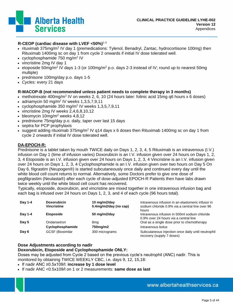
CLINICAL PRACTICE GUIDELINE LYHE-002 Version 12 Appendices
Page 5 of 44
R-CEOP (cardiac disease with LVEF <50%)1-3 rituximab 375mg/m2 IV day 1 (premedications: Tylenol, Benadryl, Zantac, hydrocortisone 100mg) then
Rituximab 1400mg sc on day 1 from cycle 2 onwards if initial IV dose tolerated well. cyclophosphamide 750 mg/m2 IV vincristine 2mg IV day 1 etoposide 50mg/m2 IV days 1-3 (or 100mg/m2 p.o. days 2-3 instead of IV; round up to nearest 50mg
multiple) prednisone 100mg/day p.o. days 1-5 Cycles: every 21 days
R-MACOP-B (not recommended unless patient needs to complete therapy in 3 months) methotrexate 400mg/m2 IV on weeks 2, 6, 10 (24 hours later: folinic acid 15mg q6 hours x 6 doses) adriamycin 50 mg/m2 IV weeks 1,3,5,7,9,11 cyclophosphamide 350 mg/m2 IV weeks 1,3,5,7,9,11 vincristine 2mg IV weeks 2,4,6,8,10,12 bleomycin 10mg/m2 weeks 4,8,12 prednisone 75mg/day p.o. daily, taper over last 15 days septra for PCP prophylaxis suggest adding rituximab 375mg/m2 IV q14 days x 6 doses then Rituximab 1400mg sc on day 1 from
cycle 2 onwards if initial IV dose tolerated well.
DA-EPOCH-R: Prednisone is a tablet taken by mouth TWICE daily on Days 1, 2, 3, 4, 5 Rituximab is an intravenous (I.V.) infusion on Day 1 (time of infusion varies) Doxorubicin is an I.V. infusion given over 24 hours on Days 1, 2, 3, 4 Etoposide is an I.V. infusion given over 24 hours on Days 1, 2, 3, 4 Vincristine is an I.V. infusion given over 24 hours on Days 1, 2, 3, 4 Cyclophosphamide is an I.V. infusion given over two hours on Day 5 On Day 6, filgrastim (Neupogen®) is started subcutaneously once daily and continued every day until the white blood cell count returns to normal. Alternatively, some Doctors prefer to give one dose of pegfilgrastim (Neulasta®) after each cycle of dose-adjusted EPOCH-R Patients then have labs drawn twice weekly until the white blood cell count has recovered. Typically, etoposide, doxorubicin, and vincristine are mixed together in one intravenous infusion bag and each bag is infused over 24 hours on Days 1, 2, 3, and 4 of each cycle (96 hours total).
Day 1-4 Doxorubicin Vincristine
10 mg/m2/day 0.4mg/m2/day (no cap)
Intravenous infusion in an elastomeric infusor in sodium chloride 0.9% via a central line over 96 hours
Day 1-4 Etoposide 50 mg/m2/day Intravenous infusion in 500ml sodium chloride 0.9% over 24 hours via a central line
Day 5 Ondansetron 8mg Oral as a single dose prior to chemotherapy
Cyclophosphamide 750mg/m2 Intravenous bolus
Day 6 GCSF (Biosimilar 300 micrograms Subcutaneous injection once daily until neutrophil recovery (supply 7 doses)
Dose Adjustments according to nadir Doxorubicin, Etoposide and Cyclophosphamide ONLY: Doses may be adjusted from Cycle 2 based on the previous cycle’s neutrophil (ANC) nadir. This is monitored by obtaining TWICE WEEKLY CBC, i.e. days 9, 12, 15,18: If nadir ANC ≥0.5x10⁹/l: increase by 1 dose level If nadir ANC <0.5x10⁹/l on 1 or 2 measurements: same dose as last

CLINICAL PRACTICE GUIDELINE LYHE-002 Version 12 Appendices
Page 6 of 44
Cycle If nadir ANC <0.5x10⁹/l on at least 3 measurements: decrease by 1 dose level If platelet nadir <25x109/l: reduce by 1 dose level regardless of ANC Life threatening infections: decrease by 1 dose level
Neurotoxicity: If the patient complains of significant constipation or sensory loss in fingers and/or toes, consider dose reduction of vincristine:
introduced at 75% of the normal dose thereafter. If ≥ grade 3 ileus recurs, vincristine should be discontinued
Additional medicines that may be prescribed: Septra 480mg Oral once daily Valacyclovir 500mg Oral once daily Fluconazole 50mg Oral once daily Omeprazole 20mg Oral once daily for 5 days Metoclopramide 10mg Oral four times daily as
needed Ondansetron 8mg Oral as a single dose prior to
chemotherapy, then twice daily as needed
Docusate/Senna (Senna-S®) to prevent constipation from vincristine
Consider intrathecal prophylaxis for patients with >1 extranodal site and elevated LDH

CLINICAL PRACTICE GUIDELINE LYHE-002 Version 12 Appendices
Page 7 of 44
II. INITIAL THERAPY FOR INDOLENT HISTOLOGY NON-HODGKIN LYMPHOMA
B-R bendamustine 90 mg/m2 IV day 1, 2 rituximab 375 mg/m2 IV day 1 then Rituximab 1400mg sc on day 1 from cycle 2 onwards if initial IV dose
tolerated. Cycles: repeated every 3-4 weeks depending on blood counts (usually administered every 28 days) for
a maximum of 6 cycles
CVP cyclophosphamide 800 mg/m2 IV day 1 (or 400 mg/m2/day p.o. days 1-5) vincristine 2mg IV day 1 prednisone 100mg/day p.o. days 1-5 Cycles: every 21 daysR-CVP rituximab 375mg/m2 IV day 1 (premeds: Tylenol, Benadryl, Zantac, hydrocortisone 100mg), then
Rituximab 1400mg sc on day 1 from cycle 2 onwards if initial IV dose tolerated. cyclophosphamide 750 mg/m2 IV day 1 vincristine 2mg IV day 1 prednisone 100mg/day p.o. days 1-5 Cycles: every 21 days
Maintenance Rituximab in First or Second Remission Following Chemotherapy + Rituximab Follicular and other indolent B-cell lymphoma: rituximab 1400mg sc (or 375mg/m2 IV if cannot tolerate
sc) x 1 dose q3 months x 2 years (8 doses total) Mantle cell lymphoma option: rituximab 1400mg sc ( or 375mg/m2 IV if cannot tolerate sc) q2months
until progression
Outpatient R-DHAP Cycle 1: Day1: Rituximab 375mg/m2 IV (if no rituximab in past 3months and cannot recieve sc rituximab) Day 2: 500mL NS pre, cisplatin 35 mg/m2 in 500 mL NS/mannitol, 500 ml NS post, AraC 2g/m2 in 500 mL NS. Day 3: 500mL NS pre, cisplatin 35 mgm2 in 500 ml NS/mannitol, AraC 2g/m2 in 500 mL NS. Total 5 hrs
Cycle 2 onwards: Day1: Rituximab 1400mg sc, 500mL NS pre, cisplatin 35 mg/m2 in 500 mL NS/mannitol, 500 ml NS post. Then AraC 2g/m2 in 500 mL NS. Total 5 hrs Day2: 500mL NS pre, cisplatin 35 mgm2 in 500 ml NS/mannitol, AraC 2g/m2 in 500 mL NS. Total 5 hrs
Chlorambucil (options) 0.1-0.2 mg/kg/day for 4-8 weeks then usually reduce for maintenance 10-14 mg/m² for 5 to 7 days each 28 days 0.5 mg/kg days 1 and 15 q28d cycle

CLINICAL PRACTICE GUIDELINE LYHE-002 Version 12 Appendices
Page 8 of 44
Fludarabine 25mg/m2 IV days 1-5 q28 days (days 1-3 only if frail elderly or renal dysfunction) 40mg/m2 p.o. days 1-5 q28 days (round down to nearest multiple of 10mg) (d1-3 only if frail or renal
dysfunction)
FND fludarabine 25mg/m2 IV days 1-3 or 40mg/m2 p.o. days 1-3 mitoxantrone 10mg/m2 day 1 dexamethasone 40mg p.o. days 1-3 septra for PCP prophylaxis Cycles: every 28 days
III. INITIAL THERAPY FOR PERIPHERAL T-CELL LYMPHOMA
CHOP cyclophosphamide 750 mg/m2 IV adriamycin 50 mg/m2 IV day 1 vincristine 2mg IV day 1 prednisone 100mg/day p.o. days 1-5 Cycles: every 21 days
CHOEP1-3
cyclophosphamide 750 mg/m2 IV adriamycin 50 mg/m2 IV day 1 vincristine 2mg IV day 1 etoposide 100mg/m2 IV days 1-3 (or 200mg/m2 p.o. days 2-3 instead of IV; round down to nearest 50mg
multiple) prednisone 100mg/day p.o. days 1-5 G-CSF days 7-11 or neulasta day 4 of each cycle Cycles: every 21 days
VIPD (Nasal NK/T-cell lymphoma) etoposide 100mg/m2 days 1-3 ifosfamide 1.2g/m2 days 1-3 cisplatin 33mg/m2 days 1-3 dexamethasone 40mg days 1-4 Cycles: 3 cycles after initial radiotherapy
GOLD (14 day cycle)4
gemcitabine 1000mg/m2 on day 1
oxaliplatin 100mg/m2 on day 1
L-asparaginase 10,000U/m2 on days 1-5*
dexamethasone (20mg b.i.d.) on days 1-4*An intradermal test was required prior to the administration of L-ASP

CLINICAL PRACTICE GUIDELINE LYHE-002 Version 12 Appendices
Page 9 of 44
SMILE (28 day cycle)5
Methotrexate 2g/m2 on day 1
Leucovorin 15mg x 4 on day 2, 3, and 4
Ifosfamide 1500mg/m2 on day 2, 3, and 4
Mesna 300 mg/m2 x 3 on day 2, 3 and 4
Dexamethasone 40mg/d on day 2, 3 and 4
Etoposide 100mg/m2 on day 2, 3 and 4
L-asparaginase 6000U/m2 on day 8, 10, 12, 14, 16, 18 and 20GCSF should be given from day 6 and discontinued if the leukocyte count exceeds 5000/μL. Antibiotic prophylaxis with sulfamethoxazole-trimethoprim is recommended.

CLINICAL PRACTICE GUIDELINE LYHE-002 Version 12 Appendices
Page 10 of 44
IV. HODGKIN DISEASE CHEMOTHERAPY REGIMENS
Initial Therapy
ABVD adriamycin 25 mg/m2 IV days 1 and 14 ChlVPP chlorambucil 6mg/m2 p.o. days 1-14
bleomycin 10 mg/m2 IV days 1 and 14 vinblastine 6mg/m2 IV days 1 and 8 vinblastine 6 mg/m2 IV days 1 and 14 procarbazine 100mg/m2 p.o. days 1-14 dacarbazine 375 mg/m2 IV days 1 and 14 prednisone 40mg/m2 p.o. days 1-14 Cycles: every 28 days Cycles: every 28 days
BEACOPP (escalated) MOPP nitrogen mustard 6mg/m2 days 1 & 8
bleomycin 10mg/m2 IV day 8 vincristine 1.4mg/m2 IV days 1 & 8 etoposide 200mg/m2 IV days 1-3 procarbazine 100mg/m2 po days 1-14 adriamycin 35mg/m2 IV day 1 prednisone 40mg/m2 po days 1-14 cyclophosphamide 1250mg/m2 IV day 1 Cycles: every 28 days vincristine 1.4mg/m2 IV day 8 procarbazine 100mg/m2 p.o. days 1-7 COPP cyclophosphamide 650mg/m2 IV days 1&8
prednisone 40mg/m2 po days 1-14 vincristine 1.4mg/m2 IV days 1 & 8
G-CSF 300-480g sc d9-19 (to ANC>1.5) or Neulasta d9 procarbazine 100mg/m2 po days 1-14 Cycles: every 21 days prednisone 40mg/m2 po days 1-14
Cycles: every 28 days BEACOPP (baseline)
bleomycin 10mg/m2 IV day 8 etoposide 100 mg/m2 IV days 1-3 adriamycin 25mg/m2 IV day 1 cyclophosphamide 650mg/m2 IV day 1 vincristine 1.4mg/m2 IV day 8 procarbazine 100mg/m2 p.o. days 1-7 prednisone 40 mg/m2 p.o. days 1-14
V. LYMPHOMA SALVAGE REGIMENS
Aggressive Histology Hodgkin and Non-Hodgkin Lymphomas*
DICE dexamethasone 10mg IV q6 hours days 1-4 ifosfamide 1g/m2 (max 1.75g) over 15 minutes days 1-4 cisplatin 25mg/m2 IV over 1hour days 1-4 etoposide 100mg/m2 over 1 hour days 1-4 mesna 200 mg/m2 over 5-10 min prior to first dose of ifosfamide, then 200 mg/m2 IV at 4 hours and
400mg/m2 p.o. (or 200 mg/m2 IV) at 8 hours post-ifosfamide x 4 days Cycles: every 21-28 days
CEPP cyclophosphamide 600 mg/m2 IV days 1 and 8 etoposide 70mg/m2 days 1-3 procarbazine 60mg/m2 p.o. days 1-10 prednisone 100mg/day p.o. days 1-10
Cycles: every 28 days
GDP gemcitabine 1000mg/m2 IV days 1 and 8 dexamethasone 40mg p.o. days 1-4

CLINICAL PRACTICE GUIDELINE LYHE-002 Version 12 Appendices
Page 11 of 44
cisplatin 75mg/m2 IV
DICEP dexamethasone 10mg IV q8 hours x 10 doses cyclophosphamide 1.75 g/m2 IV over 2 hours days 1-3 etoposide 350mg/m2 IV over 2 hours days 1-3 cisplatin 35mg/m2 IV over 2 hours days 1-3 mesna 1.75g/m2 IV over 24 hours days 1-3 septra for PCP prophylaxis Cycles: Once only
*Add rituximab to salvage regimens for transplant eligible patients with relapsed B-cell lymphomas
Indolent Histology Non-Hodgkin Lymphoma
As above, plus:
Rituximab 375mg/m2 IV days 1,8,15, and 22 (Rituximab 1400mg sc from day 8 onwards if initial IV dose tolerated). Pre-medicate with hydrocortisone 100mg IV, Benadryl, Zantac, and Tylenol Infuse 50mg/hour initially, then increase by 50mg/hour increments q30 minutes as tolerated to a
maximum of 400mg/hour Subsequent infusions can begin at 100mg/hour and increase by 100mg/hour increments as tolerated to
maximum of 400mg/hour
FND fludarabine 25mg/m2 IV days 1-3 or 40mg/m2 p.o. days 1-3 mitoxantrone 10mg/m2 day 1 dexamethasone 40mg p.o. days 1-3 septra for PCP prophylaxis Cycles: every 28 days
R-FCM fludarabine 25mg/m2 IV days 1-3 or 40mg/m2 p.o. days 1-3 cyclophosphamide 200mg/m2 IV days1-3 mitoxantrone 8mg/m2 IV day 1 rituximab 375mg/m2 IV day 1 (Rituximab 1400mg sc on day 1 from cycle 2 onwards if initial IV dose
tolerated). Cycles: every 28 days

CLINICAL PRACTICE GUIDELINE LYHE-002 Version 12 Appendices
Page 12 of 44
VI. BURKITT LYMPHOMA 6,7
Modified Magrath Regimen of R-CODOXM/R-IVAC (Blood 2014; 124:2913-2920)
Regimen A (R-CODOX-M) Days: 1 2 3 4 5 6 7 8 9 10 11 12 13 14 15 16 17 18 19
rituximab 1400mg sc x
cyclophosphamide 800mg/m2 IV x x
doxorubicin 50mg/m2 IV x
vincristine 1.5mg/m2 IV cap 2mg x x
allopurinol 300mg/day po x x x x x x x x x x
methotrexate 3000mg/m2 IV over 2 hour IV**
x
leucovorin 25mg IV @ 24 hours, then 25mg IV q6h until methotrexate<10-8 M
xxxx
xxxx
xxxx
xxxx
IT methotrexate 12mg X
IT cytarabine 50mg * X
Peg-filgrastim 6mg X
*if CNS disease, give extra IT AraC 50mg d5 cycle 1 only**HDMTX administered once urine pH>7, and diuresis established with hydration including D5-0.2%NS plus 2-3 amps sodium bicarbonate. Continue hydration and alkalinization until MTX cleared.
Regimen B (R- IVAC) Days: 1 2 3 4 5 6 7 8 9 10 11 12 13 14 15 16 17 18
rituximab 375mg/m2 IV X
cytarabine 2g/m2 IV q12h x 4 doses
Xx xx
ifosfamide 1500mg/m2 IV X x x x x
mesna 360mg/m2 IV q3 hours xxx xxx xxx xxx xxx xxx
etoposide 60mg/m2 IV X x x x x
IT methotrexate 12mg x
Peg-filgrastim 6mg x
*if CNS disease, give extra IT AraC 50mg d3 cycle 1 only
Low risk patients: Single extra-abdominal mass <10cm, or completely resected abdominal disease and normal LDH Modified regimen A x 3 cycles (cytarabine IT day 1 and methotrexate IT day 3 each cycle)
High risk patients: All others Alternate regimen A with regimen B for a total of 2 each or 4 cycles total
Start next cycle once ANC>1.0 and platelets>50

CLINICAL PRACTICE GUIDELINE LYHE-002 Version 12 Appendices
Page 13 of 44
VII. PRIMARY CNS LYMPHOMA PROTOCOL
A. Transplant-Eligible Patients: age < 65years, no significant co-morbidities, no immune suppression
Step 1 *Step 2 **Step 3 **Step 4
Week 1 2 3 4 5 6 7 8 9 10 12-14
Rituximab iv/sc d0 and d 4, 14 high-dose methotrexate 3.5 g/m2 d1&15 procarbazine 100 mg/m2 d1-7
x x x
x x
rituximab 1400mg SC d1 cytarabine 3 g/m2 x d1&2 G-CSF 5-10 µg/kg d8-13 Apheresis ~d14 or 15
x x
x x
Rituximab 1400mg SC d0 high-dose methotrexate 3.5 g/m2 d1 cytarabine 2 g/m2 twice daily days 2-3 all q21d for 2 cycles
x x x
thiotepa 300 mg/m2 IV days -6,-5 busulfan 3.2 mg/kg IV days -4 to -2, ASCT day 0
X X
* Step 2 may begin either week 4 or 5 depending upon patient status and apheresis scheduling*Step 3 may be omitted in patients who have achieved some response and are phsycially fit to proceed directly to ASCT on week 9.
* Male IBW = 50kg + 2.3kg x inches > 5ft, Female IBW = 45.5 kg + 2.3kg x inches> 5ft. Adjusted BW = IBW + [40% x (actual – IBW)]
Step 2. Rituximab/high-dose cytarabine x 1 cycle for stem cell collection after 2 cycles of methotrexate
Day Medications Other Orders
1 0900hr -Premeds : Hydrocortisone 100mg IV, Benadryl 50mg IV, Zantac 50mg IV, Tylenol 650mg p.o.
-rituximab 1400mg SC
Weight
CBC & Diff, EP, creatinine, glucose
ALT,AlkP,LDH,bilirubin,Alb,Ca,Mg
2 & 3 0800hr – Kytril 2mg IV, dexamethasone 10mg IV 0800hr – IV N/S 500mL/hour x 2 hours 1000hr – cytarabine 3g/m2 IV over 3 hours (2g/m2 if >60yrs or creatinine >100)
9-14 1000hr – G-CSF 480-600 μg subcutaneous daily until apheresis completed (plan for apheresis approximately day 13-15, once ANC>5, Plt >75 and CD34>20)
Daily CBC & differential starting day 10
Step 1. Induction: High-dose methotrexate/procarbazine q14 days x 2 cycles
Day Medications Other Orders
ADMISSION 0
0900hr-Rituximab 375mg/m2 (1st infusion protocol) 2000hr – IV D5W + 20meq KCL/L + 2 amps NaHCO3/L @ 200ml/hour x 5d
Daily weights
Daily CBC & Diff, EP, Creat, glucose
ALT,AlkP,LDH,bilirubin,Alb,Ca,Mg
LFTs, Ca, lipase, every Monday &Thursday
1 0800hr - Kytril 1mg IV 0800hr - methotrexate 3500mg/m² IV over 2 hour cycles 1 and 2 0800hr - procarbazine 100mg/m² po daily x 7days only cycle 1
(round down to nearest 50mg multiple)
0700hr - Urine pH twice daily, call MD if <7.0
2-3 0800hr - folinic acid (leucovorin) 25 mg IV q6 hours until methotrexate level < 0.05 Continue hydration until methotrexate level <0.05
0500-0800hr – methotrexate Level daily (expect level < 10 d2, <1 d3)
4 0900hr- Rituximab 1400mg SC on Cycle 1 only and continue folinic acid) 0500-0800hr – methotrexate Level daily
5 Discharge once methotrexate level <0.05
If level 0.01-0.05, discharge on leucovorin 5mg p.o. q6 hours x 2-3 days
Discharge meds: septra DS 1 daily or dapsone 50mg daily x 6-9 months; consider dexamethasone taper if on dexamethasone
Remember coumadin/LMWH and Dilantin if patient is on these medications

CLINICAL PRACTICE GUIDELINE LYHE-002 Version 12 Appendices
Page 14 of 44
* Male IBW = 50kg + 2.3kg x inches > 5ft, Female IBW = 45.5 kg + 2.3kg x inches> 5ft. Adjusted BW = IBW + [40% x (actual – IBW)]**NB: Step 3 may be omitted in patients who have achieved some response and are phsycially fit to proceed directly to ASCT on week 9.
Step 4. TBu/ASCT consolidation after response to methotrexate and high-dose cytarabine
Day Medications Other Orders
ADMISSION Day -7
Allopurinol 300 mg p.o. daily until day 0
2200hr - D5 ½ N/S + 20 mEq KCL/L @ 125 mL/hour until day -1
Consult dietician
Consult physiotherapy
Low bacteria diet
24 hour intake
Mouth protocol
Record intake and output
-6 & -5 0800 – thiotepa 300 mg/m² IV over 3 hours x 2 days (use IDEAL BSA)
0800hr – granisetron 2 mg IV daily x 8 days
EP daily x 31days
Shower/bath q6 hours x 3 days; avoid skin creams
-4 to -2 0900 - busulfan 3.2 mg/kg IV daily x 3 days (use Ideal weight)
lorazepam prophylaxis x 4 days
CBC & differential daily x 31 days
ALT, Alk Phos, bili, alb, Ca, Mg, every Monday &Thursday
PT, PTT, every Monday
-1 Rest day mycostatin 500,000 units q2-4 hours
septra RS 1 tab p.o. daily
acyclovir 5 mg/kg bid IV or 400 mg p.o. four times daily
0 Autologous Blood Stem Cell INFUSION
+7 G-CSF 300µg (if less than 70kg) or 480µg (if over 70kg) subcutaneous daily until post-nadir ANC > 1.5
**Step 3. High-dose methotrexate/cytarabine consolidation q21 days x 1 cycles after stem cell collection
Day Medications Other Orders
ADMISSION 0
16:00hr- Rituximab 1400mg subcutaneously 2000hr – IV D5W + 20meq KCL/L + 2 amps NaHCO3/L @ 200ml/hour x 5 days
Daily weights
Daily CBC & differential, EP, creatinine, glucose
ALT,AlkP,LDH,bilirubin,Alb,Ca,Mg
ALT, Alk P, bili, Ca, lipase, every Monday &Thursday
1 0800hr - Kytril 1mg IV 0800hr - methotrexate 3500mg/m² IV over 2 hours
07:00 - Urine pH bid, call MD if <7.0
2-3 080hr- folinic acid (leucovorin) 25 mg IV q6 hours until methotrexate level < 0.05 -Continue hydration until methotrexate level <0.05
0800hr – Kytril 2mg IV, dexamethasone 10mg IV 1000hr – cytarabine 2g/m2 IV over 2 hours twice daily x 2 days
Reduce to 1.5g/m2 if age >60 years or creatinine >100
0500-08:00 – methotrexate level daily (expect <10 d2, <1 d3, <0.1 d4, <0.05 d5)
5 Discharge once methotrexate level <0.05 If level 0.01-0.05, discharge on leucovorin 5mg p.o. q6 hours x 2-3 days (other meds as step 1 above)
8-12 1000hr – G-CSF 480-600 μg subcutaneous daily until post-nadir ANC >1.5 Daily CBC & differential starting day 10

CLINICAL PRACTICE GUIDELINE LYHE-002 Version 11 Appendices
Page 15 of 44
B. Transplant-Ineligible Patients: age >65 years. Should be CIRS 0-6 and ECOG 0-2.
Step 1 Step 3
Week 1 2 3 4 5 6 7 8 9 10 11 12 15 or 16
Rituximab d0, 4 high-dose methotrexate 3.5 g/m2 d1 cytarabine 2g/ m2 q12h x 2 d2 Thiotepa 30mg/ m2 d4
x x x x
x x x x
x x x x
x x x x
Ifosfamide 2g/m2 daily days 1-3 X
Methotrexate should be omitted if creat clearance < 50 mL/min or if renal dysfunction with prior cycle Cytarabine should be reduced to q24hr if creat clearance < 50 mL/min or complications of myelosuppression
* Male IBW = 50kg + 2.3kg x inches > 5ft, Female IBW = 45.5 kg + 2.3kg x inches> 5ft. Adjusted BW = IBW + [40% x (actual – IBW)]
* Male IBW = 50kg + 2.3kg x inches > 5ft, Female IBW = 45.5 kg + 2.3kg x inches> 5ft. Adjusted BW = IBW + [40% x (actual – IBW)]
Step 2. Ifosfamide consolidation after response to MATRIX (optional)
Day Medications Other Orders
15 or 16 0800hr – Kytril 2mg IV, dexamethasone 10mg IV daily x 3d 0800hr – N/S IV 500mL/hour x 1 hour daily x 3d 0900hr – Mesna 400mg/m2 IV daily x 3d 0900hr – Ifosfamide 2g/m2 with 1g Mesna IV over 3 hours daily x 3d 1200hr – Mesna 400mg/m2 IV daily x 3d 1200hr – 1L NS IV 250mL/hour x 4 hours daily x 3d 1600hr – Mesna 400mg/m2 IV daily x 3d
weight (call MD if >2kg above day 1)
CBC & differential, EP, creatinine, glucose
ALT,AlkP,LDH,bilirubin,Alb,Ca,Mg
Step 1. Induction: MATRIX x 4 cycles
Day Medications Other Orders
ADMISSION 0
0900hr-Rituximab 375mg/m2 (1st infusion protocol) 2000hr – IV D5W + 20meq KCL/L + 2 amps NaHCO3/L @ 200ml/hour x 5 days
Daily weights
Daily CBC & differential, EP, creatinine, glucose
ALT,AlkP,LDH,bilirubin,Alb,Ca,Mg
LFTs, Ca, lipase, every Monday & Thursday
1 0800hr - Kytril 1mg IV 0800hr - methotrexate 3500mg/m² IV over 3 hours
0700hr - Urine pH twice daily, call MD if <7.0
2-3 0800hr - folinic acid (leucovorin) 25 mg IV q6hr until MTX level < 0.05 Continue hydration until methotrexate level <0.05 1000hr –Cytarabine 2mg/m2 by 1 hour infusion q12 hr x 2
0500-0800hr – methotrexate level daily (expect level < 10 d2, <1 d3)
4 0900hr- Rituximab 1400mg subcutaneously 1000hr- Thiotepa 30mg/m2 by 30min infusion
0500-0800hr – methotrexate Level daily
5 Discharge once methotrexate level <0.05
If level 0.01-0.05, discharge on leucovorin 5mg p.o. q6 hours x 2-3 days
Discharge meds: septra DS 1 daily or dapsone 50mg daily x 6-9 months; consider dexamethasone taper if on dexamethasone
Remember coumadin/LMWH and dilantin if patient is on these medications

CLINICAL PRACTICE GUIDELINE LYHE-002 Version 12 Appendices
Page 16 of 44
VIII. SECONDARY CNS LYMPHOMA PROTOCOL
A) Transplant-eligible patients (age <65 years, no significant co-morbidities, no immune suppression) withisolated CNS relapse/progression following complete response of systemic lymphoma to RCHOP.
Step 1 Step 2 Step 3
Week 1 2 3 4 5 6 7 8 9 10 11 12 13 14
high-dose methotrexate 3.5 g/m2 d1 Rituximab 375 mg/m2 d2 procarbazine 100 mg/m2 x 7 days d1-7 vincristine 1.4 mg/m2 d1
x x x x
x x
x
x x x
x
rituximab 1400mg sc days 1,4 dexamethasone 20 mg days 1-4 cisplatin 35 mg/m2 days 1,2 cytarabine 2 g/m2 x1 dose, days 1,2 G-CSF 5-10 µg/kg day 8-13 Apheresis day 13 or 14
x x x x
x x
R-TbuM/ ASCT (ritux d-7 + thiotepa 250mg/m2 d -6,-5 busulfan 3.2 mg/kg day -4 to -2, melphalan 100 mg/m2 d-1, ASCT d 0
x
* Male IBW = 50kg + 2.3kg x inches > 5ft, Female IBW = 45.5 kg + 2.3kg x inches> 5ft.
Step 2. Rituximab/DHAP x 1 cycle for stem cell collection after 4 cycles of methotrexate
Day Medications Other Orders
1 0800hr - hydrocortisone 100mg IV, Benadryl , Zantac, Tylenol 0900hr - rituximab 1400mg sc 0900hr -IV 1L NS 0900hr – dexamethasone 20mg p.o./IV daily x 4 days 0900hr – Kyrtil 1mg IV or 2mg p.o. x 3-4 days 0900hr – aprepitent protocol p.o. x 3 days 1000hr – cisplatin 35mg/m2 IV over 2 hours with mannitol 25g and 500mL NS 1200hr- cytarabine 2g/m2 IV over 2 hours x 1 doses (1.5g/m2 if >60yr)
Weight
CBC & differential, EP, creatinine, glucose
ALT,AlkP,LDH,bilirubin,Alb,Ca,Mg
2 0800hr – dexamethasone Kytril, Aprepitent continued 1000hr – cisplatin 35mg/m2 IV over 2 hours with mannitol 25g and 500mL NS 1200hr- cytarabine 2g/m2 IV over 2 hours x 1 doses (1.5g/m2 if >60yr)
4 Rituximab 1400mg sc
8-13 1000hr – G-CSF 480-600μg subcutaneous daily until apheresis completed (plan for apheresis approximately day 13-15, once ANC>5, Plt >75 and CD34>20)
Daily CBC & differential starting day 10
Step 1. Induction: high-dose methotrexate/vincristine/procarbazine q14 days x 4 cycles
Day Medications Other Orders
ADMISSION 0
2000hr - IV D5W + 20meq KCL/L + 2 amps NaHCO3/L @ 200mL/hour x 5 days Daily weights
Daily CBC & differential, EP, creatinine,glucose
ALT,AlkP,LDH,bilirubin,Alb,Ca,Mg
LFTs, Ca, lipase, every Monday & Thursday
1 0800hr - Kytril 1mg IV 0800hr - methotrexate 3500mg/m² IV over 2 hours cycles 1-4 0800hr - procarbazine 100mg/m² p.o. daily x 7days cycles 1 and 3
(round down to nearest 50mg multiple) 1000hr - vincristine 1.4mg/m2 IV only cycles 1 and 2
0700hr - Urine pH twice daily, call MD if <7.0
2-4 0800hr- folinic acid (leucovorin) 25 mg IV q6 hours until methotrexate level < 0.05 Continue hydration until methotrexate level <0.05 1000hr – Rituximab 375mg/m² IV (first 3 cycles HDMTX)
0500-0800hr – methotrexate level daily (expect level < 10 today)
5 Discharge once methotrexate level <0.05
If level 0.01-0.05, discharge on leucovorin 5mg p.o. q6 hours x 2-3 days
Discharge meds: septra DS 1 daily or dapsone 50mg daily x 6-9 months; consider dexamethasone taper if on dexamethasone
Remember coumadin/LMWH and dilantin if patient is on these medications

CLINICAL PRACTICE GUIDELINE LYHE-002 Version 12 Appendices
Page 17 of 44
Step 3. R-TBuM/ASCT consolidation after response to MTX and RDHAP Induction
Day Medications Other Orders
ADMISSION Day -7
Allopurinol 300 mg p.o. daily until day 0 Premeds: Hydrocortisone 100mg IV, Benadryl 50mg IV, Zantac 50mg IV, Tylenol 650mg p.o. -rituximab 375mg/m2 IV (first dose long infusion protocol)
2200hr - D5½ N/S + 20 mEq KCL/L @ 125 mL/hour until day -1
Consult dietician, physiotherapy
Low bacteria diet. 24hour intake
Mouth protocol; record intake and output
-6 & -5 0800hr – thiotepa 250 mg/m² IV over 2 hours x 2 days (use ideal BSA)
0800hr – Granisetron 2 mg IV daily x 8 days
EP daily x 31days
Shower/Bath q6 hours x 3 days; avoid skin creams
-4 to -2 0900 - busulfan 3.2 mg/kg IV daily x 3 days (use Ideal weight)
lorazepam prophylaxis x 4 days
CBC & differential daily x 31 days
ALT, Alk Phos, bilirubin, alb, Ca, Mg, every Monday &Thursday
PT, PTT every Monday
-1 10:00 -melphalan 100mg/m2 (actual BSA) IV over 5 minutes 10:15 – Lasix 20mg IV 10:30 - mannitol 20% 250 mL IVPB over 1 hour 11:30 - IV 1L NS @ 500 mL/hour for 3 hours 14:30 -IV 1L NS with 40 mEq KCL/L @ 125 mL/hour x 18 hours
Mycostatin 500,000 units q2-4 hours
Septra RS 1 tab p.o. daily
Acyclovir 5 mg/kg twice daily IV or 400 mg p.o. four timesdaily
0 Autologous Blood Stem Cell INFUSION
+7 G-CSF 300 µg (if less than 70kg) or 480µg (if over 70kg) subcutaneous daily until post-nadir ANC > 1.5

CLINICAL PRACTICE GUIDELINE LYHE-002 Version 12 Appendices
Page 18 of 44
B) Transplant-eligible patients (age <65 years, no significant co-morbidities, no immune suppression) withearly Systemic and CNS lymphoma (prior to completing RCHOP x6): RCHOP and HDMTX x4 cycles then RDHAP for stem cell mobilization and collection, then R-TBuM/ASCT.
Step 1 Step 2 Step 3
Week 0 1 2 3 4 5 6 7 8 9 10 13 14 15 16 17 18
Methotrexate 3.5 g/m2 q14d X* X** X X X X
R-CHOP X X X X
rituximab 1400mg sc days 1,4 dexamethasone 20 mg days 1-4 cisplatin 35 mg/m2 days 1,2 cytarabine 2 g/m2 x1 dose, days 1,2 G-CSF 5-10 µg/kg day 8-13 Apheresis day 13 or 14
x x x x
x x
R-TbuM/ ASCT (ritux d-7, thiotepa 250mg/m2 d-6,-5 busulfan 3.2 mg/kg day -4 to -2, melphalan 100 mg/m2 d -1, ASCT d 0
x
*HDMTX prior to RCHOP#1 if CNS and systemic lymphoma both identified at time of initial diagnosis.**If CNS lymphoma identified after RCHOP initiated but systemic disease responding to RCHOP, then plan for at least 4 doses HDMTX q14d with subsequent cycles RCHOP before proceeding to R-DHAP.
* Male IBW = 50kg + 2.3kg x inches > 5ft, Female IBW = 45.5 kg + 2.3kg x inches> 5ft.
Step 2. Rituximab/DHAP x 1 cycle for stem cell collection after 4 cycles of methotrexate
Day Medications Other Orders
1 0800hr - hydrocortisone 100mg IV, Benadryl , Zantac, Tylenol 0900hr - rituximab 1400mg sc 0900hr -IV 1L NS 0900hr – dexamethasone 20mg p.o./IV daily x 4 days 0900hr – Kyrtil 1mg IV or 2mg p.o. x 3-4 days 0900hr – aprepitent protocol p.o. x 3 days 1000hr – cisplatin 35mg/m2 IV over 2 hours with mannitol 25g and 500mL NS 1200hr- cytarabine 2g/m2 IV over 2 hours x 1 doses (1.5g/m2 if >60yr)
Weight
CBC & differential, EP, creatinine, glucose
ALT,AlkP,LDH,bilirubin,Alb,Ca,Mg
2 0800hr – dexamethasone Kytril, Aprepitent continued 1000hr – cisplatin 35mg/m2 IV over 2 hours with mannitol 25g and 500mL NS 1200hr- cytarabine 2g/m2 IV over 2 hours x 1 doses (1.5g/m2 if >60yr)
4 Rituximab 1400mg sc
8-13 1000hr – G-CSF 480-600μg subcutaneous daily until apheresis completed (plan for apheresis approximately day 13-15, once ANC>5, Plt >75 and CD34>20)
Daily CBC & differential starting day 10
Step 1. Induction: RCHOP q21d as well as high-dose methotrexate q14 days x 4 cycles
Day Medications (HDMTX component) Other Orders
ADMISSION 0
2000hr - IV D5W + 20meq KCL/L + 2 amps NaHCO3/L @ 200mL/hour x 5 days Daily weights
Daily CBC & differential, EP, creatinine,glucose
ALT,AlkP,LDH,bilirubin,Alb,Ca,Mg
LFTs, Ca, lipase, every Monday & Thursday
1 0800hr - Kytril 1mg IV 0800hr - methotrexate 3500mg/m² IV over 2 hours cycles 1-4
0700hr - Urine pH twice daily, call MD if <7.0
2-4 0800hr- folinic acid (leucovorin) 25 mg IV q6 hours until methotrexate level < 0.05 Continue hydration until methotrexate level <0.05
0500-0800hr – methotrexate level daily (expect level < 10 today)
5 Discharge once methotrexate level <0.05
If level 0.01-0.05, discharge on leucovorin 5mg p.o. q6 hours x 2-3 days
Discharge meds: septra DS 1 daily or dapsone 50mg daily x 6-9 months; consider dexamethasone taper if on dexamethasone
Remember coumadin/LMWH and dilantin if patient is on these medications

CLINICAL PRACTICE GUIDELINE LYHE-002 Version 12 Appendices
Page 19 of 44
Step 3. R-TBuM/ASCT consolidation after response to MTX and RDHAP Induction
Day Medications Other Orders
ADMISSION Day -7
Allopurinol 300 mg p.o. daily until day 0 Premeds: Hydrocortisone 100mg IV, Benadryl 50mg IV, Zantac 50mg IV, Tylenol 650mg p.o. -rituximab 375mg/m2 IV (first dose long infusion protocol)
2200hr - D5½ N/S + 20 mEq KCL/L @ 125 mL/hour until day -1
Consult dietician, physiotherapy
Low bacteria diet. 24hour intake
Mouth protocol; record intake and output
-6 & -5 0800hr – thiotepa 250 mg/m² IV over 2 hours x 2 days (use ideal BSA)
0800hr – Granisetron 2 mg IV daily x 8 days
EP daily x 31days
Shower/Bath q6 hours x 3 days; avoid skin creams
-4 to -2 0900 - busulfan 3.2 mg/kg IV daily x 3 days (use Ideal weight)
lorazepam prophylaxis x 4 days
CBC & differential daily x 31 days
ALT, Alk Phos, bilirubin, alb, Ca, Mg, every Monday &Thursday
PT, PTT every Monday
-1 10:00 -melphalan 100mg/m2 (actual BSA) IV over 5 minutes 10:15 – Lasix 20mg IV 10:30 - mannitol 20% 250 mL IVPB over 1 hour 11:30 - IV 1L NS @ 500 mL/hour for 3 hours 14:30 -IV 1L NS with 40 mEq KCL/L @ 125 mL/hour x 18 hours
Mycostatin 500,000 units q2-4 hours
Septra RS 1 tab p.o. daily
Acyclovir 5 mg/kg twice daily IV or 400 mg p.o. four timesdaily
0 Autologous Blood Stem Cell INFUSION
+7 G-CSF 300 µg (if less than 70kg) or 480µg (if over 70kg) subcutaneous daily until post-nadir ANC > 1.5

CLINICAL PRACTICE GUIDELINE LYHE-002 Version 12 Appendices
Page 20 of 44
C) Transplant-eligible patients (age <65 years, no significant co-morbidities, no immune suppression) withlate relapse (prior RCHOP x6) with systemic and CNS lymphoma: HDMTX-Ifosfamide-etopside x2 then RDHAP for stem cell mobilization and collection, then R-TBuM/ASCT
Step 1 Step 2 Step 3
Week 1 2 3 4 5 6 7 8 9 10 11 12 13 14
high-dose methotrexate 3.5 g/m2 d1 Rituximab 375 mg/m2 d2 Ifosfamide 1.5 g/m2 d3-5 Etoposide 100 mg/m2 d3-5
X X X X
X X X X
X
rituximab 1400mg sc days 1,4 dexamethasone 20 mg days 1-4 cisplatin 35 mg/m2 days 1,2 cytarabine 2 g/m2 x1 dose, days 1,2 G-CSF 5-10 µg/kg day 8-13 Apheresis day 13 or 14
x x x x
x x
R-TbuM/ ASCT (ritux d-7 + thiotepa 250mg/m2 d -6,-5 busulfan 3.2 mg/kg day -4 to -2, melphalan 100 mg/m2 d-1, ASCT d 0
X
* Male IBW = 50kg + 2.3kg x inches > 5ft, Female IBW = 45.5 kg + 2.3kg x inches> 5ft.
Step 1. Induction: R-IE and high-dose methotrexate x 2 cycles (HDMTX x3)
Day Medications Other Orders
ADMISSION 0
2000hr - IV D5W + 20meq KCL/L + 2 amps NaHCO3/L @ 200mL/hour x 5 days Daily weights
Daily CBC & differential, EP, creatinine,glucose
ALT,AlkP,LDH,bilirubin,Alb,Ca,Mg
LFTs, Ca, lipase, every Monday & Thursday
1 0800hr - Kytril 1mg IV 0800hr - methotrexate 3500mg/m² IV over 2 hours cycles 1-4
0700hr - Urine pH twice daily, call MD if <7.0
2 0800hr- folinic acid (leucovorin) 25 mg IV q6 hours until methotrexate level < 0.05 Continue hydration until methotrexate level <0.05 1000hr – Rituximab 375mg/m² IV
0500-0800hr – methotrexate level daily (expect level < 10 today)
3-5 0800hr – Kytril 2mg IV, dexamethasone 10mg IV daily x 3d 0800hr – N/S IV 500mL/hour x 1 hour daily x 3d 0900hr – Mesna 0.5 g IV daily x 3d 0900hr - Ifosfamide 1.5g/m2 with 1g Mesna IV over 3 hours daily x 3d 1200hr – Mesna 0.5 g IV daily x 3d 1200hr – 1/2NS IV 250mL/hour x 4 hours daily x 3d 1200hr – Etoposide 100 mg/m2 IV daily x 3d 1600hr – Mesna 1.0 g IV daily x 3d 1000hr
5 or 6 Discharge once methotrexate level <0.05
If level 0.01-0.05, discharge on leucovorin 5mg p.o. q6 hours x 2-3 days
Discharge meds: septra DS 1 daily or dapsone 50mg daily x 6-9 months; consider dexamethasone taper if on dexamethasone
Remember coumadin/LMWH and dilantin if patient is on these medications

CLINICAL PRACTICE GUIDELINE LYHE-002 Version 12 Appendices
Page 21 of 44
Step 2. Rituximab/DHAP x 1 cycle for stem cell collection after 4 cycles of methotrexate
Day Medications Other Orders
1 0800hr - hydrocortisone 100mg IV, Benadryl , Zantac, Tylenol 0900hr - rituximab 1400mg sc 0900hr -IV 1L NS 0900hr – dexamethasone 20mg p.o./IV daily x 4 days 0900hr – Kyrtil 1mg IV or 2mg p.o. x 3-4 days 0900hr – aprepitent protocol p.o. x 3 days 1000hr – cisplatin 35mg/m2 IV over 2 hours with mannitol 25g and 500mL NS 1200hr- cytarabine 2g/m2 IV over 2 hours x 1 doses (1.5g/m2 if >60yr)
Weight
CBC & differential, EP, creatinine, glucose
ALT,AlkP,LDH,bilirubin,Alb,Ca,Mg
2 0800hr – dexamethasone Kytril, Aprepitent continued 1000hr – cisplatin 35mg/m2 IV over 2 hours with mannitol 25g and 500mL NS 1200hr- cytarabine 2g/m2 IV over 2 hours x 1 doses (1.5g/m2 if >60yr)
4 Rituximab 1400mg sc
8-13 1000hr – G-CSF 480-600μg subcutaneous daily until apheresis completed (plan for apheresis approximately day 13-15, once ANC>5, Plt >75 and CD34>20)
Daily CBC & differential starting day 10
Step 3. R-TBuM/ASCT consolidation after response to MTX and RDHAP Induction
Day Medications Other Orders
ADMISSION Day -7
Allopurinol 300 mg p.o. daily until day 0 Premeds: Hydrocortisone 100mg IV, Benadryl 50mg IV, Zantac 50mg IV, Tylenol 650mg p.o. -rituximab 375mg/m2 IV (first dose long infusion protocol)
2200hr - D5½ N/S + 20 mEq KCL/L @ 125 mL/hour until day -1
Consult dietician, physiotherapy
Low bacteria diet. 24hour intake
Mouth protocol; record intake and output
-6 & -5 0800hr – thiotepa 250 mg/m² IV over 2 hours x 2 days (use ideal BSA)
0800hr – Granisetron 2 mg IV daily x 8 days
EP daily x 31days
Shower/Bath q6 hours x 3 days; avoid skin creams
-4 to -2 0900 - busulfan 3.2 mg/kg IV daily x 3 days (use Ideal weight)
lorazepam prophylaxis x 4 days
CBC & differential daily x 31 days
ALT, Alk Phos, bilirubin, alb, Ca, Mg, every Monday &Thursday
PT, PTT every Monday
-1 10:00 -melphalan 100mg/m2 (actual BSA) IV over 5 minutes 10:15 – Lasix 20mg IV 10:30 - mannitol 20% 250 mL IVPB over 1 hour 11:30 - IV 1L NS @ 500 mL/hour for 3 hours 14:30 -IV 1L NS with 40 mEq KCL/L @ 125 mL/hour x 18 hours
Mycostatin 500,000 units q2-4 hours
Septra RS 1 tab p.o. daily
Acyclovir 5 mg/kg twice daily IV or 400 mg p.o. four timesdaily
0 Autologous Blood Stem Cell INFUSION
+7 G-CSF 300 µg (if less than 70kg) or 480µg (if over 70kg) subcutaneous daily until post-nadir ANC > 1.5

CLINICAL PRACTICE GUIDELINE LYHE-002 Version 12 Appendices
Page 22 of 44
VIII. SECONDARY CNS LYMPHOMA PROTOCOL
D) Transplant-ineligible patients (age >65 years, significant co-morbidities, or immune suppression) withisolated CNS relapse/progression following complete response of systemic lymphoma to RCHOP. (consider only for highly motivated patients who wish curative intent therapy. Otherwise palliation with IT chemotherapy, radiotherapy, or supportive care).
Step 1 Step 2 Step 3
Week 1 2 3 4 5 6 7 8 9 10 11 12 15 or 16
Rituximab 375mg/m2 d0, 4 high-dose methotrexate 3.5 g/m2 d1 procarbazine 100 mg/m2 x 7 days d1-7
x x x
Rituximab 375mg/m2 d0 high-dose methotrexate 3.5 g/m2 day 1 cytarabine 1.5-2 g/m2 bid days 2-3
x x x
x x x
x x x
x x x
Ifosfamide 2g/m2 daily days 1-3 X
* Male IBW = 50kg + 2.3kg x inches > 5ft, Female IBW = 45.5 kg + 2.3kg x inches> 5ft. Adjusted BW = IBW + [40% x (actual – IBW)]
* Male IBW = 50kg + 2.3kg x inches > 5ft, Female IBW = 45.5 kg + 2.3kg x inches> 5ft. Adjusted BW = IBW + [40% x (actual – IBW)]
Step 1. Induction: high-dose methotrexate/procarbazine x 1 cycle
Day Medications Other Orders
ADMISSION 0
0900hr-Rituximab 375mg/m2 (1st infusion protocol) 2000hr – IV D5W + 20meq KCL/L + 2 amps NaHCO3/L @ 200ml/hour x 5 days
Daily weights
Daily CBC & differential, EP, creatinine, glucose
ALT,AlkP,LDH,bilirubin,Alb,Ca,Mg
LFTs, Ca, lipase, every Monday & Thursday
1 0800hr - Kytril 1mg IV 0800hr - methotrexate 3500mg/m² IV over 2 hours 0800hr - procarbazine 100mg/m² p.o. daily x 7days only cycle 1
(round down to nearest 50mg multiple)
0700hr - Urine pH twice daily, call MD if <7.0
2-3 0800hr - folinic acid (leucovorin) 25 mg IV q6hr until MTX level < 0.05 Continue hydration until methotrexate level <0.05
0500-0800hr – methotrexate level daily (expect level < 10 d2, <1 d3)
4 0900hr- Rituximab 375mg/m2 (subsequent infusion protocol)on cycle 1 only and continue folinic acid)
0500-0800hr – methotrexate Level daily
5 Discharge once methotrexate level <0.05
If level 0.01-0.05, discharge on leucovorin 5mg p.o. q6 hours x 2-3 days
Discharge meds: septra DS 1 daily or dapsone 50mg daily x 6-9 months; consider dexamethasone taper if on dexamethasone
Remember coumadin/LMWH and dilantin if patient is on these medications
Step 2. High-dose methotrexate/cytarabine consolidation q21 days x 4 cycles
Day Medications Other Orders
ADMISSION 0
1600hr- Rituximab 375mg/m2 (subsequent infusion protocol)on cycle 1 only and continue folinic acid) 2000hr – IV D5W + 20meq KCL/L + 2 amps NaHCO3/L @ 200mL/hour x 5 days
Daily weights
Daily CBC & differential, EP, creatinine, glucose
ALT,AlkP,LDH,bilirubin,Alb,Ca,Mg
ALT, Alk P, bilirubin, Ca, lipase, every Monday &Thursday
1 0800hr - Kytril 1mg IV 0800hr - methotrexate 3500mg/m² IV over 2 hours
07:00 - Urine pH bid, call MD if <7.0
2-3 0800hr- folinic acid (leucovorin) 25 mg IV q6 hours until methotrexate level < 0.05
-Continue hydration until methotrexate level <0.05 0800hr – Kytril 2mg IV, Decadron 10mg IV 1000hr – cytarabine 2g/m2 IV over 2 hours twice daily x 2 days; reduce to 1.5g/m2 if age >60 years or creatinine >100
0500-08:00 – Methotrexate Level daily (expect <10 d2, <1 d3, <0.1 d4, <0.05 d5)
5 Discharge once methotrexate level <0.05 If level 0.01-0.05, discharge on leucovorin 5mg p.o. q6 hours x 2-3 days (other meds as step 1 above)
8-12 10:00 – G-CSF 480-600 μg subcutaneous daily until post-nadir ANC >1.5 Daily CBC & diff starting d10

CLINICAL PRACTICE GUIDELINE LYHE-002 Version 12 Appendices
Page 23 of 44
Step 3. Ifosfamide consolidation after response to methotrexate and high-dose cytarabine
Day Medications Other Orders
15 or 16 0800hr – Kytril 2mg IV, dexamethasone 10mg IV daily x 3d 0800hr – N/S IV 500mL/hour x 1 hour daily x 3d 0900hr – Mesna 1.0 g IV daily x 3d 0900hr – Ifosfamide 2g/m2 with 1g Mesna IV over 3 hours daily x 3d 1200hr – Mesna 0.5 g IV daily x 3d 1200hr – 1/2NS IV 250mL/hour x 4 hours daily x 3d 1600hr – Mesna 1.0 g IV daily x 3d
weight (call MD if >2kg above day 1)
CBC & differential, EP, creatinine, glucose
ALT,AlkP,LDH,bilirubin,Alb,Ca,Mg

CLINICAL PRACTICE GUIDELINE LYHE-002 Version 12 Appendices
Page 24 of 44
E) Transplant-ineligible patients (age >65 years, significant co-morbidities, or immune suppression) withearly Systemic and CNS lymphoma prior to completing initial RCHOP x6. (consider only for highly motivated patients who wish curative intent therapy. Otherwise palliation).
Step 1 Step 2 Step 3
Week 0 1 2 3 4 5 6 7 8 9 10 13 14 15 16 17 18
Methotrexate 3.5 g/m2 q14d X* X** X X X X
R-CHOP X X X X
rituximab 1400mg sc days 1,4 dexamethasone 20 mg days 1-4 cytarabine 2 g/m2 x1 dose, days 1 and 2 G-CSF 5-10 µg/kg day 8-13
x x x
x
Ifosfamide 2g/m2 daily days 1-3 X
*HDMTX prior to RCHOP#1 if CNS and systemic lymphoma both identified at time of initial diagnosis.**If CNS lymphoma identified after RCHOP initiated but systemic disease responding to RCHOP, then plan for at least 4 doses HDMTX q14d with subsequent cycles RCHOP before proceeding to R-AraC. * Male IBW = 50kg + 2.3kg x inches > 5ft, Female IBW = 45.5 kg + 2.3kg x inches> 5ft.
Step 2. Rituximab/DHAP x 1 cycle for stem cell collection after 4 cycles of methotrexate
Day Medications Other Orders
1 0800hr - hydrocortisone 100mg IV, Benadryl , Zantac, Tylenol 0900hr - rituximab 1400mg sc 0900hr -IV 1L NS 0900hr – dexamethasone 20mg p.o./IV daily x 4 days 0900hr – Kyrtil 1mg IV or 2mg p.o. x 3-4 days 0900hr – aprepitent protocol p.o. x 3 days 1000hr – cisplatin 35mg/m2 IV over 2 hours with mannitol 25g and 500mL NS 1200hr- cytarabine 2g/m2 IV over 2 hours x 1 doses (1.5g/m2 if >60yr)
Weight
CBC & differential, EP, creatinine, glucose
ALT,AlkP,LDH,bilirubin,Alb,Ca,Mg
2 0800hr – dexamethasone Kytril, Aprepitent continued 1000hr – cisplatin 35mg/m2 IV over 2 hours with mannitol 25g and 500mL NS 1200hr- cytarabine 2g/m2 IV over 2 hours x 1 doses (1.5g/m2 if >60yr)
4 Rituximab 1400mg sc
8-13 1000hr – G-CSF 480-600μg subcutaneous daily until apheresis completed (plan for apheresis approximately day 13-15, once ANC>5, Plt >75 and CD34>20)
Daily CBC & differential starting day 10
Step 3. Ifosfamide consolidation after response to methotrexate and high-dose cytarabine
Day Medications Other Orders
15 or 16 0800hr – Kytril 2mg IV, dexamethasone 10mg IV daily x 3d 0800hr – N/S IV 500mL/hour x 1 hour daily x 3d 0900hr – Mesna 1.0 g IV daily x 3d 0900hr – Ifosfamide 2g/m2 with 1g Mesna IV over 3 hours daily x 3d 1200hr – Mesna 0.5 g IV daily x 3d 1200hr – 1/2NS IV 250mL/hour x 4 hours daily x 3d 1600hr – Mesna 1.0 g IV daily x 3d
weight (call MD if >2kg above day 1)
CBC & differential, EP, creatinine, glucose
ALT,AlkP,LDH,bilirubin,Alb,Ca,Mg
Step 1. Induction: RCHOP q21d as well as high-dose methotrexate q14 days x 4 cycles
Day Medications (HDMTX component) Other Orders
ADMISSION 0
2000hr - IV D5W + 20meq KCL/L + 2 amps NaHCO3/L @ 200mL/hour x 5 days Daily weights
Daily CBC & differential, EP, creatinine, gluc
ALT,AlkP,LDH,bilirubin,Alb,Ca,Mg
LFTs, Ca, lipase, every Monday & Thursday
1 0800hr - Kytril 1mg IV 0800hr - methotrexate 3500mg/m² IV over 2 hours cycles 1-4
0700hr - Urine pH twice daily, call MD if <7.0
2-4 0800hr- folinic acid (leucovorin) 25 mg IV q6 hours until methotrexate level < 0.05 Continue hydration until methotrexate level <0.05
0500-0800hr – methotrexate level daily (expect level < 10 today)
5 Discharge once methotrexate level <0.05
If level 0.01-0.05, discharge on leucovorin 5mg p.o. q6 hours x 2-3 days
Discharge meds: septra DS 1 daily or dapsone 50mg daily x 6-9 months; consider dexamethasone taper if on dexamethasone
Remember coumadin/LMWH and dilantin if patient is on these medications

CLINICAL PRACTICE GUIDELINE LYHE-002 Version 12 Appendices
Page 25 of 44
F) Transplant-ineligible patients (age >65 years, significant co-morbidities, or immune suppression) withlate relapse (prior RCHOP x6) with relapsed systemic and CNS lymphoma.
This situation is unfortunately associated with extremely poor prognosis, and generally should be treated with palliative intent. Treatments could include IT chemotherapy, radiotherapy, decadron, or best supportive care.
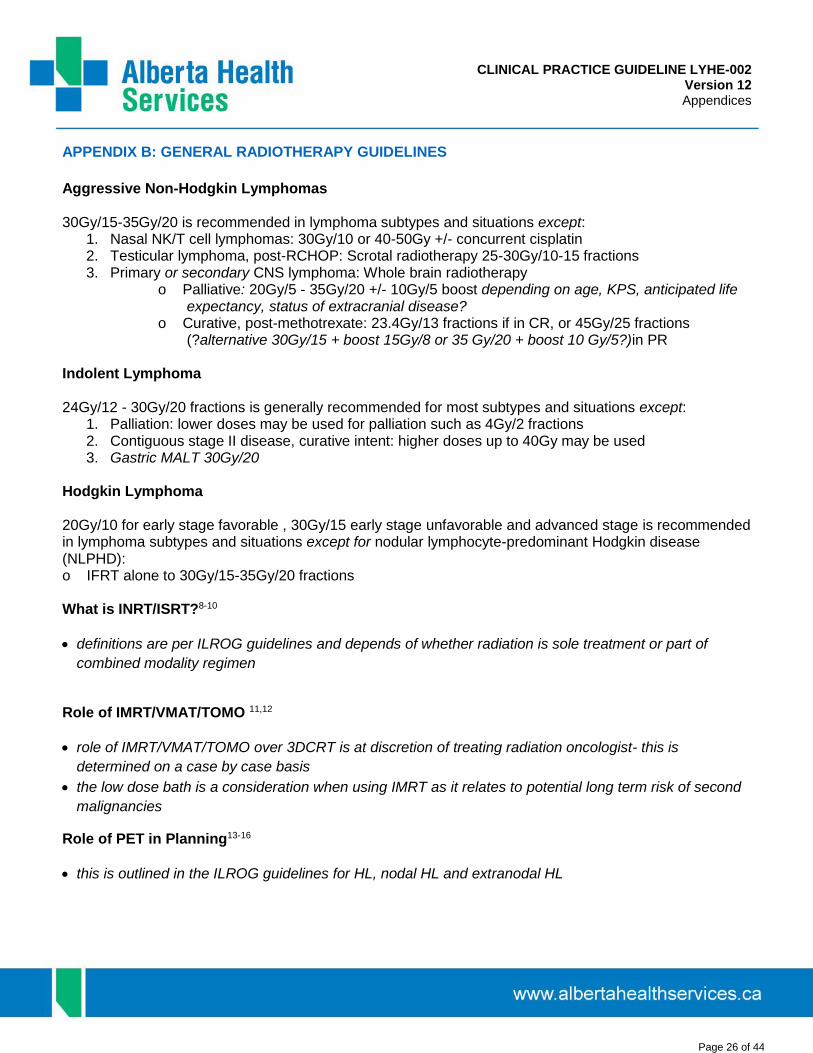
CLINICAL PRACTICE GUIDELINE LYHE-002 Version 12 Appendices
Page 26 of 44
APPENDIX B: GENERAL RADIOTHERAPY GUIDELINES
Aggressive Non-Hodgkin Lymphomas
30Gy/15-35Gy/20 is recommended in lymphoma subtypes and situations except: 1. Nasal NK/T cell lymphomas: 30Gy/10 or 40-50Gy +/- concurrent cisplatin2. Testicular lymphoma, post-RCHOP: Scrotal radiotherapy 25-30Gy/10-15 fractions3. Primary or secondary CNS lymphoma: Whole brain radiotherapy
o Palliative: 20Gy/5 - 35Gy/20 +/- 10Gy/5 boost depending on age, KPS, anticipated lifeexpectancy, status of extracranial disease?
o Curative, post-methotrexate: 23.4Gy/13 fractions if in CR, or 45Gy/25 fractions(?alternative 30Gy/15 + boost 15Gy/8 or 35 Gy/20 + boost 10 Gy/5?)in PR
Indolent Lymphoma
24Gy/12 - 30Gy/20 fractions is generally recommended for most subtypes and situations except: 1. Palliation: lower doses may be used for palliation such as 4Gy/2 fractions2. Contiguous stage II disease, curative intent: higher doses up to 40Gy may be used3. Gastric MALT 30Gy/20
Hodgkin Lymphoma
20Gy/10 for early stage favorable , 30Gy/15 early stage unfavorable and advanced stage is recommended in lymphoma subtypes and situations except for nodular lymphocyte-predominant Hodgkin disease (NLPHD): o IFRT alone to 30Gy/15-35Gy/20 fractions
What is INRT/ISRT?8-10
definitions are per ILROG guidelines and depends of whether radiation is sole treatment or part of
combined modality regimen
Role of IMRT/VMAT/TOMO 11,12
role of IMRT/VMAT/TOMO over 3DCRT is at discretion of treating radiation oncologist- this is
determined on a case by case basis
the low dose bath is a consideration when using IMRT as it relates to potential long term risk of second
malignancies
Role of PET in Planning13-16
this is outlined in the ILROG guidelines for HL, nodal HL and extranodal HL

CLINICAL PRACTICE GUIDELINE LYHE-002 Version 12 Appendices
Page 27 of 44
APPENDIX C: PROGNOSTIC MODELS
ECOG Performance Status
0 Fully active, able to carry on all pre-disease activities without restriction. 1 Restricted in physically strenuous activity but ambulatory and able to carry out work of a light or sedentary nature
(e.g. light housework, office work). 2 Ambulatory and capable of all self-care but unable to carry out any work activities. Up and about more than 50% of
waking hours. 3 Capable of only limited self-care. Confined to bed or chair more than 50% of waking hours. 4 Completely disabled. Cannot carry on any self-care. Totally confined to bed or chair.
International Prognostic Index (IPI) for DLBCL Following CHOP-Type Chemotherapy17
Factors # of Factors 5 year PFS
Age > 60 years ECOG 2-4 Stage III/IV ENS > 1 Increased LDH
0-1 60%
2-3 30%
4-5 15%
Revised IPI for DLBCL Following R-CHOP Chemotherapy18
Factors # of Factors % of Patients 4 year PFS 4 year DSS 4 year OS
Age > 60 years ECOG 2-4 Stage III/IV ENS > 1 Increased LDH
0 11 96% 95% 95%
1-2 48 81% 83% 79%
3-5 41 55% 56% 55%
R-CHOP for DLBCL by Elevated LDH and Stage 3-418
# of Factors % of Patients 4 year PFS 4 year DSS 4 year OS
0 27 92% 90% 84%
1 38 78% 79% 77%
2 35 53% 56% 55%
An online prognostic calculator is available at: http://www.qxmd.com/calculate-online/hematology/prognosis-large-b-cell-lymphoma-r-ipi
Modified IPI for Non-Bulky Stage I-IIA DLBCL Treated with CHOP x 3 cycles and IFRT
Factors # of Factors 5 year PFS 10 year PFS
Age > 60 yrs ECOG 2-4 Stage II Increased LDH
0 94% 89%
1-2 79% 73%
3-4 60% 50%

CLINICAL PRACTICE GUIDELINE LYHE-002 Version 12 Appendices
Page 28 of 44
Salvage Age-Adjusted IPI for Relapsed DLBCL19
Factors # of Factors ~ PFS for HDCT/ASCT Patients
Stage III/IV Elevated LDH ECOG 2-4
0 70%
1 50%
2 30%
3 10%
Primary CNS Lymphoma (Memorial Sloan Kettering Cancer Center Model)20
Risk Group mOS 5 year OS mFFS 5 year FFS
Age < 50 years 5-8 years 50-60% 2-5 years 35-40%
Age >50 years, KPS > 70% 2-3 years 15-35% 1.5 years 10-20%
Age >50 years, KPS < 70% 1 year 10% 0.5-1 year 5-10%
Simplified IELSG Primary CNS Lymphoma (Leon Berard Cancer Centre Model)21
Factors # of Factors mOS 5 year OS
Age > 60 years Elevated LDH Deep Tumour
o Cerebellumo Periventricularo Basal gangliono Brainstem
0 6 years 60%
1 4 years 40%
2 1 year 23%
3 0.5 years 0%
Follicular Lymphoma Internacional Prognostic Index (FLIPI) Pre-dated Rituximab-Chemotherapy (Survival with Non-Rituximab Containing Therapy)22
Factors Prognosis # Factors % Patients 5 year OS 10 year OS
Age > 60 years Stage III-IV Increased LDH Hb < 120 g/L 5+ nodal sites
Good 0-1 36 90% 70%
Intermediate 2 37 78% 50%
Poor 3-5 27 53% 35%
An online prognostic calculator is available at: http://www.qxmd.com/calculate-online/hematology/follicular-lymphoma-international-prognostic-index-flipi
FLIPI 2 23
Factors Prognosis # Factors % Patients 3 year PFS 5 year PFS
Age > 60 years Marrow involvement Increased B2M Hb < 120 g/L Node >6cm longest diameter
Good 0 20 91% 80%
Intermediate 1-2 53 69% 51%
Poor 3-5 27 51% 19%

CLINICAL PRACTICE GUIDELINE LYHE-002 Version 12 Appendices
Page 29 of 44
Hodgkin Lymphoma International Prognostic Score (IPS) for Advanced Disease24
Factors # of Factors 5 year FFS with ABVD
Age >45 years Male Stage IV Albumin <40 g/L Hb<105 g/L WBC>15 x 109/L Lymphocyte < 0.6 x 10⁹/L or < 8% WBC
0-1 80%
2 70%
3 60%
4-7 50%
An online prognostic calculator is available at: http://www.qxmd.com/calculate-online/hematology/hasenclever-hodgkins-prognosis-score-ips
Prognosis of Hodgkin Lymphoma Relapsed After Prior Chemotherapy25
Factors # of Factors 2nd Line Chemo HDCT/ASCT
Time to relapse <1 year Relapse stage III-IV Hb<105 female, 120 male
0 70% 100%
1 60% 70%
2 30% 50%
3 0% 50% * 5yr OS by second line therapy.* Freedom from second failure was 50% for 0-1 factor, 35% for 2 factors, and 15% for 3 factors.
Mantle Cell Lymphoma (MIPI)26
Points Age ECOG LDH WBC
0 <50 0-1 <0.67 ULN <6.7
1 50-59 - 0.67-0.99 ULN 6.7-9.99
2 60-69 2-4 1-1.49 ULN 10.0-14.99
3 70+ - >1.5 ULN >15.0
Points Age ECOG LDH (ULN 235) WBC
0 <50 0-1 <157 <6.7
1 50-59 - 157-235 6.7-9.99
2 60-69 2-4 235-352 10.0-14.99
3 70+ - >352 >15.0
Risk # Points ~Median OS ~5 year OS
Low 0-3 6 years 60%
Intermediate 4-5 4 years 40%
High 6-11 2 years 20%
An online prognostic calculator is available at: http://www.qxmd.com/calculate-online/hematology/prognosis-mantle-cell-lymphoma-mipi
Post-Transplantation Lymphoproliferative Disease (PTLD) Prognostic Scoring Systems
1. Evens et al., 201027
Score 1 point for each: hypoalbumenia, bone marrow involvement, CNS involvement # of Factors Overall 3 year PFS Overall 3 year OS
0 84% 93%
1 66% 68%
2-3 7% 11%

CLINICAL PRACTICE GUIDELINE LYHE-002 Version 12 Appendices
Page 30 of 44
Patients who received rituximab-based therapy as part of their initial treatment had a 3-year PFS of 70% and an OS of 73% compared with a 3-year PFS of 21% (p<0.0001) and an OS of 33% (p=0.0001) for patients who did not receive rituximab.
2. Leblond et al., 200128
Risk Group PS and/or # of Sites mOS
low-risk PS < 2 and 1 >5 years
intermediate risk PS > 2 or 2 or more 3 years
high risk PS > 2 and 2 or more 1 month

CLINICAL PRACTICE GUIDELINE LYHE-002 Version 12 Appendices
Page 31 of 44
Waldenström Macroglobulinemia
Study Prognostic Factors Stratification Survival
Gobbi et al, 1994 29 Hb<9 g/dL Age >70 years Weight loss Cryoglobulinaemia
0-1 factor 0-2 2-4 factors
mOS 80 months mOS 48 months
Morel et al, 2000 30 Age > 65 years Albumin <40 g/L 1 cytopenia (1-point) >1 cytopenia (2-points)
0-1 factor 2 factors 3-4 factors
5 year survival 87% 5 year survival 62% 5 year survival 25%
Dhodapkar et al, 2001 31 β2M >3 mg/L Hb <12 g/dL IgM >40 g/L
β2M<3 mg/L + Hb>12 g/dL β2M<3 mg/L + Hb<12 g/dL β2M>3 mg/L + IgM<40 g/L β2M>3 mg/L + IgM>40 g/L
5 year survival 87% 5 year survival 63% 5 year survival 53% 5 year survival 21%
Merlini et al, 2003 32 Age>60 years Hb<100 g/L Albumin <35 g/L
<60 years, Hb>100, Alb>35 >60 years, Hb <100, Alb<35 Other combinations
mOS 178 months mOS 33 months mOS 84 months
CLL Prognostic Score from MD Anderson Cancer Center
Factors # of Factors # of Patients 5 year OS
Age >60 years B2M >2 mg/L Alb < 35 Creatinine > 1.6 17p mutations
0 364 96%
1 623 79%
2 497 69%
3 70 30%
4-5 10 16%
CLL Internation Prognostic Score: Bahlo 2015ASCO, J Clin Oncol 33, 2015 (suppl; abstr 7002)
Factors Points Risk Group 5 year OS
Age >65 years 1 Low (0-1 points) 93% (~90%)
Clinical Stage >1 1 Intermeidate (2-3 points) 79% (~80%)
IGHV unmutated 2 High (4-6 points) 64% (~60%)
B2M >3.5 mg/L 2 Very high risk (7-10 points) 23% (~25%)
17p deletion or TP53 mutations 4
The full analysis set was collected from eight phase 3 trials in France, Germany, the United Kingdom, the United States, and Poland (n=3,472 patients, median age 61 years (27-86 yrs)). 89% of patients had received treatment for CLL and median overall survival (OS) was 95 months. The model was externally validated in a third dataset comprising 845 patients with newly diagnosed CLL from the Mayo Clinic; 39% had received treatment for CLL. The final model of multivariate analysis identified 5 independent predictors for OS: TP53 (17p) mutation (deleted and/or mutated; hazard ratio [HR]: 4.2); IGHV mutation status (unmutated, HR: 2.6); B2M (>3.5 mg/L; HR: 2.0); clinical stage (Binet B/C or Rai I-IV, HR: 1.6); and age (>65 years, HR: 1.7). Using weighted grading, a prognostic score from 0 to 10 was derived that separated the patients into four different groups: low risk (score 0-1), intermediate risk (score 2-3), high risk (score 4-6), and very high risk (score 7-10). At 5 years, significantly different rates of OS were observed for the low to the very high risk group, 93%, 79%, 64%, and 23%, respectively (P<0.001; C-statistic c=0.72 [95% CI: 0.69, 0.76]). The multivariable model was confirmed on the internal validation datasets; in addition, the four risk groups were reproduced with on the Mayo dataset, with 5-year OS rates of 97%, 91%, 68% and 21%, respectively (P<0.001; C-statistic c=0.79 [95% CI: 0.74, 0.85]).

CLINICAL PRACTICE GUIDELINE LYHE-002 Version 12 Appendices
Page 32 of 44
APPENDIX D: LYMPHOMA RESPONSE CRITERIA
Response Definition Nodal Masses Spleen, Liver Bone Marrow
CR Disappearance of all evidence of disease
(a) FDG-avid or PET positive prior to therapy; mass of any size permitted if PET negative
(b) Variably FDG-avid or PET negative; regression to normal size on CT
not palpable, nodules disappeared
Infiltrate cleared on repeat biopsy; if indeterminate by morphology, immuno-histochemistry should be negative
PR Regression of measurable disease and no new sites
> 50% decrease in SPD of up to 6 largest dominant masses; no increase in size of other nodes (a) FDG-avid or PET positive prior to therapy; one or more PET positive at previously involved site (b) Variably FDG-avid or PET negative; regression on CT
> 50% decrease in SPD of nodules (for single nodule in greatest transverse diameter); no increase in size of liver or spleen
Irrelevant if positive prior to therapy; cell type should be specified
SD Failure to attain CR/PR or PD
(a) FDG-avid or PET positive prior to therapy; PET positive at prior sites of disease and no new sites on CT or PET (b) Variably FDG-avid or PET negative; no change in size of previous lesions on CT
Relapsed Disease or PD
Any new lesion or increase by 50% of previously involved sites from nadir
Appearance of a new lesion(s) > 1.5 cm in any axis, 50% increase in SPD of more than one node, or 50% increase in longest diameter of a previously identified node > 1 cm in short axis
Lesions PET positive if FDG-avid lymphoma or PET positive prior to therapy
> 50% increase from nadir in the SPD of any previous lesions
New or recurrent involvement
Abbreviations: CR=complete response, FDG-PET=(18)F-fluorodeoxyglucose positron emission tomography, CT=computed tomography, PR=partial response, SPD=sum of the product of the diameters, SD=stable disease, PD=progressive disease.
LYMPHOMA RESPONSE CRITERIA33
Complete Response (CR)
The designation of CR requires the following:
1. Complete disappearance of all detectable clinical evidence of disease and disease-related symptoms ifpresent before therapy.
2a. Typically FDG-avid lymphoma: in patients with no pretreatment PET scan or when the PET scan was positive before therapy, a post-treatment residual mass of any size is permitted as long as it is PET negative.
2b. Variably FDG-avid lymphomas/FDG avidity unknown: in patients without a pretreatment PET scan, or if a pretreatment PET scan was negative, all lymph nodes and nodal masses must have regressed on CT to normal size (<1.5 cm in their greatest transverse diameter for nodes >1.5 cm before therapy). Previously involved nodes that were 1.1 to 1.5 cm in their long axis and more than 1.0 cm in their short axis before treatment must have decreased to <1.0 cm in their short axis after treatment.
3. The spleen and/or liver, if considered enlarged before therapy on the basis of a physical examinationor CT scan, should not be palpable on physical examination and should be considered normal size by

CLINICAL PRACTICE GUIDELINE LYHE-002 Version 12 Appendices
Page 33 of 44
imaging studies, and nodules related to lymphoma should disappear. However, determination of splenic involvement is not always reliable because a spleen considered normal in size may still contain lymphoma, whereas an enlarged spleen may reflect variations in anatomy, blood volume, the use ofhematopoietic growth factors, or causes other than lymphoma.
4. If the bone marrow was involved by lymphoma before treatment, the infiltrate must have cleared onrepeat bone marrow biopsy. The biopsy sample on which this determination is made must be adequate(with a goal of >20 mm unilateral core). If the sample is indeterminate by morphology, it should benegative by immunohistochemistry. A sample that is negative by immunohistochemistry but thatdemonstrates a small population of clonal lymphocytes by flow cytometry will be considered a CR untildata become available demonstrating a clear difference in patient outcome.
Partial Response (PR)
The designation of PR requires all of the following:
1. At least a 50% decrease in sum of the product of the diameters (SPD) of up to six of the largestdominant nodes or nodal masses. These nodes or masses should be selected according to all of thefollowing: they should be clearly measurable in at least 2 perpendicular dimensions; if possible theyshould be from disparate regions of the body; and they should include mediastinal and retroperitonealareas of disease whenever these sites are involved.
2. No increase should be observed in the size of other nodes, liver, or spleen.
3. Splenic and hepatic nodules must regress by >50% in their SPD or, for single nodules, in the greatesttransverse diameter.
4. With the exception of splenic and hepatic nodules, involvement of other organs is usually assessableand no measurable disease should be present.
5. Bone marrow assessment is irrelevant for determination of a PR if the sample was positive beforetreatment. However, if positive, the cell type should be specified (e.g., large-cell lymphoma or smallneoplastic B cells). Patients who achieve a CR by the above criteria, but who have persistentmorphologic bone marrow involvement will be considered partial responders. When the bone marrowwas involved before therapy and a clinical CR was achieved, but with no bone marrow assessmentafter treatment, patients should be considered partial responders.
6. No new sites of disease should be observed.
7. Typically FDG-avid lymphoma: for patients with no pre-treatment PET scan or if the PET scan waspositive before therapy, the post-treatment PET should be positive in at least one previously involvedsite.
8. Variably FDG-avid lymphomas/FDG-avidity unknown: for patients without a pre-treatment PET scan, orif a pre-treatment PET scan was negative, CT criteria should be used.

CLINICAL PRACTICE GUIDELINE LYHE-002 Version 12 Appendices
Page 34 of 44
9. In patients with follicular lymphoma or mantle-cell lymphoma, a PET scan is only indicated with one orat most two residual masses that have regressed by more than 50% on CT; those with more than tworesidual lesions are unlikely to be PET negative and should be considered partial responders.
Stable Disease (SD)
Stable disease is defined as the following:
1. A patient is considered to have SD when he or she fails to attain the criteria needed for a CR or PR, butdoes not fulfill those for progressive disease (see Relapsed Disease [after CR]/Progressive Disease[after PR, SD]).
2. Typically FGD-avid lymphomas: the PET should be positive at prior sites of disease with no new areasof involvement on the post-treatment CT or PET.
3. Variably FDG-avid lymphomas/FDG-avidity unknown: for patients without a pre-treatment PET scan orif the pre-treatment PET was negative, there must be no change in the size of the previous lesions onthe post-treatment CT scan.
Relapsed Disease (after CR)/ Progressive Disease (after PR or SD)
1. Lymph nodes should be considered abnormal if the long axis is more than 1.5 cm regardless of theshort axis. If a lymph node has a long axis of 1.1 to 1.5 cm, it should only be considered abnormal if itsshort axis is more than 1.0. Lymph nodes <1.0 x <1.0 cm will not be considered as abnormal forrelapse or progressive disease.
2. Appearance of any new lesion more than 1.5 cm in any axis during or at the end of therapy, even ifother lesions are decreasing in size. Increased FDG uptake in a previously unaffected site should onlybe considered relapsed or progressive disease after confirmation with other modalities. In patients withno prior history of pulmonary lymphoma, new lung nodules identified by CT are mostly benign. Thus, atherapeutic decision should not be made solely on the basis of the PET without histologic confirmation.
3. At least a 50% increase from nadir in the SPD of any previously involved nodes, or in a single involvednode, or the size of other lesions (e.g., splenic or hepatic nodules). To be considered progressivedisease, a lymph node with a diameter of the short axis of less than 1.0 cm must increase by >50%and to a size of 1.5 x 1.5 cm or more than 1.5 cm in the long axis.
4. At least a 50% increase in the longest diameter of any single previously identified node more than 1cm in its short axis.
5. Lesions should be PET positive if observed in a typical FDG-avid lymphoma or the lesion was PETpositive before therapy unless the lesion is too small to be detected with current PET systems (<1.5cm in its long axis by CT).
6. Measurable extranodal disease should be assessed in a manner similar to that for nodal disease. Forthese recommendations, the spleen is considered nodal disease. Disease that is only assessable(e.g., pleural effusions, bone lesions) will be recorded as present or absent only, unless, while an

CLINICAL PRACTICE GUIDELINE LYHE-002 Version 12 Appendices
Page 35 of 44
abnormality is still noted by imaging studies or physical examination, it is found to be histologically negative.
7. In clinical trials where PET is unavailable to the vast majority of participants, or where PET is notdeemed necessary or appropriate for use (e.g., a trial in patients with MALT lymphoma), responseshould be assessed as above, but only using CT scans. However, residual masses should not beassigned CRu status, but should be considered partial responses.

CLINICAL PRACTICE GUIDELINE LYHE-002 Version 12 Appendices
Page 36 of 44

CLINICAL PRACTICE GUIDELINE LYHE-002 Version 12 Appendices
Page 37 of 44

CLINICAL PRACTICE GUIDELINE LYHE-002 Version 12 Appendices
Page 38 of 44
APPENDIX E: New Lymphoma Patient Data Sheet
Identification:
Name DOB (d/m/y) AHN ACB# Gender: male female Age at Diagnosis
Diagnostic Information:
Date Diagnosis (d/m/y) Surgical accession # Biopsy type: open surgical core needle fine needle bone marrow blood Diagnosis:
Stage: I II III IV B sx: yes no Bulk>10cm: yes no
Marrow +ve: yes no Other Extranodal Sites:
LDH elevated: yes no ECOG Status: 0 1 2 3 4
Prognosis Score by Histology:
Large Cell Lymphoma: #IPI Factors: 0 1 2 3 4 5
Age > 60yr Stage III/IV LDH>ULN ECOG 2-4 >2 Extranodal Sites
# FLIPI Factors: 0 1 2 3 4 5
Age > 60yr Stage III/IV LDH>ULN Hb<120g/L >5 Nodal Sites
# IPS Factors: 0 1 2 3 4 5 6 7
Circle if present:
Follicular:
Circle if present:
Hodgkin:
Circle if present: Age > 45 yr Stage IV Male Lymphocyte<0.6 (or < 8%WBC) Albumin < 40 g/L
Hb < 105g/L WBC > 15
Initial Treatment:
Therapy Plan Regimen / Radiation Site Start Date d/m/y
Chemotherapy yes no
Maintenance Rituximab yes no
Radiotherapy yes no
Stem Cell Transplant yes no
First Relapse Information:
Relapse/progression after treatment 1: yes no Date relapse (d/m/y)
2nd Treatment: Regimen Radiation yes no HDCT/ASCT yes no
Survival Information:
Dead: yes no Date death or last follow-up(d/m/y) Cause of death: lymphoma other (specify)

CLINICAL PRACTICE GUIDELINE LYHE-002 Version 12 Appendices
Page 39 of 44
Ann Arbor Staging Nodal Sites
FLIPI Nodal Sites
Ann Arbor Staging System
Stage I Single lymph node region (I) or one extralymphatic organ (IE)
Stage II >2 lymph node regions (II) or local extralymphatic extension plus lymph nodes (IIE), same side of diaphragm.
Stage III Lymph node regions both sides of diaphragm, either alone (III) or with local extralymphatic extension (IIIE)
Stage IV Diffuse involvement of one or more extralymphatic organs or sites.
ECOG Performance Status
0 Fully active, able to carry on all pre-disease activities without restriction
1 Restricted in physically strenuous activity but ambulatory and able to carry out work of a light or sedentary nature.
2 Ambulatory, capable of all self-care but unable to carry out any work activities. Up and about >50% waking hours.
3 Capable of only limited self-care. Confined to bed or chair more than 50% of waking hours.
4 Completely disabled. Cannot carry on any self-care. Totally confined to bed or chair.
Revised-International Prognostic Index for Diffuse Large B-Cell Lymphoma Following R-CHOP Chemotherapy
Factors:
Age > 60yr #Factors %pts 4yr PFS
ECOG 2-4 0 11 95%
Stage III/IV 1-2 48 80%
ENS > 1 3-5 41 55%
LDH
FLIPI (Follicular Lymphoma International Prognostic Index) Factors Survival with Non-Rituximab Containing Therapy
Age > 60yrs Prognosis # %pt 5yr 10yr
Stage 3-4 Good 0-1 36 90% 70%
Increased LDH Intermed 2 37 75% 50%
Hb < 120g/l Poor 3-5 27 50% 35%
5+ nodal sites
Primary CNS lymphoma Prognostic IndexOverall Survival Failure-Free Survival
Adverse Factors mOS 5yr OS mFFS 5yr FFS Age < 50 yrs 5-8 yrs 50-60% 2-5yrs 35-40% Age > 50 yrs KPS > 70% 2-3 yrs 15-35% 1.5 yrs 10-20% Age > 50 yrs KPS < 70% 1 yr 10% 0.5-1yr 5-10%
Hodgkin Lymphoma International Prognostic Score for Advanced Stage Disease Factors # Factors 5yr FFS with ABVD Age > 45 yrs 0-1 80% Male 2 70% Stage 4 3 60% Albumin < 40 g/L 4-7 50% Hb < 105g/L WBC > 15 x 109/L Lymphocyte < 0.6 x 10⁹/L or < 8% WBC
Mantle Cell Lymphoma (MIPI)
Points Age ECOG LDH(ULN 235) WBC 0 <50 0-1 < 157 <6.7 1 50-59 - 157-235 6.7-9.99 2 60-69 2-4 235-352 10.0-14.99 3 70+ - > 352 >15.0
Risk # points ~Median OS ~5yr OS Low 0-3 6yr 60% Intermediate 4-5 4yr 40% High 6-11 2yr 20%

CLINICAL PRACTICE GUIDELINE LYHE-002 Version 12 Appendices
Page 40 of 44
APPENDIX F: Ideal Body Weight
Males – Weight (kg) Females – Weight (kg)
Height (cm) Small Frame Medium Frame Large Frame Small Frame Medium Frame Large Frame
145 47.1 51.0 55.4
146 47.3 51.3 55.8
147 47.6 51.7 56.2
148 47.9 52.1 56.7
149 48.2 52.5 57.1
150 48.5 52.9 57.5
151 48.9 53.4 58.1
152 49.3 53.8 58.7
153 49.8 54.4 59.2
154 50.3 55.0 59.7
155 57.3 59.6 63.3 50.7 55.6 60.3
156 57.7 59.9 63.7 51.2 56.1 60.9
157 58.0 60.3 64.1 51.7 56.6 61.5
158 58.4 60.7 64.5 52.3 57.0 62.1
159 58.8 61.0 65.0 52.8 57.6 62.7
160 59.1 61.4 65.5 53.4 58.2 63.4
161 59.5 61.8 66.0 53.9 58.7 64.0
162 59.8 62.2 66.5 54.4 59.2 64.6
163 60.2 62.7 67.0 55.0 59.7 65.2
164 60.5 63.1 67.6 55.5 60.2 65.9
165 60.9 63.6 68.1 56.0 60.8 66.5
166 61.3 64.1 68.7 56.5 61.4 67.1
167 61.7 64.6 69.4 57.1 61.9 67.8
168 62.2 65.2 70.0 57.7 62.5 68.4
169 62.6 65.7 70.7 58.2 63.0 69.0
170 63.1 66.3 71.3 58.8 63.5 69.6
171 63.5 66.8 71.9 59.3 64.0 70.2
172 64.0 67.3 72.5 59.8 64.6 70.7
173 64.4 67.8 73.2 60.3 65.2 71.2
174 64.9 68.4 73.7 60.8 65.7 71.8
175 65.3 68.9 74.3 61.4 66.2 72.3
176 65.7 69.5 75.0 61.9 66.8 72.8
177 66.2 70.0 75.6 62.5 67.3 73.4
178 66.7 70.6 76.2 63.1 67.8 73.9
179 67.2 71.2 76.9 63.6 68.4 74.5
180 67.8 71.8 77.5 64.1 69.0 75.0
181 68.4 72.4 78.2 64.7 69.6 75.6
182 69.0 73.1 78.9 65.2 70.1 76.1
183 69.6 73.7 79.6
184 70.2 74.4 80.4
185 70.8 75.2 81.3
186 71.4 75.8 82.0
187 72.1 76.4 82.8
188 72.7 77.0 83.6
189 73.4 77.8 84.5
190 74.1 78.7 85.4
191 74.8 79.5 86.3

CLINICAL PRACTICE GUIDELINE LYHE-002 Version 12 Appendices
Page 41 of 44
COPYRIGHT DISCLOSURE
Copyright © (2019) Alberta Health Services.
This material is protected by Canadian and other international copyright laws. All rights reserved. This material may not be copied, published, distributed or reproduced in any way in whole or in part without the express written permission of Alberta Health Services (please contact the Guideline Resource Unit Manager at CancerControl Alberta at [email protected]).
This material is intended for general information only and is provided on an "as is", "where is" basis. Although reasonable efforts were made to confirm the accuracy of the information, Alberta Health Services does not make any representation or warranty, express, implied or statutory, as to the accuracy, reliability, completeness, applicability or fitness for a particular purpose of such information. This material is not a substitute for the advice of a qualified health professional. Alberta Health Services expressly disclaims all liability for the use of these materials, and for any claims, actions, demands or suits arising from such use.

CLINICAL PRACTICE GUIDELINE LYHE-002 Version 12 Appendices
Page 42 of 44
REFERENCES
1. Pfreundschuh MG, Trümper L, Kloess M, Schmits R, Feller AC, Rudolph C. Two-weekly or 3-weekly CHOP
chemotherapy with or without etoposide for the treatment of young patients with good-prognosis (normal LDH)
aggressive lymphomas: results of the NHL-B1 trial of the DSHNHL. Blood 2004 2004 Aug;104(3):626-33.
2. Gang AO, Strom C, Pedersen M, d'Amore F, Pedersen LM, Bukh A, et al. R-CHOEP-14 improves overall survival
in young high-risk patients with diffuse large B-cell lymphoma compared with R-CHOP-14. A population-based
investigation from the Danish Lymphoma Group. Ann Oncol 2012 Jan;23(1):147-153 PubMed ID 21460380.
3. Pfreundschuh M, Zwick C, Zeynalova S, Duhrsen U, Pfluger KH, Vrieling T, et al. Dose-escalated CHOEP for the
treatment of young patients with aggressive non-Hodgkin's lymphoma: II. Results of the randomized high-CHOEP
trial of the German High-Grade Non-Hodgkin's Lymphoma Study Group (DSHNHL). Ann Oncol 2008
Mar;19(3):545-552 PubMed ID 18065407.
4. Guo HQ, Liu L, Wang XF, Lin TY, Yao SN, Yao ZH, et al. Efficacy of gemcitabine combined with oxaliplatin, L-
asparaginase and dexamethasone in patients with newly-diagnosed extranodal NK/T-cell lymphoma. Mol Clin
Oncol 2014 Nov;2(6):1172-1176 PubMed ID 25279218.
5. Yamaguchi M, Kwong YL, Kim WS, Maeda Y, Hashimoto C, Suh C, et al. Phase II study of SMILE chemotherapy
for newly diagnosed stage IV, relapsed, or refractory extranodal natural killer (NK)/T-cell lymphoma, nasal type:
the NK-Cell Tumor Study Group study. J Clin Oncol 2011 Nov 20;29(33):4410-4416 PubMed ID 21990393.
6. Magrath I, Adde M, Shad A, Venzon D, Seibel N, Gootenberg J, et al. Adults and children with small non-cleaved-
cell lymphoma have a similar excellent outcome when treated with the same chemotherapy regimen. J Clin Oncol
1996 Mar;14(3):925-934 PubMed ID 8622041.
7. Barnes JA, Lacasce AS, Feng Y, Toomey CE, Neuberg D, Michaelson JS, et al. Evaluation of the addition of
rituximab to CODOX-M/IVAC for Burkitt's lymphoma: a retrospective analysis. Ann Oncol 2011 Aug;22(8):1859-
1864 PubMed ID 21339382.
8. Girinsky T, Ghalibafian M. Radiotherapy of hodgkin lymphoma: indications, new fields, and techniques. Semin Radiat
Oncol 2007 Jul;17(3):206-222 PubMed ID 17591568.
9. Girinsky T, van der Maazen R, Specht L, Aleman B, Poortmans P, Lievens Y, et al. Involved-node radiotherapy
(INRT) in patients with early Hodgkin lymphoma: concepts and guidelines. Radiother Oncol 2006 Jun;79(3):270-
277 PubMed ID 16797755.
10. Yahalom J. Hodgkin’s and non-Hodgkin’s lymphoma radiotherapy indications, field and dose decisions and
implementation: refresher course. American Society for Therapeutic Radiology and Oncology 47th Annual
Meeting, Denver, Colorado, October 16-19, 2005.
11. Girinsky T, Pichenot C, Beaudre A, Ghalibafian M, Lefkopoulos D. Is intensity-modulated radiotherapy better than
conventional radiation treatment and three-dimensional conformal radiotherapy for mediastinal masses in patients
with Hodgkin's disease, and is there a role for beam orientation optimization and dose constraints assigned to
virtual volumes? Int J Radiat Oncol Biol Phys 2006 Jan 1;64(1):218-226 PubMed ID 16169675.
12. Goodman KA, Toner S, Hunt M, Wu EJ, Yahalom J. Intensity-modulated radiotherapy for lymphoma involving the
mediastinum. Int J Radiat Oncol Biol Phys 2005 May 1;62(1):198-206 PubMed ID 15850922.
13. van Baardwijk A, Baumert BG, Bosmans G, van Kroonenburgh M, Stroobants S, Gregoire V, et al. The current
status of FDG-PET in tumour volume definition in radiotherapy treatment planning. Cancer Treat Rev 2006
Jun;32(4):245-260 PubMed ID 16563636.
14. Bujenovic S. The role of positron emission tomography in radiation treatment planning. Semin Nucl Med 2004
Oct;34(4):293-299 PubMed ID 15493006.
15. Kahn ST, Flowers C, Lechowicz MJ, Hollenbach K, Johnstone PA. Value of PET restaging after chemotherapy for
non-Hodgkin's lymphoma: implications for consolidation radiotherapy. Int J Radiat Oncol Biol Phys 2006 Nov
15;66(4):961-965 PubMed ID 17145526.

CLINICAL PRACTICE GUIDELINE LYHE-002 Version 12 Appendices
Page 43 of 44
16. Specht L. 2-[18F]fluoro-2-deoxyglucose positron-emission tomography in staging, response evaluation, and
treatment planning of lymphomas. Semin Radiat Oncol 2007 Jul;17(3):190-197 PubMed ID 17591566.
17. A predictive model for aggressive non-Hodgkin's lymphoma. The International Non-Hodgkin's Lymphoma
Prognostic Factors Project. N Engl J Med 1993 Sep 30;329(14):987-994 PubMed ID 8141877.
18. Sehn LH, Berry B, Chhanabhai M, Fitzgerald C, Gill K, Hoskins P. The revised International Prognostic Index (R-
IPI) is a better predictor of outcome than the standard IPI for patients with diffuse large B-cell lymphoma treated
with R-CHOP. Blood 2007 Mar;109(5):1857-61.
19. Hamlin PA, Zelenetz AD, Kewalramani T, Qin J, Satagopan JM, Verbel D, et al. Age-adjusted International
Prognostic Index predicts autologous stem cell transplantation outcome for patients with relapsed or primary
refractory diffuse large B-cell lymphoma. Blood 2003 Sep 15;102(6):1989-1996 PubMed ID 12676776.
20. Abrey LE, Ben-Porat L, Panageas KS, Yahalom J, Berkey B, Curran W, et al. Primary central nervous system
lymphoma: the Memorial Sloan-Kettering Cancer Center prognostic model. J Clin Oncol 2006 Dec
20;24(36):5711-5715 PubMed ID 17116938.
21. Ghesquieres H, Sunyach MP, Drouet Y, Sebban C, Chassagne C, Jouanneau E, et al. Long-Term Follow-Up of
132 Immunocompetent Primary Central Nervous System Lymphomas Treated at Leon Berard Cancer Centre:
Proposition of a New Prognostic Model. ASH Annual Meeting Abstracts 2007 November 16;110(11):521.
22. Solal-Celigny P, Roy P, Colombat P, White J, Armitage JO, Arranz-Saez R, et al. Follicular lymphoma international
prognostic index. Blood 2004 Sep 1;104(5):1258-1265 PubMed ID 15126323.
23. Federico M, Bellei M, Marcheselli L, Luminari S, Lopez-Guillermo A, Vitolo U, et al. Follicular lymphoma
international prognostic index 2: a new prognostic index for follicular lymphoma developed by the international
follicular lymphoma prognostic factor project. J Clin Oncol 2009 Sep 20;27(27):4555-4562 PubMed ID 19652063.
24. Hasenclever D, Diehl V. A prognostic score for advanced Hodgkin's disease. International Prognostic Factors
Project on Advanced Hodgkin's Disease. N Engl J Med 1998 Nov 19;339(21):1506-1514 PubMed ID 9819449.
25. Josting A, Franklin J, May M, Koch P, Beykirch MK, Heinz J, et al. New prognostic score based on treatment
outcome of patients with relapsed Hodgkin's lymphoma registered in the database of the German Hodgkin's
lymphoma study group. J Clin Oncol 2002 Jan 1;20(1):221-230 PubMed ID 11773173.
26. Hoster E, Dreyling M, Klapper W, Gisselbrecht C, van Hoof A, Kluin-Nelemans HC, et al. A new prognostic index
(MIPI) for patients with advanced-stage mantle cell lymphoma. Blood 2008 Jan 15;111(2):558-565 PubMed ID
17962512.
27. Evens AM, David KA, Helenowski I, Nelson B, Kaufman D, Kircher SM, et al. Multicenter analysis of 80 solid organ
transplantation recipients with post-transplantation lymphoproliferative disease: outcomes and prognostic factors
in the modern era. J Clin Oncol 2010 Feb 20;28(6):1038-1046 PubMed ID 20085936.
28. Leblond V, Dhedin N, Mamzer Bruneel MF, Choquet S, Hermine O, Porcher R, et al. Identification of prognostic
factors in 61 patients with posttransplantation lymphoproliferative disorders. J Clin Oncol 2001 Feb 1;19(3):772-
778 PubMed ID 11157030.
29. Gondim JA, Schops M, de Almeida JP, de Albuquerque LA, Gomes E, Ferraz T, et al. Endoscopic endonasal
transsphenoidal surgery: surgical results of 228 pituitary adenomas treated in a pituitary center. Pituitary
2010;13(1):68-77.
30. Durando X, Thivat E, Gilliot O, Irthum B, Verrelle P, Vincent C, et al. Temozolomide treatment of an adult with a
relapsing medulloblastoma. Cancer Invest 2007 Sep;25(6):470-475 PubMed ID 17882660.
31. Colao A, Di Sarno A, Sarnacchiaro F, Ferone D, Di Renzo G, Merola B, et al. Prolactinomas resistant to standard
dopamine agonists respond to chronic cabergoline treatment. J Clin Endocrinol Metab 1997 Mar;82(3):876-883.
32. Deutsch M, Thomas PR, Krischer J, Boyett JM, Albright L, Aronin P, et al. Results of a prospective randomized
trial comparing standard dose neuraxis irradiation (3,600 cGy/20) with reduced neuraxis irradiation (2,340 cGy/13)
in patients with low-stage medulloblastoma. A Combined Children's Cancer Group-Pediatric Oncology Group
Study. Pediatr Neurosurg 1996;24(4):167-176; discussion 176-7 PubMed ID 8873158.

CLINICAL PRACTICE GUIDELINE LYHE-002 Version 12 Appendices
Page 44 of 44
33. Cheson BD, Pfistner B, Juweid ME, Gascoyne RD, Specht L, Horning SJ, et al. Revised response criteria for
malignant lymphoma. J Clin Oncol 2007 Feb 10;25(5):579-586 PubMed ID 17242396.