Lymphocytes ExpressingType ComplementReceptors Proliferate ... ·...
8
Lymphocytes Expressing Type 3 Complement Receptors Proliferate in Response to Interleukin 2 and Are the Precursors of Lymphokine-activated Killer Cells J. Dixon Gray and David A. Horwitz University ofSouthern California School of Medicine, Clinical Immunology and Rheumatic Disease Section, Los Angeles, California 90033 Abstract In the absence of antigenic or mitogenic stimulation, certain peripheral blood lymphocytes exhibit proliferative and lym- phokine-activated killer (LAK) cell activities when cultured with recombinant IL-2. Both activities were found to be an exclusive property of lymphocytes expressing type 3 comple- ment receptors (CR3) identified by anti-CD1 1 monoclonal an- tibodies. CD11+ lymphocytes were then fractionated into three subsets by two-color flow cytometry. These included CD16+ cells, which display distinctive Fc receptors for IgG (CD16). Using anti-CD5, the CD11+ CD16- lymphocytes were separated into non-T cell and T cell subsets. The two non-T cell subsets (CD11+ CD16+ and CD11+ CD16- CD5-), but not the T cell subset (CD11+ CD16- CD5+), could proliferate in response to IL-2. Both CD11+ non-T cell subsets, but not the CD11+ T cell subset, had the capacity to mediate natural killer cell activity. However, all three CD1 1 + lymphocyte subsets were capable of generating LAK activity. These findings are consistent with the concept that two signals are required to stimulate T cells to proliferate. However, at least a small subset of blood T cells can be activated by IL-2 to become LAK cells. Introduction IL-2 is a T cell-derived lymphokine that is the principal growth factor for T cells (1, 2). It has been postulated that IL-2 acts upon T cells that have received a first signal provided by antigen or mitogen, but not upon resting T cells (3-5). How- ever, in the absence of a first signal, "resting" peripheral blood T cells have been reported to proliferate in response to IL-2 (6-1 1). Natural killer (NK)' cells can proliferate in response to IL-2 (8, 11, 12), but if T cells also have this capacity, this finding would conflict with the two-signal theory of T cell activation. Lymphocytes mediating cytotoxic activity after culture with IL-2 have been called lymphokine-activated killer cells (LAK) (13). This cytotoxicity is non-MHC restricted and the Received for publication 27 April 1987 and in revised form 2 October 1987. 1. Abbreviations used in this paper: CR3, type 3 complement receptor; IL-2R, IL-2 receptor; LAK, lymphokine-activated killer cell; NK, nat- ural killer; rIL-2, recombinant IL-2; UNF, unfractionated PBMC. target cells killed include those that are resistant to NK cells (13). The precursors of LAK activity are less well defined. Although LAK activity can be derived from NK cells (1 1), lymphocyte populations devoid of or depleted of NK activity also demonstrate LAK activity upon culture in the presence of IL-2 (14, 15). In the present study, we have characterized the phenotype of lymphocytes which proliferate in response to IL-2 and which are the precursors of LAK cells. PBMC were separated using anti-CD 11 monoclonal antibodies that react with re- ceptors for the iC3b component of complement (16, 17). These receptors have been called type 3 complement receptors (CR3) (18). Activities associated with the lymphocyte popula- tions identified by anti-CD 11 monoclonal antibodies include NK cell activity (19, 20), antibody-dependent cellular cyto- toxic activity ( 19), and the suppression of IgG production in- duced by pokeweed mitogen (21). We report that all IL-2 re- sponsiveness was contained exclusively within the CD 11 + lymphocyte populations. Methods PBMC. Peripheral blood, obtained from healthy volunteers by veni- puncture, was separated by Ficoll-Hypaque (Isolymph; Gallard-Schle- singer Corp., Carle Place, NY) density centrifugation. Mononuclear cells were then washed three times and resuspended in RPMI 1640 medium (Irvine Scientific, Santa Ana, CA) containing glutamine (2 mM), gentamicin (20 ,g/ml), and Hepes buffer (10 mM), and supple- mented with 10% human AB serum (Irvine Scientific). Monoclonal antibodies. The monoclonal antibodies used in this study were anti-CD 1 1 OKM 1 (Ortho Diagnostics Systems Inc., Rari- tan, NJ) or Leu 15 phycoerythrin conjugated (Becton-Dickinson & Co., Mountain View, CA); anti-CD 16 Leu 1 la; anti-CD5 Leu 1; and anti-CD25 (a chain), anti-IL-2 receptor phycoerythrin conjugated (all from Becton-Dickinson & Co.). Isolation of lymphocyte subsets. PBMC were stained with mono- clonal antibodies under saturating conditions at 4°C for 30 min. When PBMC were stained with unconjugated monoclonal antibodies the cells were subsequently incubated with FITC-F(ab')2 sheep anti-mouse IgG (Cappell Laboratories, Cochranville, PA) at 4°C for 30 min. Cells were then washed and resuspended at 5 X 10⁶/ml. Stained PBMC were analyzed and sorted by flow cytometry using a FACS IV (Becton-Dickinson & Co.) or a cytofluorograph 50H (Ortho Diagnostics, Westwood, MA). Both flow cytometers are equipped with an argon laser which was operated at 488 nm for excitation of fluores- cein and phycoerythrin. Lymphocyte-sized cells were electronically separated from debris and other cells on the basis of forward and right angle scatter. In all studies with two-color staining, the percentage of cells positive for each marker was confirmed by single-color analysis. When CD 1I + CD 16- lymphocytes were further fractionated with anti-CD5 monoclonal antibodies, a second sort was performed to isolate the CD5+ and CD5- subsets. Cell sorting provided populations of lymphocytes of > 95% purity. Interleukin 2-responsive Lymphocytes 1247 J. Clin. Invest. © The American Society for Clinical Investigation, Inc. 0021-9738/88/04/1247/08 $2.00 Volume 81, April 1988, 1247-1254
Transcript of Lymphocytes ExpressingType ComplementReceptors Proliferate ... ·...

Lymphocytes Expressing Type 3 Complement Receptors Proliferatein Response to Interleukin 2 and Are the Precursorsof Lymphokine-activated Killer CellsJ. Dixon Gray and David A. HorwitzUniversity of Southern California School of Medicine, Clinical Immunologyand Rheumatic Disease Section, Los Angeles, California 90033
Abstract
In the absence of antigenic or mitogenic stimulation, certainperipheral blood lymphocytes exhibit proliferative and lym-phokine-activated killer (LAK) cell activities when culturedwith recombinant IL-2. Both activities were found to be an
exclusive property of lymphocytes expressing type 3 comple-ment receptors (CR3) identified by anti-CD1 1 monoclonal an-
tibodies. CD11+ lymphocytes were then fractionated intothree subsets by two-color flow cytometry. These includedCD16+ cells, which display distinctive Fc receptors for IgG(CD16). Using anti-CD5, the CD11+ CD16- lymphocyteswere separated into non-T cell and T cell subsets. The twonon-T cell subsets (CD11+ CD16+ and CD11+ CD16-CD5-), but not the T cell subset (CD11+ CD16- CD5+),could proliferate in response to IL-2. Both CD11+ non-T cellsubsets, but not the CD11+ T cell subset, had the capacity tomediate natural killer cell activity. However, all three CD11 +lymphocyte subsets were capable of generating LAK activity.These findings are consistent with the concept that two signalsare required to stimulate T cells to proliferate. However, atleast a small subset of blood T cells can be activated by IL-2 tobecome LAK cells.
Introduction
IL-2 is a T cell-derived lymphokine that is the principalgrowth factor for T cells (1, 2). It has been postulated that IL-2acts upon T cells that have received a first signal provided byantigen or mitogen, but not upon resting T cells (3-5). How-ever, in the absence of a first signal, "resting" peripheral bloodT cells have been reported to proliferate in response to IL-2(6-1 1). Natural killer (NK)' cells can proliferate in response toIL-2 (8, 11, 12), but if T cells also have this capacity, thisfinding would conflict with the two-signal theory of T cellactivation.
Lymphocytes mediating cytotoxic activity after culturewith IL-2 have been called lymphokine-activated killer cells(LAK) (13). This cytotoxicity is non-MHC restricted and the
Received for publication 27 April 1987 and in revised form 2 October1987.
1. Abbreviations used in this paper: CR3, type 3 complement receptor;IL-2R, IL-2 receptor; LAK, lymphokine-activated killer cell; NK, nat-ural killer; rIL-2, recombinant IL-2; UNF, unfractionated PBMC.
target cells killed include those that are resistant to NK cells(13). The precursors of LAK activity are less well defined.Although LAK activity can be derived from NK cells (1 1),lymphocyte populations devoid of or depleted of NKactivityalso demonstrate LAK activity upon culture in the presence ofIL-2 (14, 15).
In the present study, we have characterized the phenotypeof lymphocytes which proliferate in response to IL-2 andwhich are the precursors of LAK cells. PBMCwere separatedusing anti-CD 11 monoclonal antibodies that react with re-ceptors for the iC3b component of complement (16, 17).These receptors have been called type 3 complement receptors(CR3) (18). Activities associated with the lymphocyte popula-tions identified by anti-CD 11 monoclonal antibodies includeNK cell activity (19, 20), antibody-dependent cellular cyto-toxic activity ( 19), and the suppression of IgG production in-duced by pokeweed mitogen (21). Wereport that all IL-2 re-sponsiveness was contained exclusively within the CD11 +lymphocyte populations.
Methods
PBMC. Peripheral blood, obtained from healthy volunteers by veni-puncture, was separated by Ficoll-Hypaque (Isolymph; Gallard-Schle-singer Corp., Carle Place, NY) density centrifugation. Mononuclearcells were then washed three times and resuspended in RPMI 1640medium (Irvine Scientific, Santa Ana, CA) containing glutamine (2mM), gentamicin (20 ,g/ml), and Hepes buffer (10 mM), and supple-mented with 10% human AB serum (Irvine Scientific).
Monoclonal antibodies. The monoclonal antibodies used in thisstudy were anti-CD 1 1 OKM1 (Ortho Diagnostics Systems Inc., Rari-tan, NJ) or Leu 15 phycoerythrin conjugated (Becton-Dickinson &Co., Mountain View, CA); anti-CD 16 Leu 1 la; anti-CD5 Leu 1; andanti-CD25 (a chain), anti-IL-2 receptor phycoerythrin conjugated (allfrom Becton-Dickinson & Co.).
Isolation of lymphocyte subsets. PBMCwere stained with mono-clonal antibodies under saturating conditions at 4°C for 30 min. WhenPBMCwere stained with unconjugated monoclonal antibodies thecells were subsequently incubated with FITC-F(ab')2 sheep anti-mouseIgG (Cappell Laboratories, Cochranville, PA) at 4°C for 30 min. Cellswere then washed and resuspended at 5 X 10⁶/ml.
Stained PBMCwere analyzed and sorted by flow cytometry using aFACSIV (Becton-Dickinson & Co.) or a cytofluorograph 50H (OrthoDiagnostics, Westwood, MA). Both flow cytometers are equipped withan argon laser which was operated at 488 nm for excitation of fluores-cein and phycoerythrin. Lymphocyte-sized cells were electronicallyseparated from debris and other cells on the basis of forward and rightangle scatter. In all studies with two-color staining, the percentage ofcells positive for each marker was confirmed by single-color analysis.When CD 1 I + CD16- lymphocytes were further fractionated withanti-CD5 monoclonal antibodies, a second sort was performed toisolate the CD5+and CD5- subsets. Cell sorting provided populationsof lymphocytes of > 95% purity.
Interleukin 2-responsive Lymphocytes 1247
J. Clin. Invest.© The American Society for Clinical Investigation, Inc.0021-9738/88/04/1247/08 $2.00Volume 81, April 1988, 1247-1254
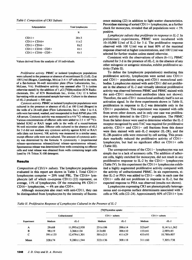
Table . Composition of CR3Subsets
Subpopulation Total lymphocytes
CDI 1+ 20±3CD11+ CD16+ 11±2CDll+ CD16- 8±2CDI 1+ CD16- CD5+ 4±1CDI 1+ CD16- CD5- 4±1
Values derived from the analysis of 10 individuals.
Proliferative activity. PBMCor isolated lymphocyte populationswere cultured in the presence or absence of recombinant IL-2 (rIL-2) at100 U/ml (Biogen, Cambridge, MA) at 3-4 X 10⁴ cells/well in the wellsof a flat-bottom 96-well microtiter plate (Flow Laboratories, Inc.,McLean, VA). Proliferative activity was assessed after 72 h (unlessotherwise stated) by the addition of 1 MACi [3H]thymidine (ICN Radio-chemicals, Div. of ICN Biomedicals Inc., Irvine, CA) 12 h beforeharvesting with an automated sample harvester. Culture in the absenceof rIL-2 produced < 500 cpm.
Cytotoxic activity. PBMCor isolated lymphocyte populations werecultured in the presence or absence of rIL-2 at 100 U/ml (Biogen) inthe wells of a 24-well plate (Flow Laboratories, Inc.). After 3 to 4 d,cells were harvested, washed, and resuspended in fresh RPMI plus 10%AB serum. Cytotoxic activity was measured in a 4-h 5'Cr release assay.Various concentrations of effector cells were added to 2.5 X 10³ 5'Cr-labeled K562 or RAJ1 target cells in the wells of a round-bottom96-well microtiter plate. Effector cells cultured in the absence of rIL2for 3 d did not mediate any cytotoxic activity against K562 or RAJ1cells (data not known). NKactivity was measured in a similar assay,except effector cells were not cultured. The amount of cytotoxic activ-ity was determined by the formula: % cytotoxicity = (experimentalrelease-spontaneous release)/(total release-spontaneous release).Spontaneous release was determined from wells containing no effectorcells and total release was obtained from wells containing target cellslysed by 1% Triton X-100 detergent.
Results
Composition of CDI l+ subsets. The lymphocyte populationsevaluated in this report are shown in Table I. Total CD11+lymphocytes comprise 20% total PBL. The CD16+ lym-phocyte (all of which co-express CD11) (22) represent, onaverage, 11% of lymphocytes. Of the remaining 8% CDl 1 +CD16- lymphocytes, 4%are also CD5+.
Although monocytes also react with anti-CD 11, they canbe distinguished from lymphocytes by the intensity of fluores-
cence staining (22) in addition to light scatter characteristics.Peroxidase staining of sorted CD11+ lymphocytes, as a furthercheck for monocytes, revealed that all populations were < 5%positive.
Lymphocyte subsets that proliferate in response to IL-2. Inpreliminary experiments, PBMCwere incubated with10-10,000 U/ml of IL-2 for 3 d. The proliferative responseobserved with 100 U/ml was at least 80% of the maximalresponse observed at higher concentrations, and 100 U/ml wasselected for further studies unless stated otherwise.
Consistent with the observations of others (6-1 1), PBMCcultured for 3 d in the presence of rIL-2, in the absence of anyother mitogenic or antigenic stimulus, exhibit proliferative ac-tivity (Table II).
To define the lymphocyte population(s) mediating thisproliferative activity, lymphocytes were sorted into CD1 +and CD11- populations using anti-CD 11 monoclonal anti-bodies. Lymphocytes stained with anti-CD 1I1 did not prolifer-ate in the absence of IL-2 and virtually identical proliferativeactivity was observed between PBMCand PBMCstained withthe anti-CD 11 monoclonal antibodies (data not shown).Therefore, the anti-CD1 1 monoclonal antibody itself is not anactivation signal. In the three experiments shown in Table II,proliferation in response to IL-2 was detectable only in theCD1 + population. This experiment was repeated with cellsfrom seven other donors, and in only one case was prolifera-tive activity detected in the CDl 1- population. The PBMCfrom the latter donor were used to determine whether the IL-2receptor recognized by anti-TAC was required for proliferativeactivity. CD1 1 + and CDl 1- cell fractions from this donorwere then stained with anti-IL-2 receptor (IL-2R), and theIL-2R-positive cells were removed by cell sorting. This proce-dure markedly reduced the proliferative activity of CD 11-lymphocytes, but had no significant effect on CDl 1 + cells(Table III).
The unresponsiveness of the CD 11- lymphocytes was notsimply due to a lack of accessory cells. The addition of adher-ent cells, highly enriched for monocytes, did not result in anyproliferative response to IL-2 by the CD 11- lymphocytes(Table IV). In this experiment the CD11+ lymphocytes exhib-ited a highly augmented proliferative activity compared withthe activity of unfractionated PBMC. In six experiments, ei-ther IL-2 or PHAwas added to CD11- cells: in each case theCD11- cells did not proliferate in response to IL-2, but theexpected response to PHAwas observed (results not shown).
Lymphocytes expressing CR3are phenotypically heteroge-neous and co-express surface determinants associated with Tcells or NKcells (22-24). Approximately 60% of CDl 1 + lym-
Table II. Proliferative Response of Lymphocytes Cultured in the Presence of IL-2
[3H]Thymidine uptake
Unfractionated CD 11- subsets CDI 1 + subsets
Experiment Medium rIL-2 Medium rIL-2 Medium rIL-2
A 29±68 11,995±2,930 251±106 206±14 323±47 8,141±1,362B 98±19 3,366±134 213±88 306±10 155±41 2,989+45C 591±135 12,479±1,301 501±213 411±29 455±95 10,780+807
Mean 328±74 9,280+1,544 322±136 308±18 311±60 7,305±738
1248 J. D. Gray and D. A. Horwitz
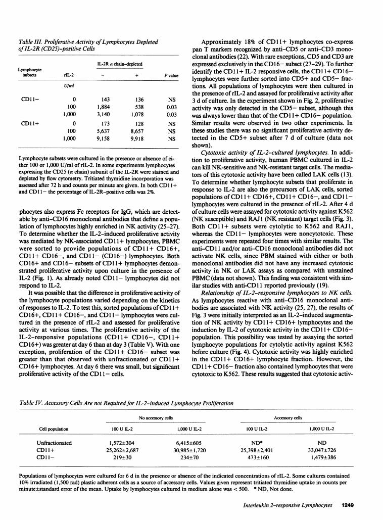
Table III. Proliferative Activity of Lymphocytes Depletedof IL-2R (CD25)-positive Cells
IL-2R a chain-depletedLymphocyte
subsets rIL-2 - + P value
U/mi
CD11- 0 143 136 NS100 1,884 538 0.03
1,000 3,140 1,078 0.03
CD11+ 0 173 128 NS100 5,637 8,657 NS
1,000 9,158 9,918 NS
Lymphocyte subsets were cultured in the presence or absence of ei-ther 100 or 1,000 U/ml of rIL-2. In some experiments lymphocytesexpressing the CD25 (a chain) subunit of the IL-2R were stained anddepleted by flow cytometry. Tritiated thymidine incorporation wasassessed after 72 h and counts per minute are given. In both CDl 1 +and CDI I - the percentage of IL-2R-positive cells was 2%.
phocytes also express Fc receptors for IgG, which are detect-able by anti-CD 16 monoclonal antibodies that define a popu-lation of lymphocytes highly enriched in NKactivity (25-27).To determine whether the IL-2-induced proliferative activitywas mediated by NK-associated CDl 1 + lymphocytes, PBMCwere sorted to provide populations of CD11 + CD16+,CDl 1+ CD16-, and CD1 1- (CD 16-) lymphocytes. BothCD16+ and CD16- subsets of CDl 1 + lymphocytes demon-strated proliferative activity upon culture in the presence ofIL-2 (Fig. 1). As already noted CDl 1- lymphocytes did notrespond to IL-2.
It was possible that the difference in proliferative activity ofthe lymphocyte populations varied depending on the kineticsof responses to IL-2. To test this, sorted populations of CD11+CD16+, CD11+ CD16-, and CD11- lymphocytes were cul-tured in the presence of rIL-2 and assessed for proliferativeactivity at various times. The proliferative activity of theIL-2-responsive populations (CDl1+ CD16-, CDll+CD16+) was greater at day 6 than at day 3 (Table V). With oneexception, proliferation of the CD11 + CD16- subset wasgreater than that observed with unfractionated or CD11+CD16+ lymphocytes. At day 6 there was small, but significantproliferative activity of the CD 11- cells.
Approximately 18% of CD11+ lymphocytes co-expresspan T markers recognized by anti-CD5 or anti-CD3 mono-clonal antibodies (22). With rare exceptions, CD5and CD3areexpressed exclusively in the CD16- subset (27-29). To furtheridentify the CDl 1 + IL-2 responsive cells, the CDl 1 + CD16-lymphocytes were further sorted into CD5+ and CD5- frac-tions. All populations of lymphocytes were then cultured inthe presence of rIL-2 and assayed for proliferative activity after3 d of culture. In the experiment shown in Fig. 2, proliferativeactivity was only detected in the CD5- subset, although thiswas always lower than that of the CDl 1 + CD16- population.Similar results were observed in two other experiments. Inthese studies there was no significant proliferative activity de-tected in the CD5+ subset after 7 d of culture (data notshown).
Cytotoxic activity of IL-2-cultured lymphocytes. In addi-tion to proliferative activity, human PBMCcultured in IL-2can kill NK-sensitive and NK-resistant target cells. The media-tors of this cytotoxic activity have been called LAK cells (13).To determine whether lymphocyte subsets that proliferate inresponse to IL-2 are also the precursors of LAK cells, sortedpopulations of CDl 1+ CD16+, CDl 1+ CD16-, and CDl 1-lymphocytes were cultured in the presence of rIL-2. After 4 dof culture cells were assayed for cytotoxic activity against K562(NK susceptible) and RAJ 1 (NK resistant) target cells (Fig. 3).Both CD11+ subsets were cytolytic to K562 and RAJ1,whereas the CD11 - lymphocytes were noncytotoxic. Theseexperiments were repeated four times with similar results. Theanti-CD 11 and/or anti-CD 16 monoclonal antibodies did notactivate NK cells, since PBMstained with either or bothmonoclonal antibodies did not have any increased cytotoxicactivity in NK or LAK assays as compared with unstainedPBMC(data not shown). This finding was consistent with sim-ilar studies with anti-CD 11 reported previously (19).
Relationship of IL-2-responsive lymphocytes to NKcells.As lymphocytes reactive with anti-CD 16 monoclonal anti-bodies are associated with NKactivity (25, 27), the results ofFig. 3 were initially interpreted as an IL-2-induced augmenta-tion of NKactivity by CDl 1 + CD16+ lymphocytes and theinduction by IL-2 of cytotoxic activity in the CDl 1 + CD16-population. This possibility was tested by assaying the sortedlymphocyte populations for cytolytic activity against K562before culture (Fig. 4). Cytotoxic activity was highly enrichedin the CD1 I+ CD16+ lymphocyte fraction. However, theCDl 1 + CD16- fraction also contained lymphocytes that werecytotoxic to K562. These results suggested that cytotoxic activ-
Table IV Accessory Cells Are not Requiredfor IL-2-induced Lymphocyte Proliferation
No accessory cells Accessory cells
Cell population 100 U IL-2 1,000 U IL-2 100 U IL-2 1,000 U IL-2
Unfractionated 1,572±304 6,415±605 ND* NDCD11+ 25,262±2,687 30,985±1,720 25,398±2,401 33,047±726CDI 1- 219±30 234±70 473±160 1,479±386
Populations of lymphocytes were cultured for 6 d in the presence or absence of the indicated concentrations of rIL-2. Somecultures contained10% irradiated (1,500 rad) plastic adherent cells as a source of accessory cells. Values given represent tritiated thymidine uptake in counts perminute±standard error of the mean. Uptake by lymphocytes cultured in medium alone was < 500. * ND, Not done.
Interleukin 2-responsive Lymphocytes 1249

I
L-
UNF CD1CD11
LYMPHOCYTE
1+ CDI1+ CDII-8+ CD16-
SUBPOPULATION
Figure 1. The variouslymphocyte populationsindicated were added tothe wells of microtiterplates at 4 X 10⁴ cells/well. Cells were cul-tured in the presence ofrIL-2 at 100 U/ml for72 h with [3H]TdRadded for the last 18 hof the culture. Indicatedis the mean±SEMpro-liferative activity. UNF,Unfractionated PBMC.
ity, after culture in IL-2, results from lymphocyte populationspresumably capable of mediating NKactivity.
To further define the CD 1I+ CD16- lymphocytes me-diating NK activity, cells were sorted into CD5+ and CD5-subsets and assayed for NKactivity (Fig. 5 and Fig. 6 A). Thiscytotoxic activity was detectable only in the CD5- fraction ofthe CDl 1 + CD16- lymphocytes. No cytotoxic activity wasfound in the CD11+ CD16- CD5+ or CD11- populations.No effect on the level of NKactivity was observed when PBLstained with anti-CD5 were assayed, indicating that anti-CD5by itself did not result in any modulation of NKactivity.
Each of the sorted lymphocyte populations was then as-sessed for cytotoxic activity against K562 (Fig. 6 B) and RAJ 1(Fig. 6 C) after culture in the presence of IL-2. All the CD 1 +fractions mediating endogenous NK activity (Fig. 6 A) werealso cytotoxic after culture in IL-2 (Fig. 6 B and C). However,the CD 1 I + CD16- CD5+ subset, which did not have endoge-
nous NK activity, exhibited cytotoxic activity against bothK562 and RAJ1 after culture in IL-2. Similar results wereobtained in two other experiments. Moreover, when assayedfor cytotoxic activity against another NK-resistant cell line,Daudi, the same CD11+ lymphocyte subpopulations me-diated LAK cell activity in three separate experiments. Onesuch experiment is shown in Table VI. Thus, after culture withIL-2, all three CD11 + lymphocyte subpopulations had cyto-toxic activity against NK-susceptible and NK-resistant targetcells.
Discussion
The lymphocyte populations that respond to IL-2 in short-term cultures in the absence of any mitogenic or antigenicstimulus have been identified. Whether we assayed responsive-ness to IL-2 as proliferative activity or cytotoxic activity, thiswas an exclusive characteristic of lymphocytes displaying CR3as detected by anti-CD 11 monoclonal antibodies. Two non-TCD1 I+ lymphocyte subsets responding to IL-2 were identi-fied. These were CD16+ lymphocytes with Fc receptors forIgG (26, 27), and CD16- CD5- cells. T cells in the CDl 1-fraction and CD 1 I + CD5+ lymphocytes did not proliferate inresponse to IL-2, a finding consistent with the two-signal con-cept of T cell activation. After 7 d of culture, there was smallbut significant proliferative activity by CD11- lymphocytes,but this finding was possibly associated with the generation ofan autologous mixed lymphocyte culture or by activation dueto serum components.
While proliferation in the presence of IL-2 was found to bean exclusive property of CD 1 + lymphocytes, this populationof lymphocytes are heterogeneous in their expression of othercell surface antigens. Approximately half of the CD 1I+ lym-phocytes co-express the CD2 antigen or sheep erythrocyte re-ceptor (23, 24). Therefore, previous studies demonstratingIL-2-induced proliferative activity in both erythrocyte-posi-tive and erythrocyte-negative fractions would not be unex-pected, and probably does not reflect T cell proliferation (6, 7).As approximately one-third of CD1 + lymphocytes also ex-press the CD8 antigen, the reported proliferation of both
Table V. Lymphocyte Subsets That Proliferate in Response to IL-2 [3H] Thymidine Uptake
Experiment
3-d incubation periodABC
Mean
6-d incubation periodABC
Mean
1250 J. D. Gray and D. A. Horwitz
20
16 _
-
0
x
ECtU
w
0.
wz
-
I-I
C
12F
81
4
0
Unfractionated
1,580±32ND
4,284±271
2,932±151
13,612+2,96818,825±77179,129±5,016
31,189+2,918
CD1 1+ CD16+
4,165±407ND
3,305±3823,735±395
18,510±8588,384±714
18,778±3,169
15,224+1,580
CDI 1+ CD16-
11,499±768ND
11,690±48211,594±625
57,258+5,80836,840±2,26274,711+2,42656,270+3,499
CD1 1- CD16-
435±69ND
311±53
373±51
1,442±3561,283+1991,541+3851,422+313
PBMCor sorted lymphocyte subsets were incubated for the indicated days with IL-2 (100 U/ml) or medium alone. Tritiated thymidine uptakeby unstimulated lymphocytes ranged between 200 and 600 cpm after 6 d of culture.

A
j-. 80
O 60I--0)- 40C.)zw 200trwa.
r
20:1 10:1 5:1
80 -
60 -
40 _^^
20 -
20:1 10:1 5:1
EFFECTORTO TARGETRATIO
Figure 4. CD1 + lymphocyte subsets mediate NKactivity. PBMC(.), purified populations of CDI 1+ CD16+ (m), CDl 1 + CD16- (-),or CD11- CD16- (o) lymphocytes were assayed for cytotoxic activ-ity against K562 at the effector to target ratios indicated. The resultsof two different experiments (A and B) are shown.
UNF CD11+ CD1I
CD16+ CD16- CDl6- CD16-CD5- CD5+
LYMPHOCYTESUBPOPULATIONFigure 2. See legend toFig. 1.
CD8+ and CD8- lymphocytes is again consistent with ourfindings (9, 1 1). Finally, as only 4-5% of CD 1 + lymphocytesco-express the CD4 antigen (22), the observation that CD4+lymphocytes do not proliferate in response to IL-2 is also con-sistent with our results (6, 1 1).
Interestingly, in the present study the proliferative activityof the CD5- subset of CD11+ CD16- lymphocytes wasalways less than that of the unfractionated CDl 1 + CD16-population. This finding suggests that the CD5+ subset, al-though demonstrating little or no proliferation in the presenceof IL-2, was capable of amplifying the response to IL-2 of theCD5- subset. Itoh et al. also documented the ability of anIL-2-nonresponsive population to augment the proliferationof an IL-2-responsive population (1 1).
In addition to the proliferative activity, LAK activity wasan exclusive property of CD1 1 + lymphocytes. Grimm et al.reported previously that LAK activity could develop fromCD11 - precursors (14), but these conclusions were based
80 r
CD11+CD1f+ CDO1+ CDOl- CDII-CD16-
LYMPHOCYTESUBPOPULATION
Figure 3. The lympho-cyte populations indi-cated were isolated andthen cultured in thepresence of rIL-2 at 100U/ml. After 3 d, cellswere assayed for cyto-toxic activity againstK562 (filled column) orRAJ1 (hatched column)tumor target cells in a4-h 51Cr release assay atan effector to targetratio of 1:1. Columnsshow the percentage ofcytotoxic activity.
upon complement-dependent cytolysis of CDl 1 + cells usingOKM1, a method much less efficient than the flow cytometryprocedure used in the present studies. Consistent with ourfindings, Itoh et al. found that cytotoxic activity after culturein IL-2 was attributable to CD16+ lymphocytes (11). How-ever, our studies also revealed that CD16- (CD 11+) lympho-cytes mediate cytotoxic activity after culture in IL-2. The in-ability of Itoh et al. (11) to detect cytotoxic activity in theCD16- population was possibly due to the relatively low con-centration (10 U) of rIL-2 used.
50 I
40 p
x00I-
30 F
20 I
10 F
UNF CD1 1+
CD16+ CD16- CDOl- CD16-CD5- CD5+
LYMPHOCYTESUBPOPULATION
Figure 5. PBMCor the indicated purified lymphocyte subpopula-tions were assayed for cytotoxic activity against K562 in a 4-h 5'Crrelease assay at an effector to target ratio of 15:1.
Interleukin 2-responsive Lymphocytes 1251
I28
7 240
a200
w
< 16
wa120
28
I2:
4
0
70h
601-
>. 50I-
x40R
0 4030
20
10-
0'
CD1 1-
B
-A
F-1-
0 L I\ySS
I

A ~~~~~~~~~~~~70B70 ...60
60
6050-
5050
I-~~~ ~ ~ ~ ~ ~ ~ ~ ~~40-~40
40 x~~~~~~~~~~~~~~~~~0
0 30I- ~~~~~~~~~~~~~~>.30-3000
20 ~~20 m
20
10 10 10
0-A 0 ~OHLLJtNF CDII'+ CD11- LUNF CDI1¶' CD11- UNF CDI 1+ CDII-
CDIS' Cole- CDl6- CD16- CDl6* Col6- CD16- COl6- C016* CD16- CD1e- C016-CO5- CO5. COO- COO. COO- COO'
LYMPHOCYTESUBPOPULATION LYMPHOCYTESUBPOPULATION LYMPHOCYTESUBPOPULATION
Figure 6. The lymphocyte subpopulations indicated were assayed for cytotoxic activity against K562 on the day of isolation (A) or were cul-tured for 3 d in the presence of rIL-2 at 100 U/ml, then assayed for cytotoxic activity against K562 (B) or RAJ 1 (C) tumor target cells. In A theeffector to target ratio shown is 20:1, whereas in B and Can effector to target ratio of 10:1 is shown.
Although the CD5+ subset of CR3+ lymphocytes did notproliferate in response to IL-2 or mediate NK activity, thissubset did develop LAK activity in the presence of IL-2. Thisminor T cell subset was the only T cell subset capable of re-sponding to IL-2. These results differ from the observations ofPhillips and Lanier, who reported that all the precursors ofLAK cells in blood were non-T cells (30). These workersfound that CD5+ Leu 19+ cultured with IL-2 killed NK-sus-ceptible, but not NK-resistant, target cells (30). Possible expla-nations of the differences between the observations of Phillipsand Lanier (30) and our findings may reflect the target cellsused and the time of culture in the presence of IL-2. Phillipsand Lanier (30) cultured cells for 7 d, whereas we have found
Table VI. Cytotoxic Activity of Lymphocyte Subsetsagainst Daudi Cells
Effector to target cell ratio
Cell subset 20:1 10:1 5:1
Unfractionated 63 53 45CD11+CD16+ 57 44 27CD11+ CD16- 65 44 27CD11-CD16- 3 0 0CD11+ CD16-CD5+ 54 41 36CD11+ CD16-CD5- 70 53 46
Various lymphocyte populations were cultured in the presence of100 U rIL-2. After 3 d lymphocytes were assayed for cytotoxicityagainst Daudi target cells in a 4-h 5'Cr release assay.
that 3-5 d cultures generate the maximal cytotoxic activityagainst both NK-susceptible and NK-resistant target cells.
Recently, Damle et al. (31) reported that IL-2-activatedkiller cell activity could be derived from populations consistingof T cells as well as non-T cells. Since CD 11 is expressed on
both T and non-T cells, it is possible that these IL-2-respon-sive cells are also CDl 1+.
The present studies revealed two phenotypically identifi-able lymphocyte populations capable of mediating NKactiv-ity. Besides the highly active CDl 1 + CD1 6+ population, NKactivity was also detected with CD 1I+ CD1 6- effector cells.Upon further fractionation of the latter population into CD5+and CD5- subsets, cytotoxic activity was found only in theCD5- subset. Recently, Lanier et al. also reported phenotypicheterogeneity in NKeffector cells, with activity being detectedin CD16+ and CD16- populations (32). However, these au-
thors demonstrated that NK activity of the latter populationwas associated with a CD5+ subset. This apparent conflictwith our findings may reflect the different monoclonal anti-bodies used to isolate the various lymphocyte populations.The monoclonal antibody used by Lanier et al. (32), Leu 19,identifies a population of lymphocytes of which - 80% co-ex-
press the CR3 receptor (data not shown). Thus, - 20%of Leu19+ lymphocytes are CD11-. It is, therefore, possible that a
CD5+ subset of NK cells is contained within the Leu 19+CD11- population.
An important functional difference between CD11 + andCD - lymphocytes is the role of the IL-2 receptor recognizedby anti-CD25 monoclonal antibodies. On one occasion wherewe did observe spontaneous proliferation of CD l 1- lympho-cytes in response to IL-2, this activity was reduced by remov-
1252 J. D. Gray and D. A. Horwitz
K562 CELLS K562 CELLS RAJI CELLS

ing cells stained by anti-CD25. Removal of this subset fromCD1 + lymphocytes did not affect proliferation in responseto IL-2.
Recently, a 70-kD IL-2 binding protein distinct from theCD25 (Tac) IL-2 receptor has been described which binds IL-2with an intermediate affinity (33-36). It has been reported thatnatural killer cells (35, 37) and resting T cells (37) can expressthis 70-kD protein. Our studies would suggest that expressionof the 70-kD protein on CD1 + lymphocytes is sufficient forIL-2 responsiveness, whereas expression of this molecule byitself is insufficient for responsiveness by CDl 1-lymphocytes.
AcknowledgmentsThe authors wish to thank Ms. Lille Hsu for excellent technical assis-tance and Ms. Vickie Wongand Ms. Alma Wade for skillful operationof the flow cytometer. Wealso thank Dr. William Stohl for his helpfulcomments. Ms. Jewelean McCovery and Ms. Kathy Schaivone arethanked for their assistance with the preparation of the manuscript.
This work was supported in part by grants from National Instituteof Health (AM-29846) and the Hastings Foundation.
References
1. Smith, K. A., and F. Ruscetti. 1981. T cell growth factor and theculture of cloned functional T cells. Adv. Immunol. 31:137-175.
2. Smith, K. A. 1984. Interleukin 2. Ann. Rev. Immunol. 2:319-333.
3. Cantrell, D., and K. A. Smith. 1983. Transient expression ofinterleukin 2 receptors. Consequences for T cell growth. J. Exp. Med.158:1895-1911.
4. Depper, J. M., W. J. Leonard, M. Kronke, P. D. Noguchi, R. E.Cunningham, T. A. Waldman, and W. C. Greene. 1984. Regulation ofinterleukin 2 expression: effects of phorbol diester phospholipase Cand reexposure to lectin or antigen. J. Immunol. 133:3054-3061.
5. Hemler, M., M. Brenner, J. McLean, and J. Strominger. 1984.Antigenic stimulation regulates the level of expression of interleukin 2receptor on human T cells. Proc. Natl. Acad. Sci. USA. 81:2172-2175.
6. Lifson, J., A. Raubitschek, Benike, K. Koths, A. Ammann, P.Sondel, and E. Engleman. 1986. Purified interleukin 2 induces prolif-eration of fresh human lymphocytes in the absence of exogenous stim-uli. J. Biol. Response Mod. 5:61-72.
7. Hammer, S. M., and J. M. Gillis. 1986. Effects of recombinantinterleukin 2 on resting human T lymphocytes. J. Biol. Response Mod.5:36-44.
8. Talmadge, J. E., R. H. Wiltrout, D. F. Counts, R. B. Herberman,T. McDonald, and J. R. Ortaldo. 1986. Proliferation of human periph-eral blood lymphocytes induced by recombinant interleukin 2: contri-bution of large granular lymphocytes and T lymphocytes. Cell Im-munol. 102:261-271.
9. Bich-Thuy, L. T., H. C. Lane, and A. S. Fauci. 1986. Recombi-nant interleukin 2 induced polyclonal proliferation of in vitro unstim-ulated human peripheral blood lymphocytes. Cell Immunol. 98:396-410.
10. Taylor, DS., J. A. Kern, and P. C. Nowell. 1986. 11-2 alone ismitogenic only for TAC-positive lymphocytes in human peripheralblood. J. Immunol. 136:1620-1624.
11. Itoh, K., A. B. Tilden, K. Kumagai, and C. M. Balch. 1985. Leu11+ lymphocytes with natural killer (NK) activity are precursors ofrecombinant interleukin 2 (rIL-2) induced activated killer (AK) cells.J. Immunol. 134:802-807.
12. Trinchieri, G., M. Matsumoto-Kobayashi, S. C. Clark, J.Seehra, L. London, and B. Perussia. 1984. Response of resting human
peripheral blood natural killer cells to interleukin 2. J. Exp. Med.160:1147-1169.
13. Grimm, E. A., A. Mazumder, H. Z. Zhang, and S. A. Rosen-berg. 1982. Lymphokine activated killer cell phenomenon. Lysis ofnatural killer resistant fresh solid tumor cells by interleukin 2 activatedautologous human peripheral blood lymphocytes. J. Exp. Med.155:1823-1841.
14. Grimm, E. A., K. M. Ramsey, A. Mazumder, D. J. Wilson,J. Y. Djeu, and S. A. Rosenberg. 1983. Lymphokine activated killercells phenomenon. II. Precursor phenotype is serologically distinctfrom peripheral lymphocytes memory cytotoxic thymus derived natu-ral killer cells. J. Exp. Med. 157:884-897.
15. Gray, J. D., H. Shau, and S. H. Golub. 1985. Functional studieson the precursors of human lymphokine activated killer cells. CellImmunol. 96:338-350.
16. Wright, S. D., P. E. Rao, W. C. Van Voorhis, L. S. Craigmyle,K. lida, M. A. Talle, E. F. Westberg, G. Goldstein, and S. C. Silver-stein. 1983. Identification of the C3bi receptor of human leukocytesand macrophages by using monoclonal antibodies. Proc. Natl. Acad.Sci. USA. 80:5699-5703.
17. Ross, G. D., J. A. Cain, and P. Lachmann. 1985. Membranecomplemented receptor type three (CR3) has lectin-like propertiesanalogous to bovine conglutinin and functions as a receptor for zymo-san and rabbit erythrocytes as well as a receptor for iC3b. J. Immunol.134:3307-3315.
18. Ross, G. D., and M. E. Medof. 1985. Membrane complementreceptors specific for bound fragments of C3. Adv. Immunol. 37:217-267.
19. Kay, H. D., and D. A. Horwitz. 1980. Evidence by reactivitywith hybridoma antibodies for a probable myeloid origin of peripheralblood cells active in nature cytotoxicity and antibody-dependent cell-mediated cytotoxicity. J. Clin. Invest. 66:847-851.
20. Zarling, J. M., and P. C. Kung. 1980. Monoclonal antibodieswhich distinguish between human NKcells and cytotoxic T lympho-cytes. Nature (Lond.). 288:394-396.
21. Abo, W., J. D. Gray, A. C. Bakke, and D. A. Horwitz. 1987.Studies on human blood lymphocytes with iC3b (type 3) complementreceptors. II. Characterization of subsets which regulate pokeweed mi-togen-induced lymphocyte proliferation and immunoglobulin synthe-sis. Clin. Exp. Immunol. 67:544-555.
22. Bakke, A. C., J. D. Gray, W. Abo, F. P. Quismorio, A. Lash,S. M. Cooper, and D. A. Horwitz. 1986. Studies of human bloodlymphocytes with iC3b (type 3) complement receptors. I. Granular,Fc-IgG receptor positive and negative subsets in healthy subjects andpatients with systemic lypus erythematosus. J. Immunol. 136:1253-1259.
23. Zarling, J. M., K. A. Clouse, W. E. Biddison, and P. C. Kung.1981. Phenotypes of human natural killer cells populations detectedwith monoclonal antibodies. J. Immunol. 127:2575-2580.
24. Griffin, J. D., T. Hercend, R. Beveridge, and S. F. Schlossman.1983. Characterization of an antigen expressed by human naturalkiller cells. J. Immunol. 130:2947-2951.
25. Lanier, L. L., A. M. Le, J. H. Phillips, N. L. Warner, and G. F.Babcock. 1983. Subpopulations of human natural killer defined byexpression of the Leu 7 (HNK-1) and Leu 11 (NK-15) antigens. J.Immunol. 131:1789-1796.
26. Perussia, B., S. Starr, S. Abraham, V. Fanning, and G. Trin-cheri. 1983. Humannatural killer cells analyzed by B73. 1, monoclonalantibody blocking Fc receptor functions. I. Characterization of thelymphocyte subsets reactive with B73.1. J. Immunol. 130:2133-2141.
27. Perussia, B., G. G. Trinchieri, A. Jackson, N. L. Warner, J.Faust, H. Rumpold, D. Kraft, and L. L. Lanier. 1984. The Fc receptorfor IgG on human natural killer cells: phenotypic, functional andcomparative studies with monoclonal antibodies. J. Immunol.133:180-189.
28. Phillips, J. H., N. Warner, and L. L. Lanier. 1984. Correlationof biophysical properties and cell surface antigenic profile of percoll
Interleukin 2-responsive Lymphocytes 1253

gradient separated human natural killer cells. Nat. Immun. CellGrowth Regul. 3:73-86.
29. Lanier, L. L., T. J. Kripps, and J. H. Phillips. 1985. Functionalproperties of a unique subset of cytotoxic CD3+ T lymphocytes thatexpress Fc receptors for IgG (CD16/Leu 11 antigen). J. Exp. Med.162:2089-2106.
30. Phillips, J. H., and L. L. Lanier. 1986. Dissection of the lym-phokine activated killer cell phenomenon. Relative contribution ofperipheral blood natural killer cells and T lymphocytes to cytolysis. J.Exp. Med. 164:814-825.
31. Damle, N. K., L. V. Doyle, and E. C. Bradley. 1986. Interleukin2-activated human killer cells are derived from phenotypically hetero-geneous precursors. J. Immunol. 137:2814-2822.
32. Lanier, L. L., A. M. Le, C. I. Civin, M. R. Loken, and J. H.Phillips. 1986. The relationship of CD16 (Leu 11) and Leu 19(NkH-1) antigen expression on human peripheral blood NKcells andcytotoxic T lymphocytes. J. Immunol. 136:4480-4486.
33. Sharon, M., R. D. Klausner, B. R. Cullen, C. Chezzonite, R.
and W. J. Leonard. 1986. Novel interleukin 2 receptor subunit de-tected by crosslinking under high affinity conditions. Science.234:859-863.
34. Tsudo, M., R. W. Kozak, C. K. Goldman, and T. A. Wald-mann. 1986. Demonstration of a non-Tac peptide that binds interleu-kin 2: a potential participant in a multichain interleukin 2 receptorcomplex. Proc. Natl. Acad. Sci. USA. 83:9694-9698.
35. Teshigawara, K., H. Wang, K. Kato, and K. A. Smith. 1987.Interleukin 2 high-affinity receptor expression requires two distinctbinding proteins. J. Exp. Med. 165:223-238.
36. Robb, R. J., C. M. Rusk, J. Yodoi, and W. C. Greene. 1987. Aninterleukin 2 binding molecule distinct from the Tac protein: analysisof its role in formation of high affinity receptors. Proc. Natl. Acad. Sci.USA. 84:2002-2006.
37. Dukovich, M., Y. Wano, Le Bich-Thuy, P. Katz, B. R. Cullen,J. H. Kehrl, and W. C. Greene. 1987. Identification of a second humaninterleukin-2 binding protein and its role in the assembly of the highaffinity IL-2 receptor. Nature (Lond.). 327:518-522.
1254 J. D. Gray and D. A. Horwitz