Psychology Saundra K. Ciccarelli J. Noland White Third Edition
Upload
evan-adamsCategory
view
217download
0
Lung SoundsAn Assessment of the Patient in Respiratory
Distress
Michael Ciccarelli, DO
December 12, 2006

Introduction
• Lungs major function– Provide continuous gas exchange between
inspired air and blood in the pulmonary circulation

Anatomy of Respiratory System
• Nasopharynx
• Larynx
• Trachea
• Bronchi
• Bronchioles
• Alveoli

Anatomy
• Respiratory tract extends from mouth/nose to alveoli
• Upper airway filters airborne particles, humidifies and warms inspired gases
• Lower airway serves for gas exchange

Anatomy

Blood Supply
• Lungs have a double blood supply– Pulmonary circulation for gas exchange with
the alveoli (pulmonary artery with subdivisions)
– Bronchial arteries arising from descending aorta supplies lung parenchyma

Contributors of Respiration• Controlled in the brainstem• Mediated by muscles of respiration
– Diaphragm primary muscle of inspiration– Accessory muscles of inspiration
• SCM• Scalenes• Intercostals
• Expiration is a passive process from elastic recoil of lung and chest wall, with passive diaphragm relaxation

Mechanism for Breathing
• Pressure gradient required to generate air flow– Diaphragm contracts, descends and enlarges
thoracic cavity– Intra-thoracic pressure decreases– Air flows through tracheobronchial tree into
the alveoli expanding lungs


Technique for Respiratory Exam• NEED ORDERLY PROCESS• Before beginning, if possible:
– Quiet environment– Proper positioning (patient sitting for posterior thorax exam,
supine for anterior thorax exam) – Bare skin for auscultation– Patient comfort, warm hands and diaphragm of stethoscope, be
considerate of women (drape sheet to cover chest)
• Inspect• Palpate• Percuss• Auscultate

Initial Respiratory Survey
• Observe the patient’s breathing pattern– Rate (normal vs. increased/decreased) – Depth (shallow vs. deep)– Effort (any sign of accessory muscle use,
inspect neck)
• Assess the patient’s color– cyanosis
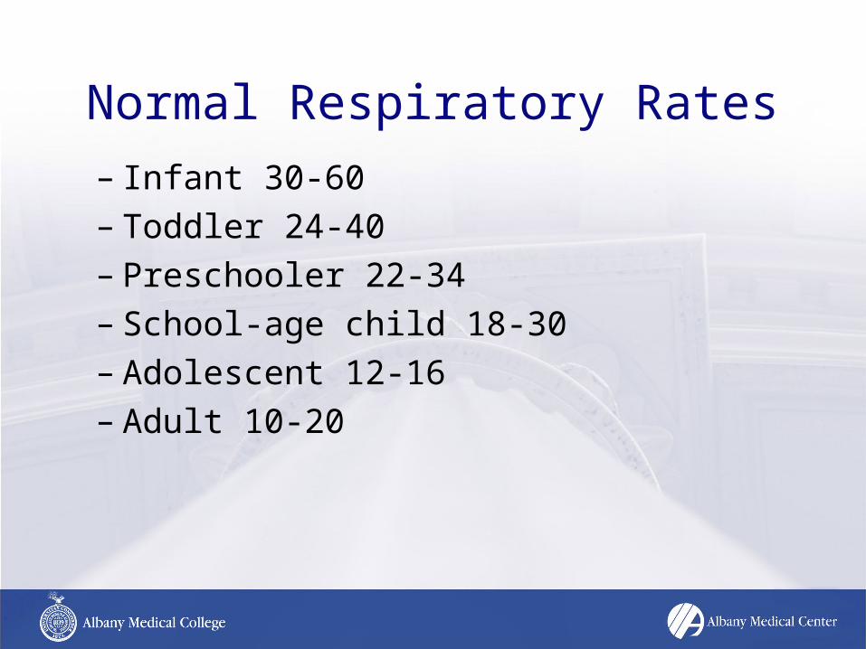
Normal Respiratory Rates
– Infant 30-60– Toddler 24-40– Preschooler 22-34– School-age child 18-30– Adolescent 12-16– Adult 10-20

Pertinent History– Any chronic conditions
• Asthma, COPD, CHF, DM
– Exposure to new medication• ACE-Inhibitor, Abx
– Recent change in diet• Peanuts, Strawberries
– Substance abuse/Overdose• Opioid abuse, ASA toxicity
– Prior DVT, PE– Recent trauma to chest

Inspection• Note the shape of the chest and the way it
moves– Deformities or asymmetry
• Increased AP diameter in COPD
– Abnormal retractions of interspaces during respiration
• Lower interspaces, supraclavicular in acute asthma exacerbation
– Impaired respiratory movement • Flail Chest and paradoxical movement with rib fx’s

Palpation
• Identify tender areas – Bruising with rib fx
• Observe for appropriate chest wall expansion
• Feel for tactile fremitus symmetrically – palpable vibrations transmitted to chest wall– use ulnar surface of hand, say “ninety-nine”– decreased with COPD, pleural effusions, PTX

Percussion
• Helps to identify if underlying tissues are air-filled, fluid-filled, or solid– Hyperextend middle finger of either hand and
press against chest wall– Strike with flexed middle finger of opposite
hand
• Always percuss symmetrically on chest wall
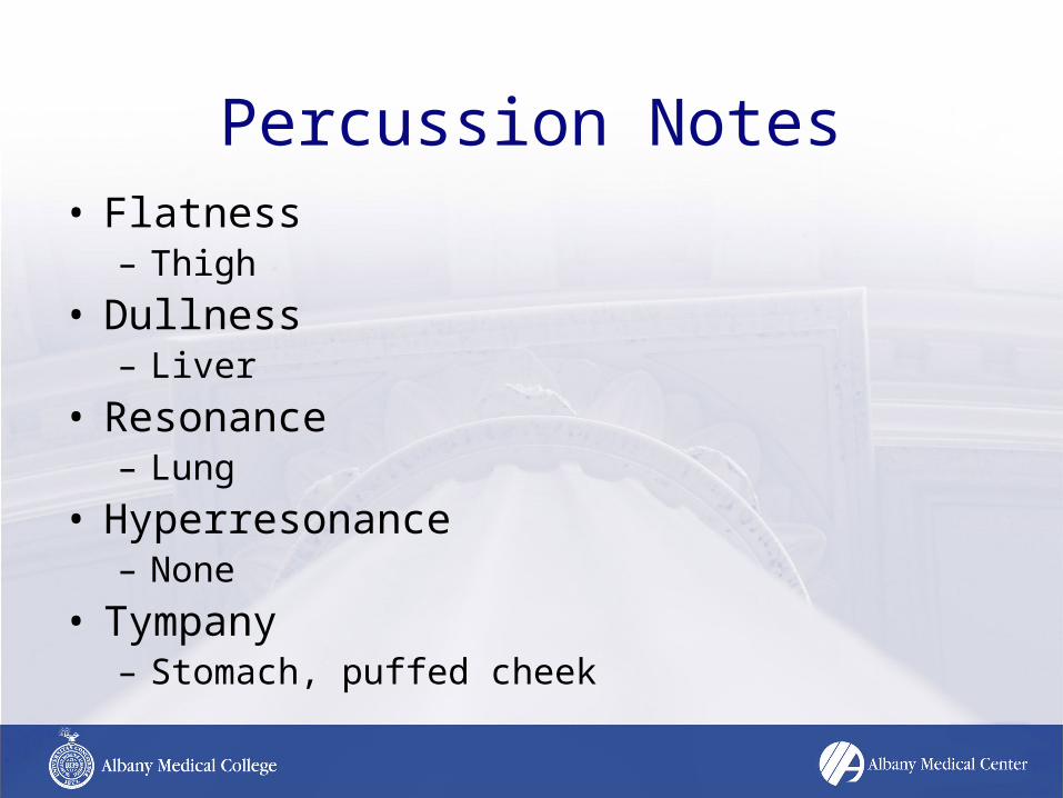
Percussion Notes• Flatness
– Thigh
• Dullness – Liver
• Resonance – Lung
• Hyperresonance – None
• Tympany – Stomach, puffed cheek

Percussion• Dullness replaces resonance when fluid or solid
tissue replaces air containing lung– PNA– Pleural Effusions– Hemothorax– Tumor
• Unilateral Hyperresonance – Pneumothorax
• Generalized Hyperresonance – COPD

Auscultation• 12 anterior locations• 14 posterior locations• Auscultate symmetrically• Should listen to at least 6 locations anteriorly
and posteriorly

Breath Sounds• Normal
– Tracheal– Bronchial– Bronchovesicular– Vesicular
• Abnormal– Absent/Decreased– Bronchial
• Adventitious – Crackles (Rales)– Wheeze– Rhonchi– Stridor– Pleural Rub

Normal Breath Sounds• Created by turbulent air flow• Inspiration
– Air moves to smaller airways hitting walls– More turbulence, Increased sound
• Expiration– Air moves toward larger airways– Less turbulence, Decreased sound
• Normal breath sounds – Loudest during inspiration, softest during expiration

Normal Breath Sounds• Tracheal
– Very loud, high pitched sound– Inspiratory = Expiratory sound duration– Heard over trachea
• Bronchial– Loud, high pitched sound– Expiratory sounds > Inspiratory sounds– Heard over manubrium of sternum– If heard in any other location suggestive of
consolidation

Normal Breath Sounds• Bronchovesicular
– Intermediate intensity, intermediate pitch– Inspiratory = Expiratory sound duration– Heard best 1st and 2nd ICS anteriorly, and between
scapula posteriorly– If heard in any other location suggestive of
consolidation
• Vesicular– Soft, low pitched sound– Inspiratory > Expiratory sounds– Major normal BS, heard over most of lungs

Transmitted Voice Sounds• If abnormally located bronchial or bronchovesicular
breath sounds assess transmitted voice sounds with stethoscope– Ask the patient to say “Ninety-nine”, should normally be muffled,
if heard louder and clearer this is bronchophony– Ask the patient to say “ee”, should normally hear muffled long E
sound, if E to A change this is egophony– Ask the patient to whisper “Ninety-nine”, should normally hear
faint muffled sound, if louder and clearer sounds are heard this is whispered pectoriloquy
• Increased transmission of voice sounds suggests that air filled lung has become airless

Adventitious Breath Sounds• Crackles (Rales)
– Discontinuous, intermittent, nonmusical, brief sounds– Heard more commonly with inspiration– Classified as fine or coarse – Normal at anterior lung bases
• Maximal expiration• Prolonged recumbency
– Crackles caused by air moving through secretions and collapsed alveoli
– Associated conditions • pulmonary edema, early CHF, PNA

Adventitious Breath Sounds
• Wheeze– Continuous, high pitched, musical sound,
longer than crackles– Hissing quality, heard > with expiration,
however, can be heard on inspiration– Produced when air flows through narrowed
airways– Associated conditions
• asthma, COPD

Adventitious Breath Sounds
• Rhonchi– Similar to wheezes– Low pitched, snoring quality, continuous,
musical sounds– Implies obstruction of larger airways by
secretions– Associated condition
• acute bronchitis

Adventitious Breath Sounds
• Stridor– Inspiratory musical wheeze– Loudest over trachea– Suggests obstructed trachea or larynx– Medical emergency requiring immediate
attention– Associated condition
• inhaled foreign body
Adventitious Breath Sounds
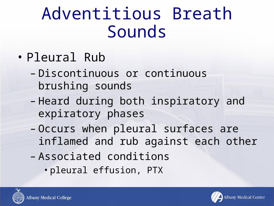
• Pleural Rub– Discontinuous or continuous brushing sounds– Heard during both inspiratory and expiratory
phases– Occurs when pleural surfaces are inflamed
and rub against each other– Associated conditions
• pleural effusion, PTX

Causes of decreased or absent breath sounds
• Asthma
• COPD
• Pleural Effusion
• Pneumothorax
• ARDS
• Atelectasis

Case #1• Dispatch Information
– 62 yo female with progressive SOB over past 48 hours• PMH
– 40 pack year smoking history– On home O2– Some type of lung problem
• VS– O2 sat 78% on 2L O2 NC, RR 26, T 98.1
• Physical Exam– Barrel shaped chest– Decreased BS B/L – Diffuse expiratory wheezing B/L lung fields– Digital cyanosis and clubbing noted

What is this patient’s condition and appropriate treatment prior to ED
arrival?
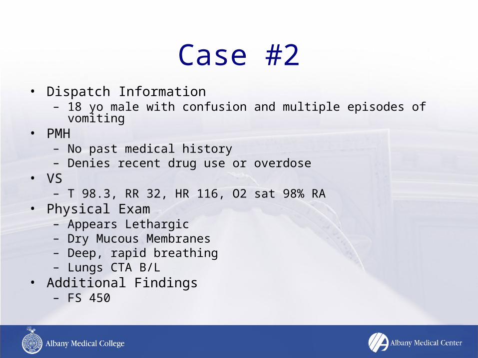
Case #2• Dispatch Information
– 18 yo male with confusion and multiple episodes of vomiting• PMH
– No past medical history– Denies recent drug use or overdose
• VS– T 98.3, RR 32, HR 116, O2 sat 98% RA
• Physical Exam– Appears Lethargic– Dry Mucous Membranes– Deep, rapid breathing– Lungs CTA B/L
• Additional Findings– FS 450

What is this patient’s condition and appropriate treatment prior to ED
arrival?
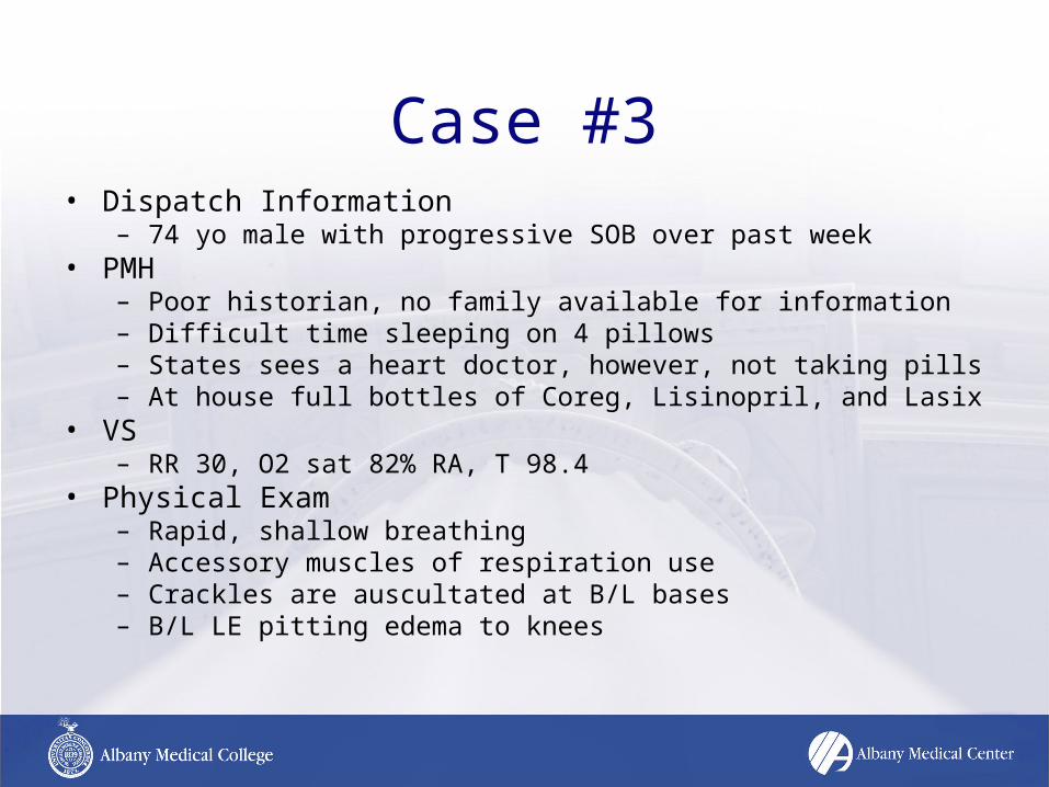
Case #3• Dispatch Information
– 74 yo male with progressive SOB over past week• PMH
– Poor historian, no family available for information– Difficult time sleeping on 4 pillows – States sees a heart doctor, however, not taking pills– At house full bottles of Coreg, Lisinopril, and Lasix
• VS– RR 30, O2 sat 82% RA, T 98.4
• Physical Exam– Rapid, shallow breathing– Accessory muscles of respiration use– Crackles are auscultated at B/L bases – B/L LE pitting edema to knees
What is this patient’s condition and appropriate treatment prior to ED
arrival?

Case #4• Dispatch Information
– MVA rollover on Rt. 4 in East Greenbush• 25 yo male • unrestrained driver• significant intrusion into driver door• + LOC, GCS 13 at present
• PMH– EtOH abuse
• VS– RR 28, O2 sat 76% RA
• Physical Exam– multiple bruises on B/L chest wall– paradoxical movement of L chest wall– absent breath sounds on L side

What is this patient’s condition and appropriate treatment prior to ED
arrival?
Case #5• Dispatch Information
– 42 yo female with difficulty breathing and facial swelling over past hour
• PMH – HTN– NKDA or food allergies– Started Lisinopril for BP 1 month ago
• VS– HR 108, RR 28, O2 sat 86% RA, T 98.4
• Physical Exam– Perioral facial and lip swelling– Inspiratory stridor on auscultation

What is this patient’s condition and appropriate treatment prior to ED
arrival?