Lung resection Nattachai Anantasit. In Ramathibodi hospital (1983-1997, N= 20) Indication for lung...
42
Lung resection Nattachai Anantasit
-
Upload
angelina-webb -
Category
Documents
-
view
220 -
download
2
Transcript of Lung resection Nattachai Anantasit. In Ramathibodi hospital (1983-1997, N= 20) Indication for lung...
Lung resection
Nattachai Anantasit

In Ramathibodi hospital (1983-1997, N= 20)
• Indication for lung resection– Congenital cystic disease 45%– Persistent pneumonia and/or 25%
atelectasis with bronchiectasis – Lung abscess 20%– Abnormal mass with 10% recurrent hemoptysis
Srisan P. A thesis submitted for the diploma of thai subboard of pediatric pulmonology 1998.

Lung resection
• Pneumonectomy: Surgical removal of an entire lung
• Lobectomy: Surgical excision of a lobe
• Segmentectomy: Surgical excision of segment of lung
• Wedge resection: A surgical procedure to remove a triangle-shaped slice of tissue. It may be used to remove a tumor and a small amount of normal tissue around it

Preoperative evaluation
• Pulmonary function• Calculation of predicted
postoperative pulmonary function
• Measurement of gas exchange• Exercise testing

Pulmonary function test
• FEV1 <60%predicted was strongest predictor of postoperative complication
• DLCO

Current guidelines
• Preoperative FEV1 >80%predicted can tolerate pneumonectomy
• Exertional dyspnea or coexistent interstitial lung disease DLCO
• Preoperative FEV1 and DLCO > 80% predicted not need further testing
Colice GL, et al. Chest 2007

Predicted postoperative PFTs
• Combination of Preoperative spirometry and quantitative perfusion lung scanning to estimate the degree of functional loss

Predicted postoperative PFTs
• FVCc = FVCpreop x S x 5.26/100
FVCc = FVC calculated
FVCpreop = FVC pre operation
S = segment left

Current guidelines
• Increased risk for lung resection with predicted postoperative values for either FEV1 or DLCO < 40% predicted
• Preoperative exercise testing is recommended
Colice GL, et al. Chest 2007

Gas exchange
• PaO2; not important predictor of postoperative complication
• PaCO2; not correlate with postoperative complication
Marshall MC, et al. Clin Chest Med 1993.Wyser C, et al. Am J Respir Crit Care Med 1999.

Cardiopulmonary exercise testing (CPET)
• Correlate with postoperative complication
• Maximal oxygen consumption (VO2max)
• VO2max < 15ml/kg/min or < 50%predicted correlated with postop complication1
1Walsh GL, et al. Ann Thorac Surg 1994.

Current guidelines
• VO2 max < 10 ml/kg/min
or
• VO2 max < 15ml/kg/min and both predicted postoperative FEV1 and DLCO < 40% predicted
increase risk of perioperative death and complication
Colice GL, et al. Chest 2007

Physiology of lung resection
Olsen GN. Chest 1998.

Postoperative lung resection

Postoperative lung resection
Insert ICD ?

Postoperative lung resection
Pleurocentesis ?

Anatomic changes
• Immediately; – air fills the space previously occupied by
lung– Chest tube is not inserted
• Over time; – Elevation of hemidiaphragm, hyperinflation
of the remaining lung and shifting of mediastinum to postpneumonectomy space (PPS)
– Fluid accumulating in PPS (2 rib space per day)

Anatomic changes
– Complete opacification of hemithorax after pneumonectomy (3wks-7mo)
• Unexpected rapid accumulation of fluid hemorrhage, infection or chylothorax
• Vital organs shifted position

Anatomic changes
Day 1 Day 2
Day 14 Day 30
Chae EJ, et al. RSNA 2006.

Early mortality
• 30 days mortality 2.4-11.6%• Risk factors for early mortality
– Right-sided pneumonectomy– Specific type of surgical resection– Underlying disease– Emergency surgery– The level of experience of surgeon

Postoperative pulmonary outcome
• FEV1, FVC are decreased
• DLCO is decreased but normal corrected DLCO/lung volume ratio
• Lung compliance is decreased, airway resistance is increased
• Arterial oxygen saturation, PO2, PCO2 not changed

Postoperative cardiovascular outcome
• Right pneumonectomy– Rt.ventricular end diastolic volume is low
but left ventricular function is normal
• Left pneumonectomy– Opposite Rt.pneumonectomy
Smulders SA, et al. Ann Thorac Surg 2007.

Postoperative quality of life
• Quality of life scores (pain, physical function and dyspnea) decrease after pneumonectomy
• Lobectomy and wedge resection are normal
Balduyck B, et al. Lung Cancer 2007.

Postoperative complication
• Hemorrhage complication• Cardiac complications• Pulmonary complications

Postoperative complication
• Hemorrhage complication: – Inadequate hemostasis of the bronchial
artery or a systemic vessel in the chest wall– Infrequently, slipping of a ligature or an un-
recognized injury is a cause– Bleeding related to coagulation is rare
• Re-exploration is indicated if – failed response to blood replacement– a large amount of blood in the hemithorax– persistent massive bleeding from the chest
tube
Postoperative complication
• Cardiac complications: – arrhythmias, cardiac herniation, cardiac
temponade
• Pulmonary complications

Postoperative pulmonary complication
• Early complications:– Pulmonary edema, ARDS – Bronchopleural fistula– Postpneumonectomy empyema– Pneumonia of contralateral lung
• Late complications:– Postpneumonectomy syndrome– Late onset bronchopleural fistula– Infections

Postpneumonectomy pulmonary oedema (PPO)
• Incidence ~5% but high mortality >50%
• Histopathology: – The first 5 day; endothelial integrity lost
with extravasation of fluid, protein and inflammatory cells into alveolar spaces
– First few days; marked proliferation of fibroblasts and type II pneumocytes
– After 10 days; interstitial and intraalveolar fibrosis, thrombotic and obliterative change
Jordan S, et al. Eur Respir J 2000.

Postpneumonectomy pulmonary oedema (PPO)
Jordan S, et al. Eur Respir J 2000.
Postpneumonectomy pulmonary oedema (PPO)
• Risk factors;– Fluid balance ?
Jordan S, et al. Eur Respir J 2000.

Postpneumonectomy pulmonary oedema (PPO)• A dog pneumonectomy model:
– Higher fluid input and urine output not developing PPO if left heart filling pressure remained normal
• An intraoperative fluid input > 2L risk of PPO1
Jordan S, et al. Eur Respir J 2000.1Parquin F, et al. Eur J Cardiothorac Surg 1996.
“ Increased infusion of fluids in high permeability patients may be relevant in exacerbating or prolonging the clinical condition”

Postpneumonectomy pulmonary oedema (PPO)
• Risk factors;– Fluid balance ? – Surgical technique ?
• Degree of parenchymal injury inflammatory reaction
• Duration of surgery does not be implicated
Hayes JP, et al. Thorax 1995.

Postpneumonectomy pulmonary oedema (PPO)
• Risk factors;– Fluid balance ? – Surgical technique ? – Tidal volume ventilation ?
• Low tidal volume pressure limited technique can improve outcome
– Age and preoperative lung function ?• Not correlate

Postpneumonectomy empyema
• Early empyema; 10-14days after surgery, associated with bronchopleural fistula or /and esophagopleural fistula
• Late empyema; more than 3 months, infection (via hematogenous route)– S.aureus and P.aeruginosa are common

Postpneumonectomy pulmonary oedema (PPO)
• Onset: 1-3 days postoperative• Clinical presentation: same as
pulmonary edema, ARDS• May be difficult to differentiate between
PPO and pneumonia
Jordan S, et al. Eur Respir J 2000.

Postpneumonectomy pulmonary oedema (PPO)
• Pathophysiology: – Panendothelial inflammatory vascular injury
release of inflammatory mediators – Vasoconstrictor endothelins (ETs) lead to
pulmonary vascular remodelling pulm. HT
– Others; vascular obstruction and positive pressure ventilation
Jordan S, et al. Eur Respir J 2000.
Pulmonary vascular control

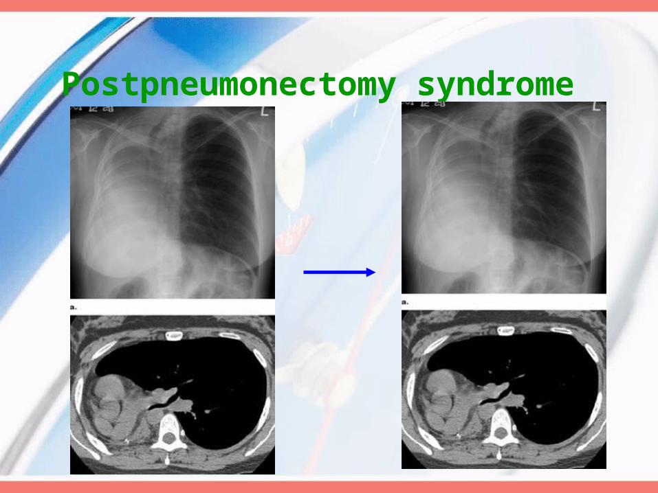
Postpneumonectomy syndrome (PPS)
Valji AM, et al. Chest 1998.

Postpneumonectomy syndrome (PPS)
• Extrinsic compression of distal trachea and mainstem bronchus: Left( right ) PPS: (counter) clockwise rotation of great vessels and trachea→compression of right (left) main bronchus and right(left) pulmonary artery
• Shifting of the mediastinum and hyperinflation of remaining lung
Valji AM, et al. Chest 1998.

Postpneumonectomy syndrome (PPS)
• Occur more than 6 months following surgery
• Progressive dyspnea, cough, inspiratory stridor and recurrent pneumonia
• PFTs: obstructive pattern (bronchial obstruction leads to decrease in
flow rate and air trapping)
• Diagnosis: CXR, CT chest and awake fiberoptic bronchoscope
Valji AM, et al. Chest 1998.

Postpneumonectomy syndrome (PPS)
• Surgical repositioning of mediastinum and filling of PPS with a non absorbable material– Saline solution-filled prosthesis and anterior
pericardiorrhaphy
• Early diagnosis and treatment of PPS should prevent tracheobronchomalacia
Valji AM, et al. Chest 1998.
Postpneumonectomy syndrome

Postpneumonectomy syndrome
Podevin G,et al. J Pediatr Surg 2001.