Lung-protective Ventilation Strategies and … Strategies and Adjunctive Treatments for ... acute...
17
Lung-protective Ventilation Strategies and Adjunctive Treatments for the Emergency Medicine Patient with Acute Respiratory Failure Brian J. Wright, MD, MPH a,b, * INTRODUCTION Invasive mechanical ventilation (MV) is an essential component of critical care and emergency medicine (EM). Successful resuscitation requires an expedient and parallel assessment of airway maintenance, efficiency and effectiveness of breathing me- chanics, and adequacy of circulation and perfusion. Management should be directed at ensuring sufficient oxygenation, ventilation, and prompt reversal of the inciting dis- ease process if possible. This article reviews the evidence for safe MV strategies in the critically ill patient in the emergency department (ED) and provides treatment options for patients who are difficult to oxygenate and ventilate or cannot safely be managed with standard MV strategies. Disclosure: None. a Department of Emergency Medicine, Stony Brook University School of Medicine, 101 Nicolls Road, Stony Brook, NY 11794, USA; b Department of Surgery, Stony Brook University School of Medicine, 101 Nicolls Road, Stony Brook, NY 11794, USA * Corresponding author. E-mail address: [email protected] KEYWORDS Acute respiratory failure Acute respiratory distress syndrome Mechanical ventilation KEY POINTS Respiratory failure is a frequent disease process encountered in the emergency department. There is significant need for improvement in the care of patients on mechanical ventilation. If not contraindicated, lung-protective ventilation strategies should be used. Patient specific disease pathophysiology is important to consider when treating patients that are difficult to oxygenate, ventilate or when PaO 2 , PaCO 2 , and/or pH can only be main- tained at unsafe ventilator settings. Emerg Med Clin N Am - (2014) -–- http://dx.doi.org/10.1016/j.emc.2014.07.012 emed.theclinics.com 0733-8627/14/$ – see front matter Ó 2014 Elsevier Inc. All rights reserved.
Transcript of Lung-protective Ventilation Strategies and … Strategies and Adjunctive Treatments for ... acute...

Lung-protectiveVenti lation Strategies and
Adjunctive Treatments for theEmergency Medicine Patient withAcute Respiratory FailureBrian J. Wright, MD, MPHa,b,*
KEYWORDS
� Acute respiratory failure � Acute respiratory distress syndrome� Mechanical ventilation
KEY POINTS
� Respiratory failure is a frequent disease process encountered in the emergencydepartment.
� There is significant need for improvement in the care of patients on mechanical ventilation.If not contraindicated, lung-protective ventilation strategies should be used.
� Patient specific disease pathophysiology is important to consider when treating patientsthat are difficult to oxygenate, ventilate or when PaO2, PaCO2, and/or pH can only be main-tained at unsafe ventilator settings.
INTRODUCTION
Invasive mechanical ventilation (MV) is an essential component of critical care andemergency medicine (EM). Successful resuscitation requires an expedient and parallelassessment of airway maintenance, efficiency and effectiveness of breathing me-chanics, and adequacy of circulation and perfusion. Management should be directedat ensuring sufficient oxygenation, ventilation, and prompt reversal of the inciting dis-ease process if possible. This article reviews the evidence for safe MV strategies in thecritically ill patient in the emergency department (ED) and provides treatment optionsfor patients who are difficult to oxygenate and ventilate or cannot safely be managedwith standard MV strategies.
Disclosure: None.a Department of Emergency Medicine, Stony Brook University School of Medicine, 101 NicollsRoad, Stony Brook, NY 11794, USA; b Department of Surgery, Stony Brook University School ofMedicine, 101 Nicolls Road, Stony Brook, NY 11794, USA* Corresponding author.E-mail address: [email protected]
Emerg Med Clin N Am - (2014) -–-http://dx.doi.org/10.1016/j.emc.2014.07.012 emed.theclinics.com0733-8627/14/$ – see front matter � 2014 Elsevier Inc. All rights reserved.

Wright2
EPIDEMIOLOGY/STATEMENT OF THE PROBLEM
Acute respiratory failure (ARF) requiring MV is a common clinical scenario. Wunschand colleagues1 suggested that approximately 3% of all hospital admissions in theUnited States require invasive MV. MV costs approximately $27 billion dollars nation-ally.1 Nearly one-third of patients who are placed on MV die in the hospital and amongsurvivors only 30% are discharged home after their admission.1 Recent data suggestopportunities for improvement because many patients in the ED and intensive careunit (ICU) do not get optimal MV therapy for ARF.2,3
PATHOPHYSIOLOGY
Breathing is essential for homeostasis. In critically ill patients the demands for oxygensupply and carbon dioxide removal are often increased. This increased demand canbe superimposed on prior impaired cardiopulmonary reserve. In ARF the cardiopul-monary system fails to oxygenate or ventilate adequately or inefficient breathing me-chanics put excessive loads on the cardiopulmonary system. MV is used to offloadrespiratory muscle work and assist in oxygen delivery and ventilation.
OXYGENATION AND HYPEROXIA
Most oxygen is carried by hemoglobin molecules in red blood cells. MV is often usedto correct hypoxemia (low oxygen saturation) to improve total blood oxygen content.In emergent situations it is beneficial to increase the fraction of inspired oxygen (FiO2)to increase blood oxygen content. Increased FiO2 should only be considered as a tem-porary fix because there are downsides to high concentrations of FiO2.First, in patients with normal lungs, supernormal FiO2 concentrations lead to hyper-
oxia or a PaO2 greater than or equal to 200.4 Multiple studies suggest that hyperoxialeads to the formation of reactive oxygen species that can cause tissue damage.4–8
More recent data in brain-injured patients4,8 and patients after cardiac arrest9 suggestworse outcomes with hyperoxia versus normal oxygen concentrations. Hyperoxia isan iatrogenic entity that can be avoided by turning the FiO2 down.Second, in patients with abnormal lungs, an FiO2 of 1.0 (100%) can mask the degree
of pulmonary dysfunction.3 A normal oxygen saturation on pulse oximetry can befalsely reassuring.3 On a FiO2 of 1.0, a saturation of 100% may correspond with aPaO2 of between 100 and 500, the latter being normal and the former evidence of sig-nificant respiratory dysfunction. Having a through understanding of the magnitude ofpulmonary dysfunction may allow better patient care3 because these patients may becandidates for other adjunctive therapies and lung-protective ventilation strategies(LPVS). Once stabilized and resuscitated through the peri-intubation period, dialdown the FiO2 using pulse oximetry. An oxygen saturation around 95% is sufficientand in certain conditions (chronic obstructive pulmonary disease [COPD], obesityhypoventilation syndrome, and obstructive sleep apnea) a saturation of between88% and 92% may be more appropriate.10 Titrating the FiO2 helps to determine thedegree of respiratory dysfunction and helps limit oxygen toxicity.
OXYGEN DELIVERY AND HEART-LUNG INTERACTIONS
Placing a critically ill patient on MV can have serious untoward effects on cardiacoutput (CO) and oxygen delivery and can lead to adverse events if not appropriatelyanticipated and managed proactively.11 In conditions associated with high afterload,MV can be beneficial by decreasing the force opposing left ventricular contraction andleft ventricular transmural wall pressure.11 In preload-dependent states (hypovolemic

Lung-protective Ventilation in ARF 3
and distributive shock) and diseases with right ventricular (RV) dysfunction (pulmonaryembolism, pericardial tamponade, tension pneumothorax) the increase in intratho-racic pressure and RV afterload during MV can impede venous return to the rightside of the heart and CO.11 This decrease in venous return can manifest as a precip-itous decrease in blood pressure and even cardiac arrest after initiation of MV. Thereader is referred to recent work by Funk and colleagues12,13 for a more detaileddescription of RV function and venous return. Focused bedside echocardiographyassessing inferior vena cava collapsibility and RV size and function before intubation(if feasible) may clue the clinician into potential postintubation complications anddirect the need for aggressive fluid resuscitation and early vasopressor support duringthe induction and early MV period.
HYPOXEMIA
Hypoxemia is low arterial oxygen saturation. There are multiple physiologic mecha-nisms for hypoxemia: hypoventilation (airway obstruction or sedative overdose), lowFiO2 or low partial pressure of O2 (high altitude), diffusion-limited processes (interstitiallung disease), ventilation/blood flow (V/Q) mismatch (COPD), shunt (pulmonaryedema), and low venous oxygen content (shock).14 During MV, persistent hypoxemiacan usually be attributed to a combination of shunt physiology and low venous admix-ture of blood.15
The cardiac and pulmonary systems match ventilation and blood flow. Shunt phys-iology is a V/Q mismatch in which blood passes from the right to the left side of theheart without participating in gas exchange. Desaturated venous blood mixes withoxygenated arterial blood, decreasing the overall arterial oxygen saturation. Supple-mental oxygen only partially corrects hypoxemia or has no effect on systemic oxygensaturation because the shunted blood bypasses the delivered supplemental oxygen.16
VENTILATION AND HYPERCAPNEA
Effective MV must provide for adequate CO2 excretion and O2 saturation while notexposing the patient to excessive airway pressures (barotrauma) or tidal volumes(Tv) (volutrauma). The clearance of CO2 depends on the relationship between CO2 pro-duction and alveolar ventilation (Va)
17:
PaCO2 z (CO2 production)/(Va).
Va 5 minute ventilation (Vm) � Dead space ventilation (Vds).
The typical MV adjustment to hypercarbia is to increase the Vm by increasing the res-piratory rate (RR) or Tv. In most patients this increase in Vm leads to an increase in Va
and lowering of the PaCO2. However, in low cardiac output states, acute respiratorydistress syndrome (ARDS), advanced COPD, massive pulmonary embolism, andabdominal compartment syndromes,17 Vds can increase. In these instances, merelyadjusting the set Vm may not improve CO2 clearance if Va does not improve. Instead,the clinician should attempt to reverse the underlying process if possible and/orattempt to decrease CO2 production. Rechecking arterial blood gases (ABGs) afterMV adjustments is important. Continuous end-tidal CO2 monitoring (ETCO2) is anotherhelpful tool for the patient needingMVbut does not obviate ABGs. ETCO2 approximatesthe PaCO2. The difference between the PaCO2 and ETCO2 depend on Vds. Therefore, it isimportant to obtain an ABG to correct for patient-specific relationships between theETCO2 and PaCO2 and not rely solely on the ETCO2. The clinician should be cognizant
Wright4
of the value of the ETCO2 and actual Vm (notmerely the set or prescribed Vm if the patientis breathing spontaneously at more than the set ventilator rate) when the ABG is drawn.A high ETCO2 is an assurance of a high PaCO2 but a lowor normal ETCO2 can be seenwitha normal PaCO2 or an increased PaCO2 in the setting of high Vds. Large differences be-tweenPaCO2 and ETCO2 can be seenwith excessive Vds or lowCO.18 Changes in clinicalcondition, severity of illness, or lungmechanics should prompt the clinician to considerwhether a repeat ABG is necessary to recalibrate the ETCO2 with the PaCO2.
ARDS AND LUNG-PROTECTIVE VENTILATION
ARDS was first described by Ashbaugh and colleagues19 in 1967. They described 12patients with a diffuse alveolar infiltrative chest radiograph pattern that manifestedacute onset of tachypnea, hypoxemia, and cyanosis that was refractory to oxygentherapy. ARDS was defined in 1994,20 and this definition was revised in 2012 (the Ber-lin definition21) to address some limitations of the earlier definition. The major changesand new ARDS criteria are as follows:
1. The term acute lung injury was eliminated and the 2-tiered model was replaced by a3-tiered scale based on the degree of hypoxemia as measured by the PaO2/FiO2(P/F) ratio: mild (�300–200 mm Hg), moderate (�200–100 mm Hg), and severe(�100 mm Hg). A minimum continuous positive airway pressure (CPAP) or positiveend-expiratory pressure (PEEP) must be present.
2. Onset within 1 week of inciting event.3. Bilateral opacities consistent with pulmonary edema that are nonhydrostatic or
noncardiogenic in origin. A wedge pressure is no longer required, but the respira-tory failure must not be fully explained by cardiac failure or fluid overload.21
The new severity scale that uses the degree of hypoxemia is the most importantchange to the new definition. Worsening P/F ratio predicts mortality and duration ofMV in survivors and can help triage patients who may benefit from more aggressivetherapies.21
With a few exceptions, MV is usually required in ARDS. Over the last 5 decades, ithas been recognized that MV can be injurious. In the 1970s, ventilator settings weretitrated to normalize blood gas values. Clinicians used Tv of 12 to 15 mL/kg of bodyweight.22 Gross barotrauma in the form of pneumothorax, pneumomediastinum,and pneumoperitoneum was common, and mortality in severe ARDS was as highas 90%.23 Work by Amato and colleagues24 and then the landmark ARDSNet (AcuteRespiratory Distress Syndrome Network)25 trial in 2000 showed improved outcomeswith low Tv ventilation strategies (4–6 mL/kg of ideal body weight [IBW]) versus highertraditional Tv (10–12 mL/kg IBW). IBW is determined by the patient’s height and sexand predicts lung volume better than actual weight.A better understanding of ventilator induced lung injury (VILI) in recent years has
created a renewed impetus on providing LPVS.26 LPVS was first shown to be benefi-cial in patients with ARDS but more recent data suggest that lowering the tidal volumein patients without ARDS may be beneficial.27–30 There are 4 major theories for venti-lator-induced lung injury: barotrauma, volutrauma, atelectrauma, and biotrauma.27
Barotrauma is lung damage secondary to high airway pressure (ie, pneumothoraxor pneumomediastinum). Volutrauma is injury induced by high Tv causing overdisten-tion of alveoli. Atelectrauma is damage from the shear and strain of collapsible lungunits opening and closing, and biotrauma is damage from the release of proinflamma-tory cytokines and immune-mediated injury that occurs when lung tissue is exposed tounphysiologic stress or strain.26

Lung-protective Ventilation in ARF 5
SELECTION OF OPTIMAL TIDAL VOLUME
The mainstays of LPVS are to (1) limit tidal volume; (2) limit end-inspiratory plateaupressure (Pplat); (3) provide adequate PEEP to keep the lung open and prevent alveolarcollapse, and (4) limit FiO2.
30
The optimal Tv for patients without ARDS who require MV is not known.30–32 Animaldata suggest that normal Tv for mammals is 6.3 mL/kg.33 Data from ARDSNet25 as wellas multiple additional trials27–29 suggest that Tv greater than 10 mL/kg IBW is harmful.Lellouche and colleagues27 showed a lower incidence of organ failure and shorter ICUlength of stay (LOS) when using Tv less than 10 mL/kg IBW in patients having cardiacsurgery. The IMPROVE28 study group similarly showed less postoperative respiratorysupport, less pneumonia, and shorter hospital LOS among patients receiving lowintraoperative Tv (6–8 mL/kg IBW) after abdominal surgery. In addition, Serpa Netoand colleagues29 performed a meta-analysis of the use of LPVS with low Tv and clin-ical outcomes among patients without ARDS and showed less subsequent lung injury(RR, 0.33; [0.23–0.47], number needed to treat [NNT], 11), less mortality (RR, 0.64;[0.46–0.89]; NNT, 23), and shorter hospital LOS.If using LPVS and low Tv, it is imperative that the RR is turned up to maintain an
adequate Vm. Adequate Vm is especially important in the ED environment when para-lytics like succinylcholine and longer acting agents like rocuronium are used for rapidsequence induction and patients are heavily sedated to facilitate management. Inthese instances respiratory drive is blunted and patients cannot increase their RR tocompensate for low Tv. This can lead to alveolar hypoventilation, hypercarbia, andworsening acidosis. Normal Vm is approximately 100 mL/kg. This requirement isgreater in critically ill patients. It is helpful to use the patients pre-intubation respiratoryrate and depth of ventilation to gauge the post-intubation Vm requirements. A patientwith Kussmaul breathing will need a high RR and appropriate Tv tomaintain respiratorycompensation for their metabolic acidosis post-intubation. Alternatively, if a patient ison Non-Invasive Ventilation (NIV) prior to intubation, post-intubation Vm can be esti-mated by using the Vm from the NIV respirator.Maintaining adequate pH should be weighed against the need to provide safe MV
settings and safe airway pressures. In many clinical scenarios the goal should notbe to normalize blood gas values but to provide LPVS while accepting permissive hy-percapnia and a mild acidosis. Some data even suggest that mild hypercapnea is pro-tective against lung injury30 and improves red blood cell oxygen delivery.14 ARDSnet25
tolerated pH levels down to 7.15. However, in certain clinical scenarios (eg, brain injurywith increased intracranial pressure, toxicologic emergencies, refractory shock) anincreased PaCO2 may not be safe. In addition, in ARDSNet, some patients required bi-carbonate drips to maintain pH and the low Tv group had a mean respiratory rate ofapproximately 30 breaths per minute.25 Higher respiratory rates shorten the expiratorytime. In certain patient populations with increased airway resistance and obstructivelung disease this can potentially lead to air-trapping and auto-PEEP. Auto-PEEP inpreload-dependent states can decrease venous return and lead to hemodynamicinstability, increased vasopressor and fluid needs, and even cardiovascular collapsein severe cases.In summary, with regard to Tv selection, in patients who meet criteria for ARDS, a
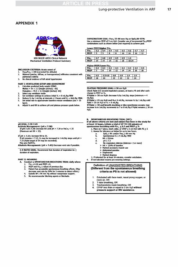
low-Tv strategy that replicates the original ARDSNet trial (4–6 mL/kg IBW) should beused (Appendix 1).3,25 In patients without ARDS, expert opinion and clinical data sug-gest that LPVS with a Tv of 6 to 8 mL/kg of IBW is prudent.31 Using Tv greater than10 mL/kg of IBW is associated with worse clinical outcomes. Whether the optimal tidalvolume is 6 or 8 mL/kg is still open to debate.32,33 When using LPVS, it is important to

Wright6
ensure an adequate Vm by providing an appropriate RR. ABG, in addition to pulse ox-imetry and ETCO2, should be used to ensure adequate oxygenation and ventilation.
LIMITING INSPIRATORY Pplat
An additional goal of LPVS is to limit airway pressure to avoid barotrauma. The inspi-ratory hold or Pplat estimates the pressure distending the alveolus. This maneuver isdone by pausing the flow of air at the end of inspiration. There is no definitive safePplat.
34 In ARDS, the goal should be a Pplat less than 30 cm H2O.25 Hager and col-leagues,34 in their analysis of the ARDSNet data, saw improved outcomes with lowerPplat values. Whether this was causal or coincidental and whether a lower Pplat shouldbe actively sought by lowering Tv is currently unclear. In obese patients or patients withstiff chest walls, Pplat may not accurately reflect transpulmonary pressure or the pres-sure distending the alveoli. In these instances it may be acceptable to tolerate higherPplat.
31 In patients without ARDS, targeting a Pplat less than 20 cmH2O is suggested bysome experts.31 Lowering Pplat is accomplished primarily by decreasing Tv but, again,acceptable (albeit not necessarily normal) PaCO2 and pH should be ensured. In additionto lowering Tv, providing adequate sedation to facilitate patient synchrony, adjustingPEEP to optimal levels, ensuring that auto-PEEP is not present, and ruling out pneu-mothorax or mucous plugging are other measures that can help lower Pplat.
SELECTION OF OPTIMAL PEEP AND OPEN LUNG RECRUITMENT
As stated earlier, most oxygenation problems that occur on MV are secondary toshunt physiology. Shunts are caused by pulmonary (pneumonia, pulmonary contu-sion, pulmonary edema, and so forth) or cardiac (patent foramen oval, atrial or ventric-ular septal defects) causes.The typical treatment of hypoxemia from pulmonary shunts is to attempt to recruit
collapsed lung units by increasing PEEP. The optimal PEEP settings, or even the bestmethod to go about choosing PEEP, are controversial.35,36 To simplify management inthe ED, I recommend using the PEEP table provided by ARDSNet37 because it hasbeen validated and is easy to use35 (see Appendix 1). There is no evidence favoringa high-PEEP versus a low-PEEP strategy in terms of survival, but a high-PEEP strategyhas been associated with improved oxygenation.38 PEEP goals should be reassessedif vasopressor or fluid requirements increase after increasing PEEP, Pplat is greaterthan 30 to 35 cm H2O (depending on body habitus and clinical condition), or thereis evidence of worsening tissue oxygen delivery or worsening oxygen saturation.If increasingPEEP fails tocorrectoxygenation,or oxygenationworsens, it is important
for the clinician to troubleshoot and not continue with treatment strategies that do nothelp and potentially complicate the clinical scenario. Cardiac shunts can worsen withincreased PEEP because the increase in RV afterload increases right-to-left shunt.39
Dessap and colleagues40 in their cohort of 203 patients with ARDS found a prevalenceofamoderate to largepatent foramenovale (PFO) shunt in19.2%of their patients.Thesepatientshadapoorer P/F ratio response toPEEPandhad longer ICUandventilator dayscompared with their counterparts without PFO shunting. Inhaled pulmonary vasodila-torsandpronepositioning (discussed later)maybeamoreappropriate treatment optionin patients with PFO shunts. A transthoracic echocardiogram (or ideally transesopha-geal echocardiography) bubble study can help to detect cardiac shunts.In addition, unilateral lung diseases (pneumonia, mucous plugging, pulmonary contu-
sions, and so forth) may have a paradoxic response to increasing levels of PEEP.41 Inthese situations the increased airway pressure from applied PEEP may overdistendhealthy lung tissue. This will divert perfusion away from aerated lung and through the

Lung-protective Ventilation in ARF 7
shuntworseningoxygenation. Theseconditionscanbedifficult to predict, butwhenapa-tient hasworsening hypoxemiawith initiation ofMV (ie, after intubation) orwith increasinglevels ofpositive airwaypressure, it is important for theclinician toconsider thesediseaseprocesses. The treatment of patients with unilateral lung shunts and cardiac shunts isdifferent than the standard open lung recruitment strategies used in most patients withhypoxemic respiratory failure. In these infrequent instances, blindly following a PEEP/FiO2 table is likely to worsen the clinical scenario. In these patients, limiting PEEP, pronepositioning, and pulmonary vasodilators might be more effective techniques.Fig. 1 summarizes a stepwise strategy for selection of appropriate Tv, RR, FiO2 and
PEEP. In addition to ensuring adequate PaO2, PaCO2, and/or pH, every effort should bemade to provide safe LPVS. Fig. 2 summarizes a stepwise strategy for a patient with aventilator crisis or with a failure to oxygenate or ventilate.
ADJUNCTIVE MANEUVERS
Multiple new and promising developments that focus on non-MV adjunctive treatmentmeasures for patients with refractory ARDS and respiratory failure have recently been
Fig. 1. Algorithm for initiating initial MV settings in the ED. (Ventilator Waveform fromSantanilla JI, Daniel B, Yeow ME. Mechanical ventilation. Emerg Med Clin N Am 2008;26:849–62. PEEP table modified from Haas CF. Lung protective mechanical ventilation inacute respiratory distress syndrome. Respir Care Clin N Am 2003;9(3):363–96. Adaptedfrom The Acute Respiratory Distress Syndrome Network. Ventilation with lower tidal vol-umes compared with traditional tidal volumes for acute lung injury and the acute respira-tory distress syndrome. N Engl J Med 2000;342:1301–8 and Slutsky AS, Ranieri VM.Ventilator-induced lung injury. N Engl J Med 2013;369:2126–36.)

Fig. 1. (continued)
Wright8
developed. These treatments may find a role in the EM management of ARDS and se-vere respiratory failure. Further information on each can be found in references.38,42,43
NEUROMUSCULAR BLOCKING AGENTS
Neuromuscular blocking agents (NMBAs) are used in patients with severe respiratoryfailure to facilitate ventilator synchrony.44 A patient who is bucking the ventilator canbe exposed to barotrauma, volutrauma from breath stacking, and other serious com-plications (eg, tube dislodgment). NMBAs have been used to facilitate ventilator syn-chrony and are associated with improvements in P/F ratio.31 The rational for thisbenefit is not fully clear because NMBAs seem to help even in patients who do not

Fig. 1. (continued)
Lung-protective Ventilation in ARF 9
have ventilator dysynchrony.31 However, concern for ICU-acquired weakness haslimited the use of NMBAs, especially in patients receiving steroids.44 The ACURASYSstudy,45 a multicenter trial, recently assessed the use of cisatracurium in the earlymanagement (6–29 hours from diagnosis) of patients with severe ARDS (P/F ratio<150) and showed an improvement in 90-day mortality (hazard ratio, 0.68; confidenceinterval, 0.48–0.98). The incidence of ICU-acquired weakness did not differ betweenthe two groups.45 EM physician familiarity with NMBA and the early timing of interven-tion make this a promising study for the ED; however, it is important to ensure that thepatient is adequately sedated before initiating NMBA because it is challenging toassess the level of sedation in a paralyzed patient.
INHALED PULMONARY VASODILATORS
Inhaled nitric oxide (INO) and inhaled prostacyclin (IP) are the inhaled pulmonary va-sodilators (IPVs) currently used for salvage therapy in refractory hypoxemia. The

Fig. 2. An algorithm for an approach to the crashing patient on MV. (Adapted from Diaz JV,Brower R, Calfee CS, et al. Therapeutic strategies for severe acute lung injury. Crit Care Med2010;38(8):1646–48.)
Wright10
reader is encouraged to read a more in-depth review from the clinics series.46 IPVswork by improving blood flow to ventilated lung units and decreasing shunt magni-tude. In many cases this leads to an improvement, albeit transient (24–96 hours), inoxygenation.42,43,46
In addition to better V/Q matching, IPV may be beneficial in patients with acute rightheart failure (ie, pulmonary embolism) and right-to-left cardiac shunts. Fig. 2 showshow to titrate INO and IP. The major benefit of IP compared with INO is cost: $275/d versus $3000/d.46 Tachyphylaxis can occur with both so the lowest effective doseshould be used. The beneficial effects should be seen immediately. INO has been
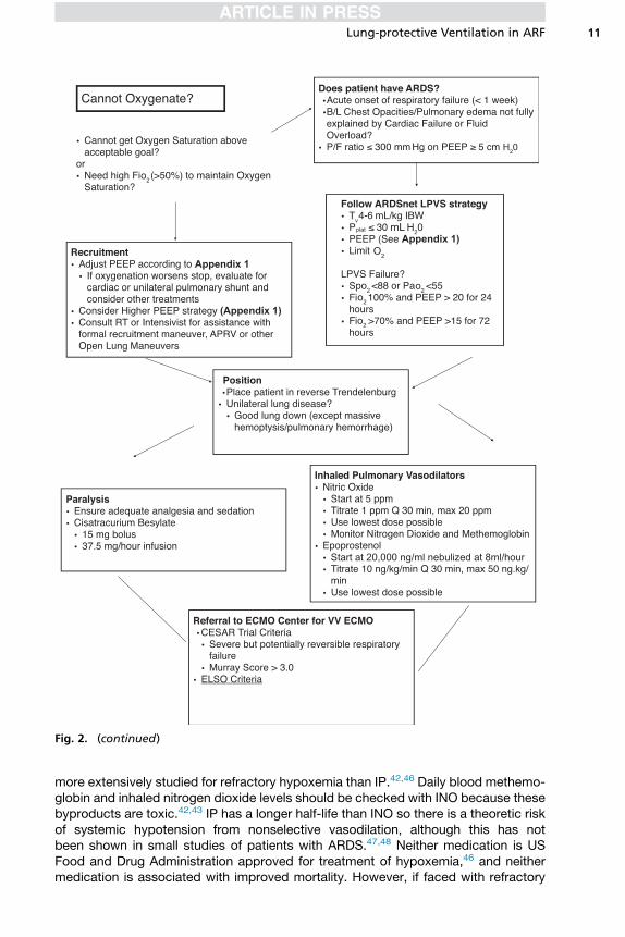
Fig. 2. (continued)
Lung-protective Ventilation in ARF 11
more extensively studied for refractory hypoxemia than IP.42,46 Daily blood methemo-globin and inhaled nitrogen dioxide levels should be checked with INO because thesebyproducts are toxic.42,43 IP has a longer half-life than INO so there is a theoretic riskof systemic hypotension from nonselective vasodilation, although this has notbeen shown in small studies of patients with ARDS.47,48 Neither medication is USFood and Drug Administration approved for treatment of hypoxemia,46 and neithermedication is associated with improved mortality. However, if faced with refractory

Fig. 2. (continued)
Wright12
hypoxemia, INO and IP can be considered as salvage therapy while planning other in-terventions (see Fig. 2).
PRONE POSITIONING
Prone positioning (PP) involves rotating the patient from the standard supine positionto a face-down or prone position. When supine, the inferior and posterior portions ofthe lung can become atelectatic from compression by the heart, thorax, and dia-phragm. Atelectasis can worsen gas exchange, decrease compliance, and increasethe risk for volutrauma because the Tv prescribed is now directed to a smaller lung vol-ume. PP in some individuals allows reexpansion of these posterior lung units. Previousdata have suggested that PP improved oxygenation and P/F ratio, but did not neces-sarily improve outcomes across all patients with ARDS.49–51 However, a trend towardimproved survival was seen in the sickest patient with ARDS.52
Recent data by the PROSEVA group53 examined PP in severe ARDS (P/F ratio<150 mm Hg) and found a reduction in 28-day mortality (16.0% vs 32.8%) and no in-crease in complications in the prone group. The investigators who participated in thistrial had significant experience with PP that potentially minimized complications(airway and central line dislodgment, pressure ulcers). They also required a stabiliza-tion period of 12 to 24 hours before PP was attempted.If considering PP, appropriate staff (especially nursing) should be available. If the
ICU has experience with this technique they should be consulted for assistance.The reader is encouraged to refer to these practical resources on the logistics of PPand for additional information on indications and contraindications.53–55 In ED patientswith difficult oxygenation, a trial of reverse Trendelenburg or placing the good lungdown (provided there is not massive pulmonary hemorrhage) can be attempted.
EXTRACORPOREAL MEMBRANE OXYGENATION
The use of venovenous extracorporeal membrane oxygenation (ECMO) in severeARDS and refractory hypercarbic respiratory failure has gained renewed interest afterrecent encouraging data from the United Kingdom56 and from experience with ARDSduring the 2009 H1N1 pandemic.57 Work from the late twentieth century showeddismal survival23 and no benefit in patients with ARDS treated with ECMO58 but im-provements in technology have made this a potential modality for severe ARDS.ECMO has risks and costs so only the sickest patients with potentially reversible
lung injury should be considered for this treatment. The Extracorporeal Life SupportOrganization (ELSO) has guidelines for consideration of ECMO in respiratory failurethat can be viewed online (http://elso.org/resources/guidelines).59 ECMO has risks

Lung-protective Ventilation in ARF 13
and costs so only the sickest patients with potentially reversible lung injury should beconsidered for this treatment. Indications for VV ECMO include: failure to oxygenate,severe CO2 retention or the ability maintain acceptable CO2 levels at unsafe airwaypressures (PPlat <30 cm H2O) or severe air leak syndromes.59
The CESAR trial56 is the major trial to date showing the potential benefit of ECMO inARDS. A controversy surrounding the study is that some of the benefits found in theCESAR trial might be secondary to treatment of ARDS in a high-volume center withexperience in providing best-evidence medicine (ie LPVS) to patients with ARDSand not necessarily related solely to treatment with ECMO.60
Current expert opinion suggests that clinicians consider referral of adult patients withsevere ARDS or severe respiratory failure with high risk of mortality, as defined byCESAR56 entry criteria or ELSOguidelines,59 to an ARDScenterwith ECMOcapabilitiesif available. Prolonged treatment with unacceptableMV strategies (high FiO2 or Pplat) is acontraindication so these patients should be referred early in the disease process.The main adverse events associated with ECMO are bleeding, hemolysis, dissem-
inated intravascular coagulation, and infection. In addition, there is significant financialcost associated with ECMO.56,60
SUMMARY
Respiratory failure is a frequent disease process encountered in the ED. Better carecan lead to better outcomes for patients on MV.61 If not contraindicated, LPVS shouldbe used. It is important to consider disease pathophysiology when formulating treat-ment strategies in patients that are difficult to oxygenate or ventilate, or when PaO2,PaCO2, and pH can only be maintained with unsafe ventilator settings. If MV adjust-ments do not produce the expected clinical outcome then different treatment strate-gies should be considered. There are multiple adjunctive therapies for ARDS andsevere respiratory failure. Some strategies are straightforward and can potentiallybe applied in the ED (IPV for refractory hypoxemia or NMBA for severe ARDS),whereas other treatments require a collaborative multidisciplinary approach and theassistance of other health care providers (PP and referral to ECMO-capable ARDScenters). Preemptively developing multidisciplinary treatment protocols assists intreating this complicated patient population.
REFERENCES
1. Wunsch H, Linde-Zwirble WT, Angus DC, et al. The epidemiology of mechanicalventilation use in the United States. Crit Care Med 2010;38:1947–53.
2. Fuller BM, Mohr NM, Dettmer M, et al. Mechanical ventilation and acute lunginjury in emergency department patients with severe sepsis and septic shock:an observational study. Acad Emerg Med 2013;20(7):659–69.
3. Manoach S. Mechanical ventilation in the emergency department: a call to ac-tion in a resource-constrained era. Acad Emerg Med 2013;20(7):746–8.
4. Brenner M, Stein D, Hu P, et al. Association between early hyperoxia and worseoutcomes after traumatic brain injury. Arch Surg 2012;147(11):1042–6.
5. Davis WB, Rennard SI, Bitterman PB, et al. Pulmonary oxygen toxicity: earlyreversible changes in human alveolar structures induced by hyperoxia.N Engl J Med 1983;309(15):878–83.
6. Kistler GS, Caldwell PR, Weibel ER. Development of fine structural damage toalveolar and capillary lining cells in oxygen-poisoned rat lungs. J Cell Biol1967;32(3):605–28.

Wright14
7. Yusa T, Beckman JS, Crapo JD, et al. Hyperoxia increases H2O2 production bybrain in vivo. J Appl Physiol (1985) 1987;63:353–8.
8. Rincon F, Kang J, Maltenfort M, et al. Association between hyperoxia and mor-tality after stroke: a multicenter cohort study. Crit Care Med 2014;42(2):387–96.
9. Kilgannon JH, Jones AE, Shapiro NI, et al, Emergency Medicine ShockResearch Network (EMShockNet) Investigators. Association between arterialhyperoxia following resuscitation from cardiac arrest and in-hospital mortality.JAMA 2010;303:2165–71.
10. O’Driscoll R. Emergency oxygen use. BMJ 2012;345:e6856.11. Feihl F, Broccard AF. Interactions between respiration and systemic hemody-
namics. Part II: practical implications in critical care. Intensive Care Med2009;35(2):198.
12. Funk DJ, Jacobsohn E, Kumar A. The role of venous return in critical illness andshock—part I: physiology. Crit Care Med 2013;41(1):255–62.
13. Funk DJ, Jacobsohn E, Kumar A. Role of the venous return in critical illness andshock: part II—shock and mechanical ventilation. Crit Care Med 2013;41(2):573–9.
14. West JB. Pulmonary pathophysiology, the essentials. 7th edition. Philadelphia:Lippincott Williams & Wilkins; 2005.
15. Takala J. Hypoxemia due to increased venous admixture: influence of cardiacoutput on oxygenation. In: Pinsky MR, Brochard L, Mancebo J, et al, editors.Applied physiology in intensive care medicine 1. Berlin Heidelberg (Germany):Springer; 2012. p. 67–70.
16. Budinger GS, Mutlu GM. Balancing the risks and benefits of oxygen therapy incritically III adults. Chest 2013;143(4):1151–62.
17. Schwartzstein RM, Parker MJ. Rising PaCO2 in the ICU: using a physiologicapproach to avoid cognitive biases. Chest 2011;140(6):1638–42.
18. Frankenfield DC, Alam S, Bekteshi E, et al. Predicting dead space ventilation incritically ill patients using clinically available data. Crit Care Med 2010;38(1):288–91.
19. Ashbaugh DG, Bigelow DB, Petty TL, et al. Acute respiratory distress in adults.Lancet 1967;2(7511):319–23.
20. Bernard GR, Artigas A, Brigham KL, et al. The American-European ConsensusConference on ARDS: definitions, mechanisms, relevant outcomes, and clinicaltrial coordination. Am J Respir Crit Care Med 1994;149(3 pt 1):818–24.
21. The ARDS Definition Task Force. Acute respiratory distress syndrome. JAMA2012;307(23):2526–33.
22. Gattinoni L, Vagginelli F, Chiumello D, et al. Physiologic rationale for ventilatorsetting in acute lung injury/acute respiratory distress syndrome patients. CritCare Med 2003;31(4 Suppl):S300–4.
23. Zapol WM, Snider MT, Hill JD, et al. Extracorporeal membrane oxygenation insevere acute respiratory failure. A randomized prospective study. JAMA 1979;242(20):2193–6.
24. Amato MB, Barbas CS, Medeiros DM, et al. Effect of a protective-ventilationstrategy on mortality in the acute respiratory distress syndrome. N Engl J Med1998;338:347–54.
25. The Acute Respiratory Distress Syndrome Network. Ventilation with lower tidalvolumes as compared with traditional tidal volumes for acute lung injury andthe acute respiratory distress syndrome. N Engl J Med 2000;342:1301–8.
26. Gattinoni L, Protti A, Caironi P, et al. Ventilator-induced lung injury: the anatom-ical and physiological framework. Crit Care Med 2010;38(10 Suppl):S539–48.

Lung-protective Ventilation in ARF 15
27. Lellouche F, Dionne S, Simard S, et al. High tidal volumes in mechanically venti-lated patients increase organ dysfunction after cardiac surgery. Anesthesiology2012;116(5):1072–82.
28. Futier E, Constantin J-M, Paugam-Burtz C, et al. A trial of intraoperative low-tidal-volume ventilation in abdominal surgery. N Engl J Med 2013;369:428–37.
29. Serpa Neto A, Cardoso SO, Manetta JA, et al. Association between use of lung-protective ventilation with lower tidal volumes and clinical outcomes among pa-tients without acute respiratory distress syndrome: a meta-analysis. JAMA 2012;308:1651–9.
30. Sinclair SE, Kregenow DA, Lamm WJ, et al. Hypercapnic acidosis is protectivein an in vivo model of ventilator-induced lung injury. Am J Respir Crit Care Med2002;166(3):403–8.
31. Slutsky AS, Ranieri VM. Ventilator-induced lung injury. N Engl J Med 2013;369:2126–36.
32. Mohr NM, Fuller BM. Low tidal volume ventilation should be the routine ventila-tion strategy of choice for all emergency department patients. Ann Emerg Med2012;60(2):215–6.
33. Wright B, Slesinger TL. Low tidal volume should not routinely by used for emer-gency department patients requiring mechanical ventilation. Ann Emerg Med2012;60(2):216–7.
34. Hager DN, Krishnan JA, Hayden DL, et al, ARDS Clinical Trials Network. Tidalvolume reduction in patients with acute lung injury when plateau pressuresare not high. Am J Respir Crit Care Med 2005;172(10):1241.
35. Miller RR, MacIntyre NR, Hite RD, et al. Chest 2012;141(6):1379–82.36. Schmidt GA. Counterpoint: should positive end-expiratory pressure in patients
with ARDS be set based on oxygenation? No. Chest 2012;141(6):1382–4.37. Available at: http://www.ardsnet.org/system/files/Ventilator Protocol Card.pdf.
Accessed on August 22, 2014.38. Esan A, Hess DR, Raoof S, et al. Severe hypoxemic respiratory failure part
1–ventilatory strategies. Chest 2010;137(5):1203–16.39. Michard F, Alaya S, Medkour F. Monitoring right-to-left intracardiac shunt in
acute respiratory distress syndrome. Crit Care Med 2004;32(1):308–9.40. Dessap AM, Boissier F, Leon R, et al. Prevalence and prognosis of shunting
across patent foramen ovale during acute respiratory distress syndrome. CritCare Med 2010;38(9):1786–92.
41. Broccard A. Challenges of mechanical ventilation in unilateral pneumonia: isPEEP the answer? Intensive Care Med 2004;30(4):530–2.
42. Raoof S, Goulet K, Esan A, et al. Severe hypoxemic respiratory failure part 2—nonventilatory strategies. Chest 2010;137(6):1437–48.
43. Diaz JV, Brower R, Calfee CS, et al. Therapeutic strategies for severe acute lunginjury. Crit Care Med 2010;38(8):1644–50.
44. Puthucheary Z, Rawal J, Ratnayake G, et al. Neuromuscular blockade and skel-etal muscle weakness in critically ill patients: time to rethink the evidence? Am JRespir Crit Care Med 2012;185(9):911–7.
45. Papazian L, Forel JM, Gacouin A, et al. Neuromuscular blockers in early acuterespiratory distress syndrome. N Engl J Med 2010;363:1107–16.
46. Puri N, Dellinger RP. Inhaled nitric oxide and inhaled prostacyclin in acute res-piratory distress syndrome: what is the evidence? Crit Care Clin 2011;27(3):561–87.
47. van Heerden PV, Barden A, Michalopoulos N, et al. Dose-response to inhaledaerosolized prostacyclin for hypoxemia due to ARDS. Chest 2000;117(3):819–27.

Wright16
48. Zwissler B, Kemming G, Habler O, et al. Inhaled prostacyclin (PGI2) versusinhaled nitric oxide in adult respiratory distress syndrome. Am J Respir CritCare Med 1996;154(6):1671–7.
49. Gattinoni L, Tognoni G, Pesenti A, et al. Effect of prone positioning on the survivalof patients with acute respiratory failure. N Engl J Med 2001;345:568–73.
50. Sud S, Sud M, Friedrich JO, et al. Effect of mechanical ventilation in the proneposition on clinical outcomes in patients with acute hypoxemic respiratory fail-ure: a systematic review and meta-analysis. CMAJ 2008;178(8):1153–61.
51. Taccone P, Pesenti A, Latini R, et al. Prone positioning in patients with moderateand severe acute respiratory distress syndrome: a randomized controlled trial.JAMA 2009;302:1977–84.
52. Gattinoni L, Carlesso E, Taccone P, et al. Prone positioning improves survival insevere ARDS: a pathophysiologic review and individual patient meta-analysis.Minerva Anestesiol 2010;76(6):448–54.
53. Guerin C, Reignier J, Richard JC, et al. Prone positioning in severe acute respi-ratory distress syndrome. N Engl J Med 2013;368(23):2159–68.
54. Messerole E, Peine P, Wittkopp S, et al. The pragmatics of prone positioning. AmJ Respir Crit Care Med 2002;165(10):1359–63.
55. Gattinoni L, Taccone P, Carlesso E, et al. Prone position in acute respiratorydistress syndrome: rationale, indications and limits. Am J Respir Crit CareMed 2013;188(11):1286–93.
56. Peek GJ, Mugford M, Tiruvoipati R, et al. Efficacy and economic assessment ofconventional ventilatory support versus extracorporeal membrane oxygenationfor severe adult respiratory failure (CESAR): a multicentre randomised con-trolled trial. Lancet 2009;374(9698):1351–63.
57. Davies A, Jones D, Bailey M, et al. Extracorporeal membrane oxygenation for2009 influenza A (H1N1) acute respiratory distress syndrome. JAMA 2009;302(17):1888–95.
58. Morris AH, Wallace CJ, Menlove RL, et al. Randomized clinical trial of pressure-controlled inverse ratio ventilation and extracorporeal CO2 removal for adult res-piratory distress syndrome. Am J Respir Crit Care Med 1994;149(2 Pt 1):295–305.
59. Available at: http://elso.org/resources/guidelines. Accessed on August 22, 2014.60. Wallace DJ, Milbrandt EB, Boujoukos A. Ave, CESAR, morituri te salutant! (Hail,
CESAR, those who are about to die salute you!). Crit Care 2010;14(2):308.61. Fuller BM, Mohr NM, Hotchkiss RS, et al. Reducing the burden of acute respira-
tory distress syndrome: the case for early intervention and the potential role ofthe emergency department. Shock 2014;41(5):378–87.
Lung-protective Ventilation in ARF 17
APPENDIX 1